International Research Journal of Engineering and Technology (IRJET)
e-ISSN: 2395-0056
Volume: 04 Issue: 07 | July -2017
p-ISSN: 2395-0072
www.irjet.net
Segmentation and Classification of MRI Brain Tumor Mukambika P. S.1, Uma Rani K.2 M.Tech (2nd year) Student, Biomedical Signal Processing and Instrumentation, Dept. of IT, SJCE, Mysuru, Karnataka, India Associate Professor, Dept. of IT, SJCE, Mysuru, Karnataka, India ---------------------------------------------------------------------***---------------------------------------------------------------------
Abstract - Bio-medical image processing is the most
and time-consuming task. Manual classification is highly prone to error due to inter observer variability and human error. As a result, the classification results are highly inferior leading to fatal results.
challenging and emerging field in medical diagnosis. Processing of MRI images is one of the difficult parts of this field. The present work presents the comparison study of two techniques used for tumor detection of MRI images. One is based on the Level set method that uses the non parametric deformable models with active contour to segment the brain tumor from the MRI brain images. The other one is the K-means segmentation algorithm. After the segmentation decision making is performed in two stages: Feature extraction using Discrete Wavelet Transform and Gray Level Co-occurrence Matrix, and classification using the Support Vector Machine. It is observed that the results of segmentation accuracies from the proposed methods are comparatively high with the existing method.
2. DATASET Dataset of MRI brain tumor images includes 17 benign and 24 malignant tumor images of different patients which are DICOM in nature. These images T2 weighted sequence images obtained from Philips , 3T machine, and are provided by JSS hospital, Mysuru. These images are labeled by the expert radiologist.
3. METHODOLOGY
Key Words: Magnetic Resonance Imagining (MRI), Brain Tumor, Level set, K-means, Performance Evaluation, Discrete Wavelet Transform (DWT), Gray Level Co-occurrence Matrix (GLCM), Support Vector Machine (SVM).
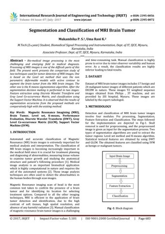
Detection and classification of MRI brain tumor images involve four modules: Pre processing, Segmentation, Feature Extraction and Classification. The steps followed for the implementation are depicted in Fig-1. Skull removal is used for preprocessing, and the preprocessed image is given as input for the segmentation process. Two types of segmentation algorithms are used to extract the tumor regions: Level set method and K-means algorithm. Statistical textural features are obtained by using DWT and GLCM. The obtained features are classified using SVM as benign or malignant tumors.
1. INTRODUCTION Automated and accurate classification of Magnetic Resonance (MR) brain images is extremely important for medical analysis and interpretation. The classification of MR brain images is becoming increasingly important in the medical field since it is crucial for treatment planning and diagnosing of abnormalities, measuring tissue volume to examine tumor growth and studying the anatomical structure and patientâ€&#x;s following procedure [1]. Medical image analysis is an important biomedical application which is highly computational in nature and requires the aid of the automated systems [2]. These image analysis techniques are often used to detect the abnormalities in the human bodies through scan images. Magnetic Resonance imaging scan of head is the most common test taken to confirm the presence of a brain tumor and for identifying its location for selecting treatment options. Compared to all the other imaging techniques, MRI is efficient in the application of brain tumor detection and identification, due to the high contrast of soft tissues, high spatial resolution, and absence of any harmful radiation [3]. Manual classification of magnetic resonance brain tumor images is a challenging
Š 2017, IRJET
|
Impact Factor value: 5.181
Fig -1: Block diagram
|
ISO 9001:2008 Certified Journal
|
Page 683