International Research Journal of Engineering and Technology (IRJET)
e-ISSN: 2395 -0056
Volume: 04 Issue: 03 | Mar -2017
p-ISSN: 2395-0072
www.irjet.net
A Review on Label Image Constrained Multiatlas Selection Ms. VAIBHAVI NANDKUMAR JAGTAP1, Mr. SANTOSH D. KALE2 1PG
Scholar , Department of Electronics and Telecommunication, SVPM College of Engineering, Malegaon (BK), Maharashtra, India.
2Assistant
Professor , Department of Electronics and Telecommunication, SVPM College of Engineering, Malegaon (BK), Maharashtra, India.
-------------------------------------------------------------------------------------------------------------------------------------------Abstract: Multiatlas selection method is a powerful and
brain images. In the method of atlas-based segmentation,
frequently used for medical image segmentation when a
the atlas is registered to the individual brain image by
standard atlas is available. In recent era of image
discovering the best spatial alteration, and then mapping
segmentation manifold ranking based methods are very
the structural information in the atlas on the individual
useful. Magnetic resonance imaging (MRI) provides an
brain image.
attractive way for confined mapping of the anatomical structure of the thing.
In multiatlas based image
segmentation,
key
the
two
factors
affecting
the
performance are atlas selection and combination. It is difficult to receive the correct atlas selection result due to prostate structure in raw images. In this paper we try to solve the problem of manual segmentation by proposing a
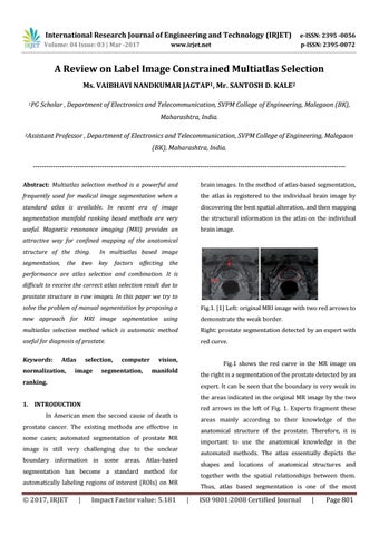
Fig.1. [1] Left: original MRI image with two red arrows to
new approach for MRI image segmentation using
demonstrate the weak border.
multiatlas selection method which is automatic method
Right: prostate segmentation detected by an expert with
useful for diagnosis of prostate.
red curve.
Keywords:
Atlas
normalization,
selection,
image
computer
segmentation,
vision,
Fig.1 shows the red curve in the MR image on
manifold
the right is a segmentation of the prostate detected by an
ranking.
expert. It can be seen that the boundary is very weak in the areas indicated in the original MR image by the two
1. INTRODUCTION
red arrows in the left of Fig. 1. Experts fragment these
In American men the second cause of death is
areas mainly according to their knowledge of the
prostate cancer. The existing methods are effective in
anatomical structure of the prostate. Therefore, it is
some cases; automated segmentation of prostate MR
important to use the anatomical knowledge in the
image is still very challenging due to the unclear
automated methods. The atlas essentially depicts the
boundary information in some areas. Atlas-based
shapes and locations of anatomical structures and
segmentation has become a standard method for
together with the spatial relationships between them.
automatically labeling regions of interest (ROIs) on MR
Š 2017, IRJET
|
Impact Factor value: 5.181
Thus, atlas based segmentation is one of the most
|
ISO 9001:2008 Certified Journal
|
Page 801