In this issue: 2G uest editorial A cautionary tale of regulatory overreach
10 P residential addresses Regional vascular societies hear swansongs from departing leaders
8 I BE Iliac branch device ‘equally effective’ in both IDE and registry studies, new data show
OCTOBER 2025 Volume 21 Number 9
THE OFFICIAL NEWSPAPER OF THE
19 Advocacy Vascular surgeons descend on Washington, D.C. www.vascularspecialistonline.com
RANDOMIZED DATA DEBUNKS WIDELY USED NEGATIVE PRESSURE WOUND THERAPY
COMPLEX AORTIC
By Jocelyn Hudson
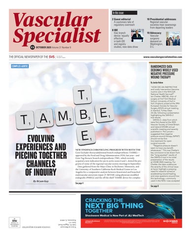
T
1
E B M T A E evolving 1
1
experiences and piecing together channels of inquiry By Bryan Kay
3
3
1
1
NEW FINDINGS CHRONICLING PROGRESS WITH BOTH THE Gore Excluder thoracoabdominal branch endoprosthesis (TAMBE)— approved by the Food and Drug Administration (FDA) last year—and Gore Tag thoracic branch endoprosthesis (TBE), which recently acquired a new indication for use in aortic zones 0 and 1, dotted the programs of some of the regional vascular society meetings in September. Data gathered from the Mayo Clinic in Rochester, Minnesota, and the University of Southern California Keck Medical Center in Los Angeles for a comparative analysis between fenestrated and branched endovascular aneurysm repair (F/BEVAR) using physician-modified endografts (PMEGs) and the off-the-shelf TAMBE device for complex
See page 4
“HOW DID AN INEFFECTIVE and costly intervention become routine care in the NHS [UK National Health Service]?” Ian Chetter, MBChB, chair of surgery at Hull York Medical School, University of Hull in Hull, England, posed at the 39th European Society for Vascular Surgery (ESVS) annual meeting in Istanbul, Turkey (Sept. 23–26), following a presentation highlighting the SWHSI-2 trial results. SWHSI-2—data from which were first shared at the 2024 Vascular Society of Great Britain and Ireland (VSGBI) annual scientific meeting and recently published in The Lancet— suggested that negative pressure wound therapy (NPWT) should not be firstline treatment for open surgical wounds. “Negative pressure doesn’t seem to have any benefit whatsoever.” This was Chetter’s key conclusion to be drawn from the “long-awaited” results of the SWHSI-2 trial in his initial presentation of the results at VSGBI 2024 meeting. For Chetter, the data underscore the severity of the clinical issue at hand, highlight a pressing need for research aimed at accelerating wound healing, and raise questions around how this technology—commonly used in the NHS—was accepted
See page 8
CRACKING THE
NEXT BIG THING TOGETHER
Shockwave Medical is Now Part of J&J MedTech In the US: Rx Only. Prior to use, please reference the Important Safety Information on indications, contraindications, warnings, precautions, and adverse events. www.shockwavemedical.com/IFU. Please contact your local Shockwave representative for specific country availability. SPL 76714 Rev. A
Vascular Specialist 9400 W. Higgins Road, Suite 315 Rosemont, IL 60018
IM
PAID
CHANGE SERVICE REQUESTED
PRESORTED STANDARD MAIL U.S. POSTAGE