I am pleased to reflect on both a remarkable legacy and an exciting period of growth for our community. This year marks the 60th anniversary of the Jules Stein Eye Institute—a milestone that honors six decades of innovation, discovery, and unwavering commitment to preserving and restoring vision. Since its founding, the Institute has helped shape modern ophthalmology by advancing patient care and scientific knowledge on a global scale.
That tradition of excellence continues today. The Jules Stein Eye Institute and Doheny Eye Institute are ranked #1 in Los Angeles and California and among the top five ophthalmology programs in the nation. These distinctions reflect the extraordinary work taking place every day across our clinics, laboratories, and educational programs, and they speak to our collaborative culture.
This spring brings new energy and expertise to our Department, with the addition of two outstanding faculty members whose clinical and research contributions will further strengthen our mission. At the same time, our investigators have secured more than $8.6 million in new research funding, supporting innovative studies that span basic science, translational research, and clinical investigation. These investments accelerate our ability to address the most pressing challenges in eye disease and vision loss.
To our residents and fellows: thank you for your tireless dedication, curiosity, and resilience. You represent the future of our field, and we are fortunate to support your growth. To our faculty and staff: your expertise, hard work, and unwavering support form the foundation of everything we accomplish—whether in the clinic, the lab, or behind the scenes. And to our donors, thank you for your continued generosity and belief in our mission.
Each of you plays an essential role in what we do. Your commitment allows us to provide exceptional care, expand knowledge, and make a meaningful difference in the lives of our patients. As we celebrate 60 years of impact and look ahead to the opportunities before us, I am confident that our momentum will continue.
The best truly do get better together.
Bradley R. Straatsma, MD, Endowed Chair in Ophthalmology Chair, UCLA Department of Ophthalmology Director, Jules Stein Eye Institute Affiliation Chair, Doheny Eye Institute
FEATURE
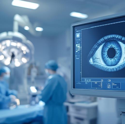
Creating Innovation in Clinical Care
Jules Stein Eye Institute faculty are leading the mission to advance less invasive eye treatments and surgical techniques while also empowering patients with clear, informed understanding of their care. PAGE 2
PHILANTHROPY
Gift Inspired by Hope for a Future Without Stargardt Disease
the Paradigm for Stargardt Research Institute investigators are optimistic that models they have created will significantly advance understanding of Stargardt disease.
PAGE 7
Diagnosed with Stargardt disease at age nine and with multiple family members affected, David Thompson made a $10,000 gift and named the Jules Stein Eye Institute as a beneficiary in his will to support research toward therapies and a cure.
PAGE 9
INNOVATION Creating in Clinical Care
THE CONSTANT CHALLENGE
Faculty in the UCLA Jules Stein Eye Institute lead the ongoing mission to create new treatments and surgical methods for the eye that are simpler and less invasive, while empowering patients with knowledge about their procedure.
For researchers at the Jules Stein Eye Institute, driving innovation in clinical practice is a constant quest—a search to solve the challenges in patient treatment by advancing surgical and therapeutic solutions that are simpler, safer, and are more comfortable for the patient, while also reducing recovery time.
“We are always on the hunt for new approaches to patient care,” says Anne L. Coleman, MD, PhD, chair of the UCLA Department of Ophthalmology, director of the Jules Stein Eye Institute, and affiliation chair of Doheny Eye Institute. “Every challenge opens new opportunities.”
Three examples of key questions in clinical practice, and recent innovations at the Institute, illustrate how advances in vision treatment created by Institute doctors are influencing medical practice worldwide:
Boris E. Malyugin, MD
Joan and Jerome Snyder Chair in Cornea Diseases
How to reduce complications in the world’s most common surgery?
Cataract removal is performed more than six million times annually in the United States. The vast majority of cataract removal and lens replacement is routine, but a notable number—some 200,000 cases each year—are complicated by pupils that are smaller than average.
“A basic problem in cataract surgery is dilating the pupil, so we have good access while replacing the lens,” says Dr. Boris Malyugin, a renowned surgeon for cataract and corneal procedures with surgical experience of more than 30 years. “We use several medications that help in most cases. But the smaller the pupil, the more challenging the operation; each millimeter of pupil constriction increases the chance of complications by about 10 percent.
“As surgeons our goal is to reduce the amount of physical trauma during surgery. With a smaller pupil eye, most likely having various comorbidities, reducing trauma is even more important than for a normal eye that can better tolerate these manipulations.”
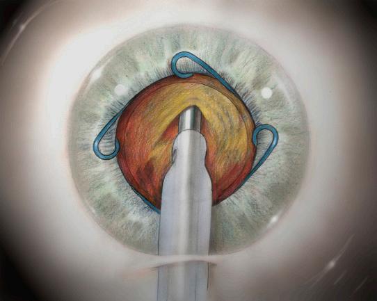
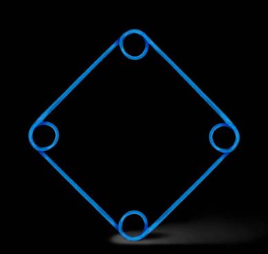
A Ring for Simplifying Surgery
To counter this problem, Dr. Malyugin developed a ring that dilates and stabilizes the iris in patients with small pupils, simplifying a challenging operation and reducing surgical risk.
Known as the “Malyugin Ring,” the device has been adopted by surgeons worldwide, and has been used several million times for cataract procedures.
The Malyugin Ring adds simplicity to cataract removal and with minimal effects on the eye.
“Because the ring can be gently implanted and then removed after surgery, it decreases the number of manipulations during the procedure,” Dr. Malyugin says. “The result is faster recovery and reduced need for postoperative medications.”
Creation of the ring was one of Dr. Malyugin’s objectives in decreasing surgical trauma, especially important for the delicate eye anterior segment.
“For cataract surgery in particular, reducing trauma means decreasing the incision size, as well as the number of incisions,” says Dr. Malyugin. “Each improvement such as the ring continues to simplify the work and reduce the recovery time; we have moved from surgeries that a few decades ago required multiple days in the hospital and weeks of recovery down to today’s outpatient procedures that result in excellent vision, reading, or watching TV the next day.”
Even with such dramatic changes in methods and outcomes, Dr. Malyugin is working towards improvement in the ring and other devices to lessen surgical trauma.
“We are now in our second generation of the ring; compared to the original device, the current ring is thinner, more compact, and flexible, and in general, more ‘friendly’ to the eyes,” says Dr. Malyugin. “We are brainstorming about a third generation of the ring that will be even easier to apply and remove, and as a result save time and ease recovery for the patient.”
As surgeons our goal is to reduce the amount of physical trauma during surgery.
BORIS E. MALYUGIN, MD
Left: The Malyugin Ring shown inserted in the eye. Below: The Malugin Ring as a stand-alone device.
Shawn R. Lin, MD
Health Sciences Assistant Clinical Professor of Ophthalmology
How to increase connection between doctors and patients to improve communication and build patient confidence before and after surgery?
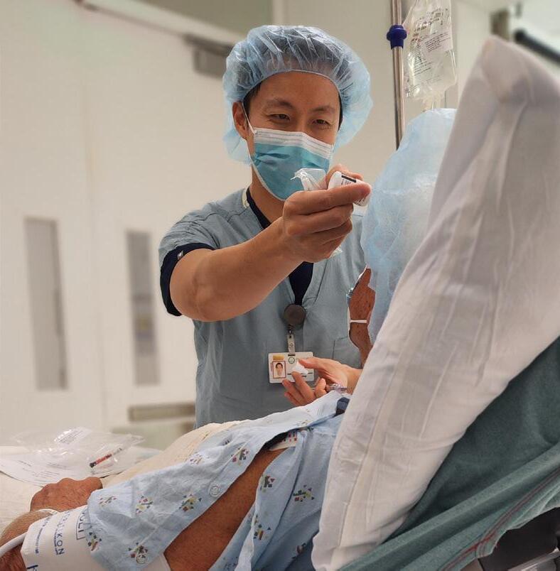
Dr. Shawn Lin performs more than 1,000 surgeries a year—primarily precision cornea, cataract, and refractive surgeries. Dr. Lin is also a leader in creating education about ophthalmology as a co-creator of EyeGuru, an educational platform visited more than one million times each year by ophthalmologists in 125 countries.
Dr. Lin’s interests in doctor-patient communications, along with his formidable surgical schedule, led him to build a new approach to doctor-patient connection.
“Any surgery is psychologically stressful, especially involving the eyes and vision,” says Dr. Lin. “If doctors had all the time in the world, we could spend hours walking each patient through every detail about the procedure and recovery.
“Individual consultations are indeed vital to doctor-patient communication. But the challenge for doctors is to increase connection by building a methodical, regular schedule of communication tools that empowers the patient.”
Moving closer to that goal, during the COVID-19 pandemic Dr. Lin created the Practice Optimization Dashboard (POD), a platform of information and delivery schedules to link patients to information they need at every step before and after surgery.
A Catalog of Doctor-Patient Communication
Rather than relying on a large packet of printed information delivered all at once to inform patients, doctors use POD to selectively time the delivery of communication precisely when patients need them. Developed by Dr. Lin and his father (an accountant experienced in refining management systems), POD includes custom-built schedules of emails, texts, videos, and other communications to connect patients with information in the weeks before their operation and during recovery.
POD can include any information a physician chooses, from background about the procedure and patient preparation to routine logistics such as parking information and postoperative instructions.
“POD ensures that we reach our patients at precisely the times they need to hear from us, so they feel assured and fully informed about their surgery,” says Dr. Lin. “The result is that patients are enlightened about their procedure, which increases their confidence and reduces stress.”
A typical POD experience can include information sent weeks before the surgery, reminders as the date nears, follow-up instructions and reminders about medication, status reports, expectations about recovery, and a library of videos explaining the most asked questions (for Dr. Lin, his library now includes some 50 videos, with plans for an additional 100).
“Every medical institution works hard to offer the best patient experience for health care, and one of the best methods to improve the experience comes from the patients themselves—when they fully understand the procedure and feel empowered by information,” Dr. Lin says. “POD helps us to accomplish this.”
Dr. Shawn Lin meets with his patient to identify the intended surgical site before anesthesia is given.
POD ensures that we reach our patients at precisely the times they need to hear from us, so they feel assured and fully informed about their surgery.
SHAWN
R. LIN, MD
A Boon for Surgical Planning
The POD system also extends beyond patient communication and into the details of surgical scheduling.
“Everything about patient communication is built around the surgical date,” says Dr. Lin. “So an important element of POD is using the system to streamline surgical scheduling that works across all the UCLA Department of Ophthalmology offices throughout Southern California.”
Not coincidentally, increased patient knowledge about their surgical procedures has been accompanied by a substantial drop in surgical cancellations.
“I’m seeing a 50 percent decreased cancellation rate for surgeries,” says Dr. Lin.
A Thriving System of Connections
POD has already been used to communicate with more than 5,000 patients, with enthusiastic reviews.
“Dr. Lin and his support staff are so amazing that all fear subsides,” says one cataract patient. “Every one of my fears and concerns was addressed ahead of time.”
The POD system is being used by Dr. Lin and other UCLA physicians, with a goal of expanding the platform to other medical organizations.
“The patient experience is a huge part of measuring the success of a healthcare system,” says Dr. Lin. “This is especially important for young patients who expect a smooth, digitized experience.
“Patient trust that comes from increased knowledge and communication improves healing, because wellinformed patients are more empowered and confident about the results.
“I believe a system like POD should be a standard of care in medicine, because every surgery has issues before and after the procedure that should be coordinated to optimize healing. As the process is refined, I see tools like these becoming more and more useful.”
Robert Alan Goldberg, MD
Bert O. Levy Endowed Chair in Orbital and Ophthalmic Plastic Surgery
Chair of the Orbital and Ophthalmic Plastic Surgery Division
How to refine the tools for treatment of the delicate regions surrounding the eye?
The issues of clinical care for vision extend beyond the eye itself.
For Dr. Robert Goldberg much of this work focuses on refining minimally invasive surgery and tools for procedures on the tissues surrounding the eye.
“The ongoing challenge is to innovate with new surgical instruments and methods that are simpler, more efficient, and more accurate,” says Dr. Goldberg.
“It has been a hallmark of our program to create advances in surgery and instruments. We have been innovating with that goal for decades.”
Dr. Goldberg’s work centers on oculoplastic surgery, a specialized field of ophthalmology for treatment of the eye socket, eyelids, tear ducts, and the surrounding facial structures.
“These tissues are especially delicate and require special surgical techniques and instruments to support the eye,” says Dr. Goldberg, whose research also explores Graves’ disease
(thyroid eye disease), and minimally invasive surgical techniques for orbital tumors and tear duct obstruction.
The methods produced by Goldberg and his team are taught and practiced internationally.
“Our goal is continuous improvement—to always be working toward smaller incisions and less invasive procedures for more accurate and effective surgery,” Dr. Goldberg says. “Part of the process of making surgery better often involves improving the instrumentation itself.”
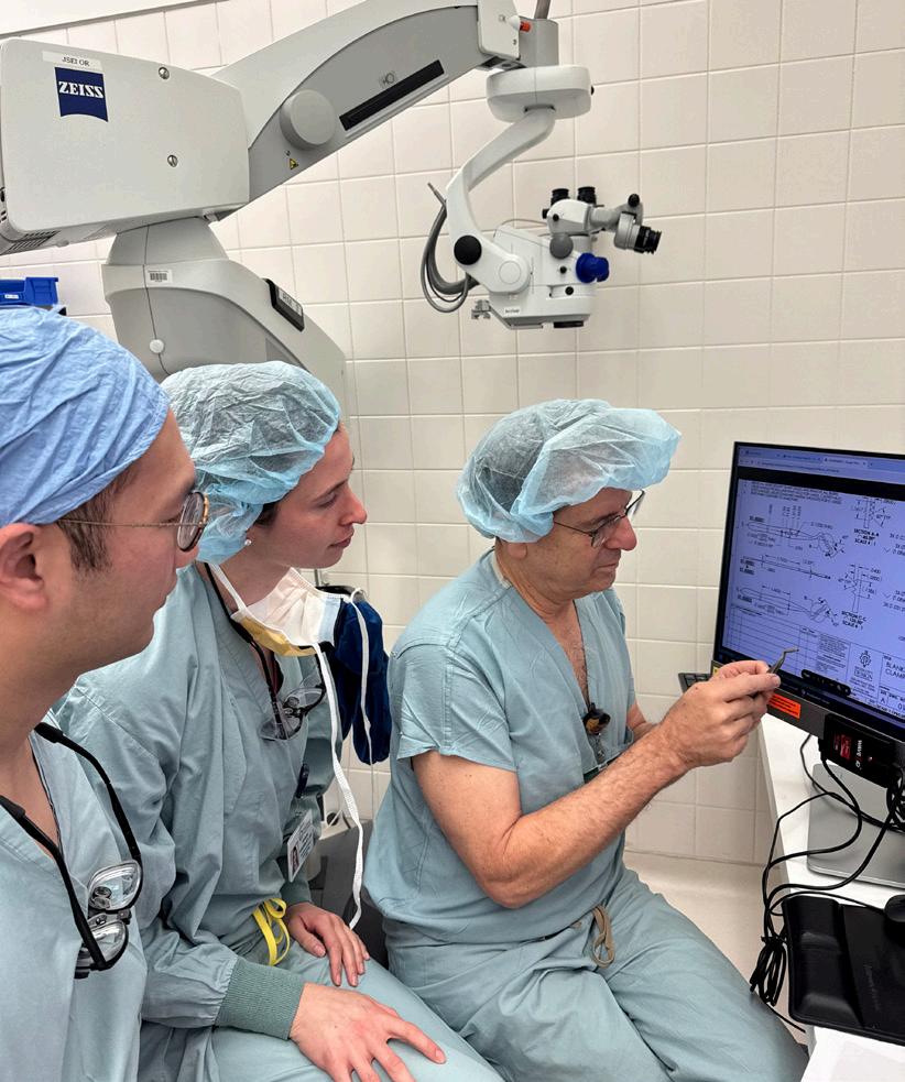
Dr. Goldberg’s team works with instrument makers and precision machinists at Bausch + Lomb, translating an exchange of ideas or back-of-the-napkin sketches into practical surgical instruments that are ultimately licensed and used for thousands of procedures every year.
Among the instruments created by Dr. Goldberg’s team is a cutting needle holder that aids in surgical efficiency, a retractor with new design elements for increased effectiveness in work around the eye, and an instrument (now in development) to improve accuracy in eyelid surgery.
“These instruments were initially created for surgeries on the eyelids, tear ducts, and other tissues around the eye, but also can be used for many types of procedures,” Dr. Goldberg says.
At the Jules Stein Eye Institute, the philosophy of continuous improvement is instilled in the next generation of ophthalmologists; Drs. Goldberg and Malyugin organize an annual summit for Institute residents and fellows to generate new ideas and planning for development of surgical tools and methods.
“We encourage a culture of innovation,” says Dr. Goldberg, “working together as a team to explore the challenges of surgery and inspire a process of always thinking of better ways to work.
“We train our residents to think of themselves as innovators, to learn to see the process of identifying a clinical problem, think about a solution, and then work with industry to bring a product or technique to life so it can benefit thousands of patients.
“It’s important to foster a culture where people are always thinking about those goals, and feel empowered to work on new ideas. It’s a philosophy within the Institute, and a culture that we encourage.”
The ongoing challenge is to innovate with new surgical instruments and methods that are simpler, more efficient, and more accurate. It has been a hallmark of our program to create advances in surgery and instruments. We have been innovating with that goal for decades.
ROBERT ALAN GOLDBERG, MD
Dr. Robert Goldberg (right) reviews the design of the Gout Ptosis Clamp with Oculoplastic Fellows Dr. Frank Mei (left) and Dr. Tatiana Rosenblatt (center).
A Heritage of Clinical Innovation
Developing innovation in patient care has been integral to the UCLA Jules Stein Eye Institute since its founding in 1966. Institute faculty have been responsible for a range of milestones that affect vision practice worldwide, among them:
Leonard Apt, MD: “A Man of Firsts”
Dr. Apt, who died in 2013, invented diagnostic tests, including the widely used Apt test, which distinguishes between fetal and maternal blood after birth. He was the first to use plastic tubing for blood transfusions, to develop a method for predicting allergy to catgut and collagen sutures prior to surgery, to pinpoint a formula for the eyes’ proper position under anesthesia, and to identify several new diseases.
Joseph L. Demer, MD, PhD: Eye Movement
Dr. Demer, Arthur L. Rosenbaum, MD, Chair in Pediatric Ophthalmology and chief of the Pediatric Ophthalmology and Strabismus Division, discovered the role of the orbital connective tissues in ocular motility and strabismus. He identified the first description and naming of “sagging eye syndrome,” and multiple innovations and surgeries arose from this insight. From Dr. Demer’s work, it is now understood that sagging eye syndrome is the most common cause of acquired double vision in people over age 50 years, and the condition no longer requires complex and costly clinical neurological investigations.
Jean-Pierre Hubschman, MD: Robotics for Ophthalmic Surgery
Dr. Hubschman, past Institute faculty, founded the Institute’s Advanced Robotic Eye Surgery Laboratory. He spearheaded breakthroughs in ophthalmic robotics, from developing the Intraocular Robotic Interventional and Surgical System (IRISS), which has performed automated cataract removal in porcine eyes, to leading the creation of the Polaris AI-enabled microsurgical platform.
Sherwin Isenberg, MD: Preventing Eye Disease in Newborns
Dr. Isenberg, distinguished professor of ophthalmology and pediatrics emeritus, together with Dr. Apt, established the use of povidone iodine as a safe topical antimicrobial agent. It is now used worldwide before eye surgery, applied to the eyes of newborn babies, and utilized to treat infections. Dr. Isenberg also led a team that produced extraordinary success in preventing blindness in Sub-Saharan Africa by ensuring precise levels of oxygen therapy for premature babies—work that contributed to Dr. Isenberg receiving the 2025 Parks Medal for a lifetime of achievement in pediatric ophthalmology.
Kevin M. Miller, MD: Artificial Iris
Dr. Miller, Kolokotrones Chair in Ophthalmology and chief of the Cataract and Refractive Surgery Division, pioneered work on artificial iris implantation that led to the first prosthetic iris approved by the FDA. Coining the term, “Astigmatism Management,” Dr. Miller organized a step-by-step approach to astigmatism standard of care that is now used worldwide. He developed toric markers, lens injectors, anterior capsule polishers, and suture removal forceps with industry partners. Dr. Miller has also played a key role in bringing phacoemulsification instruments, the first capsule tension ring, and the first postoperative, power-adjustable intraocular lens to market.
Steven D. Schwartz, MD: Embryonic Stem Cell Breakthrough
We train our residents to think of themselves as innovators, to learn to see the process of identifying a clinical problem, think about a solution, and then work with industry to bring a product or technique to life so it can benefit thousands of patients. It’s a philosophy within the institute, and a culture that we encourage.
Robert Alan Goldberg, MD
Dr. Schwartz, past Institute faculty and past chief of the Retina Division, led the first documented study showing that human embryonic stem cells could improve vision in patients with otherwise untreatable eye diseases, marking a pivotal moment in regenerative medicine. In the preliminary trial, published in The Lancet, Dr. Schwartz and his team injected embryo-derived stem cells into the retinas of two patients, both of whom experienced measurable and meaningful vision gains.
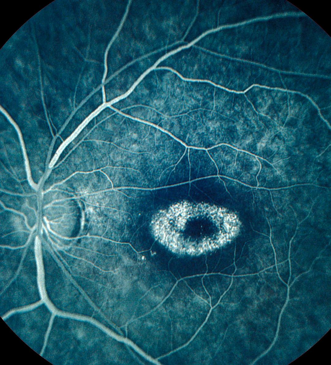
With Key Discovery and Development of New Disease Models, UCLA Team Changes the Paradigm for Stargardt Research
STARGARDT DISEASE is the leading cause of genetic blindness—resulting in the progressive loss of central vision, most commonly beginning in the first or second decade of life. The symptoms of this juvenile macular degeneration resemble those of age-related macular degeneration (AMD), leading to devastating results for young people, with no cure.
For many years, it’s been known that Stargardt is associated with a mutation in the ABCA4 gene, affecting the vitamin A transport in the retina. In the mid1990s, researchers localized the protein encoded by ABCA4 and described its role in recycling the visual chromophore to prevent toxicity within photoreceptor cells. But more than two decades after that important finding, a team led by Roxana A. Radu, MD , Vernon O. Underwood Family Chair in Ophthalmology, and director of the Retina Biochemistry and Disease Modeling Laboratory at the UCLA Jules Stein Eye Institute, identified additional novel pathogenic pathways, with significant clinical implications both for patients with recessive Stargardt disease (STGD1) and a subgroup of AMD patients.
Publishing in PNAS in 2018, Dr. Radu and her colleagues reported that the ABCA4 gene and its protein product are expressed not only in the photoreceptor cells, but also in the retinal pigment epithelium (RPE). “This has changed the paradigm in the study of Stargardt disease pathology,” Dr. Radu says. “The idea that there are multiple types of cells—rod and cone photoreceptors and RPE—contributing to the pathology opens up a major new direction toward a better understanding of this disease.”
Dr. Radu explains that a key pathological feature of STGD1 RPE cells is complement dysregulation, reflecting an inappropriate innate immune response to cellular stressors such as vitamin A dimers, oxidized lipids, and undegraded
proteins. While complement involvement had previously been associated only with AMD, Dr. Radu’s lab was the first to identify inefficient complement activity in RPE cells from STGD1 patients. The significance of the findings was reflected in the 2017 PNAS, as her group developed a gene-based therapy to demonstrate the effect of complement on the RPE homeostasis.
Building on that discovery, Dr. Radu’s lab pioneered the development of experimental disease models using both mice and human stem cell lines. With human-derived cells from STGD1 patients, she and her team validated what they had found in the mouse model. Publishing in Cells in 2022, Dr. Radu and her colleagues introduced STGD1 as a
I’m very optimistic. I have no doubt that with the models we have developed, we will learn a great deal more about the pathology of this disease.
ROXANA A. RADU, MD
“disease in a dish,” using the induced pluripotent stem cells from an STGD1 patient to describe RPE’s role in the pathology in the absence of photoreceptors. They found that compared to a healthy patient, the RPE cells in STGD1 showed reduced ABCA4 protein levels and diminished activity. This provided strong evidence in human-derived cells that complement dysregulation in the RPE cells plays a critical role in visual loss associated with STGD1, as well as late-onset macular degeneration.
From there, Dr. Radu’s group analyzed three patient-derived cell lines with distinct ABCA4 mutations. In their 2024 FASEB Journal paper, they presented findings showing that each displayed impaired protein degradation—indicating a cell-autonomous RPE-driven pathology—and suggesting the potential value of future research targeting RPE cells to treat ABCA4-mediated retinopathies. “This study validates that, irrespective of the mutation, RPE membranes have a deficient lipid composition that bears on the balance of processing, recycling, degradation, and clearance of internalized material,” Dr. Radu explains. “Basically, RPE becomes like the garbage can of all this material, losing the ability to clear properly.”
Dr. Radu believes the implications of her group’s research are still not fully appreciated. “Many biotech companies continue to target the photoreceptor cells only for therapeutics,” she says. “Our findings, endorsed by other labs, suggest that recessive Stargardt disease is largely a retinal pigment epithelium disease.” Ultimately, she hopes this understanding will lead to gene therapy approaches that target all cells expressing ABCA4, such as RPE cells and the light-sensitive rod and cone cells.
Originally from Romania, where she obtained her medical degree, Dr. Radu came to UCLA in 2001 for a postdoctoral fellowship in retinoid biochemistry under the mentorship of Jules Stein Eye Institute faculty member, Dr. Gabriel Travis Her initial project as a postdoctoral fellow involved working on the first mouse model generated for Stargardt disease. “I became so fascinated by the vitamin A biochemistry in the eye, and at the end of my fellowship, I knew I wanted to dedicate my career to understanding the cause of blindness in Stargardt at the molecular level, focusing on the innate immunity and pathogenic role of vitamin A dimers,” Dr. Radu says.
Nearly 25 years later, her team continues to make significant headway as it learns more about the disease. “We hope we can show that by intervening along these new pathways specific to RPE, we can rejuvenate the RPE cells and lengthen their life. Our working hypothesis is that a gradual decline in RPE functional and structural aspects has a subsequent deleterious impact on the photoreceptors’ activity. Therefore, maintaining a healthier RPE cell, we can promote the survival of the photoreceptor to prevent loss of sight,” Dr. Radu says.
“I’m very optimistic,” she adds. “I have no doubt that with the models we have developed, we will learn a great deal more about the pathology of this disease.”
A Gift Inspired by Hope for a Future Without Stargardt Disease
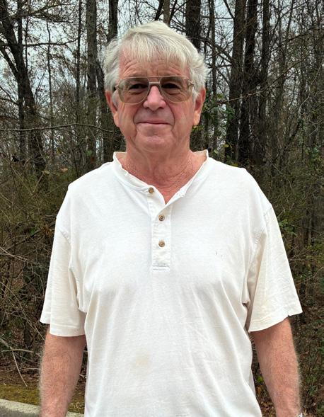
David Thompson was nine years old when he was diagnosed with Stargardt disease and told he would “just have to live with it.” More than 50 years later, his eye doctor in Georgia mentioned a clinician-researcher at UCLA who was exploring the use of stem cells to treat degenerative retinal disease. “That was the first glimmer of hope I had heard about for Stargardt,” David said. After learning more about the research, he came to the UCLA Jules Stein Eye Institute to see if he was a candidate for the clinical trials, only to be told he was too old.
David remained in contact with Rosaleen Ostrick, MPH, MA , administrative director of Retina Clinical Research, and stayed informed about clinical trials. During one discussion, he mentioned wanting to donate to the Institute’s research. Rosaleen connected him to Susan Lee DeRemer, CFRE , director of development, and after several conversations, during which David shared that other family members have Stargardt disease, he decided to increase his impact by naming the Jules Stein Eye Institute as a beneficiary in his will. “I am not a wealthy man,” he said, “but I have always saved my money, and since I am single with no children, this seemed like the best use of it.”
David also wanted to donate while he was alive. For a recent eye appointment, he flew in from Georgia with his brother and niece to present the Institute with a $10,000 check. It was an amazing experience, he said, to see how welcoming and grateful everyone was. He was looking forward to sharing the experience with his friends when he returned home, saying many had mentioned wanting to donate as well to support his quest to advance potential therapies for Stargardt disease and, ultimately, a cure.
David Thompson was nine years old when he was diagnosed with Stargardt disease.
David shared that other family members have Stargardt disease. He decided to increase his impact by naming the Jules Stein Eye Institute as a beneficiary in his will. “I am not a wealthy man,” he said, “but I have always saved my money, and since I am single with no children, this seemed like the best use of it.”
Advancing Dr. Allan Kreiger’s Legacy by Strengthening Retinal Care for Underserved Communities
Allan E. Kreiger, MD, founding chief of the Retina Division at the UCLA Jules Stein Eye Institute, passed away in 2024. To honor his legacy, the Kreiger Retinal Support of Medically Underserved Populations Fund was established, reflecting his lifelong belief that every patient deserves access to high-quality retinal care. The strong response to the Fund inspired the launch of a $1 million campaign in 2025 to build on this momentum and create the Allan E. Kreiger, MD, Endowed Chair in Retinal Diseases.
This campaign endowed the Kreiger Retinal Support of Medically Underserved Populations Fund, allowing it to expand its impact in meaningful and lasting ways. While the campaign ultimately did not reach the threshold required to establish the endowed chair, the Fund is now providing increased support for individuals with limited access to retinal care and stands as a powerful tribute to Dr. Kreiger’s lifelong commitment that all patients, regardless of circumstances, receive the care they need. Through this expanded support, the Fund continues to strengthen our mission of preserving and restoring vision through exceptional clinical care, innovative research, education, and community outreach.
On behalf of everyone at the Jules Stein Eye Institute, we are deeply grateful for the generous partnership of the faculty, former students, colleagues, and patients who helped pay tribute to Dr. Kreiger and carry forward his promise to make a difference in the lives of those who need it most.
Dr. Natik Piri: A Career Dedicated to Preserving Vision
Natik Piri, PhD, professor of ophthalmology, retired January 1, 2026. Dr. Piri joined the Department as assistant professor in the Glaucoma Division in March 2003. He received his doctorate in molecular biology from the Institute of Biorganic Chemistry, U.S.S.R. Academy of Sciences in Moscow. He began postdoctoral studies at the UCLA Jules Stein Eye Institute in 1991, working in the laboratory of Dr. Debora Farber. He was honored as a Distinguished Postdoctoral Fellow in Neuroscience by the UCLA Brain Research Institute in 1994. The following year he joined the Institute as an assistant research ophthalmologist.
Dr. Piri’s research aimed toward understanding the molecular mechanisms leading to retinal ganglion cell (RGC) death in glaucoma. Due to his expertise in this field, he became the director of the Molecular Biology of RGC Laboratory at the Institute, where his efforts in defining the molecular mechanisms underlying RGC degeneration, developing strategies for preserving RGC loss in glaucoma, and identifying novel genes expressed in RGC whose encoded proteins are critical for RGC function will remain impactful. Dr. Piri also worked closely with Dr. Joseph Caprioli and was a member of the Glaucoma Center for Excellence in Care and Research, working alongside researchers and statisticians from the UCLA School of Public Health to identify individuals at greatest risk for vision loss, to develop and assess therapeutic strategies, and implement new treatments to preserve vision. Dr. Piri was also a long-standing contributor to the Core Class for the Undergraduate Neuroscience Majors at David Geffen School of Medicine, teaching about sensory systems.
Dr. Piri received grants for his research, including support from the National Eye Institute and the Gerald Oppenheimer Family Foundation. He was the recipient of the 2015 Spitzer Grant Research Program award for support of groundbreaking medical research at UCLA. In addition to publishing his work in well-respected peer-reviewed journals, such as Investigative Ophthalmology & Visual Science and Cells, Dr. Piri served as an Editorial Board Member for the Open Journal of Synthesis Theory and Applications, Advances in Medicine: Ophthalmology, and the Journal of Neurophysiology and Neurological Disorders
Dr. Piri’s achievements have not only pushed the boundaries of scientific inquiry but have also inspired countless colleagues and students. We are grateful for Dr. Piri’s contributions to the Department over the past three decades and wish him the best in this next chapter.
UCLA Ophthalmology Named Top Five Program for Academic Excellence
The UCLA Department of Ophthalmology has been ranked among the top five programs in the nation for academic achievement, according to a peer-reviewed study published in the Journal of Academic Ophthalmology in December 2025. The analysis evaluated U.S. ophthalmology departments using objective measures, including faculty research impact, lifetime scholarly influence, and leadership roles on editorial boards of leading scientific journals.
This national recognition underscores UCLA Ophthalmology’s strength in research, education, and innovation, and reflects the Department’s ongoing commitment to advancing vision science and improving patient outcomes worldwide.
Congratulations to our clinical fellows, Alejandro Itzam Marin, MD, and Brian Soetikno, MD, PhD, on receiving the prestigious Heed Fellowship. Supported by the Heed Ophthalmic Foundation and the Society of Heed Fellows, this award provides funding and national recognition to outstanding early- career ophthalmologists committed to leadership in academic medicine.
Join us June 12, 2026, for the UCLA Department of Ophthalmology Annual Seminar
Held at the Jules Stein Eye Institute’s RPB Auditorium, the event highlights clinical and research advances across ophthalmic subspecialties, along with distinguished named lectures.
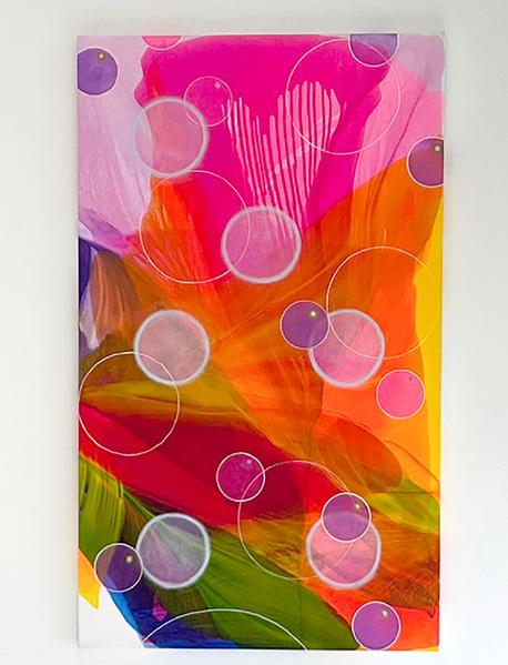
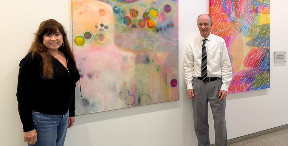
Where Art and Healing Meet
An installation of 20 original contemporary paintings by artist Linda Stelling adorns the once-barren white walls of the Edie & Lew Wasserman Building, providing a cohesive display of color and motif. Ms. Stelling, a patient of Dr. Kevin Miller’s since 2012, says it is her hope that people appreciate the art that embellishes the area and integrates joy into the space.
The Edie & Lew Wasserman Building is an award-winning six-story structure highlighted by clean lines, white terracotta, and pale oak. It features floor-to-ceiling windows that flood spacious rooms with natural light and reveal dramatic views of the campus. It was designed by Richard Meier and Partners, the same architects who created the Getty Center.
2025 American Academy of Ophthalmology Meeting
UCLA Department of Ophthalmology faculty had an outstanding showing at the American Academy of Ophthalmology annual meeting held October 18 to October 20, 2025, in Orlando, Florida. Our team contributed to symposia, instructional courses, rapid-fire sessions, posters, and panel discussions spanning retina, glaucoma, imaging, inflammation, and artificial intelligence applications in ophthalmology. Drawing worldwide participation, the educational meeting is a global sharing of knowledge that advances our field.
Extending our successful collaboration, the Jules Stein Eye Institute and Doheny Eye Institute once again partnered on a shared exhibit booth, which highlighted our programs and showcased groundbreaking research and clinical excellence. The booth was a perfect hub for connecting with alumni, industry colleagues, and collaborators, and served as an anchor for conversations about current research and future clinical innovations.
Artist Linda Stelling joins Dr. Kevin Miller, chief of the Cataract and Refractive Surgery Division, which is housed in the Edie & Lew Wasserman Building.
Major Grants Fuel Research Innovation
Vision scientists from the UCLA Department of Ophthalmology have earned significant research grants, underscoring the Department’s national leadership in advancing eye and vision science. Spanning a broad range of investigations, these competitive awards will accelerate groundbreaking work aimed at preventing vision loss, restoring sight, and improving patient outcomes. Together, these grants, representing more than $8.6 million in new research funding, reflect both the depth of the Department’s scientific expertise and the continued momentum behind its mission to transform the future of eye care through research excellence.
Adrian Au, MD, PhD
$1,221,895
NIH K23 Career Development Award. PROJECT: Genetic Variation in AMD Progression and Treatment Response
Sophie X. Deng, MD, PhD
$3,140,138
NIH R01 Research Project Grant
PROJECT: Development of Stem Cell–Based Therapies for Limbal Stem Cell Deficiency
Greg D. Field, PhD, and David S. Williams, PhD
$2,518,452
NIH/NEI P30 Vision Science Core Award
PROJECT: Support of the Vision Research Core within the Jules Stein Eye Institute
Kaustabh Ghosh, PhD
$1.5 million
NIH R01 Grant (three-year award)
PROJECT: Mechanobiology of Retinal Vascular Inflammation and Degeneration in Diabetes
David S. Williams, PhD
$75,000
Research to Prevent Blindness International Collaborators Award
PROJECT: Establishing Better Models for Studying Retinal Degeneration and Developing Therapies for Usher Syndrome Type 1B
Luke Nelson, Medical Student, Howard University College of Medicine
$38,000
Research to Prevent Blindness Medical Student Fellowship
with David S. Williams, PhD
PROJECT: The Role of Mitochondria in Supporting Photoreceptor Outer Segment Phagosome Degradation in Retinal Pigment Epithelium
Edmund Tsui, MD, MS
$129,604
NIH Small Business Technology Transfer Award in collaboration with ChromoLogic LLC
PROJECT: NGS-Based
Metagenomic Screening of Ocular Infections
FACULTY HONORS
Anthony J. Aldave, MD, Bartly J. Mondino, MD, Endowed Chair in Ophthalmology, was the recipient of the Jules Stein Eye Institute’s 2025 Golden Eye Award. The award recognizes tremendous surgical capabilities, as well as the ability to collaborate exceptionally with the operating room (OR) team. The award voting committee is comprised of OR nurses, scrub techs, and staff.
Saba Al-Hashimi, MD, health sciences associate clinical professor, has been appointed as the director of ophthalmology at UCLA West Valley Medical Center.
Clémence Bonnet, MD, PhD, health sciences clinical professor, received a 2025 Research to Prevent Blindness Stein Innovation Award.
Joseph Caprioli, MD, FACS, David May II Chair in Ophthalmology, was Guest of Honor at the American Glaucoma Society (AGS) annual meeting in Rancho Mirage, California, on February 20, 2026. He was selected unanimously by the AGS Board of Directors for his exceptional leadership and his outstanding scientific and academic contributions to the field.
Vikas Chopra, MD, The Charles Stewart Warren and Hildegard Warren Endowed Research Chair, was appointed to the Board of Directors of the American Board of Ophthalmology, the organization that oversees certification for ophthalmologists. In this role, he will shape the future of eye care by helping to design and oversee ophthalmology expertise exams and guide efforts to improve how doctors deliver safe, high-quality care.
Joseph L. Demer, MD, PhD, Arthur L. Rosenbaum, MD, Chair in Pediatric Ophthalmology, delivered the 2025 Vancouver John PrattJohnson Lecture, Treating Strabismus Caused by Pathology of the Orbital Pulley System, at the University of Vancouver, Canada, on September 7, 2025.
Deborah A. Ferrington, PhD, The Stephen J. Ryan–Arnold and Mabel Beckman Foundation Endowed Presidential Chair, was featured in the September/October 2025, issue of Ophthalmology Times. In the interview, Dr. Ferrington highlighted how breakthroughs in imaging, artificial intelligence, and stem cell research are reshaping ophthalmology.
JoAnn A. Giaconi, MD, and Gary N. Holland, MD, have been named Department of Ophthalmology Specialty Advisors for Career Exploration. In this role, they serve as experienced faculty advisors, facilitating opportunities for students of all levels to explore the ophthalmology specialty and provide support for students through their residency application process.
Kouros Nouri-Mahdavi, MD, MSc, Kay K. Pick Endowed Chair in Glaucoma Research, served as chair of the American Glaucoma Society’s (AGS) 2026 annual meeting in Rancho Mirage, California, February 19–22, 2026. Dr. Nouri-Mahdavi was also chair of the AGS 2025 annual meeting.
Pradeep S. Prasad, MD, MBA, health sciences associate clinical professor, received a 2025 American Society of Retina Specialists (ASRS) Honor Award for his time and contributions to the scientific programs of the ASRS annual meeting.
Alfredo A. Sadun, MD, PhD, Flora L. Thornton Endowed Chair in Vision Research, served as chair of the Leber’s Hereditary Optic Neuropathy (LHON) Forum held virtually on November 17, 2025. The event featured an international faculty presenting the latest research and clinical perspectives on LHON.
Jayanth Sridhar MD, chief of Olive View-UCLA Medical Center, gave the Ching J. Chen, MD Memorial Lecture at the University of Mississippi Medical Center in Jackson, Mississippi, on August 16, 2025. He also presented the Robert Haimovici Visiting Retina Lecture at the Bascom Palmer Eye Institute in Miami, Florida, on January 29, 2026.
Irena Tsui, MD, associate professor, has been appointed as Hilel Lewis Family Chair in Ophthalmology. Dr. Lewis, recognized as a pioneer in the treatment of retina diseases and Jules Stein Eye Institute alumnus, established this chair in 2020 via Columbia University to support an outstanding clinician-investigator in retina.
Xian-Jie Yang, PhD, professor of ophthalmology, has been appointed Dolly Green Chair in Vision Science. The endowed chair was established in 2021 to support a vision scientist.
Victoria H. Yom, MD, health sciences assistant clinical professor, has been appointed as Cornea Fellowship Director for the Doheny Eye Centers UCLA.
NEW FACULTY APPOINTMENTS
Giulia Corradetti, MD
Health Sciences Clinical Instructor of Ophthalmology
Ali Mahdavi Fard, MD
Assistant Clinical Professor of Ophthalmology
Dr. Corradetti is a retina specialist and physician-scientist with expertise in retinal diseases, advanced retinal imaging, functional endpoints, and oculomics. Born and raised in Italy, she was trained at the San Raffaele Research Institute in Milan. She completed an international medical retina fellowship at the UCLA Jules Stein Eye Institute and Doheny Eye Centers UCLA. During her fellowships, she became interested in clinical research in the applications of cutting-edge retinal imaging to early diagnosis of age-related macular degeneration, and in 2024, she was awarded the prestigious Evangelos Gragoudas Award at the Macula Society for her work in this field. Along with her research being featured in peer-reviewed journals, Dr. Corradetti is also a lecturer at national and international conferences. She is passionate about education, and she is excited to mentor medical students, residents, and fellows.
LOCATION: Doheny Eye Center UCLA–Pasadena
Dr. Mahdavi Fard is a fellowship-trained cornea specialist with over 15 years of clinical ophthalmology experience. He completed two ophthalmology residencies (Tabriz University of Medical Sciences and SUNY Buffalo) and two cornea fellowships (Doheny Eye Center UCLA and Tabriz University). Board certified by the American Board of Ophthalmology and a Fellow of the American Academy of Ophthalmology, he served as assistant professor of ophthalmology and medical director at Loma Linda University and SAC Health. He has received multiple honors, including the “Healing Hands” award, a Certificate of Appreciation for clinical teaching, the award for Outstanding Academic Performance, and induction into the Gold Humanism Honor Society. A researcher with over 30 peer-reviewed publications, Dr. Mahdavi Fard also serves as a reviewer for leading journals. Fluent in English, Farsi, and Turkish, he is devoted to compassionate patient care, academic excellence, and teaching— embodying the ideal of a “teacher with healing hands.”
LOCATIONS: Doheny Eye Centers UCLA–Pasadena and Orange County
COMMUNITY OUTREACH
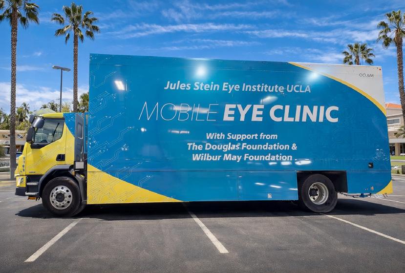
Powering Sight, Sustainably: New Electric Mobile Eye Clinic
We are pleased to announce the addition of our one-of-a-kind electric vehicle to the UCLA Mobile Eye Clinic (UMEC) fleet! This new electric vehicle clinic represents our commitment to sustainability, innovation, and community health. Partnering with Odulair and Topcon, the team spent 16 months perfecting the functions and operation of our custom-made vehicle, ensuring the most comfortable and advanced experience for our patients.
With contributions from The Douglas Foundation and Wilbur May Foundation, this investment supports our mission by reaching patients where care is needed most, while also aligning the UCLA Mobile Eye Clinic with UCLA’s environmentally responsible practices.
We are excited to put the new all-electric vehicle out into the field this spring, and are grateful to all of UMEC’s supporters, partners, patients, students, and friends for making this a reality.
For more information on the UCLA Mobile Eye Clinic please visit https://www.uclahealth.org/departments/eye/mobile-eye
EDUCATION
Aesthetic Eyelid and Facial Rejuvenation Course
The Aesthetic Eyelid and Facial Rejuvenation course, chaired by Robert A. Goldberg, MD, was presented at the UCLA Jules Stein Eye Institute July 11–12, 2025. The course showcases the work of UCLA faculty, and presents safe, innovative, and minimally invasive techniques. Attendees from throughout the world met for the two-day course. The laboratory dissection day concentrated on anatomic and surgical pearls of core aesthetic procedures and included a live audiovisual feed of course faculty performing high-definition dissections. The second day featured lectures and discussions highlighting conceptual approaches, nuances of facial rejuvenation, and new surgical and nonsurgical techniques for facial rejuvenation. Ronald Mancini, MD, FACS, presented the Shorr Lecture. Course directors were Dr. Goldberg, Jonathan A. Hoenig, MD, Justin N. Karlin, MD, and Daniel Rootman, MD
For information about upcoming orbital and ophthalmic plastic surgery courses, email Dr. Karlin at jkarlin@mednet.ucla.edu
The two-day Aesthetic Eyelid and Facial Rejuvenation Course is taught by recognized leaders in the field. Top right: Dr. Ronald Mancini (R) presented the Norman Shorr Lecture, and is shown with Dr. Shorr (L).
Women in Ophthalmology Summer Symposium
The Women in Ophthalmology Summer Symposium was held August 7–10, 2025, in Amelia Island, Florida. The meeting was an opportunity for attendees to learn about subjects of importance to women ophthalmologists, and UCLA faculty presented at the meeting in multiple capacities, including:
f Anne L. Coleman, MD, PhD: Speaker, “Academic All Stars” Breakout Session: “How to Set Yourself Up for Success: Negotiating During Academic Hire”
f Victoria L. Tseng, MD, PhD: Instructor, Phacoemulsification Wet Lab and Minimally Invasive Glaucoma Surgery Wet Lab. The Summer Symposium was an enriching and educational experience for all who attended.
Innovation and Engagement at the 7th Annual Glaucoma Symposium
The 7th Annual Doheny-UCLA Glaucoma Symposium was a great success with more than 80 attendees. It was held on September 27, 2025, at Doheny Eye Institute. We extend our sincere thanks to the UCLA Department of Ophthalmology glaucoma faculty, including Reza Alizadeh, MD, Judy Chen, MD, JoAnn Giaconi, MD, Simon Law, MD, PharmD, Kouros Nouri-Mahdavi, MD, MS, Vivian Qin, MD, and Victoria Tseng, MD, PhD, for their superb presentations, which covered a range of important topics in glaucoma care and innovation. The hands-on wet labs were also a highlight, giving participants the opportunity to gain practical experience and engage directly with new surgical techniques.
A special thank you goes to symposium co-directors Vikas Chopra, MD, and Brian Francis, MD, MS, for their leadership in organizing this outstanding program.
Cataract Surgery Essentials Course
Kevin M. Miller, MD, chief of the Cataract and Refractive Surgery Division, and Boris E. Malyugin, MD, PhD, Joan and Jerome Snyder Chair in Cornea Diseases, presented the annual Cataract Surgery Essentials Course sponsored by Zeiss on November 1, 2025, in Costa Mesa, California.
The course featured labs and workstations where approximately 60 trainees from UCLA, USC, UCI, Loma Linda, UCSD, and the Naval Medical Center gained hands-on experience learning about essential surgical instruments and equipment used in cataract surgery.
Exploring the Future of Retina Care at Doheny-UCLA International Retina Symposium
The 4th annual Doheny-UCLA International Retina Symposium was held January 31, 2026, at Doheny Eye Institute. It featured international keynote speakers Sobha Sivaprasad, MD, FRCOphth, Moorfields Eye Hospital, UK, and Mark W. Johnson, MD, University of Michigan, who shared insights on retinal disease management and emerging innovations in clinical practice. Attendees gained a deeper understanding of new treatments for age-related macular degeneration, surgical advancements, and the latest in translational research—all in one dynamic, information-packed day designed to enhance patient care and professional growth. Course directors were Michael Ip, MD, and Giulia Corradetti, MD
Jules Stein Eye Institute Distinguished Lecture Series
The UCLA Jules Stein Eye Institute Distinguished Lectures Series, coordinated by Roxana A. Radu, MD, was host to two notable speakers at the Institute’s RPB Auditorium in November 2025.
Rui Chen, PhD, professor of ophthalmology at the Gavin Herbert Eye Institute Center for Translational Vision Research in the Department of Ophthalmology at University of California, Irvine, spoke on November 7. Dr. Chen utilizes genetics, genomics, model systems, and bioinformatics to unravel the genetic basis of human retinal diseases. Chen is also leading the effort of creating the human eye’s single-cell atlas for the Human Cell Atlas project, employing cutting-edge single-cell omics and spatial transcriptomics technologies.
Aaron Nagiel, MD, PhD, assistant professor of ophthalmology at the Keck School of Medicine of USC, gave a presentation at the Institute on November 21. His research focuses on understanding how connections between nerve cells in the human retina form and function, both in health and disease, by studying lab-grown human retinal tissue to inform new treatments such as gene and cell therapies.
Doheny Distinguished Lecture Series
Doheny Eye Institute closed 2025 and opened 2026 in their Distinguished Lecture Series with presentations from vision researchers who shared groundbreaking insights.
Machelle Pardue, PhD, professor and vice chair of research in ophthalmology at Emory University and senior research career scientist at the Atlanta VA Healthcare System, spoke on December 12. Dr. Pardue focuses on developing clinically relevant treatments for retinal diseases that improve patients’ quality of life. Her lab is advancing novel screening and treatment strategies for earlystage diabetic retinopathy and exploring the retinoscleral mechanisms of myopia.
Connie Cepko, PhD, Bullard Professor of Genetics and Neuroscience at Harvard Medical School and investigator at the Howard Hughes Medical Institute, gave a presentation on January 9, 2026. Dr. Cepko’s research looks at the development and diseases of the retina, addressing questions regarding the mechanisms of cell fate determination, and developing gene-agnostic gene therapy to preserve vision.
The lecture series can be joined via Zoom. For information on how to attend and future speakers, email Yolanda Mercado at YMercado@doheny.org
Alumni Reconnect at AAO 2025 in Orlando
Alumni from the UCLA Jules Stein Eye Institute and Doheny Eye Institute gathered October 18 in Orlando, Florida, for a lively alumni reception held in conjunction with the 2025 American Academy of Ophthalmology annual meeting. The event was an opportunity to reconnect with longtime colleagues, build new professional relationships, and celebrate the collective achievements of our distinguished alumni community.
Throughout the evening, conversations flowed around shared experiences, fresh ideas, and the innovations shaping the future of ophthalmology. A highlight of the night was a high-energy DJ set by AJA—also known as Dr. Anthony Aldave—which added a memorable and unexpected flair to the celebration.
Thank you to everyone who joined us and helped make this event both engaging and impactful.
Stay connected with the UCLA Ophthalmology Alumni Association by sharing your preferred contact details for digital Institute news and event invitations!
Email us at alumni@jsei.ucla.edu or scan the following QR code to confirm your preferred contact information.
We look forward to connecting with you at future alumni gatherings!
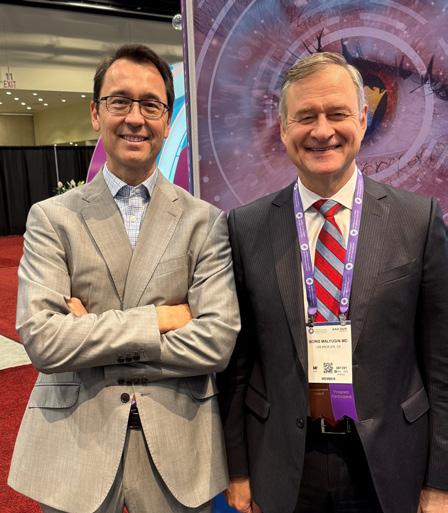
L to R: Drs. Brian Francis and Boris Malyugin.
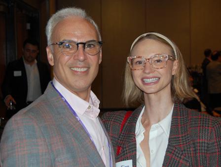
L to R: Drs. Peter Quiros and Kim Firn.
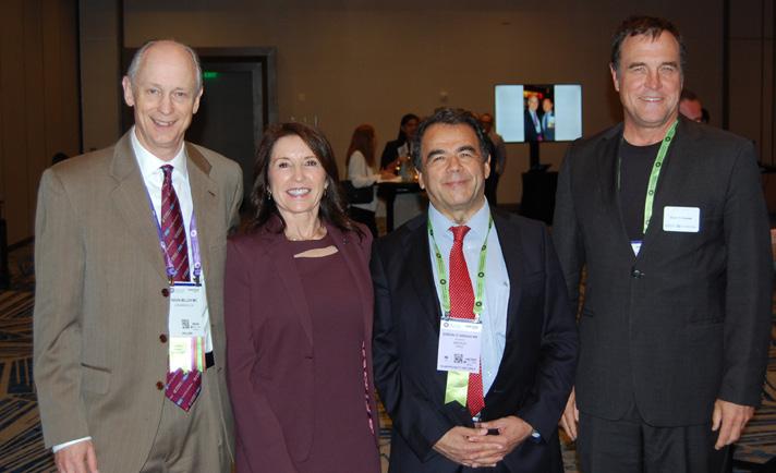
L to R: Drs. Kevin Miller, Anne Coleman, Gonzalo Vargas, and Colin McCannel.
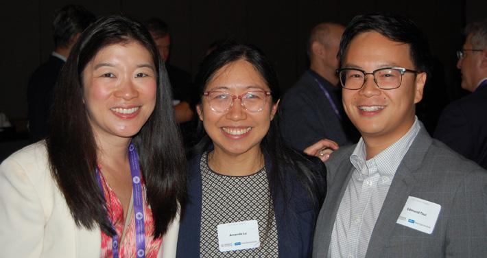
L to R: Drs. Victoria Tseng, Amanda Lu, and Edmund Tsui.
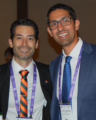
L to R: Drs. Chris Alabiad and Jayanth Sridhar.
iPads as Translators: Helping Every Patient Be Heard
At the heart of excellent eye care is something simple but essential: being able to communicate clearly with your ophthalmologist. When patients can describe what they’re experiencing, ask questions, and truly understand their treatment options, they’re better equipped to take an active role in protecting their vision. Yet for many in our community, language differences can make these conversations challenging.
Recognizing this, the Department’s Inclusive Excellence (IE) Committee, led by Vice Chair Sophie Deng, MD, PhD, is committed to making sure that every patient has equal access to exceptional eye care and can communicate comfortably with their care team at every step of their visit.
Bringing real-time interpretation to the clinic
The Committee previously surveyed patients about barriers to receiving vision care, and one message came through clearly: language is one of the biggest obstacles. In response, two members of the IE Committee, Yolanda McNair and Laura Bonelli, MD, were key in launching an initiative to make communication faster and easier for patients who prefer a language other than English.
Supported by the Department of Ophthalmology and the David Geffen School of Medicine at UCLA, Yolanda spearheaded the effort to purchase and distribute iPad interpreter devices at all clinic sites of the UCLA Jules Stein Eye Institute and Doheny Eye Centers UCLA, as well as the UCLA Stein Eye Surgery Center. These iPads connect patients and providers with professional medical interpreters, via video or audio, within moments. This real-time access helps:
f Reduce delays in care
f Improve accuracy when sharing symptoms or discussing treatment
f Support patients who are deaf or hard of hearing
f Ensure every patient receives informed and compassionate care.
Progress so far
Early feedback from both patients and clinicians has been overwhelmingly positive, particularly regarding the ease of use and the clarity of video interpretation. Staff are relying less on family members or coworkers to translate, helping protect patient privacy and ensuring medical accuracy.
Looking ahead
A formal evaluation is currently underway to better understand how the devices are being used and how effectively they are easing communication barriers, a process led by Monica Khitri, MD, Dr. Deng, and medical student Adam Alnihmy. An IRB-approved survey will be launched later this year to gather insights from patients and clinicians, and Simon Law, MD, is leading a complementary survey examining how vision care providers view and deliver care to older adults, with analysis nearly complete and a manuscript expected next spring.
Taken together, these efforts will shape the next steps in expanding language services and strengthening support for aging patients. What remains unchanged is the Department’s commitment to ensuring that every person who walks through our doors feels heard, valued, and confident in their care.