WGO Executive Secretariat 555 East Wells Street, Suite 1100 Milwaukee, WI 53202 USA info@worldgastroenterology.org Editorial Message from the Editors 3 Expert Point of View
INTERESTED IN WRITING FOR E-WGN?
SUBMIT YOUR ARTICLE TODAY!
WGO is accepting article submissions for upcoming issues of e-WGN. Articles reach a global audience and are disseminated through WGO’s mailing list and social media platforms.
Article Instructions
To find more information on article and author instructions, visit our e-WGN submission portal! This portal contains all necessary instructions to help you complete your e-WGN contribution and allows you to submit all information in one place. There are many ways to contribute to our newsletter! Expert Point of View, meeting promotions and summaries, testimonials and more are all welcome inside of e-WGN! Explore our submission portal at:
e-WGN Submission Portal
Please note that the Co-Editors of e-WGN may make edits and changes to your article.
For more information, please email info@worldgastroenterology.org
Consultant of Gastroenterology, Hepatology & Endoscopy
Police Authority Hospitals Cairo, Egypt
Dao Viet Hang, MD, PhD
Director, Endoscopy Center, Hanoi Medical University Hospital (HMUH)
Senior Researcher, Institute of Gastroenterology and Hepatology (IGH) Hanoi, Vietnam
Welcome to this special issue of e-WGN, our first edition dedicated exclusively to Expert Point-of-View articles.
In response to the growing interest from the global gastroenterology community, e-WGN has launched a series of special issues focused on emerging and clinically relevant topics that are shaping contemporary practice. This issue presents five expert perspectives addressing key developments in multidisciplinary management of acute severe ulcerative colitis, liver transplantation, dysphagia, optimization of endoscopy reporting, and chronic hepatitis B guidelines.
Acute severe ulcerative colitis (ASUC) remains one of the most challenging emergencies in gastroenterology, requiring timely diagnosis and coordinated multidisciplinary care. Dr. Fabián Juliao-Baños and Dr. Viviana Parra Izquierdo from Colombia provide a comprehensive overview of current approaches to initial assessment and risk stratification, while reviewing contemporary rescue therapies and surgical indica-
tions based on the latest evidence and guideline recommendations.
Liver transplantation continues to evolve as a life-saving option for a broad spectrum of liver diseases. Dr. Burcu Gürbüz, Dr. Berat Baran, and Dr. Hatice Yasemin Balaban present a detailed review of current indications for liver transplantation, encompassing cirrhosis, acute liver failure, acuteon-chronic liver failure, metabolic diseases, and selected oncological conditions. Their article also highlights critical donor-related considerations, including donor selection, ethical challenges, organ allocation, pre- and post-transplant evaluation, and management of transplant-related complications. Importantly, the authors emphasize that ethical principles remain fundamental to all aspects of transplantation practice.
The issue also features an intriguing discussion on festive dysphagia, authored by Prof. Nazish Butt, Prof. Amanullah Abbasi, Dr. Sabir Ali, and Dr. Kanwal Butani from Karachi, Pakistan. This phenomenon emerged from the increasing incidence of food bolus obstruction observed during the
Islamic festival of Eid-ul-Adha, often associated with hurried eating, inadequate mastication, distraction during meals, and ingestion of large pieces of meat. In addition to outlining endoscopic management strategies, the authors present illustrative clinical cases and underscore the importance of public education and preventive measures to reduce the burden of this avoidable condition.
Standardization of endoscopic reporting is increasingly recognized as an essential component of high-quality gastrointestinal care. Prof. Sandie R. Thomson, Dr. Sabina Beg, and Dr. Shivangi T. Kothari provide a comprehensive narrative review of published guidance on endoscopy reporting, including recent recommendations from the American Society for Gastrointestinal Endoscopy (ASGE), the British Society of Gastroenterology (BSG), and the European Society of Gastrointestinal Endoscopy (ESGE). Their analysis highlights the value of structured reporting systems in enhancing diagnostic accuracy, facilitating communication among healthcare professionals, and improving clinical decision-making. The authors also examine the challenges and opportunities associated with implementation across diverse healthcare settings and compare international reporting frameworks and supporting data infrastructures.
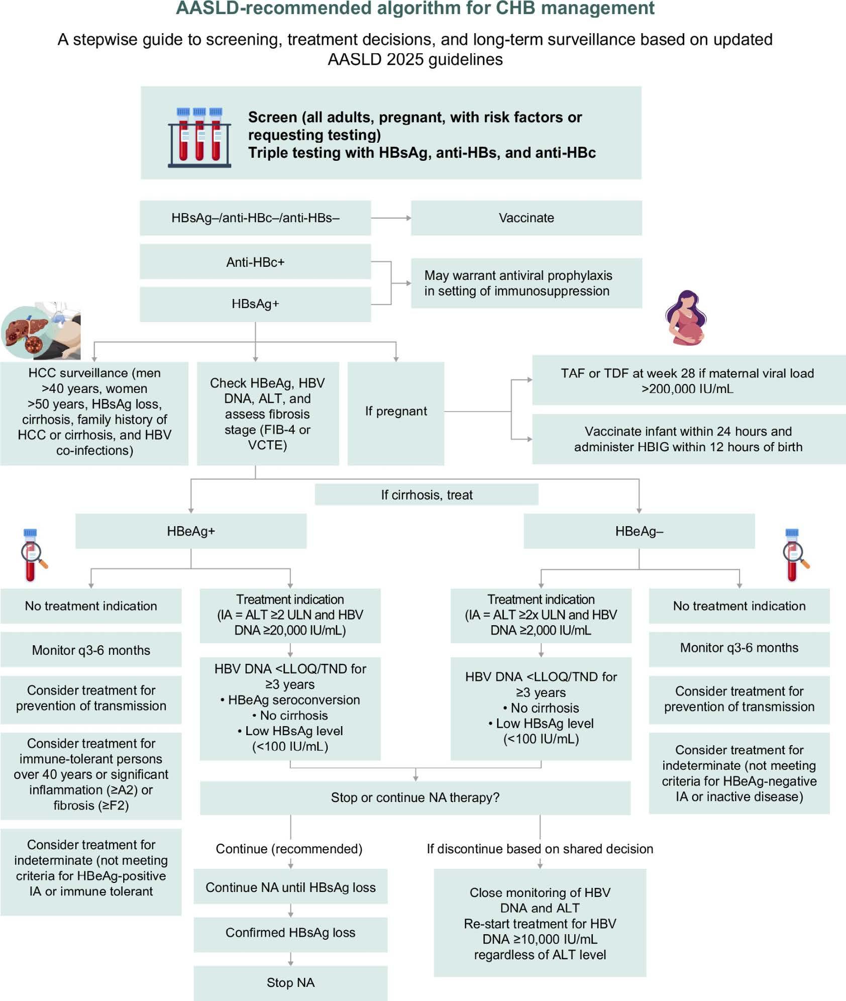
Finally, Dr. Leandro Sierra from Cleveland Clinic and Dr. Nikki Duong from Stanford University Medical Center present a comprehensive review of the 2025 AASLD/IDSA Practice Guideline on Chronic Hepatitis B. Key concepts discussed include the emerging definition of “partial cure,” management of patients within
the clinical “grey zone,” and treatment indications for special populations. The authors also provide a detailed comparison between the 2025 guideline and previous recommendations from AASLD (2018), as well as those from EASL and APASL. This timely review offers practical insights that will benefit not only hepatologists but also clinicians across multiple specialties involved in the care of patients
with chronic hepatitis B.
We hope that this special issue, dedicated entirely to expert perspectives, will provide valuable insights, stimulate discussion, and inspire future innovation in clinical practice and research. We warmly invite our readers to share their recent publications, professional experiences, society news, and educational activities with the World Gastroenterology Organ-
isation (WGO) for consideration in future editions of e-WGN. We also encourage you to follow WGO through its official communication channels to stay informed about the latest developments and activities.
Sincerely,
Dr. Dao Viet Hang, Vietnam
Dr. Nancy Fanous, Egypt
Severe Acute Ulcerative Colitis (ASUC): The Importance of Multidisciplinary Management
Introduction
Fabián Juliao-Baños, MD
Gastroenterology and Digestive Endoscopy
IBD Center of Excellence
Pablo Tobón Uribe Hospital
Medellín, Colombia
Viviana Parra Izquierdo, MD
Gastroenterologist — Rheumatologist
IBD Center of Excellence
International Hospital of Colombia
Cellular and Molecular Immunology Group (InMubo), Universidad El Bosque
Medellín, Colombia
Ulcerative colitis (UC) is a chronic inflammatory disorder of the gastrointestinal tract that primarily involves the rectum and extends proximally through the colon in a continuous pattern. It is thought to result from a dysregulated immune response to intraluminal antigens in genetically susceptible individuals. Clinically, bloody diarrhea is the hallmark symptom, and the disease course is typically characterized by alternating periods of remission and exacerbation.1 Acute severe ulcerative colitis (ASUC) represents the most severe clinical presentation of UC and is defined by the presence of bloody diarrhea associated with systemic inflammation. Because it is a potentially life-threatening condition, ASUC requires prompt recognition and timely multidisciplinary management in specialized centers.2 It has been estimated that approximately 25% of patients with UC will experience at least one severe flare requiring hospitalization during their lifetime, and nearly 20% of these cases may occur at the time of diagnosis. The risk
of colectomy during the first hospitalization is around 20%, increasing to as much as 40% after recurrent hospitalizations.3 Furthermore, a systematic review reported mortality rates of 0.84% and 1.01% at 3 and 12 months of follow-up, respectively, with mortality increasing significantly with advancing age and the presence of comorbidities.4
Definition
The latest version of the European Crohn’s and Colitis Organization (ECCO) guidelines5 has adopted the Truelove & Witts classification,5 proposed more than 60 years ago, to define ASUC.6 This includes the presence of more than 6 bloody bowel movements per day, plus at least one of the following criteria: tachycardia (heart rate greater than 90 beats per minute), fever (temperature greater than 37.5 °C), hemoglobin less than 10.5 g/dL, and erythrocyte sedimentation rate (ESR) greater than 30 mm/h. A new classification proposed by the American College of Gastroenterology (ACG) proposes the term ful-
minant colitis, and in addition to the previous criteria, adds the following severity criteria: presence of continuous fecal urgency, fecal calprotectin levels > 150-200 mcg/g, endoscopic Mayo index of 3 and a wall thickening > 3 mm, along with submucosal edema and hyperperfusion on intestinal ultrasound (IUS).7
Initial Evaluation and Management
The initial evaluation and support of these individuals require management by a multidisciplinary team that includes a gastroenterologist, colorectal surgeon, nutritionist, nurse, and the support of an emergency physician, general surgeon, internist, and intensivist, according to the patient’s needs arising from their clinical condition. The goal of treatment in acute colitis is to achieve a clinical response, defined as a decrease (< 3) or normalization of the number of daily bowel movements, without visible bleeding. These criteria must be met when deciding on outpatient management, according to expert opinion.8
Upon admission, many patients are present with dehydration and hypokalemia due to their diarrhea, thus requiring adequate support with intravenous fluids and potassium supplementation. The oral route appears to be appropriate for nutrition, unless the patient presents with toxic megacolon or paralytic ileus. The advent of total parenteral nutrition (TPN) in the late 1960s sparked interest in whether bowel rest could eliminate the antigenic properties and mechanical trauma of ingested food. The prevailing view at the time was that TPN would promote the repair of damaged bowel, provide nutritional support to a frequently malnourished patient population, and lead to improved outcomes for pa tients with inflammatory bowel disease (IBD). Two UK-led randomized controlled trials (RCTs) in the 1980s, which used TPN as an adjunct to intravenous steroids, showed an increase in TPN-related complications but no improvement in colectomy rates or mortality.33 There is renewed interest in considering nutritional enrichment (NE) as a nutritional approach for the treatment of severe acute ulcerative colitis (ASUC) after Sahu et al. conducted an open-label RCT to demonstrate its efficacy in a cohort of 62 patients randomized 1:1 to semi-elemental NE or standard treatment. A per-protocol analysis showed a significant reduction in inflammatory markers (C-reactive protein [CRP] and fecal calprotectin), length of hospital stay, and mortality. Although this trial was prematurely terminated due to the COVID-19 pandemic and recruited just over 50% of the planned sample size, the study demonstrated an enhanced response to corticosteroids, underscoring the need for further trials to confirm these
findings.34
Infectious causes of colitis must be ruled out. Avoid the use of medications that decrease colonic motility in patients at risk of toxic megacolon, including opioid derivatives, antidiarrheals, and anticholinergics, as well as nonsteroidal anti-inflammatory drugs (NSAIDs) due to the risk of exacerbation. A plain abdominal radiograph is part of the initial evaluation to rule out the presence of ileus, colonic dilation, or perforation.
In cases of suspected abdominal sepsis or doubt regarding
biopsies within the first 72 hours of hospitalization results in a lower rate of colectomy (17% vs. 28%), fewer days of hospitalization, and less use of rescue therapy.11
The routine use of antibiotics in the absence of an infectious focus in patients with ASUC is controversial. A recent meta-analysis of 13 studies and 785 patients showed that they are not effective in inducing a clinical response (OR: 0.94, 95% Confidence Interval CI: 0.51–1.74).12 Given that ASUC is a hypercoagulable inflammatory state, thromboprophylaxis is recommended in hospitalized patients with ASUC. The presence of rectal bleeding is not a contraindication, and some suggest continuing thromboprophylaxis on an outpatient basis for up to 8–12 weeks, the duration of the hypercoagulable state.9, 13
colonic perforation, an abdominal CT scan should be performed.9
Furthermore, a flexible rectosigmoidoscopy (RSC) without bowel preparation should be performed within the first 24–72 hours of admission, with minimal insufflation and biopsy sampling, to establish endoscopic severity, rule out the presence of pseudomembranes associated with Clostridioides difficile infection, and obtain biopsies to exclude cytomegalovirus (CMV) infection, especially in patients with pre-existing immunosuppression, according to ECCO recommendations.10 A recent retrospective study demonstrated that performing flexible RSC with
Initial Treatment
For over 50 years, Truelove & Jewell have changed the natural history of patients with ASUC with the use of intravenous (IV) steroids.14 The current recommended dose is 100 mg of hydrocortisone every six hours or 60 mg of methylprednisolone daily. A review of 32 randomized controlled trials (RCTs) and cohort studies, conducted between 1974 and 2006 in patients requiring IV steroids, found an overall response rate of 67%, with a colectomy rate of 27% and a mortality rate of 1%.15 One RCT demonstrated that patients with ASUC do not require continued mesalazine during hospitalization; there is no difference in clinical response to IV steroids with or without 5-ASA at seven days of follow-up.16
Risk Stratification
Several composite indices exist that allow the identification of patients with ASUC who may require “rescue” therapy after IV steroid use. One
index, called the ACE score, is applied upon patient admission and includes CRP ≥50 mg/L, albumin ≤30 g/L, and a severe Mayo endoscopic index of 3, predicting a 78% steroid failure rate.17 A more recent index, called the ADMIT-ASC score, is also applied upon admission and assigns a score, including CRP ≥100 mg/L = 1 point, albumin ≤25 g/L = 1 point, UCEIS ≥4 = 1 point, and UCEIS ≥7 = 2 points; a score ≥3 predicts an 84% steroid failure rate.18 A study from Oxford (UK) found that having more than 8 bowel movements per day on day 3, or the combination of 3–8 bowel movements per day with a CRP greater than 45 mg/L, is associated with an 85% risk of steroid failure. This is known as the Oxford or Travis index.19
Rescue Therapy
Historically, patients with ASUC who did not respond to IV steroids ended up undergoing colectomy. The first drug used as rescue therapy after steroid failure was cyclosporine (Cys), an immunosuppressant macrolide that inhibits the activation of interleukin-2 (IL-2) by activated T lymphocytes through a calcineurindependent pathway. The first randomized controlled trial (RCT) with Cys was conducted by Lichtiger et al. in 20 patients with ASUC who had not responded to hydrocortisone for 7 days. Cys was used at a dose of 4 mg/ kg/day, achieving a clinical response rate of 82% vs. 0% with placebo (P < 0.001), with a colectomy rate at 2 weeks of follow-up of 18% vs. 44%, compared with placebo (RR: 0.6, 95% CI: 0.18–2.06).20 Since then, Cys has been used in patients with ASUC at a dose of 2 mg/kg/day. Infliximab (IFX), an anti-tumor necrosis factor alpha (anti-TNF α) agent, has been used for individuals with ASUC for several years. The first RCT with IFX was conducted in centers in Sweden and Denmark, using a single dose of 5 mg/kg as “rescue”
therapy in 45 patients (24 IFX, 21 placebo) refractory to IV steroid treatment. It found a 3-month colectomy rate of 29% with IFX and 67% with placebo (P: 0.017; OR: 4.9; 95% CI: 1.4–17.0).21
It has been documented that individuals with ASUC have high circulating and tissue levels of tumor necrosis factor (TNF), which acts as a “sponge” that rapidly absorbs standard doses of anti-TNFs. Furthermore, it has been established that mononuclear cells of the reticuloendothelial system phagocytize and proteolytically degrade the complex formed by the anti-TNF-α drug with TNF. Finally, increased mucosal permeability associated with severe inflammation may act as a “sieve,” allowing fecal loss of anti-TNF.22
Additionally, in an Italian multicenter study, multivariate analysis demonstrated that CRP levels ≥ 3 mg/ dL (RR = 2.15, 95% CI 1.05–4.36, p = 0.003) and the presence of severe endoscopic lesions (RR = 5.13, 95% CI 1.55–16.96, p = 0.007) at the time of the acute attack were associated with a higher risk of colectomy.23 For all the above reasons, some authors suggest that patients with ASUC and CRP ≥ 3 mg/dL, serum albumin < 30 g/L, and severe endoscopic lesions (deep ulcers and spontaneous bleeding) require individualized management with a higher initial dose of IFX.24 However, a recent RCT from Australia (PREDICT-UC) compared IFX 5 mg/kg vs. 10 mg/kg in subjects with ASUC refractory to IV steroids and found no significant difference in the proportion of patients who had a clinical response by day 7 between the 10 mg/kg and 5 mg/kg groups (30 [65%] of 46 vs. 56 [61%] of 92, p=0.62).25
The choice between IFX and Cys as “rescue” therapy in ASUC has been highly controversial. A recent systematic review (SR) with 18 RCTs and 26 cohort studies suggests that IFX is superior to Cys in reducing the
risk of colectomy at 3 months (RR, 0.67; 95% CI, 0.48–0.92) and 12 months of follow-up (RR, 0.56; 95% CI, 0.42–0.75).26
On the other hand, recent studies in hospitalized patients with ASUC already exposed to anti-TNFs have demonstrated the efficacy of Janus kinase inhibitors (anti-JAKs), such as tofacitinib at a dose of 10 mg every 8 hours orally and upadacitinib at a dose of 45 mg daily or 30 mg every 12 hours. A recent systematic review with 35 studies and 664 individuals, in the short term (<1 month), showed that the pooled clinical response and colectomy rate with tofacitinib was 77.9% (95% CI, 67.1%-86%) and 11.5% (95% CI, 7.1%-18.4%), whereas for upadacitinib, it was 86.5% (95% CI, 72.3%-94.1%) and 11.2% (95% CI, 7.2%-16.9%), respectively. The adverse events reported were venous thromboembolism (2.2%; 95% CI, 1.1%–4.7%), major adverse cardiovascular events (0.7%; 95% CI, 0.1%–10.3%), and herpes zoster (3.4%; 95% CI, 1.9%–6.1%).27
An Indian RCT (TACOS) compared patients with ASUC, all receiving IV hydrocortisone, to a group that also received tofacitinib at a dose of 10 mg every 8 hours from day 1 and compared them to placebo. At day 7, response to treatment was achieved in 44/53 (83.01%) patients receiving tofacitinib vs. 30/51 (58.82%) patients receiving placebo (odds ratio 3.42, 95% CI 1.37–8.48, P 0.007).28
Sequential therapy with cyclosporine induction followed by vedolizumab maintenance has emerged as a valuable rescue strategy in ASUC, particularly in patients with steroid-refractory disease who have failed or are not candidates for anti-TNF therapy. The rationale for this approach lies in the rapid onset of action of cyclosporine, which can induce short-term clinical response within days, thereby serving as a bridge to vedolizumab, a gut-selective biologic with a slower
onset but favorable efficacy and safety for long-term maintenance. Available evidence suggests that this strategy can achieve high rates of remission while reducing colectomy risk and maintaining an acceptable safety profile. The most relevant prospective study supporting this approach showed that, among steroid-refractory ASUC patients treated with cyclosporine followed by vedolizumab, most patients achieved initial response, with sustained clinical, endoscopic, and even histologic remission during followup, as well as a high colectomy-free survival rate at 1 year, without serious adverse events.29
In a recent review, only three studies, including a total of 13 patients (the largest study included 10 patients), were found to report the efficacy of ustekinumab (an IL-12 and IL-23 p40 subunit antagonist) in ASUC. All three studies were retrospective and used sequential treatment in all patients, starting with a calcineurin inhibitor for induction (cyclosporine or tacrolimus), followed by ustekinumab as maintenance therapy. With this sequential treatment strategy, colectomy was avoided in all patients. Furthermore, the rate of serious adverse events and the mortality rate attributable to ustekinumab
were 0%.30
Surgical
Treatment
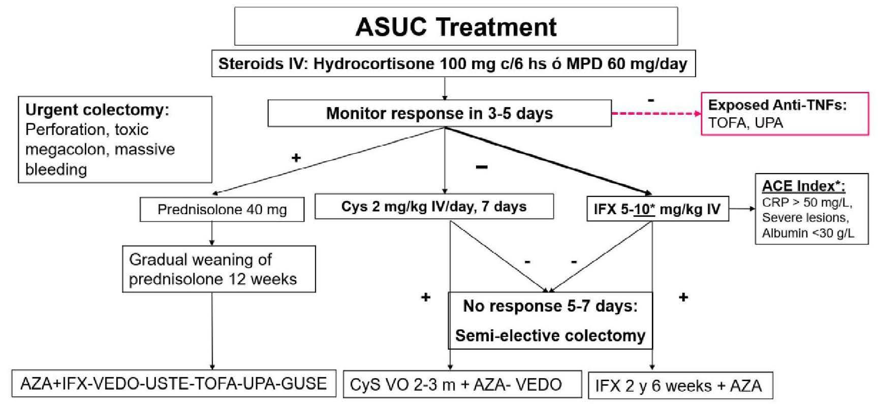
Although medical treatment is the first-line approach for managing patients with ASUC, there is a risk of colectomy, and a gastroenterologist or colorectal surgeon should be involved in the patient’s management from the time of admission. Indications for urgent colectomy include perforation, toxic megacolon, massive bleeding, and lack of response to medical treatment.5 An Oxford study of 80 patients with refractory colitis who underwent colectomy demonstrated that those who experienced more complications had a longer duration of medical treatment prior to surgery (8 vs. 5 days, P = 0.036).31 Additionally, a recent systematic review and meta-analysis, including 67,075 patients with colitis, found that postoperative mortality is lower in subjects undergoing elective surgery (0.7%, 95% CI 0.6%–0.9%) versus emergent surgery (5.3%, 95% CI: 3.8%–7.4%).32 Therefore, surgery should ideally always be semi-elective. A flowchart of the management of patients with ASUC is shown in Figure 1.
Conclusion
In summary, ASUC remains a lifethreatening condition that demands rapid recognition, close monitoring, and coordinated multidisciplinary care in specialized centers. Although intravenous corticosteroids remain the cornerstone of initial treatment, early risk stratification is essential to identify patients unlikely to respond and to expedite rescue therapy. Advances in medical management, including the preferential use of infliximab in selected cases and the emerging role of JAK inhibitors, have expanded the therapeutic options available for this challenging condition. Nevertheless, surgery remains a fundamental component of care, and early colorectal surgical consultation is crucial to optimize outcomes and avoid delays in intervention. Ultimately, successful management of ASUC depends on an evidence-based strategy that balances aggressive medical therapy with timely colectomy when indicated, always adhering to the principle of save the patient, not the colon.
References
Readers may access the full list of references here.
Figure 1
Liver Transplantation and Donor Health
Burcu Gürbüz, MD
Department of Internal Medicine, Gastroenterology and Hepatology
Hacettepe University
Ankara, Türkiye
Berat Baran, MD
Department of Internal Medicine
Hacettepe University
Ankara, Türkiye
Hatice Yasemin Balaban, MD
Department of Internal Medicine, Gastroenterology and Hepatology
Hacettepe University
Ankara, Türkiye
Introduction
Liver transplantation (LT) is the definitive treatment for end-stage liver disease, acute liver failure, certain metabolic disorders, and selected primary and secondary liver malignancies. Since the first successful human LT performed by Dr. Thomas Starzl in 1963, advances in surgical technique and perioperative care.1 Initially plagued by poor outcomes due to technical challenges and limited immunosuppressive options, the advent of calcineurin inhibitors in the 1980s marked a turning point, enabling dramatic improvements in graft and patient survival rates.2 The first successful living-related donor liver transplantation, performed by Strong et al. in 1989, represented another major milestone.3
More recent developments include expanded indications such as acuteon-chronic liver failure and transplant
oncology, improvements in organ preservation through machine perfusion, and refined allocation systems such as MELD 3.0. Contemporary transplantation practice increasingly emphasizes individualized candidate assessment, frailty, careful donor selection, and comprehensive pre- and post-transplant management. Nevertheless, organ demand continues to exceed supply worldwide, requiring innovation alongside ethical vigilance regarding donor protection and organ trafficking.
Indications
LT is a life-saving procedure for patients with decompensated cirrhosis, acute liver failure (ALF), acute-onchronic liver failure (ACLF), some metabolic diseases (tyrosinemia, urea cycle defects, or wilson’s disease), and oncological indications, such as hepatocellular carcinoma (HCC).4
Although hepatitis C virus (HCV)related cirrhosis was historically the leading indication, alcohol-related liver disease (ALD) and metabolic dysfunction-associated steatohepatitis (MASH) have become increasingly prominent.5 Other indications include hepatitis B virus (HBV), autoimmune hepatitis (AIH), primary biliary cholangitis (PBC), primary sclerosing cholangitis (PSC), alpha-1 antitrypsin deficiency, and inherited metabolic disorders. According to the OPTN/ SRTR 2023 Annual Data Report, ALD accounts for approximately 40% of adult LT candidates in the United States, while MASH is the second most common indication. This transition reflects the declining burden of viral hepatitis following effective antiviral treatment. The availability of direct-acting antivirals has also enabled HCV-negative recipients to receive grafts from HCV-infected donors, thereby expanding the donor pool.6
i. Cirrhosis
Cirrhosis remains the principal indication for LT worldwide. Progressive architectural distortion and portal hypertension lead to complications such as ascites, spontaneous bacterial peritonitis, hepatic encephalopathy, and variceal bleeding. Once decompensation occurs, median survival sharply declines, with estimates ranging from one to two years in the absence of transplantation.7 LT is the only curative intervention capable of correcting both hepatic insufficiency and portal hypertension. Timely referral improves survival and quality of life, whereas delayed assessment increases the likelihood of infection, renal dysfunction, frailty, sarcopenia, and waitlist mortality. Early identifica-
tion and multidisciplinary management are therefore essential. Transplant referral and organ allocation are guided primarily by prognostic scores.8 The original Model for End-Stage Liver Disease (MELD) score incorporates serum creatinine, bilirubin, and international normalized ratio (INR) to estimate threemonth mortality. Serum sodium was subsequently added to create MELDNa because hyponatremia independently predicts mortality. MELD 3.0 further incorporates serum albumin and female sex to improve prediction and address disadvantages experienced by women because of lower muscle mass and serum creatinine.9 Contemporary evidence supports MELD 3.0 as the most accurate available tool for determining urgency and guiding organ allocation in LT.
ii. Acute Liver Failure
Acute liver failure (ALF) is characterized by the sudden onset of hepatic dysfunction, coagulopathy, and hepatic encephalopathy in a person without preexisting liver disease.10 Common causes in developed countries include acetaminophen toxicity, acute viral hepatitis, autoimmune hepatitis, and idiosyncratic drug-induced liver injury.
ALF is distinguished by its rapid progression and unpredictable course, with many patients developing multiorgan failure within days to weeks.11 Spontaneous recovery varies according to etiology, age, and the severity of extrahepatic organ dysfunction; acetaminophen-related ALF generally has a more favorable prognosis. For patients with progressive encephalopathy and severe coagulopathy, however, LT remains the only definitive treatment.12 Listing decisions must be made rapidly to prevent irreversible neurological injury or sepsis. Prognostic tools such as the King’s College criteria are commonly used to guide listing decisions, but clinical judg-
ment remains paramount due to the dynamic nature of ALF.13 Although ALF accounts for fewer than 5% of liver transplants in the United States and Europe, one-year post-transplant survival generally exceeds 80%. Early referral to a transplant center is therefore crucial.
iii. Acute on Chronic Liver Failure
Acute-on-chronic liver failure (ACLF) is characterized by acute decompensation of chronic liver disease, accompanied by organ failures and increased short-term mortality. ACLF is most commonly precipitated by events such as bacterial infection, gastrointestinal bleeding, or active alcohol consumption.14 Severity is graded according to the number and type of organ failures; grade 3 ACLF representing the most severe form, frequently involving renal, circulatory, respiratory, or cerebral dysfunction, and may carry a 28-day mortality exceeding 75% at 28 days without LT.15
Although patients with ACLF were previously considered poor transplant candidates because of perioperative risk and concerns about futility, current evidence indicates that carefully selected patients can achieve substantial survival benefit. Potentially reversible extrahepatic organ failures, a controllable precipitating event, and timely transplantation are key considerations. Selected patients, including some with ACLF grade 3, may achieve one-year post-transplant survival comparable to recipients without ACLF.16
ACLF should be differentiated from acute decompensation (AD) of cirrhosis. The term AD is used if one or more significant consequences of liver disease occurs in a short time. Unlike ACLF, AD is not accompanied by an inflammatory state. The European Association for the Study of LiverChronic-Liver Failure (EASL-CLIF) Group had modified the Sequential Organ Failure Assessment score
(SOFA) that is used for the intensive care patients into the prognostic scores for cirrhotic patients with acute organ failure in cirrhotic patients:
• CLIF-SOFA (for organ failure in cirrhosis in general, and includes bilirubin for liver failure, creatinine for renal failure, West-Haven hepatic encephalopathy grade for neurological failure, INR for hematology, mean arterial pressure (MAP) for circulation, partial pressure of arterial oxygen/ fraction of inspired oxygen (PaO2/ FiO2) for respiratory system).
• CLIF-C ACLF (for ACLF, and include age and WBC in addition to CLIF-SOFA score).
• CLIF-C AD (for acute decompensation, and includes age, creatinine, INR, WBC and Na).
These scores may aid in identifying suitable candidates for transplantation, though expert multidisciplinary assessment remains crucial.17
iv. Metabolic Diseases
Inherited metabolic diseases constitute a significant and expanding indication for LT, particularly in pediatric practice. Disorders such as Wilson’s disease, hereditary hemochromatosis, alpha-1 antitrypsin deficiency, primary hyperoxaluria, and a variety of urea cycle and organic acidemias can result in progressive hepatic dysfunction, acute liver failure, or life-threatening systemic complications despite maximal medical management. In many disorders, LT replaces the deficient hepatic enzyme, prevents further toxicmetabolite accumulation, and may provide a definitive metabolic cure.18 Timing is critical because delayed transplantation can lead to irreversible neurological or renal injury. Inherited metabolic diseases requiring LT can be broadly categorized according to their pathophysiological mechanisms and the extent of hepatic
versus extrahepatic involvement. Group A includes disorders such as Wilson’s disease, hereditary tyrosinemia type I, and alpha-1 antitrypsin deficiency, where progressive hepatocellular injury leads to cirrhosis and liver failure. In these conditions, LT restores normal hepatic function and prevents further hepatic decompensation. Group B includes urea cycle disorders, porphyrias and primary hyperoxaluria. Here, these patients rarely have ESLD; LT is performed for extrahepatic organ involvement. LT not only replaces deficient enzymes to correct the metabolic defect but also halts the accumulation of toxic metabolites, thereby preventing irreversible neurological damage or other systemic complications. Group C includes maple syrup urine disease, mitochondrial hepatopathies. In such cases, transplantation can stabilize or slow disease progression but may not completely reverse extrahepatic manifestations.19
Overall, disease-specific assessment
and timely referral are essential for optimal outcomes.
v. MELD Exceptions
Classic examples of standardized MELD exceptions include patients with HCC meeting specific criteria (such as the Milan criteria), cholangiocarcinoma (CCA), familial amyloid polyneuropathy (FAP), cystic fibrosis, hepatopulmonary syndrome (HPS), or portopulmonary hypertension (POPH).20 Exception requests are reviewed by regional or national committees to promote consistency and prevent misuse.
MELD 3.0 improves allocation equity, particularly for women and candidates with hypoalbuminemia. Nevertheless, adjudicating exceptions remains difficult for diseases with variable natural histories or limited outcome data. Exception policies should therefore be periodically reviewed using registry evidence to ensure that allocation reflects both medical urgency and expected transplant benefit.
vi. Transplant Oncology
Liver transplantation for oncologic indications is firmly established selected patients with HCC who satisfy criteria such as the Milan or University of California, San Francisco criteria. For eligible patients with early-stage HCC and underlying cirrhosis, LT not only removes both the tumor and the underlying liver disease, resulting in five-year survival rates above 70% in many series. Locoregional ablation and transarterial chemoembolization may be used as bridging therapies while patients await transplantation.21 Recent studies have broadened the oncologic indications for LT beyond conventional HCC. Selected patients with intrahepatic cholangiocarcinoma have achieved promising outcomes under strict protocols incorporating neoadjuvant therapy.22 LT has also been investigated for unresectable colorectal liver metastases and selected neuroendocrine tumors.23 Because non-HCC malignancies generally carry greater recurrence risk, expansion of transplant oncology must be balanced against organ scarcity, utility, justice, and expected survival. Candidate selection should involve multidisciplinary tumor boards and prospective registries. Future progress will likely depend on improved molecular profiling, risk stratification, and careful integration of systemic and immune-based therapies.
Donor Issues
i. Organ Trading and Trafficking
Organ trafficking is a critical global health and human rights issue, and is estimated to account for up to 10% of all solid organ transplants performed worldwide.23 It is driven by the persistent imbalance between organ supply and demand, socioeconomic inequality, and unregulated transplant markets. Migrants, refugees, and people from low-income settings are
particularly vulnerable to coercion, deception, and commercial exploitation. These practices are frequently associated with inadequate medical assessment, poor postoperative care, and increased infectious and surgical complications.24
The Declaration of Istanbul, endorsed by major transplant societies and the World Health Organization (WHO), prohibits commercial organ trade and emphasizes transparency, traceability, voluntariness, and donor protection.25 Nevertheless, trafficking networks remain difficult to detect and prosecute because of their transnational structure, limited reporting systems, and occasional involvement of healthcare professionals. Transplant teams must therefore verify the legitimacy of donation and ensure that consent is informed and free from coercion.
Effective prevention requires international cooperation, harmonized legal standards, reliable donor registries, professional accountability, and public education. Combating organ trafficking is both a legal obligation and an ethical requirement for preserving trust and integrity in liver transplantation field.
ii. Donor Selection Criteria
a. Living Donor Criteria
Living Donor Liver Transplantation (LDLT) offers several advantages, including the reduction of waitlist mortality and the opportunity to schedule transplantation electively. Donor safety, voluntariness, and autonomy are the central principles of the evaluation process. Potential donors must receive comprehensive information about operative risks, possible effects on quality of life, and available alternatives.
and absence of contraindicating anatomical variants. Typically, donors are aged 18–55, though carefully selected older individuals may also be suitable candidates. Candidates should be in good overall health, without significant cardiovascular, pulmonary, metabolic, infectious, or chronic liver disease. Serological testing, CT or MRI assessment of vascular and biliary anatomy, and evaluation for hepatic steatosis and fibrosis by noninvasive methods or biopsy are required. Psychosocial assessment should confirm informed motivation and exclude coercion.
Surgical planning for LDLT is technically complex, involving selection of the optimal liver graft (right lobe, left lobe, or left lateral segment) based on recipient needs and donor safety. Threedimensional imaging and virtual surgical planning are increasingly used to estimate graft and future liver remnant volumes, which are critical for minimizing the risk of small-for-size syndrome in the recipient and ensuring adequate residual function for the donor. Detailed mapping of vascular and biliary anatomy allows for safe resection and reconstruction. The graft-to-recipient weight ratio should ideally be ≥0.8%, and the donor’s future liver remnant should exceed 30% of their original liver volume.26
LDLT is associated with low donor mortality of approximately 0.1–0.2%, and available evidence does not indicate reduced longterm life expectancy compared with the general population. Nevertheless, donors remain at risk of biliary complications, infection, bleeding, venous thromboembolism, and, rarely, acute liver failure. Long-term complications may include incisional hernia, fatigue, chronic pain, depression, or
adjustment difficulties, although most donors report high satisfaction with their decision.
b. Cadaveric Donor Criteria
Cadaveric (deceased) donor selection aims to maximize graft viability while minimizing the transmission of infection or malignancy. Evaluation includes review of the donor’s medical and social history, laboratory testing, and imaging, ensuring there are no active systemic infections, high-risk malignancies, or other contraindications such as uncontrolled sepsis, untreated HIV, or recent high-risk behaviors. Exclusion criteria may also encompass hemodynamic instability, prolonged hypotension, and severe steatosis or fibrosis.6
Deceased donation are classified as either donation after brain death (DBD) or donation after circulatory death (DCD). DBD remains the main source of cadaveric livers globally, but the increasing acceptance of DCD has significantly expanded the donor pool. While DCD grafts are associated with a higher risk of ischemic cholangiopathy and primary nonfunction, improved procurement methods and the implementation of normothermic machine perfusion have markedly enhanced graft preservation, reduced complications, and allowed for successful transplantation from donors previously deemed marginal.
Successful donor-recipient matching is achieved by careful consideration of ABO blood group compatibility and size (especially in pediatric and small adult recipients), and human leukocyte antigen (HLA) typing, when possible. Additional factors such as donor age, liver function tests, cold ischemia time, and presence of anatomical variants further
guide allocation and perioperative planning. The integration of comprehensive donor risk indices, such as the Donor Risk Index (DRI), aids transplant teams in stratifying grafts and predicting post-transplant outcomes, thus supporting clinical decision-making.
In summary, deceased donor selection requires multidisciplinary assessment that balances recipient safety, organ utility, equitable allocation, and the risks associated with broader donor acceptance.
iii. Living Donor Follow-up
Post-donation monitoring protocols require structured lifelong follow-up addressing both physical and psychological outcomes. Early monitoring includes liver function tests and assessment for bile leaks, bleeding, infection, thromboembolism, vascular complications, and, rarely, liver failure. Longer-term surveillance should evaluate biliary or vascular abnormalities, incisional hernia, persistent fatigue, chronic pain, metabolic changes, and occult liver dysfunction.
Psychosocial follow-up is equally important, particularly when the recipient experiences graft failure or death. Donors should be assessed for anxiety, depression, adjustment difficulties, or regret, using structured interviews or validated tools where available. Access to dedicated psychosocial support should form part of routine donor care.
With appropriate selection and follow-up, living liver donors generally have survival comparable to the general population, and most report high satisfaction and willingness to donate again. Centralized registries and national reporting systems are essential for identifying late complications and refining donor selection, surgical practice, and long-term monitoring. Multidisciplinary followup therefore remains fundamental to maintaining the safety and ethical
integrity of LDLT programs.
Pretransplant Evaluation of Recipient
i. Routine Tests
Multidisciplinary pretransplant evaluation aims to confirm transplant eligibility, identify modifiable risks, and reduce perioperative complications. The standard evaluation protocol begins with a comprehensive laboratory work-up, including liver biochemistry and synthetic function, renal function and electrolytes, complete blood count, and metabolic parameters. Abdominal imaging, typically with ultrasound and multiphasic CT or MRI, evaluates the liver parenchyma, vasculature, and biliary system, and screens for hepatocellular carcinoma, portal vein thrombosis, and other structural abnormalities.
Cardiopulmonary evaluation generally includes electrocardiography and transthoracic echocardiography, with stress testing, myocardial perfusion imaging, or coronary assessment in selected patients. Chest imaging, pulse oximetry, and pulmonary function testing are used to identify underlying pulmonary disease. Arterial blood gas analysis, contrast echocardiography, or right-heart catheterization may be required when hepatopulmonary syndrome or portopulmonary hypertension is suspected.
Infectious disease screening is mandatory for all candidates and includes serologies for HBV and HCV, HIV, cytomegalovirus (CMV), Epstein-Barr virus (EBV), and screening for tuberculosis, syphilis, and, where relevant, endemic infections (e.g., Strongyloides, Chagas disease). Identification of active infection is a contraindication to transplantation until adequately treated. Vaccination status should also be reviewed, and indicated vaccines— including hepatitis A and B, pneumococcal, influenza, and varicella vaccines—should ideally be administered before immunosuppression.
Nutritional status, frailty, substance use, psychological health, treatment adherence, and social support must also be assessed because they influence perioperative and long-term outcomes. Evaluation therefore requires collaboration among hepatology, transplant surgery, cardiology, infectious diseases, nutrition, and psychosocial care teams.
ii.
Treatment Targets for Underlying Liver Disease
• Viral Hepatitis (HBV and HCV): For patients with chronic HBV infection, achieving and maintaining viral suppression is essential before LT using potent nucleos(t) ide analogues, such as entecavir or tenofovir, which minimize the risk of HBV reactivation and graft reinfection after LT. Prophylactic strategies —including hepatitis B immunoglobulin in selected cases— may be individualized according to virological risk profile and institutional practice.
Direct-acting antivirals (DAAs) achieve virological response in nearly all patients with HCV, and may be administered pre- or post-transplant. HCV-positive grafts can also be used in HCVnegative recipients when prompt post-transplant antiviral therapy is available, thereby expanding the donor pool.
• Alcohol-Associated Liver Disease (ALD):
Alcohol-related liver disease is a leading indication for LT in Europe and North America. Pretransplant management emphasizes a comprehensive psychosocial evaluation and multidisciplinary care, including addiction specialists, psychiatry, and social work. Although traditional protocols required a 6-month period of abstinence before transplantation, recent evidence and guidelines
support early transplantation in carefully selected patients with severe ALD who fail medical therapy. Selection criteria focus on psychosocial stability, family support, absence of comorbid psychiatric illness, and demonstrated motivation for sustained abstinence. Continued addiction care and relapse monitoring are essential after transplantation.
• Autoimmune Liver Diseases (AILD):
In AIH, PBC, and PSC, the diagnosis should be confirmed and disease-specific treatment optimized before LT. Patients with acute severe AIH should receive early corticosteroid therapy, but nonresponders and those with progressive hepatic failure require prompt transplant referral. Adherence, social support, associated diseases, and the risk of post-transplant recurrence should be assessed in all patients with autoimmune liver disease.
iii. Sarcopenia, Cardiomyopathy
Evaluation
Assessment of sarcopenia and cardiomyopathy has become an integral component of pretransplant evaluation in candidates for LT, as both conditions have significant negative impact on perioperative risk and longterm outcomes.
Sarcopenia (defined as the loss of skeletal muscle mass and function) is highly prevalent in patients with advanced liver disease and is associated with increased waiting list mortality, higher rates of postoperative complications, and impaired quality of life. (27) Guidelines recommend systematic screening for sarcopenia using objective tools such as cross-sectional imaging (e.g., CT or MRI to quantify psoas muscle area), handgrip strength, and functional performance tests. Identification of sarcopenia should
prompt prehabilitation interventions, including tailored nutritional support, resistance exercise programs, and management of underlying factors such as chronic inflammation or hormonal disturbances. Early recognition and intervention can improve physical function and potentially enhance transplant candidacy and post-transplant recovery.28
Cardiomyopathy and, more broadly, cardiac dysfunction is another critical aspect of pretransplant assessment. Patients with cirrhosis are at increased risk for cirrhotic cardiomyopathy, characterized by blunted cardiac contractility, diastolic dysfunction, and electrophysiological abnormalities, which may be unmasked by the hemodynamic shifts of transplantation. The guidelines advise a comprehensive cardiac work-up, including echocardiography for structural and functional assessment, electrocardiography for arrhythmias or conduction defects, and, when indicated, stress testing or advanced imaging to evaluate for coronary artery disease or high-risk features. Early detection of cardiomyopathy or significant cardiac disease may necessitate further evaluation, medical optimization, or, in some cases, consideration of combined heart-liver transplantation.29
Early Complications of Liver Transplantation
i. Surgical Complications: Vascular and Biliary Complications
Vascular complications occur in approximately 7% of LT recipients and can lead to early graft loss and mortality, particularly if diagnosis is delayed. Early detection (primarily using doppler ultrasound, and when needed, angio-CT or angiography) is critical for optimal management.30
Hepatic artery thrombosis (HAT) is the most frequent arterial complication and remains the leading cause of graft failure. Risk factors include endothelial injury, extended cold isch-
emia, transfusions, hypercoagulability, technical problems and pediatric transplantation. Early HAT can present with fever, mental changes, and rapid liver enzyme elevation, often leading to graft ischemia, biliary necrosis, abscesses, or multiorgan failure. Without intervention, graft failure and high mortality are likely. Early detection protocols such as serial doppler ultrasounds are recommended, and endovascular interventions (thrombolysis/thrombectomy) are increasingly successful.31 Other arterial issues include hepatic artery stenosis, hepatic artery pseudoaneurysm (rare but life-threatening), and arterial conduit occlusion. Stenosis is treated with angioplasty or stenting, while pseudoaneurysm may require surgical intervention or retransplantation if rupture occurs.
Portal vein complications such as portal vein thrombosis or stenosis are often due to technical problems and low portal flow. These may present with acute liver failure or portal hypertension. Treatment may involve anticoagulation, angioplasty, or surgical revision. Hepatic vein or inferior vena cava outflow obstruction can also occur, especially in the early postoperative period, requiring angioplasty, stenting, or occasionally surgical revision.32
Biliary complications are frequent after liver transplantation, with reported incidences ranging from 2% to 19%. They are major causes of post-transplant morbidity and can be life-threatening.31 Bile leaks commonly develop in the first month after transplantation, with risk factors including technical errors, ischemic injury, and T-tube placement. Most leaks can be managed conservatively or with endoscopic or radiological drainage, but surgical revision is occasionally necessary. Biliary strictures are the most common biliary issue post-transplant, particularly extrahepatic anastomotic strictures, which
typically present within the first year. Endoscopic management with ERCP and stenting is the preferred first-line therapy, with surgery reserved for refractory cases. Intrahepatic biliary strictures (ischemic cholangiopathy) are especially associated with ischemic injury, prolonged preservation, or arterial problems (e.g., HAT), and are more frequent after DCD transplantation. These strictures are difficult to treat and often result in the need for retransplantation.
Machine-perfusion techniques may reduce ischemic injury, but close postoperative surveillance and rapid intervention remain essential because vascular and biliary complications may initially present with subtle clinical findings.
ii. Hyperacute and Acute Rejection
Hyperacute rejection is a rare but catastrophic complication that manifests within hours of transplantation triggered by preformed recipient antibodies directed against donor antigens, most often in the context of ABO blood group incompatibility or pre-existing anti-donor HLA antibodies. Rapid complement activation and endothelial injury cause diffuse vascular thrombosis and immediate graft failure. Clinically, hyperacute
rejection presents with signs indistinguishable from severe ischemic injury, with coagulopathy, hemodynamic instability, and rapid graft failure. Due to its fulminant course, urgent re-transplantation is typically the only therapeutic option. Improved compatibility testing and antibody screening have markedly reduced its incidence.
Acute rejection occurs in approximately 15–25% of recipients, most commonly during the first two to six weeks, although it may develop later. It is predominantly T-cell mediated and is associated with inadequate immunosuppression, younger age, and autoimmune or viral liver disease. Calcineurin inhibitors have substantially reduced its frequency and severity.33 Patients may present with nonspecific symptoms such as fever, malaise, right upper quadrant pain, jaundice, and altered liver biochemistry, especially elevation of transaminases and cholestatic enzymes. Diagnosis relies on a combination of clinical suspicion, laboratory findings, and most definitively histological confirmation via liver biopsy, which typically reveals portal inflammation, bile duct damage, and endothelitis. The percutaneous or transjugular approach may be employed depending on coagulopathy and patient
stability. First-line therapy consists of high-dose corticosteroids along with intensification of baseline immunosuppressive regimens. The majority of cases respond to standard therapy, but steroid-resistant rejection may require additional immunosuppressants such as mycophenolate mofetil or antithymocyte globulin.
iii. Infections
Infections remain a major cause of post-transplant morbidity and mortality. Risk is determined by the intensity of immunosuppression, surgical complexity, high MELD score, prolonged intensive care, retransplantation, invasive procedures, and prior antimicrobial exposure. Bacterial infections are the most prevalent, accounting for up to 70% of post-OLT infections especially in the first month postoperatively when nosocomial pathogens, surgical site infections, and devicerelated infections predominate. The growing challenge of multidrug-resistant organisms (MDROs), including methicillin-resistant Staphylococcus aureus (MRSA), Vancomycin-resistant Enterococci (VRE), and Extendedspectrum beta-lactamase (ESBL)producing Enterobacteriaceae, has complicated management and increased mortality, particularly among
patients with prior antibiotic exposure or invasive procedures.
Invasive fungal infections, notably candidiasis and aspergillosis, are more common among high-risk patients especially those requiring retransplantation, prolonged ICU care, or reoperation. Prophylactic antifungal regimens and early intervention with echinocandins or azoles have improved outcomes, yet mortality rates remain significant in invasive cases. Viral pathogens, including cytomegalovirus (CMV), Ebstein-Barr virus (EBV), herpes simplex virus (HSV), and varisella zoster virus (VZV), pose ongoing threats, particularly within the first six months, and often manifest as systemic or graft-complicating disease. In particular, CMV is associated with direct allograft injury, chronic rejection, and increased susceptibility to other opportunistic infections. Antiviral prophylaxis or pre-emptive viralload monitoring is standard practice. Latent infections such as tuberculosis, strongyloidiasis, and toxoplasmosis must be screened for and treated pretransplant in endemic populations to prevent life-threatening reactivation after the transplantation.34
Long Term Follow-up of Transplanted Patients
i. Immunization
Liver transplant recipients have an increased risk of vaccine-preventable infections complications because of chronic immunosuppression and pre-existing cirrhosis-associated immune dysfunction. The principle of vaccination strategy is to maximize protection against vaccine-preventable diseases, recognizing that both innate and adaptive immune responses are attenuated not only as a result of immunosuppressive therapy but also due to pre-existing cirrhosis-associated immune dysfunction (CAID). Importantly, live attenuated vaccines are generally contraindicated after LT because of the risk albeit theoretical of
vaccine-derived infection. Recombinant and inactivated vaccines remain the mainstay. Administration of all indicated vaccines should ideally be completed prior to transplantation. Despite concerns, real-world evidence indicates that vaccines including those for hepatitis A and B, pneumococcus, influenza, and COVID-19 are safe in LT recipients, with serious adverse events and episodes of allograft rejection being exceedingly rare.
Indeed, the responses to vaccines such as hepatitis B, hepatitis A, pneumococcus, influenza, and COVID-19 are frequently suboptimal, and vaccine efficacy diminishes further with advancing liver disease and in the immediate post-LT setting. Although blunted, the immune responses are still clinically meaningful, and observational studies demonstrate significant reductions in disease incidence, complications, and mortality in vaccinated LT cohorts. Strategies to improve vaccine immunogenicity in LT recipients include using newer adjuvanted formulations (such as Heplisav-B for HBV), booster dosing, and optimizing vaccine timing (ideally at least 3 months post-LT if not done pre-LT). Importantly, COVID-19 vaccination has proven highly effective in reducing symptomatic infection, severe disease, and mortality, and current guidelines endorse a complete primary series with an mRNA vaccine and regular boosters for all LT patients.
Given persistent gaps in vaccine uptake often due to provider hesitancy, lack of awareness, or unfounded safety concerns, transplant teams must prioritize pre-LT immunization and ensure that post-LT patients remain up to date on recommended schedules.35
ii. Recurrence
Recurrence of the underlying disease (e.g., HBV, HCV, AILD) is a known risk and requires long-term monitor-
ing. Antiviral prophylaxis and periodic surveillance are recommended. In autoimmune and metabolic liver diseases, close coordination with hepatology is necessary to promptly detect and manage relapse.
iii. Metabolic Complications
Metabolic syndrome, including posttransplant diabetes, hypertension, dyslipidemia, and obesity, is increasingly prevalent and impacts both patient and graft survival. Long-term management includes dietary counseling, pharmacologic therapy, and lifestyle interventions.
iv. Malignancy Screening
The risk of de novo malignancy, particularly skin cancers and post-transplant lymphoproliferative disorder (PTLD), is elevated due to chronic immunosuppression. Regular screening (dermatologic, colonoscopic, and imaging as indicated) is essential for early detection and improved outcomes.
Conclusion
LT is a life saving method and standard of care in patients with liver diseases. However, knowledge based and delicate evaluation of both recipient and donor has paramount importance for a successful LT. The follow up of living donors and all liver transplantation recipients must be according to updated protocols. Each transplantation center might have their own approaches determined by their experience and facilities, but national and international hepatology societies must be guiding and supervising them.
References
Readers may access the full list of references here
Festive Dysphagia: Eid-ul-Adha–Associated Food Bolus Obstruction and the Need for Public Awareness
Introduction
Nazish Butt, MBBS, FCPS
Head of Gastroenterology Department
Jinnah Postgraduate Medical Centre Chair, WGO Endoscopy Committee Karachi, Pakistan
Meritorious Professor Amanullah Abbasi, MBBS, FCPS, MRCPI, FRCPO
Chairperson, Medicine
Dow University of Health Sciences Karachi, Pakistan
Sabir Ali, MBBS, FCPS
Consultant Gastroenterologist
Department of Gastroenterology
Jinnah Postgraduate Medical Centre Karachi, Pakistan
Kanwal Butani, MBBS, FCPS
Consultant Gastroenterologist
Lyari General Hospital Karachi, Pakistan
Festive dysphagia is a descriptive term for the seasonal rise in cases of food bolus obstruction observed during Eid-ul-Adha. This Islamic festival involves the ritual sacrifice of livestock and widespread consumption of beef and mutton among families and communities. While the celebration symbolizes sacrifice, generosity, and unity, it is also associated with an increase in emergency presentations related to esophageal meat impaction.
Unlike chronic dysphagia, which is usually associated with neurological, structural, or motility disorders, festive dysphagia is predominantly triggered by behavioral factors such as hurried eating, inadequate mastication, distraction during meals, and swallowing large pieces of meat. Emergency departments in Pakistan, such as the Department of Gastroenterology at Jinnah Postgraduate Medical Centre (JPMC), Civil Hospital (Dow University Hospital), and Lyari
General Hospital in Karachi, often experience a surge in patient volume during Eid. This pattern highlights the connection between cultural practices and acute gastrointestinal emergencies.
Epidemiology and Risk Factors
Food bolus obstruction can occur across all age groups but is most commonly observed in adults aged 18–50 years.1 In younger adults, underlying structural abnormalities such as Schatzki rings, eosinophilic esophagitis, or peptic strictures are frequently implicated. Elderly individuals over 60 years are at higher risk because of poor dentition, impaired chewing efficiency, decreased esophageal motility, and associated neurological diseases. Children are less commonly affected, although accidental ingestion of bone fragments or inadequately chewed meat may occasionally cause obstruction.
Several risk factors predispose individuals to festive dysphagia, including esophageal strictures, Schatzki rings, eosinophilic esophagitis, gastroesophageal reflux disease, stroke, Parkinson’s disease, poor dentition, rapid eating habits, and inadequate chewing of meat.
Clinical Presentation
Patients typically present with sudden-onset dysphagia immediately after meat ingestion, including sensation of food stuck in the throat or chest, inability to swallow saliva, drooling, retrosternal chest discomfort, anxiety and restlessness, recurrent vomiting, choking or coughing, or, in severe
cases, respiratory distress.2
Complete esophageal obstruction with inability to handle secretions constitutes a medical emergency requiring urgent intervention.
Diagnosis
The diagnosis of festive dysphagia is primarily clinical and based on a detailed dietary history along with symptom onset following meat consumption. Upper gastrointestinal endoscopy confirms the diagnosis, identifies the location of obstruction, and enables simultaneous therapeutic intervention.
Plain radiographs may identify radiopaque foreign bodies such as bones but are often normal in soft meat impactions. Computed tomography is reserved for suspected perforation, aspiration, or mediastinal complications.
Because eosinophilic esophagitis is increasingly recognized as an important underlying cause of recurrent food impaction, esophageal biopsies should be considered during endoscopy whenever clinically indicated.3
Management
Endoscopic intervention remains the gold standard for the management of food bolus obstruction. Early endoscopy provides both therapeutic relief and diagnostic evaluation of underlying pathology.
Patients with complete obstruction, drooling, or inability to swallow saliva require emergent endoscopy, preferably within 2–6 hours. Stable patients with partial obstruction should undergo urgent endoscopy within 24 hours to prevent complications.4, 5
Retrieval Techniques
Retrieval devices such as rat-tooth
forceps, alligator forceps, polypectomy snares, and Roth Net baskets are used to safely grasp and extract impacted meat boluses.
Push Technique
In selected patients with soft meat impaction, the bolus may be gently advanced into the stomach using the endoscope tip or overtube. This
method should be avoided in cases involving sharp bone fragments or suspected strictures.
Fragmentation
Large boluses may be fragmented into smaller pieces to facilitate safe extraction.
Treatment of Underlying Pathology
Endoscopy also permits simultaneous dilation of esophageal strictures and biopsy of suspicious lesions or of eosinophilic esophagitis, thereby reducing the risk of recurrence.
Success rates of endoscopic management exceed 95% when performed promptly by experienced gastroenterologists. Pharmacological therapies such as glucagon or effervescent drinks have shown limited benefit and should not delay definitive intervention.
Surgical management is rarely required and is reserved for failed endoscopic retrieval or perforation.
Complications
Delayed diagnosis or prolonged obstruction may result in serious complications, including aspiration pneumonia, esophageal ulceration, mucosal ischemia, esophageal perforation, mediastinitis, and airway compromise.6, 7 Prompt intervention significantly reduces morbidity and mortality. Clinical experience at JPMC, Dow University Hospital, and Lyari General Hospital reflects the seasonal burden of festive dysphagia during Eid-ul-Adha.
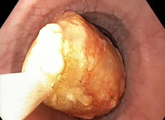
The following are a few illustrative cases that were presented during Eid: A 45-year-old male arrived with acute dysphagia after consuming meat. An endoscopic examination revealed a large meat bolus lodged in
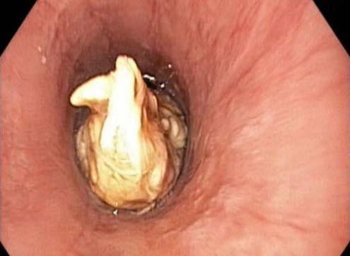
Figure 1. A large meat bolus lodged in the middle of the esophagus.
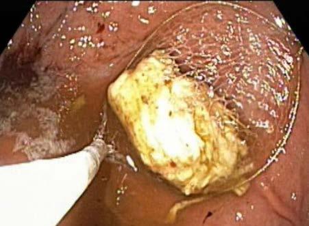
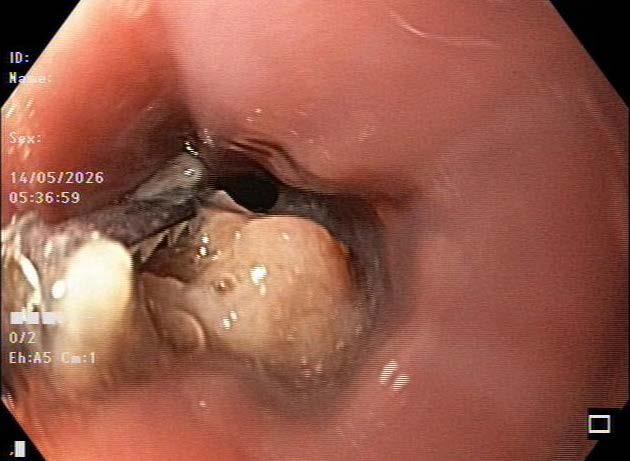
Figure 2. A large piece of meat and bone was carefully introduced into the stomach and subsequently extracted using a Roth Net basket.
Figure 3. A meat and bone bolus were extracted using a Roth Net basket.
the distal esophagus, which was successfully removed using a gentle push technique (Figures 1–3).
A 62-year-old female, with a known history of benign peptic esophageal stricture, experienced an obstruction after quickly eating chunks of meat. Endoscopic removal, followed by dilation of the stricture, effectively alleviated her symptoms.
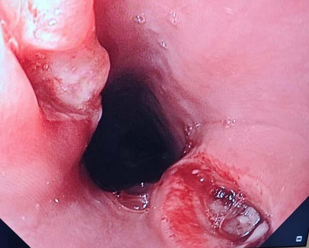
A 30-year-old female presented with severe chest discomfort and difficulty swallowing after ingesting bone fragments. Endoscopy revealed mucosal injury, which required careful extraction and the application of a hemoclip at the site of a minor perforation (Figures 4 and 5).
These cases highlight the importance of rapid endoscopic access and multidisciplinary coordination during Eid-ul-Adha.
Prevention and Public Awareness
Festive dysphagia is largely preventable. Public health campaigns before Eid-ul-Adha should focus on safe eating practices and early recognition of warning symptoms.
Preventive recommendations include eating slowly and mindfully, chewing meat thoroughly, avoiding swallowing large chunks of meat, carefully removing bone fragments, and avoiding talking or laughing while swallowing. Seek immediate medical care if food becomes stuck.
Mosques, community gatherings, television programs, and social media campaigns can serve as effective platforms for awareness dissemination.
Hospitals should also prepare for increased caseloads during Eid by ensuring availability of emergency endoscopy services, anaesthesia support, trained staff, and multidisciplinary
coordination between gastroenterologists, surgeons, and emergency physicians.
Summary
Festive dysphagia is a significant yet preventable gastrointestinal emergency that arises during the Eid-ulAdha celebrations. While most cases can be effectively managed through endoscopy, a delay in treatment can lead to serious complications. Raising public awareness, promoting mindful eating habits, ensuring rapid access to emergency endoscopy, and enhancing hospital preparedness are essential measures that can substantially reduce the burden of this condition while
preserving the spirit and joy of the festival. As the saying goes, “Prevention is better than cure.”
References
1. Chilukuri P, Odufalu F, Hachem C. Dysphagia. Mo Med. 2018;115(3):206–210.
2. Leopard D, Fishpool S, Winter S. Management of oesophageal soft food bolus obstruction: A systematic review. Ann R Coll Surg Engl. 2011;93(6):441–444.
3. Dellon ES, Gonsalves N, Hirano I, et al. ACG Clinical Guideline: Evidencedbased approach to the diagnosis and management of eosinophilic esophagitis. Am J Gastroenterol. 2013;108(5):679–692.
4. European Society of Gastrointestinal Endoscopy (ESGE). Guideline for removal of foreign bodies in the upper gastrointestinal tract. Endoscopy. 2016;48:489–496.
5. Longstreth GF, et al. Esophageal food impaction: Epidemiology and management. Gastroenterol Clin North Am. 1991;20(4):691–701.
6. Triadafilopoulos G. Esophageal food bolus impaction: Epidemiology, diagnosis, and management. UpToDate. 2025.
7. ASGE Standards of Practice Committee. Management of ingested foreign bodies and food impactions. Gastrointest Endosc. 2021;93(5):1085–1091.
Figure 4. The impacted bone above the gastroesophageal junction has been removed using alligator forceps.
Figure 5. Small perforation with ulcer above GEJ due to impacted sharp bone.
Optimizing Endoscopy Reporting: Structure, Standards, and Practical Applications in Clinical Practice
Emeritus Professor Division of Medical Gastroenterology
Department of Medicine
University of Cape Town Cape Town, South Africa
Sabina Beg, MBChB, MRCP, PhD, FRCP
Consultant Gastroenterologist and Endoscopist
Department of Gastroenterology
East and North Hertfordshire NHS Trust Stevenage, UK
Shivangi T. Kothari, MD, FACG, FASGE
Associate Professor of Medicine
Director, Advanced Endoscopy and Endoscopy
Innovation Programs Division of Gastroenterology and Hepatology University of Rochester Medical Center Rochester, New York, USA
Abstract
Background: High-quality endoscopy reporting is essential for patient safety, quality assurance, research, and clinical education. Variability in documentation and lack of standardization can limit audit, training, and communication between clinicians. The introduction of structured electronic endoscopy reporting systems (EERS), quality frameworks such as the UK Joint Advisory Group (JAG) standards, and validated lesion classification systems has substantially improved the completeness and reproducibility of endoscopy reports.
Objective: This educational review summarizes the essential components of a high-quality endoscopy report, compares structured reporting frameworks used in the United Kingdom with those employed internationally, and highlights practical approaches to integrating classification systems, image documentation, and digital quality tools into routine clinical practice.
Methods: A narrative synthesis of published guidelines, peer-reviewed studies, and expert consensus statements from professional societies including the British Society of Gastroenterology (BSG), American Society for Gastrointestinal Endoscopy (ASGE), and European Society of Gastrointestinal Endoscopy (ESGE) was undertaken, together with literature describing electronic reporting platforms and national endoscopy registries.
Results: Comprehensive endoscopy reports include structured pre-procedure, intra-procedure, and post-procedure documentation. High-quality reporting incorporates validated clas-
sification systems, systematic photographic documentation of anatomical landmarks and pathology, and clear documentation of therapeutic interventions and follow-up recommendations. In the United Kingdom, reporting platforms integrated with the National Endoscopy Database (NED) enable automated extraction of key performance indicators and benchmarking across centers. Comparable national databases and digital reporting frameworks have been implemented in other regions, including the GIQuIC registry in the United States, the Japan Endoscopy Database, and the Norwegian Gastronet program. Emerging technologies such as artificial intelligence-assisted documentation may further enhance reporting efficiency and standardization.
Conclusion: Structured and standardized endoscopy reporting is fundamental to high-quality gastrointestinal practice. Integration of validated classification systems, digital reporting platforms, and national quality registries improves communication, training, audit, and research capability. Adoption of these principles across diverse healthcare settings, including resource-limited environments, will be essential to improving endoscopy quality and patient outcomes globally.
Endoscopy has evolved from a purely diagnostic procedure to a cornerstone of modern gastrointestinal therapy, underpinning the detection, prevention, and management of malignancy and numerous benign conditions.¹ As procedural complexity has increased, so too has the need for robust and reproducible documentation.²-⁴ The endoscopy report serves as both a
clinical record and a medicolegal document, summarizing the indication, findings, interventions, and recommendations.³-⁵ Its quality directly influences patient outcomes, interdisciplinary communication, and institutional quality assurance.³-⁵
Despite the centrality of the endoscopy report, variability in content and format remains widespread. Studies from the UK, Europe, North America, and Africa reveal significant inconsistencies in how procedural completeness, lesion description, and follow-up plans are documented.⁵-⁸
Such variation undermines clinical governance, hampers audit, and complicates inter-center comparison.
In response, professional societies such as the British Society of Gastroenterology (BSG), Joint Advisory Group on Gastrointestinal Endoscopy (JAG), and American Society for Gastrointestinal Endoscopy (ASGE) have issued detailed guidance defining minimum reporting datasets.⁹-¹² Meanwhile, the advent of electronic endoscopy reporting systems (EERS) has transformed the process, enabling structured data entry, automated quality monitoring, and integration with hospital information systems.13, 14
A high-quality endoscopy report should not only document what
was observed and performed but also reflect adherence to procedural standards and support data-driven learning. The use of established classification and grading systems, such as the Los Angeles classification for esophagitis, the Paris classification for superficial neoplasia, and the Forrest classification for bleeding lesions, further enhances objectivity and reproducibility.¹⁵-¹⁷
This review outlines the essential components of an optimal endoscopy report, discusses classification systems and digital innovations, and compares reporting frameworks used in the UK with those internationally. It also explores educational and practical considerations for implementation across diverse healthcare settings, including low- and middle-income countries (LMICs).
Core Components of a HighQuality Endoscopy Report
A comprehensive endoscopy report provides a structured, objective, and reproducible record of the entire procedural episode. Although requirements may differ depending on whether the procedure is diagnostic, screening, or therapeutic, three universal phases exist: pre-procedure, intra-procedure, and post-procedure
documentation. High-quality reporting systems such as the UK Joint Advisory Group’s standards, ASGE quality indicators, and ESGE performance measures emphasize these domains as the backbone of quality assurance.⁹-¹² These three phases are summarized in Table 1
Pre-Procedure
Accurate pre-procedure documentation ensures patient safety, traceability, and informed consent.
Patient identification and demographics are fundamental. Each report should include the patient’s full name, hospital or national identification number, date of birth, gender, and contact details. Integration with hospital electronic health records enhances continuity and auditability. The indication for procedure must be clearly stated, specifying whether it is diagnostic, screening, surveillance, or therapeutic. Documenting the clinical indication ensures justification, facilitates outcome comparison, and aligns the report with service commissioning and audit frameworks.
Relevant medical history and comorbidities should be noted, particularly anticoagulant or antiplatelet use, bleeding disorders, previous gastrointestinal surgery, or cardiovascular instability. These details guide pre-procedure preparation and inform procedural risk stratification.
Informed consent must be documented as being obtained, explicitly confirming that risks, benefits, and alternatives were discussed with the patient or guardian.
Sedation and monitoring details are critical for patient safety and medicolegal compliance. The report should specify the drugs used, dosage, vital sign monitoring, and any adverse events. If an anaesthetist or sedationist is responsible for separate sedation records, the main report should reference their documentation.
Table 1. Core Components of a High-Quality Endoscopy Report
Phase/ Component
Pre-procedure
Patient demographics
Indication
Medical history
Consent
Sedation
Intra-procedure
Procedure type
Completeness
Findings
Key Details
Name, ID, DOB, gender, contact details
Diagnostic / therapeutic / surveillance
Co-morbidities, anticoagulation, prior surgery
Risks, benefits, and alternatives recorded
Drug, dose, monitoring, complications
EGD, colonoscopy, ERCP, EUS.
Extent reached (e.g., caecum, D2)
Lesion size, location, morphology
Classification LA, Paris, Forrest, Mayo, NICE, JNET, etc.
Imaging
Intervention
Complications
Post-procedure
Impression
Specimens
Recommendations
Discharge
Intra-Procedure Details
Photo and fluoroscopic documentation
Technique, device, outcome
Description, severity, management
Visual vs histological diagnosis
Site, number, pathology linkage
Follow-up, MDT, surveillance
Warning signs, advice and follow up
This section forms the core of the endoscopy report, describing the examination in sufficient detail to allow independent understanding of what was performed and observed. Structured templates or EERS prompts help ensure completeness.
These include the type and extent of examination by defining the procedure performed and its completeness, and the names and roles of the endoscopists, assistants, and trainees. It is necessary to document both normal and abnormal observations using consistent and objective terminology specifying size, shape, color, surface characteristics, location, and disease extent. To ensure reproducibility and inter-observer consistency, endoscopic findings should be categorized using validated classification and systems.15-17 These frameworks allow uniform interpretation and facilitate multidisciplinary interaction research,
training, and audit. Where available, reporting software should integrate these classifications into drop-down or prompted fields to reduce variability and promote standardized terminology.10-12
Photographic and fluoroscopic documentation is essential. High-quality imaging supports quality assurance, training, and peer review. Image documentation should include key anatomical landmarks (e.g., esophagogastric junction, ampulla, caecum, ileocecal valve) to confirm completeness, and representative images of the pathological findings to convey accurate morphology and intervention images, before and after therapeutic procedures, confirming technical success. For interventional procedures such as ERCP or EUS-guided drainage, fluoroscopic images should be archived alongside endoscopic images. These should capture key steps such as contrast injection, cannulation, stent
placement, or retrieval. All images should be linked to the endoscopy report via the EERS or hospital PACS, ensuring permanent traceability.
Therapeutic interventions should be documented comprehensively, detailing the type of intervention (e.g., biopsy, polypectomy, dilation, hemostasis, stenting), the devices and accessories used (e.g., snare type, balloon size, stent dimensions), the outcome, supported by imaging, any intra-procedural complications (bleeding, perforation, sedation-related reactions), and the management steps taken. Structured digital templates can prompt mandatory fields for intervention and complication documentation, improving audit quality.
Post-Procedure Documentation and Follow-up
The post-procedure section of the report should include a provisional diagnosis, clearly differentiating visual assessment from pending histology. Specimen details of site, number, and lab identification of all biopsies or resected tissue is mandatory. Discharge instructions regarding red flag symptoms and post-procedure diet, activity, and medication should be part of the record as are recommendations for follow-up, which should clearly identify the further management pathway. Each report should be signed and time-stamped electronically, confirming authorship and completion.
Summary
The ideal endoscopy report combines accurate pre-procedure data, standardized intra-procedure documentation, and clear post-procedure instructions. Incorporating validated classification systems and photographic or fluoroscopic evidence enhances reproducibility, supports quality assurance, and facilitates meaningful comparison between operators and centers.
Standardization, Reporting Systems, and International Comparisons
Standardization is the cornerstone of quality assurance in gastrointestinal endoscopy. Without consistent terminology, structure, and reporting metrics, data become difficult to interpret, benchmark, or compare across centers and regions.³ Disparities in report content, particularly in low- and middle-income countries (LMICs), remain a significant barrier to quality improvement and research.⁷ ²⁵ ²⁶
Historically, endoscopy reports were handwritten, variable in format, and often incomplete.⁵-⁷ With the transition to electronic systems, opportunities have emerged for standardization through structured fields, predefined classifications, and automated data extraction. These systems improve completeness, legibility, and enable the generation of quality metrics such as adenoma detection rate and caecal intubation rate.¹³ ¹⁴
International organizations including the World Endoscopy Organization (WEO), the American Society for Gastrointestinal Endoscopy, and the European Society for Gastrointestinal Endoscopy have therefore promoted structured digital reporting and minimal standard terminology frameworks.¹⁸, ¹⁹
WEO Minimal Standard Terminology (MST)
The World Endoscopy Organization Minimal Standard Terminology
framework provides a unified system to describe endoscopic findings and supports semantic interoperability between reporting platforms.⁴ MST specifies that reports should include the following domains:
1. Anatomical structure and landmarks — defined by organ, site, and modifiers.
2. Endoscopic findings — described using a consistent set of terms, attributes (appearance), and attribute values (e.g., benign, malignant).
3. Reason for endoscopy — categorized by symptoms or diseases and qualified by duration, degree, or surveillance context.
4. Endoscopic diagnosis — visual diagnosis linked to standardized codes such as ICD-10 (e.g., C16 for gastric cancer, K26 for duodenal ulcer).
5. Procedures performed — interventions categorized by intent (diagnostic vs. therapeutic).
6. Adverse events — described by type, site, and severity.
7. Actions and outcomes — intraprocedural and post-procedural management, resolution, or complications.
By integrating MST into endoscopy reporting systems, clinicians can produce structured, analyzable data, improving the comparability of reports across centers.
Essential components of an ideal EERS
• Support structured data entry with minimal free text.
• Incorporate drop-down lists for standard terminology and classification systems.
• Enable real-time photo and video documentation.
• Integrate seamlessly with
° Hospital Information Systems (HIS)
° Picture Archiving and Communication Systems (PACS).
Centralized storage and management of endoscopic images and video, data archiving and analytics
Structured reporting templates, integration with hospital electronic records, audit data export
Endoscopy reporting modules, integration with hospital information systems, audit and KPI monitoring
Abbreviations: PACS, Picture Archiving and Communication System; EHR, Electronic Health Record; KPI, Key Performance Indicator; HICCS, Healthcare Information and Clinical Coding Systems. System capabilities and registry integration depend on institutional configuration. Product information and access details are available from the respective vendors.
Structured and Electronic Endoscopy Reporting Systems (EERS)
Electronic endoscopy reporting systems are now standard in most high-income countries. These systems support clinical documentation, image archiving, quality monitoring, and research data capture. Structured reporting templates improve accuracy and enable automated extraction of procedural metadata including completeness, timing metrics, and complication rates.¹³, ¹⁴
Electronic Endoscopy Reporting Systems in High-Income Countries
Table 2 details the core functions and features of endoscopy reporting systems in high-income countries. The United Kingdom has developed one of the most integrated national frameworks for electronic endoscopy reporting, driven by the Joint Advisory Group on Gastrointestinal Endoscopy (JAG) and the National
Endoscopy Database (NED).¹¹ ¹⁴
The JAG accreditation program, established under the Royal College of Physicians, defines national quality standards for endoscopy training, service delivery, and documentation. A key requirement of JAG accreditation is the use of electronic endoscopy reporting systems capable of exporting structured procedural data to NED.
Launched in 2016, NED functions as a centralized national registry that automatically extracts standardized data from participating endoscopy units. Metrics including caecal intubation rate, polyp detection rate, polyp retrieval rate, complication rates, and procedure completion are captured directly from reporting platforms without additional manual data entry.¹³ ¹⁴ This automated data pipeline enables continuous benchmarking of key performance indicators across units and individual endoscopists.
The integration of structured reporting systems with a national quality registry has created a powerful
framework for audit, accreditation, and service improvement.¹³, ¹⁴ By linking routine clinical documentation with automated data extraction, the UK model allows real-time monitoring of endoscopy quality, facilitates training oversight, and supports national quality improvement initiatives. As a result, the UK system is frequently cited as a model for the development of coordinated endoscopy reporting and quality assurance programs internationally.
International Endoscopy Databases
Other high-income regions have also developed national endoscopy registries. These include the GI Quality Improvement Consortium (GIQuIC) in the United States, which collects colonoscopy quality metrics across thousands of endoscopists; the Japan Endoscopy Database (JED), a nationwide database supporting quality assurance and research; and Norwegian Gastronet, which
2. Examples
integrates colonoscopy quality data with national registries.²⁰-²² These systems vary in sophistication, cost, and interoperability, but all facilitate benchmarking, audit, and large-scale clinical research using structured procedural data.
Artificial Intelligence in Endoscopy Reporting
Artificial intelligence (AI) is increasingly influencing both the optical detection of pathology and the documentation of endoscopic findings, creating opportunities to integrate lesion recognition with structured reporting. Computer-vision systems are already capable of assisting with the real-time detection and characterization of gastrointestinal lesions during endoscopy, while natural language processing (NLP) and generative AI tools address the complementary challenge of translating procedural observations into standardized clinical documentation.²³-²⁶
When applied to endoscopy reporting, NLP-based systems can convert spoken procedural narration
into structured, guideline-compliant reports in real time. During a procedure, an endoscopist may describe findings verbally, for example, “normal gastric mucosa” or “5 mm sessile polyp in the sigmoid colon,” while AI software trained on large medical datasets transcribes, interprets, and populates predefined reporting fields within the electronic endoscopy reporting system. This approach has the potential to reduce documentation time, enhance completeness and standardization of reports, and enable automated capture of quality indicators such as bowel preparation quality, withdrawal time, and lesion detection metrics.²⁴-²⁶
Importantly, the greatest potential of AI lies in the integration of optical diagnosis with structured reporting workflows. Computer-vision algorithms capable of detecting or characterizing lesions may automatically generate corresponding report entries, link annotated images to specific anatomical descriptions, and populate classification systems within the report. In this way, AI can bridge
the gap between visual interpretation and structured clinical language, ensuring that endoscopic observations are recorded in a reproducible and analyzable format.
Early implementations of AIassisted documentation are being explored within commercial reporting platforms, demonstrating promising improvements in efficiency, reporting completeness, and data extraction for quality monitoring and research.²⁵, ²⁶ However, further validation through multi-center studies is required, particularly to evaluate user acceptance, interoperability with existing reporting systems, and governance of the large datasets required to train and maintain these algorithms.
Reporting in Low- and MiddleIncome Countries
In many low- and middle-income countries (LMICs), structured endoscopy reporting systems and national quality registries remain uncommon. Endoscopy reports are frequently handwritten, inconsistently structured, and often limited to the
Abbreviations: ACG, American College of Gastroenterology; AI, artificial intelligence; ASGE, American Society for Gastrointestinal Endoscopy; GIQuIC, Gastrointestinal Quality Improvement Consortium; JAG, Joint Advisory Group on Gastrointestinal Endoscopy; JED, Japan Endoscopy Database; JGES, Japan Gastroenterological Endoscopy Society; LMIC, low- and middle-income countries; MST, Minimal Standard Terminology; NED, National Endoscopy Database; PACS, Picture Archiving and Communication System. LMIC models represent typical features reported in resource-limited healthcare systems rather than a single unified national framework.
Table 3. International Comparison of Endoscopy Reporting Systems and Data Infrastructure
documentation of positive findings rather than a comprehensive procedural record.⁷, ²⁷-²⁹ These limitations restrict opportunities for audit, benchmarking, training feedback, and research.
Efforts to implement electronic endoscopy reporting systems (EERS) in regions such as Sub-Saharan Africa and the Pacific Islands have encountered multiple barriers. These include the high cost of commercial software platforms, limited hospital information technology infrastructure, lack of local technical support, and competing healthcare priorities in resource-constrained health systems. In addition, limited clinician engagement and the absence of national quality frameworks or registries can slow adoption of standardized reporting practices.²⁷-²⁹
Despite these challenges, several locally developed digital reporting solutions have demonstrated feasibility in resource-limited environments. Examples include the University of Cape Town FileMaker® endoscopy reporting system and REDCap-based databases, which provide adaptable and relatively low-cost alternatives to commercial platforms.²⁷, ²⁸ These systems typically incorporate structured fields for demographic and procedural data, drop-down menus
aligned with Minimal Standard Terminology (MST), integrated image storage, and the ability to generate trainee logbooks and audit datasets. Such platforms illustrate how pragmatic digital solutions can support quality improvement and data capture even in settings with constrained resources.
Successful implementation of structured reporting systems in LMICs depends on several key factors. Institutional leadership and clinician engagement are essential to ensure sustained adoption. Endoscopists and nursing staff require training in structured data entry and standardized terminology, and reporting templates must be adapted to local procedural profiles, language requirements, and information technology capacity. Regular audit of key performance indicators can reinforce adherence to reporting standards and drive continuous quality improvement, while compliance with national data protection regulations remains essential.²⁹ Through these measures, structured reporting can become a realistic and scalable component of endoscopy quality improvement in resourcelimited healthcare systems.
Training Implications
Beyond immediate clinical communication, standardized reporting systems
provide an objective framework for training and competency assessment. Systems with integrated Direct Observation of Procedural Skills (DOPS) such as Unisoft and EndoSoft, or similar criteria-based assessments modules, enhance formative feedback.³¹ Consistent data capture allows supervisors to review key performance indicators, offer targeted feedback, and monitor progression against defined benchmarks. At a service level, structured datasets enable meaningful benchmarking of performance against peers, institutional targets, and national quality standards. For departments, structured reports improve audit readiness and accreditation compliance.
Conclusion
The transition from handwritten documentation to structured digital reporting represents a major shift in endoscopic practice. Structured reporting improves communication between endoscopists and referring clinicians while supporting continuity of care and evidence-based clinical decision-making.
At a systems level, electronic reporting platforms transform routine clinical activity into high-quality data for audit, research, and performance monitoring. National endoscopy databases are an example that further enable benchmarking and quality assurance across healthcare systems.
Broader global adoption of structured reporting systems adapted to local resource contexts will be essential to improving endoscopy quality, safety, and research capacity. Ultimately, structured reporting is not merely an administrative exercise but a foundation for measurable performance improvement and improved patient outcome and research.
References
Readers may access the full list of references here
AASLD/IDSA
2025 Practice Guideline
on Chronic Hepatitis B: Key Updates and Clinical Implications
I – Introduction
Leandro Sierra, MD
Resident, Internal Medicine
Cleveland Clinic Cleveland, Ohio, USA
Nikki Duong, MD
Assistant Professor of Medicine
Division of Gastroenterology & Hepatology
Stanford University Medical Center Stanford, California, USA
Chronic hepatitis B virus (CHB) infection remains a major global health challenge, affecting approximately 258 million individuals worldwide and accounting for an estimated 1.1 million deaths annually due to cirrhosis and hepatocellular carcinoma (HCC).1 Despite the availability of safe and effective antiviral therapies and a highly efficacious vaccine, significant gaps persist in diagnosis, monitoring, and treatment, particularly in low- and middle-income countries.2
The American Association for the Study of Liver Diseases (AASLD), in collaboration with the Infectious Diseases Society of America (IDSA), recently published an updated practice guideline for CHB management.3 This update, the first since 2018,4 was developed using the Grading of Recommendations, Assessment, Development, and Evaluation (GRADE) approach. It addresses six critical PICO questions covering prevention, HCC surveillance, and treatment across specific clinical scenarios that have evolved considerably since the prior guidance.3
This article provides a focused overview of the most clinically relevant updates, highlighting the areas in which the 2025 guideline diverges from or significantly expands upon prior recommendations.
II – Core Concepts
CHB is a dynamic disease characterized by five phases: immune-tolerant, HBeAg-positive immune active, HBeAg-negative immune active, inactive carrier, and HBsAg-negative immune clearance. The 2025 guideline reinforces the concept of “indeterminate phases,” also referred to as the “grey zone,” to describe patients whose ALT and/or HBV DNA levels fall outside the thresholds defining immune-tolerant, immune active, or inactive CHB. Notably, up to 40% of adults with CHB fall into this category.5, 6
The preferred antivirals remain entecavir (ETV), tenofovir disoproxil fumarate (TDF), and tenofovir alafenamide (TAF), selected based on availability, cost, renal and bone profile, pregnancy status, HIV coinfection, and prior treatment history.3
One of the goals of treatment is functional cure, defined as HBsAg loss with undetectable HBV DNA on at least 6 months off therapy. A new concept, “partial cure,” is introduced for patients who achieve HBsAg <100 IU/mL with undetectable HBV DNA sustained off therapy.7 The key updates across these six domains are summarized in Table 1
III – Prevention: Mother-to-Child and Horizontal Transmission Prevention of Mother-to-Child Transmission (MTCT)
The 2025 guideline issues a strong recommendation to initiate TDF or TAF at gestational week 28, continuing until delivery (if the sole indication is MTCT prevention), for all HBsAg-positive pregnant individuals with HBV DNA >200,000 IU/mL, regardless of HBeAg status.8-10 This represents a refinement from the 2018 conditional recommendation, which suggested initiation between weeks 28 and 32 with continuation up to 3 months postpartum.4
A key practical addition addresses settings where hepatitis B immune
Topic AASLD 2018
MTCT prevention TDF at weeks 28–32; continue up to 3 months postpartum
Horizontal transmission
Immune-tolerant phase
HBeAg-negative indeterminate phase
Not addressed
Observation; treatment not routinely recommended
Observation; treat upon transition to immune-active disease
NA withdrawal Not explicitly addressed for HBeAg-negative non-cirrhotic patients
HCC surveillance after HBsAg loss
HCC surveillance in HBV-HDV
HCC surveillance in HBV-HIV
Limited guidance; focused on cirrhotic patients
Not specifically addressed
TDF or TAF at week 28; discontinue at delivery if sole indication is MTCT; TDF from week 16 if HBIG unavailable
Shared decision-making for viremic patients in highrisk scenarios; non-pharmacologic measures remain cornerstone
Consider antiviral therapy for patients >40 years or with fibrosis ≥F2 or inflammation ≥grade 2 on non-invasive testing or biopsy
Consider antiviral therapy via shared decision-making; reassess at each visit; factors favoring treatment: male sex, age >40, platelet count <180,000/mm³
Suggest against NA withdrawal until HBsAg loss; requires HBsAg <100 IU/mL if cessation desired
Continue surveillance in cirrhosis, family history of HCC, men after age 40, women after age 50
Surveillance for all adults regardless of cirrhosis status; individualize in children
Not specifically addressed Surveillance for men ≥18 years and women ≥40 years of age
Conditional Very low
Conditional Very low
Conditional Very low
Conditional Very low
Conditional Very low
Conditional Very low
Conditional Very low
Abbreviations: HBIG, hepatitis B immune globulin; HBeAg, hepatitis B e antigen; HBsAg, hepatitis B surface antigen; HCC, hepatocellular carcinoma; HDV, hepatitis D virus; HIV, human immunodeficiency virus; MTCT, mother-to-child transmission; NA, nucleos(t)ide analogue; TAF, tenofovir alafenamide; TDF, tenofovir disoproxil fumarate.
globulin (HBIG) is unavailable for the infant: based on a recent randomized controlled trial, TDF may be initiated as early as gestational week 16, with the infant receiving the HBV vaccine series at birth as the sole neonatal prophylactic intervention in lieu of HBIG.11 For individuals whose sole indication for antiviral therapy is MTCT prevention, discontinuation at delivery is now explicitly suggested, representing a meaningful change from prior practice. Post-cessation monitoring of HBV DNA and ALT every 1 to 3 months for up to 6 months is recommended, with reinitiation of treatment for significant withdrawal flares (ALT ≥5× ULN).4, 12 Breastfeeding on TDF or TAF is safe for both mother and infant.8
Antiviral Prophylaxis for Horizontal Transmission
For viremic HBsAg-positive individuals who do not meet standard
treatment indications but are in high-risk scenarios for horizontal transmission (e.g., unprotected sex with multiple partners, injection drug use, healthcare workers performing SHEA Category III exposure-prone procedures), the guideline issues a conditional recommendation favoring a shared decision-making approach regarding antiviral therapy. Evidence for this recommendation is graded as very low, and non-pharmacologic measures such as vaccination of susceptible contacts, universal precautions, and harm reduction remain the cornerstone of prevention.13
IV
– Treatment of the ImmuneTolerant Phase: A Paradigm Shift Immune-Tolerant Phase
Perhaps the most impactful update involves the immune-tolerant phase (HBeAg-positive, HBV DNA ≥10⁷ IU/mL, persistently normal ALT). Prior guidance recommended observa-
tion for most patients in this phase4; the 2025 guideline now conditionally suggests antiviral therapy for patients over age 40 or with significant liver inflammation (grade ≥2) or fibrosis (F2 or greater) on non-invasive testing or liver biopsy.14 For individuals under 40 without these risk features, shared decision-making is recommended, weighing HCC risk reduction against the need for long-term therapy.15
This shift is driven by accumulating evidence linking persistently high HBV DNA levels with HCC risk even in the absence of elevated transaminases, as well as data showing that subclinical histologic disease (significant fibrosis or inflammation) can be present in up to one-third of immune-tolerant patients over age 40 despite normal ALT.14 Vibrationcontrolled transient elastography (VCTE) ≥8 kPa is suggested as the threshold for identifying F2 or higher fibrosis in this population.16 Monitor-
Table 1. Key recommendation changes: AASLD 2018 vs. AASLD/IDSA 2025
ing every 6 months remains appropriate for patients in whom treatment is deferred, with the primary goal of detecting transition to the immuneactive phase.13 An integrated management algorithm based on the updated 2025 recommendations is presented in Figure 1.
V – The Indeterminate Phase: Moving Toward Individualized Therapy
HBeAg-Negative Indeterminate Phase
The HBeAg-negative indeterminate phase comprises patients who are HBsAg-positive, HBeAg-negative, without cirrhosis, and with HBV DNA and/or ALT levels that do not meet criteria for immune-active or inactive CHB. Previous guidance recommended observation for this group with initiation of treatment upon transition to immune-active disease.4
The 2025 guideline conditionally suggests considering antiviral therapy using a shared decision-making approach, with re-evaluation at each follow-up visit if treatment is deferred.3 Key factors favoring treatment include male sex, age >40 years, and platelet count <180,000/mm³ as surrogates for higher fibrosis risk.17 A systematic review supporting this recommendation demonstrated that antiviral therapy was associated with a significantly lower annual HCC incidence (adjusted incidence rate ratio 0.36, 95% CI 0.16 to 0.81) in this population.17 However, the certainty of evidence is very low, largely due to the observational nature of available studies and wide heterogeneity in outcomes.17
VI – Nucleos(t)ide Analogue Withdrawal: When (if ever) to Stop
Nucleos(t)ide Analogue Discontinuation
Among HBeAg-negative, non-cirrhotic patients with sustained undetectable
HBV DNA on nucleos(t)ide analogue (NA) therapy, the 2025 guideline conditionally suggests against withdrawing NA therapy until HBsAg loss is achieved.18 This position differs notably from EASL 2025 and APASL guidance, which allow consideration of NA cessation in selected patients with prolonged viral suppression.19
The guideline acknowledges that NA discontinuation can lead to HBsAg loss in a subset of patients (approximately 10% over 2 years in RCTs), but weighs this benefit against the substantial risks of virologic relapse, ALT flares, and hepatic decompensation: risks that appear even higher in real-world cohort studies than in RCTs.18 For patients who strongly desire to stop therapy, eligibility requires HBsAg <100 IU/mL, undetectable HBV DNA for ≥2 years, no cirrhosis or prior decompensation, and no HIV or HDV co-infection, with post-cessation monitoring of HBV DNA and ALT every 1–3 months for the first 6 months and immediate retreatment if HBV DNA ≥10,000 IU/mL or ALT ≥5× ULN.7
VII – Hepatocellular Carcinoma
Surveillance: Expanding the Net Hepatocellular Carcinoma Surveillance After HBsAg Loss and in Coinfected Patients
Hepatocellular Carcinoma Surveillance in Special Populations
The 2025 guideline provides refined and expanded HCC surveillance recommendations for several previously under addressed populations.3
For patients who have achieved HBsAg loss, surveillance is conditionally suggested for those with cirrhosis, a family history of HCC, men who experienced HBsAg clearance after age 40, and women after age 50.20
For HBV-HDV co-infected adults, HCC surveillance is conditionally suggested independent of cirrhosis status, reflecting data showing that HDV co-infection significantly amplifies
HCC risk, even in the absence of cirrhosis, with an incidence rate of 18.65 per 1,000 person-years compared to 4.84 for HBV monoinfection.21
For HBV-HIV co-infected individuals, HCC surveillance is suggested for men ≥18 years and women ≥40 years of age. For HBV-HCV co-infection, HCV treatment with direct-acting antivirals is strongly recommended, and HCC surveillance follows the same criteria as for HBV monoinfection after HCV cure.22, 23
In all populations, surveillance with abdominal ultrasound and serum AFP at 6-month intervals remains the recommended approach.24
VIII – Future Directions
Several important areas are anticipated to shape future guideline iterations. These include defining the role of novel biomarkers, quantitative HBsAg, HBV RNA, and hepatitis B core-related antigen (HBcrAg) as tools to guide treatment initiation and monitoring.25 The potential benefit of antiviral therapy in inactive carriers with HBsAg levels >1,000 IU/mL, the role of peginterferon add-on or switch strategies to enhance functional cure rates, and the diagnostic performance of non-invasive tests as treatment decision guides remain unaddressed.13, 26 As new finite therapies targeting functional cure advance through clinical trials and reach regulatory approval, updated guidelines are anticipated, including those from the World Gastroenterology Organisation (WGO).27
IX – Conclusion
The 2025 AASLD/IDSA practice guideline represents a meaningful evolution in the management of CHB, moving beyond the established framework of treating immune-active disease toward a more proactive, individualized approach for immunetolerant and indeterminate phase patients. The expanded attention to
Figure 1. AASLD/IDSA 2025 recommended algorithm for the management of chronic hepatitis B. Abbreviations: ALT, alanine aminotransferase; anti-HBc, antibody to hepatitis B core antigen; anti-HBs, antibody to hepatitis B surface antigen; HBeAg, hepatitis B e antigen; HBsAg, hepatitis B surface antigen; HBV, hepatitis B virus; HCC, hepatocellular carcinoma; HDV, hepatitis D virus; HIV, human immunodeficiency virus; HBIG, hepatitis B immune globulin; NA, nucleos(t)ide analogue; TAF, tenofovir alafenamide; TDF, tenofovir disoproxil fumarate; ULN, upper limit of normal.
MTCT prevention, HCC surveillance in co-infected and post-HBsAg-clearance populations, and the nuanced guidance on NA withdrawal reflect a maturation of the HBV field. Implementation of these recommenda-
tions, particularly in resource-limited settings, will require adaptation to local contexts and continued emphasis on equitable access to diagnosis and treatment.
References
Readers may access the full list of references here
Calendar of Events
Please check the WGO Meetings and Events Calendar for the latest updates at https://www.worldgastroenterology. org/meetings/meetings-and-eventscalendar
WGO RELATED EVENTS
World Congress of Gastroenterology 2026
When: September 30, 2026 – October 3, 2026
Location: India International Convention & Expo Centre
Address: New Delhi, India
Organizers: World Gastroenterology Organisation and Indian Society of Gastroenterology
Are you a WGO Member Society wanting to share your event with WGO readers? Visit https:// www.worldgastroenterology.org/ forms/submit-event.php to submit your event for publication in WGO’s website conference calendar as well as the quarterly e-WGN calendar of events!
CALENDAR OF EVENTS
Annual Meeting of the Asociación Hondureña de Gastroenterología
When: July 30, 2026 – August 1, 2026
Location: Centro de Convenciones
Hotel Copantl Sula
Address: San Pedro Sula, Honduras
Organizer: Asociación Hondureña de Gastroenterología
Annual Scientific Conference and General Meeting (SOGHIN)
When: August 12, 2026 – August 15, 2026
Location: Continental Hotel, Zone 4
Address: Abuja, Nigeria
Organizers: SOGHIN, Africa HepatoPancreato-Biliary Cancer Consortium (AHPBCC), and Nigerian Cancer Society (NCS)
WEGE (Women Empowerment in GI Endoscopy) Program
When: August 20, 2026 – August 21, 2026
Location: Cairo
Country: Egypt
Organizer: WEGE Summer School Website: https://www.linkedin.com/ company/wege-program
ECOS International 2026
When: August 20, 2026 – August 22, 2026
Location: Hospital Español, CDMX
Country: Mexico
Organizer: AMG
Website: https://www.gastro.org.mx/
XI Semana de Enfermedades Digestivas
When: August 27, 2026 – August 29, 2026
Location: Machala, El Oro
Country: Ecuador
Organizers: Ecuadorian Society of Gastroenterology – Manabí Chapter Website: https://seg-manabi.com
AGW 2026
When: August 28, 2026 – August 31, 2026
Location: Perth, WA
Country: Australia
Organizer: Gastroenterological Society of Australia
www.biocodexmicrobiotainstitute.com/pro: an international hub of knowledge dedicated to microbiota!
Biocodex Microbiota Institute is an international scientific institution that aims to foster health through spreading knowledge about the human microbiota. To do so, the Institute addresses both healthcare professionals and the general public to raise their awareness about the central role of this still little-known organ of the body.
It is designed to provide you with reliable, updated, and adapted content. It is also designed to reflect the dynamism and innovation of the human microbiota.
Available in 7 languages (English, French, Spanish, Russian, Polish, Turkish, and Portuguese), this online international hub provides Healthcare Professional with the latest scientific news and data about microbiota including the Institute’s exclusive content such as Microbiota magazine, thematic folders, continuing medical education (CME) courses and interviews with experts. Check them out!