FROM WITS
to
the
A tribute to Health Sciences at the University of Witwatersrand, Johannesburg
As told by Chaim M. Rosenberg & the MBBCh Class of 1960
Edited by Geraldine Auerbach, MBE.
Chapter
Chapter
the
A tribute to Health Sciences at the University of Witwatersrand, Johannesburg
As told by Chaim M. Rosenberg & the MBBCh Class of 1960
Edited by Geraldine Auerbach, MBE.
Chapter
Chapter
When I first learned of the newsletters the class of 1960 had put together, I thought it was a superb endeavour to document the anecdotes and stories of the time. Archives and memory-keeping are important parts of an institution’s longevity. Our alumni help inform us of our way forward. Their experiences and life lessons shape how we teach, learn, research, and, indeed, market the Wits Faculty of Health Sciences to students.
Reading the rich stories in the newsletters, expertly curated by Dr Chaim Rosenberg and edited by Geraldine Auerbach, helped me understand the zeitgeist and spirit of Wits Medical School in the 1950s and 1960s. I was touched by the humanity each student showed and how their stories came alive to paint a picture of the time.
The 50s and 60s were tumultuous times in South Africa. Apartheid looked like it would never end, and those who protested and fought against it paid dearly, some with their lives. In this e-book, you’ll read the stories of two such activists who luckily survived that dark time.
You will also notice that the student body in the 50s and 60s was mostly white and male. Times have drastically changed. It’s heartening to see that change does happen, however slow and impossible it seemed at the time. I think the gains prove what a dynamic and brave institution Wits is.
I was also touched by the humour in the anecdotes: it’s hilarious that poor Professor Raymond Dart was flummoxed by the second-year anatomy students and lamented that after millions of years of evolution, students still made very sloppy mistakes. Other lecturers were stern and embodied the “academic of the age”, meaning students were terribly frightened of them.
The class of 1960 is indeed a special one. We have so many illustrious alumni from that year. I can’t even begin to list their collective accomplishments of which you will read about in the book.
Lastly, I’d like to extend my immense gratitude to this class. Your generosity of spirit and kind donations will keep the critical work of science alive and thriving. Our health institutions are the canaries in the coalmine. With your support, our students will be exemplary professionals to continue serving South Africans – poor and wealthy.
We hope other classes will follow suit in gathering the rich stories of their time at Wits Faculty of Health Sciences. After all, the world is made of stories (and science).

Dean: Faculty of Health Sciences University of the Witwatersrand, Johannesburg
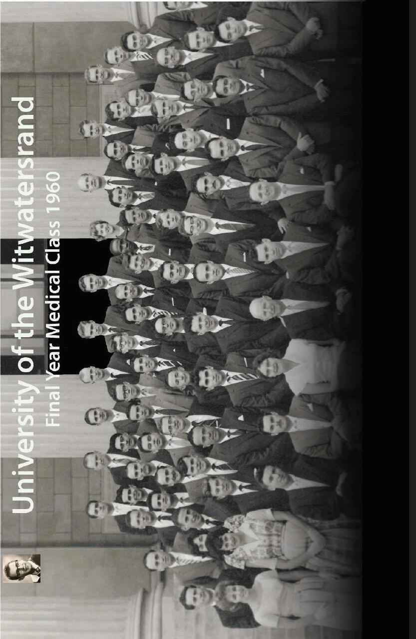
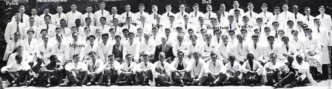
Back row: G Dimopoulos, W Wilson, R Palmer, KR Edge, EJB. van Veen, VI McCusker, J Stuart, R Otten, P Landsberg, A van As.
5 th row: E Agrotis, A Kalell, C Bosman, L Willies, L Kahn, D Bongani, H Silver, A Meyers, A Mauff, A Heynes.
4 th row: Zeilinga, R Prentice, P Urbani, R Jackson, M Plit, D Rossouw, A Crosley, P Botha, R Hollis, A Vink.
3 rd row: S Zail, G Shulman, A Ravdel, A Gottlieb, H Glazer, I Lissoos, M Howell, I Shapiro, T Kretzmar, B Kuming, G Lampert, G Katz, V Lee, G Maass, D Parry.
2 nd row: N Livni, M Dawes, G Davis, G Schneider, R Cochrane, M Nissenbaum, E Dove, M Berger, F Sims, M Cohen, G Boner, CM Rosenberg, M Hurwitz, E Jassat, LB Kahn, D Paton, S Ou Tim.
Sitting: J Kussel, A Milunsky, Mrs E C Phillips (Assistant Registrar) Prof E Cluver (Dean), A Rubenstein A Fanaroff.
Sitting: J Kussel, A Milunsky, Mrs E C Phillips (Assistant Registrar) Prof E Cluver (Dean), A Rubenstein A Fanaroff.
In March 2020, when we were all suddenly locked up in our homes because of the COVID-19 pandemic, I came across my Wits Medical School (now Faculty of Health Sciences) graduation class photograph of December 1960. Looking at the fresh faces of the new doctors, I began to wonder who all these people were, and what had become of them. I thought about my own travels since then, working on four continents and mused about where others might have landed. I thought about what our thorough medical training at Wits had enabled us all to do in our careers.
I spoke to a few colleagues with whom I had kept in touch, and they were equally intrigued. They agreed, in our diamond jubilee year, to work with me to try to find our contemporaries. Class members Avroy Fanaroff, a neonatologist in Cleveland; Len Kahn, a pathologist in New York; Andre Van As, a pulmonologist in South Africa, Albuquerque, New Mexico and Philadelphia; Gary Katz, a paediatrician in London; Ronnie Auerbach, an ENT surgeon in London; and Geoff Boner, a nephrologist in Israel all climbed on board. With much sleuthing and cajoling, we assembled quite a list and reached out to all the classmates we could find.
The Class of 1960 project thus progressed faster than I could ever have imagined. In the first three months, from March to June 2020, I must have sent or received nearly a thousand emails. I collected ten biographical stories with many pictures. We were honoured to have the support of the then Dean of the Faculty of Health Sciences, Professor Martin Veller.
We compiled a dozen newsletters between July and December. They were beautifully edited, illustrated and formatted by Geraldine Auerbach, another Wits alumna, though not in medicine, the wife of classmate Ronnie Auerbach and sister of classmate Theo Kretzmar. We approached the Wits Alumni Relations Office, who were excited about the project. Their North American Representative, Nooshin Erfani-Ghadimi, offered to distribute the newsletters and also agreed to create a dedicated website for us as a repository for the material we were collecting.
These newsletters rekindled the sights and smells of the anatomy halls and hospital corridors. We thought again about our memorable faculty members and colleagues, as well as the institutions, libraries, hospitals and clinics in which we trained. After our first newsletter was launched and circulated to class members in South Africa, the UK, USA, Israel and Australia in July 2020, the response was quick to follow. Classmates appreciated being contacted after such a long time and reading these bulletins. Alf Mauff, a pathologist of Lancet Laboratories in Johannesburg, said: “I look forward to these newsletters that have after 60 years brought me into contact with my class in my twilight years. As I read their history, so many memories flood back from the furthest reaches of my memory bank. They would certainly never have seen the light of day but for these newsletters”. Another, Jack
Kussell, a paediatrician in Johannesburg, said it was really good to see the Class of 1960 become a cohesive and proud unit.
Dean of the Faculty of Health Sciences, Martin Veller, thanked us for sharing our newsletters with him. “It is absolutely fabulous,” Professor Veller said. He recalled that he had worked closely with several of the members of our class. “In particular”, he said, “I spent a number of years working with Tony Meyers in the kidney transplant unit at the then Johannesburg General Hospital. Irving Lissoos was also involved and Clive Rosendorff was head of physiology when I studied.”
We consider our ‘cohort’ to be those people who either started their Wits medical studies with us in 1955 or graduated with us in 1960. Some joined our class along the way, having repeated a year or completed other courses first. Others dropped out for similar reasons. Some of our cohort spent a year or two doing an intercalated medical science degree and graduated a year or two later.
Our class started with 115 students in 1955. Nine of them achieved first-class passes in all four subjects in their first year. 29 of them, however, failed their first year. Several were allowed to re-sit the subjects they had failed, and a few were permitted to repeat the year. A significant number, though, went straight through the six years. Eighty-four of us graduated at the end of 1960.
In those days, the mid-1950s, you were accepted to study medicine on the merit of your matric results. There was no interview. Your character or other achievements were not taken into account. It seems that it was immaterial if you were a senior prefect, cricket captain or played the cello. Nobody asked.
Looking at the picture of our graduation class, we see that it was mostly young men at medical school. There were only about five women and five people of colour (indicating a possible quota system); the rest were white males aged about 23. They were in a very privileged position. They knew it and took this responsibility very seriously, aiming to be the best doctors they could be.
Many of us belonged to the National Union of South African Students (NUSAS), participated in its anti-apartheid activities and served on the Academic Freedom Committee. This was fraught with danger as students could be banned, detained without trial, and there were government spies on campus to report us to the police. Some were willing to follow their convictions and sadly suffered for their actions, which we describe later.
In our searches, we were delighted to discover how many of our classmates were still alive—now in their early to mid-eighties. Many were still at work (in 2020), and a few were even working full-time! Of the known domiciles, the majority of our classmates, 59, practised in South Africa; 17 worked in the United States; eight had spent their medical careers in the United Kingdom; three in Israel, three in Australia; and one in New Zealand.
In those dark days of lockdown all over the world in 2020, reconnecting and learning about each other’s pathways and successes became a highlight for many of us. We were all having to take stock of our lives and contemplate our mortality during the COVID-19 pandemic, which was particularly dangerous for people in our age group. Sadly, COVID-19 caught and claimed a few of us.
We were impressed to see how much our classmates had contributed to medical teaching and practice wherever they had settled. Several had achieved higher degrees of Doctor of Medicine, Doctor of Science and PhDs, both in South Africa and overseas. Many had specialised and joined the Royal Colleges of surgeons, physicians, paediatricians, or equivalent bodies.
During the span of our careers, from 1960 to 2020, the world had seen the most breathtaking and far-reaching
advances in medicine and healthcare, more than at any equivalent period in history. Therefore, we invited colleagues to write about ‘Medicine, Then and Now 1960–2020’ in their own fields. In this book we will lead the reader to a series of short articles by classmates regarding the technical, pharmacological and practical advances they had experienced in their medical practice. These covered: babies, children, blood, psychiatry, waterworks, surgery, genetics, pathology, allergy, cardiology and pulmonology.
Once we had got to know more about each other than we ever had known, or even dreamed we would know, many expressed the wish to meet again. Of course, the pandemic made that impossible ― but on the other hand, the magic of Zoom made it even more possible than ever before. In December 2020, exactly 60 years after our graduation, we gathered together from the far corners of South Africa and indeed of the world, travelling by our fingertips instead of in aeroplanes. It was a really heart-warming meeting, enjoyed from the comfort of our homes, addressed by the current Dean of Health Sciences at Wits, Professor Shabir Madhi and several representatives of our class. It was a fitting culmination of our diamond jubilee year
Much to our delight, in 2022 we were contacted, out of the blue, by a final year Wits MBBCh student, Taariq Hassim. He told us that he had stumbled upon our website and found the history in it very interesting. He said that he loved reading the newsletters and the individual stories about our personal experiences of the developments over the years. Knowing that our story could inspire new generations of the Wits Faculty of Health Sciences students made our project all the more worthwhile
This prompted us to meet again on Zoom, two years after our diamond jubilee, in December 2022, inviting both Taariq and the current Dean, Professor Madhi to address us. We also invited members of our class as well as representatives of other classes of the early 1960s to give a short talk. A recording of this meeting can be found under ‘videos’ on the dedicat
This further reunion also prompted the wish from the Dean of Health Sciences that our class of ‘60 stories should be highlighted in some way and made more accessible ― hence this eBook. In it, we hope we have created an exciting narrative, introducing our classmates from their schooldays onwards and following them (in their own words) through their pre-clinical and clinical training. We record their interactions with each other and with their memorable teachers and mentors, and their adventures out into the wide world. In doing so, this book reveals the outstanding medical training at the University of Witwatersrand and the appreciation the class members of the 1960s feel for this outstanding start to their medical careers. This book also shows what this group of graduates in turn (like many others before and after them) have been able to contribute to the development of medical knowledge and practice around the globe.
We discovered that no less than 22 of our classmates had become professors and heads of departments, and teachers in various specialities. Three of our class members, Clive Rosendorff, Laurence Geffen and Arthur Rubenstein, had been appointed deans of medical schools in South Africa, Australia, and the USA. Martin Bobrow had become a Fellow of the Royal Society ― a rare and high honour indeed. We describe their stories in more detail in the chapter on ‘academic medicine’.
We feature in this book the contribution that our class members made to medicine in South Africa: Alf Mauff and Stan Zail’s participation in establishing Lancet Laboratories and Anthon Heyns’s role in running the South African Blood Transfusion Services as well as Mervyn Hurwitz establishing the first sex therapy clinic. Mike Plit became President Nelson Mandela’s personal physician, and Jack Kussel was the paediatrician to Madiba’s
grandchildren.
There are chapters devoted to memorable professors and teachers such as Raymond Dart, Phillip Tobias, Guy Elliott, Mosie Suzman and Sonny Du Plessis. We write about the hospitals we worked in. A whole chapter is devoted to Baragwanath. We were delighted to have elicited fascinating biographical sketches from more than 50 of our classmates. This book, while not comprehensive as there were many people and stories we could not discover, nevertheless gives us an illuminating snapshot of Johannesburg and Wits in the late 1950s. We include a chapter on what sustains us – the pastimes and activities that absorb us especially in our later years.
This has been an international project, and I want to express my enormous thanks to all those mentioned above who were so deeply involved. I am grateful too, for the encouragement and patronage of the Deans of the Faculty of Health Sciences at Wits; first, Professor Martin Veller when we started the project, and currently Professor Shabir Madhi, who shared a reunion Zoom meeting with members of our class.
We are very grateful to Dean Madhi for suggesting the creation of this book to make our story more widely known. We thank Geraldine again, who undertook the task of re-editing and compiling the material we had collected on our website into a compelling narrative for this book. We express our warm thanks to Didi Fombad and Beth Amato in the Dean’s Office who have worked with Geraldine on the process. Our warm thanks go also to Peter Maher director of the Alumni Relations Office for his enthusiastic support and for putting us in touch with Nooshin Erfani-Ghadimi, the Wits representative in the United States.
I am glad to say that this diamond jubilee project was not just about ourselves. While connecting with and learning about the members of our very impressive and privileged class of 1960, we also made sure that we all contributed to help current Wits Faculty of Health Sciences students to achieve their MBBCh by making donations to the Phillip Tobias Bursary Fund.
We hope our initiative will encourage other classes to reconnect with each other and also help all readers to want to support the Wits Faculty of Health Sciences, which gave us such a magnificent start in life.
Class Member: Wits MBBCh 1960
MBBCh MD (Wits), PhD (University of New South Wales)
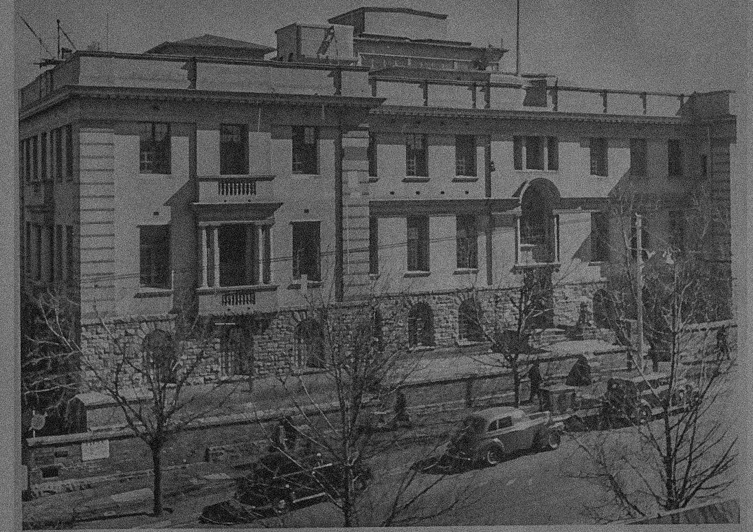

A very fine way to begin this story is to look at the photograph of the Students Medical Council (SMC) of 1959 (a year before our graduation). It was taken at the front door of our Medical School on Hospital Street. If ever a picture could foretell the future of achievement, this is surely the one.
The Wits Students Medical Council (SMC) was the official representative body of candidates for the MBBCh degree programme. Twelve members of our cohort who either started with us in 1955 or graduated with us in 1960, were members of the SMC for 1959: Peter Arnold, Martin Bobrow, Avroy Fanaroff, Essop Jassat, Basil Kuming, Gerald Lampert, Irving Lissoos, Gary Katz, Jeffrey Maisels, Aubrey Milunsky, Clive Rosendorff and Arthur Rubenstein.
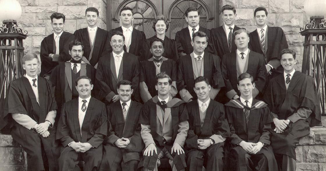
Seven members of our cohort had already served on the SMC the previous year, 1958. Many were active on other student representative bodies, and several were involved with student political action for academic freedom in the face of government attempts to force black students out of the major universities, which occurred in 1959 during our studies.
Subsequently, Arthur Rubenstein and Clive Rosendorff were appointed as deans of medical schools; Martin Bobrow and Aubrey Milunsky became world-renowned geneticists; Jeffrey Maisels and Avroy Fanaroff are neonatal paediatric chairmen in the USA; Gary Katz became a consultant paediatrician and senior lecturer in London; Peter Arnold in Sydney was recently awarded the Order of Australia for ‘achievement and meritorious service’; Essop Jassat, a leading fighter against injustice, was awarded one of South Africa’s highest accolades, the Order of Luthuli in Silver. Sadly, South African urologist Irving Lissoos, ophthalmic surgeon Basil Kuming and general practitioner Gerald Lampert who served on this committee and who conducted their practices with distinction in South Africa, have passed away. We look forward to celebrating and commemorating them with many other classmates that we have been able to trace, throughout the narrative of this book.
We begin with our colleagues with a conscience:
Two of our cohort of students, Essop Jassat and Costa Gazidis, took up the fight for democracy regardless of the dire consequences to themselves and their careers. Those were the intimidating days of detention without trial, spies on campus and banning orders for any activity against apartheid. Both were to be banned and imprisoned, which seriously impacted their lives and their ability to carry out their medical practices.
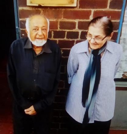
Essop Jassat was a lifelong worker for democracy in South Africa. He followed his father into the Transvaal Indian Congress and served as the president of the Indian Youth Congress. This led to his first 5-year ban in 1955. He was sentenced in 1964 for failing to comply with his next banning order. Although he appealed that he was unable to report to the police while attending a medical emergency, he lost the appeal and served time in prison. This was just the beginning.
Jassat remained active in the fight against the injustice of apartheid. He became the president of the Transvaal Indian Congress and served on the executive of the United Democratic Front (UDF) that was set up in 1983 to coordinate all the opposition to the new constitution – which entrenched the apartheid system .
Following the 1984 boycott of the Tricameral Parliament, UDF co-presidents Albertina Sisulu and Archie Gumede, together with 14 other members, including Jassat, were arrested on the serious charge of treason. Bail was initially denied. When this ruling was overturned by Justice Friedman of the Supreme Court in Natal, very severe bail restrictions were applied. Later that year, charges against 12 of the 16 accused were withdrawn due to “insufficient and unconvincing evidence”.
Essop Jassat’s steadfast work culminated in him being invited to serve as a delegate at the Convention for a Democratic South Africa, set up to negotiate the end of apartheid. In the 1994 elections, he was elected a Member of Parliament, serving until 2004. He then returned to his medical practice on Bree Street in Fordsburg. Jassat was also awarded one of the county’s highest accolades, the Order of Lithuli in Silver, “for his excellent contribution to the struggle for liberation and advancing democracy in South Africa”.
Costa Gazidis, a member of our anatomy class in 1956, was incensed at the start of our anatomy year at learning that black students were forbidden to dissect white cadavers, while white students were permitted to dissect both white and black.
When he graduated in 1962, Costa was under the surveillance of the police special branch for his political views and activities. He was not allowed to complete house jobs at Queen Victoria Maternity Hospital in Johannesburg and King Edward VII Hospital in Durban. He was not permitted to work at Baragwanath Hospital either.
Costa eventually found a job in the medical department of the West Rand Consolidated Mines. In 1962, he joined the Communist Party and was the health secretary of the Pan-African Congress (PAC). He remembered: “People were astonished that the PAC had a white member.” Costa was arrested in 1964 and imprisoned for two years, serving time in solitary confinement. Following his release in 1966, he was promptly banned. He said: “I became very isolated and was not allowed to further my studies.”
Gazidis then spent 22 years continuing the struggle in exile in the UK. Determined to serve the people of
South Africa, he returned to the country in 1990. He stood for election to Parliament in 1994. When this was unsuccessful, he took a position running community clinics. He was soon at loggerheads with the ANC government over his determination to use the drug AZT in treating AIDS-affected pregnant patients. Fined for insubordination, Costa took his case to the Pretoria High Court where he won after a long-drawn-out trial.
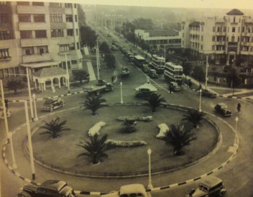
I wonder if people remember this familiar landmark on the way to Medical School?
Clarendon Circle, built in 1931, directed traffic into and out of Hillbrow, where the Wits Medical School was located. Here we see this iconic landmark in 1938, close to the time when most of us were born. It was where East Avenue intersected with Empire Road and Bruce, Twist and Klein Streets. The island contained palm trees and shrubs. It was named in honour of George Herbert Hyde Villiers, 6th Earl of Clarendon, who presided over the Union as the Governor General from 1931 until 1937. In time, Clarendon Circle was abandoned, and replaced by a direct intersection with traffic lights.
Not many of us might have realised at the time the international stature of many of our Wits medical faculty teachers. We were particularly fortunate to be guided and inspired by anatomists like our Professor Raymond Dart and lecturer Phillip Tobias. We were mostly unaware of the groundbreaking discoveries they were making that were changing our understanding of how and where humans had developed.
In this book, we are pleased to include tributes from our class members who had spent time with Raymond Dart and Phillip Tobias on the intercalated medical science degree. They were able to make us and others aware of their enormous achievements and honours. In the next chapter, we look at Raymond Dart’s life and achievements.
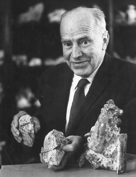
Our classmate Laurence Geffen had the privilege of working with Raymond Dart when he (and a few other classmates) took a year or two out of the medical course in 1957 to study for an intercalated medical science degree. He wrote this appreciation of our anatomy professor and renowned palaeontologist, Raymond Dart who set the standard for the subject at Wits and was a world-renowned pioneer of African palaeontology.
Raymond Arthur Dart was a Queenslander by birth. He migrated from Australia via the UK to South Africa as a young man of 30, to become the first Professor of Anatomy at the University of Witwatersrand. There, for the next sixty-three years, he worked as a medical educator and paleoanthropologist, achieving world-wide fame, as well as some notoriety, as the discoverer of Australopithecus Africanus.
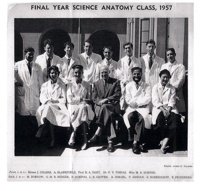
When I first met Professor Dart as my anatomy lecturer in 1956, he was in his 60s, small, balding, wiry, and with an agile posture. He had piercing blue eyes, and a very resonant voice that could strike fear into the hearts of his students. I will remember Raymond Dart as a great teacher who imbued all who were privileged to be his pupils with a love of science and a suspicion of received truth.
He was a larger-than-life character who was at times the terror of the dissection hall, bemoaning the fact that millions of years of evolution had produced such clumsiness in second-year medical students. At other times, he transfixed lecture audiences with graphic demonstrations of how reptiles emerged from swamps onto dry land by performing his famous crocodile crawl, or how primates descended from an arboreal to a savannah habitat by brachiating along the service pipes in the ceiling of the lecture theatre.
To my knowledge, he made only two return visits to Australia, both at the invitation of the University of Sydney, in 1950 and again in 1972. He was invited on a third occasion in 1981, again to Sydney, at the age of 88, but had to cancel due to failing eyesight.
He died in Johannesburg in 1988 at the age of 95. He was survived by his second wife Marjorie, his daughter, Diana, and son, Galen. His memory is revered in his adopted country and was actively perpetuated in Johannesburg by his equally famous pupil and successor, Phillip Tobias.
Raymond Dart was born in Brisbane on 4 February 1893, the year of disastrous floods that washed away the Victoria Bridge and deposited ships in the Botanic Gardens. It also washed away the plantation and sugar mill that his grandfather William had built on the St. Lucia site of what is now the University of Queensland. It is said that the infant Raymond was floated out of a second-storey window of their inundated house.
Six years later, his parents, who were devout Baptists, moved to a bush farm at Blenheim in Queensland to remove their children from the temptations of city life. Here, Raymond Dart had a typical Australian farm boyhood. Later, at boarding school, he soon showed his brilliance and in 1911, on receipt of a scholarship, he became a foundation student at the nascent University of Queensland, where he achieved his BSc degree with honours in 1913 and his MSc two years later.
Despite, as he put it, “the discrepancies between fundamentalism and the facts of evolution,” he intended to become a medical missionary in China. He enrolled in Medicine at the Sydney University in the fateful year 1914, and soon fell under the influence of the Professor of Anatomy, J.T. Wilson, an authority on the comparative anatomy of the marsupial brain.
In 1914, the British Association for the Advancement of Science held its first-ever congress outside the British Isles, arriving en-masse by ship in Sydney. At the Congress, Dart attended lectures by such luminaries as Sir Grafton Elliot Smith, an Australian who had become a leader of British anthropology, and an expert on Ancient Egypt and the Dawn of Civilisation.
At one of these lectures in 1914, the Talgai skull, a fossil excavated in Southeast Queensland and the oldest to be discovered in Australia, was exhibited by Professor Wilson. Dart was hooked!
However, when the First World War broke out, the entire congress packed up and set sail for England. Dart
finished his medical degree in Sydney, and then served briefly in military medical service in France. In 1918, he joined the Anatomy Department at University College, London (UCL) under the distinguished Australian neuroanatomist, Sir Grafton Elliot Smith whom he had heard lecture in Sydney. Dart’s interest in developmental neuroscience flourished at UCL for the next five years, with an illuminating interlude in the US on a Rockefeller Fellowship. He was one of the twenty protégés of Elliot Smith, who were to occupy chairs of anatomy around the world!
In 1923, Dart was selected and sent (somewhat reluctantly) to the “colonies”. He was to become the new Professor of Anatomy at the fledgling medical school at Wits University in Johannesburg.
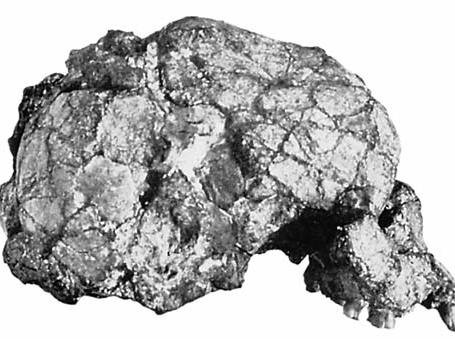
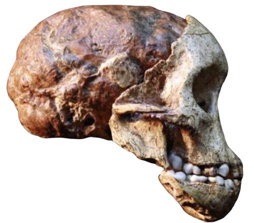
This turned out to be a lucky choice for him. Within two years of his arrival in Johannesburg, Dart had identified the Taung skull as a “missing link” in hominid evolution. The consequences of this discovery were to preoccupy him for the rest of his life. This skull ranks as one of the most important hominid fossils ever found, not because it was the oldest or most complete, but because of its critical position in the transition to hominid status. Dart’s recognition of this transition radically altered our concept of human ancestry and the forces at play in the evolution of upright posture and the development of the brain. But Dart’s insight was not fully accepted for decades and was subject to derision by his peers.
The circumstances surrounding the discovery and publication of the Taung fossil are fascinating and reveal the character and temperament of this pioneer. When he arrived at Wits, Dart was appalled by the lack of facilities in his new Department of Anatomy (barely five years old). He was dismayed at the absence of an anatomical museum, having just come from one of the great collections in London and seen some in America. He, therefore, urged his students to look out for fossils that might form the basis of a collection.
Within a year, he had struck, not gold, but limestone. The limestone was being extracted in a blasting operation at the Northern Lime Company near a remote village called Taung, in what was then the Northern Cape Province of South Africa. Embedded in the limestone were large numbers of primate and other fossils.
Dart’s attention was drawn by one of his enthusiastic students, Ms Josephine Salmons, to a fossil baboon skull with a neat hole in the top as if it had been struck by a sharp implement. She had obtained it from a miner, Mr De Bruyn, who had collected many such fossils as a hobby, with no knowledge of their significance.
Through the intercession of a colleague, two crates of bone-bearing limestone were dispatched by rail to Dart’s home in Johannesburg. Within minutes of opening one of the crates, Dart had found two astonishing pieces. One was a brain-shaped lump of agglomerated sand, the other a piece of breccia that appeared to contain a skull. The two pieces articulated. Using his wife’s knitting needles as chisels, he carefully cleaned the promising block of limestone, and on the twenty-third day, it is said, the rock parted to reveal the face of the Taung child. (Breccia is a sedimentary rock, composed of a mixture of existing rock fragments held together by a fine-grained matrix. Fossils may be found in such circumstances).
Dart was to write in his popular book Adventures with the Missing Link: “I knew at a glance that what lay in my hands was no ordinary anthropoidal brain. The skull cavity was three times larger than that of a baboon and considerably bigger than that of an adult chimpanzee, [but] it was not big enough for primitive man.” In his report,
submitted to Nature, Dart claimed that the skull was “humanoid” rather than ape-like and that this early human, named Australopithecus Africanus, “had walked upright, with its hands free for the manipulation of tools and weapons...providing clear evidence that Africa was the cradle of man.”
When Dart published his paper describing the Taung child fossil in “Nature” in 1925, entitled, “Australopithecus africanus: the man-ape of South Africa”, he stated that “it is manifest that we are in the presence here of pre-human stock, neither chimpanzee nor gorilla, which possesses a series of differential characters not encountered hitherto in anthropoid stock…” He further suggested that the expansion of the brain followed the adoption of an upright posture which had first freed the hands to become specialised for manipulation. However, it took a long time before his theories were accepted because it was the antithesis of the prevailing view that the expansion of the brain preceded a fully upright posture.
To understand the reactions of both the British public and scientific establishment to Dart’s assertions about both the African origins of early humans and their form, I need to digress to another young man with a very different temperament to Dart, Charles Darwin. Sixty-six years earlier, Darwin, in his classic work ‘On the Origin of Species’ written in 1859, had cautiously made one solitary reference to the human species, stating. “Light will be thrown on the origin of Mankind and his History”. I will not recapitulate the creationist controversies that followed, and which forced Darwin into seclusion and reticence. It took a further 20 years before he again wrote with exemplary caution, “It is somewhat more probable that our early progenitors lived on the African continent than elsewhere.”
In 1925, when Dart’s paper was published, the reverberations of Darwin’s statement were still being felt in the public arena, with teachers such as Scopes in Tennessee being on trial for teaching evolution in schools. Also, the anthropological establishment was still in ferment over the interpretation of the discovery in 1912 of what came to be known as “Piltdown Man”.
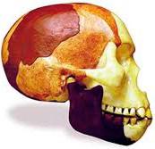
When, by the start of the 20th century, the anthropology establishment had fully accepted Darwin’s theories, it sought a European origin for mankind, preferably British. They seemed to have found what they were looking for some fourteen years before Dart’s discovery – in Piltdown Man!
The amateur archaeologist the Reverend Charles Dawson, in collaboration with one of the doyens of British anthropology, Sir Arthur Keith, claimed they had discovered the “missing link” saying that they had unearthed fragments of a jaw, a cranium and some teeth of apparent great antiquity in a quarry in Sussex called Piltdown.
The Piltdown fossils suggested a hominid with a large brain and an ape-like face. It was named Eonanthropus Dawsoni, New Dawn Man.
Piltdown Man turned out to be one of the biggest scientific frauds ever perpetrated. Sadly, it set anthropology back nearly 50 years. This was because it reinforced the preconception that it was the expansion of the human brain that had preceded the development of other human characteristics, such as upright bipedal posture, and that this had determined the subsequent course of human evolution.
Under the influence of Piltdown, Dart was pilloried in the scientific as well as popular press. It took until 1953 to establish that Piltdown was a fraud that consisted of a doctored modern human skull and an ape jaw. Dawson, Keith and even Conan Doyle, Sherlock Holmes’ creator, who lived nearby, have all been implicated but not
proven as the perpetrators.
In the late 1930s, adult specimens of Australopithecines were discovered at Sterkfontein, only 30 miles west of Johannesburg. The eccentric Dr Robert Broom, a Scottish physician who had come to South Africa earlier, supported Dart’s theories and was an enthusiastic if sometimes difficult, collaborator.
As early as 1938 Dr William King Gregory, of the American Museum of Natural History, declared after an inspection of the Australopithecine finds: “It is the missing link no longer missing. It is the structural connecting link between ape and man”. And as late as 1957, in a letter published in Nature, Sir Arthur Keith confessed, “I am now convinced on the evidence presented by Dr Robert Broom that Professor Dart was right, and I was wrong”. Over the next 50 years Dart was to be further vindicated by numerous further discoveries of up to four species of Australopithecines, some associated with evidence of tool using, and possibly tool making.
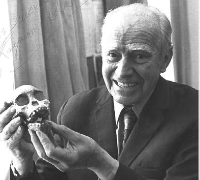
Dart took his students out on field trips across Africa to make life and death masks of all the people of Africa he could came across. These 1,110 masks (397 life, 487 deaths, 226 not stated) were created between 1927 to the 1980s and form one of the most comprehensive facemask collections in Africa. These made an impact on all of us as they lined the corridors outside the anatomy halls of our medical school. The faces of former Wits students, once engaged in facial casting workshops, were also included. It made us aware of the wonderful diversity of mankind.
Raymond Dart held the position of Professor of Anatomy at the Witwatersrand University Medical School for 35 years, from 1923 to 1958.
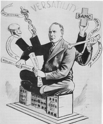
Dart was also appointed Dean of the Faculty of Medicine a post he held for nearly 20 years from 1925 to 1943. Dart’s versatility was depicted in a student magazine showing Dart as a many-armed Hindu deity, sitting on top of the medical school.When I became Dean of Medicine at Dart’s alma mater, the University of Queensland, I invited his successor and colleague, Professor Phillip Tobias, to open an exhibition arranged by the University Anthropology Museum in 1992/93 commemorating the centenary of Professor Raymond Dart’s birth in Brisbane in 1893. (see Chapter 5 on Phillip Tobias)
We were all somewhat “children of Dart” as we pursued our pathways in our studies and out into the world. Laurence Geffen AM BSc (Hons 1) , MBBCh, MSc (all Wits), BA (Hons 1) DPhil (both Oxon), MD Hons (Flinders) FRACP (Hons) FRANZCP, Brisbane, Australia. August 2020.
In our second year, we were introduced to the worlds of anatomy and physiology. At last, we entered the medical school, then on Hospital Street, and were introduced to the superb anatomy lecturers, Phillip Tobias and Maurice Arnold.
The Wits Medical School building on Hospital Street, where we studied in the 1950s, had opened in the early 1920s. When this picture was taken nearly a hundred years later, in 2017, it had become the University of the Witwatersrand’s Department of Forensic Medicine, while the medical school had moved to Parktown in the late 1970s.
Laurence Geffen, who spent much time with him as a student in his BSc course, has written our tribute to our dynamic anatomy lecturer, Phillip Tobias. Chaim Rosenberg compiled a bio-sketch of Maurice Arnold.
Anatomy Teacher, Professor of Anatomy, Palaeoanthropology and Zoology (1951 – 2012). Dean of Wits Medical School
By Laurence Geffen and others
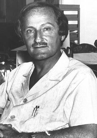
There can be few more memorable teachers of the Class of 1960 than Phillip Vallentine Tobias. Most people will remember him primarily for his lectures to our spellbound second-year anatomy class in 1956, although he only joined us mid-year, fresh from his studies in the UK and USA. Slight of build, in a crisp white coat, with glittering eyes and an aquiline profile, balancing on the balls of his feet, his presence filled the main lecture theatre located at the entrance to the Medical School. A torrent of words, slides and gesticulations projected his passion for the topic of the day. Not even the growing impatience of the next class, crowded outside in the foyer, seemed capable of stemming his flow.
Three years later, in 1959, when most of us were well into our clinical training, Tobias was to assume the mantle of Professor of Anatomy at Wits University, handed on to him by his mentor, Raymond Dart. The bond between Dart and Tobias was profound. First, Tobias had the role of pupil, then they became colleagues. Tobias was later to assume the role of Dart’s “bulldog”, defending Dart’s views on human evolution against a hostile reception, much as Huxley had guarded Darwin. Not that Dart was as reticent as Darwin on the topic!
Phillip Vallentine Tobias was born in Durban, Natal, on 14 October 1925. His father, Joseph Newman Tobias, was born in Portsmouth, England, and emigrated to South Africa, where for many years, he ran a shop. His mother, Fanny Rosendorff, was a piano teacher born in Edenburg, in the Orange Free State. Joseph Tobias’s great-grandfather was the Anglo-Jewish publisher Isaac Vallentine. Phillip Tobias apparently learned to read at the age of three, but his childhood was disrupted by his parents’ divorce.
He attended Durban Preparatory Boys’ School from 1933 to 1935, but spent 1936 at President Brand School in Bloemfontein. He completed his primary schooling at St Andrews School in Bloemfontein. Tobias then entered Durban Boys High School in 1939, where he matriculated in 1942. During those years, he frequently visited the
Durban Natural History Museum, where he was fascinated by the exhibits on genetics, zoology, and archaeology.
When Tobias was 15, his sister Valerie, then only 21, died of diabetes. This was a major blow to him and a significant factor in his decision to study medicine. In 1944, he enrolled at Wits medical school, and the following year 1945, as a student, he was appointed as a demonstrator in histology and instructor in physiology. Tobias studied under the anatomist Dart and the histologist Joseph Gillman, and received his BSc hons in histology and physiology in 1946/7. He was introduced to paleo-anthropology through Dart, who described the first Australopithecus africanus fossil. In 1945, Tobias led a group of fellow students to Sterkfontein, where Robert Broom had discovered australopithecine fossils in the 1930s.
Tobias also became involved in student politics at a time when national politics were affecting university education. He was elected president of the National Union of South African Students three times (1948-50), and in this role, led some of the earliest campaigns against the apartheid government. In 1948, the National Party came to power in South Africa, and one consequence of its apartheid policies was that the student union, which had previously been non-racial, was now racially segregated. Tobias’ opposition to this policy marks the beginning of a life-long effort to combat apartheid.
He obtained his MBBCh in 1950. Tobias inclination, though, was towards science and research rather than practising medicine. In 1951, a year after qualifying as a doctor, Tobias was appointed as a lecturer in anatomy. Under Gillman’s influence, Tobias began researching chromosomes, obtaining his PhD in 1953. He devoted much of his career to groundbreaking research in paleo-anthropology. In 1955, he undertook postgraduate research in physical anthropology with Jack Trevor in the Duckworth Laboratory at Cambridge University, in England, where he was a Nuffield Dominion Senior Travelling Fellow. He undertook further study in the United States in anatomy, physical anthropology, human genetics, dental anatomy and growth, as a Rockefeller Travelling Fellow.
On his return to South Africa, in mid-1956, as well as teaching anatomy to our year, he analysed and described hominid fossils from such geographically diverse places as Ubeidiya in Israel, Chemeron in Kenya, and Haua Fteah in Libya. In 1959, he succeeded Dart and was appointed professor and head of the anatomy and human biology; a position he held until he retired in 1990. Thereafter, appointed Professor Emeritus and was head of the research department at the Sterkfontein Caves. Tobias was given many honours all over the world and was nominated three times for a Nobel Prize. He was appointed a Fellow of the Royal Society in London in 1996. Although he is best known for his work on hominid fossils from Olduvai Gorge, in Tanzania, and from various sites in South Africa, it is as an inspirational lecturer, tour leader and mentor that he is remembered by generations of students.
I got to know Tobias on a personal level when, along with a few other members of the second-year medical class of 1956, I undertook intercalated studies in anthropological and physiological sciences in the years from 1956 to 57. Instead of the formaldehyde suffused atmosphere of the dissecting hall, we were introduced to the twin challenges of living physical anthropology and paleo-anthropology. We also had our first heady encounters with the disciplines of genetics, comparative anatomy, evolutionary biology and other cognate (or related) sciences. Not to mention courses on the main University campus ‘down the hill’ on the history and philosophy of science and a smattering of scientific German!
Tobias, the polymath, was our main guide to these wonders, although Dart would, to our terror, also appear regularly at our student seminar presentations. Martin Bobrow has remarked that making a presentation at those student seminars was a trial by fire. But they were also transformative. Ever since, I have subjected my own research students to equivalent in-house rehearsal(s) before they were allowed to present to the public.
Absorbing though the daily challenges at medical school were, nothing compared to the excitement of the annual field camping trip to Makapansgat, in what was then the Northern Transvaal. While still a medical student in 1945, Tobias had initiated a series of annual research visits to this fossil-rich site. The fruits of these expeditions formed
the core of the collection of bone, tooth and horn artefacts that Dart adduced in support of his controversial australopithecine osteo-donto-keratic culture.
On our Makapansgat expedition in 1957, Tobias’ enthusiasms were in full flow, and he relished holding court in the evenings around the campfire. Back at home, he would invite us to his apartment for reunions at which he would serve his speciality, giant fluffy omelettes!
I subsequently had less contact with Tobias, as I completed the medical course and then left South Africa in 1964, going first to the UK and subsequently to Australia. However, when the Apartheid era ended, I occasionally returned to South Africa to visit family and always took the opportunity to visit Phillip. In turn, he, an inveterate traveller, visited me in Australia several times.
Saul David Nathanson, who graduated in 1966 wrote: I spent a week at Makapansgat with Tobias exploring 2-million-year-old fossils in the Northeast Transvaal caves in the winter of 1962. Raymond Dart, Tobias’ mentor, friend, and predecessor in the anatomy chair at Wits, visited for lunch one day and seeing the two of them banter and discuss various aspects of paleoanthropology was simply extra-ordinary. This stimulated me to learn more deeply by associating with the excitement of inquisitive minds. My classmates with the best memories remembered all the new facts of the week. Their results stood out when the examination results were posted on the message board in the lobby.
“Phillip Tobias taught us to think like a scientist and to observe everything intensely. One day, walking from our campsite at the river in Makapansgat, he suddenly stopped and bent down and dug a little with a small spade in the dirt. He had seen and unearthed the skeletal remains of a small mammal. Like an excited child he explained the process by which an eagle or similar bird swooped and swallowed an animal and later regurgitated the undigested bony parts onto the ground, where they became fossilized. I would not have seen the specimen, but he was always alert to his surroundings, an attribute that I have tried to mimic.
“My year with him as a ‘table doc’ was priceless. His knowledge of anatomy and paleo-anthropology was obviously extensive, but he also knew history, opera, philosophy, politics, and human behavior. I thought I knew Darwin until I heard him talk to the graduate students about the subject. I measured bones with calipers, Phillip breathing over my shoulder to make absolutely sure my measurement was accurate enough for his obsessive nature. He visited me in Detroit in 1993 and I saw how he had aged but his mind was sharp enough to hold forth with the graduate students in anthropology at Wayne State University. My extra-ordinary year with Tobias not only gave me a vast knowledge of anatomy but also an introduction to graduate students who learned how to think.”
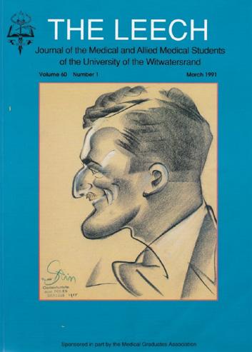
When in 1990, Tobias retired from the Department of Anatomy, after over forty years on its staff, thirty-two of them as Head, I was approached by the editors of The Leech, to produce a tribute to Tobias.
The Leech was a remarkable quarterly journal founded in 1928 and run by the Wits Medical Student Council. It published scientific papers, local news and letters submitted by under-graduates and graduates. (In our time, Irving Lissoos served as editor of The Leech).
For this tribute, I wrote a detailed account of his career which was published in March 1991, in volume 60 (1)1-4 of The Leech. What follows are a few excerpts from my account of Phillip Tobias as I knew him.
I recounted an episode from our expedition to the Batonga in 1957, that was revealing of Tobias’ qualities as a scholar. I still remember, to my shame, sulking in my tent because he would not allow me to keep as a memento even a single stone arrow head that I had picked up on an ancient river bank, despite the fact that the gravel bed was littered with thousands of (to me) identical artefacts. Tobias insisted it be properly labelled (with its exact location) and put in a museum collection:
the object did not matter but the principle did!
In that same article, I also described: The crowds of pot-bellied Tongan children who followed him and fell about with hilarity when he peered into their mouths and guessed their ages from their erupting dentition. He was later to amaze my own sons with this party trick on a visit to Adelaide in 1979 and almost launched them into careers in dentistry. (They eventually did medicine instead).
I was to get to know Tobias even better in July 1957, when he invited me and fellow BSc student, Martin Bobrow (we were known as ‘Tobias’ Angels’) to join him on that expedition to study the Batonga tribe. They had lived isolated for centuries in the valleys lining the Zambesi river. However, their villages were to become part of the flooded bed of Lake Kariba, when the river was dammed for what was then Africa’s largest hydroelectric scheme. The Batonga were to be moved, not always willingly, onto the higher ground of the escarpment that would form the lake shore, where it was anticipated they would convert from a hunting to a fishing community.
I recall one dramatic episode when a tsetse fly (Glossina species), a vector of sleeping sickness (Trypanosoma brucei), found its way into Tobias’ shirt. We were going about our daily anthropometric exercises when Phillip let out a yell and frantically tried to remove his shirt, only to become entangled in the straps of his camera, binoculars and other devices adorning his neck. Martin and I did not help matters in the ensuing confusion by pulling in opposite directions on the various straps until his eyes nearly popped out. Realising the futility of this manoeuvre, we then saved him from a nasty bite and from a tropical disease by beating the fly to death on his chest – for which Tobias was not particularly grateful.
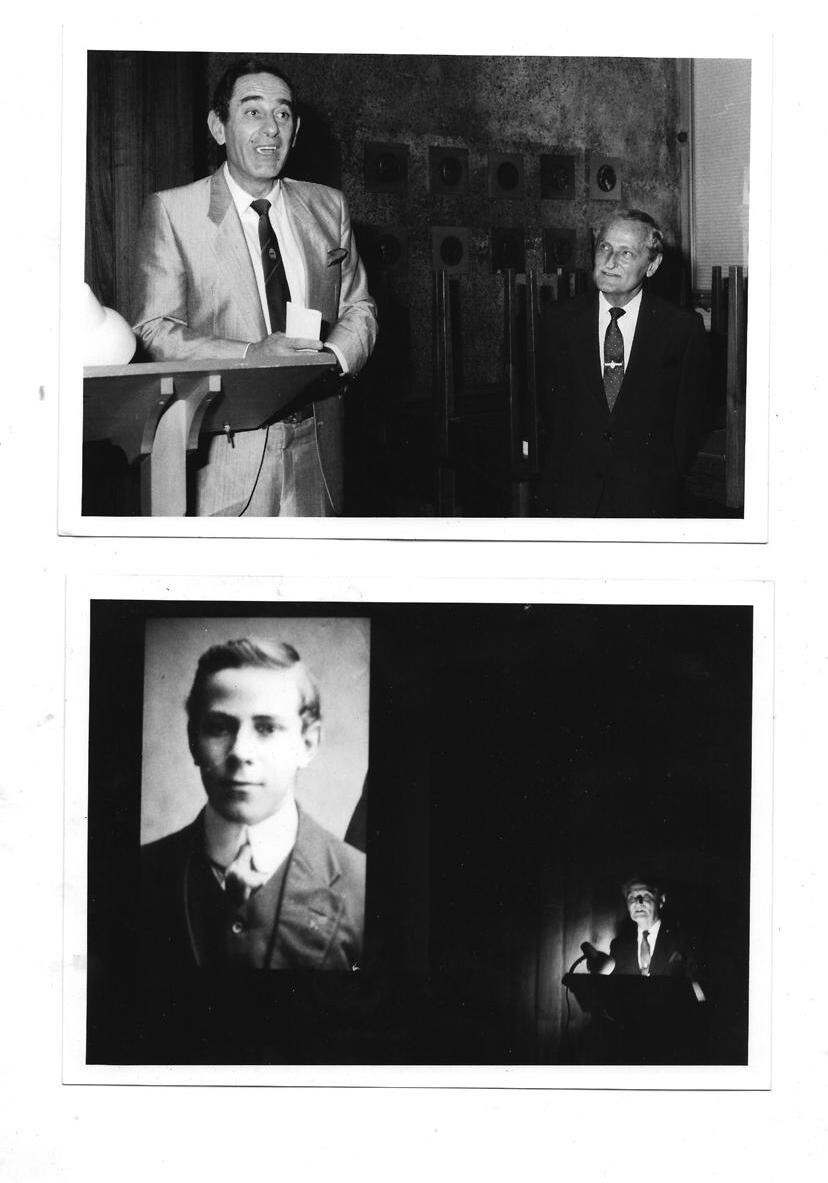
In 1979 when I was Dean in Adelaide, we invited Tobias to deliver the inaugural Andrew Abbie memorial lecture, named after an eminent Australian anthropologist who also happened to have been our neighbour.
As I wrote in the article: It was vintage Tobias. Before a packed, be-gowned audience in the Howard Florey Theatre (so named by the Governor-General in Tobias’ presence that very morning) he delivered an epic lecture worthy of his gifts: ‘The Evolution of the Human Brain, Intellect and Spirit.’ It was epic in scope, epic in delivery and epic in duration. More than two hours later, the audience emerged, exhausted but exalted, to seek revival in cold tea and curled sandwiches. I was very proud of him.
When I became Dean of Medicine at Dart’s alma mater, the University of Queensland, I invited Tobias again, this time to open an exhibition arranged by the University Anthropology Museum, in 1992/93 commemorating the centenary of Professor Raymond Dart’s birth in Brisbane in 1893.
Tobias’ talk was delivered with the same passion, fluency, erudition and theatricality that I remembered from nearly forty years earlier. And yes, it ran so much over time that again the post-lecture tea was cold, and the sandwiches curled.
The first precept of the Oath of Hippocrates of Kos enjoins us ‘to consider dear to me as my parents him who
taught me this art’. I am but one of thousands of Wits medical and science graduates who would immediately think of Phillip Tobias in this regard.
Lawrence Geffen, who wrote this memoir, had three overlapping careers: Lab work, Medical Education, and Clinical Psychiatry. He was appointed Professor of Physiology and then Dean of two great Universities in Australia: Flinders University in Adelaide and Queensland University in Brisbane. This latter was Professor Raymond Dart’s Alma Mater in Australia.
Geoffrey Boner, of our class of ‘60, who became a nephrologist in Israel, remembers: The lateral incisor on the left side of my upper jaw never developed, while the lateral incisor on the right side of my upper jaw, was small and pointed. As luck would have it, I had Professor Tobias for my oral examinations in anatomy. He had noticed my small tooth and we spent the allotted time discussing my teeth. Many years later I was present at a lecture he gave in Israel. I went up to him after the lecture and asked him if he remembered me. His answer was that he remembered me, not my name, but that I had a small, sharp incisor tooth on one side. Our next meeting was at the re-union in 2005. He had recently published his memoir. I bought the book and had him sign it for me.I will always remember him as one of our great teachers and one with an outstanding memory.
Ronnie Auerbach, member of our class of ‘60 who became an ENT Surgeon in London says: I travelled to London by mail ship after my matric in December 1954. At my table for dinner each evening were Trevor Huddleston and Phillip Tobias. I can’t recall what must have been scintillating conversations. I was visiting paternal grandparents that I had never met, before embarking on my medical studies. Tobias was on his way to Cambridge to spend a year at the Duckworth Laboratory, and further study in America. He only arrived back at Wits 18 months later when we were halfway through our anatomy course in 1956. Towards the end of 1956, nearly two years after we had met on the ship, I was walking down the anatomy corridor when a man popped out of his office and without hesitation, pointed to me and said ‘Auerbach, Transvaal Castle!’ It was Phillip Tobias.
Nathanson, also recalled his Wits student days with pleasure. He said: “Wits Medical School was wonderful. Standout memories include great teachers and memorable characters. Who can forget Philip Tobias that first day in the anatomy hall, where four prominent local theologians, while conducting a funeral according to each of their faiths, clearly articulated the importance of respect for the dead while we stood in silence next to the covered cadavers, many of us experiencing dead people for the first time. Or the days when the ‘little professor’ dropped in unexpectedly during a dissecting session in the Vesalius Anatomy lab and performed like a Shakespearean actor. Short stature, wearing a meticulously ironed white coat, forceps, and scalpel in hand, he passionately demonstrated to the gathered students the anatomy of the day, bringing a ‘dead’ subject to life.
Another ’66 graduate Robert Jacobson wrote: “Second year Medical School was enthralling. The professor of anatomy, Phillip Tobias was a most dynamic, erudite, and brilliant teacher and leader. When I first entered the anatomy department it was overwhelming. There must have been about fifty cadavers with four students assigned to each one. Dental students and physiotherapists joined us for the long and detailed anatomy course. With his predecessor Dart, Tobias had made Wits the leading centre in the world for paleontology and physical anthropology research and teaching“.
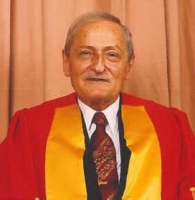
Tobias was simultaneously a professor at Wits in the fields of anatomy, palaeoanthropology and zoology. He held the positions from 1951 almost until his death in 2012, of demonstrator, lecturer, professor, dean, emeritus professor, honorary professorial research fellow and director of the Sterkfontein Research Unit.
Over the years, he was offered posts around the world, but always turned them down, happy to stay at his alma mater, from where he had obtained five degrees.
Sydney Brenner, a fellow student of Tobias at Wits and then eminent scientist and Nobel laureate, wrote in the foreword to Tobias’s autobiography: “Unlike many of his contemporaries who left South Africa in the 1950s, Phillip stayed on and committed himself to maintaining high standards of scholarship and personal integrity during the difficult years,”
In his own words: Tobias somewhat regretted not having married or had children – but said "that he had had 10,000 children, whom he had taught at Wits over many decades". Tobias said: “In a very real sense, I have passed on my legacy to the bright, eager-eyed young students, not biologically, not by my DNA, but as tutor, mentor, exemplar and friend. I was one of those strange professors who loved his students. By being available to them at all times to help them with their problems and with constructive, creative advice and trying to widen their horizons. I like to think that I have had a moderately good impact on some of them, and bless them, they’re always telling me this when I meet them in Edmonton in Canada, Sydney, Nairobi, Hong Kong, New York and Cambridge.”
Anantomy Teacher and Textbook Auther
By Chaim Rosenberg
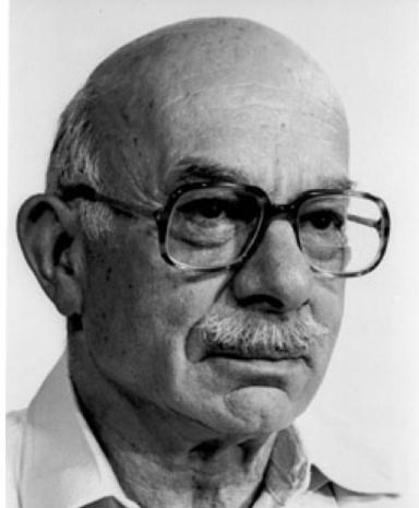
I compiled this story of the life of Maurice Arnold (MBBCh (Wits), FRCS (Edin), MD (New South Wales)) based on reports from his son – our classmate, Peter Arnold, and obituary notices written by Phillip V Tobias [Journal of Anatomy (1995):187, p.253] and the South African Medical Journal Volume 85, No.4, April 1995, p.296. There are added comments from classmates, Avroy Fanaroff and Ronnie Auerbach.
Morris Amoils was fast. He was the fastest runner in his primary school, and his schoolmates nicknamed him ‘Toby’ to link him with ‘Toby’ Betts, the schoolboy sprinter, who in 1924, represented South Africa in the Summer Olympic Games. To his family, he was known as Meish. Later, he changed his original family name from ‘Morris Amoils’ to ‘Maurice Arnold’.
He was born in Grahamstown in 1907, the son of Lithuanian-Jewish immigrants. The family moved to Johannesburg, where he attended Jeppe Boys’ High School. None of his siblings finished high school. Instead, they went to work to support little Morris, who was very bright, to attend medical school.
The University of the Witwatersrand Medical School had only opened in 1919. Morris was one of eighteen students in the seventh year of the school. Ironically, he had to repeat Anatomy (or maybe that was a contributing factor to his later career). He graduated MBBCh in 1931.
After a few years in medical practice, he went to Great Britain where, in 1938, he obtained his Fellowship of the Royal College of Surgeons in Edinburgh. Returning to South Africa, he signed up in the Second World War and served for five years as a surgeon in the Union Defence Force in field hospitals in North Africa, Palestine, and Italy.
After the War, he approached the Professor of Anatomy, Raymond Dart for a position in the Anatomy Department at Wits Medical School. Jobs were scarce, so for the first year he taught anatomy without pay. The next year he was rewarded with a salaried position as Senior Lecturer in Gross Anatomy and Lecturer in Clinical, Applied and Surgical Anatomy.
Avroy Fanaroff remembers Toby Arnold as a superb anatomy teacher. He spent a lot of time at the chalkboard reconstructing anatomical sites which he would build up in layers. He would use coloured chalk and draw with both hands. Ronnie Auerbach remembers that he would illustrate comparative anatomy by drawing a human spine with one hand and a baboon spine with the other – simultaneously! Ronnie says the students had to work in pairs if they wanted to take notes – one to write the notes and the other to draw the pictures. Toby
clearly very much enjoyed teaching and was always polite and friendly.
Arnold’s book Man’s Anatomy: A Study of Human Dissection, published by Wits University Press, was reprinted and used by generations of medical students (though this was attributed to Phillip Tobias as well – or even alone). Always wearing white gloves, Arnold would start with a meticulous drawing on the chalk board of the bony underlay of a region. He would add to this the ligaments and tendons, then the muscles, nerves, blood vessels and viscera. This teaching method was the basis of his second book, published in 1968, 'Reconstructive Anatomy To Australia in 1961'.
In 1961, Maurice Arnold and his family, including his son, our classmate Peter, emigrated to Australia. From 1961-63, Maurice Arnold was Reader in Anatomy at the University of Queensland, Brisbane, Australia. From 1966 until his death in 1994, he served in the anatomy departments of the University of Sydney and the University of New South Wales. He also held visiting professorships in anatomy at the Hebrew University in Jerusalem (1952), Wayne State University, Detroit (1960) and St. Louis University (1964-65).
‘Thousands of students and colleagues,’ wrote Phillip Tobias, ‘benefitted from his single-minded enthusiasm, encyclopaedic knowledge, integrity and accessibility. Despite his failing health, Toby Arnold continued to foster the study of anatomy. Heart and circulatory conditions led to the amputation of both his legs, but he remained uncowed and, from his wheelchair, he indomitably continued daily trips to the anatomy department, cataloguing and upgrading the museum collections, teaching, counselling and writing. His remarkable memory for detail remained fully functional until he died aged 87 years.’
With his ‘Arnold’s Anatomy’ feature in the monthly newsletter of the Anatomical Society of Australia and New Zealand (ASANZ), Toby encapsulated his approach to the understanding of anatomy. He served for many years as a Member of the Board of Examiners for the Diploma of Anatomy of Australia and New Zealand.
The University of New South Wales awarded him the degree of Doctor of Medicine honoris causa and, in 1989, the title of Professor Emeritus. The citation for his honorary doctorate described him as ‘one of the world’s leading anatomists whose knowledge of the subject is extensive and probably unrivalled.’ He was known as ‘Mr. Anatomy of Sydney.’
Professor Phillip Tobias considered Toby Arnold ‘A man of singular modesty, great personal integrity, humility and gentle humour appreciated and loved by thousands of former students, colleagues and friends.’ Both of Toby’s sons are doctors, as are two granddaughters. His great-grandson is a medical student.
St. John’s College, Parktown Boys’ and Girls’, Jeppe Boys’, and Athlone Boys’ High Schools and the German School Hillbrow
In our day, the South African school leaving examination was known as the ‘matric exams’ and by extension, students in the final year of high school (grade 12) were known as ‘matriculants’ or, more commonly, ‘matrics.’ Once the matric exams have been passed, students are said to have ‘matriculated.’
Most of the graduating class of 1960, had matriculated in 1954. Some of our class came from high-achieving schools in the affluent suburbs of Johannesburg. There were six medical entries from St. John’s College and nine from King Edward VII School, both in leafy Houghton. Parktown Boys (six) and Parktown Girls’ School (four) provided ten outstanding students. Other excellent Johannesburg boys’ schools were Highlands North (from where there was a bumper crop of 11 medical students in our cohort). There were two boys from Jeppe and four from Athlone. Some students came from gold mining towns to the east and west of Johannesburg, and a few from further afield across the country like Durban, Bloemfontein and Kimberley. Let us introduce you to those we have been able to reach, who are the cast of characters in this book.
In this chapter we look at the schools and meet some of the students from St. John’s College, (six) Parktown Boys and Girls, Jeppe, and Athlone High Schools, as well as the German School in Hillbrow.
St. John’s College: There were six boys from St. John’s College who enrolled in the 1955 medical class. They were Anthony Molyneux Meyers, better known as Tony Meyers, Colin Robertson, L U Hall, David Paton, Rodger Edge and Andre van As.
The College is perhaps the oldest and certainly one of the most prestigious schools in Johannesburg. It was founded in 1898 as an Anglican Christian School. Many of the teachers were recruited from British universities. The general tone of the school, in the 1950s, was one of a traditional British public school such as Eton and Harrow. Andre van As told us: “At St. John’s, we all wrote the higher standard of Matric, the Joint Matriculation Board Exam (JMB), which fulfilled an important function in controlling and maintaining standards for British university admission, although this was not offered at many Johannesburg schools who wrote the local Provincial school leaving exams.
Being a private fee-paying school, faced stiff competition after the second Anglo-Boer war when Lord Alfred Milner’s colonial administration expanded state schools in the Transvaal and created high-achieving government schools such as Jeppe, Parktown and King Edward VII School (KES).
Being cash-strapped in the early 1900s, It sought the help of the Community of the Resurrection (CR) an AngloCatholic order of missionary priests and lay brothers based at Mirfield in Yorkshire, to come to assume control of the College, which they did until 1934.
The new headmaster, Father James Okey Nash decided to move the school from the centre to the rustic northern outskirts of the city. He chose a site on Houghton Ridge, which the school was able to purchase through the generosity of diamond magnate Sir Thomas Cullinan, who donated £5000, and Johannesburg Consolidated Investments, Barney Barnato’s company, which sold the land to St John’s at half its market value.
They commissioned the distinguished British architect Herbert Baker to design the new school buildings. He created a blueprint for much of what was to materialise on the site over the ensuing decades. St. John’s celebrated its 125th anniversary in grand style in 2023. It is still among South Africa’s pre-eminent schools, with a reputation for excellence extending well beyond the country’s borders.
Not only did we have six St. John’s starter students in 1955, but a number of the faculty who taught them in the late fifties were also St. John’s alumni. These included John Barlow and Mozie Suzman, as well as Tim Barlow and the Gear Brothers. In his address to the College in December 1978, our renowned professor of surgery, Prof DJ ‘Sonny’ du Plessis, who was then also the Vice-Chancellor of Wits University, with tongue firmly in his cheek, coined the term “the St. John’s Medical Mafia”, referring to an incredible production line of Old Johannian medical doctors that has continued since. This group was remarkable not only in its number but for its impact on the field of medicine.
Andre van As wrote: “Avroy Fanaroff has written that the boys from King Edward School had a close relationship at school and university which endured for many years, before and after graduation. Regarding the St. John’s boys, however, apart from being in the same Clinical ‘Firm’ with Paton and Meyers, I never had any contact with the others during our time at Wits.
I took advantage of the extra year in the sixth form at St. John’s to facilitate getting a head start for my first year in medicine.
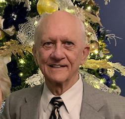
Andre van As became a pioneering pulmonologist and physician in internal medicine, first in South Africa, and later in the USA. He also played a part as a clinical development leader for pharmaceutical companies.
In Andre’s biography, there is a picture of the so-called ‘St. John’s Mafia’ on the Faculty at Wits Medical School in our time. Andre also sent this picture of the third-year Pharmacology class of 1957―where all six of the Johannians are indicated.
After spending 17 satisfying years as a single-handed General Practitioner, Roger specialised in anaesthetics. Asked to set up ongoing postgraduate anaesthetic teaching, he organised lectures at the College of Medicine in Parktown with drinks and dinners. His piece de resistance was his annual educational meeting for GP anaesthetists held at the Kruger National Park.
When he worked at the Chamber of Mines hospital, he insisted on being taken 2.5 Kilometres deep underground to see how miners became injured.
Later, Roger spent six years at the King Khaled Eye Specialist Hospital in Riyadh, Saudi Arabia (KKESH). When he returned to South Africa he organised refresher courses for GPs. With lockdown, this went virtual, with monthly lectures on an encrypted TV Channel. Andre said: I last saw Rodger Edge in 1989 when I was invited to give presentations on the treatment of asthma in the United Arab Emirates. He was coincidentally attending another conference and he shared with me the fact that he was an anaesthetist in Riyadh.
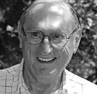
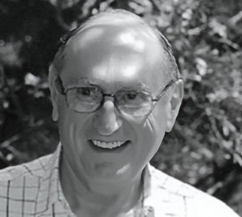
Tony Meyers had matriculated from St John’s in 1950 and spent some years as a student in various faculties at Wits before settling on Medicine.
Tony became the first Professor of Nephrology at Wits and chief of the department at the Johannesburg General Hospital. As such he was a teacher and mentor to numerous physicians and nephrologists. Wits awarded him a Doctorate in Medicine in 2020 on a topic that he was passionate about – kidney stones. He also undertook the mammoth task of recording the history of nephrology in South Africa.
David Paton – David Paton was the son of Alan Paton who wrote Cry the Beloved Country. He had graduated from Natal University in Psychology before starting medicine in 1955. Andre said: “After 1960 I never saw Paton again and had intermittent contact with Meyers when he was delegated to run the renal dialysis unit that we set up at the JG Strijdom Hospital in the mid-1970s”.
We believe he became an obstetrician
Parktown Boys’ High School: There were five Parktown boys in our class starting in 1955: Michael Berger, Martin Bobrow, Laurence Geffen, Aubrey Milunsky and Farrol Sims.
Remarkably all of the Parktown boys became distinguished professors in some aspect of medicine somewhere in the world. (There were a further seventeen members of our class who also became professors and heads of departments whom we will introduce you to in more detail in the chapter on ‘Academic Medicine’.)
In compiling this Parktown section, we wondered whether they might have been inspired by teachers at Parktown Boys’ High. However, on enquiring, they all reported that their science master was a stern man who did little to instil an interest in the subject in his students. Laurence Geffen who together with Parktown mates Martin Bobrow and Michael Berger spent a year or two completing an intercalated BSc degree, before completing the medical studies, wrote – probably for all – when he said: “My interest in research was first catalysed in second year Medicine by Benny Kaminer, a physiologist and Phillip Tobias an anatomist, whom I looked up to as my role models. If I were to generalise about our collective high achievement in medical science, I would ascribe most credit to the quality of the Wits medical faculty.”
Martin Bobrow agreed, saying, “School and first years were not memorable, but when I did the BSc, it was very different, with distinctive, even florid characters like Phillip Tobias, Joe Gilman and Benny Kaminer.
There was even a connection with the later Nobel Prize winner, Sydney Brenner, who was taxing as a teacher, but he was a truly great mind whom I admired immensely. Philip Tobias in particular had a major influence on my future.
The Parktown boys all had distinguished careers in academic and clinical practice. We will meet them again in a little more detail in the chapter on ‘Academic Medicine’ but you can read their fuller bio-sketches on the links with their names: Mike Berger became professor of chemical pathology at the University of Natal Berger, Michael Benjamin; Martin Bobrow became a professor of human genetics at Amsterdam, London (Guys Hospital, now subsumed into Kings College) and Cambridge universities and was elected a Fellow of the Royal Society in England Bobrow, Martin (BSc); Laurence Geffen became the dean of two medical schools in Australia and professor of psychiatry Geffen, Laurence; Aubrey Milunsky became professor of genetics at the University of Boston Milunsky, Aubrey; Farrol Sims became director of research and associate professor in family medicine at
the University of Wisconsin Medical School Sims, Farrol Hyman.
Another Parktown boy who matriculated earlier and joined the medical class, which graduated in 1960, was Leopold (known as Leon) Kahn. Leon had already spent two years in dentistry and one in geology before he was accepted into the second year of Medicine and thus joined the 1960 graduation class in 1956. Leon also had a distinguished career, practising surgery and urology in South Africa and the UK. Leon was born in the East End of London and came to South Africa as a small boy during the war. He spent two years very happily in the Johannesburg Jewish Orphanage, Arcadia, where he says he learned to swim and climb trees. He loved his time at Rosebank Primary School where he played in the school football team and was Victor Ludorum in athletics in 1947.
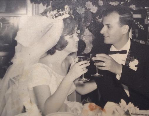
He moved on to Parktown Boys’ High School, where apart from being in the school swimming and athletic teams, he also captained the school hockey team in matric. He got the best matric results of his year, 1952. Unsure which career path to follow Leon did spent two years in the faculty of Dentistry and one year in the faculty of Science [Geology was amazing, he said]. Once in the Medical School, he teamed up with classmate Naomi Livni a brilliant student and sportswoman who had matriculated with flying colours at Parktown Girls’ High School. They were married in 1961, two weeks after graduation. Leon became a general surgeon specialising in urology first in South Africa, then the UK.
Parktown Girls’ High School: Remarkably, four of the five or so girls in the class of ‘60, matriculated in 1954 at Parktown Girls’ High School. Apart from Naomi Livni, Gloria Davis, Adele Blankfield, and Joan Feldman, were all accepted to study medicine at Wits, starting in 1955.
Naomi spoke of her pathway to study medicine. She said: “I loved school and all the country schools I had attended. I got excellent matric results and was accepted at Wits University. I went to enrol for nuclear physics on the appointed day. In those days, enrolment was done in person on a set date, and students simply queued for the faculty of their choice. I was the only person in the queue for nuclear physics. When I approached the desk, the gentleman seated there said to me, ‘We have only two places for students in nuclear physics, and you aren’t going to get either one of them – so run along, little girl’.
This was my first (and not the last) encounter with the glass ceiling! I left the room with not much of a clue as to what to do. I joined the queue for medicine and was accepted.” Naomi became a sought-after clinical pathologist, also specialising in later years in urological histology.
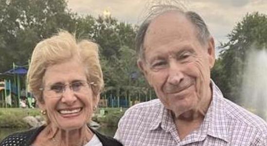
Gloria Davis who lives in Johannesburg and specialised in allergy wrote: “I enjoyed my school days at Parktown Girls’ High school in Parkview Johannesburg. It was a prestigious school and one where behaviour and dress code were strictly enforced. Even the length of our skirts was measured to be long enough to meet the required distance above the knee. Imagine this nowadays. I can hear the laughter now! We also wore lovely Panama hats. Nobody can afford this now. I loved mathematics, geometry, biology and
Latin.
We had Miss Sturgess as our biology teacher. She was Eric Sturgess (of tennis fame) sister. She was a wonderful lady and teacher. Our vocational guidance teacher was a tiny lady, but her temper was not in proportion to her size. There were the four of us who had applied to do medicine at Wits. She looked at us and turned to me and said, ‘I do not believe that you Gloria will manage and qualify’. Oh, how I would have loved to show her that I was chosen as the best woman graduate of Wits for that year. All four of us who applied completed our degrees. I was asked to address the matriculants at the school on their speech night many years later. My message to them was to always believe in themselves and never listen to others who doubt you. Incidentally, my prize for the award was a textbook of my choice. It’s now completely outdated! I use it to lift my computer to a better height! “Naomi Livni and I were in the same group of eight throughout our clinical years. We lost touch with each other, but since our class renewed our acquaintance through the graduation anniversary, we have been in constant touch with one another. We reminisce fondly about our student days. Life was so male-dominated, but we still had wonderful female lecturers like Shirley Siew, who was a great pathologist. I was happy to be in the same class as my cousin Arthur Rubenstein. There have been anxious times and many challenges, but the satisfaction and joy of helping my patients feel better have been the cherry on top.We were not able to trace any of the other women in our class, however we learned that Joan Feldman had gained a PhD and became a Psychiatrist in the United Kingdom.
Jeppe High School for Boys: The two boys who joined our class from Jeppe High School were both among the nine who got four first-class passes in our first year – Pieter Landesberg and Hilton Silver. Jeppe High is Johannesburg’s oldest state school, established in 1890 in Fairview, Johannesburg (just 14 years after gold was discovered in a barren place).
Pieter Landsberg, born in 1936 in Linley, Orange Free State, completed his schooling at Jeppe Boys’ High. He was a star student at Wits. Practising as a General Practitioner in Benoni, Pieter developed a great interest in emergency medicine, he became involved with the Red Cross, where he gave weekly lectures. He was also fiercely concerned about the training of the fire-fighting unit in Benoni. With the head of the Fire Service, Ted Barber, Pieter travelled abroad to learn how to improve fire stations and to better equip emergency medical services in Benoni.
He was also interested in water safety and instrumental in creating the first organisation dealing with underwater and hyperbaric medicine and physiology. Together with a few other doctors, Pieter evolved the concept of a formal diving medical association, and the Southern African Undersea and Hyperbaric Medical Association (SAUHMA) was formed in 1992. Pieter was the first president. He was revered as the father and doyen of diving medicine in South Africa.
Hilton Silver, the other pupil at Jeppe Boys High, also achieved a full house of first-class passes in his first year. Hilton writes fondly of his student days at medical school and training years. He said: “Medical school was a great adventure for me. I found the first three years very difficult and challenging. Thereafter the clinical years were a delight and represented for me the real world of medicine. Throughout the six years, we were taught by a diverse group of lecturers, each with their own idiosyncrasies. Professor Phillip Tobias was entertaining and sensational as he unravelled the origins of Man. Professor Paul Levy made sense of Physiology, whereas Dr Sydney Brenner, who would
later be awarded a Nobel Prize, made the study of cellular RNA and DNA an even greater mystery. Professor B J Becker, a dour man, made Pathology very boring.”
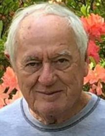
Alf Mauff was another of our nine brilliant students who got first-class passes in all subjects in the first year. He matriculated at the German School in Hillbrow. Having tried surgery with Professor Du Plessis – “that outstanding teacher and stern disciplinarian,” he decided to become a pathologist. Alf became a pioneering member of Lancet Laboratories with classmate Stanley Zail.
Athlone High School for Boys: Three boys who matriculated at Athlone in 1954 joined the Medical Class in 1955: Ronnie Auerbach, Ephraim Dove and Arnold Ravdel. They were apparently surprised to see each other when they arrived to enrol at Wits as they had had no prior inkling that they were all applying to study medicine. Ruby Zamit, a fourth classmate, had also applied but was not accepted that year. He first completed a science degree and was accepted later at medical school. And then there was Harry Glazer who also joined our class starting in 1955.
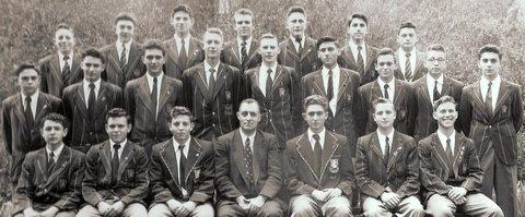
Harry Glazer had been the head boy of Athlone in 1949 and had represented the school at Rugby. He wanted to study medicine but could not afford to, so he qualified first as a pharmacist and then joined our class, working his way through his medical studies. Harry was also one of the nine who obtained a full house of first-class passes in his first year. He gained his Fellowship of the South African College of Physicians but preferred to practice family medicine as a General Practitioner – first in South Africa and then in Sydney, Australia.
Other notable old Athlonians are musician, Johnny Clegg, Sol Kerzner, businessman and hotelier (creator of Sun City); Harold Luntz, legal academic; George Bizos, human rights lawyer. Harry Levitt, also, in the 1954 matric class, worked on developing digital hearing aids at Bell Laboratories, and Gideon Shimoni (Simonowitz) became Professor of Modern Jewish History at the Hebrew University in Jerusalem.
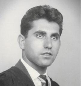
Ephraim specialised in internal medicine. He organised the 25th anniversary reunion for the class and has a photograph of that event in the chapter on ‘reunions and reflections’.
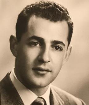
Ronnie married Geraldine Auerbach from Kimberley in 1962. They joined his family in London, who had fled there from Nazi persecution in Germany. Ronnie specialised in ENT surgery.
We were not able to trace Arnold at the time, so we do not have a biographical sketch for him. Looking him up on the internet in 2023, we discovered that he had become an orthopaedic surgeon and had practised – or indeed was still working – in Houston, Texas. He had married Maureen Barris from Port Elizabeth in 1962.
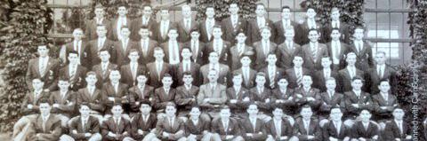
We have been looking at where the Wits students came from to start their medical studies in the mid-1950s. Though there were many high-achieving schools in Johannesburg whose pupils started medicine in 1955, there was an exceptionally bumper crop of boys from both Highlands North Boys’ School and King Edward VII Schools. Having so many boys who were already known to each other from competing in the classroom and on the sports field, might have had something to do with the cohesion and ambition of the group as a whole. In this chapter Gary Katz recalls the bumper crop of medical students from the matric year at his school, Highlands North. In the next chapter, Avroy Fanaroff conjures memories of the Old Edwardians.
Gary Katz wrote that our year was an exceptional one, when no less than ten 1954 matriculants from Highlands North Boys High School (and one Highlands boy who matriculated elsewhere) were accepted to study medicine at Wits. This was an astonishingly high number, never matched at the school, (or any other school) before or since.
Highlands North (or ‘Haalands’ as it was pronounced) had opened its doors in 1939 at no 1, Fourth Avenue Highlands North, a small suburb in the north-east of Johannesburg, established in 1903, with the suburbs of Waverley, Oaklands, Orchards, Norwood, Sydenham and Orange Grove as neighbours. Louis Botha Avenue, a major arterial road, runs from central Johannesburg via Highlands North towards Pretoria. In our day, the school drew a significant number of its pupils from immigrant families.
Three other 1954 matriculants deferred taking up a place for a year or more before entering medical school: Ivan Samson and Peter Soldin started their courses in 1956 and graduated in 1961, and Brian Bethlehem graduated in medicine in 1967. This makes a total of 11 Highlands North boys at Wits Medical School in those years! For some they were the first of their extended families to attend a university.
I have been asked whether we had inspiring schoolteachers, but I cannot recall any. There were other high-fliers in our matric year. One of our class, Max Labe, became a highly successful barrister and then a judge in Johannesburg. Another, Peter Held, became the senior partner of one of the largest accountancy firms in Canada. Several others did well at Wits in their chosen courses. Peter Byland became a successful engineer in Switzerland.
Six of these ten 1954 matriculants started their studies in 1955 and graduated in 1960, the class that celebrated its Diamond Jubilee in 2020. They are Leonard (Len) Kahn, Gerald Lampert, Gerald Shulman, Gerald Schneider, Jack Kussel, and me (Gary Katz). Two more started with us in 55 but graduated a year later: Mervyn Damelin and John Festenstein. (John had left Highlands North before matriculation.)
Three other 1954 matriculants deferred taking up a place for a year or more before entering medical school: Ivan Samson and Peter Soldin started their courses in 1956 and graduated in 1961, and Brian Bethlehem graduated in medicine in 1967. This makes a total of 11 Highlands North boys at Wits Medical School in those years! For some they were the first of their extended families to attend a university.
I have been asked whether we had inspiring schoolteachers, but I cannot recall any. There were other high-fliers in our matric year. One of our class, Max Labe, became a highly successful barrister and then a judge in Johannesburg. Another, Peter Held, became the senior partner of one of the largest accountancy firms in Canada. Several others did well at Wits in their chosen courses. Peter Byland became a successful engineer in Switzerland.
All these 11 Highlands North boys had memorable medical careers. Three (Gerald Lampert, Gerald Schneider and Mervyn Damelin) became General Practitioners, three became Paediatricians (Gary Katz and Jack Kussel and Ivan Samson) three specialised in Pathology (Len Kahn, Gerald Shulman and Peter Soldin) one in Radiology (John Festenstein) and one in Anaesthetics (Brian Bethlehem). The three GPs sadly had died by 2020. Of these eleven Highland North High School doctors, six practised in South Africa (Kussel, Lampert, Schneider, Soldin, Damelin and Bethlehem). Two went to the USA. After travelling and working in different centres, Len Kahn settled in New York and Gerald Shulman in Atlanta. John Festenstein and I, (Gary Katz) settled in London after working in America for a short while. Ivan Samson settled in St Catherines, Ontario.
Eight of the eleven us are still alive (in 2020) all in our early eighties, and though mostly retired, or furloughed due to the COVID-19 pandemic, we still lead busy lives, some consulting in our speciality and some indulging in cultural pursuits (or both). What seems to have characterised all of us is our love and care for our patients and our desire to further the knowledge and organisation of our specialities. This appears to have been appreciated by our patients and colleagues.
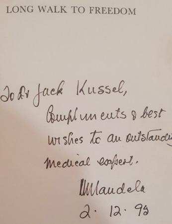
Jack has messages attesting to his warm personality and excellent care from his patients, great and small. He looked after the grandchildren and great-grandchildren of Nelson Mandela. In his story on the website, we see Mandela photographed with Jack’s own children and grandchildren. Madiba called him an “outstanding medical expert” in the inscription in his biography ‘Long Walk to Freedom’ that he gave as a gift to Jack. On his retirement, a grateful and loving patient wrote a beautiful poem of how everyone will always remember Jack Kussel which ended: Dr Kussel, your career may have come to a rest – but your legacy and the lives you saved (literally!) will live on with EVERYONE … FOREVER. We salute you; we thank you, And we will never ever forget you.
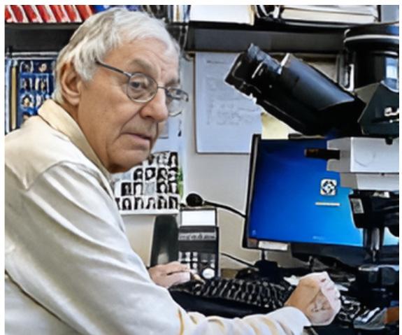
Len was a straight “A” student at Highlands North. He received several distinctions in the classroom and was an enthusiastic sportsman. On leaving school, Len was given a special prize for outstanding scholarship. He received first-class passes in all his first- and second-year medical subjects. It was in his third year that Len really found his calling. He fell in love with microscopy and the study of the microanatomy of human tissue in health and disease. In 1962, after completing his house jobs, he moved to the University of Cape Town (UCT) to specialise in pathology. After a three-year registrar-ship program, he was awarded the Degree of Master of Medicine in Pathology (MMed Path UCT). Len then went to St Louis, Missouri, where he was privileged to work with the world-renowned American physician and pathologist Lauren V Ackerman, who championed the subspecialty of surgical pathology. There followed professorships in surgical pathology at UCT and then in North Carolina and finally New York, where Len is still at work.
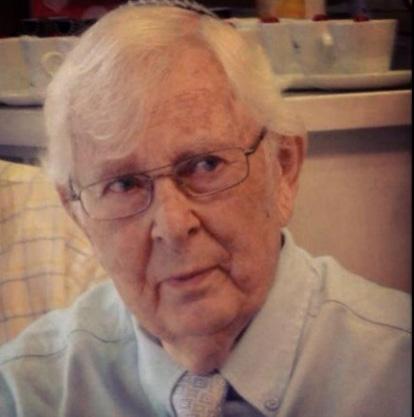
Another Gerald in our class was Gerald Schneider. On graduation, he became a general practitioner in Germistonand remained there for his whole career. He was much loved by his patients and by his family. Len Kahn, his colleague and classmate at school and Wits, attested to Gerald’s devoted attention to his mother, Rachel Kahn, who suffered from multiple sclerosis for many years, and to his father, Robert Kahn, who suffered from a salivary gland malignancy. Gerald also paid frequent visits, despite his extremely busy practice schedule, to Robert Kahn during Robert’s final years as a resident at the retirement facility in Sandringham
Gardens, Johannesburg. Gerald died in 2012, aged 75.
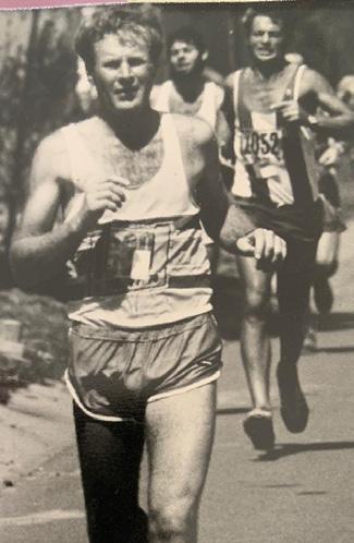
Gerald and I became friends at school and great friends at Wits. We worked together in a ‘firm’ with Len Kahn, Aubrey Milunsky, Farrol Sims and Dennis Rossouw. Gerald’s son Dr Kevin Lampert, wrote a moving essay about his father Gerald who died in 1998, at the age of 61. Gerald was head boy at Highlands North in 1954. I remember the time when Gerald and I dissected a rat in my mother’s laundry room – launching our anatomy career as well as my mother’s fury. Gerald’s son Kevin wrote of how Gerald worked at the bookies’ offices at the horse races in Turffontein on weekends to make ends meet. As Students we also served with other students as bar waiters at the Rand Easter Show. Gerald told Kevin that he and I had worked together as male nurses at West Rand Mines Hospital, where we learned to suture and apply plaster of paris from a maestro plaster technician. Like several of us, Gerald failed his first home nursing exam, but unlike some of us, he became a meticulous bed-maker and passed this skill on to his children.
Gerald was a keen squash player. When a wrist injury curtailed his squash ‘career’, he took up running – in a big way! This included countless marathons and 13 Comrades Marathons in which he was awarded 7 silver finishes. He would leave his house, rain or shine, at 04h00 and run 16 to 20 miles before starting his daily work. In his running lifetime he must have covered almost twice the circumference of our earth!
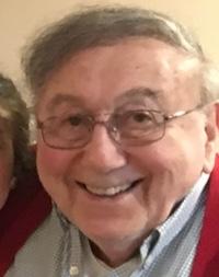
Yet another Gerald, Gerald Shulman was also hooked by microscopes early on. After his house jobs at Coronation Hospital, Gerald Shulman joined the South African Institute for Medical Research staff as a trainee pathologist. Gerald met and married Dr Cynthia Cohen in 1965. In 1968, he qualified as a Fellow of the South African Faculty of Pathologists. Cynthia followed the same path. They travelled together to London where Gerald worked at the Postgraduate Medical School at Hammersmith Hospital, in Chemical Pathology. At the same time, Cynthia furthered her career by attending the Clinical Pathology course at Hammersmith Hospital, graduating with the Diploma in Clinical Pathology. At the end of two years Gerald graduated with the Membership of the Royal College of Pathologists. They obtained a series of ‘his and hers’ jobs together in Johannesburg, Hershey Pennsylvania, Little Rock Arkansas’ and Atlanta Georgia, where Gerald became heavily involved with blood banks. Like Len and others of the many academics in the class of ‘60, Gerald has written many articles, papers and chapters in books on his subject.
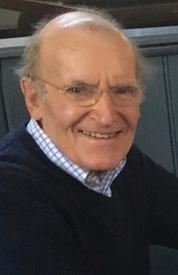
I was introduced to paediatric surgery at the age of eight when Josh Lannon removed my appendix. My student experiences the Transvaal Memorial Hospital (TMH) and a month on the children’s ward at Baragwanath Hospital before my house jobs were real inspirations and added to my ambition to pursue a career in paediatrics. I served on the Student Medical Council in 1958 and 1959. I married my wife Karen (Kirsch) in June 1960. We worked for a year in America and moved to London in 1963. Life in London was good to us. As events fell into place, we decided to settle there. During my Senior Registrar rotation, I used my ‘spare’ time to complete the research for my MD, which I submitted to Wits in 1971. I was awarded my MD in December 1971. Early in 1972, I was asked to present my work at the Transvaal Memorial Hospital. I learned to my surprise that I was the first paediatrician to be awarded an MD from
Wits. During my appointments at Edgware General and Barnet hospitals in London, I was responsible for developing a highly regarded paediatric service and a sub-regional neonatal intensive care unit. In 1996, I was appointed the Northwest Thames Regional Advisor in Paediatrics, responsible for paediatric training. The training in all specialities was undergoing significant changes nationally. With three colleagues, I was responsible for implementing a new training programme involving every paediatric department in London and its surroundings.
John was a year ahead of us at school and left Highlands North after Form 3, matriculating at Damelin College in 1952. After two years of a BA course, John started medical studies with us in January 1955. Having repeated third year, he graduated in the class of ‘61. He specialised in radiology in Boston. When he had to leave the States because his visa had expired, he relocated to London, where he had prestigious jobs and obtained his Fellowship of the Royal College of Radiologists. He became a consultant at Edgware General Hospital in 1977 which merged with Barnet Hospital in 1997. He spent the remainder of his career at Edgware and Barnet in the company of several South African colleagues, two of whom were also old Highlands North pupils: Les Bernhardt and I (Gary Katz).
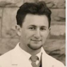
Mervyn started with us at medical school in 1955. In second year, we were anatomy partners and in third year, pathology partners. Then Mervyn had to repeat third year. He graduated with John Festenstein, Ivan Samson and Peter Soldin in 1961. After his house jobs, he practised as a GP in Mafikeng. He subsequently moved back to Johannesburg, where he was the Medical Superintendent of Edenvale Hospital. After retiring, he emigrated to Sydney to join his children and grandchildren. Sadly, he died in 2012.
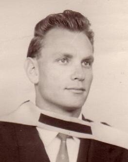
Ivan was accepted for medical school with us in 1955. His father, however, insisted that he should rather study economics and go into his diamond-cutting business in Kimberley. Having done one year of that, Ivan was able to switch and take up his calling and his medical place the following year. He sailed through medical school, was elected class representative in first year, and served on many student committees. He was also part of the organising committee of the all-African cast of the operetta, King Kong, presented so spectacularly at the university’s Great Hall. Ivan specialised in paediatrics in London and Scotland. He emigrated to Canada where he had a distinguished academic and clinical child health career in St Catharine’s General Hospital, Ontario. Ivan was sad not be included in our cohort and he took it upon himself to create a website and collect biographies of the 1961 class, working closely with the team for the 1960 Diamond Jubilee project.
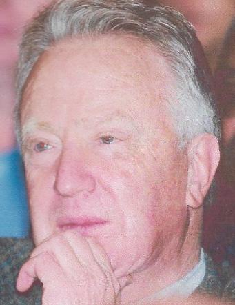
After matriculating with us in 1954, Brian completed a BA at Wits. He started medical school only in 1962, after the rest of the 1954 matriculants had already graduated, graduating himself in 1967. He specialised in anaesthetics. During his training, he spent time at the Hammersmith Hospital in London. After returning to South Africa, he practiced in Johannesburg. Brian told me that while working as an anaesthetist, he developed strong friendships with our classmates Irving Lisoos and Gerald Lampert.
Peter started at Highlands North in 1950 and did very well academically. He matriculated with us in 1954 and had a gap year. He started at Wits Medical School in 1956, graduating in 1961. He qualified as a Fellow of the SA Faculty of Pathologists and practised as a pathologist in Vereeniging.
As we have seen, the matric year of 1954 at Highlands North produced a bumper crop of ten bright students who were accepted at Wits Medical School. With John Festenstein, another former Highlands North pupil who started Medical School with us, all eleven contributed much to the student body and the medical profession in South Africa and around the world. I had wondered whether this large intake was something that happened on a regular basis. However, despite much research, this does not seem to be the case. There have certainly been many Highlands North medical graduates over the years, from the first two that were capped in 1954 (the year that our group of 10 matriculated). However, it is clear that our matric year is likely to remain an exceptional one.
We have been looking at where the Wits students came from to start their medical studies in the mid-1950s. Though there were many high-achieving schools in Johannesburg whose pupils started Medicine in 1955, there was an exceptionally bumper crop of boys from both Highlands North Boys’ School and King Edward VII School. In the previous chapter, Gary Katz wrote about his fellow students from Highlands North. Here, Avroy Fanaroff recalls the matriculants of 1954 at King Edward VII School (KES), who were our classmates at medical school.
King Edward VII School (KES) is situated on a spacious campus in the heart of Upper Houghton, Johannesburg. The red brick buildings with the clock tower create a striking façade (as seen in this painting). They have been designated a National Monument. The school is surrounded by spacious sports grounds and has many cultural facilities.
Avroy Fanaroff wrote: Of the 26 members of Form VA in 1954 (the matric class of the Latin and History scholars), eight chose careers in Medicine. They were Manley Cohen, Allan Gottlieb, Mervyn Hurwitz, Irving Lissoos, Jeffrey Maisels, Ivan McCusker, Arthur Rubenstein and myself. Another KES boy in our medical class who had matriculated earlier was Peter Arnold, the son of our brilliant anatomy lecturer Maurice Arnold.
When the second Anglo-Boer War ended in 1902, there was an urgent need for schools in the Transvaal. In search of suitable buildings to establish temporary classrooms, the Milner Administration found a vacant cigar factory in Johannesburg on the corner of Gold and Kerk Streets. This was chosen as the venue for ‘The Government High School for Boys,’ also known as the ‘Johannesburg High School for Boys.’ Thus, was born a school that ultimately became the King Edward VII School.
It grew so rapidly that, in 1904, it was moved to Barnato Park, where it was established in the mansion originally designed for the mining millionaire Barney Barnato, who died mysteriously at sea in 1897. At its new location, it referenced as “Johannesburg College,” but within seven years, the premises were deemed inadequate.
In 1911, the school was moved to its present site on Houghton Ridge, where new buildings had been impressively designed and specifically constructed for the school. The time frame, within less than a year after the founding of the Union of South Africa and the death of Queen Victoria’s eldest son and successor, Edward VII, led to the proposal that the institution’s name be changed to honour his memory, thus establishing the appellation, King Edward VII School.
In our day, approximately a third of the students were boarders, many from the farming areas of the Transvaal. The rest were day students from the upwardly mobile residents of the surrounding leafy metropolitan suburbs of Houghton, Killarney, Saxonwold and Norwood. This resulted in a fairly diverse student body. In 1954, when we matriculated, there were 84 seniors in the school and 400 to 500 students altogether. There was a strict dress code, and discipline was enforced by the school prefects, and, as was the practice at that time, ‘cuts’ with the cane from the Headmaster. The school intake has since grown in number and diversity, but it continues as a
Government Secondary School, recognising and rewarding academic achievement and cultural and sporting prowess.
As Gary Katz noted in his comments on his Highlands North School group of 11 who entered medical school, we cannot recall any really inspiring schoolteachers who might have directed us into a career in medicine. It was our own personal and family choice.
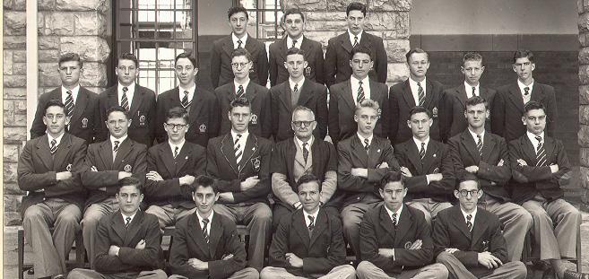
The boys entering medicine came from various backgrounds. Gottlieb and McCusker followed their fathers into the medical profession. Jeff’s father, Isie Maisels, was a leading advocate. Monty Rubenstein and Herman Hurwitz were pharmacists. Jack Fanaroff was a garage proprietor. Robert Lissoos worked at the food market, and Tevya Cohen manufactured ties.
Apart from those who chose medicine, our high school class also had many other brilliant students, most notably Robert Kwitz who now lives in Israel, (called Reuven Korvan) has three PhDs, and works in artificial intelligence; Teddy Brett and Geoffrey Hutchings, who both became Professors of English; Ian Froman, a dentist who founded the Israel Tennis Centres and was recognised by the President of Israel; and John Patten, who edited The Star newspaper in Johannesburg. There were several other successful professionals from our class as well.
The school class preceding us (matric 1953) had produced four doctors: Ronnie Joffe, a neurologist; Paul Rome, an anaesthetist; Morris Super, a paediatrician; and Michael Heymann, a paediatric cardiologist. Having eight boys enter medical school from our form seems to be a record for our school. Third
Here are some events in world history that were taking place at the time that we were preparing for our Matric exams and starting to give thought to our future careers:
• In South Africa, JG Strydom took over from DF Malan as Prime Minister
• Sasolburg started production of synthetic liquid fuel from coal
• The US Supreme Court issued a landmark decision, the ‘Brown versus Board of Education of Topeka’ ruling. Handed down by Chief Justice Earl Warren, it states that “Separated educational facilities are inherently unequal.” The decision overturns the ‘Separate but Equal’ ruling in the matter of ‘Plessy vs Ferguson’ dating back to 1896.
• A major fast-food franchise, McDonald’s, first opened in California.
• The first transistor radio was marketed on October 18.
• The Polio vaccine is given to children for the first time. In February, the first mass trials of the newly developed Salk vaccine begin in Pittsburgh, Pennsylvania. (Professor James Gear in South Africa supported the development work.)
• The First successful kidney transplant is performed.
• The first public demonstration of a machine translation system was held in New York at IBM’s headquarters.
• Roger Bannister ran the first sub-four-minute mile in recorded history. The 25-year-old from Harrow on the Hill, England, completed the distance in 3:59.4 at Oxford. At the end of the year, Bannister retired from running to pursue his medical studies full-time. He later became a neurologist. ‘There is no fuss and fanfare about Bannister. When he was asked to explain that first four-minute mile—and the art of record-breaking— he answered with original directness: “It’s the ability to take more out of yourself than you’ve got.’”
• Bell Laboratories introduced the photovoltaic cell, which converts sunlight to electricity.
When we left school and started at university, we already had an established group of friends. We had spent many years striving together in the classroom and competing with each other on the sports field. This gave us confidence and made our transition from school to university both easy and enjoyable. Our friendships lasted throughout our medical school days and into our careers, however widely we were spread around the world. We took part in student affairs and joined NUSAS (the National Union of South African Students) together.
I (Avroy) am on the left with Arthur Rubenstein and Irving Lissoos in our Wits blazers, with our tennis togs, going off to a NUSAS Conference in Grahamstown in July 1957.
We also enjoyed spending our vacation time together. On the right, I am second from the left with Irving Lissoos, left, Jeffrey Maisels and Arthur Rubenstein on Durban Beach in December 1957.
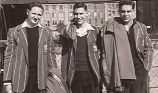
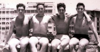
In Form 2, at age 13-14, I recall that our English teacher, one of only two female teachers in the boys’ high school, informed us that we were a class of useless, lazy individuals who would never amount to anything.
She would be surprised to learn that we have all specialised and obtained higher medical degrees. Three have chaired academic medical departments, one was a dean of a medical school, and between us, we have made significant medical discoveries and contributions. In addition, we have been recognised for our endeavours by receiving many national and international awards. For a group that she considered ‘mumblers’ who could not put a sentence together, they have lectured all over the globe and published over 1,000 peer-reviewed medical articles, innumerable chapters in books and at least twenty-five textbooks. Let’s meet them:
Manley Cohen (1937 – 2015) was kind, gentle and smart. He would look quizzically at you when you said something he disliked or misunderstood. He played no sport at school, but entertained us often at his home around the swimming pool. Manley sailed through medical school. He did an Internship in Chicago. He then specialised in gastro-intestinal disorders at the Mayo Clinic where he spent five years.
He was then recruited to be the Director of Medical Education at the new medical school in Long Beach, California. He was also the sole gastroenterologist in the area and spent the rest of his successful career in
the University of California’s system. Manley loved to fix things and taught his children plumbing and electrical repairs. He developed an interest in locks and became a certified locksmith. He loved to read and had an extensive home library, which included books on the 78 countries he and his wife, Barbara, had visited. Manley and Barbara have three children and 11 grandchildren. Their eldest son, Darien, is an emergency room physician; Ronan is a lawyer; and Gila, their daughter, has a doctorate in Social Science. The Cohens enjoyed travelling with their grandchildren. Manley, much loved by his patients, died in 2015. In keeping with the concept of ‘six degrees of separation’, Manley’s brother Hartley, also a physician, is married to Arthur Rubenstein’s sister Nola.
After Yeoville Boys Primary School, I was transferred to Athlone High School. This required a four-block walk, a tram and a bus, whereas KES was just four blocks from my house. I was 11 years old, and there were burly 15-year-olds in the Athlone class who initially bullied me mercilessly. I tolerated it and grew up rapidly, but was delighted to be transferred in the middle of the year to KES, where I enjoyed the rest of my high school days in good company.
I always knew I would like to attend medical school and was thrilled to be admitted. The first half of the second and third years seemed like a blur, and the topics made little sense until you really got into the textbooks and the vision cleared. The clinical years were very logical, and we had the privilege of learning in a golden era of clinical medicine. We had spectacular medical teachers and bountiful patients.
The clinical experience was thrilling and challenging. I decided I wanted to be a paediatrician, but as there were no specific paediatric exams, I did internal medicine and paediatrics before doing the Diploma in Child Health in London and Membership of the Royal College of Physicians (MRCP) in Edinburgh in 1964, using paediatrics as my subspecialty.
My biosketch on the website outlines a number of my career moves. My interest in neonatology began in the 1960s when I worked with Professor Sam Wayburne in the premature units at Baragwanath. This was furthered, when as a registrar at the Transvaal Memorial Hospital (TMH), I teamed up with Professor Solly Levin to start providing paediatric services at the Queen Victoria Maternity Hospital. My career pathway has been in parallel with Jeff Maisels. He and I have received much of the same recognition and have attended many conventions and research meetings together. I, too, was a competitive squash and tennis player. But now I am an avid golf duffer. I enjoy bridge and photography, having learnt bridge in the medical school canteen with Arthur Vinik. Ros and I have travelled the world and have friends and colleagues on many continents.
In high school, Allan was a great athlete and brilliant student. He was a fabulous gymnast. He was one of the top five students in a very competitive form. Always immaculately dressed and groomed, he stood out in a crowd. After qualifying and house jobs, Allan, who had set his mind on surgery, became a teacher in the anatomy department at Wits and continued attending Professor Du Plessis’ surgery lectures. He then went to the UK and became a Fellow of the Royal College of Surgeons, specialising in Thoracic Surgery. He spent some time in surgical residency at Duke University and attended an elite program in the USA. He ultimately settled in the UK, where he was a consultant thoracic surgeon at Harefield and, later, a general surgeon at Lewisham Hospital in London, teaching those Brits a thing or two, especially about clinical diagnosis.
Allan and I share a thumb anomaly (brachydactyly – short fingers and/or toes) inherited dominantly. Our anatomy
lecturer, Phillip Tobias, showed pictures of our thumbs at several of his lectures. Allan’s father, Leo Gottlieb, was a general surgeon, and his brother Paul, was a successful urologist in the United States, now retired. Allan states that he does not like nostalgia but lives for the present and future.
Mervyn (1937 – 2020) was a keen sportsman at school, playing rugby and swimming for the school teams. He was also a bright student. At lunchtime most days, we hung out near the tuck shop, eating our sandwiches and chatting. Often, we would exchange jokes. Mervyn kept a record of good jokes in his notebook. His brother Hilliard was the Medical Officer of Health in Johannesburg. A relative was Charles Hurwitz, after whom the Tuberculosis (TB) Hospital next to Baragwanath Hospital was named. Charles did much of the original research on TB and was one of the leading radiologists of his era.
Mervyn obtained his Fellowship of the Royal College of Obstetrics and Gynaecology. He returned to Johannesburg and commenced private practice with Joel Cohen and Max Bloom. He later taught at the University of Witwatersrand Medical School. Mervyn was one of the pioneers of sex therapy in South Africa, establishing the country’s first sex therapy clinic. 20 years ago, he migrated to the USA to join his children, providing services for an under-served area of Arkansas for 10 years. He received recognition from the Governor of Arkansas for organising rural obstetric services and transporting high-risk maternity cases to the University Medical Centre. A special flag flew over the Capital in his honour. Hurwitz, Mervyn Bernard
Irving Lissoos (1937 – 2011), interviewed by Mike Alfreds for the Johannesburg Heritage Portal website in 2006, said: “From our house, it was a three-minute bicycle ride to King Edward School from Yeoville. I wasn’t transferred to KES, but to Highlands North High School, much further away. KES seldom took boys from Yeoville. My mother said, ‘This is total nonsense!’ She went to see the headmaster, who said sorry, he couldn’t take me, so she went to see the school inspector. He told her there was no room at KES, and she told him she was going to sit in his office until her son was admitted. An hour or two later I was admitted to KES. The standard of education at King Edward’s was very high, with special strengths in the teaching of mathematics and science. This contributed to my first-class matric. The next step was Wits Medical School, where I qualified as a doctor.” We all find it extremely difficult to reconcile the fact that Irving died from a malignant melanoma in 2011. Irv and I were classmates from standard one (aged 6) at Yeoville Primary School, through high school at KES and medical school. He taught me to drive a truck on his grandfather ’s farm. He loved riding horses on the farm. When you went to his home, his mother was always ready to feed you, even if it was past midnight. They were a most generous family. If you mentioned a book you had heard of, Irv would get a copy and deliver it to you the next day. His grandfather was his idol and role model. He loved history and received top marks in the Transvaal for the matriculation exam. He joined the local history society and gave tours of Parktown and Westcliff, teaching the area’s history. He was the most sought-after guide. He was loved and respected by all.
During our university years, Irving planned many holiday trips for our group of friends from KES. We went to Game Parks, Lourenco Marques, the Cango Caves and East London. His planning was meticulous. Those trips were great fun and the most memorable experiences.
In the picture, we are emerging from the Cango Caves: Me (Avroy Fanaroff) on the left, then Michael Plit, Allan Gottlieb, Arthur Rubenstein and Ian Froman. Kneeling in front is Irving Lissoos.
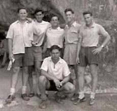
Irving was a dedicated doctor. He was committed to his patients and their well-being. He first specialised in general surgery and then in urology. He was a pioneer in kidney transplantation and secretary of the Urological Association of South Africa. He said: “From primary to high, to medical school and then on to postgraduate training, we were fortunate in having great teachers, and we lived and learnt in a very privileged society.”
For over 70 years, Jeff Maisels and I have been buddies. Our relationship has only grown stronger over the years.
To quote Jeff talking of himself, “I was a late academic bloomer, but peaked early as an athlete and entertainer.” He was a marvel at imitating Jimmy Durante and Tom Lehrer, and no party would have been complete if Jeff hadn’t taken his place at the piano and performed. He danced well doing the Charleston, Jitterbug and then the Twist. He was also a very good athlete, and as noted in his biography, he won the Intercollegiate Squash Championships and was selected to represent South Africa in squash. He was also an excellent tennis player and, like millions of others, is struggling with golf.
After a house job at Baragwanath, Jeff headed to Boston for a residency at the famous Harvard Pediatric Program. This was followed by a fellowship with Dr Clement Smith, who trained most of the pioneers in neonatology. Smith’s family tree of trainees and their progeny represents the ‘who’s who’ in neonatal and perinatal medicine and reaches across the globe.
Jeff completed his Carbon Monoxide studies in the laboratory at night because the assay was so sensitive. If the elevator ran during his experiment, it would affect the data. This research saved him a trip to the war zone in Vietnam, because he was considered a specialist. He was assigned to Walter Reed Hospital in Washington, DC, to continue his research.
After his U.S. ‘military service’, he joined Professor Nicholas Nelson, a mentor of his, in opening the Pennsylvanian State University Medical School in Hershey, developing a regional neonatal intensive care unit which he ran for 14 years. Jeff then became the chair of the Paediatric Department at Beaumont, which also developed a new medical school. In the meantime, he studied and published extensively, mainly on jaundice, bilirubin metabolism, and bilirubin neuropathy, which is the basis for his Doctor of Science from Wits. He published two books on this topic and was eagerly sought after by lawyers for consultation. He was an enthusiastic co-editor of the Yearbook of Neonatal-Perinatal Medicine, writing commentaries on diverse articles. Although officially retired, Jeff still carries out first class research.
Ivan McCusker (1937 – 2004) was born in Rouxville, a small wool and cattle farming town in the Orange Free State. He attended an Afrikaans-medium elementary school and was one of the few truly bilingual boys in our class. Born to an Irish father, a doctor, and an Afrikaans-speaking mother, he was a brilliant student and athlete. Tall, handsome, and muscular, he excelled at rugby, track events, and swimming. He achieved a first-class matriculation with several distinctions. He floated through medical school. At Wits, with medical classmate Andre van As, he was part of the victorious Wits under-19 rugby team that won the Transvaal title. Everything came naturally and easily to Ivan. After graduation and internships, he ran his general practice in Aliwal North for a while because his father was ill. He then began his surgical training in Johannesburg and Cape Town, culminating in a Fellowship of the South African College of Surgeons in 1966 and a Fellowship of the Royal College of Surgeons of Edinburgh in 1967.
Ivan ran a successful surgical practice and was affiliated with university programmes. He was a born leader. He chaired the Association of Surgeons of South Africa from 1988 to 1995, was President from 1995 to 1996, and was awarded an Honorary Life Vice Presidency. He also chaired many other committees. Ivan retired to his farm at Groot Brak River, where he bred Santa Gertrudis cattle – a hardy breed of beef cattle developed in Texas in 1940, mating Brahmin bulls with Shorthorn cows. They are known for their ability to adapt to harsh climates. He also converted hectares of wattle bush into rolling pasture, carefully preserving the indigenous flora. He found peace in farming and enjoyed a braai and coffee laced with Scotch whiskey. He died in 2004. In his obituary in the South African Medical Journal 2004 vol 94 #8, the writer says, “Ivan was one of the best minds I have known. He had the ability to assimilate information, remove irrelevance and bias and present it in a simple, clearly understood language, sometimes with caustic wit, but without jargon or political correctness… He refused to associate himself with decisions he considered dishonest, immoral or politically expedient. He was respected and loved by his students and colleagues.”
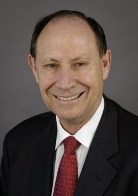
Arthur was a brilliant, straight-A student at school (even before we used that term). Together with two classmates, Robert Kwitz and John Ryder, Arthur was always at the top of the class in academic achievement. He was also an excellent sportsman. In addition to getting his colours in cricket, he enjoyed rugby, tennis and squash. In one of our school rugby matches, Arthur was knocked unconscious. Protocol dictated that if you could say what the score in the match was, you were considered fit to continue playing. They asked him for the score. His response of, “Who is playing”, got him a trip to the sideline. He matriculated with five distinctions, a great accomplishment in our day, including a distinction in Afrikaans, which was extremely unlikely if that was not your mother tongue. His medical school career was equally remarkable. Dr Jack Friedman, the head of Forensic Medicine, was the only one to deprive him of a first because he said, “he wrote too much”. Arthur graduated cum laude. Each year at medical school, Arthur and I studied one subject together. His thoughts were always crystal clear. His organisation of work and play followed a schedule that resulted in the best outcomes. He was confident and calm at all times. As interns, we had adjacent rooms at Resdoc, and no matter how late I returned, his light would still be on, and he would be going through the literature. As an intern, he became fascinated with a patient with diabetes who was treating himself by eating lucerne, the leguminous plant usually fed to cattle. This led him to a career in endocrinology and expertise in diabetes.
He worked for Professor Guy Elliott as a registrar, then trained in London before moving to the University of Chicago, where he started his collaboration with Dr Steiner. He was part of the team, led by Steiner, that reported on the structure and function of Proinsulin, a Nobel Prize-worthy quality research. After chairing the Department of Medicine in Chicago for fifteen years, Arthur was appointed Dean at Mount Sinai University in New York and then Dean and Executive Vice-President of the School of Medicine at the University of Pennsylvania. Notwithstanding his many accomplishments, awards and positions of responsibility, Arthur has remained a modest, humble, soft-spoken, humorous, hospitable, sincere friend and colleague. He and his wife Denise (nee Hack), a dermato-pathologist, who graduated from Wits in 1959, have two sons: Jeffrey, who is a scholarly Rabbi teaching in New York and Errol, who is a teacher in Minnesota. They have six grandchildren.
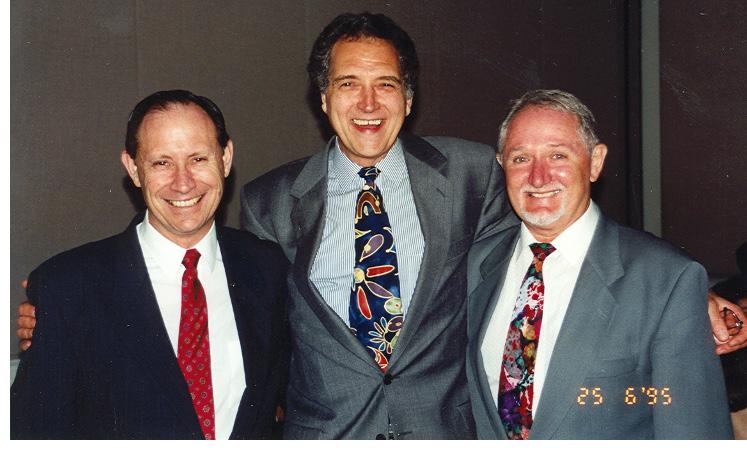
Peter was another KES boy who had matriculated a couple of years earlier but also started at medical school with us in 1955. Peter’s journey took him via the medical BSc to Australia as a family practitioner. Then, with a BA from Sydney University, majoring in Politics and Philosophy with a minor in Economics, Peter became involved in medical politics and editing medical literature. In recognition of his achievements and meritorious service, Peter was awarded the Order of Australia Medal in the 2019 Queen’s Birthday Honors.
Today, in October 2020, four of the eight schoolmates from KES are alive and well, with some actually still working every day. Arthur, Jeff and I are in the USA. Our friend Allan Gottlieb lives in London. Above all, at King Edward School, we made life-long friends. Chaim Rosenberg’s imaginative project of celebrating the graduates of 60 years ago has enabled us to connect again in a meaningful way and especially allowed us to put something back to help current and future medical students at Wits with our donations to the Phillip Tobias Bursary Fund. Looking back, I believe the order, organisation, and teamwork instilled in us at King Edward’s paved the way for our successful studies at the Wits Medical School. We had a sound education and were well prepared for the transition from school to university, for continued learning, and for life. The school motto was ‘Strenue’, meaning go forth strenuously, actively, and vigorously—and so we did!
Several of our classmates came from towns on the East and West Rand. Arthur Vinik matriculated in 1954 at the East Rand town of Benoni and Max Shaff from nearby Brakpan. Chaim Rosenberg came from the West Rand town of Randfontein and joined Costa Gazidis at Krugersdorp High School. Stan Zail did the same journey from Randfontein to Krugersdorp High School. Anton Heyns, haematologist and head of the South African Blood Transfusion Service, also matriculated in Krugersdorp at the Afrikaans medium Monument Hoërskool. Geoff Boner and Raymond Cochrane started their schooling in Roodepoort, Wolfe Rakusen grew up in Germiston, and Mike Plitt came from the coal mining town of Vereeniging, about 65 kilometres south of Johannesburg. Alec Kallel grew up in Sabie in the Eastern Transvaal, Rodwin Jackson came from Durban, Clive Rosendorff and Basil Kuming from Bloemfontein and Anthony Price and Theo Kretzmar went to school in Kimberley. Let’s meet them:
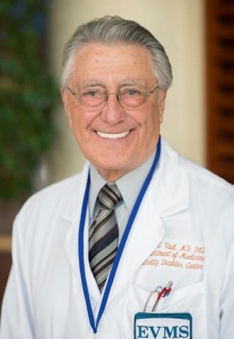
Arthur was born in a little gold mining town of Van Ryan, in the Transvaal. After his initial junior schooling, his family moved from the mining community to the East Rand town of Benoni. In addition to achieving the highest academic distinctions in his matric year at Benoni High School, Arthur was the school’s victor ludorum as he was also a great all-round sportsman. His medical school classmate, Max Shaff, from neighbouring town Boksburg, who first met Arthur when their school rugby and athletics teams competed, recalls: "He was unbeatable in the 100- and 220-yard sprint, and I would not be surprised if his inter-high school records still stand in those races."
In early 1955, Arthur and Max encountered each other again. This time, they were both signing up as students at Wits Medical School. They travelled together to and from university every day and became lifelong friends and also became brothers-in-law when each married one of the daughters of Dr Tolly Fram of
Springs. Arthur married Etta, and Max married his younger daughter, Michelle. Many students fondly remember the ongoing bridge games with Arthur as the tutor, not to mention his prowess at pinball!
Arthur went on to receive national and international recognition as a scientist and clinician in three separate areas: Clinical Diabetes and endocrinology, neuropathy and neuroscience and Neuroendocrine tumours.
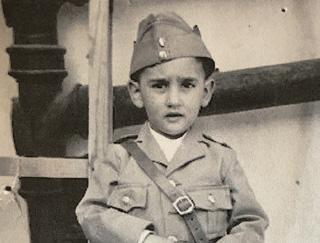
Arthur’s life-long friend and brother-in-law, Max, was born in Boksburg in 1937. His father, with the help of his mother, managed the small Boksburg North Hotel in the poorer part of town. Max’s family lived at the hotel for the first ten years of his life. The reason for the existence of Boksburg, approximately 15 miles from Johannesburg, was the huge East Rand Proprietary Mine (ERPM), which employed thousands of people. Started in 1893, this was one of the richest and largest gold mines in the world. It produced copious gold for over 100 years.
By 1993, it was also the deepest, with mining happening miles underground. In 2008, underground mining ceased, and then they started recycling the huge mine dumps using newer and better methods of extracting gold. As in most of urban South Africa at the time, English was the dominant language in Boksburg, but Afrikaans and various African languages were spoken. Max says: “At home, my parents spoke Yiddish between themselves and their friends. Thus, I was exposed to many languages in daily life, coming into contact with boarders and domestic help in the hotel and frequenters of the premises. There was also a lingua franca constructed in the mines known as Fanagalo, using words and phrases from several African languages. This was created because African workers from several different tribes and their White overseers needed a common language for training and to communicate in often dangerous conditions. Max completed his schooling at Christian Brothers College (CBC) in Boksburg. He said: “The contribution of CBC Boksburg to the Wits Medical School in my time was abundant. Clive Noble MBBCh 1961 (Orthopaedics) was in my matriculation class. Clive Segil (Orthopaedics), Bernard Kotton (ENT), Edwin Myers (Paediatrics), and Joe Rod (Cardiology) are also CBC alumni. Theodore Kopenhager (Ob-Gyn) matriculated at CBC and has an MBBCh from Pretoria University. Max also wrote a book about his early life.
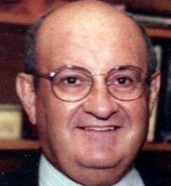
Chaim Rosenberg was the person who dreamed up the idea to contact classmates 60 years on.
From the age of twelve, he took a train from the West Rand town of Randfontein, twenty kilometres northeast, to Krugersdorp High School, where he was in the same class and also a prefect with fellow medical student Costa Gazidis. Chaim worked on four continents on his journey into psychiatry, starting in Africa (Johannesburg), then to Europe (Israel and the UK) and Australia, before settling in Boston in the USA. Since retiring, he has moved to Chicago and become a successful author of historical books.
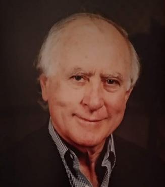
Stan Zail also grew up in Randfontein, where his father ran a concession store. He also matriculated from Krugersdorp High School and entered Wits medical school in 1955. He was awarded his medical degree cum laude – a rare distinction.
Stan Zail was able to combine his academic career with private practice, joining Clinical Laboratories and then going into partnership with his classmate, the pathologist Alf Mauff, as well as Drs Zent and Skudowitz in Lancet Hall, Jeppe Street. Together, they were founding partners of Lancet Laboratories, which became of the largest and most prestigious pathology laboratories in Africa.
There, he spent decades as a consultant in chemistry and a developer for the lab’s chemistry division. He maintained his love of research and teaching.
Geoffrey Boner’s family’s first home was in Roodepoort on the West Rand. His high school would have been Krugersdorp, where he would have been with Chaim Rosenberg, Costa Gazidis and Stan Zail. Instead, his parents sent him as a boarder to Marist Brothers College in Observatory, Johannesburg. The economy of Roodepoort was based on the Roodepoort Durban Deep gold mine within the town limits. Marist Brothers, like many of the other high schools, put an emphasis on excellence in sport. Geoffrey said: “Although I was most interested in sport, I was not a sportsman and spent time watching others and writing about them. However, I was very successful in my studies. I became the best scholar in my year and was awarded a scholarship for my studies. I was also very successful on the debating team, representing the school in debates against other schools. At that time, there was the Johannesburg Junior Council with two representatives from each high school in Johannesburg. Together with Max Green, I represented Marists on the Junior Council.
During my career in Israel, I was lucky enough to be one of the early nephrologists and to become the Head of the department at the Rabin Medical Center – Beilinson Campus. It was there that I advanced my career in nephrology and influenced the establishment of a Kidney Transplant department at Beilinson, which is now the largest department in Israel and a world leader in the field, especially of transplants using living donors.”
Anthon Heyns also matriculated in Krugersdorp, though at the Afrikaans medium Monument Hoërskool. He remembers that adapting to the culture of Wits and its Medical School was challenging. He spent a lot of his time on his hobby, chess, in which he represented South Africa in three Olympiads held in Cuba, Switzerland and Germany. He also represented South Africa at the World Senior Chess Championship held on the Isle of Man. He remembers his fourth-year ‘firm’: “Andre van As, at that time, had already shown that he would be a talented physician. Joan Feldman was clearly a budding psychiatrist/psychologist. David Paton had endless debates about religion with Fanie le Roux. Abe Zeilinga has been a friend for 66 years! He is caring and has been a successful and well-liked general practitioner. I also remember Arthur Vinik and his group of bridge players; Arthur was not able to teach me the subtleties of this game!
Wolfe Rakusen was born in Germiston in 1937 on the East Rand and went to school at the Germiston Boys High School. He was a great multi sportsman as well as prefect, house captain and cadet officer. He started medical studies with us in 1955 – but failed botany and chemistry in first year. He was lucky to be one of only four students accepted to repeat first year. He sailed on from there, graduating in 1961. He specialised in ophthalmology at St John’s Eye Hospital at Baragwanath under Professor Maurice Luntz. He was awarded a PhD in Medicine from Wits in 1975.
Raymond, who practised as a General Surgeon in South Africa, also started his schooling in Roodepoort. He was then sent as a high school border to Dale College in King William’s Town.
An only child, Alec Kallel, was raised in the rural area of Sabie, in the Eastern Transvaal. He went to an Afrikaans primary school. For high school, his parents sent him to Pretoria to live with his grandmother, aunt and uncle and their family of four children. He completed his education there, where he not only excelled academically, but developed his leadership qualities. His achievements included athletics victor ludorum and rugby 1st XV captain and being chosen as head prefect. Alex became a trusted radiologist. In 1966, he was introduced to clay target shooting. Alec played a major role in developing the sport and became the revered chairman for many years.
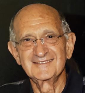
Mike Plitt grew up in the Transvaal town of Vereeniging. He matriculated from General Smuts High School. He was its first Head Boy after the school became an English medium school from the dual medium Vereeniging Hoërskool. In 1955, he started medicine at Wits and was a popular member of the class graduating in 1960. Mike worked in Pulmonary Medicine in South Africa and after Nelson Mandela was released from prison, Mike became his personal physician until Mandela’s death.
Rodwin Jackson went to Boys’ High School in Durban. Rodwin was also one of the nine 1955 entrants who got four first-class passes in the first year. After qualifying, he was a House Surgeon to Professor Du Plessis and a House Physician to Dr Agranat, and then a Medical Registrar to Professor Bothwell. After gaining his South African College of Physicians Fellowship, he went to London to study diabetes, where he and his family remained.
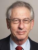
Clive Rosendorff grew up in Bloemfontein, where he matriculated from Christian Brothers College (CBC). He started at Wits in 1955. Then he did an Intercalated BSC in 1957 (Physiology and Anatomy including Physical Anthropology majors) and a BScHons in Experimental Physiology in 1958. He received his MBBCh in 1962. We will meet Clive again in the chapter on Academic Medicine as Clive became head of Physiology at Wits and then Dean of the Wits Medical School before taking up a similar position at the Icahn School of Medicine at Mount Sinai Heart Hospital, New York, NY, USA.
Basil Kuming (1937–1997) came from Bloemfontein, where he attended St Andrews School, one of the country’s oldest Anglican church schools, established in 1863. Basil won the English prize. After graduating from medical school, he trained at Baragwanath Hospital. In England, he did house jobs at Queen Victoria Hospital in East Grinstead, where he was influenced by Sir Benjamin William Ryecroft, the ophthalmic surgeon. Basil achieved his FRCS in 1967 and returned to Johannesburg to practice as an ophthalmic surgeon. His hobbies included stamp collecting and woodwork. Basil died on 5 April 1997, aged 60 years.
Anthony Price went to school in Kimberley at the Christian Brothers College. He completed his house jobs at Baragwanath and Edendale Hospital, Pietermaritzburg, and then moved to Pretoria to specialize in obstetrics before going to Durban, where he specialized in ENT. He died in Canada in 2005.
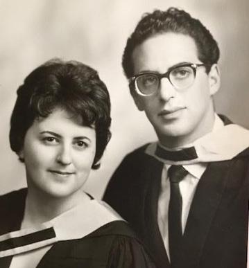
Theo Kretzmar (pictured with his sister Geraldine Auerbach, née Kretzmar) came from Kimberley, where he attended the Kimberley Boys’ High School and Geraldine attended the Girls’ High School.
Both graduated from Wits in 1960, Theo with his MBBCh and Geraldine with a BA. Geraldine writes: “Having found this graduation picture and because I am so closely connected with the project as the Editor in Chief, and also married to one of the classmates, Ronnie Auerbach, who became an ENT Surgeon in London, I thought it would be OK to sneak my picture into this book.”
Theo became a surgeon (like his father in Kimberley) in Springs and then Johannesburg, and how Baragwanath Hospital set him up for his career, and then when multiple sclerosis started to affect his legs, he was able to switch to work with Adrian Gore on the development of Discovery Health in Johannesburg, who was very grateful for his sage advice and guidance.
Many students reported that the first three years had had to be endured, but few had relished them. All were inspired in second year by the enthusiasm and acumen of the anatomy department emanating from Phillip Tobias, who had just returned from his sabbatical in the UK and America. Several members of our class joined him on an intercalated science degree, which was a life-changing experience for them, visiting African tribes and fossil sites that evidenced the birth and development of humanity and setting them on a pathway to scientific research in medicine.
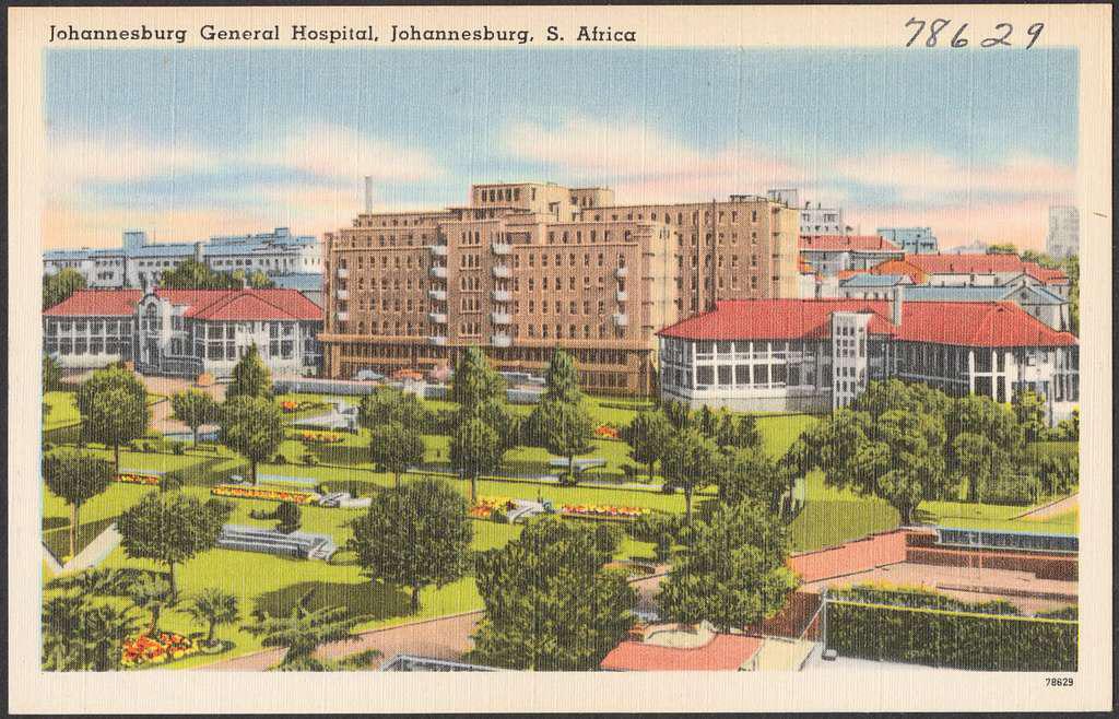
But it was really only in the fourth year when they were at last to experience all they had learned, ‘in the flesh’ as it were, that their spirits soared, and they felt that they were approaching the job they had all signed up for.
They reported: For the fourth, fifth, and sixth years of our medical training, we were divided into groups referred to as ‘firms’. In the fourth and fifth years, we had up to nine individuals, whereas in the final year, it was just four to six. The ‘firm’ would be assigned to heads of departments in designated hospitals for our clinical training in surgery, medicine, obstetrics and gynaecology, paediatrics, and other specialities. We would attend ward rounds in the mornings with the head of department or the professor known as the ‘chief’, together with his registrars and house officers, the nursing sisters and staff. The registrars, qualified doctors studying to be specialists, did most of the teaching. In the afternoons, we helped with ‘clerking’ - booking in the new patients and writing full notes of their complaints and treatment history so far. In between, we attended lectures and demonstrations at the medical school, located on Hospital Street. These experiences shaped our interests and guided us in choosing our medical careers.
Bridgman Memorial Hospital Brixton, Johannesburg, was the largest hospital in the southern hemisphere designed specifically for African women. It was forcibly closed in 1965 when the apartheid authorities objected to Black patients being treated in a white area.
Alexandra Health Centre (Alex) is situated in Alexandra, a crowded township that had grown up on what was then the edge of the white suburbs of Johannesburg and was now surrounded by them. Covering just a square mile, in our time, it was home to 20,000 or more impoverished Black families. The clinic started as a missionary charity and is still run by charities. It has had a long symbiotic relationship with Wits Medical School, and students do rotations there.
Baragwanath Hospital, Soweto (Bara) This vast hospital complex in southwest Johannesburg opened in 1942 as a convalescent hospital for British and Commonwealth troops. After the war, it was destined to serve the Black population of Soweto. We devote a whole chapter to this amazing facility—the largest hospital in Southern Africa.
JG Strijdom Hospital (JGS) This large new general hospital in Auckland Park did not exist when we were students. It opened in 1967, providing advanced care and teaching. Several of our class members were appointed to senior positions there, after graduation.
Hospital name changes: The hospital names followed the politics. ‘Queen Victoria’, ‘Coronation’, ‘Alexandra’ and ‘Baragwanath’ were given while South Africa was part of the British Empire. On May 31, 1961, the Republic of South Africa was established. The ‘new’ hospital opened in Auckland Park in 1967 and was named JG Strijdom for the second Afrikaner Nationalist Prime Minister.
After 1994, when the African National Congress gained power in South Africa’s first democratic elections – all our training hospitals received new names. Baragwanath Hospital became the Chris Hani Baragwanath Hospital in 1997. Coronation was renamed Rahima Moosa Mother and Child Hospital in 2008. The Johannesburg General Hospital is now called Charlotte Maxeke Johannesburg Academic Hospital, and the JG Strijdom Hospital became the Helen Joseph Hospital in 1997. We describe our experiences at the hospitals that served the white population.
The Johannesburg General Hospital
After World War II, there had been a significant reorganisation of academic medical services. Previously, the academic heads and other physicians were all part-timers in private practice. In 1946, full-time physicians and surgeons were put in place at the Johannesburg General Hospital, who also became professors and teachers for the Wits Medical School.
In our day, our student clinical years and house jobs from 1958 to 1962, the Johannesburg General Hospital (pictured) had grown to have 1070 beds: 328 beds for medicine, 218 for surgery, 82 for gynaecology, 92 for orthopaedics, as well as beds allocated for urology, plastic surgery, psychiatry, radiotherapy, neurology and neurosurgery. As students, we all did a rotation there at some stage, and the ablest students were offered house jobs at ‘The Gen’.
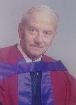
In 1946, Guy Elliott became the first full-time Professor of Medicine at Wits and the Chief Physician of the Johannesburg General Hospital. His remit extended to the staffing of Coronation and Baragwanath Hospitals, which were also significant in our training. He was quickly recognised as an inspiring teacher, a skilled physician, and excellent manager of varied activities, including research.
Andre van As wrote: “I first met Professor Elliott in 3rd year when I was his patient. He cared about solving the issue and was thorough and attentive. My next encounter with him was in 1964 when I worked in his ward as a registrar. At that time, an Intensive Care Unit (ICU) was just being formed. Together with my fellow registrars, Derek Stables, Arthur Rubenstein, and Sean Lynch we provided a centralised service for critically ill patients. I continued working with Professor
At the time we did our clinical training,1958-60, and house jobs 1961/62, the hospitals were as follows:
Johannesburg General Hospital: (The Gen) The city had only been founded in 1886, soon after the discovery of gold. Within ten years, the City of Gold had a population of 100,000, mostly black labourers. Its first hospital was a brick and thatch structure in Commissioner Street that doubled as a jail. The first permanent hospital, built on Hospital Hill, opened in 1890 with 130 beds and an operating theatre. In 1897, the Barnato wing was added. The Nurses’ Home on Klein Street opened in 1909. The physicians were largely British-born and educated, but with increasing numbers of South African-born doctors who travelled abroad to complete their medical training. In 1919, the medical school opened its doors. By 1937, the old hospital was demolished to make room for a larger Johannesburg General Hospital, designed by Gordon Leith – the imposing red brick building we knew and loved.
After our time, during the 1970s, this was superseded by a brand-new, huge hospital complex on Parktown Ridge. It was built on the site of Hohenheim, the first Parktown Randlord mansion. It was the first mansion built in the new township of Parktown. It was designed by Frank Emley and built in 1892 for Hermann Eckstein, Chairman of the ‘Corner House’ (mining company).
Hermann died very young, and it was then the home of a later Chairman of the Corner House group, Sir Lionel and Lady Florence Phillips.
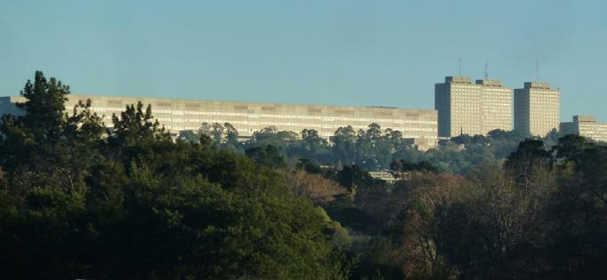
When they were banished from South Africa because of their involvement with the Jameson Raid, another director of the company lived there. This was Sir Percy Fitzpatrick (author of ‘Jock of the Bushveld’). In 1915, the house was donated to the hospital administration and converted into the Otto Beit Convalescent Home. It was run as the second branch of the Johannesburg Hospital. The first matron was Miss BG Alexander. She later became Matron of the Johannesburg General Hospital until 1931.
In the early 1970s the mansion was demolished to build the new Johannesburg General Hospital complex. A Heritage Plaque is in place to commemorate the site of this historic monument.
Transvaal Memorial Hospital for Children (TMH) This was built to memorialise those killed in the First World War and opened in 1923. From 1950 to 1968, the part- and full-time staff were appointed by the Transvaal Provincial Administration in consultation with Wits, which recognised TMH as a school of paediatrics only in 1968.
Queen Victoria Maternity Hospital (the Vic) is across the road from TMH, and the Vic was associated with the Johannesburg General.
The Fever Hospital (Fevers) opened in 1916, serving the white population of Johannesburg. The Public Health Department sent cases of infectious diseases, and admission was compulsory when isolation at home was not possible. It could accommodate 80 patients in five separate wards, two of which were isolation wards with cubicles. The non-European Tropical Diseases isolation hospital at Rietfontein (now called Sizwe) had already opened in 1895 (and has been in use ever since). It provided treatment to non-European patients with smallpox, tuberculosis, malaria, leprosy, typhoid, Congo fever, Spanish flu, bubonic plague, anthrax and Ebola, as well as venereal diseases. None of our students rotated to these hospitals.
Non-European Hospital (NEH) In our day, so-called ‘non-European’ inpatients were largely dealt with at Coronation, Bridgman, and Baragwanath Hospitals, but there was still an Outpatient Department of the Non-European Hospital on Hospital Hill, facing Esselen Street, under the control of the Johannesburg General.
Coronation Hospital (Coro) in Coronationville, near the mixed area of Sophiatown, opened in 1944 and serves a large population of Coloured, Indian, and Black people.
Elliott in developing intensive care until eventually it expanded to 12 beds run by the Pulmonary Unit. This paved the way for my specialising in Pulmonology.”
Culinary prowess was also one of Professor Elliott’s many attributes. He was not only a good cook but a gracious host and enjoyed entertaining. Andre remembers when an adventurous firm of students that included John Gear (son of the famous vaccine expert James Gear, together with the son of Professor Fassler (of Architecture), reciprocated and invited Professor Elliott and his registrars and house officers to dinner at the Fassler’s house. The evening was to feature a slide show by Professor Elliott of his overland trip from London to Johannesburg.
Source: By Egyptian Streets
The dinner party was the night after a busy intake with no sleep for Andre. He remembers a cosy fire, a tasty roast provided by the students, with plenty of good Cape wine. The last thing he remembered was Tutankhamun’s tomb in Egypt. (see picture). He awoke to the Fassler’s dog barking at his heavy snoring – and the journey had already ended. He thought he would be in the dog box – but said that Prof took it in his stride.
Andre says: “The training of our students and graduates improved as the teaching staff expanded, and new specialities, methods, devices and therapies became available.”
Elliott became Dean of the Faculty of Medicine from 1959 to 1963. He retired in 1967 to return to the Cape, where he had grown up. Wits conferred an honorary doctorate on him. He died in 1975, ironically, of an illness requiring ventilation in an ICU – the use of which he had pioneered.
Arthur Rubenstein added a few thoughts on being Professor Elliott’s Medical House Officer
In 1960, I was invited to join Professor Elliott’s ward as an ‘intern’ for the first 6 months after graduation. I was honoured and excited to be chosen to be his house officer. And so, I very much looked forward to my orientation by his registrars. Imagine my surprise when 90% of the time of the orientation was spent on instruction devoted to how to manage the ‘teatime’ period that preceded rounds and was obviously of great importance. I learned how to brew tea to perfection, which crockery to use, how and when to serve, and in what order and when it was permissible to talk. The ‘tea period’ was certainly rigorously organised, planned and executed. After imbibing all of this, I asked about patient care, ward rounds, presentations, and so forth, and was told not to worry, as the Sister would organise everything and instruct me how to proceed
Related to this were wonderful, but very long dinners at Professor Elliott’s beautiful apartment. Jack, his longstanding man-servant, would (maybe together with Professor Elliott) prepare gourmet dinners. The problem was that I invariably fell asleep at some time during these long evenings. I remember well Professor Elliott saying on one of these occasions in his very gentle voice, “Arthur, I do think you should go to the washroom and splash cold water on your face,” in the hopes that this would wake me up. Despite all of this, I learned a great deal, mainly from the amazing registrars as well as Professor Elliott, who was always a strong supporter of my career.
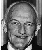
As we started our clinical years in 1958, a new Professor of Surgery was appointed at Wits. He was also appointed chief of surgery at the Johannesburg General Hospital. This was Daniel Jacob du Plessis. He, like Professor Elliott, was a UCT graduate. He had also distinguished himself in service in the Second World War as captain in the South African Field Ambulance with the British 8th Army in North Africa. He had carried out postgraduate surgical training at the universities of Cape Town and Witwatersrand, working for a time at Baragwanath Hospital. In 1951, he travelled to England on a Nuffield Scholarship, spending time at Oxford University and, in London, at St. Mary and St. Thomas hospitals, where he gained his FRCS.
He had served as a lecturer in surgery at the University of Cape Town and a staff surgeon at Groote Schuur Hospital before taking up the Chair at Wits.
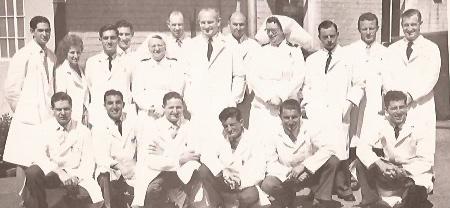
Professor Du Plessis was a hard taskmaster, expecting unswerving dedication from his staff, but he always led from the front Avroy Fanaroff, third from the left, crouching in the front of this picture above during his student rotation with Prof du Plessis from January to April 1959, remembers:
“We had to arrive at work at 6.00 am because we had to do the haemoglobin, blood count and urinanalysis on all our patients before rounds – you were forbidden to use the lab"
“When your patients went to surgery, you accompanied them. Your 10-minute surgical scrub was supervised by the operating room nurse, who invariably found some fault and started you over again, so by the time you entered the theatre, they were completing the case, and you saw nothing or else they tucked you away from the action, holding a retractor for the duration of the procedure.”
Avroy remembers that when he and Rodwin Jackson were interns. “For the first time in months, we went to the movies. Suddenly, Rodwin realised he did not have a gall bladder series which he needed for the next morning’s rounds. He left the movies, called a radiographer friend who met him at the General Hospital and had the X-rays done. So, the next morning, when Prof asked for the X-rays, they were available.”
Jack Hoffmann, who worked closely with du Plessis a few years after our time, recalls: “He was a formidable man in every respect. Physically, he was tall and well-built. He had a rugby forward’s shoulders and ears. He had receding, silvery-white hair, cold blue eyes and a prominent nose and chin.
“He was known to all as The Prof – as if he were the only professor in the medical school. He was more fondly known as ‘Dup’ – never to his face, though. His closest colleagues were allowed to call him ‘Sonny’, but when a junior addressed him, he or she would never dare to call him anything but ‘Professor ‘or ‘Sir’. His other nickname was ‘God’. And he truly was a god. He was the creator, the father, and the shaper of surgery in Johannesburg. He produced generations of disciples who spread his surgical credo throughout South Africa and to far continents.
“His lectures on surgery were masterpieces of clarity, precision and logic. His bedside teaching on his weekly ward rounds were paragons of excellence and attracted an entourage of acolytes. He rendered the most complex clinical problems into gems of simplicity by applying basic surgical principles. Each word from his mouth was a pearl of wisdom, a jewel of logical reasoning. His sage opinions would be delivered in the form of quietly spoken monologues.
“Dup would address only his senior colleagues, the ward sister or the registrar. More junior beings were ignored unless he was berating them. He was dogmatic to the extreme. No transgression of the guidelines was tolerated, even when well-founded. His anger at shoddy presentation of cases or failure to adhere to du Plessisian principles was revealed by the iciness of his tone. He never raised his voice. He never handed out compliments. Success was registered by lack of criticism.”
Jack knew that simply surviving the six months without putting a foot wrong would be a badge of merit. Nevertheless, he longed for one tiny nod of approval, one small word of praise, one scintilla of a smile from the great man. One of the houseman’s most important chores, was to write the case summary of the patients on their discharge from hospital. It had to be typed on a single sheet of size A6 paper by the ward secretary in time for the deadline at noon on Saturday. If the secretary could not manage it in time, it had to be written by hand by the houseman. As with the case presentations, it was a balancing act between providing all the necessary data and excluding irrelevant details. In complex cases, it was almost impossible to find space for all the facts within the space provided by an A6 sheet. The Professor checked each summary minutely. If it was not up to scratch, he would tear it in two without a word. If it was accepted, he would place it without comment on the pile in front of him. One of Jack’s patients had had an extremely complicated course. She had undergone an enormous barrage of tests and three operations. Jack spent most of the Friday night rendering the multitudinous array of facts into a coherent kernel. The Professor picked up the sheet of paper. He read every word. Jack was sure his heartbeat could be heard across the room. ‘This is a damn fine summary, Hoffmann’ Dup said as he added it to the pile.
Class of '60 classmate, Aaron Arthur Vinik completed his residency training in Johannesburg; his wife Etta said: “Our first son was born while we were living at Resdoc, and Arthur was doing a surgery residency with Professor du Plessis at the Johannesburg General Hospital. Arthur would be away most nights on call and come back exhausted at 6.00 am to shower and change into clean clothes, especially a well-ironed shirt and neat tie. That was the rule. Du Plessis didn’t care if the residents had no sleep. They had to look good and have clear minds to answer the knife-like questions he would often hurl at them, demeaning them for incorrect answers. Du Plessis was definitely not interested in a newborn baby being cared for by an inexperienced mother.”
Our classmate Allan Gottlieb, who, after qualifying and after his house jobs, had set his mind on surgery, became a teacher in the Anatomy Department at Wits and also continued to attend Professor du Plessis’ surgery lectures.
Heather Crew Brown, intern at the Gen with Dup in 1967, paid this tribute to Professor DJ du Plessis. She wrote: “The surgical department was well structured with high standards and ethical principles. The role of the team, particularly of senior staff and matrons/senior nursing staff, was important. From the top down, there was an awareness of necessary discipline. The daily routine for interns was demanding and early morning starts were rigorous. However, this did not deter one and had the galvanizing effect of producing greater commitment and effort. At the end of the six-month period, Professor du Plessis gave each intern a gift copy of ‘The Student Life’, the philosophy of Sir William Osler, edited by Richard Verney, with a personal message on the front page. This book had a significant influence on my further postgraduate training and has done throughout my life and career in the medical field. It is a gem with much useful wisdom and advice on the problems and responsibilities of students, reaching far beyond their medical training. I realised that Prof had been mindful of and practised much of its philosophy in his own career. He also had an influence on my decision to remain based in South Africa and to serve primarily the neediest sections of the population, where possible.”
By the time Du Plessis retired as professor of surgery in 1977, after 19 years, the department had risen from a demoralized and parlous state into a superbly organised and highly productive department recognised as one of the finest in the world. He played a major role in establishing the College of Medicine of South Africa and its surgical affairs, in establishing biennial congresses and creating the South African Journal of Surgery and the Surgical Research Society. He and his students were making important contributions to surgical meetings, research and practice throughout the English-speaking world.
He was elected to be Vice-Chancellor of the University of the Witwatersrand in 1977, where he spearheaded wide ranging thoughts and plans for the development of the University. With the Milner Park campus site rapidly becoming overcrowded, it is widely acknowledged that it was due to his leadership, determination and drive that Wits finally acquired the adjacent Rand Show site, creating the magnificent West Campus in the mid-1980s, including new hostel accommodation for students in residence. Chris Barron, in an obituary, wrote: “Seldom has a
university owed more to the vision and bloody-mindedness of one man than Wits owes to Sonny du Plessis.” Wits awarded him the degree of Doctor of Laws, honoris causa, in 1984. He died in 1999, aged 81 years.
Jack Hoffmann also had the opportunity of working as a senior registrar for Joss Lannon, who was another of the superb senior surgeons on our rotations and house jobs. Jack sums up the difference between the operative skills of Lannon and du Plessis by saying, “Dup operated like a battalion of soldiers in battle. Each step had been carefully planned; secure that road, bomb that bridge, explode that building. It was effective, but it was not elegant. Joss Lannon operated like a ballerina: rhythm, synchrony, precision. Each stroke of his scalpel, each movement of his forceps was a masterpiece. His dissections were like the illustrations in Sabota’s famous Atlas of Human Anatomy. And he never spilt a drop of blood. He anticipated, he knew, where each artery, where each vein was hiding and clamped them before they could bleed and obscure his field of dissection, The Professor was rugby. Mr Lannon was cricket. Joss was Chopin. Dup was Wagner.”
Jack went on to say: “Joss Lannon was also the one person not afraid of saying what he thought even if he knew it would not please Prof. Every Saturday morning, the capacious lecture hall of the Johannesburg General Hospital was filled for the professor’s case presentations. The rows were occupied to the last seat, according to this hierarchy: first the staff of the professorial surgical unit; the staffs of the other surgical departments of the Gen, Coro and Bara; surgeons in private practice from the city of Johannesburg and the surrounding towns; casualty officers; registrars; housemen; medical students; and nurses."
“Three or four cases were presented for discussion. The discussion usually involved a monologue by Dup, who would throw a few questions to the audience. Wise or relevant responses were received with a half-smile of approval. Nonsense was rejected with jutting jaw, a look of withering disdain and silence. Very few dared to utter an opinion, especially uninvited, in the presence of ‘God’. There was one exception. This was Joss Lannon. He was a dapper, not very tall man with silver hair. He had a never-fading sun tan so that he always looked as though he had just returned from three weeks on the French Riviera. He always had a somewhat mischievous smile on his face, which revealed a slight gap between his upper medial incisors. This added to his roguish air. He had a quick and pithy humour. Whereas all his colleagues dressed in customary dark suits and sober ties, he often wore relaxed flannel trousers, blazers with brass buttons and colourful ties. He had the reputation of being the best surgeon in Johannesburg. He was no match for Dup when it came to knowledge of the science of surgery and the analysis of complex problems, but technically, he was a genius. They said he had golden hands. He wasn’t a scientist like Dup was, but his crisp mind, sense of logic and his vast clinical experience gave him enough fire power to face the Professor. He greatly respected Dup but refused to be like the bunch of poodles that followed obediently at Dup’s heels.
“A case was presented of a patient with severe, constant abdominal pain. She had already undergone a vast battery of blood tests, urine examinations, endoscopies, x-rays, scans and periods of observation. Even the Professor of Internal Medicine had been called in to assess her. Dup, as usual, proposed an algorithm of profound insight. Esteemed members of the audience suggested an extra blood test here, another x-ray there. ‘’What about a psychiatric evaluation?’’ suggested one. Joss raised his hand, stood up and said, ‘This poor woman has been investigated from Dan to Beersheba, from Beitbridge to Cape Point. She’s had every test in the book. Now some of you are suggesting she’s nuts! Why not just go back to basics? Take a knife and open her abdomen. I’m sure you’ll find what you’re looking for.’ Even Dup bowed to this impeccable logic. She subsequently underwent a so-called exploratory laparotomy, the jargon for Joss’ suggestion. A small tumour was found in her adrenal gland that had invaded into the surrounding tissues. This was an ample explanation for her symptoms.
“On another occasion, a case was presented illustrating post-operative death by pulmonary embolism. After surgery, various factors such as changes in the coagulability of the blood and torpid flow of blood through the veins of an inactive calf, can lead to the formation of a clot of blood in these veins. This can cause a spike of fever, pain in the calf and swelling of the ankle. In extreme cases, the blood clot loosens and is carried along the venous system through the heart and into the lung where it blocks the circulation. It is then termed a pulmonary embolus, a condition which can be fatal. Dup sagely enumerated the known methods of preventing such a
catastrophe. One or two in the audience added their tuppence worth.
“Joss stood up and said, ‘My patients never get pulmonary emboli. I just give them a little shot of heparin (a substance that prevents coagulation of the blood used in various situations to dissolve blood clots in arteries or veins) before the operation.’ Dup asked, ‘Do you have statistics to back up your claim?’ ‘No, you’ll just have to take my word for it.’ Sniggers rippled across the hall. ‘But surely the patient must bleed excessively during the operation if you’ve filled them with anticoagulant?’ ‘No, it never happens because the dosage is too small.’ ‘How much do you give?’ ‘Oh, I don’t know. My nurse just gives them a little.’ More sniggers. ‘So you have no documentation of how many emboli you have per number of patients operated nor on the incidence of excessive bleeding nor of the dosage you recommend. I am afraid that we cannot take your claim seriously.’ Overt laughter now. ‘You’re right Prof. Not very scientific. But it works!’
A few years later, after others had stumbled on the idea and tested it clinically, prophylactic peri-operative heparin would become standard surgical practice throughout the world. None of the users would ever have heard of Joss Lannon.
Arthur Rubenstein was on Dr Agranat’s firm and sent us this picture which includes seven of the nine members of our class in his ‘firm’.
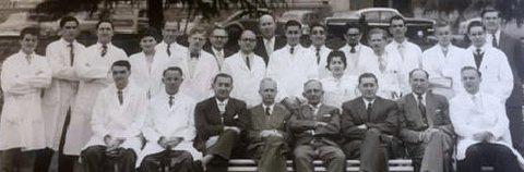
Avroy says: This picture must have been taken in 1958 when we were in fourth year, because there are final year students in the picture, including Glick and Friedman. Harold Waldbaum, who became an ENT specialist, was one of the housemen. I have no recollection of anything about Agranat, but his registrar, Hyam Isaacs, sitting in the front row on the extreme right, influenced us a lot. He was a great clinician and teacher who had developed an expertise in muscle disorders and was able to show us a variety of these patients. Our firm maintained a relationship with him and went to him for tutorials.
Rodwin Jackson adds: All the members of our firm were excellent students and colleagues, which made our three student clinical years so memorable and enjoyable. Friendships have been maintained to this day. All have done well in their careers. We had good clinical teaching from all the consultants, but for me, the most inspiring teacher was Hyam Isaacs, who was a registrar. He was an excellent diagnostician and had a special interest in congenital myopathies, about which he was writing an MD thesis.
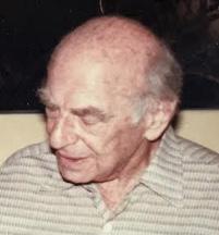
Avroy Fanaroff remembers much more about working with Mosie Suzman. Avroy and Chris Bosman had the privilege and pleasure of being housemen with him. They recall Dr Suzman as a remarkable teacher and mentor. He gave his students, interns and patients as much time as needed before leaving for his consulting rooms.
What set Moses Myer Suzman apart was his refusal to accept conventional wisdom. Voracious reading and an encyclopaedic memory combined to make him a formidable destroyer of pet medical theories. At our weekly conferences attended by many specialists, he would delight in taking a contrary view. Needless to say, he would often be proved correct.
Avroy wrote: “He was very gentle with me when I presented the ECGs of three patients who had all died after being admitted the previous night with massive myocardial infarctions. He said that based on their presentations in shock and their ECGs, they had no chance of surviving. He was not gentle, however, when I had to consult a chart during a conference because the patient had so many admissions, and I could not remember all the details. That only happened once.
Mosie was a pioneer, decades ahead of his time, in the use of anticoagulants such as Warfarin (created as a rat poison – but accepted for humans in America in 1954) for MI patients. Avroy and Andre van As both attest to the difficulties of running the weekly clinic, where they had to adjust the dose of anticoagulants for the cardiac patients. It was a massive undertaking, as 200 to 300 patients often attended the clinic. And they were dealing with a very inexact science in those days, with the risk of starting major bleeds or causing repeated clotting. Andre notes that it is interesting that currently, anticoagulation is back in vogue for the long-term management of MI.
Generations of Wits medical students found Mosie Suzman inspiring, awesome and warm. His wife was the internationally known Helen Suzman, one of South Africa’s most famous members of parliament. She said: ‘Whenever I travelled overseas on political business, some doctor would come up to me and say: ‘I learnt all my medicine from Mosie on Ward 11 of the Johannesburg General Hospital’.”
Barlow was a cardiologist of international standing at Wits. Andre van As remembers that although we had limited direct contact with him as students, his influence as a cardiologist was felt, and his opinion was widely respected. During the period after 1958, when Barlow returned to South Africa, very few of us were aware of his rapid academic and research ascension that was occurring right under our noses. While Barlow did not have frequent student teaching rounds nor delivered didactic cardiology lectures, he did spend a lot of time with his cardiology trainees during their daily activities in his Cardiac clinic. His clinical expertise was of the highest standards, and he rose to heights that most clinicians never attain. His trainees all aspired to reach his level of clinical expertise, and their interactions with the residents and interns passed on to the students what they had learned from him. The mere fact that he was part of the medical community at Wits affected anybody that had contact with him. During my academic tenure in the US, mentioning that I had contact with Barlow was enough to get the attention of my colleagues.
Classmate Tony Myers (who became a Professor of Nephrology at Wits) pointed out two names that stood out for him during his training. He said: “The holistic, humanitarian and highly skilled practice of Professor Tom Bothwell has created indelible ideals for many of us fortunate enough to have been taught by him. A second giant and one with both immense knowledge and judgement, as well as iron-fisted social graces was Dr Ben Goldberg. But there were many more before and have been after, even up to this day.
Arthur Vinik has carried into his profession, both as a physician and also into everyday life, many of the diagnostic lessons he learned from his teachers at Wits. He has incorporated the clinical skills he learned from the likes of Mosie Suzman at the Johannesburg General Hospital. From Doctors Barlow and (later Professor)
Bothwell, he learned to eavesdrop on conversations three tables away in restaurants and employ that listening technique to perfect hearing heart sounds and to observe people in everyday situations and notice how they walked, held their heads, and if they had any unusual deformities. The pressed shirt also remained an obsession with Arthur. He learned to use the Black and Decker iron (reminiscent of his woodwork tools) and re-ironed the shirts that returned from the laundry.
In his biographical sketch on our website, classmate Jack Kussel writes that his love for clinical paediatrics was born, inspired by Sam Javett, Seymour Heymann and Hessel Utian during his time at the ransvaal Memorial Hospital for Children (TMH).
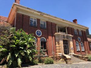
Heymann, born in Pretoria in 1902, was paediatrician-in-chief and trained generations of paediatricians. He was in private paediatric practice together with Sam Javett and Hessel Utian. At 66, Heymann emigrated to the USA to work at the Children’s Hospital Medical Centre of Northern California, Oakland, where he continued to train paediatric residents. Heymann died in California in 1997, aged 95 years. Hessel Utian was born in 1932. He was 30 years younger than his mentor. After graduating MBBCh from Wits Medical School in 1954, Utian worked at the Hospital for Sick Children in London and obtained a Diploma in Child Health and MRCP (Edin). Utian maintained a teaching and clinical position with Wits Medical School for many years. He died in 2012, aged 80.
Avroy Fanaroff remembers: “The students did six-week rotations. When we were students and house officers, there were six paediatric chiefs: Doctors Heymann, Javett, Theron, Parnell, Kessell, and Levin. Until about 1965, there were only two medical and two surgical senior house officers (SHOs) and one registrar, Dr Hessel Utian. The SHOs were on call alternate nights and rotated on Sunday, so we could be on call from 08h00 on Sunday till 17h00 on Tuesday! Furthermore, at least once a week, the SHO was responsible for the Casualty from 22h00.
The teaching was magnificent and the variety of cases remarkable because we were getting referrals from all over the Transvaal Province and also Portuguese East Africa. Sick, malformed and premature babies from the Queen Vic Maternity Hospital were also sent to TMH.
Sam Javett was the most outstanding teacher. He used algorithms before we were even familiar with the term and was often consulted by internists when they had complicated patients. The Paediatric Department got its first University chair, Boet De V Heese, in 1968.
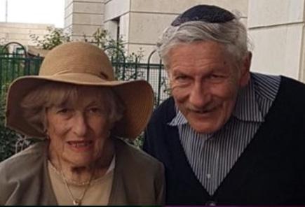
Another paediatrician who made a great impact on our class was Professor Solomon E Levin. Avroy Fanaroff and Aubrey Milunsky were both very sad to hear that their revered teacher and mentor, Solly Levin, had died of COVID-19 in July 2020, just a few days after his beloved wife Cynthia had also succumbed. (They are pictured together here.) They say: “He was the epitome of the Academic Paediatrician. He was a marvellous clinician, an excellent teacher, an investigator. Solly was extremely knowledgeable, inquisitive, a great listener,
wonderful mentor, and kept meticulous records including photographs. He did not have a mean bone in his body. They both felt certain that Solly’s interest in them and role model had contributed to their own passion for paediatrics.
In 1943 the Queen Victoria Maternity Hospital moved to a new building across the road from the Transvaal Memorial Hospital for Children.
In the shake-up of medical provision in 1946, OS Heyns was appointed first Professor of Obstetrics and Gynaecology at Wits Medical School and Chief of OBGYN at the Johannesburg General Hospital. His authority also covered the Queen Victoria Maternity and Coronation Hospitals.
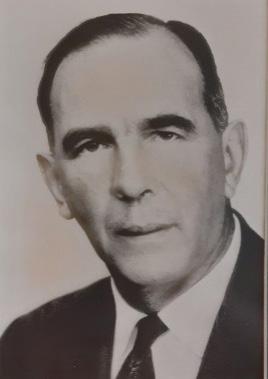
In 1959, Heyns published a paper on Abdominal Decompression during the First State of Labour. Heyns had developed the ‘birth-suit’ made of plastic with a fibre-glass dome over the abdomen. It had a pressure gauge and a vacuum pump, as used in an ordinary household vacuum cleaner. He claimed that his device brought obstetrics ‘into the age of technology’.
Experimenting on pregnant women at the Queen Victoria and Coronation Hospitals, Heyns advised that they wear the suit for half an hour during the last ten weeks of pregnancy and right through the first stage of labour. He said that with decompression, the abdominal muscles were lifted off the uterus, enabling the uterus contractions to speed the expulsion of the child into the world. Not only was the delivery shorter and less painful, but the women produced, he said, ‘a harvest of exceptionally bright children [with] IQs ranging from 110 to 173’. Professor OS Heyns was lauded as ‘one of the unsung heroes of modern obstetric care.’ His co-authored book Decompression: A New Medical Breakthrough promised easier and safer pregnancies, shorter labour, and healthier and brighter children.
OS Heyns retired in 1967. Unfortunately, the success of Heyns’ birth-suit was largely built on hype and the subjective claims of mothers that their children were exceptionally bright. In 1990, J. Justus Hofmeyr, professor of Obstetrics and Gynaecology at Wits University, reported in a carefully controlled study that ‘the manifestly unrealistic claims’ by mothers of their children’s abilities were simply ‘not the case’. Hofmeyr suggested that the birth-suit and foetal decompression be relegated to the history books. Ockert Stephanus Heyns was also Dean of Medicine in our time, from 1957 to 1958.
Medical care in racially segregated Johannesburg in the 1950s was of a very high standard across the board. Wits graduates travelled abroad to study and specialise in the most advanced centres of the world. Many returned to serve the South African population as clinicians and teachers. In the White areas, the services strove to be equal to anywhere in the developed world.
Even in the overwhelming circumstances of looking after huge and impoverished Black communities at Baragwanath, Alexandra Clinic, or the Non-European and Coronation Hospitals, we encountered the same skilled, innovative, and dedicated medical staff and gifted teachers eager to do the very best for the health of the people they cared for. These were the ‘non-European’ hospitals at which we worked and the gifted teachers who trained us.
In our day, so-called ‘non-European’ inpatients (i.e., anybody who was not White—except for some Japanese and Chinese) were largely dealt with at Coronation, Bridgman, and Baragwanath Hospitals. However, there was still an outpatient department of the Non-European Hospital (NEH) on Hospital Hill, facing Esselen Street, under the control of the Johannesburg General.
The Bridgman Memorial Hospital in Brixton, Johannesburg, was named after the revered American missionary and activist Frederick Brainerd Bridgman (1869–1925) and his wife Clara Davis Bridgman (1872–1956). It opened in 1928. It was the largest hospital in the Southern Hemisphere, designed specifically for African women. It was forcibly closed in 1965 when the apartheid authorities objected to Black patients being treated in a White area. The Bridgmans were key figures in the establishment of institutions in aid of Black and Coloured communities. Frederick Bridgman was instrumental in the founding of the Helping Hand Club in Jeppestown, the Bantu Men’s Social Club in 1924, and the Jan Hofmeyr School of Social Studies―the first institution to train Black social workers in South Africa, with many well-known alumni. He also began a clinic for migrant mine workers with the intention of eventually establishing a hospital. He died suddenly in 1925 during a visit to the USA. His wife, Clara, raised the funds required, and the Bridgman Memorial Hospital was completed in 1928.
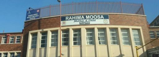
The large and busy 147-bed Coronation Hospital opened in 1944. It was built in the suburb of Coronationville, near Sophiatown, to serve the Coloured and Indian populations of Newclare, Noordgesig, and Coronationville. In our time, it also cared for Black patients from Pimville, Orlando, and Sophiatown.
The surgical chief at Coronation at our time was Boris Lewin, who was later appointed Associate Professor of Surgery at Wits in 1969. The Chief Physician at Coronation was Sydney Grieve, later appointed Professor and Head of the Department of Medicine at Wits Medical School.
Ronnie Auerbach remembers his surgical house job there with Mike Jaffe, Gerald Shulman and Kleintje van Veen. Intake nights were very busy, often stretching into the morning session as there were so many admissions for the registrars and house staff to deal with. Mostly, it was trauma: fights, stabbing, head injuries, motor or bike accidents, as well as acute illnesses like appendicitis. Jerry Jersky, the superb surgical registrar, took it in his stride, even repairing stabbed hearts (of course, with no heart-lung machines).
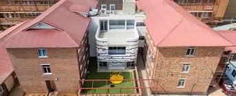
The influx of visitors had to be managed. When visiting time started and the wards filled up rapidly, the porters closed the doors and only let more in when others had left. When they once did a survey on the visitors, they found that 10% of visitors had no specific patient to visit but had come as a social activity either for themselves or for patients who had no company. On Sundays, the Salvation Army Band would come and play in the courtyard between the ward blocks (seen in the picture) for the benefit of the patients’ souls but to the annoyance of the staff! (The white bit in the picture seems to be a new addition.)
In our final year at medical school, we had a six-week stint at the Alexandra Healthcare Centre. This was situated in the crowded Alexandra Township, which had grown up on what was then the edge of white suburban Johannesburg and was now surrounded by it. Covering just a square mile, in our time, it was home to 20 000 or more impoverished Black families. There were attempts by the Apartheid Government to eradicate it altogether, but they failed.
The Health Centre had been set up in the 1920s by the American Board Missionaries (ABM). In 1939 when the ABM charity withdrew its funds to focus on other projects, a new charity took over. At this time, a symbiotic relationship was formed between Wits University and the clinic. This became a valuable training facility for students who provided free medical services for the clinic under the guidance of a full-time medical director.
The students were required to sleep there when on call. Andre says, “This was the one rotation where we had the sole responsibility for the management of patients. After 16h00, the students were the only medical staff present.”
Colleagues may remember that when they were on call, each night, the porter would come to the students’
quarters, knock on the window and hold up a number of fingers, indicating how many possibly critically ill or trauma patients had arrived to be seen. He usually held up one or two fingers at a time. But Ronnie Auerbach recalls one night when he held up a whole hand!
He said: “Arriving quickly in the treatment room, the students on call were faced with five unconscious patients, a mother, grandmother and three children. There was no sign of injury. They had never come across this before and they had no idea what to do. It took ages for them to find and pore over all the available textbooks. They were beginning to despair when they noticed that all the patients had started to come round. They had apparently been overcome by carbon monoxide poisoning (which was quite common in their cramped and ill-ventilated homes). Having spent an hour or more in the fresh air, they returned to health and were duly sent home.”
Students were meant to make home visits on bicycles, but because of the rabid dogs, they tended to travel by car, carrying medication samples donated by various drug companies.
Andre Van As remembers a nurse giving him a list of names and addresses of patients that needed to be visited for follow-up. He entered the homes that had no windows, were pitch dark and contained unattended, seriously ill patients that should have been in hospital.
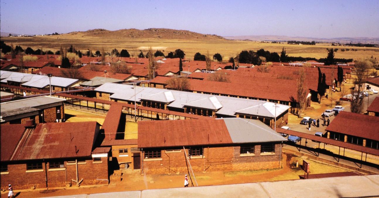
Working at Baragwanath Hospital was an invaluable learning experience because of the number of patients and the wide range of pathology encountered.
Andre van As tells us that Asher Dubb, physician at Baragwanath Hospital, and subsequently principal physician and professor of medicine, was “a classic example of the interactive teacher who made the process an adventure.” Born in 1928 in Somerset East, Dubb graduated from Wits Medical School in 1950. He did his house jobs at Baragwanath Hospital and served as medical registrar at Coronation Hospital. Dubb was a mentor and role model to generations of medical students, interns and registrars, with his inspiring bedside rounds, his courteous approach to patients, his almost obsessive attention to detail, his encyclopaedic knowledge of medicine and his dazzling deductive powers.” Asher Dubb died 2005, aged 77 years.
Harry Seftel was a much-loved and admired teacher of many students. He was appointed Wits Professor of African Diseases and chief physician at the Non-European Hospital. There he set up the Wits Carbohydrate and Lipid Metabolism Research Centre, focusing on obesity, diabetes, hyperlipidaemia and coronary heart disease, leading to many scientific papers. Harry had qualified with an MBBCh in 1952, eight years before our class. Both his parents had died of smoking-related illnesses. Harry built his career promoting a healthy lifestyle, no smoking, exercise and a fish and fibre diet. In 1976, he formed the National Council Against Smoking. Harry Seftel was able to discuss health matters in laymen’s terms. Starting in the 1990s, he took his message to the airwaves with his “A word on medical matters” show on Talk Radio 702. To many, he was the most inspiring teacher. Wits conferred an honorary doctorate on him.
Robert Jacobson, a graduate of 1966, fondly remembers being Seftel’s houseman. “The unit consisted of one
male and one female ward, with about 30 adult beds each. Ward rounds were conducted every morning between 08h00 and 11h00. In attendance were the interns, registrars, senior nurses and the master clinician and teacher, Professor Seftel. He was a most dynamic and enthusiastic man and taught his junior doctors clinical skills and judgement.
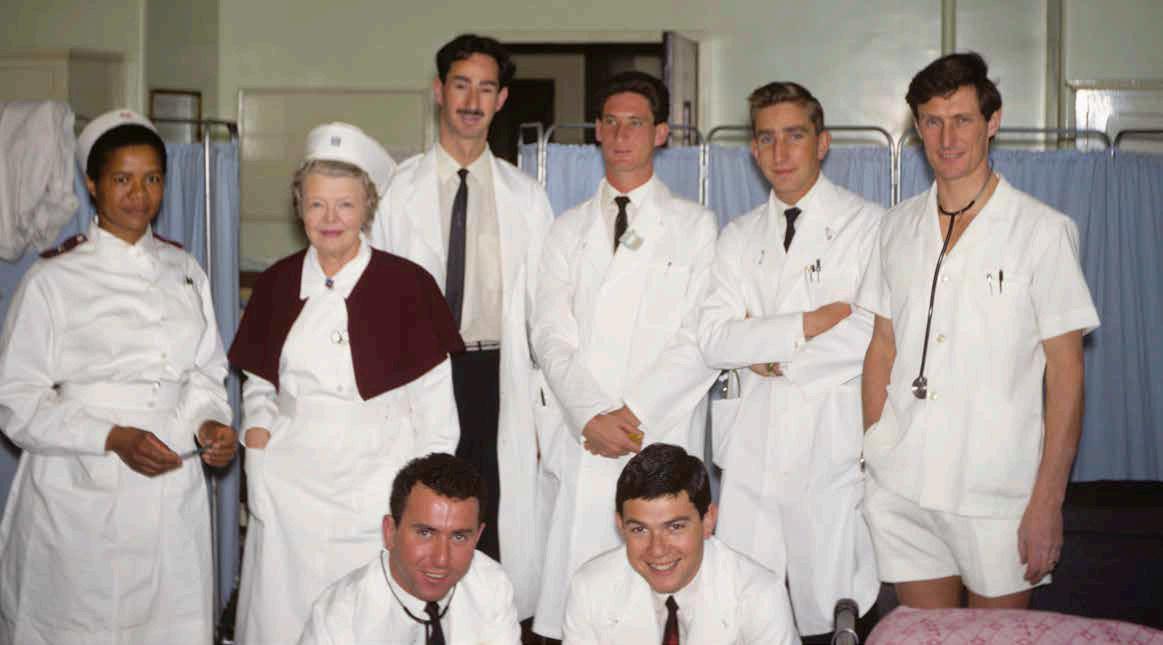
To the African nursing staff and patients, he showed respect and cultural understanding. He questioned us at the bedside about our treatment and management of the patients. Professor Seftel never embarrassed us, and I still remember his proclamations, ‘Sugar is poison’; ‘Milk is baby food’; ‘no need for taking vitamins, they end up in the toilet’. I was most fortunate to have had such an excellent role model and mentor early in my career.”
Professor Seftel conducted research on diabetes mellitus and the effect of dietary changes on the African and Indian populations in South Africa. We learnt that the western diet prevalent in Johannesburg was causing a surge of diabetes in the Indian population and that coronary heart disease, which had been rare in the African communities, was now emerging in the African population that mainly ate the food in the White homes they worked in. All these observations were made ahead of the time and were more generally accepted.
Arthur Vinik’s interest in gastrointestinal hormones was sparked when he worked with Professor Harry Seftel at Baragwanath Hospital. Later, working on a research project with a member of the surgery department, Dr Richard Fiddian Green, he developed a lifelong fascination with this field, where he has made seminal contributions. It was also this interest that provided the opportunity to join the faculty at the University of Michigan as Professor of Medicine and Surgery in 1979.
Andre van As said: “Harry Seftel epitomised the peak of clinical competence that can be achieved. To reach that in a setting where clinical excellence abounded in our mentors and fellow students is a truly remarkable feat. His clinical career is filled with remarkable episodes that were very characteristic of Harry’s style and each of which left an indelible imprint on our memories. In the first six months of 1960, I was a surgery houseman in Michael Dinner ’s unit when one of his remarkable incidents occurred. The house staff in another surgical unit heard a loud and strange sound while auscultating the chest of a patient. They asked Harry to come and see the patient to unravel the origin of the sound. When he arrived, he stood still at the foot of the patient’s bed and appeared to be listening to something. After a few moments, he cocked his head to one side and said, ‘What you are hearing is a pericardial crunch. Get an X-ray of his chest’, and he walked out. The X-ray showed a pneumopericardium that was causing the crunching sound. The patient had been admitted with a stab wound to the chest. The story of this incident went around Bara like wildfire – or, in today’s parlance, went viral.”
Leo Schamroth graduated from Wits Medical School in 1948 and joined the staff at Baragwanath in 1956. He wrote An Introduction to Electrocardiography (1957), followed by “The Disorders of Cardiac Rhythm and The Electro-cardiology of Coronary Heart Disease.” Wits awarded him an MD in 1965 and DSc in 1970. In 1972, he was appointed Professor of Medicine and Chief Physician at Baragwanath, where he remained until his retirement, owing to ill health, in 1987. He died in 1988, aged 64 years. These highly skilled physicians often subjected medical students and house officers to a lot of good-natured abuse. If a student made a mistake reading an electrocardiogram, Dr Schamroth was prone to announce: “A shame and a disgrace. He went to college and wasted his father’s money.”
Jeff Maisels says:
“As Avroy and I have said, Bara is and remains a unique institution representing at various times both the best and the worst of South Africa, although, even at the height of apartheid, it provided a remarkably high quality of care and was staffed by a truly dedicated group of physicians who were superb clinicians and teachers.”
Jeff thought you might like to play this dance video, filmed at Baragwanath Hospital with participation by Baragwanath Hospital staff – and possibly patients too. He says: “The hospital facility was less than adequate, and much of it is still that way, but the dance performance does display the remarkable spirit that still pervades that institution.’
Baragwanath Hospital is a village. This musical video, with images of the hospital, depicts the teamwork and joy of dancing in an incredibly well-coordinated manner. The choreography is spectacular, with many, many participants, and the African beat and singing make for a most enjoyable experience.
Hilton Silver summed up by saying:
“Our clinical teachers, under whom we did our rotations and housemanships, gave us a solid grounding in clinical medicine. I recall being constantly impressed by their knowledge and clinical abilities. In medicine, I learned much from Dr Mosie Suzman with his encyclopaedic knowledge, Dr A.L. Agranat, who seemed to always speak in whispers, Dr Hyam Isaacs, of the whimsical smile, and Dr Asher Dubb, who was my favourite teacher. At Baragwanath, Dr Leo ‘Zay a Mentch’ Schamroth made electrocardiography seem ridiculously simple. Dr Harry Seftel, with his booming voice, proclaimed his vast range of knowledge. Surgery, never an interest of mine, was made fascinating by Professor Bert Myburgh and Mr Jos Lannon. Paediatrics, to which I was eventually drawn, was presented as diagnostic challenges at The Children’s Hospital by the Drs Heyman and Javett's team. But for me, the more inspirational practice of paediatrics came from visits to Baragwanath Hospital, where the paediatric unit was headed by Dr Sam Wayburn and his excellent team of Drs Len Taitz, Sol Levin, Harry Stein and Monty Shnier.”
Avroy Fanaroff, Neonatologist Cleveland Ohio and Jeffrey Maisels, Neonatologist Royal Oak, Michigan and others.
As students during 1958 and ‘59, our class did rotations at the Chris Hani Baragwanath Hospital. We also served as interns and registrars there from 1962 to 1966. In 1961, some of us also did locums at ‘Bara’ before we officially started our residencies.
For those that don’t know, ‘Bara’ was, and is, widely renowned as a huge, sprawling and bustling hospital to the Southwest of Johannesburg, serving what was then known as the ‘Non-European’ population of the nearby townships of Soweto (short for: South West Townships). Students and interns who had the opportunity of working there received the most extraordinary medical training, meeting with every kind of medical problem and surgical condition in quick succession. The ‘chiefs’ who worked there were legendary and young doctors were set up for life.
Construction of the 1,544-bed hospital had begun in November 1941. It had been commissioned by the British Army to care for injuries to the allied soldiers serving ‘up North’ in Africa, the Middle East and Italy. It was speedily built on the barrack system of field hospitals. You can see the individual buildings with long outdoor corridors.
The hospital was built near where the old Wayside Inn run by a Cornishman named John Albert Baragwanath had been situated. The Inn was known as simply as ‘Baragwanath’s’, and the name transferred to the new hospital. So urgent was the need that before a year had passed, on the 23rd September 1942, Field Marshall Smuts officially opened the hospital.
He used the opportunity to indicate that after the war, the Government would use the hospital for the Black population of the Witwatersrand. Meanwhile, Baragwanath was called on to deal with war casualties, mainly from the Middle East Command. During the latter part of the war, Baragwanath treated mostly tuberculosis patients, not only from the Middle East Command, but also from the Far East Command – mainly the Burma theatre.
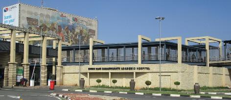
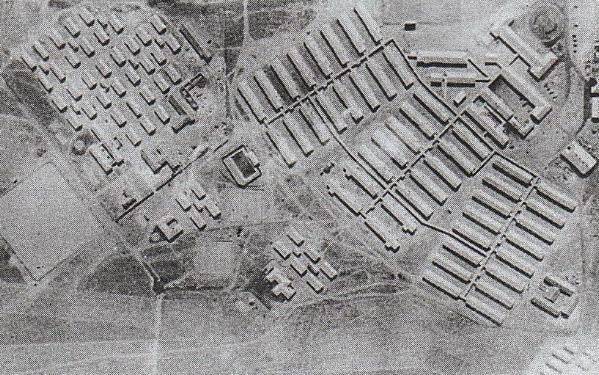
After the war, the South African Government took over the hospital and on 1 April 1948, the black section of Johannesburg General Hospital (known as Non-European Hospital) was transferred to Baragwanath, and the new stage of the hospital opened with 480 beds.
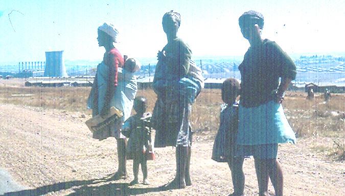
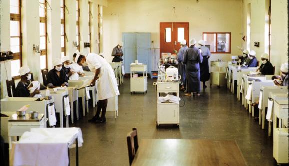
The impact of the hospital was immediate. When it opened the infant mortality was 250 per thousand. Within one year with the establishment of a rehydration centre (“drip room”) the mortality was down to 60 per thousand and within a few years down to 30 per thousand. Also, the establishment by Eric Kahn and Sam Wayburne of two 75-bed premature baby units with mothers caring for their babies lowered premature mortality to levels equivalent to the best in the world.
Over the next 30 years, Baragwanath grew in size and stature. In the early 2000s, it was the third largest hospital in the world, occupying around 173 acres (0.70 km2), with approximately 3,200 beds and about 6,760 staff members. Approximately 70% of all admissions are emergencies, including about 160 victims of gunshot wounds per month.
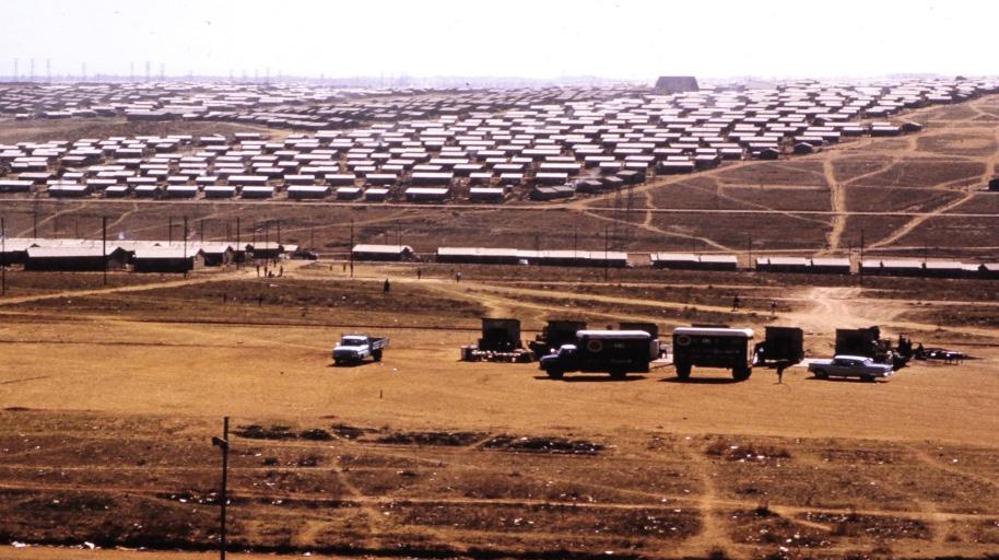
Accident, emergency and ambulance transports are the busiest services, with over 350 daily patients. Every year, about 150 000 inpatient and 500 000 outpatient cases are registered. These represent a small amount of outpatient visits because today, there are also many clinics surrounding the hospital in Soweto for primary care and referrals. Bara not only serves Soweto but is also a referral hospital for a large part of the country and also other African States.
“To get to Bara, most of us took the bus from the Johannesburg General Hospital parking lot. The contrast of the high-rise spacious buildings in Hillbrow on the ridge above Johannesburg, to the dense low dwellings of smog-filled Soweto was striking. The smog was generated from the fires used to cook and warm homes, which had no other heating.
If we were assigned to the general outpatient service, it was not unusual to be confronted with a line of 50 or more patients who had walked from Soweto and were waiting patiently to be seen. We worked extremely hard, but also made sure there was time for socialisation, unlike the frenetic pace that many of us subsequently embraced in our adopted countries.
On the adult services, the rounds would begin early in the 50-bed male and female units. At 10h00, even if you were between the first and second heart sounds, you stopped and went up to the cafeteria for cucumber sandwiches and tea, provided by Matilda. There, Professor Leo Schamroth, a world-class cardiologist and expert in ECGs, held court and read all the complex ECGs. At the same time, he would find out who had a car, and could give him a ride home in the early afternoon. He also handed out ECGs with the command, “Fanaroff, start talking.” If you came up with the (usually) incorrect answer, he would say, “A shame and a disgrace, gegannen (went) in college and wasted his father’s money.”
Bara had very basic facilities, although everything necessary for medical and surgical patient care was available, and the quality of the devoted nursing staff was excellent. There was no library or recreational facilities when you were on call overnight. We were there way before the internet, beepers or cell phones. Your biggest fear was that you would get “Bara Guts”, a dysentery-like illness from the food when you stayed overnight. There was a nice swimming pool and tennis courts, but very few opportunities to use them. It was interesting to watch the paraplegics playing wheelchair basketball. Lipschitz and Bloch had established a world-class paraplegic care and rehabilitation centre (for victims of spinal injury, usually intentionally administered with a bicycle spoke stab to the neck).
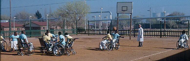
Each unit was on call every 4th day. We would start with about 10 empty beds in our 50-bed ward. At the end of 24 hours, we would have received between 40 and 60 medical admissions. The big influx of admissions came as the ambulances arrived jam-packed with patients from the surrounding healthcare facilities. Pretty soon, there would be patients in beds, patients under beds, patients on stretchers between the beds and chairs and the 50-bed unit would have expanded dramatically.
The experience was at times overwhelming, but the variety of patients presented a remarkable opportunity to further our medical skills. We were exposed to the whole gamut of medical conditions. We worked with interpreters who would often have a long interchange with a patient, and then inform you: “He says no” to the question.
On the surgical side, one could admit 60 to 100 new patients a day, including stab wounds, fractures, acute abdomens and a full array of surgical conditions. It was sometimes difficult to keep track of them. It was not uncommon for a patient scheduled for surgery to disappear only to reappear when dinner was served. Another problem was the orderlies just leaving patients in the corridor on their way to the operating suite while they went on their tea break. New operating theatres were built when we were students. The architects miscalculated badly so the place flooded with the first rain as the water came down the service supply road and into the theatres.”
“It was at a young age that I heard about Bara when I was living on the West Rand in Roodepoort. In the 1940s, after the founding of the Hospital for servicemen, my aunts would visit the soldiers recuperating at Bara. On the weekends, they would bring home a number of those convalescents
I think that, as students, we were given more responsibility for treating patients than elsewhere, and this contributed to us acquiring knowledge and confidence at an early stage of our careers. During our rotation in surgery, I arrived in the department on my first day and introduced myself as the new student to the physician on call. He welcomed me and told me to proceed to the surgical theatre and that he would send me patients to operate on. The first patient had a large, infected bursa anterior to the knee. I was at a loss as to what to do. I asked the anaesthetist if he had seen such cases, and when he replied in the affirmative, I asked him where I should incise. I completed my first independent surgical case under the instructions of the anaesthetist. I managed to complete a few more minor surgical procedures successfully. We have all come a long way since Bara, but I am sure that all of us remember our stays there.”
The obstetric service was also remarkable, with about 18 000 deliveries a year. Because many patients came from the surrounding country, the policy was to avoid caesarean section at all costs, so we were forced to witness the primitive symphysiotomy. Vaginally delivered healthy mothers and babies were discharged within 24 hours and cared for by the district nurses. The Gynaecology department took care of repairing 10 to 20 illegal abortions a day.
Geoff Boner recalls: "During our firm’s rotation in gynaecology and obstetrics, we were very impressed with the efforts of an Indian doctor in the department and his willingness to help the students understand the subject. When our rotation was drawing to a close, we were told that it was customary for the students to arrange a party for members of the department. We asked whether this Indian doctor would be allowed to participate. When we were told that he would not be invited, we announced that there would be no party. There was the absurdity of observing the apartheid rules, which allowed blacks and whites to work together and even operate together but would not allow them to drink coffee together."
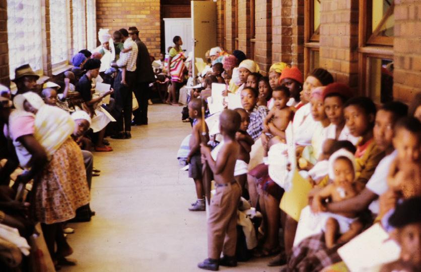
In the picture, we see the many children with their mothers waiting calmly to be seen. Doctors saw at least 40 patients in each session. There were approximately 250 paediatric beds covered by only two physicians in-house at night. Common conditions included kwashiorkor, (protein-calorie malnutrition), marasmus, rickets, scurvy, tuberculosis, tetanus, measles
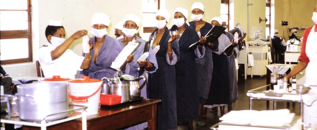
(pre-vaccine) and the full scope of paediatric disorders. There was a superb faculty of teaching staff and excellent nurses.
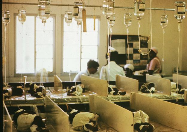
One of the key features of the Paediatric department was the premature baby units. There were two rooms with about 75 babies covered with cotton wool in wooden cribs. As they could not heat each crib, they heated the whole room with two stoves in the middle of the ward. The average temperature in each unit was 84ºF. The premature babies were cared for by their mothers. The mothers lived in an adjacent ward. They provided all of the routine care for their babies and contributed breast milk, which was pooled and pasteurised (see the picture). This system was likely
responsible for the very low nosocomial infection rates in those units. The survival rate was equal to the best units in the USA.
Another key feature of the Paediatric department was the drip rooms, where dehydrated babies lay side by side on long tables about ten in a row, and during the summer months, we had as many as 50 in each unit. The two nurses in each unit were technically superb and were skilled at starting the scalp vein intravenous drips.
It was depressing to see the amount of gross malnutrition in a country producing enough food to feed all its people.
A unique set of patients on the medical service were those who were said to have “mental confusion”. Perfectly fit young male adults would present with this disorder. We suspected they had been to the traditional healer because they often had scratch marks on their skin. We could never detect toxins, yet some of them died, with nothing abnormal revealed at autopsy. After they were stabilised, they often walked around the hospital corridors and were labelled with a piece of tape on the back of their hospital shirt, asking that they be returned to their respective wards. Some of these “confused” patients were quite violent and received an intravenous “lytic cocktail” of Demerol (pethidine), valium and another sedative. According to a possibly apocryphal story, one of these patients was being restrained, and the resident grabbed the nearest arm and injected the cocktail, whereupon one of the attendants collapsed.
An amusing response to the interpreter asking a potentially “confused” patient whether he heard voices was “Yes.” When asked, “What do they say?” he said, “Calling Dr Seftel. Calling Dr Harry Seftel. Calling Dr Harry Seftel”.
There was once a scam by the orderlies on security duty. They decided to charge the visitors a tickey (the affectionate name for the old three pence) for “admission”. This was discovered when a woman complained to the superintendent that she had already paid and had gone out through the gate, and they wanted to charge her again when she returned!.
We received a remarkable education at Bara, but it was not for the faint-hearted. The teachers were tough and not afraid to embarrass you, but they were remarkably well-informed about a wide range of subspecialty areas. With the current practice of subspecialisation, generalists of this quality simply no longer exist.
Some of the most dreaded calls you would get from the ward nurse were: “Doctor, your patient is gasping.” This was the euphemistic way of informing you of the death of a patient. Sometimes you would arrive, and they would be bathing the corpse because it had been so long since they discovered him, and rigour mortis had already set in. We look back fondly on our experience at Bara, although at the time, we often felt frustrated and overwhelmed by the sheer volume of patients.
Theo Kretzmar from our class, who became a surgeon in Springs and then Johannesburg, told us some Baragwanath Hospital-related stories in the pathway to his career. He said: “One day, walking up one of the long outdoor corridors at Baragwanath Hospital to tea, my co-registrar Abe Mizrachi stopped to chat with a young nurse, who happened to have a sinus under her jaw. Abe told me this was a median dental sinus from caries of a lower incisor tooth, and the treatment is to removing the tooth. It just so happened that my first short case in the Edinburgh Fellowship exam was just such a problem, which I correctly diagnosed. (They had him as tuberculosis). The examiner said yes, that is what it is, and promptly removed the case from the exam,
pending an X-ray which concurred. I would never have known this but for the chance passage encounter at Baragwanath Hospital.
One of the professors lecturing on the Surgical Fellowship course in Edinburgh, on thoracic surgery, was discussing treatment of the daily intake. He quizzed us about the treatment of a seaman who had been brought in stabbed in the heart. I put up a hand - “Sir, was the stab into the right or left ventricle?”
The answer was, “Who are you? And where do [you] come from?” “Baragwanath Hospital, Sir, and we operate on these frequently, if right-sided stabs.” (Stabs into the left ventricle do not get to hospital.)
I wondered what would happen if I met him in the exam. I did, and he was charming. We had a wonderful general discussion around stab heart surgery at Baragwanath Hospital in the 1960s. I passed the exam and recall that coming in from the snow to the college library for the sherry party, to welcome those who passed, was the best cherry on the top one could have.
We then moved to London, where I worked for two years before returning to South Africa. My wife cajoled me into writing the London FRCS during our time in London, which I did and passed. We lived quite close to my sister Geraldine and her husband from our class, Ronnie Auerbach, who had settled in London.
At my interview at Farnborough Hospital in South London, they warned me that intake nights could be very busy. When I enquired how busy, they said there may be at least 4 or 5 cases coming in overnight! One trauma emergency still remains in my memory. Three young men wrapped their car around a lamppost. One was a head injury and I sent him to a neurosurgical unit. The other two were a ruptured spleen and a ruptured liver and spleen, on both of whom I successfully operated. The next day, when the elderly consultant heard about this, he was concerned and asked me why I had not called him. I gently asked him when he last handled such cases. His answer was that he had never done so, and I could see that he was relieved that he had not been called.
In 1997, soon after the fall of apartheid, a new name was added to the hospital. After the tragic murder of the prominent activist, Chris Hani, his name was coupled with that of Baragwanath, to give the hospital the name “Chris Hani Baragwanath Hospital”.
Hani was a remarkable man. He was born on 28 June 1942 at Cofimvaba in the Transkei and matriculated at Lovedale College. He obtained his BA degree (Latin and English) from Fort Hare and Rhodes universities in 1961. Shortly thereafter, he joined the military wing of the African National Congress (ANC) or Umkhonto we Sizwe (MK). During 1962, he was mostly active in the Eastern and Western Cape but was soon involved in military operations in Rhodesia.
Although he spent time in Botswana and Zambia, he infiltrated South Africa again in 1973 to settle in Lesotho, where he stayed active until 1982. Repeated assassination attempts, however, forced Hani, now Deputy Commander and Commissar of MK, to leave Maseru for Lusaka.
From 1983 to 1987, he was Political Commissar and member of the National Executive Committee of the ANC (a post he had held since 1974). In 1987, he was promoted to Chief of Staff of MK, a post he held until his untimely death.
On his return to this country, he was actively involved in the negotiations towards an interim Constitution and preparations for the first democratic elections. His death on the 10th April 1993 left the nation with a great loss. Coupling his name with that of the hospital cemented the best of the past with the best of the present. A healing act and a firm step towards reconciliation.
Compiled by Geraldine Auerbach with story and paintings by Shirley
Talerman
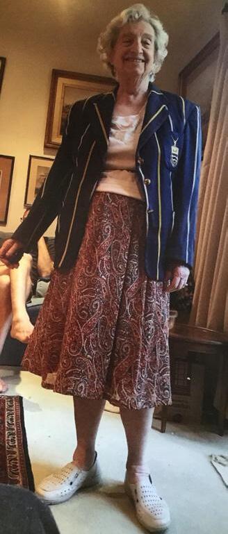
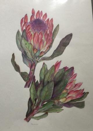
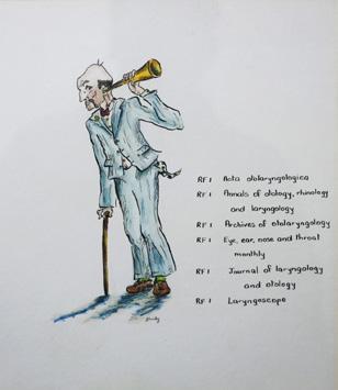
Shirley Talerman (Librarian at Wits Medical School Library, from 1956 to 1961) wrote in 2020: “Thank you so much for sending me the Class of ‘60 Newsletters with all the interesting stories of the medical students and teachers whom I knew in the medical library. They all did so well. Credit to the fantastic doctors at Wits Medical School ".
I have a copy of the drawings I created for the stacks at the Medical Library to indicate where the various journals were to be found. I believe the original ones have been preserved and are now hanging in a corridor in Parktown, I would love to send my greetings to all those who were there when I worked in the library from 1956-1961. Now I just paint flowers!”.
It seemed to us only fitting to connect Shirley and her delightful and amusing watercolour paintings that illuminated the journal stacks downstairs, to the students that she knew passing through the library, searching for articles that she helped to pin-point, by devoting a special chapter in this book to her watercolours in the library.
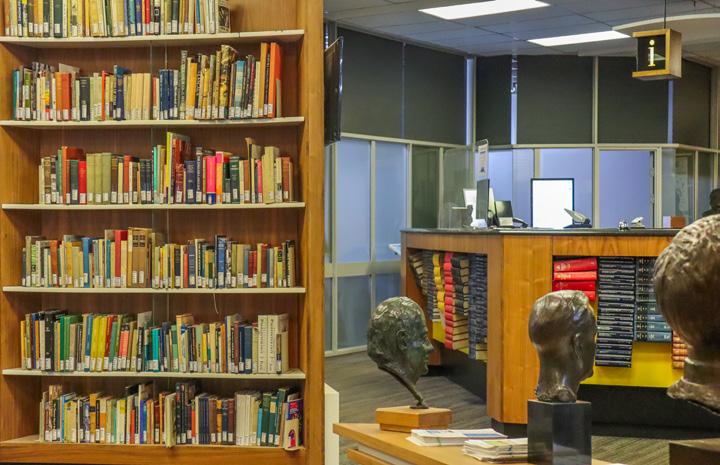
In March 1923, the Johannesburg Hospital Board, the Medical Journal of South Africa, and the University of the Witwatersrand (Wits) Medical School resolved to establish a central medical library. It was to be for the use of the medical profession based on the Witwatersrand; the staff of the Hospital; and the medical students at the university.
The library of the Johannesburg Hospital was duly transferred to the Medical School, which had been established in Esselen Street in 1917. It was to be amalgamated with the partial collection bequeathed by Sir Grafton Elliot Smith to one of his former students, our professor of anatomy, Raymond Dart.
Elliot Smith was the renowned Australian anatomist, Egyptologist and anthropologist who revolutionised anatomy teaching in the UK as Professor of Anatomy at University College London, 1919 - 1937. By 1938, over twenty of Elliot Smith’s former staff were in chairs of anatomy around the world.
It was he who had sent Raymond Dart, a fellow Australian, somewhat reluctantly, to take up the position of Professor of Anatomy at Wits – a position he held from 1923 to 1958. (Dart was also to serve as Dean of the Faculty of Medicine at the University for nearly 20 years, from 1925 to 1943).
This was fortuitous for Dart, for in 1924, at a time when Asia or Europe were believed to have been the cradle of mankind, Dart’s recognition of the humanlike features of the skull of the Taung child, changed all that. This skull had been recovered in South Africa near the Great Kalahari Desert. It substantiated Charles Darwin’s prediction that such ancestral hominin forms would be found in Africa. From this discovery, Dart declared the skull to be a specimen of a new genus and species, Australopithecus Africanus or ‘southern ape of Africa’, which lived approximately two to three million years ago, and probably strode upright. Dart believed that it might characterize the so-called ‘missing link’ between ‘humans and non-human animals’.
The Witwatersrand Medical Library (WML), with a collection of 600 books, was formally opened on the 12th July 1926 by Dart, who acted as the first librarian until 1928. By 1958, WML had completely outgrown the physical space to which it was assigned. It finally moved to new premises in 1964, when two symbolic books were carried by Dart and Dr I Goldblatt into the new premises. This was the same task that they had carried out some 40 years earlier, when Goldblatt had been one of the two medical student-librarians under the supervision of Dart, who was then the honorary librarian.
In 1982, WML, along with the faculty it served, moved to new premises in Parktown, adjacent to the new Johannesburg Hospital (now called the Charlotte Maxeke Johannesburg Academic Hospital). It is on one of Johannesburg’s many ‘white water ridges’ which gave rise to the name ‘Witwatersrand’. The name of ‘Witwatersrand Medical Library’ (WML) was changed in 1995 to Witwatersrand Health Sciences Library (WHSL) when the University’s Dental Library was also incorporated, reflecting the formal amalgamation of the Faculties of Dentistry and Medicine.
The advent of digitisation and electronic resources has enabled WHSL to downsize its initial main library, plus four physical branch libraries, at various academic hospitals, to the main physical library and print-based collections at Parktown, with only one small physical branch remaining at Chris Hani Baragwanath Hospital. Although WHSL closely resembles a traditional print-based library, it began offering its decentralised clients access to e-journals as early as 2000.
This move was made in readiness for the adoption in 2003 of a new electronic medical curriculum for the hybrid Graduate Entry Medical Programme (GEMP). Based on the enthusiastic adoption of e-journals by clients, a move to e-books followed shortly afterwards as more suitable material became available. WHSL thus still resembles an old-fashioned and traditional print-based warehouse collection, but now offers attractive physical study space with both wired and wi-fi access to current resources (including most of its former short loan collection). In 2014, another complete revamp of the library took place.
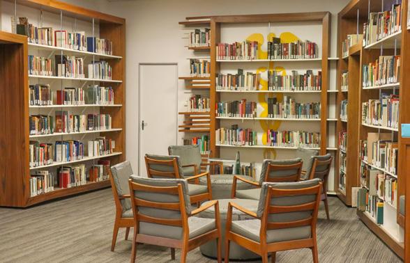
Shirley Talerman wrote about her experiences: Early mornings were a rush to climb up Hospital Hill to get to the Wits Medical School Library by 08h30. It was situated between the South African Institute for Medical Research and the Non-European Hospital, and opposite the General Hospital. Before I even got to the door of the building, I was besieged by students calling, “Shirley, please take my overnight books!”. These had to be returned by 09h00 so as to avoid a fine, and most were very heavy – Davidson’s Medicine, and Guyton’s Medical Physiology, in particular.
The Library was situated on the ground floor, on the right-hand corner, of the Medical School, next to the Librarians’ Office. By the time I arrived, weighed down by the offloaded books, Alpheus and Daniel, our Black members of staff would already be there, putting books back on the shelves.
As soon as we arrived, Lucy (Mary Lucas, Head Librarian) Daphne Reid, Juliette Back and I (Shirley Silkiner) would grab a white coat off the back of the door, and we would each be assigned to the tasks for the day: cataloguing, research, or desk duty, which was usually hectic. In fact, everyone at the Medical School had to wear a white coat. The library would soon fill with students, and we were immediately busy issuing books, answering queries, or doing research for articles in the Index Medicus (a precursor of computerised reference material).
Professor Jock Gear, Dr Daubenton, Phillip Tobias, Professor Dart, and many others would drop in every day for a cup of tea and a chat. Professor Guy Elliot, in fact, was renowned for the great parties he held in the library, when my sister, Hilda, worked there a few years earlier. There were still corks stuck on the ceiling when I was there.
Phillip Tobias attended the same synagogue as I did, the Wolmarans Street Synagogue, and sat with his father every Friday night. He once dashed into the library, passed behind me at the desk, and ran his finger down my back. I screamed. Every student sitting there looked up in shock. Phillip was most embarrassed. Once, while I was on desk duty, some years before he was world famous, Christiaan Barnard appeared.
From time to time, Lee McGregor would appear, always sporting a bow tie and leather gloves. He was a formidable character, the senior surgeon at the Johannesburg General Hospital and Lecturer in Surgery at Wits. He had written a seminal textbook: A Synopsis of Surgical Anatomy. Placing his gloves on the desk, he’d ask for a particular journal from the stacks down the stairs. We would duly rush down and bring it up to him. He would stand by the desk, glance at it, and then ask us to go down and get another journal. This could go on for an hour! One day I had the temerity to ask if he would like to go down to the stacks himself. To my surprise, he said he
would love to! Problem solved.
On my first day at Medical School, noticing that I was a new young librarian, Dov Sevel insisted I take him to the sex books section. He claimed not to be able to see where I was pointing. He insisted that I stay beside him until he found what he was looking for. He later became Professor of Ophthalmology!
Many students were about the same age as I was, and I was often invited to parties. I’d heard they were very wild, so I was too timorous to go.
There were so many students, and it was so long ago, but I remember quite a few. Amongst others, I often saw Louis Berman, Eugene Toker and his brother Cyril, as well as Selma Brodie, and Manley Smukler, who kept coming behind the desk to get me to dance with him. Harold Talerman, notably, was banned from the library in his first year for not having returned Gray’s Anatomy. (Reader, I married him. And the book is still on our shelf).
I remember Jeff Maisels and his brother Keith and his cousin Lyddon Simon, they also went to the Wolmarans Street Synagogue, and in fact, my mother and I sat next to their mothers! Other students whom I remember were Cyril Donninger, Adele Blankfield, Elliot Engelberg, Mike Plit, Gary Katz, Irvine Lissoos, Janse van Rensberg, Motala, Ian Thomson, Ian Chappell, Denise Hack, Arthur Rubenstein, and Ronnie Auerbach - who insisted on giving me a lift home on the back of his tandem after a late duty, even though I could not ride a bike.
It was often a relief just to sit and research information in the Index Medicus for medico-legal cases. Sometimes, a surgeon would rush in five minutes before closing, hoping to find some urgent information for an imminent operation. And so, at 17h00 or at 18h00 twice a week, the day would end – fun, rewarding, and tiring, but never boring.
Letter to Shirley Talerman from the Library in 2013
In December 2013, Shirley had received an email from Glenda Myers, head librarian at the Wits Health Sciences Library, the subject: Shirley the Artist. It said:
Dear Mrs Talerman (or can I call you Shirley?
I feel I know you so well, because when I was appointed as Librarian of the then Wits Medical Library in 1991, I found, in a storeroom, a box of beautifully hand-illustrated end plates (lists of journals designed to be hung at the end of the shelves as a finding tool). The framing glass of some of these delightful watercolours was damaged (apparently when the library moved from the Esselen Street Medical School to the Johannesburg Hospital premises in 1982) and the frames were old fashioned and in a dreadful condition.
The Esselen Street Library is the one you refer to in your letter as the ‘new premises’, but we now refer to it as the ‘Old-Old Medical Library’ (the original premises in Hospital Street); the ‘Old Medical Library’ is what you refer to as the ‘new’ premises in Esselen Street; and now we occupy the ‘New Medical Library’, adjacent to the Charlotte Maxeke Johannesburg Academic Hospital on top of the ridge in Parktown.
There is never any money available, but I managed to scrape together funds and had the series of watercolours reframed and the glass repaired. There was no way in which I could allow such delightful illustrations to disappear, but I have always wondered who “Shirley” was. No one at Medical School seemed to know, so you have now supplied the missing link!
We were not able to rehang the water colours on the ends of the current shelves in the current library, as we no longer subscribed to many of the original journals, and also had by this time acquired many more journals which did not feature on the original lists. The library’s users would have found these lists somewhat confusing, as they no longer described the contents of the current journal shelves. However, we have rehung them as a group in the corridor leading to the staff offices in the present library (now renamed the Witwatersrand Health Sciences Library, or WHSL for short, to incorporate the Dental Faculty Library when the two faculties amalgamated).
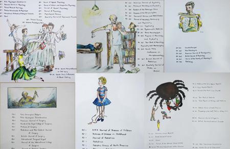
I smile every time I pass by them on my way to and from my office, because whoever painted them (and now I know this to be you) had such a wonderful sense of humour. I just love the use of poppies with a sleeping dreamer in the field to illustrate the subject of anaesthesia. And the chubby woman standing on a scale to illustrate nutrition. All of the pictures are wonderfully and gently humorous, but I think that my personal favourite is the very scary hypnotherapist who was used to illustrate psychiatry
I am so very glad that you wrote to our Alumni Office, so that I could finally learn the identity of the artist involved! I think that this needs to be documented and placed as a note beside the water colours so that future generations can know a little bit about WHSL’s history and the artist of these delightful water colours.
You would probably be delighted to know that we used an old photograph in which the original water colours can be seen in their original location (see the second page of this chapter) at a conference in February this year. We presented a paper on the topic of the embedded librarian (a concept that incorporates the changes in both the physical library as well as the way in which we interact as librarians with our library users).
At the bottom of this abstract is a PDF copy of the Powerpoint presentation in which we made mention of your end plates, and there are some photographs of them in the original library. You can also see photographs of the Esselen Street library and the current library at Medical School in this presentation.
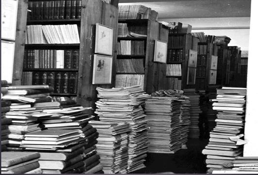
We’ve come a very long way from the cramped and overcrowded premises that you would remember (and that can be seen in the photograph) but once again, we are in the process of revamping the current premises to incorporate more ‘teaching and learning’ space. As a librarian, we now do far more teaching than we do traditional book curation, and in fact, my post is one of the first at Wits to reflect this as I have a 50% academic position (because of the amount of formal teaching I do) in addition to my 50% support service position as head of WHSL.
We are currently planning a spanking new-look library for 2014 (to use the architects’ words it’s being ‘blinged-up’) to take our students and library users into the 21st century-world of electronic information, and we’re hoping to incorporate some of the historical pictures we have of the old library premises into a permanent display at the reception/information desk in the new foyer area of WHSL. Please keep in touch with us, and I’ll be able to send you photographs of the new areas when completed next year.
With very warm wishes, Glenda Myers D Litt et Phil, MBibl, MEd Witwatersrand Health Sciences Librarian.
Of the 84 Wits University Medical Graduates of 1960, the majority, 59, practised medicine in South Africa. They became skilled physicians, surgeons, general practitioners and pathologists, in private and public medicine in the metropolises of Johannesburg and Cape Town, as well as in smaller country districts. They were innovative and entrepreneurial in developing new specialities and facilities and conducting ground-breaking research. Many became revered teachers at Wits, University of Cape Town and KwaZulu-Natal medical schools. They all served their country and community with skill and dedication.
In the 1950s, the Nationalist Party’s Apartheid policy of removing ‘non-European’ students from Wits was abhorrent to most of our graduates. Many of the class of 1960 students and their teachers served on bodies seeking academic freedom for all South Africans. Some classmates, like Essop Jassat and Costa Gazidis, actively joined the fight against these policies – with serious consequences for their own careers. Others left as soon as they could to avoid it. Most put their heads down to do the best they could for their families and for the people they served.
Many of those who did eventually decide to leave South Africa had first provided up to thirty years of valiant and valuable service to South African medicine before moving elsewhere, perhaps to be closer to their children.
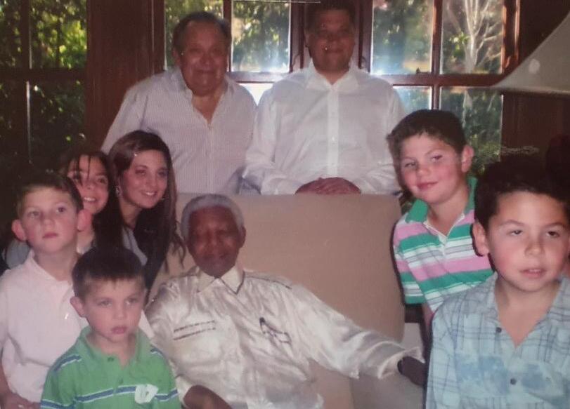
Naomi Livni did tremendous work at South African Institute for Maritime Research (SAIMR). Her husband, also our classmate Leon (Leopold) Kahn, surgeon and urologist who was part of the pioneering team working on renal dialysis and kidney transplants, practised in South Africa for nearly thirty years before settling in the UK in 1989.
After the end of Apartheid, some of our classmates were held in high esteem by the ruling African National Congress. Jack Kussel was invited to be the paediatrician for Nelson Mandela’s grandchildren, and Michael Plit was Mandela’s favourite ‘go-to’ physician for all his ailments for over 20 years. Essop Jassat and Costa Gazidis were awarded for standing up for their convictions.
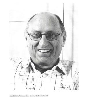
Many members of the class of 60 gained specialist training and higher degrees both in South Africa and by visiting the UK and the USA. They took a leading role in developing specialities and facilities in South Africa and created some completely new services. Anthony Meyers was the first professor of Nephrology in South Africa. He worked with urologists and physicians from our class, Leon Kahn, Irving Lissoos, Andre van As and others, in developing dialysis units and kidney transplants in Johannesburg.
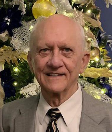
Andre van As worked as the Principal Physician and Clinical Head Internal Medicine Department at the JG Strijdom Hospital - now called the Helen Joseph Hospital (JGS/HJH). He specialised in respiratory medicine and developed the intensive care units at the Johannesburg General and the JG Strijdom Hospitals with Professor Guy Elliott. In the 1970s, Andre was thus instrumental in developing the speciality of pulmonology as well as being a pioneer in renal dialysis and nephrology in South Africa.
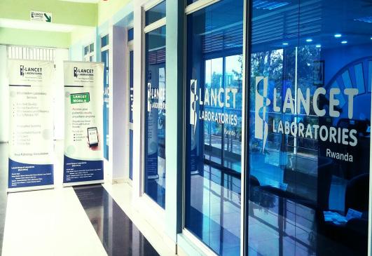
Classmates, Alf Mauff and Stan Zail, with graduates in other years, Zent and Skudowitz, set up Lancet Laboratories providing advanced pathology services.
Gloria Davis added her expertise to the new speciality of allergy in South Africa, both as a practitioner and a teacher.
Mervyn Hurwitz practised Obstetrics and Gynaecology in Johannesburg. He also lectured in the subject at Wits, as well as starting the first sex therapy clinic in South Africa.
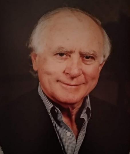
Many of the class of 1960 were involved in ground-breaking research both at Wits and at SAIMR. Alf Mauff says, “Our classmate and my partner Stan Zail (pictured) was one of the most brilliant graduates (if not the best!) of our class. His achievement, to be part of the development of Lancet Laboratories and still to find the time and energy to do basic research, is unique and should be recognised.” His work, particularly in iron and red cell metabolism, has been, and still is, internationally recognised and acclaimed. Alf Mauff, himself also a brilliant student, spent time at SAIMR doing research in Microbiology. Naomi Livni worked at SAIMR in the fields of cytology and histopathology. Anthon Heyns carried out important studies there on blood platelets and blood safety.
Many of our cohort became teachers and served on the faculty of Wits Medical School in several specialities and at other South African medical establishments.
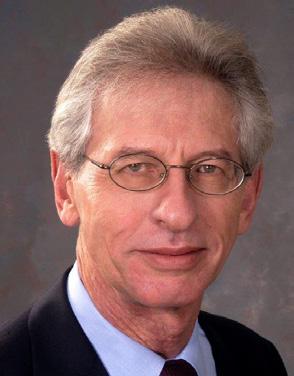
Clive Rosendorff was Head of Physiology and then Dean of the Wits Medical School. Anthony Meyers became the first Professor of Nephrology at Wits University, having helped develop renal services in South Africa. Wolfe Rakusin was the Medical Director of Undergraduate Teaching in Ophthalmology at Wits Medical School and chief examiner in ocular pathology from 1975 until 1991.
Allan Gottlieb was a teacher and demonstrator in the anatomy department. Mervyn Hurwitz taught in Obst and Gynae, Gloria Davis in Allergy, Naomi Livni, Alf Mauff, and Stan Zail in Pathology, all taught medical students at Wits and Baragwanath Hospital from time to time. Max Shaff taught students in Radiology at Groote Schuur.
Leonard Kahn and Aaron (Arthur) Vinik specialised and also taught at the Medical School of the University of Cape Town before moving to America. Leonard Kahn returned to UCT after studying in the USA, where he served as the youngest appointed Associate Professor at UCT from 1970 to 1977.
From 1962 to 1963, Arthur Rubenstein was a Senior Research Bursar at the South African Council for Scientific and Industrial Research under the direction of Professor H. Steyn. Between 1963 and 1967, he served as Tutorial Registrar in Medicine to the Professorial Unit at Johannesburg General Hospital and as a part-time Lecturer in Physiology at the College of Physicians and Surgeons of South Africa. During this time, he carried out research on diabetic patients and their blood sugar control, as well as abnormalities in their lipid metabolism in Johannesburg before continuing his endocrine and diabetes research in the USA in 1967.
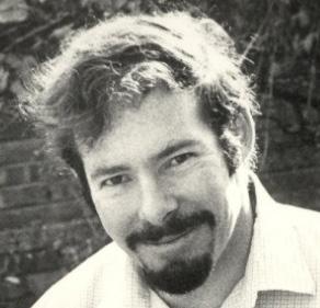
At the universities of Cape Town and KwaZulu-Natal, Mike Berger was able to collaborate in basic laboratory and therapeutic studies with a talented and ambitious cohort of clinicians located at both the main teaching hospitals Groote Schuur, the Red Cross and King Edward VII Hospitals. Though research was Mike’s primary interest, he was also heavily involved in teaching, administration, service development and delivery. At UCT, Mike was also appointed head of the Adult Lipid Clinic at Groote Schuur, and was thus able to integrate the study and management of both childhood and adult dyslipidaemias.
Mike says, “From the research point of view, South Africa was a treasure trove of clinical material. Familial hypercholesterolaemia was rife among the Afrikaans-speaking population due to the founder effect of expansion from a small population base. The South African Indian (South Asian) population, with a strong tradition of first-cousin marriages, was another reservoir of unusual recessive disease. Organophosphate pesticide poisoning: I particularly treasure the use of the serum cholinesterase assay to demonstrate widespread chronic organophosphate pesticide poisoning in rural farm workers of the Western Cape, which went entirely unrecognised by the sufferers themselves because of their generally poor health status. Our laboratory was selected as one of the national centres for the ‘statin’ studies of the 1980, which brought in much-needed funding for our other research activities.”
In 1989, Mike was appointed Professor of Chemical Pathology at the University of KwaZulu-Natal Medical School, Durban. Here, with the stage larger and the country even more turbulent, Mike and his team continued to serve the University and the community of KwaZulu-Natal with an integrated teaching, service and research department. For seven years, he endeavoured to maintain the ideal of excellence. He contributed significantly to the standing of the department by continuing to publish good work and by maintaining high teaching and service standards before retiring to Cape Town in 1996.
Anthon Heyns was recruited early on to become Professor and to establish a Department of Haematology at the new medical school of the University of the Free State in Bloemfontein.
He established the Bloemfontein academic hospitals as reference centres for the diagnosis and treatment of patients with haematological malignancies, coagulation and bleeding disorders and created an academic
research environment. He was also responsible for the Department of Nuclear Medicine and thus interacted closely with members of the Department of Biophysics. The team developed a novel technique to label blood platelets with In111-oxine. This radionuclide made it possible to image and quantitatively measure and model the kinetics, in vivo distribution, and sites of destruction of the radiolabelled platelets.
The transfusion of blood and blood components was essential for the treatment of haematological disorders. To successfully establish such a facility in Bloemfontein, Anton Heyns worked closely with Dr Maurice Shapiro and Dr Robert Crookes of the SABTS. In 1990, when Dr Shapiro retired, he persuaded Anton to accept the position of CEO/Medical Director of SABTS, which later became the South African National Blood Service (SANBS)
Almost all South African specialists of the class of ‘60 have at some time been office bearers and leaders in the South African professional bodies for their specialities. They have developed and maintained international connections.
Professor Michael Berger was appointed National Representative to the International Federation of Clinical Chemistry from 1992-1993. Later, he was elected President of the Lipid and Atherosclerosis Society of Southern Africa and the South African Association of Clinical Biochemistry.
Professor Anthony Meyers served as President of the South African Renal Society and the South African Transplant Society. He had been the official South African representative on the African Association of Nephrology (AFRAN) and had served on the executive committee and as chairman. He was also the African representative on the International Society of Nephrology (ISN) executive committee. He has served as chair and vice-chair of the Johannesburg Hospital and Transvaal Provincial Pharmacy and Therapeutic Committee and on the Colleges of Medicine Examination Executive Committee (Johannesburg Branch).
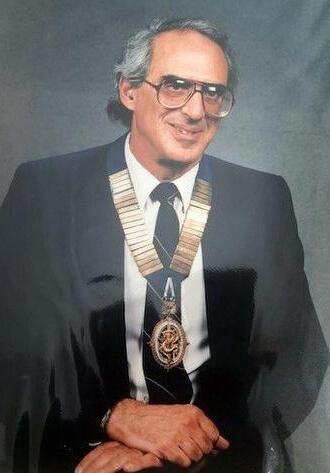
Professor Anthon Heyns was a member and past president of the South African Society for Haematology, South African Society for Pathologists, and a life member and past president of the African Society for Blood Transfusion. He was also a Councillor of the SA Medical Research Council, a member and chairman of the board of directors of the National Bioproducts Institute, and chairman of the SA Bone Marrow Registry. He served on the SA National AIDS Council and was a member and chairman of the SA Advisory Committee on HIV. He served as a member of several expert committees and workgroups of the World Health Organisation. He is a member of the USA Training Advisory Committee of the National Institute of Health Fogarty Institute, focusing on research training in HIV-related Transfusion Medicine and Haematology.
Ivan McCusker was a born leader. He chaired the Association of Surgeons of South Africa from 1988 to 1995, was President from 1995 to 1996, and was awarded Honorary Life Vice-Presidency. He chaired many other committees. Theo Kretzmar (pictured wearing the chain of office) was President of the East Rand branch of the Federal Council of Surgeons of South Africa.
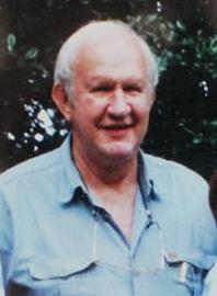
Pieter Landsberg was a star student, gaining first-class passes in all four subjects in his first year. After graduation, he set up in general practice in Benoni. He developed a passion for emergency medicine. He lectured weekly to the Red Cross and worked with the head of the Fire Service to improve fire safety in Benoni.
His interest in diving and power boating led him, in 1992, together with a few other doctors, to create a formal diving medical association – The Southern African Underwater and Hyperbaric Medical Association (SAUHMA).
Pieter was its first president. He was also key in bringing the NSRI (National Sea Rescue Institute) inland, to monitor water safety in the dams around Benoni and the Vaal. As a Lieutenant Commander in the South African Navy, Pieter was a lecturer on the only diving and submarine medicine course held annually at the naval base in Simonstown.
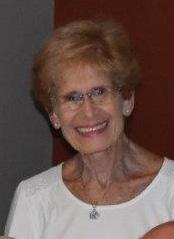
Gloria David embarked on the study of Allergy in 1963 when her second son was clearly allergic to eggs and nuts, and she had to find a remedy.
In 1982, she and her family moved back from Port Elizabeth to Johannesburg, where she took a post at the JG Strydom Hospital in the Respiratory Unit run by Mike Plit. Gloria said, “I became involved in doing drug trials and treating asthma patients as well as learning about respiratory illnesses and general medicine. There were now wonderful new products available for treating asthma. I started to attend conferences on respiratory problems, of which asthma featured prominently. Then, to my delight, the Allergy Society of SA was established. This gave me the impetus to start doing work on allergy and the role it played in all the conditions that made up the syndrome of atopy, namely asthma, rhinitis, conjunctivitis, eczema and food allergy.”
Gloria became involved in a teaching programme for anaesthetic registrars on how to examine patients so that they could diagnose their illnesses and then assess risk for anaesthesia. She continued this teaching programme at Bara. She continued: “I was also involved with asthma patients at Bara, improving their diagnosis and management, and begging the authorities to improve the facilities for these patients and the doctors treating them. I joined the paediatric asthma unit, and my entire research now took on meaning as I studied allergy in asthmatic black children, as well as all the other conditions from which they suffered in the realm of allergy. I managed to organise and perform skin-prick testing on all the patients, as well as on cardiac patients as a control group.”
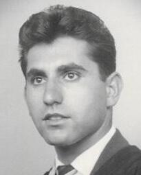
Ephraim commenced work in July 1968, initially in Jeppe Street and later at the Garden City Clinic, Johannesburg. He was, for many years, the visiting Consulting Internist to the Boksburg-Benoni Hospital, and for a few years was also consulting Internist to the Mines Benefit Society. He served as Medical Consultant to Roussel Laboratories and a variety of Life Insurance companies. He was appointed Chief Medical Officer to Munich Reinsurance Company of Africa in 1975, and currently still serves as a consultant to this company. For a few years, he was the Medical Advisor to an insurance settlement company based in New York.
From being a successful General Practitioner for 17 years, Roger was encouraged by his family to specialise. He chose anaesthetics. While in the Department, he was given the task of setting up the ongoing postgraduate anaesthetic teaching. This evolved into monthly meetings, during which he was able to move from the hospital to the College of Medicine in Parktown. He said: “We had a good format, an hour-long lecture followed by drinks and dinner. The meetings were well attended, and there was no problem getting drug companies to sponsor them. We hoped that the lectures would promote a better relationship between town and gown. I also organised the two-day annual anaesthetic update meetings in the Department. I was asked by doctors in some outlying areas if I could take some of the speakers we had to outlying areas. This would have been far too expensive, and alternative modes of communication were investigated. I suggested an ‘update’ course for GP anaesthetists in the Kruger Park. Prof Dave Morrell said that he didn’t think it would work, but in spite of this, I went ahead and we had a great meeting. This became an annual event for a few years after that.
After nine years in the anaesthetics department, Roger Edge spent a year at the Chamber of Mines Specialist Hospital on Eloff Street extension. He found that an illuminating experience. He was able to observe how miners became injured. As part of his responsibilities, he was taken underground to see what happened at the rockface. This involved travelling 2.5 kilometres underground: 1.5 kilometres in the first skip, then changing to another skip and going down the rest of the way. The skip carried a large number of people, all packed together with arms at their sides like sardines. In the event of a problem with the skip, it was difficult for anyone to panic and go wild, causing injuries to others. He saw how miners drilled holes in the rockface into which they placed explosives. The mine would then be cleared, and the explosives would be activated. After a reasonable amount of time, the miners would return to the rockface. Some of the charges would not have detonated. Some miners could not resist fiddling with these and, of course, were injured in the ensuing explosion.
In 2002, Roger Edge developed ‘Matters Medical’ on Television, He says: “On retirement, I joined my wife in our newly built home in Knysna. But after a year of making gardens, steps and cupboards, I organised a refresher course for General Practitioners in Brenton. It was well-attended, and they asked for more. I decided that it would be well worthwhile organising a monthly lecture on a TV channel for medical practitioners only. An accountant friend and I set about planning it. In 2002, Matters Medical Television made its debut on an encrypted channel. It was very exciting to see the idea come to life. We broadcast 22 one-hour lectures, which were well received. We saw this as a way to bring some of the best speakers in the country to all practitioners in Southern Africa, while sitting in their favourite armchairs at home and at no cost to them. At that time, medical practitioners had to pay high fees for CPD-accredited lectures.Television is an expensive medium and we needed more funds. We approached the universities, the Department of Health and the Health Ministry to no avail, so after three years, we sadly had to abandon the project. I enjoyed my life in medicine and would not have changed any of it. My life was fulfilling and gave me great satisfaction.
Tony Meyers reflects on some colleagues and teachers: “How wonderful it is to read all the news about our class’s contributions to medicine in South Africa. A few events remain clearly etched in my memory, such as the contributions of Dr Leon (Leopold) Kahn. During his training as a urologist, he spent eight months with me as a renal registrar. On qualification, he and his wife Naomi (Livni) (also from our class) settled in Vereeniging, where he became the local guru in nephrology – not only dialysis. We spent many hours on the old (and very efficient) telephone discussing renal biopsies. When Leon went to London to practice, his name as a ‘no mean general nephrologist’ was widespread. In fact, at that time, he probably knew more about renal histopathology than most nephrologists! Naomi, of course, is famous in her own right as a histopathologist. This makes me wonder how many other ‘husband and wife’ doctors there may be in our and/or other classes and disciplines from Wits. What an interesting list this would make.
Irving Lissoos, who sadly died of a malignant melanoma in 2011, practised as a Urologist in Johannesburg at Milpark Hospital. After qualifying at Wits Medical School, he first specialised in general surgery, gaining his Fellowship of the Royal College of Surgeons in Edinburgh. He then branched into urology, where he worked with me and was a pioneer in kidney transplantation, serving as secretary of the Urological Association of South Africa.
On a less happy note, we should spare a moment of our thoughts for our deceased colleagues. One terrible tragedy concerns the fate, soon after graduation, of our classmate Dr Kleintjie van Veen. He was an extremely hardworking and knowledgeable young man. Soon after graduating with overall high marks, he and his newly married wife set out for Durban on their honeymoon. They were both tragically killed when driving at night on the highway near Heidelberg. He could so easily have been one of our leading practitioners.
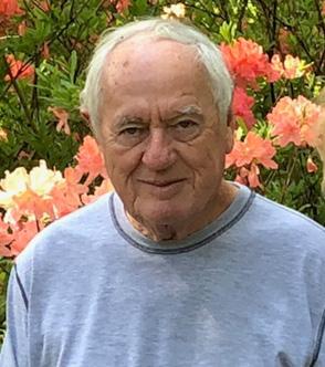
Alf Mauff, pathologist of Lancet Laboratories, describes the changes in medical practice in Johannesburg that occurred during his practice life.
“When I joined my small practice in Lancet Hall, the heart of medicine was concentrated in Jeppe Street. This was the location of the famous ‘doctor buildings’: Lancet Hall, Medical Centre and the new pride of Jeppe Street, Lister Buildings. This was where we all had our consulting rooms and offices.
“Some small hospitals were located in central Johannesburg, the Lady Dudley, Florence Nightingale, Brenthurst, with the Kensington Clinic the most peripheral. However, medical practice was dominated by the state academic Johannesburg General Hospital and for pathology, the SAIMR.
“Rapid changes, over the years, saw the growth of solid medical aid systems, the prosperity of suburbia and the influx of clinicians and new hospitals. Initially, they were centrally located, such as the Kenridge Hospital and Millpark, followed by suburban hospitals in Sandton, Morningside, Sunninghill, and Garden City. This witnessed the complete transfer of clinicians from ‘doctor buildings’ in the city centre to hospitals closer to where the patients lived.
“With the ever-increasing and sophisticated expansion of private practice, the class of 1960, although having lost many of our talented members to distant shores, as well as to our own academic institutions, nevertheless contributed significantly to the maintenance of outstanding and sophisticated private medical services to the Johannesburg community. The names that come to my mind from our class who did their bit were: Irving Lissoos, Mike Plit, Jack Kussel, Anthony Crosley, Raymond Palmer, Christopher Bosman, Raymond Cochrane, Ephraim Dove, Gerald Lampert and my medical partner, Stan Zail, among others.”
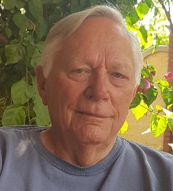
Anthon Heyns, who became Professor of Haematology at the new medical school of the University of the Orange Free State and later the CEO of the South African National Blood Transfusion service, sums up by saying:
“Medicine is a wide-ranging discipline and offers the opportunity after graduation to shape one’s career to fulfil a personal interest. Our class illustrates this: our passions range from highly specialised disciplines to general practice, and from doctors who are interested in treating and interacting with patients to those with a more scientific and laboratory leaning. In our class, some pursued an academic career and some preferred private practice; a few successfully combined the two.
‘It is notable that the common thread through most of these stories is that our teachers have instilled in us a value system and ethical code that is central to the practice of medicine. This exposure to the culture and standards of the Wits Medical School in our formative years has shaped our lives and attitude to our profession. We have served all the people of South Africa equitably to the best of our ability and in a special ‘Wits graduate’ way. One
can only wonder what could have been achieved if there had been a just and equitable government in the era that we qualified, and more members of our class could have contributed to the development of medicine in our country.”
We have counted 22 members of our class who held senior academic posts as professors, associate professors and senior lecturers, in medical schools in South Africa and across the world from Albuquerque to KwaZulu-Natal. They were involved in research, clinical practice, teaching, publications, faculty and international affairs as well as conferences. Wits, and other universities, conferred well-earned higher degrees, such as MDs and PhDs on many of them for outstanding research.
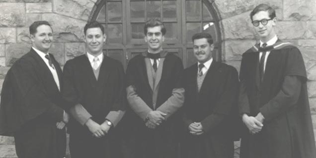
Several seminal textbooks have been authored and edited by members of our class. Yet, other classmates also lectured from time to time, imparting their knowledge to students, colleagues, general practitioners and nurses.
Two members of the class became professors in the early days of Nephrology. Two became professors of the burgeoning discipline of Human Genetics. Another two high-flyers became internationally renowned professors of Endocrinology. Two became Professors of Neonatal Paediatrics, and two taught Pulmonology. Our class produced three professors of Pathology and two professors of Psychiatry. One taught Family Medicine, while the other taught Ophthalmology. One was involved in Radiology, and another, in Anaesthetics. One was in Obstetrics and Gynaecology – opening the first sex therapy clinics in South Africa. There were others who were appointed lecturers in Allergy and Paediatrics.
Below follows a list, in alphabetical order and very short summaries of their careers.
• Michael Berger, Professor of Chemical Pathology first at the University of Cape Town and then later at the University of KwaZulu-Natal.
• Martin Bobrow, Commander of the Order of the British Empire (CBE) and Fellowship of the Royal Society (FRS), Professor of Genetic Medicine at the University of Amsterdam, as well as at the Guy's Hospital and the Cambridge University.
• Geoffrey Boner, Associate Professor of Nephrology at Tel Aviv University.
• Gloria Davis, taught Allergy and risk assessment to anaesthetic registrars in South Africa.
• Avroy Fanaroff, Chair of Neonatology, Department Chair and subsequently tenured Professor of Paediatrics and Reproductive Biology at the Case Western Reserve University in Cleveland.
• Laurence Geffen, Foundation Professor and Head of Human Physiology at Flinders University in Adelaide, Dean of Medicine and then Pro-Vice Chancellor. Dean of Medicine and Professor of Psychiatry at the University of Queensland in Brisbane.
• Anthon Heyns, inaugural Professor of Haematology at the University of the [Orange] Free State and Director of the South African National Blood Transfusion Service.
• Mervyn Hurwitz, served in Obstetrics and Gynaecology at the then Wits Medical School.
• Leonard Kahn, Professor and Chair of the Department of Pathology at the Albert Einstein College of Medicine in New York City.
• Gary Katz, Senior Lecturer in Paediatrics at the University College London.
• Jeffrey Maisels, Professor of Paediatrics at the Oakland University in Michigan.
• Anthony M Meyers, Professor of Nephrology at the Wits Medical School.
• Aubrey Milunsky, Professor of Human Genetics, Paediatrics, Obstetrics and Gynaecology, and Pathology at Boston University.
• Michael Meyer Nissenbaum, is listed as a professor, although we have not found any further information about him and his career.
• Michael Plit, teacher in Respiratory Medicine at the then Johannesburg General Hospital. Head of the Academic Teaching Unit at the then JG Strydom Hospital in 1980.
• Chaim Rosenberg, Associate Professor of Psychiatry, Boston University.
• Clive Rosendorff, Professor of Physiology at Wits and then Dean of the then Wits Medical School and later Dean of the Mount Sinai Medical School in New York City.
• Arthur Rubenstein, Professor of Medicine in Chicago, and then Dean of Medical schools in New York City and thereafter in Pennsylvania.
• Max Shaff, Associate Professor of Radiology at the Vanderbilt University Medical Centre in Nashville.
• Farrol Sims, Associate Professor in Geriatrics at the University of Wisconsin Medical School from 1989 until his retirement.
• Andre van As, Principal Physician, Chief of Internal Medicine and Pulmonary Division at then JG Strijdom Hospital. He was also a pioneer in the early days of renal dialysis and nephrology. He became Professor of Medicine and Chief of Pulmonary Division at the University of New Mexico in Albuquerque, USA.
• Aaron (Arthur) Vinik, Professor of Medicine at the Eastern Virginia Medical School in Norfolk, Virginia.
We first look at the exceptional careers of Clive Rosendorff, Arthur Rubenstein and Laurence Geffen, (pictured) the three members of our class who became Deans of prestigious medical schools in South Africa, the USA and Australia.
They each showed early drive and ability with high achievement at school, and excellent marks in their medical studies. Clive and Laurence spent time in scientific research to complete a BSc with anatomy lecturer Dr Philip Tobias (who was not yet a professor in 1956). Arthur was the top student in our medical school class. In addition to their heavy academic load, all three were involved in student political activities, including membership in the Student Medical Council and the fight for academic freedom for all in South Africa.
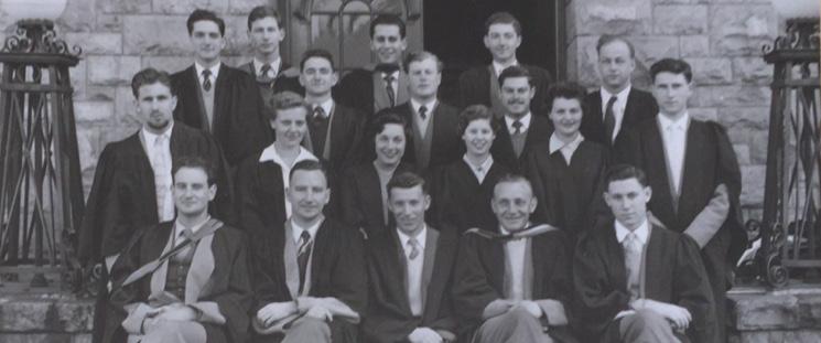
In this photograph of the Wits Student Medical Council of 1958, we see the three future deans together: Clive Rosendorff is standing in the back row, second from left. To his right in the back row is Laurence Geffen with his head in front of the window glass.
Arthur Rubenstein is sitting in the front row extreme right. They are assembled in front of the entrance to Wits Medical School on Hospital Street. Also, in the picture are some of our other illustrious colleagues: Jeffrey Maisels, the tall head at the back on the extreme left, Gerald Lampert (in the middle – head half in and half out of the door). Aubrey Milunsky is next to him to the right and Gary Katz is on the extreme right. Also in the picture, in front of Gerald to the left is Denise Hack, Arthur’s future wife. Gloria Davis’s future husband, Len Myers, is seated next to Arthur.
MBBCh, MD, PhD, DSc Med, FACC, FAHA, FACP, FRS SA, FRCP
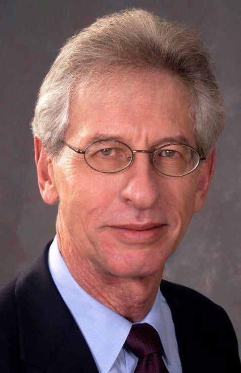
1970 Professor of Physiology at Wits
1987 to 1989 Dean of Wits Medical School
1992 Professor and Vice-Chairman of the Department of Medicine at Mount Sinai School of Medicine and Mount Sinai Heart (Hospital)
During his student days, Clive was prominent in university anti-apartheid activities as chair of the Academic Freedom Committee, a member of the national executive of the National Union of South African Students (1957), president of the Association of Medical Students of South Africa (1959), and president of the Student Representative Council (1960-61). He completed his BSc (Honours) in 1958 and graduated MBBCh in 1962. After an intern year at the Johannesburg General Hospital (the Gen), Clive went to London for further research at King’s College Hospital. He became a lecturer at St Thomas’s Hospital Medical School, where he completed training in internal medicine and cardiology, gaining his Membership of the Royal College of Physicians of London (MRCP) in 1966. He was awarded a PhD in 1969 for his work on cerebral and coronary blood flow. From 1969 to 1970 Clive was a research fellow at Yale University School of Medicine, where he was a visiting scientist and visiting Assistant Professor in the Department of Internal Medicine.
While he was at Yale, in 1970, Clive was head-hunted at the age of just 32, to return to South Africa to be Professor and Chairman of the Department of Physiology at Wits University. He was also appointed Senior Physician in the Department of Medicine. He served in those two capacities for 17 years. Then, from 1987 to 1989, he became the Dean of Wits Medical School – giving back to his alma mater the expertise he had gained. At the end of that period, after further research in Paris, in 1992, Clive moved to the United States to become Professor and Vice-Chairman
of the Department of Medicine at Mount Sinai School of Medicine and Mount Sinai Heart (Hospital) as well as the Chairman of Medicine at the Bronx Veterans Administration Hospital in New York City. In 2020, Clive was still working full-time. It may be of interest to know that Clive and the late Phillip Tobias were first cousins.
MBBCh (Wits) 1960, DSc (Wits) 2001, FCP (SA) 1964, FRCP (London) 1977, MACP (US) 1987
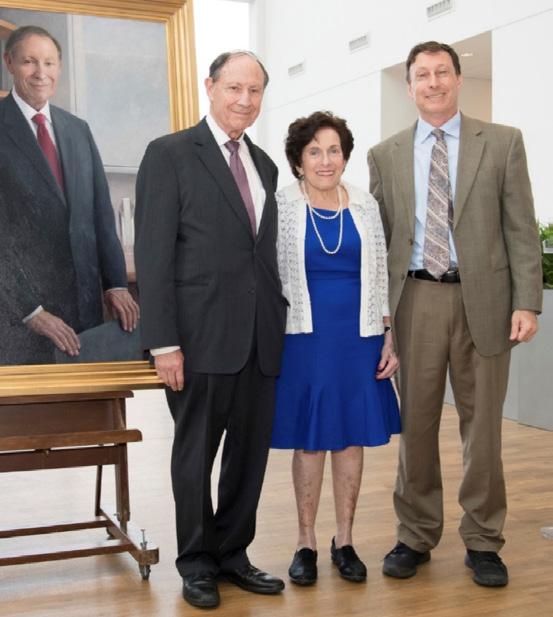
1981: Professor and Chairman of the Department of Medicine, University of Chicago
1997: Dean of Mount Sinai School of Medicine, New York City 2001: Dean of the Perelman School of Medicine and Executive Vice President for the Health System, University of Pennsylvania
Arthur Rubenstein was awarded the medal for the most distinguished graduate of the year 1960. He completed his house jobs and registrar training at the Johannesburg General Hospital, after which he spent a year at Hammersmith Hospital in London, conducting research in endocrinology, which led to a distinguished career in diabetic research. From London, he returned briefly to Johannesburg. In 1967, Arthur and his family left for the United States, where he began a research fellowship at the University of Chicago. Arthur rose rapidly up the academic ladder. In 1981, he was appointed Professor and Chairman of the Department of Medicine at the University of Chicago, where he made seminal contributions to research in the field of diabetes mellitus. He published numerous research papers and received many distinguished awards. Upon his departure, the University of Chicago established the Arthur H. Rubenstein MD Mentorship Award in his honour. In 1997, after 30 years in Chicago, Arthur moved to New York as the Gustav L. Levy Distinguished Professor of Medicine at Mount Sinai School of Medicine. He served as President of the Association of Professors of Medicine and President of the Association of American Physicians. After four years at Mount Sinai, in 2001 and aged 63 years, Arthur was recruited by the University of Pennsylvania as Executive Vice-President of its health system and Dean of the Perelman School of Medicine, America’s first ever medical school founded in 1765. Arthur was lauded as ‘an exceptional educator and accomplished physician, scientist and leader.’ During his decade-long tenure as Dean, he successfully built up the University of Pennsylvania into one of America’s leading medical teaching and clinical centres. On his retirement, the grateful university commissioned a portrait of him. (See the portrait above with Arthur, his wife Dr Denise (née Hack, MBBCh Wits 1959) and their son Jeffrey, a Professor of Talmud). The University also endowed the Arthur H. Rubenstein MBBCh Professorship. In August 2020, Arthur was still active in research and teaching at the Perelman School of Medicine.
BSc (Hons 1), MBBCh, MSc (all Wits), BA (Hons 1), DPhil (both Oxon), MD hon (Flin) FRACP (hon) FRANZCP
1967 Reader in Physiology and Pharmacology at Monash University, Melbourne. Dean of Medicine, Flinders University, Adelaide 1991-1994, Dean of Medicine at the University of Queensland, Brisbane Professor of Psychiatry, University of Queensland
Laurence, born in Johannesburg in 1937, was the first in his family to attend a university, graduating with the MBBCh in 1962. He joined the Nuffield Dominions Fellowship at Oxford University and later received Fulbright Fellowship to the Roche
Institute of Molecular Biology, in the USA. In 1967 he moved to Australia to serve as a Reader in Physiology and Pharmacology at Monash University in Melbourne. He then moved to Adelaide to become the Dean of Medicine at Flinders University, and from 1991-1994, to Brisbane, to be Dean of Medicine at the University of Queensland. Laurence Geffen played a key role in reforming Australia’s medical training and extended that role to the Western Pacific, including efforts to modernise the Chinese medical system.
In the picture, Laurence introduces his old Wits Professor Phillip Tobias as an honoured guest at the University of Queensland’s celebration of 100 years since the birth of Professor Raymond Dart – who was born and had studied in Brisbane).
In 2010, Laurence was included in the Queen’s Birthday Honours as a Member of the Order of Australia (AM): “For services to neuroscience, as a clinician and researcher, and a medical educator.” Leaving room for yet another career, Laurence Geffen served as Professor of Psychiatry at the University of Queensland and practised in Brisbane as a clinical psychiatrist.
Clive, Arthur and Laurence have crowded so much into their lives as researchers, administrators, clinical physicians, innovators, thinkers and family men. Each of them is still engaged. They remain into their ninth decade, reachable, likeable and flexible; easily recognisable from their joint 1958 photograph at Medical School in 1960.
MBBCh 1960 (Wits) MD 1970 (Wits) PhD, FCP, MACP, FACE
1979 Professor of Medicine and Surgery, University of Michigan 1990 Professor and creator of a state-of-the-art Diabetes Center and Research Program at Eastern Virginia Medical School.
Aaron Arthur Vinik received national and international recognition – as a scientist and clinician in three different areas: Clinical Diabetes and Endocrinology; Neuropathy and Neuroscience; and Neuroendocrine Tumours. He translated research findings into the clinic and laid the foundations for applied clinical research from the bench to the clinic. He received life-time awards in all three areas. Vinik wrote books and articles, pitched successfully for major grants, received numerous awards, and was invited to lecture around the world. He issued three patents. He was also an obsessive sportsman and loved waterskiing, kayaking and windsurfing as well as walking, cycling and dancing.
Aaron (Arthur) Vinik sadly passed away in 2023 after suffering a severe stroke a couple of years earlier. His legacy was affirmed and celebrated locally by Eastern Virginia Medical School (EVMS) by establishing a named Professorship in his honour. The annual Aaron Vinik Lectureship was also established in his name and designed to invite one of his national or international esteemed colleagues to present on one of their previously shared scientific interests. Worldwide, major USA and European societies are recognising his contributions to Diabetes, Endocrinology, Neuropathy and Neuroendocrine Tumours at their annual conferences. Some entire sessions
have been devoted to displaying his seminal essays and early scientific advances that resulted in modern life-saving therapies. These are enduring tributes to his life’s work as a sparkling, fun teacher, scientist and caring physician!
CBE, FRS, FMed Sci, DSc Med (Wits), MBBCh, FRCP, FRCPath
Professor of Genetics – Amsterdam, London and Cambridge
Martin took the intercalated BSc Hons and was awarded a DSc (Med) after working with the inspiring teachers and mentors Philip Tobias and Ben Kaminer. He became firm friends with fellow BSc students Laurie Geffen and Clive Rosendorff.
Martin says: “As medical genetics in the UK grew up around us, a handful of colleagues and I grew up within it. I effortlessly became part of a new speciality, acquiring membership in various professional colleges and organisations. I was appointed the Professor of Human Genetics at the University of Amsterdam. Then an opportunity came up (and in such a narrow field that does not happen often) in London, to be Professor of Paediatric Research (effectively a Department of Human Genetics) at Guys Hospital.”
Martin developed a deep interest in ethics as related to medical genetics. He asked: “How could anyone watch genetic testing and prenatal diagnosis emerging without being interested in the social and ethical issues arising?” He was appointed an advisor on human genetics to governmental and philanthropic bodies, including the Nuffield Council of Bioethics (the closest thing the UK had to a national bioethics committee) thinking through the future implications of what they were doing. In 1995, he was offered the newly established Chair of Medical Genetics at Cambridge University, where he remained from 1995 to 2005. He is still Professor Emeritus. Martin Bobrow’s work in the field was recognised when he was elected a Fellow of the Royal Society in 2004―a rare honour. Martin is also a founding Fellow of the Academy of Medical Sciences, a UK body that champions excellent research and provides expert information in many fields of science. He received the honour of CBE from the Queen in 1995 for his “services to science”.
BSc, MBBCh (Wits) 1960 PhD Minn 1969, FRCP FCP(SA)
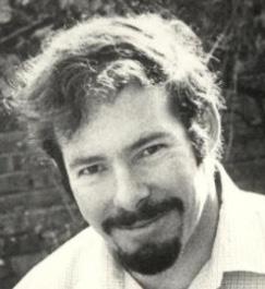
1980s Associate Professor, UCT, and 1989: Professor of Chemical Pathology, University of Natal
Another of the bright and energetic students who chose a scientific pathway, Mike Berger, also took the intercalated BSc with Clive Rosendorff, Lawrence Geffen and Martin Bobrow. After graduation and house jobs, Mike was accepted into the Mayo Clinic Clinical Pathology Residency program starting in Nuclear Medicine in mid-1964. While there, he was accepted into the Mayo Clinic-University of Minnesota PhD track. He and his wife Sheila left for the USA in 1965, where he completed an arduous but rewarding five-year program of advanced coursework and laboratory experience from 1965 to 1969.
Returning to South Africa in 1970, Mike Berger took a lectureship at the Red Cross War Memorial Children’s Hospital (RCH), as it was known at the time, which served as the paediatric teaching hospital. He spent 20 years there, developing the tiny service laboratory at the RCH into a centre for the study and treatment of paediatric genetic dyslipidaemias, primarily hyperchylomicronaemias. Mike was also appointed Head of the Adult Lipid Clinic and Associate Professor at Groote Schuur and was thus able to integrate the study and management of
both childhood and adult dyslipidaemias. In 1989, He was appointed Professor and Head of the Department of Chemical Pathology at the University of KwaZulu-Natal, where he worked for the next seven years, maintaining the ideal of excellence and contributing significantly to the standing of the Department by continuing to publish good work and by maintaining high teaching and service standards.
MBBCh (Wits), MMed Path (UCT), FRCP Path (London)
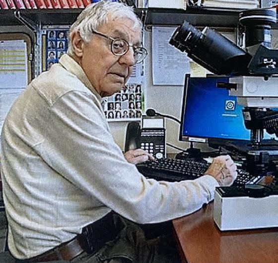
1980-2009 Chairman of Pathology Long Island Jewish Medical Center
Leonard Kahn received first-class passes in all his first- and second-year medical subjects. It was in his third year that Len really found his calling. He fell in love with microscopy and the study of microanatomy in human tissue, particularly in health and disease. In 1962, after completing his house jobs, he moved to specialise in pathology. After three-years in a registrar programme, he was awarded the Degree of Master of Medicine in Pathology at UCT. Len then went to St. Louis University School of Medicine where he was privileged to work with the world-renowned American physician and pathologist Lauren V. Ackerman, who championed the subspecialty of surgical pathology.
Following this two-year fellowship, Leonard returned to UCT where he served as the youngest appointed Associate Professor at the university from 1970 to 1977. He was then recruited as a Full Professor at the University of North Carolina, where he served from 1977 to 1980 and as Surgical Pathology. His next move was to the Long Island Jewish Medical Center, where he was appointed the Departmental Chair of Pathology and Professor of Pathology at the Stony Brook University. He also held professorships at the City College of New York, Albert Einstein College of Medicine and the Zucker School of Medicine. Len stepped down from the Chair in 2009 after a 28-year stint and is currently a full-time Senior Pathologist at the much-expanded multi-hospital Northwell Health System. Len was still at work in 2020.
MBBCh (Wits), DTM &H (Wits), MC Path (SA), MD, DSc, MD (hc)
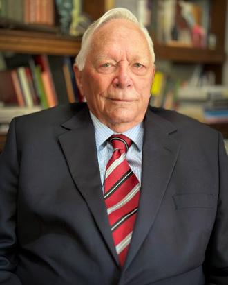
Professor of Haematology, University of the Free State; CEO and Medical Director of the South African National Blood Transfusion Service
Shortly after Anthon Heyns attained his Fellowship of the College of Pathologists of South Africa in Clinical Pathology (FC Path (SA) Clin), he was recruited to become the first Professor and Head of the Department of Haematology at the newly established medical school of the University or the [Orange] Free State. This entailed developing undergraduate and postgraduate curricula from scratch, establishing the Bloemfontein academic hospitals as reference centres for the diagnosis and treatment of patients with haematological malignancies, coagulation, and bleeding disorders, and creating an academic research environment.
In the early days of the Medical Faculty, Anthon was also responsible for the Department of Nuclear Medicine and thus interacted closely with members of the Department of Biophysics. This turned out to be a very fortunate and productive collaboration. The team developed a novel technique to label blood platelets with In-111 oxine. This radionuclide made it possible to image and quantitatively measure and model the kinetics, in
vivo distribution, and sites of destruction of the radiolabelled platelets.
The transfusion of blood and blood components was essential for the treatment of haematological disorders and as an aid to surgery. To successfully establish such a facility in Bloemfontein, Anthon worked closely with Drs Maurice Shapiro and Robert Crookes of the South African Blood Transfusion Service (SABTS). In 1990, when Dr Shapiro retired, he persuaded Anthon to accept the position of Medical Director and CEO of SABTS.
Anthon was awarded the MD and DSc degrees by the University of the Free State for his research in blood platelets. Later, in 2005, the University awarded him an honorary MD for academic excellence and his contributions to blood transfusion. He was also awarded the Havenga Prize for Medicine by the South African Academy for Science and Arts.
In the 1960s, members of our class, Avroy Fanaroff and Jeffrey Maisels, arrived in the United States. Avroy and Jeff gave us a vivid account of their experiences at Baragwanath Hospital in Chapter 10, specifically recalling the arrangements for the premature baby unit. This must have made a big impression on them as both became leaders in their field and ‘Legends in Neonatology’. They have been good friends ever since their early school days even respectively, winning the Apgar Award for outstanding contributions to perinatal medicine: Avroy in 2002 and Jeff in 2007.
MBBCh DSc (Wits)
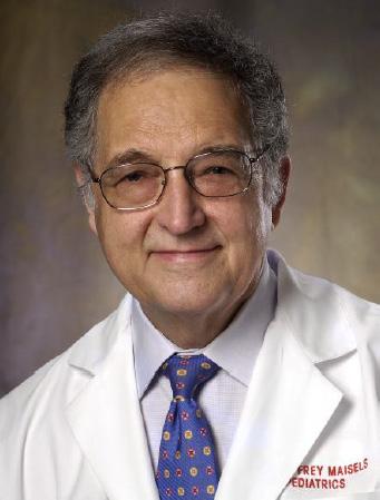
Professor of Paediatrics at Oakland University William Beaumont School of Medicine
After house jobs in Johannesburg and a period of paediatric training at Baragwanath Hospital, the Maisels family moved to the United States. Jeffrey completed his residency training in Paediatrics at the Boston Children’s Hospital in 1966. He trained in Neonatal-Perinatal medicine at Harvard Medical School, Laboratory for Neonatal Research, Boston Lying-In Hospital, where he conducted his first studies on neonatal jaundice. From 1972 to 1986, Jeffrey was the Chief of the Division of Newborn Medicine and Professor of Paediatrics at the Milton S. Hershey Medical Centre at the Pennsylvania State University College of Medicine.
In 1986, he became Chairman of the Department of Paediatrics at the William Beaumont Hospital and Clinical Professor of Paediatrics at the University of Michigan and at Wayne State University in Detroit. From 2009 to 2013, he was the founding Professor and Chair of the Department of Paediatrics of the then new Oakland University William Beaumont School of Medicine and Physician in Chief at the Beaumont Children’s Hospital. In (2020) he was serving as Professor, Chair Emeritus, and Director of Academic Affairs.
Jeffrey is recognised internationally for his studies in neonatal jaundice and his publications on this topic, which extend over five decades. He was the first to develop a technique for measuring endogenous carbon monoxide production in newborns using a rebreathing system. A Wits University and Springbok squash player, Jeff started to play squash again in 1967 and played the US ‘hardball’ game as the number 1 for the Harvard Medical School team, which won the local B league in 1968.
MBBCh (Wits 1960), MRCP (Edinburgh 64), FRCPCH (London 96), MD (Honoris causa) Wits 2004
Chairman of the Department of Paediatrics and Professor of Paediatrics and Reproductive Medicine at Case Western Reserve University Medical School.
Avroy completed his house jobs at the Johannesburg Gen and started paediatric training in Durban followed by the Transvaal Memorial Hospital for Children (TMH) and Baragwanath Hospital. He chose paediatrics believing that if a child was cured, their whole life was ahead of them. He also chose to subspecialise in neonatology he felt he could make a difference as little was known about it and the mortality for premature babies was so high, at that time.
In London, he furthered his studies at St Mary’s Hospital and the Middlesex Hospital, gaining his membership of the Royal College of Physicians in Edinburgh in 1964. He subsequently received a Fellowship of the Royal College of Paediatrics and Child Health (London) in 1998.
Avroy married Roslyn Drusinsky in 1968, and the following year they moved to Cleveland, Ohio, for him to take up a fellowship in Neonatal Perinatal Medicine under the mentorship of Dr Marshall Klaus. It was Klaus who had pioneered studies in maternal-infant interactions, which made ‘bonding’ a household word. He opened the delivery suites to fathers and families. Avroy joined the faculty at Rainbow Babies and Children’s Hospital (RB&CH) and Case Western Reserve University in Cleveland and became a tenured professor of paediatrics and reproductive biology. From 1975 to 1995, Avroy was director of neonatology at RB&CH. He built the unit into a nationally recognised centre of excellence. From 2003 to 2008, he was the Chair of the Eliza Henry Barnes Neonatology. Avroy has co-authored and edited numerous papers and journals, as well as three leading texts in neonatology: Klaus and Fanaroff’s Care of the High Risk Neonate, now in the 7th edition and edited with his son Jonathan Fanaroff. Other books include Neonatal Perinatal Medicine and Neonatology at a Glance.
Avroy concluded: “I am deeply grateful to the University of the Witwatersrand [Faculty of Health Sciences], and my teachers for preparing and launching me on this journey.”
MBBCH MD (Wits) FRCP FRCPCH DCH (London)
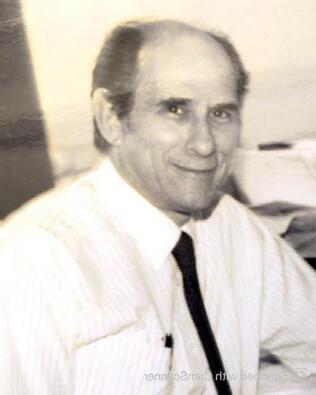
Paediatrician, Senior lecturer, University College Medical School
Gary says of his academic life: “While doing my senior registrar rotation, I debated (mainly with myself) whether to pursue an academic career or to follow a clinical path. I eventually decided to apply for a clinical post, and I was appointed as a Consultant Paediatrician at Edgware General and Barnet Hospitals, in conjunction with an honorary senior lectureship at University College Medical School, with a responsibility for medical student teaching. He was responsible for the development of a highly regarded paediatric service and a sub-regional neonatal intensive care unit at Edgware. Where he was tasked with implementing a new training programme involving every paediatric department in London and its surroundings.
MBBCh (Wits), MD, DSc, FRCP, FACMG, DCH Boston
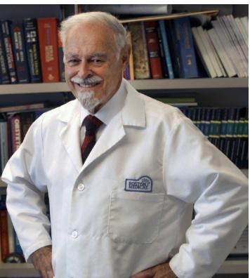
Professor of Obstetrics and Gynaecology, Tufts University School of Medicine Professor of Human Genetics, Paediatrics, Obstetrics and Gynaecology, and Pathology at Boston University School of Medicine.
Aubrey Milunsky who served on the Wits Students Medical Council in 1958 and 1959, became Professor of Human genetics, Paediatrics, Obstetrics and Gynaecology, and Pathology at Boston University. On his retirement, Boston University named the Chair in Human Genetics the ‘Aubrey Milunsky Chair’. Previously, he had served as a Medical Geneticist at Harvard Medical School and the Massachusetts General Hospital for 12 years. In 2020 in his 80s, he is an Adjunct Professor of Obstetrics and Gynaecology at Tufts University.
Aubrey has written or co-edited hundreds of papers and 26 books. These include his major reference work, Genetic Disorders and the Fetus: Diagnosis, Prevention, and Treatment, now written and co-edited with his son, Jeff, who was Professor of Paediatrics and Genetics and Genomics at Boston University. In 1982, Aubrey foundE the Centre for Human Genetics where Jeff joined him as a co-director. Aubrey has also written seminal books about medical negligence.
In 1983, he established a postgraduate continuing medical and legal education course. He has testified as an expert witness in over 100 trials in the USA. In 1982, Aubrey Milunsky was honoured by his election as a Fellow of the Royal College of Physicians of England. In that year, Wits University, his alma mater, conferred a DSc degree for his work on the prenatal detection of genetic disorders.
Our class also produced two early professors of nephrology, namely, Tony Meyers at Wits and Geoff Boner at the at Tel Aviv University. When we qualified in 1960, this was not known as a separate speciality. Kidney disease was usually dealt with either in ‘internal medicine’ or in urology. But because of the development of renal biopsy, dialysis, and kidney transplantation, it became obvious that specialist expertise was needed. Tony Meyers in South Africa and Geoff Boner in Israel were both pioneers of this new speciality in their respective countries. Both were involved in setting up and running renal units with dialysis centres and early kidney transplantation. Both spent some time in the USA to learn the latest thinking, skills and technology. They both wrote many articles and books on the subject and lectured and taught around the world. They were both active on national and international bodies concerned with the speciality becoming some of the earliest professors in their field of study. Andre van As, as head of internal medicine at the JG Strijdom and the Gen Hospitals, was another of the early developers in this field.
MBBCh (Wits) FCP(SA) FRCP
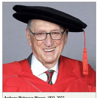
Director of Renal Services at the Johannesburg General Hospital and Wits University. 1989: Professor of Nephrology, Wits Medical School
In South Africa, Tony Meyers took an early interest in the subject and, after his house jobs, went straight into the emerging renal units, first established by Ben Goldberg in the early 60s. Andre was the first to set up renal dialysis at the Gen for Ben. Tony Meyers, rose up the ladder until he was the chief in this speciality and director of renal services at the Johannesburg General Hospital.
In 1989, he was appointed Professor of Nephrology at Wits University,, a position he retained until 2013. He was a founding member of the SA Renal Society as well as the South African Transplant Society, the African Association of Nephrology and the National Kidney Foundation of South Africa. He drew attention to the country’s chronic kidney disease burden, helping to establish early intervention programmes, educating health providers and setting up regional dialysis centres.
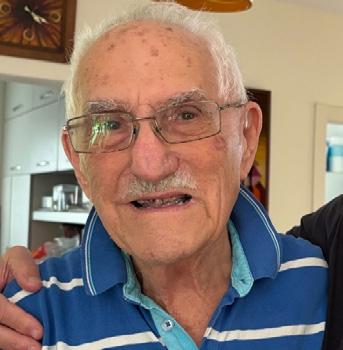
Associate Professor of Nephrology,Tel Aviv University
After his internship at Tel Hashomer Hospital (then a military barrack hospital like Baragwanath, and now the stunning new Sheba Medical Centre) and a few other medical jobs there, Geoffrey Boner served two years of compulsory army service and then signed up for the permanent army. Thereafter, he worked in renal departments as Nephrology was only recognised as a speciality in 1970. There were no special qualifications and very few specifically qualified people in Israel, so Geoffrey, as someone active in the field, was registered as a
specialist in Nephrology.
In 1977, he joined a large department of nephrology at the Beilinson Hospital in Petah Tikva, near Tel Aviv. From 1992 to 2002, Geoffrey was Director of the Institute of Hypertension and Kidney Diseases, at the Beilinson Hospital, which is now part of the large and distinguished Rabin Medical Centre. While there, he was appointed Associate Professor at the University of Tel Aviv
MBBCh (Wits), FCP(SA), PhD (1976 Wits), FCCP, FACP Professor of Medicine, Pulmonologist
Andre says, that doing the paediatrics rotations with Eric Kahn and Monty Schneier at Baragwanath Hospital, was a wonderful experience after Surgery and Medicine. "I wanted to become a paediatrician, but at the end of my six months no registrar position was available. So, I became a pioneer with Professor Guy Elliott at the Johannesburg General Hospital (Gen) in building and running and teaching about – intensive care units (ICUs)," he said.
Andre spent the next 26 years running ICUs in South Africa and USA. He was appointed Chief of Internal Medicine and Principal Physician at the JG Strijdom Hospital in 1973 where he established a pulmonary division. He also ran a very active pulmonary teaching programme and transformed the teaching and patient care standards of the Hospital. Andre was also an early pioneer in setting up haemodialysis units at the JG Strijdom and the Johannesburg Gen. In 1980, he left South Africa for the USA, where he was appointed Chief of the Pulmonary Division at the University of New Mexico and the Veterans Administration Medical Centre in Albuquerque. Here, he was soon promoted to the rank of Professor of Medicine. He was active in student, resident and fellow teaching, as well as serving on faculty and organisational teaching committees, and being Chairman of the ICU/Coronary Care Unit committee.
MBBCh (Wits) 1960, FCP (SA) FCCP Respiratory Physician
Mike was a part-time teacher in respiratory medicine at the Johannesburg General Hospital. In 1980, he was appointed as Head of the new, modern, medical, respiratory and academic teaching unit at the JG Strydom Hospital – the same unit that was originally developed by Andre van As and others.
In 1990, Mike Plit was appointed personal physician to Nelson Mandela, a position he held for 21 years, spanning the time Mandela was President of South Africa (1994-1999) and until his death in 2013, aged 95 years. During this time, Mike was introduced to a number of luminaries, including European Royalty and President Bill Clinton.
In 1992, Mike became President of the Pulmonology Society. In 1999, President of the National Asthma Education Programme and was awarded an honorary fellowship by the Pulmonology Society. His colleagues paid tribute to him for helping to promote the sub-speciality and nurturing the careers of many of the past and current pulmonologists.
MBBCh (Wits), MD (Wits), PhD (University of New South Wales, Australia)
1973: Chief of the alcohol programme at Boston City Hospital, affiliated with Harvard Medical School and then Boston University School of Medicine.
“My journey took me from Johannesburg to Israel, then to the UK and Australia, and finally to the USA, where I developed my research interests in Psychiatry.
After a couple of years in Boston treating drug addicts, I was appointed Chief of the alcohol program at Boston City Hospital. With a wonderful group of clinicians, we offered care to many patients and established a training program for alcoholism counsellors. It was a good time in my career. I was affiliated with Harvard Medical School and then Boston University School of Medicine. But good times don’t last forever. Several of my colleagues left to start a drug and alcohol abuse training program at Boston University. Middle-age irritability had set in, and it was time for me to move. I joined a federal funded mental health program for the City of Lynn in Massachusetts. Later, I formed a partnership with three other psychiatrists to establish the Psychiatric Group of the North Shore. These fellows became a second family to me: skillful, empathic, resourceful, hard-working and intelligent; we worked effectively together for many years. Once a flourishing town based on shoe manufacturing, and a vast General Electric plant, Lynn had fallen on hard times as its key industries moved away. A visitor to Lynn will be struck by the many derelict factory buildings that once housed its vast shoe industry. In fact, the skeletal remains of industry can be found in many Massachusetts towns. I asked myself who built these huge factories, why did these industries collapse, and what was the link between psychiatric disorders and the loss of employment? This led me to research on the history of these situations and the publication of many books on American industrial history."
MBBCh (Wits), MD (Milwaukee)
1989: Associate Professor of Family Medicine at the University of Wisconsin Medical School
Between undergoing major cardiac surgery, Farrol taught and supervised residents and medical students at the Waukesha Family Practice Centre in Milwaukee. In 1980, Farrol, with a new lease of life, after his coronary bypass, moved to St. Lukes’s Family Practice Residency Program in Milwauke. Here he was promoted to Associate Director of clinical medicine and medical director of Mercy Residential and Rehabilitation Centre.
He was the Geriatric Education Director for the Department of Family Practice, and a member of the Medical College of Wisconsin Interdepartmental Geriatric/Gerontology Group for Education and Research. After ten years at the Medical College of Wisconsin, Farrol focused on the field of geriatrics. In 1989, he was offered a position as Associate Professor at the University of Wisconsin Medical School. He remained there until his retirement. Farrol defied his physician’s initial prediction because of his heart problems and died in June 2020 at the age of eighty-two – caught eventually by the coronavirus pandemic.
General Practitioner, Anaesthetist and TV Teacher MBBCh (Wits), FFA (SA)
While in the Department of Anaesthetics, Rodger was tasked with setting up an ongoing postgraduate anaesthetic teaching programme. He said: “This evolved into monthly meetings, which I was able to move from the hospital to the College of Medicine in Parktown. We had a good format, an hour-long lecture followed by drinks and dinner. The meetings were well attended. I also organised the two-day annual anaesthetic update meetings in the department. I was asked by doctors in some outlying areas if I could take some of the speakers
that we had to outlying areas. This would have been far too expensive, and alternative modes of communication were investigated. I suggested an updated course for GP anaesthetists in the Kruger National Park, and we had a great meeting. This became an annual event for a few years after that.” Roger also arranged medical tutorials and lectures via television. He learnt the technique of extraocular injection for eye surgery while working at the King Khaled Eye Specialist Hospital in Riyadh, Saudi Arabia (KKESH), which undertook all kinds of eye surgery and also some ENT. Roger became very competent at administering them. His special interest was the complications of these injections. Back in Johannesburg he passed on the technique to others.
MBBCh Wits (1960), Diploma in Allergy (Wits) 2003
Created and ran allergy teaching courses at Baragwanath and JG Strijdom Hospitals, Johannesburg
Classmate Gloria Davis, who was one of the pioneers of the speciality of allergy in South Africa, was invited to prepare and run teaching programmes at the JG Strijdom Hospital respiratory unit for anaesthetic registrars. She taught how to examine patients so that they could diagnose their allergic illnesses and then assess any risk for anaesthesia. She continued this teaching at Baragwanath. Gloria said: “I not only gave talks to all staff on the poor results of asthma management at the hospital, but also arranged lectures and teaching ward rounds for the anaesthetic registrars. I then joined the paediatric asthma unit, and my entire research now took on meaning as I studied allergy in asthmatic black children, as well as all the other conditions from which they suffered in the realm of allergy. I managed to organise and perform skin-prick testing on all the patients, as well as on cardiac patients, as a control group.”
MBBCh (Wits), FRCOG (Lon)
Obstetrics and Gynaecology, Wits Faculty of Health Sciences
Mervyn Hurwitz was another of our colleagues who served at the Wits Medical School. He told us “It’s hard to believe that 60 years have passed. I am fortunate to have remained friends with several classmates across the globe".
He described his specialisation in Obstetrics and Gynaecology in Johannesburg and London and wrote: “I joined the practice of Joel Cohen and Max Bloom and later I served on the faculty at Wits. I was one of the pioneers of sex therapy in South Africa, establishing the country’s first sex therapy clinic. I remained in private practice for almost 30 years until I emigrated to the US in 1997 to be closer to our children”. Sadly, Mervyn died on August 25 2020, in Florida.
Chaim Rosenberg sums up that it was a revelation to discover that so many of the class had risen to chair positions, department heads, and professorial status. Members of the Medical School's entry class of 1955 were already high achievers in high school. After graduating from medical school, largely in 1960, more than half remained in South Africa, and others settled in Great Britain, the United States, Israel, Canada, or Australia. The desire to contribute to the good of society continued throughout their long professional careers. One in four of the class held senior academic posts and their careers contributed to medical progress through teaching, writing scientific papers and authoring important medical textbooks. Several were directors of medical departments or deans of medical schools. These endeavours brought great credit to the individuals, their graduating class, and the University of the Witwatersrand.
Our 60th Anniversary year evoked many memories. Almost miraculously, we reunited our class at this late stage in our lives. We have been able to relive our own journeys and share over 50 biographical- sketches with classmates.
What became crystal clear is that we have all lived and practised through some of the swiftest and most fundamental changes ever experienced in the practice of medicine. It is hard to believe the exponential growth of medical knowledge and technology from when we graduated in 1960 to our diamond jubilee in 2020.
Avroy Fanaroff invited several of our classmates to write a short piece on how the changes have affected them in their professional lives. We have pleasure to read these glimpses into the changes they witnessed and embraced as they progressed in their work.
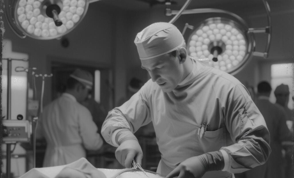
By Clive Rosendorff
Modern cardiology was born in the first half of the twentieth century, and it is sobering to recall that we were medical students not long after, in the 1950s. I was privileged to be an intern in Guy Elliott’s unit at the old Johannesburg Gen. I do wish I could remember more of that than I have, but I do recall one Elliott therapeutic triumph. On rounds one morning, a hallucinating and very agitated patient complained that there was a large dog on his bed. Without hesitating for a second, Prof. Elliott calmly went to the end of the bed, making a show of picking up the invisible animal, taking it to the door of the ward, and throwing it out. The patient immediately relaxed, and we were mightily impressed.
Myocardial infarction (MI): I do remember, however, how we treated patients who came in with a myocardial infarction. Very simply, morphine, digitalis sometimes and bed rest for 5-7 days. Anticoagulation with heparin
was very new and very controversial. I still recall the horrendous sight of Clive Prowse, then my registrar, doing a quick and courageous, and very bloody, bedside thoracotomy on a patient who had just had a cardiac arrest, and pumping the exposed heart with his hand.
Coronary Care Units: How things have changed! Today, there are Coronary Care Units (CCUs), cardiac and coronary catheterisation, percutaneous coronary intervention with angioplasty and stents, and coronary artery bypass grafting (first performed by DeBakey in Houston in 1964). The hospital mortality rate from MI was about 35% in 1960; it is less than 10% now.
The CCU was pioneered by Desmond Julian, a registrar in cardiology at the Royal Infirmary in Edinburgh, in 1961. The concept spread rapidly throughout the globe, but I do not recall that we had one in Johannesburg in 1962. The CCU allows for continuous ECG monitoring, alarms for serious arrhythmias, closed-chest cardiopulmonary resuscitation with external defibrillation, and, perhaps most importantly, the concentration of highly trained staff.
Cardiac Catheterisation: Cardiac catheterisation was first carried out in 1929 by Werner Forssman, a 24-year-old urology resident, who wanted to be able to inject drugs into the heart. He worked with catheters in the urinary tract, so he wondered whether he could insert a catheter into the heart. One evening, he returned to the clinic and, with the assistance of a nurse, exposed the basilic vein in the arm, inserted the catheter, and pushed to what he hoped was the right atrium.
He then walked down a flight of stairs and asked a technician in the X-ray department to take the now-famous film. He then walked down a flight of stairs and asked a technician in the X-ray department to take the it now-famous film.
By 1960, cardiac catheterisation laboratories had sprung up around the world, and at that stage, they were primarily utilised for the diagnosis of structural heart abnormalities.
Mason Sones was the director of the cardiac catheterisation laboratory at the Cleveland Clinic. In 1958, while performing an angiogram of the aortic root, the tip of the catheter slipped into the ostium of the right coronary artery, so that the entire volume of the contrast material entered this vessel. This was truly an ‘aha!’ moment.
Stents: Years later, cardiac catheterisation with coronary angiography became standard management of acute MI, with angioplasty of occlusive plaques and stenting, usually with stents that elute anti-inflammatory drugs, accompanied by one or two of a variety of modern anticoagulants. I recall the first description of coronary artery stenting at an American Heart Association meeting in the early 1990s, and the general horror at the idea that we would put a tubular wire cage into a coronary artery!
Heart failure: The other condition in which there have been spectacular advances is heart failure. In 1960, the diagnosis was based on those clinical symptoms and signs we knew so well: dyspnea on exertion, orthopnea, elevated jugular venous pressure, third heart sound, basal lung “crepitations” (does anyone still use that word?), and ankle swelling. I still teach those traditional diagnostic skills to medical students, but they are less and less receptive and just want to “get to the echo”. The chest X-ray often showed a big heart. Treatment was digitalis and a diuretic, and more bed rest. That was it!
Echocardiography: Since then, we have seen the development of much better diagnostic criteria, echocardiography and isotope imaging, which have enabled us to measure ejection fraction, regional contraction abnormalities, as well as useful estimates of cardiovascular function, based on exercise tolerance.
Modern Drug Therapy: We still use diuretics, but modern drug therapy reflects a new understanding that much of the cardiac dysfunction in heart failure is due to neuro-hormonal-induced inflammation. This has led to the use of a whole cocktail of drugs to reduce the effects of the renin-angiotensin-aldosterone system (angiotensin converting enzyme inhibitors, angiotensin receptor blockers with and without neutral endopeptidase inhibitors, aldosterone blockers), the sympathetic nervous system (beta- blockers) and now SGLT2 (sodium glucose co-transporter-2) inhibitors, which are anti-diabetic drugs serendipitously found to reduce mortality in heart failure.
Pacemakers and advanced implantable defibrillators: In advanced heart failure, we also have implantable cardioverter defibrillators (ICD) and cardiac resynchronisation therapy (CRT).
Heart Transplants: And when all else fails, cardiac transplantation, performed first, as we all know, by Chris Barnard in Cape Town in early December 1967.
The key pioneers of heart surgery, Christiaan Barnard of South Africa (who performed the first human heart transplant), Michael deBakey of Texas, and Adrian Kantrowitz of Brooklyn, conferred later in December 1967 in Washington, DC, before appearing on ‘Face the Nation’ broadcast.
Techniques are much the same today; only the post-operative drugs available to suppress the immune system, which rejects the new heart, have been significantly improved. All of these were too late to save all the patients I watched die in the medical wards of the Johannesburg Gen in 1962.
Valve disease and replacement: I do recall that there were large numbers of African patients who suffered the ravages of rheumatic fever, following streptococcal infection, which mainly damaged mitral and aortic valves.
There was no effective therapy, and cardiopulmonary bypass to enable open heart surgery for the repair of badly damaged mitral or aortic valves did not really expand and develop until the 1960s. We have now advanced even further, with catheter-based procedures that were unimaginable in the 1960s, such as valvuloplasty, aortic and mitral valve replacement, and even the closure of atrial and ventricular septal defects. We can also cure atrial fibrillation using percutaneous catheter ablation.
Where to from now? And now we are in the age of cell and molecular biology, stem cell therapy, gene transfer therapy, pharmacogenomics, increased understanding of the role of microRNAs, and the use of RNA interference to silence harmful genes. And above all, a very clear understanding of risk factors for the development of coronary heart disease and heart failure.
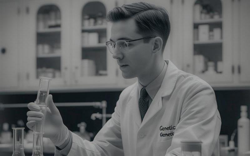
By Martin Bobrow and Aubrey Milunsky
Martin Bobrow writes:
Descriptions of diseases running in families date back to biblical times. Although inherited diseases were known, they were ‘black boxes’ – described but not understood. There was generally no idea as to what went wrong to cause them, at any cellular or biochemical level, and treatment was unimaginable.
Human Chromosomes: It was not until 1956 that the number of human chromosomes was determined as 46. Cytogenetics must have been an interesting field because Phillip Tobias earned his PhD and Sydney Brenner his MSc in the study of chromosomes.
In 1959, just one year before we graduated, Lejeune in France, reported that Down syndrome was due to the presence of an extra chromosome 21, and the medical world started paying attention.
In 1953, Watson and Crick showed that DNA is a double helix and has the structure of a coded chemical message. During the 1960s, pioneering scientists, including Sydney Brenner, elucidated the mechanisms of molecular biology – the way in which the information coded by DNA (deoxyribonucleic acid) leads to the production of proteins. These and other technological developments made it increasingly possible to define specific genetic defects and their consequences at a molecular level.
In 1960, the study of genetics was just beginning: The paint on these epochal developments was just about dry when this medical school class graduated in 1960. When I joined a Medical Research Council research unit in 1965, there were only four or five genetics clinics in the UK that were developing at the time. There were no training courses, no specific qualifications, and no established national organisations. Things were similar in the United States and other countries. Medical genetics was not a recognised discipline.
Differential chromosome staining: These medical genetics groups would typically have people offering genetic advice to referrals, often from paediatric or obstetric units. They had cytogenetics laboratories analysing chromosomes from samples of blood or small skin biopsies grown in culture. In the early 1970s, differential chromosome staining was discovered, hugely improving the accuracy of cytogenetic diagnosis. Fundamentally, the same techniques, with important technical improvements, are in use today.
In 1966, cells were cultured from amniotic fluid, leading to the possibility of diagnosing Down syndrome and similar conditions during early pregnancy.
This was the first time that geneticists could offer anything other than advice on risks. Although it remains controversial for some, it is widely seen as an important option for people in the unenviable position of possibly having an abnormal pregnancy. Interest in medical genetics within the medical profession has increased sharply. Prenatal screening and diagnosis of many genetic conditions, including counselling before and after the procedures, became part of the workload of genetic centres, although this was always shared with obstetrics departments, and in many places, they now take the lion’s share of the workload. Aubrey Milunsky is an expert in this field.
1980s, molecular genetic laboratories: Starting in the 1980s, molecular genetic laboratories grew up within genetic centres. They used DNA analysis to identify genetic markers characterising specific conditions. Two broad groups of diseases are pretty well directly caused by mutations in DNA: (a) Mutations present in the germcell cause the inherited diseases that often follow the patterns described by Mendel; (b) Mutations occurring in other cells within an individual’s body can lead to cancer.
Unsurprisingly, these are the areas where DNA analysis has had its biggest impact. They brought a fine diagnostic detail, and a certainty of diagnosis to conditions such as the Muscular Dystrophies, Huntington’s disease, familial breast and colon cancer, and many others. Asymptomatic gene carriers, who are at risk of either developing the disorder or passing it on to their family, can now be diagnosed with precision.
Hundreds of previously unrecognised heritable conditions were identified, mostly rare, but one or two (such as inherited breast cancer) are surprisingly common. The sophistication of genetic technologies increased dramatically after the Human Genome Project (2001), and the standard text now lists about 6000 conditions caused by defects in known genes. Prenatal screening is increasingly carried out by analysing minute amounts of foetal DNA in the maternal blood circulation. Similar technology is being applied to detect small amounts of abnormal DNA from cancers in the circulating blood, as a means towards cancer screening.
Cancer genetics: Cancer genetics has become a vibrant speciality in its own right. Boveri and others showed in the early 1920s that cancerous cells had abnormal chromosomes. Cancer is a truly genetic disease in the sense that virtually all cancers acquire genetic abnormalities as an integral part of their causation. In the 1970s, discovering that a chromosome change characteristic of chronic myeloid leukaemia (the Philadelphia chromosome) caused an abnormal activation of a specific kinase enabled the development of a treatment that transformed the prognosis of the condition. Molecular techniques have since revealed highly specific DNA signatures for many cancer types and subtypes, and have identified the changes that accompany tumour progression, as well as elucidated the underlying biochemical defects leading to cancer formation and propagation. Innovative treatments are appearing in significant numbers.
Precise diagnosis: A person who gets ill wants a clear and accurate diagnosis; an informed and sympathetic discussion with someone who understands what is wrong, why it has happened, what is likely to happen in the future, and some form of treatment. For diseases caused by genetic changes, genetic techniques now often offer precise diagnoses, which enable an understanding of the prognosis for the affected individual and the likelihood of recurrence within the family. Until very recently, however, medical genetics has not had much to say about treatment.
Understanding the underlying biology of disease: Perhaps the most important contribution of recent molecular genetic research in medicine is in the detailed understanding of the underlying biology of disease. As predicted decades ago, a deep understanding of what causes disease will eventually (but very slowly) lead to innovative treatments. Muscular dystrophies, spinal muscular atrophy, cystic fibrosis, Huntington’s disease, and so many others were simply ‘black boxes’ a few decades ago – we knew the clinical presentation and familial clustering but had no idea what was actually going on under the hood.
Promising treatments: There is now an exquisitely detailed understanding of the molecular basis of these conditions and many others. Early but promising treatments for cystic fibrosis, spinal muscular atrophy and some muscular dystrophies are now available and beginning to be used to treat patients (although unfortunately
currently at excruciating cost – up to $500k/patient/year, for a treatment that must be continued for life). The biochemistry of Huntington’s disease is now reasonably well worked, and it may also become treatable in the foreseeable future. There has been extraordinary progress in understanding the cellular basis of many cancer types, and a significant number of innovative new treatments have appeared.
Predisposition: Another major area of work has been attempting to understand what underlies the familial clustering observed in most “common” diseases, which do not ‘run in families’ but where the likelihood of a close relative getting the same condition is higher than the population risk. This is more the rule than the exception across all areas of medicine and was observed in family and twin studies long before the development of molecular techniques. Unsurprisingly, this clustering is due partly to shared genes, which cause “predisposition” to the disorder, and partly to environmental factors. Despite a significant amount of effort, genetic techniques have, in general, not yet made a substantial contribution to the diagnosis or prediction of these disorders; however, there are increasing examples of identifying biochemical pathways important in disease causation. There are now so many genetic markers available that new efforts are being made to combine these individually weak predictors into clinically applicable “polygenic risk scores”. These certainly define at-risk sub-populations; whether and in which cases the approach proves to be clinically useful remains to be seen.
The pace of change: When I started my career, genetics was not a recognised part of medicine. Today it is part of everyday practice. Specialist genetic centres are present in virtually all large hospitals. More importantly, genetic knowledge and techniques are becoming part of the practice in many different specialities, including oncology and pathology, haematology and ophthalmology, obstetrics and paediatrics, and others. It is widely discussed (and often somewhat overhyped) in the public media. There are formal training and accreditation courses, professional associations, and national and international meetings that attract thousands or tens of thousands. The same techniques have been transformative in forensics (consider how they managed before DNA profiling), anthropology, zoology, bacteriology, and agriculture – in all life sciences. The pace of change remains breathtaking, but much remains to be learned.
Aubrey Milunsky writes:
Prevention and avoidance through prenatal diagnosis and pre-implantation genetic testing is now available for over 5000 genetic disorders.
The discovery of the double helix in 1953 heralded the real beginning of modern medical genetics. Rosalind Franklin, PhD, a talented English chemist and X-ray crystallographer, did the fundamental work that led to the understanding of the molecular structure of DNA and RNA (ribonucleic acid).
Rosalind Franklin: Her X-ray diffraction images of DNA all but revealed the helical structure of DNA for which Watson, Crick, and Wilkins, but not her, received the Nobel prize. (They did not credit her in their publications.)
It was in 1958, the year Rosalind Franklin died at 37 years of age from ovarian cancer (possibly as a result of inadequate radiation safety measures), that, with fresh faces and open minds and with a burgeoning vocabulary, we, the graduating class of ‘60, all eagerly entered the patient’s realm. Armed with a fleeting memory of Drosophila melanogaster (the common fruit fly) taught by an eccentric, if not alcoholic, biologist, our limited knowledge of human genetics was topped by the extant knowledge that each of our cells had 46 chromosomes (as determined in Sweden in 1956), and not 48, as we had been taught on entry into medical school.
Dr Marthe Gautier: Unbeknownst to us at the time of our clinical training was the ongoing work by Dr Marthe Gautier in Paris. This was especially focused on the chromosome complement in the cells of normal individuals and those with Down syndrome. On a fellowship at Harvard, Marthe Gautier attempted to grow human cells (even using her own serum) and created slides for visual examination of the chromosomes. Unable to achieve adequate quality images, she sent her slides to an intern friend, Dr Jérôme Lejeune, in Paris. He never returned her slides, publishing the photographs and observations as his own and revealing for the first time the extra chromosome 21 associated with Down syndrome, for which he became famous, receiving France’s highest honours. He never acknowledged Dr Marthe Gautier. Many years later, the story emerged, and she was duly honoured.
Genetic diagnosis: The nascent ability to culture and examine human cells, including amniotic fluid cells, slowly
gained steam in the mid-to late sixties, setting the stage for prenatal genetic diagnosis. In a stepwise fashion, it became possible to recognise and characterise individual chromosomes, their structural aberrations including deletions and duplications, all shown by fluorescent in-situ hybridisation. Chromosomal syndromes, prenatal diagnosis, cancer genetics, newborn screening, understanding of the common chromosomal cause of early miscarriage (at least 40%), and advances in biochemical genetics and preimplantation genetic testing followed and dominated the next three decades.
Aubrey and his son, Jeff, published the 8th edition of ‘Genetic Disorders and the Fetus: Diagnosis, Prevention and Treatment’. It comprises 37 chapters and is authored by senior international experts from nine countries.
Sequencing the Human Genome: In 2000, the ‘political’ announcement in the US that the human genome had been sequenced caught the world’s attention. The fact is, however, that the claim became realistic in 2003. However, even now, the genomic sequence is incomplete, with an estimated complement of 19,000 to 20,000 genes. The remarkable exponential growth of gene analysis has revolutionised the practice of medicine in all specialities.
Technological advances in gene analysis enabled more precise diagnosis: Technological advances in gene analysis have been truly amazing. Direct and reliable sequencing of individual genes enabled precise clinical syndromic and cancer diagnoses, prenatal genetic diagnosis, preimplantation genetic testing, and carrier detection for a handful of genetic disorders. The advent of next-generation sequencing (NGS) led to the introduction of whole-exome sequencing, which allows simultaneous analysis of all coding genes. Incredibly, these genes occupy only 1-2% of our genome. This methodology also encompasses targeted sequencing of disorders with shared and similar phenotypes, but due to different genes (eg cardiomyopathy, epilepsy, ataxias, breast cancer, intellectual disabilities, etc.), using gene panels. NGS further enabled simultaneous expanded carrier detection of a few hundred monogenic disorders.
Diagnostic whole-genome sequencing has also become available, but its interpretations are complicated by the many variants (100-300) in our coding genes, in addition to the huge number of unexplored introns we all have (parts of a DNA or RNA molecule that do not code for proteins).
Chromosomal microarray (CMA): Chromosomal microarray (CMA) has been developed in parallel with gene analysis techniques. This test has enabled the detection of even tiny (50kb) microdeletions and microduplications from blood, tissues, or amniotic fluid cells, as well as the accurate determination of chromosome number. CMA has increasingly displaced chromosome analysis as the preferred first test for prenatal diagnosis, intellectual disability, etc.
Ethical considerations: The direct analysis of genes now poses a significant challenge to physicians in all specialities. Pause for a moment to consider the following facts:
• ≥ 7,000 rare genetic disorders are known
• 1 in 12 are carriers of a genetic disorder
• 4,331 genes are known this far that cause recognisable phenotypes
• 6,739 phenotypes, some due to the same gene, are diagnosable
• ≥ 2,000 congenital syndromes are known
• CARRIER tests are available for over 1,000 genetic disorders
Given these facts, failures by physicians to inform their patients with known or likely genetic disorders, about the available tests and options for timely treatment (e.g. the cardiac Long QT syndrome), prevention (e.g. familial polyposis coli), and avoidance (prenatal diagnosis or preimplantation genetic testing) will inevitably spawn malpractice litigation, which has already begun.
We are in the golden era of human genetics. It is startling to realise that virtually everything I know today about medical genetics, I learned during the decades after medical school. What I did learn there was how to learn.
By Theodore Kretzmar
Surgery over the years – where do you begin and end? What to include and what not? This review cannot be academic; it can only be anecdotal with large gaps, but I will attempt a general overview with a personal perspective, even though I retired from surgical practice many years ago.
General Surgery and Super Specialities: At the start of my career, general surgery encompassed almost everything, but with the advent of massive investigative and therapeutic knowledge, as well as newly developed instruments and subsequent procedures, super specialities split off. This process is still rapidly progressing, with huge benefits to patients in all fields.
Diagnostic and Treatment Equipment: As a registrar at Baragwanath Hospital, in about 1964, if we were faced with a head injury requiring a brain scan for the diagnosis of a subdural haemorrhage, this involved taking X-ray plates in cassettes to a dark developing room, processing them in various solvents and then drying them on hangers in front of a light above a driptray. We did this all ourselves, looking for a shift in midline structures! And then, if there was one, we used something like a carpenter's drill, hand-held, to place burr holes in the skull to drain the clot, carefully avoiding the middle cerebral artery.
Compare this to the present neurosurgeon, who uses CT/MRI scans for diagnosis and scan guidance, along with new-generation instruments, to accurately and minimally perform the same procedure, only much more accurately and safely.
I have used this example to generalise the breadth of change which applies across the entire medical spectrum. I can highlight only some of the surgical advances.
Cancer Surgery – Past and Present: Can you envisage the performance of hindquarter amputation for melanoma of the foot? A desperate measure, but horrific. This was done as there was little else. Now, lesser surgery is but a part of the total treatment. This same principle applies across most cancers.
Radical mastectomy following “frozen section biopsy“, and histology only, is no longer performed as this has been replaced by a new protocol starting with a mammogram, scan-directed biopsy, histology, minimal surgery, with
reconstruction if possible. And now adjuvant biological assessment is used for the best combination of various oncology measures. The advances in oncology are significant but are outside the scope of this article.
A recent new consideration, and the jury is still out on this, is prophylactic mastectomy if precancerous genes are present.
Gastric Surgery: When I qualified, peptic ulcer treatment involved various permutations of radical Bilroth gastrectomy/gastroenterostomy, followed and replaced by vagotomy and drainage procedures, all eventually replaced by a pill -- Tagamet /Cimetidine (which inhibits acid secretions), which has itself now been replaced by PPI pills. (Proton Pump Inhibitors).
Gastric surgery, other than for malignancy, has now resurfaced into gastric by-pass surgery, as treatment for morbid obesity, with many differing procedures, all done with minimally invasive endoscopic surgery by endoscopic surgeons.
Scopes – Gastroscopy Colonoscopy Laparoscopy: The new gastroscope was first used for ulcer diagnosis, replacing barium studies. Scopes then advanced from diagnostic to therapeutic instruments for ulcer biopsy, fulguration for bleeding ulcers and moving down a little for diagnosis and treatment of bile and pancreatic duct problems.
I personally benefitted when, as an outpatient, I underwent the removal of a common bile duct stone, replacing what would have been major and difficult surgery!! So out with the old surgery and barium studies and in with the new generation surgeons and their "tools”.
Colonoscopy, which, like gastroscopy, used to involve looking into a small scope eyepiece. Now the scope produces an image of the insides of the examined organ as large as the monitor screen or multiple screens, which will allow.
The laparoscope also made its entrance, initially serving as a diagnostic tool, but rapidly enabling minimal access surgery for many abdominal conditions, replacing the longstanding open operations. This has led to the emergence of a new speciality in endoscopic surgery, which has developed and expanded gastroenterology.
This expansion is now extending into robotic surgery, where the surgeon operates on a computer keyboard with the procedure actually carried out by an octopus-like instrument.
Transplant Surgery: Early transplants were cornea (1905), kidney in 1954, lung and liver transplants in 1963, followed by the dramatic first heart transplant in Cape Town in 1967. This is now followed by transplantation of an increasing number of failing organs, all made possible by the ongoing and improving immunological advances. Transplant surgery is now a routine surgery, limited only by the availability of organs.
Cardiac Cath Lab: The Cath Lab has likewise completely changed cardiac and vascular treatment. Cardiac by-pass surgery is now reserved for selected cases, as some ‘Cardiac Surgery’ is now performed via an incision, or two, in the groin, stenting coronary arteries and even closing septal defects and replacing diseased valves. Aortic aneurysms can be replaced this way, too, and peripheral arteries can and are repaired, as are the coronaries. Even varicose vein surgery has undergone a laser change. No longer the Trendelenburg and strip, but now, minimally invasive laser ablation of the problem.
Eye Surgery: Eye surgery has also undergone significant changes made possible by technology, such as laser correction, replacing glasses or contact lenses. Cataract surgery is now a quick outpatient procedure, with patients going home within hours, rather than days.
In conclusion, the list is, by its nature, grossly incomplete and endless, rapidly extending, and leading us to ponder where surgery will be when the class of 2020 turns 60.
By Geoff Boner
Nephrology becomes a speciality: Our graduation in 1960 marked the year when Nephrology became established as a speciality. Prior to this, researchers had investigated renal physiology, and clinicians had described various kidney diseases. Attempts had been made to use the principle of dialysis through a semipermeable membrane to treat patients with kidney failure.
The successful use of haemodialysis in the treatment of patients was described by Willem Kolff and Nils Alwal in the mid-1940s. This treatment was used only in patients with acute kidney failure, as there were difficulties in achieving repeated access to the vascular circulation.
The turning point came in 1960, when Belding Scribner developed an external arteriovenous shunt, followed by the creation and use of an arteriovenous fistula in 1966, which allowed for the repeated treatment of patients with end-stage renal failure (ESRF).
In 1960, a group of 400 researchers led by Jean Hamburger of France held a conference and decided on the formation of the International Society of Nephrology, thus creating the speciality.
Wisconsin Madison: From 1971 to 72, I spent an instructive and productive year on a clinical-research fellowship at the University of Wisconsin, Madison, where the renal unit was very active in all aspects of nephrology, including kidney transplantation. Following my return to Hadera in Israel, in 1976, I took a post at the Beilinson Hospital (now part of the major Rabin Medical Center). This had one of the largest Nephrology units in Israel, and I went on to become Director of one, which included an appointment at the Tel Aviv University Medical Faculty. The Kidney Transplant department there performs about two-thirds of all kidney transplants in Israel and works closely with the nephrology unit.
My main clinical interest was diabetic nephropathy and, together with Mark Cooper of Melbourne, I published a book on the ‘Management of Diabetic Nephropathy’. I participated in clinical work, research projects, and, in particular, the development of kidney transplantation at Beilinson Hospital.
Over the past sixty years, there have been many advances in all fields of Nephrology which I discuss below.
Treatment of patients with end-stage renal failure (ESRF): In 1972, the American Congress decided that Medicare would cover the costs of dialytic treatment for most patients. This led to widespread dialytic treatment of ESRF patients in the USA and then throughout the world. By 2018, 1,000 to 2,500 patients per million population, depending on the ethnic makeup and prevalence of renal disease, were receiving treatments for ESRF in the developed countries of the world. This number was about 100 per million in the poorer countries, where the cost of treatment is the limiting factor.
Hemodialysis: There have been significant innovations in the treatment of end-stage renal failure (ESRF) using hemodialysis, making it the most widely used modality for managing the condition. Today, most patients receive dialysis in large, community-based units rather than in hospitals, a shift made possible by several key advancements. First, improvements in vascular access—particularly the development of arteriovenous fistulae and dual-lumen catheters for central vein catheterization—have enhanced the efficiency and safety of treatment. Second, the introduction of synthetic membrane hollow-fiber dialyzers has improved the effectiveness of the dialysis process. Third, the emergence of sophisticated dialysis machines equipped with multiple treatment options and monitoring capabilities has allowed for more personalized and controlled care. Fourth, the synthesis of erythropoietin and other erythropoiesis-stimulating agents has provided effective treatment for the anemia commonly associated with kidney failure. Finally, the establishment of standards for the preparation of purified water used in dialysate production has ensured safer and more consistent treatment outcomes.
Peritoneal Dialysis: Although peritoneal dialysis is not as widely utilised as hemodialysis, the introduction of certain techniques, continuous ambulatory peritoneal dialysis (CAPD) and the use of cycling equipment, continuous cycling peritoneal dialysis (CCPD) has allowed suitable patients to treat themselves at home.
Kidney Transplantation: Following the first successful kidney transplantation in 1954, this form of treatment has evolved into the most effective therapy for end-stage renal failure (ESRF), offering patients longer survival and an improved quality of life compared to those on hemodialysis. Several key advances have contributed to this progress. First, the identification of the human leukocyte antigen (HLA) complex, encoded by the major histocompatibility gene complex, has significantly enhanced the ability to match donors with recipients. Second, the development of more specific and efficacious anti-rejection medications has made it possible to perform transplants even in cases where tissue matching is suboptimal. Third, the formal definition of brain death has facilitated greater access to organs from deceased donors. Finally, the use of organs from both deceased donors and non-related living donors has expanded the donor pool, further improving the availability and success of kidney transplantation.
Clinical Nephrology
• Treatment of patients with Acute Kidney Injury (Acute Tubular Necrosis)
Intermittent and continuous renal replacement therapy using hemodialysis, hemofiltration and hemodialfiltration have become accepted forms of treatment in acute kidney injury.
• Diagnosis and treatment of water, electrolyte and acid-based disorders
These conditions have been well defined, and treatment guidelines have been established.
• Diagnosis and Treatment of Kidney Disease and Hypertension
Over the past sixty years, the introduction of imaging techniques such as ultrasound, Doppler ultrasound, angiography, computerised tomography, magnetic resonance imaging (MRI) and isotope scanning has played a major role in improving the diagnosis of conditions within the kidney, in the arterial supply and in the urinary tract.
In addition, the microscopic examination of kidney biopsies, using light, immunofluorescence, and electron microscopy, has resulted in an improvement in the definition of kidney diseases. This has allowed the development of treatment protocols using corticosteroids and immunosuppressive agents, and deciding on the best treatment for each disease.
During the latter part of the 20th century, several formulae for the clinical estimation of the glomerular filtration rate (eGFR) were described. This has resulted in a clear definition of the various stages of chronic kidney disease and in the investigation of the beneficial effects of different medications at different stages of disease. This estimation is also of importance in adjusting the dosage of many medications. The measurement of urinary albumin excretion in the urine, expressed as a ratio of urine albumin to creatinine concentrations, in conjunction with the eGFR, has enabled the prediction of kidney disease prognosis.
The development of angiotensin-converting enzyme inhibitors and angiotensin receptor blockers has become the basis for treating most hypertensive patients. These two groups of drugs were then shown to have beneficial effects on cardiac disease and renal disease in patients with diabetic nephropathy and albuminuria. During the last decade, the inhibitors known as gliflozins of the sodium/glucose cotransporter 2 protein have been shown to be even more successful in the prevention of progressive cardiac and renal disease in diabetic patients and, recently, in non-diabetic patients.
• Renal Physiology
The use of clearance techniques, micropuncture of nephrons in the kidneys, and studies of individual cell lines using microbiology and genetic techniques has enabled researchers to define the physiology of different areas along the nephron and even to describe the various transporters and pumps in the different tubular cells along the nephron. In the almost 60 years since I began my training in nephrology, there has been tremendous progress, similar to that in many other specialities in medicine. We, the graduates of 1960, have been lucky to have been part of this progress and have played a role in it.
By Chaim Rosenberg
Psychiatry was a relatively small part of our training at the University of the Witwatersrand (Wits) Medical School. I vividly recall a visit to the Witrand Institution for the Feeble Minded, near Potchefstroom. We were “entertained” by a musical band of Down Syndrome residents, in the belief that Trisomy 21 has an affinity for music and group activities. Such people were once classified as idiots (IQ 0-25), imbeciles (IQ 25-50) and morons (IQ51-75). The lifelong residents were trained to work on the farm or in the workshops. More distressing was the visit to Sterkfontein Hospital to gawk at institutionalised schizophrenics.
Lunatic Asylums: The first half of the 20th century witnessed the building of gigantic lunatic asylums (later called psychiatric hospitals). Westkoppies served Pretoria, and Sterkfontein served Johannesburg.
I began my psychiatric training in 1962 in England at the 2,000-bed Napsbury Hospital (formerly known as the Middlesex County Asylum), near St. Albans, followed by two years at a similar Institution, the Netherne Hospital in Surrey.
By the 1950s, psychiatric hospitals in the USA held nearly 600,000 patients, and 150,000 In the UK, permanently housing mentally retarded (now labelled intellectually disabled), demented or psychotic people. Many of the schizophrenics had been subjected to draconian psychosurgery, induced fevers, and/or insulin shock “therapy”. The one-way ticket into these vast asylums failed to provide effective treatment for their residents.
In South Africa, change occurred in 1949, when the University of the Witwatersrand initiated the first postgraduate training program in South Africa, leading to the Diploma in Psychological Medicine. Five years later, Dr LA Hurst was appointed part-time lecturer in psychiatry at Wits Medical School. In 1959, Hurst became a full-time professor of psychiatry, with the aim of integrating psychiatry into the teaching of general medicine and paving the way for similar departments in other South African medical schools. Psychiatry, in South Africa as elsewhere, has since grown into a major medical discipline.
Chlorpromazine, and other drug treatments: Chlorpromazine (called Largactil in England, Thorazine in the USA) for schizophrenia and lithium salts for bipolar disorders hastened the closing of the asylums and moved treatment into the community. Now, most psychiatric illnesses can be managed in mainstream society.
Disability programs, such as the Americans with Disabilities Act of 1990, provide cash payments, health benefits, and housing grants, allowing individuals with mental disabilities to remain in the community.
Studying the brain and the mind has proven more challenging than researching the liver, lungs, kidneys, or the heart. Neuroimaging (MRI and CT scans) shows a higher rate of pathology among psychiatric patients than in the population at large. Psychiatry and Neurology are drawing closer together.
Happiness: The assessment of happiness by the General Social Survey or Gallup still relies on self-reporting. These studies show that a third of people rate themselves “very happy”, a third not-too-happy to moderately happy, and a third rate themselves as unhappy and dissatisfied with their lives. The Diagnostic and Statistical Manual of Mental Disorders (DSM-5) is largely based on clusters of symptoms and signs. In recent years, much attention has been paid to the Autism Spectrum Disorders, including Asperger’s Syndrome, observed in young children, considered to have genetic and environmental causes.
Big Pharma: Big Pharma has positioned a plethora of drugs to treat each psychiatric disorder and has moved Psychiatry boldly into the biological model of illness. The older tricyclic antidepressants (amitriptyline, imipramine and nortriptyline) have been steadily replaced by the selective serotonin reuptake inhibitors (SSRIs) such as fluoxetine, citalopram and sertraline. For anxiety, there are the benzodiazepines. The older antipsychotic medications include chlorpromazine, fluphenazine and tri-fluphenazine. The newer antipsychotic drugs include clozapine, risperidone and olanzapine. It takes time to find the “right” medication. Electro-convulsive treatment (ECT) for major depression or bipolar disorders is still used in some centers, especially when medication fails to alleviate symptoms.
Talking Therapies: The psychosocial aspects of mental distress have largely been taken over by psychologists, social workers and other talking therapists. The availability of treatment for the mentally ill depends largely on the economic and social development of each nation. With a population of 328 million, the United States today has 28,000 psychiatrists, 106,000 licensed psychologists and 700,000 social workers.
The United Kingdom, with a population of 67 million, has 11,800 psychiatrists. South Africa, with 50 million people, has only 762 registered psychiatrists (2014 figures), with 25-30 newly trained added each year. Most South African psychiatrists work in the private sector. Despite a more humane approach to mental illness, we are far from curing the ills of society. Many psychiatrically ill people become homeless or end up in prison.
The ancient Greeks advised: ‘Know Thyself’: I expect in the near future, scientific research will establish the biological cause and treatment of manic-depression (also called bipolar disorder), schizophrenia and disorders of memory (dementia), much like Diabetes and insulin or Pernicious anaemia and vitamin B12.
The increased use of genetic testing may lead to ever fewer cases of Down Syndrome, Developmental Disability (DD) and Autism Spectrum Disorders (ASD). Such testing must be voluntary and not imposed by the state.
Each person is unique. Emotions like anxiety, sadness, stress, alienation, dissatisfaction, disappointment, difficulties in getting along with others, bearing grudges, jealousy, differ widely from person to person. They are not akin to blood pressure levels, pulse rate, blood sugar, sodium or potassium levels that can be measured and treated with medication.
The complexities of human thought and behaviour take flight in the writings of Tolstoy, Dostoevsky, Jane Austen, and Shakespeare, but lose their potency when described in cold, medical language. I do not believe that Psychiatry is served with only a brief evaluation aimed at prescribing medication. The too-free prescribing of oxycodone, benzodiazepines, barbiturates and sleep aids can lead to addiction.
The relief of suffering and helping in the pursuit of emotional balance and happiness are better served by carefully combining medicines with listening and understanding. A caring psychiatrist should help patients to know themselves.
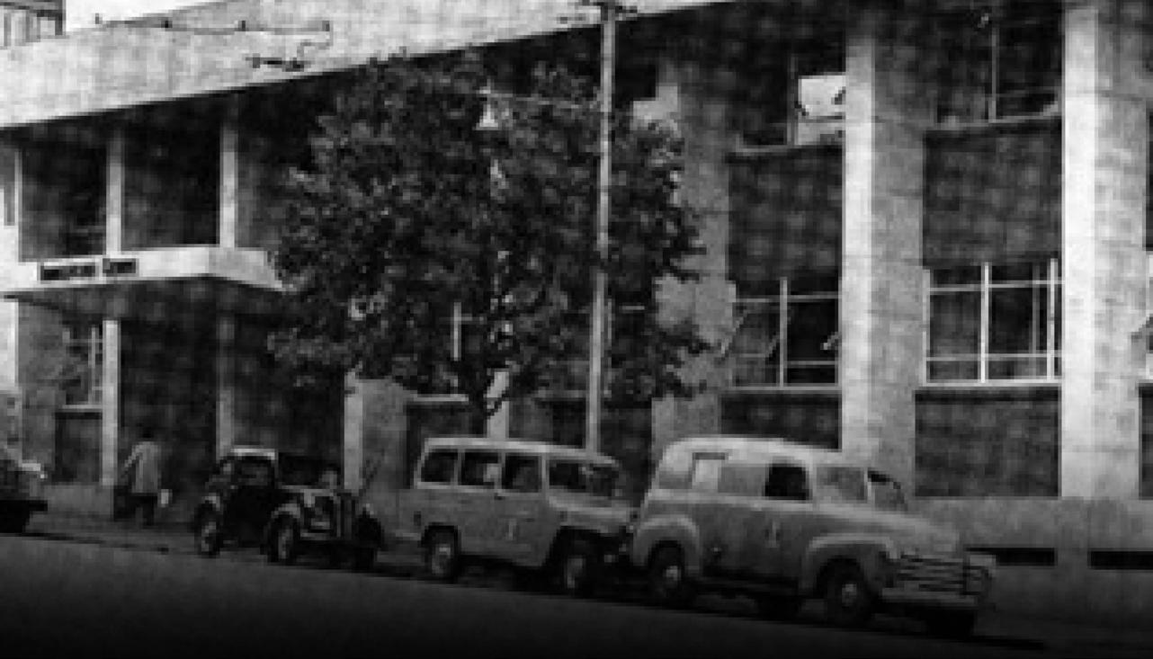
By Anton Heyns and Gerald Shulman
In the year 1900, Karl Landsteiner at the University of Vienna discovered why some blood transfusions were successful while others could be deadly.
Landsteiner discovered the ABO blood group system by mixing the red cells and serum of each of his staff members. He demonstrated that the serum of some people agglutinated the red cells of others. From these early experiments, he identified three types, called A, B and C (C was later to be re-named O for the German “Ohne”, meaning “without”, or “Zero”, “null” in English). The fourth, less frequent blood group, AB, was discovered a year later. In 1930, Landsteiner received the Nobel Prize in physiology and medicine for his work.
It was then discovered that group O blood can be given to any blood group, and all blood groups can be given to group AB. Testing compatibility prior to transfusion was developed, and the first blood bank was established in 1918.
A major advance in 1950 was the replacement of glass with plastic bags to store and transfuse blood. Drs Heyns and Shulman give us some insight into the advances in human blood transfusions and banking since our graduation in 1960.
Anthon Heyns writes:
The Rand Blood Transfusion Service (RBTS), later to become the South African Blood Transfusion Service (SABTS), was established by medical graduates and the Blood Donors Association of Wits University on 23 December 1937. Interestingly, this is also the year of birth of most of the members of our class. The meeting where the RBTS was launched was attended by Drs James Gear and Maurice Shapiro, two famous Wits graduates.
The first home of the transfusion service was on the veranda of Toc H in Doornfontein, and in 1950, SABTS moved into its own for-purpose built building in Esselen Street, Hillbrow, a building well-known to all of us.
Dr Maurice Shapiro: From its inception until his retirement in 1990, Maurice Shapiro was the Medical Director and head of SABTS. After his retirement, he continued to serve as a consultant to the service until his death in 2000.
He was a remarkable man. He received his medical training at Wits (MBBCh 1930). In 1967, Wits awarded him a DSc for his Contributions to the study of the genetics of human blood and the promotion of safety in blood and the promotion of safety in blood transfusion. In 1974, his alma mater awarded him the LLD, Doctor of Laws (Honoris Causa). He made important contributions in the field of human genetics, including the discovery of two new Rh factors.
Quality Systems: One of the most significant advances in blood transfusion in South Africa was the introduction of quality systems in the practice of blood transfusion in 1990. This was formalised by achieving the ISO 15189 standard and accreditation from the South African National Accreditation System.
These quality systems encompassed all facets of blood transfusion, from the selection and management of blood donors to the collection, processing of whole blood into components according to Good Manufacturing Practices, the issuance of blood to patients, and monitoring post-transfusion blood safety with a haemovigilance system.
More recently, SANBS has been accredited to the FACT-JACIE international standards (FACT - Foundation for Accreditation of Cellular Therapy and JACIE - Joint Accreditation Committee of the International Society of Cell and Gene Therapy) for the collection, processing, and administration of haemopoietic cellular therapy products. The transfusion service is actively involved with the administration of cellular products for haematological malignancies.
This includes transplants, donor lymphocyte infusions and immune effector cells. South African National Blood Service) (SANBS) has a close association with the SA Bone Marrow Registry and organ transplant units. SANBS actively recruits prospective bone marrow donors and has introduced genotyping of 13 blood group systems and high-resolution Human Leucocyte Antigen (HLA) typing to identify potential matches for both solid organ and bone marrow transplants.
The attainment of such high-quality standards could not have been achieved by the regional blood service structures established around major cities and provinces over the years. These small services could not afford the newly available technology for screening blood for viruses and other pathogens and lacked the capacity to collect sufficient blood and manage the logistics of distributing blood products to all patients in the country.
Following discussions with the Department of Health and with the assistance of the World Health Organisation, five of the six regional services amalgamated in December 2000 to form the South African National Blood Service (SANBS). SANBS provides about 90% of the blood to the country. Western Cape Blood Service (WCBS) elected to retain its autonomy. The SANBS head office is now housed in a new building located in the North-west of Johannesburg, and SANBS has a sophisticated testing and blood processing facility in Durban.
Providing Safe Blood - AIDS: The greatest challenge that SANBS faced was to provide safe blood products to patients. With the emergence of HIV (the Human Immunodeficiency Virus) in 1981, the prevalence of HIV in blood donors increased from 0.06% in 1991 to 0.26% in 1998. It was estimated that 1:29,000 blood collections might be contaminated by this virus.
In response to the threat, SANBS adopted a standardised blood safety policy that included the closure of donation sites with high HIV prevalence. These closures mirrored the high prevalence of HIV in the black population of South Africa. The implementation of the safety policy decreased the residual risk for collecting an HIV-1 window-period infectious unit by about 24% despite an 18% increase in HIV-1 prevalence in the South African population, and it was estimated that about 1,240,000 blood collections may be infected with undetectable HIV. The ‘window period’ is the time between infection occurring and the time it becomes detectable in the blood by a positive test – a period of about 4-12 weeks.
Race as a risk factor: In December 2004, the South African Department of Health stated that the use of race as a risk indicator was politically unacceptable. The Department recognised that the policy was not racist and accepted that the selection of blood donors was in the interests of recipients of blood, 80% of whom were black. SANBS was fortunate that, just then, technology became available to screen blood donations for HIV and hepatitis with highly sensitive nucleic acid tests (NAT) using automated laboratory systems.
SANBS took up the challenge posed by the Minister of Health and introduced and validated the tests within six months. These tests almost eliminated the risk of the window period. Since its inception and the issuing of
about one million blood products per year, hepatitis B has been transmitted to one patient, and there have been no cases of transfusion-associated HIV. The introduction of NAT screening has paved the way to recruit blood donors from all population groups and subgroups.
Quality of blood supply: In the long term, the education of blood donors will be a key factor in determining blood safety. There was a need for a structured programme that is culturally attuned and presented in the multiple languages commonly used in South Africa. SANBS was awarded funding under the President’s Emergency Plan for AIDS Relief (PEPFAR) to work with the USA Centres for Disease Control and Prevention, the American Association of Blood Banks, and the American Red Cross to achieve this end. The systems put in place have significantly increased the black donor base. In 1990, only 5% of blood donors were black, and in 2020, this has increased to 35%. The new black donors now comprise 58% of all blood donors.
All these initiatives have improved the quality of the blood supply. Currently, there are more than 600,000 active blood donors who donate over 1.1 million units of blood annually. SANBS also supplies the National Bioproducts Institute with approximately 220,000 kg of plasma annually. This is used for the manufacture of plasma-derived blood products, which are used for the treatment of bleeding and immune disorders. Historically, SANBS has struggled to maintain blood stock levels of more than two to three days, resulting in an unreliable supply to patients in need. This has led to the constant need to collect, test, process, and deliver blood on an emergency basis. This has now improved significantly to a supply of five days, equivalent to the international norm.
A challenge has always been to get blood to rural areas and patients who live far from blood centres. An exciting development has been the collaboration of SANBS with the National Department of Health and Civil Aviation Authority to introduce a drone programme that employs ground-breaking Vertical Take Off and Landing (VTOL) technology to transport blood products to patients in need, where traditional methods of transport cannot reach the patient in the so-called golden hour when medical and surgical treatment is more likely to prevent death.
Research projects: The blood services are also actively engaged in research projects focusing mainly on the safety of the blood supply. SANBS and WCBS participate in the REDS (Recipient Epidemiology and Donor Evaluation Study), the premier research programme in blood collection and transfusion safety in the United States. In its third phase — REDS-III — the programme focuses on conducting research on all elements of the transfusion chain, from the blood donor to the blood products received by the patients. SANBS received a Fogarty D43 HIV Research Training Grant in 2015. As a result, the University of California, the San Francisco Blood Systems Research Institute, University of Cape Town, and SANBS will be able to train blood bankers and haematologists to perform highquality HIV research that will benefit public health.
World-class transfusion service: None of this would have been possible without the implementation and development of sophisticated information systems. In 1992, a blood establishment computer system was installed and used for the development of quality systems and to manage and optimise the blood supply and its safety. In 2020, SANBS will replace this with a more sophisticated system that will enable the digitalisation of all business processes central to a modern vein-to-vein transfusion service.
In conclusion, I have highlighted some of the changes and achievements in blood transfusion in South Africa over the past sixty years or so. I may not have done justice to this success story, which has resulted in a world-class transfusion service that may well be the largest service, taking responsibility from blood collection to patient issuance (vein-to-vein). The illustration of a vein-to-vein transfusion in the 1920s is broadly speaking still applicable, but the tubing and the pump between the donor and the recipient have become much more complex and have made it possible to provide safe blood and blood components to all patients in need of this important therapeutic agent.
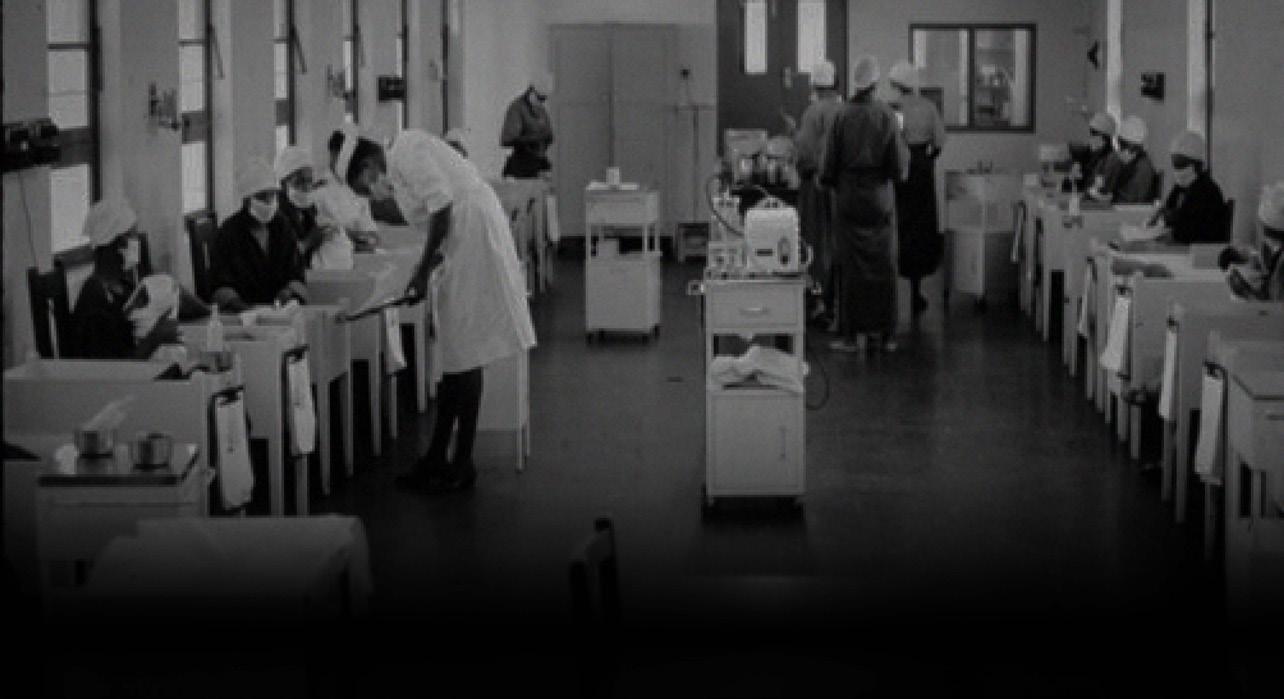
By Jeff Maisels and Avroy Fanaroff
Neonatology is the History and Science of the Care of Newborn Infants Jeffrey Maisels and Avroy Fanaroff have had a closely shared experience in their medical careers. They both trained as paediatricians before completing fellowships in Neonatal-Perinatal Medicine. Thereafter, they each directed one of the most advanced neonatal intensive care units and fellowship training programs in the USA. As tenured Professors at prestigious universities, they both conducted pioneering critical research. They subsequently became Department Chairmen. They both chaired the Neonatal Examination sub-board, worked together as editors of the Yearbook of NeonatalPerinatal Medicine, and were recipients of the Apgar Award for their contributions to Neonatal-Perinatal Medicine. Additionally, they appeared together as speakers at many local, regional, national and international conferences.
They say they started their professional careers at a time when the approach to the care of very small premature infants amounted to the ‘survival of the fittest.’ Since then, they have both been observers and active contributors to changes in the care of the tiniest premature newborns, as well as the foetus, changes that could not have been imagined when we took the Hippocratic Oath.
Human History: In an article that deals with the history of neonatology, it is appropriate to quote from one of the fathers of this field, William Silverman, MD. In his classic text on Retrolental Fibroplasia (Monographs in Neonatology, Oliver TK Jr Ed, Grune & Stratton, New York, 1980) Silverman writes: “Humankind is not exempt from the general biological rule of reproductive extravagance… In all species,…only a small fraction of those launched at conception actually survive the foetal period, birth and early infancy. Compared with the precocious status of the newborn of many other mammals.”
Human newborns are immature and completely dependent, and homo sapiens requires much additional attention to nurture a newborn infant. Without relatively elaborate techniques of hygiene and supportive care, few human infants would survive. The outcome is much worse for those born prematurely.
Our Stone Age ancestors and many civilisations practised infanticide on some infants to keep the population of those who might require additional care low. Roman law required a father to destroy a deformed child, and the Japanese practised infanticide between the early 18th and 19th centuries in order to stabilise the population. The modern increase in populations is, to some extent, due to a decrease in infanticide in most parts of the world.
Warm: It is fitting that the terms "neonatology" and "neonatologist" were coined in 1960, the year of our graduation. Care of the newborn and especially the preterm infant was primitive, the mortality rates were high, and the long-term survival of these infants was characterised
by poor neurodevelopmental outcomes. Indeed, in South Africa, newborn babies were often cared for by the obstetric house staff who lacked both the necessary knowledge and interest. Paediatrics was an orphan department, housed in the Department of Medicine, and the first Professor of Paediatrics at Wits was H. de Villiers Heese, known affectionately as Boet, appointed only in 1967.
Baragwanath Hospital: At Baragwanath Hospital, where we did some of our training, two young doctors, Eric Kahn and Sam Wayburn, created a robust and effective neonatal unit. They applied the principles first enunciated in France in the 1880s by Dr Pierre Budin: Keep babies warm, free from infection, and feed them human milk.
They pioneered the ‘drip room’ where infants were rehydrated through scalp vein drips and breastfed by their mothers. We were taught to accurately calculate fluid and electrolyte replacements for these severely dehydrated children, and with satisfaction, verified that all the calculations were spot on when the serum electrolytes were measured after rehydration was complete.
A unique practice that differentiated Baragwanath from the White obstetric hospitals in South Africa (and, indeed, the rest of the Western world) was that the mother of every premature baby stayed in a ward adjacent to the premature nursery until the baby could be discharged – a period that could be days or many weeks.
The mothers actually provided the routine care for each infant in a large toom warmed in the winter with centrally placed slow combustion stoves, thus limiting contact by a cadre of nurses and therefore the risk of infection.
The practice of having mothers of premature infants even enter a premature nursery was strictly forbidden in all US and South African centers at that time, a practice that was completely reversed in 1972. This is after Marshall Klaus and John Kennell (Avroy’s mentors) demonstrated the benefits and safety of close maternal-infant contact for both mother and infant. In fact, the survival rates of premature infants at Baragwanath Hospital were comparable to those of the best institutions in the US.
The need to keep a newborn infant warm has been recognised for centuries, and in ancient societies, weak newborns were wrapped in wadding or sheepskin. The first incubator for newborn infants, designed in 1835 by von Ruhl in St. Petersburg, Russia, and Denucé in Bordeaux, France, was followed by a similar device in 1857.
But it was only following the appalling loss of life in the Franco-Prussian War of 1870-1871 and the steadily declining birth rate in England from 1871 onward that physicians recognised the (then novel) need to conserve infant lives. The Lancet published a commentary in 1897 drawing attention to the ‘need to conserve lives…even the prematurely born… for economic as well as sentimental reasons.’
During a visit to a zoo in Paris in 1878, Tarnier, a French obstetrician, observed a warming chamber for rearing poultry and asked the designer, M. Odile Martin, if he could construct a similar box large enough for one or two premature infants. This was duly done, and in 1880, the first warm-air incubators were introduced at the Paris Maternité Hospital. A report to the Academy of Medicine of France in 1895 stated that ‘The minute and delicate care which these weekly (prematurely born) infants require, especially in winter, to protect them from the cold is so great that till now most of them have died…… Since Dr Tarnier introduced… the ingenious contrivance, called a “couveuse,” a large number of these infants have been saved.’ In 1884, Tarnier also introduced the use of gavage feedings of human milk.
The US was not far behind. Dr Julius Hess established the first premature infant nursery in 1898 at Michael Reese Hospital in Chicago. One of the earliest neonatologists to publish real data was Pierre Budin, who, in 1888, published his experience in the care of premature infants at the Paris Maternité Hospital. Budin taught that the 3 areas requiring constant attention in the care of the premature infant were their body temperature, their feeding, and ‘the diseases to which they were prone.’ He also advocated breastfeeding or the use of expressed breast milk fed by spoon or intermittent gavage.
The Incubator Doctor: World expositions, now commonly referred to as “expos”, have been held since 1851. Their purpose is to demonstrate technological innovations to the interested public and provide cultural exchanges between countries.
Tarnier believed that the upcoming 1896 Berlin World Exposition would be an ideal opportunity to exhibit his incubator and asked Martin Couney, a young associate of Pierre Budin, to do so. Couney was happy to oblige but took Tarnier's idea a step further.
He conceived the idea of exhibiting live premature infants in their incubators. He received permission to do this from the Empress Augusta Victoria, who was the Protectress of Berlin's Charity Hospital and knew that these infants had little chance of survival. Couney brought six incubators and some of Budin's nurses to the exposition, naming the exhibit the "Kinderbrutanstalt" (Child Hatchery). Several ‘batches of infants’ were reared at the show and, according to Couney, “there were no deaths”.
Couney went on to exhibit premature infants at the 1897 Victorian Era Exhibition in Earls Court, London, and at the 1901 Pan-American Exposition in Buffalo, New York. In 1903, he settled in New York and continued exhibiting at Coney Island every summer for the next 4 decades, charging $0.25 admission. The last incubator baby exhibit was shown at the 1939 New York World's Fair, and Couney died in March 1950. His obituary (March 2, 1950) in the New York Times was entitled ‘Martin A. Couney, Incubator Doctor.’
Couney was a smart operator. He knew that if a premature baby was to die, death would occur in the first 72 hours, usually from the respiratory distress syndrome (RDS), so he only accepted premature infants to his incubator exhibits if they were at least 3 days old. They were, nevertheless, well cared-for, kept warm, and fed by experienced nurses. After learning of his success and very low mortality rates, parents were quite happy to have their infants placed in his care.
Neonatal Intensive Care Units: Before 1950, neonatal care was referred to as the era of ‘benign neglect and disastrous interventions’. The smaller the baby, the longer they were starved, resulting in dehydration and hypoglycemia with deleterious effects on their long-term outcomes. Human milk was not used sufficiently, and infections were rampant. Misguided efforts to prevent infection included antibiotic prophylaxis with chloramphenicol, leading to death from the ‘grey baby syndrome’ (a form of circulatory failure from the effect of that drug on the newborn’s heart and sulfonamides that led to kernicterus, a form of brain injury from the entry of bilirubin into the brain.
By the early 1950s, special units for the care of premature newborns were established in all high-resourced countries, but perhaps the first fully equipped Neonatal Intensive Care Unit (NICU) in the USA was established at Yale only in 1965.
Positive events in the 1950s included the Apgar score, a score devised by Virginia Apgar, an anesthesiologist, that documented the condition of the baby at one and 5 minutes after birth, now administered to every baby worldwide to determine their status, and randomised trials proving that keeping babies in the thermoneutral zone would reduce mortality.
Kangaroo, or ‘skin-to-skin’ care: In the 1970s, Kangaroo mother care (KMC), also known as skin-to-skin care (SSC) with the father, was developed in Colombia as a new approach to caring for low-birth-weight infants.
In comparison to the usual cautious practice of incubator nursing with severely restricted parental access and discharge only when a weight of 1700g was attained, KMC involved ‘strapping the baby upright to the mother's chest in skin-to-skin contact, frequent [exclusive or nearly exclusive] breastfeeding, formula supplements if weight gain did not exceed 20 g/day, and early discharge.
Studies including over 3000 babies concluded that KMC for (stabilised) low birth weight infants in low-resource settings were preferable to conventional neonatal care, with lower risk of death and severe infection and higher rates of breastfeeding at term postmenstrual age or discharge from the hospital. Such care improves maternal attachment, promotes the participation of the mother and father in the infant’s care, strengthens the family’s role in the care of a fragile infant, and decreases feelings of helplessness.
Respiratory Distress Syndrome and Assisted Ventilation: In the 1950s and 60s, the leading cause of neonatal mortality in the premature infant, killing some 25,000 babies annually in the US, was ‘hyaline membrane disease’, subsequently known as the Respiratory Distress Syndrome (RDS). This disease is unique to the premature infant whose lungs have not yet produced sufficient surfactant to prevent alveolar collapse. As a result,
the infant becomes hypoxic and hypercarbic and, in the absence of some form of assisted ventilation, most will die.
In the 1960s, in most NICUs, infants with RDS were given intravenous fluids and oxygen, and Darwinian logic prevailed, resulting in the ‘survival of the fittest’. In the mid-to-late 1960s, pioneering neonatologists such as Paul Swyer and Maria Delivoria Papadopoulis in Toronto, and Mildred Stahlman at Vanderbilt, achieved some success with assisted ventilation. However, it was not until the early 1970s that mechanical ventilators were widely used in the NICU.
At that time, a newborn baby weighing 1kg (2 lbs 3 ozs), an average weight for a baby born at 27 weeks of gestation (13 weeks too early), had a 10% chance of surviving, and babies below 27 weeks were considered nonviable. Today in the US, 40% of those born at 24 weeks and weighing 500g survive intact. In Japan, where active care of babies begins even at 22 weeks of gestation, the results are even better.
Patrick Bouvier Kennedy dies of RDS: Perhaps the most famous infant in the world to die from RDS was Patrick Bouvier Kennedy, the son of President John F. and Jackie Kennedy. He was a 34 4/7’ week premature infant born on 7 August 1963 at Cape Cod Hospital. He weighed 2098g (4lb 10oz). He was transferred to the Boston Children's Hospital, where he received the best care available then, but died on 9 August.
Today, a similar infant would have more than a 98% chance of surviving unscathed, although because of contemporary obstetric management (see below), infants of that gestation with severe RDS are no longer seen in US NICUs.
There was one positive outcome of this sad event. The death of the President’s son led to a dramatic increase in funding of multiple areas of research involving the pregnant mother, the foetus and the newborn (including long-term outcomes) by the then newly named Eunice Kennedy Shriver Institute for Child Health and Human Development (NICHD). The result of this research has been a significant and worldwide reduction in perinatal, neonatal, and infant mortality, as well as improved long-term outcomes for extremely premature infants, over the last 50 years.
Continuous positive airway pressure (CPAP): In addition to mechanical ventilation, advances include the use of continuous positive airway pressure (CPAP), which dramatically improved oxygenation in babies with RDS. In 1971, George Gregory, an anesthesiologist, described the use of CPAP in newborns with RDS, and CPAP rapidly took off. Avroy’s chief, Marshall Klaus, had contracted polio as a resident and found endotracheal intubation to be difficult. Working with biomedical engineers, they developed the first silicone nasal prongs for delivering CPAP, thereby avoiding endotracheal intubation. Avroy published the first randomised trial on CPAP in the USA. These same prongs are now used in adults for the treatment of sleep apnea as well as by adult pulmonologists and anesthesiologists. CPAP, introduced by neonatologists, is now universally used in adults and children who are receiving assisted ventilation.
Ventilators with sophisticated controls over all aspects of human ventilation were built specifically for babies. By the late 1980s, the development of artificial surfactants had revolutionised the prevention, treatment, and outcome of infants with RDS.
Regionalisation: In the 1970s, the concept of regionalisation (another new idea introduced by neonatologists) was promoted by the March of Dimes and adopted throughout the US. This involved the referral of both mothers with complicated pregnancies or in preterm labour and sick or preterm newborns to high-risk obstetricians (known as perinatologists) and NICUs staffed by neonatologists and other paediatric subspecialists. Regionalisation dramatically improved the care and outcomes for newborn infants, and a new science of safe transportation of sick newborns by ambulance, helicopter, or fixed-wing aeroplane was born.
New Equipment and Therapeutic Agents: One groundbreaking development in the 1980s was the pulse oximeter, which enabled continuous, non-invasive monitoring of oxygen levels. Pulse oximeters are now used routinely in all sick and premature newborns, as well as to effectively screen for critical cyanotic congenital heart disease, and for home monitoring of adults with COVID-19 infection.
Other equipment and agents specifically designed for neonatal intensive care included micro-samples of blood
to determine blood gases and chemistries, infusion pumps, incubators and incubators compatible for magnetic resonance imaging (MRI) studies, ventilators, LED phototherapy units, bilirubin blankets, cooling blankets, prostaglandin E1, indomethacin, surfactant, and imaging modalities including ultrasound, CT scans and MRI all of which have aided in diagnosis and treatment of critically ill neonates and infants.
Fetal Diagnosis and Treatment - Rh Hemolytic Disease (erythroblastosis fetalis): It is remarkable that the use of fetal diagnosis and subsequent treatment for these two conditions, which would benefit the newborn, was introduced to the world by two creative New Zealanders, both knighted by the Queen for their services to Medical Research. William Liley was a perinatal physiologist who became interested in obstetric issues and, in particular, Rh hemolytic disease. In utero, the bilirubin normally produced by the fetus is transferred across the placenta into the mother so that the immature fetal liver does not need to take care of conjugating the bilirubin which is easily handled by the mother's liver.
A Rh-positive fetus of a Rh-negative mother, who has been sensitised to the Rh antigen, will, however, suffer from severe hemolysis in utero and produce significantly more bilirubin than normal. Some of this bilirubin makes its way into the amniotic fluid and increases as the fetus matures. Liley, in the early 1960s, was the first to describe the relationship between the amniotic fluid bilirubin level and the gestational age and the severity of the Rh disease in the fetus as manifested by fetal anemia.
For many years, measurement of bilirubin in the amniotic fluid plotted on a ‘Liley curve’ was used to determine when it was advisable to deliver the fetus rather than to continue the pregnancy. The only way of preventing fetal death from anemia and hydrops fetalis was to transfuse the fetus with packed red blood cells, and in 1963, Liley mastered the technique of inserting, blindly, a needle into the peritoneal cavity where the blood was absorbed. Unfortunately, this rather primitive technique led to many injuries, including lacerations of the liver and spleen and insertion of the needle into the thorax. Today, a catheter is inserted directly into the umbilical vein (cordocentesis), under ultrasound guidance, and packed red cells are then transfused directly into the fetus so that Liley’s curves are no longer needed, but Liley’s pioneering work saved the lives of thousands of Rh-sensitised fetuses and newborns.
Graham ‘Mont’ Liggins Respiratory Distress Syndrome: The second New Zealand physician to change the way we practice was another obstetrician, Graham ‘Mont’ Liggins. In 1969, he was studying the effects of dexamethasone on premature delivery in fetal sheep and observed, quite serendipitously, that these prematurely born lambs had several inflated alveoli rather than the airless lungs that he expected to see. (Prematurely delivered lambs were an established animal model for RDS). Liggins concluded that the corticosteroid given to the mother had crossed the placenta into the fetus and stimulated the production of surfactant. In 1972, Liggins and Howie published the first randomised controlled trial of the use of dexamethasone in human mothers who were in premature labour and demonstrated a significant reduction in the expected incidence of RDS in their babies.
Although numerous trials subsequently confirmed that antenatal corticosteroids reduced the incidence and mortality from RDS, it took over 20 years before this treatment was widely used and, today, is responsible for the essential disappearance of RDS in larger preterm infants (those 32– 36 weeks of gestation) and the survival of extremely immature infants. Indeed, before 1994, less than twenty per cent of babies delivered before 34 weeks of gestation were the beneficiaries of antenatal corticosteroids, whereas today in the USA and high-resource countries, over ninety per cent of these infants receive antenatal corticosteroids
The Jaundiced Newborn: Because about 80% of normal, breastfed, full-term newborns will become jaundiced during the first week of their lives, most mothers are quite familiar with this syndrome. This jaundice, which has been termed "physiologic," resolves by itself in a term baby over the first couple of weeks and has no ill effects but, in rare instances, the serum bilirubin level can exceed 30 mg/dL, and in about 1 in 100,000200,000 newborns go even higher and enter the baby's brain and cause permanent brain damage, (known as kernicterus), and even death.
In premature babies, this type of brain damage can occur at much lower bilirubin levels Because of these rare events in term newborns and the concern in premature infants, neonatal jaundice is the most common problem dealt with by paediatricians and family practitioners and a source of much concern and, indeed, irritation to
mothers and practitioners.
In the 1940s, the relationship between an elevated serum bilirubin level and the development of kernicterus was recognised, and the majority of infants so affected were suffering from Rh hemolytic disease.
The introduction of exchange transfusions in the late 1940s dramatically reduced the incidence of kernicterus in these infants, saving an estimated 10,000 lives and 1,000 cases of cerebral palsy each year in the US. In the late 1960s, Rh disease was the leading cause of stillbirth at the Boston Lying-in Hospital.
The recognition that kernicterus could occur at much lower bilirubin levels in preterm infants and the development of neonatal intensive care in the 60s and 70s also led to the frequent use of exchange transfusions in NICUs, particularly in infants with birth weights of less than 1500g. In Johannesburg, for many years, Dr Maurice Shapiro, the remarkable head of the South African blood transfusion service, did all of the exchange transfusions in all hospitals.
Preventing Rh disease: Based on earlier observations, Vincent Freda and others wondered whether one could give a mother an anti-RhD antibody and prevent her from becoming sensitised by Rh-positive red cells. They confirmed this hypothesis using volunteer Rh-negative male prisoners in the ‘Sing-Sing’ prison in Ossining, New York.
Those given the anti-D gammaglobulin and then the Rh-positive red cells did not become sensitised. In 1962-3, they went on to confirm that administration of Rh-D Gamma globulin after the volunteer had received Rh-positive red cells also prevented Rh sensitisation. They then carried out a further human trial administering anti-D gamma globulin (Rhogam) to Rh-negative mothers directly after they had delivered an Rh-positive fetus and reduced the incidence of Rh sensitization in the mothers from 14% to 1.5%. In later work, they showed that it was safe to administer the gamma globulin during pregnancy at 28 weeks of gestation, thus preventing the remaining 1.5% of “first pregnancy immunised” mothers from becoming sensitised. This postnatal and, then, prenatal therapy essentially eliminated neonatal Rh hemolytic disease in high-resource countries, although it is still a major problem in low-resource parts of the world.
Phototherapy and Exchange Transfusion: It is remarkable that the widespread use of Rhogam, which began in the late 1950s, more or less corresponded with the introduction of phototherapy in the treatment of jaundiced newborns in 1968. Although phototherapy was first used in the UK in 1958 and in the 1960s in Central and South America as well as in parts of Europe, it was not accepted as a therapy in the US until a study by Jerold Lucey was published in 1968. A remarkably simple and non-invasive technique, phototherapy converts bilirubin in the superficial capillaries into isomers that can be excreted both by the liver without requiring conjugation and directly into the urine.
The use of Rhogam dramatically reduced the need for exchange transfusions in full-term and near-term infants, and the addition of phototherapy reduced that need even further by eliminating exchange transfusion in the vast majority of ABO incompatible infants.
In premature infants, the reduction in exchange transfusion was even more dramatic. Prior to the introduction of phototherapy, approximately one in three infants with birth weights below 1500g received an exchange transfusion. In a recent Neonatal Network study of infants.
Pediatric Cardiology: Congenital heart disease is present in 9 /1000 live births. It is the most common cause of infant death due to a congenital malformation in the United States. Critical congenital heart disease (CCHD) occurs in 25% of newborns with congenital heart disease and is defined as a disease requiring surgery or procedural intervention in the first year of life. Screening for CCHD using pulse oximetry is universal and highly effective. For every 10,000 babies screened, 5 cases of CCHD will be detected, and 1 will be missed. False positives are often found in babies with other respiratory disorders, such as pneumonia.
A major advance is the use of Prostaglandin E1 to maintain patency of the ductus arteriosus and keep babies with ductal-dependent CCHD alive till surgical intervention can be carried out. Michael Heymann, a 1959 Wits graduate, pediatric cardiologist in California, published the first report of this intervention in collaboration with two other notable Wits graduates, Abraham Rudolph and Julian Hoffman, pioneers and leaders in pediatric
cardiology.
Echocardiography (right) is the major imaging technique used for pediatric cardiology diagnosis and is replacing the stethoscope.
None of these therapies, imaging or screening procedures were available when we graduated or during our training.
Over the past 60 years, the outcomes for extremely premature infants have changed dramatically. Fifty years ago, a baby with a birth weight of 1kg or born at 27 weeks of gestation had a 10% chance of survival; today, more than 90% survive, and some 40% of those born at 24 weeks survive intact.
Today, neonatologists are confronted with complex and soul-searching decisions involving infants born as early as 22 weeks, some of whom survive, but with increasing risks of long-term cognitive and developmental disabilities.
Contemporary intensive care has certainly led to the survival of infants who previously would have died and better outcomes for selected populations. The net result, however, has been an increase in the absolute number of both healthy and neurologically impaired children in the populations of high-resource countries. Over the past fifty years, neonatology has evolved from simple, anecdotal and empirical care to evidence-based, high-tech medicine. This has been accompanied by dramatic declines in mortality and intact survival of many frail, immature, low birth weight infants. Key elements of care include prenatal administration of corticosteroids, prevention of Rh disease and GBS infection; well-trained staff present at all deliveries for appropriate resuscitation, improved thermal control, better nutritional support, including the use of human milk, human milk supplementation and fortification, total parenteral nutrition and better stewardship of antibiotic therapy and better management of respiratory disorders. The ultimate challenge, however, remains in the hands of the obstetricians how to reduce prematurity and, therefore, global infant mortality.
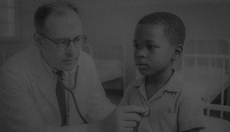
By Gary Katz and Jack Kussel
Gary Katz writes:
When we graduated, my knowledge of Paediatrics was limited. We had excellent teaching at Baragwanath and the Transvaal Memorial Hospital (TMH). We were exposed to a vast array of clinical cases, unmatched by what I have seen in the UK or the USA. Although the medical care in South Africa was excellent, the psycho-social needs of hospitalised children were not adequately met.
Medical care had advanced considerably in the first half of the 20th century. I never anticipated that this would be followed by exponential advances in most branches of medicine, including Paediatrics, in the second half.
As this is a personal rather than an academic review of the changes, this account of the changes in Paediatrics will not, by any means, be comprehensive. Several of our classmates (Avroy Fanaroff, Jeff Maisels, Martin Bobrow and Aubrey Milunsky) have been at the forefront of the astonishing advances in Neonatal Paediatrics and Genetics. We have had a glimpse of this in their biographies and notes on medical advances in their respective spheres.
During my Paediatric journey, I’ve been interested in several aspects of Paediatrics. The advances in many of the specialities have been breathtaking, but in some, the changes have been limited. Unexpected and unwelcome new diseases have also emerged.
Paediatric cardiology: In our student days, Rheumatic Heart Disease and Paediatric Cardiology were virtually synonymous. We soon became skilled in auscultation, able to distinguish the murmurs of mitral and aortic valvular disease. We were taught about congenital heart disease and ‘Blue Babies’ and their poor prognosis, but we hardly saw any patients. Paediatric cardiac surgical success was limited to ‘extra-cardiac’ conditions. The successful ligation of patent ductus arteriosus (PDA) in the late 1930s marked the ‘birth’ of Paediatric Cardiology. During the 1940’s more complicated extra-cardiac procedures followed, including surgery for Tetralogy of Fallot, Coarctation of the Aorta and Pulmonary Stenosis.
In the year we started Medical School, the first successful repair of an intracardiac defect was achieved in the USA using cardiopulmonary bypass. As a Resident in New York in 1962, I was given the dubious honour of assisting the Paediatric Cardiologist at his 06h00 Friday cardiac catheterisation sessions. Diagnostic catheterisation is now almost redundant in Paediatric Cardiology, following the introduction of echocardiography,
colour flow Doppler and MRI. The high mortality rate of Paediatric Cardiac Surgery has dropped dramatically following the contributions of cardio-pulmonary physiologists, greater knowledge about the cause and pathogenesis of cardiac malformations and referral to specialist cardiac centres. Prevention is coming to the fore; children at risk for adult cardiovascular disease are being identified, and appropriate preventive strategies are being followed.
Bacterial meningitis: When we were students, intensive care units did not exist in the hospitals in which we trained. Several of our fellow graduates became intensivists. Andre Van As was involved in the development of Intensive Care at The General. Avroy and Jeff, as indicated earlier, have made major contributions to neonatal intensive care. In the UK, Paediatric Intensive Care followed the introduction of Paediatric Cardiac and Neonatal Intensive Care, resulting in major improvements in the treatment of children with acute respiratory and neurological diseases as well as sepsis.
Meningitis has always been one of the major acute illnesses that General and Neonatal Paediatricians have had to treat. We have been fortunate to train and work in the antibiotic era. With the introduction of antibacterial Sulphonamides in the 30’s and the antibiotics in the 40’s, it became possible to cure children with Meningococcal, Pneumococcal and Haemophilus meningitis. Nevertheless, when we were students and trainees, the mortality and morbidity rates remained high. Improvements followed as new intravenous antibiotics were developed. Despite early diagnosis and prompt antibiotic treatment, many children, particularly those with meningococcal septicaemia, continued to die.
After antibiotics, the next major breakthrough was the introduction of vaccines for Haemophilus B, Pneumococcal and Meningococcal C infections and in 2015, Meningococcal B and MenACWY (which protects against 4 strains of the meningococcal bacteria A, C, W and Y). Although the incidence of meningitis is at an all-time low, cases of meningococcaemia still occur.
The mortality rate has fallen as a result of the early and vigorous management of sepsis in Paediatric and Neonatal ICUs. In the UK, the overall incidence in 2017/2018 had fallen to 1 in 100,000. In infants, there has been an increase from 11 in 100,000 in 2016/17 to 16 in 100,000 (MenB is the most common pathogen). In children, there has been a decrease from 5 in 100,00 to 4 in 100,000. In 2020, a Paediatric Houseman could spend 6 months in a busy children’s ward without seeing a child with meningitis; unthinkable in the 60s!
Cancer: When we were students and trainees, the diagnosis of cancer in children was usually a death warrant. In the 70s, improvements in chemotherapy, radiotherapy, and surgery brought incremental improvements in the outlook. Between the 70s and the 90s, life expectancy increased by almost a decade. Over the past 30 years, the five-year survival rate has increased to more than 80% of the children treated for cancer.
The greatest advances have been made in the treatment of leukaemia and lymphoma. Younger people have a better prognosis. Over 90% of children aged 14 or less will survive Acute Lymphoblastic Leukaemia for 5 years, compared to 70% of patients between 15 and 24. The 5-year survival rate for Acute Myelogenous Leukaemia is lower (60-70%). Despite these advances, adult survivors of childhood cancer have an increased risk of long-term morbidity and premature death. A recent study showed as much as a 28% gap in life expectancy for survivors of childhood cancers diagnosed between 1970 and 1980. Recent advances in treatment, based on DNA analysis, bone marrow transplantation, and tailored and targeted treatments, are leading to improved cure rates and life expectancy.
Preventive measures: Although the emphasis during our training was on treating children’s illnesses, the focus now also includes preventive measures. Over the past 60 years, we have witnessed dramatic breakthroughs in disease prevention. This has been achieved through two major approaches:
• Immunisation and Screening: As students, we learnt about the use of vaccines to reduce the incidence of smallpox, rabies, whooping cough, diphtheria, and tetanus, although the adverse effects of some early vaccines limited their use. During our medical student years, the introduction of the Salk vaccine resulted in a dramatic reduction in polio cases. Since graduating, we have witnessed improvements in DPT vaccines (Diphtheria, Pertussis, and Tetanus) and the introduction of vaccines for common diseases such as measles, mumps, rubella, hepatitis A and B, chickenpox, and rotavirus. Screening has also helped to change the face of Paediatrics. In my early years as a Neonatal Registrar, I spent considerably more time
performing exchange blood transfusions for Rhesus incompatibility than ventilating neonates. The injection of anti-D immunoglobulin to Rhesus-negative mothers had an immediate and major impact, a breakthrough detailed in Jack Kussel’s article below.
• Screening: Screening has also helped to change the face of Paediatrics. In my early years as a Neonatal Registrar, I spent considerably more time doing exchange blood transfusions for Rhesus incompatibility than ventilating neonates. The injection of anti-D immunoglobulin to Rhesus-negative mothers had an immediate and major impact (This breakthrough is detailed in Jack Kussel’s article, below).
Pre-natal, intra-uterine and neonatal screening (clinical and blood spot) has led to the early detection and treatment of many conditions, including congenital heart disease, congenital dislocation of the hip, hypothyroidism, sickle cell disease, cystic fibrosis and several rare metabolic diseases, which have fatal outcomes without early intervention.
• Improving Maternal Health: Involves steps mothers take to stay healthy before and during pregnancy, which significantly increases the chances of having a child without a birth defect. A striking example is the reduction in the incidence of spina bifida in infants whose mothers take folic acid. However, the foetus can still be harmed by other nutritional deficiencies, as well as exposure to smoking, alcohol, drugs, and harmful substances.
What about other changes, and what does the future hold?: Major breakthroughs have occurred in many other specialities. The outlook for children with surgical, respiratory, gastro-enterological, haematological, neurological, metabolic and locomotor disorders has improved significantly. Specialities such as immunology and transplantation surgery, which did not feature in our curriculum, play a major part in 2020 Paediatrics. Relatively simple measures have striking benefits. The incidence of ‘Sudden Infant Death’ fell dramatically when the sleeping position of infants was changed from prone to supine. Death as a result of car accidents in infants fell by 70% after the introduction of car seats and by 45% in children aged 4-8, using booster seats. In older children, as in adults, seatbelts have significantly reduced the risk of death or serious injury.
Clinicians have been challenged by a number of viral infections that did not exist in 1960, such as HIV, Ebola and Zika, and I am sure others. Antibiotic resistance is now a major problem. Covid has demonstrated how international collaboration between scientists can result in the development of an effective vaccine in less than a year and bodes well for future developments.
What about the downside?: The major causes of childhood mortality when we were students were malnutrition, TB and Malaria. Sadly, these remain the major causes in Africa, Asia and Latin America. Malnutrition still causes 3.1 million deaths a year in children under 5. That is 45% of all deaths in this age group. The survivors of malnutrition are likely to be stunted, wasted and/or underweight. In so-called developed countries, food poverty and obesity are growing problems. Nearly 250,000 children still die of TB annually. BCG does not prevent TB, although it does protect children from major complications. A truly effective vaccine is urgently needed. The antimicrobial drugs developed in the 1950s and 1960s are still in use today, but are less effective due to drug resistance. Malaria is still responsible for 400,000 deaths a year. The Oxford Vaccine Research Group, which speedily developed an effective Covid vaccine, hope to have a suitable vaccine available for Malaria in 2021/22.
Mental health: Although the progress in this branch of Paediatrics has not matched the rapidity of the advances we have seen in other Paediatric specialities, Child Psychiatrists and Psychologists have worked tirelessly to improve the mental health and welfare of children and adolescents. The teamwork that now exists between Psychiatrists, Psychologists, Paediatricians, and Social Workers has led to a major improvement in the psychological needs of children and adolescents in hospitals and the community, in contrast to the provisions we witnessed during our student days. The tasks that lay ahead are nevertheless daunting. Our newspapers highlight issues on a daily basis. Despite attempts at ‘Safeguarding’ physical, sexual and emotional child abuse, sadly, it is rife. Eating disorders have increased in developed countries, and there is worldwide child extortion and slavery. I am optimistic that the efforts of Child and Adolescent Psychiatrists, Psychologists, Paediatricians, and Social Workers will ensure that, in time, these issues will decrease significantly.
Jack Kussell writes:
Highlights of the changes that have occurred in my career over the past 50 years. I practised general Paediatrics with Neonatology as a sub-speciality in my career, and here are some of the marked changes I have witnessed.
Neonatal Jaundice: The management of severe Haemolytic Jaundice of the newborn was an exchange transfusion. It was not uncommon for up to 5 exchange transfusions to be performed per week on different babies. At times, as many as 3 transfusions would be performed on the same baby over a period of three days. Over the past 10 years, I have not performed any exchange transfusions. What has brought about the change?
Recognising sensitised mothers after the birth of their first infant and giving them antiserum postnatally, the monitoring of antibody titres in sensitised infants during pregnancy, and the checking of the blood group status of every baby born at birth of a Rh-negative or Group O mother are essential steps. Bilirubin must be fractionated into indirect or direct bilirubin rather than just relying on the total level of bilirubin, and the serum proteins must be adequately maintained, with neonatal plasma given at times. Phototherapy has now become one of the main factors in managing all forms of neonatal jaundice. Finally, the introduction of Polygam (intravenous human globulin) therapy early on in badly sensitised infants has further reduced the need for exchange therapy.
These factors completely changed my approach to the management of haemolytic jaundice of the newborn.
Neonatal Respiratory Distress: The changes in the management of both Hyaline Membrane Disease and PFC (Persistent Foetal Circulation) have seen marked improvements in management over the years. In Hyaline Membrane Disease the advent of surface tension lowering factors such as Surfactant and Curosurf used early on by intubation have certainly decreased the need for prolonged ventilation and, according to some experts, claims have been made as to why ventilation would be necessary at all in a great majority of cases My early experiences of treating Hyaline Membrane ranged from giving oxygen and intravenous therapy. Then came the “Bird ventilator”, and with further advances in ventilation therapy, the introduction of intermittent positive and negative pressure ventilation.
As the realisation dawned on us that too much pressure or ventilation for too long could be a factor in contributing to subsequent broncho-pulmonary dysplasia, we started to introduce oscillation with or without nitrous oxide therapy. Now, finally, the pendulum has swung full circle, and we are managing many of our cases of Respiratory Distress with the quick introduction of intubation and the administration of surface tension-lowering factors, such as Survanta or Curosurf, followed by detubation and possibly no need for ventilation at all.
The management of the PFC infant with Respiratory Distress has also undergone many changes. The previous practice was administering oxygen as almost a routine part of the resuscitation. Today, the practice is rather withholding oxygen and allowing the saturation levels to fall in the first one or two minutes, thereby allowing the circulation to become balanced following the closure of the ductus.
We have changed from the previous practice of withholding fluids to now administering boluses of saline in the shunted infant. How dramatically the management has changed in both Hyaline Membrane and PFC over the past 50 years.
Vaccinations: I will quote a radio and TV advertisement regarding one specific type of vaccine. “Don’t hesitate –vaccinate”. The slogan should really be amended to “Hesitate before you vaccinate”. No one will argue that if we use vaccination to prevent mortality or severe morbidity, there can be no contraindication to that type of vaccine. I refer specifically to: Polio, TB (BCG), Diphtheria, Whooping cough (Pertussis), Tetanus 121, Meningitis (including meningococcal Menactra vaccine), Hepatitis and Measles.
In my opinion, these vaccines are totally overprescribed and should only be used for selective criteria. I believe that vaccines have impacted the ability of our T-cells and Lymphocytes ability to cope with more serious conditions due to our immune systems being bombarded with vaccines for diseases where it might be better off relying on normal immune mechanisms to cope with the disease processes themselves, instead of giving this to every child, and only use these vaccines for children at risk.
With the emergence of wild strains of Polio, Measles, and, undoubtedly, the constant mutations of many severe viral infections, such as COVID-19, it may be prudent in less compromised children to allow their own immune
systems to take over, thereby also helping to build herd immunity. Chicken Pox in our youth was considered an illness that we should all get in childhood so that we could then be conferred with lifelong immunity. Today, the chickenpox vaccine is routinely administered to some children as part of the vaccination programme. The result is cases are now being reported of chickenpox and Herpes infections in adults who have been immunised, and some are becoming seriously ill. To conclude, I have no argument against the first group of vaccines that I mentioned; however, in my practice, I “hesitate before I vaccinate” with the second group.
Avroy Fanaroff adds on immunisation: Immunisation is one of the most cost-effective and life-saving interventions, preventing over 2.5 million deaths annually. Unfortunately, over 20 million children living in underdeveloped countries still do not have access to vaccines or are under-immunised. Hepatitis B is a chronic infection that ultimately leads to cirrhosis, liver failure and hepatic carcinoma. Mothers can pass the hepatitis B virus to their baby during childbirth without knowing it, since it is passed through blood and bodily fluids. Immunisation, starting at birth, is highly effective in protecting the baby from hepatitis B, protecting others from the disease, and preventing the child from developing liver disease and cancer. The hepatitis B vaccine is both safe and effective. Greater efforts are needed to ensure that this vaccine is available worldwide.
Hilleman’s role: Today, few people outside the immunology field recognise Maurice Ralph Hilleman’s name. Unlike Louis Pasteur, who developed vaccines for rabies and anthrax, or Jonas Salk, who created one for polio, Hilleman (pictured right in about 1958) never basked in fame’s limelight, but he is credited with saving more lives than any one person in modern history. He created more than 40 vaccines, including the staple trio of measles, mumps, and rubella (MMR), as well as hepatitis A and B, influenza, and chickenpox, to name a few. Experts estimate that Hilleman’s vaccines save about 8 million lives a year. He was motivated by one thing — he wanted to try and make vaccines for every disease that could possibly hurt or kill a child.”
By Len Kahn and Alf Mauff
Leonard Kahn writes:
Pathology highlights from 1960 to 2020, per decade, include the transition from monocular microscopy to electronic whole-slide imaging sine microscopes; from evaluation of hematoxylin and eosin-stained tissue sections to molecular diagnosis; and from morbid anatomy/autopsy pathology to multiple subspecialties in pathology.
1950s to 1960s: During our medical school years in the 1950s, our practical sessions in pathology required attendance at autopsy dissections, many of which were conducted under the tutelage of the feared and fearsome Dr Chatkidaikis; examination of histologic tissue slides was performed by peering down antiquated monocular microscopes under the benevolent guidance of mentors such as Dr Shirley Siew. Gerald Lampert and I enjoyed the challenge of reviewing the slides from our personal assigned slide boxes at my home, an exercise that initially sparked my interest in the field of pathology.
During my registrar years in the Department of Pathology at the University of Cape Town, we were provided with the then state-of-the-art binocular microscopes, featuring wide-angle eyepieces, and even those with attached cameras for photomicrography. Diagnoses were rendered in nearly all cases following evaluation of the H&E-stained sections with the addition of a few ancillary histochemical stains, such as mucicarmine for epithelial mucins when necessary. Difficult diagnostic cases were resolved following evaluation by senior staff members and, in some cases, by submission to so-called outside ‘experts’ in the field. Who could challenge their expertise? Until the advent of more sophisticated diagnostic methods, such as electron microscopy, immunohistochemistry, and molecular pathology, in the following decade.
1970s: Electron microscopic evaluation of tissue was introduced into the diagnostic repertoire in the 70s, enabling examination of cellular organelles at magnifications a quantum leap above the x1000 of the light microscope. In addition to its importance in advancing the diagnosis of glomerular lesions, it facilitated more stringent diagnoses of lesions such as amelanotic melanoma (characterised by intracytoplasmic melanosomes) and neuroendocrine lesions (characterised by intracytoplasmic dense core granules). I especially enjoyed performing EM examinations in the hushed and darkened ambience of the EM suite. I recall the excitement of identifying intracytoplasmic neurosecretory dense core granules and neurofilaments in a nasal
esthesioneuroblastoma and having the findings documented in one of the early editions of a newly published journal, “Human Pathology” (1).
1980s to 1990s: The 1980s and 1990s witnessed the introduction and development of immunologic evaluation of tissue sections by targeting them with antibodies to specific antigenic cellular components. The coupled dye used to visualise a positive reaction was usually brown, hence the appellation ‘brown stains’. The identification of cellular antigenic components rendered electron microscopic evaluation obsolete, with the exception of renal disease. For example, the diagnosis of an amelanotic melanoma no longer required time-consuming and expensive evaluation by an electron microscope; the simple application of immunohistochemical antibody stains to melanocytic antigenic cellular components, such as Melan A and HMB45, does the job. We currently have a plethora of such antibodies in daily use. I may add that during this period, the complexities of the immune system were facilitated by the development of a variety of antibodies to our immunocytes, which spawned new classifications of neoplastic diseases of the lymphoid system.
The new millennium – 2000 and following: The advances in molecular technology in the new millennium has added additional armamentarium to tissue diagnosis. By techniques such as fluorescent in situ tissue hybridisation (FISH), it is now possible to identify genetic aberrations associated with specific diseases and neoplastic conditions, e.g., the translocation X-18 is a definitive diagnostic marker for synovial sarcoma. As with immunohistochemistry, we now have access to a wide range of such molecular alterations to assist in definitive tissue diagnosis. Current practice in pathology resorts to a combination of the above-described methodologies. Initial examination of the H&E-stained tissues is usually all that is required to render an accurate diagnosis (‘the gold standard’); further evaluation not infrequently requires the addition of immunohistochemistry, and in a small number of the most difficult cases, molecular evaluations are performed. We have thus moved beyond the ‘old days’ when difficult diagnoses required the imprimatur of ‘world experts’ to a more scientific approach to such cases.
A recent innovation in both radiology and pathology involves the use of electronic telepathology for teaching and diagnostic services. It is now possible to prepare whole-slide images and then view them electronically at various magnifications in exquisite detail. Such images can be shared in real-time from the frozen section laboratory and even between institutions at remote locations. There are currently laboratories in multihospital systems that have deemed the microscope outdated and practice exclusively from electronic computer monitors in their offices. In the not-too-distant future, microscopes will no longer be required to practice our profession!
A word about the burgeoning of subspecialties in pathology, from the primacy of the autopsy in the 50s to the development of subspecialties such as ‘surgical pathology’ to meet the needs of the general surgeon [guiding the surgeon’s hand (2)] in the 70s as well as the evolution of fields such as cytopathology, neuropathology, hematopathology, dermatopathology, gastrointestinal, genitourinary, musculoskeletal, oral and maxillofacial, renal and hepatopathology, as well as prior well established fields including hematology, microbiology, chemical pathology and blood banking. Several of these specialities have board-certified fellowship programs in the USA with related qualifying examinations.
My own interest in musculoskeletal and oral and maxillofacial disease was stimulated by my fellowship training at Washington University School of Medicine under the tutelage of a giant in surgical pathology and especially skeletal disease, the late Dr Lauren V Ackerman and at Long Island Jewish Medical Center by Dr Leon Eisenbud. It led to my decades-long membership in the prestigious International Skeletal Society. I was the organising chairman of the international meetings for nearly two decades and was awarded the Society's highest accolades, the Medals of the Society and the Founders medal in 2002 and 2008, respectively.
In conclusion, a career in pathology offers a career in medicine with a strong clinical component including interaction with surgical colleagues during performance of frozen sections as well as a strong scientific bent. It is no wonder that so many of our class of 1960 chose pathology for their careers – Stan Zail, Alf Mauf, Naomi Livni, Gerald Shulman, Michael Berger and myself - considering that a relatively small number of pathologists are required in relation to other fields in Medicine.
Alf Mauff writes:
Progress in Pathology: When I joined the small practice of Irving, Schrire and Zent in Lancet Hall, Jeppe Street in 1966 our pathology procedures at this stage were based on such technical procedures as flame photometry, calorimeters, van Slyke apparatus for CO2, no histology or cytology, white cell counting chambers, and immunology consisted of VDRL and TMX agglutination and pregnancy tests using Xenopus frogs.
Venesections were done using glass syringes, and all reports were made by typists. Our support base was confined to a small central area of Johannesburg and a few small central hospitals. About a year after I joined Lancet (Drs Irving, Zent, Schrire and Gon), I remember the day when we celebrated our 300th requisition for the day.
These days, The Lancet receives more than 26,000 requisitions per day. Monthly, they get 550,00 requisitions and carry out 3.1 million tests.
I am certain that today their volume and range of tests offered is bigger than the South African Institute for Medical Research, which was the very big brother of pathology for many years.
I loved the laboratory work and am always grateful for the massive technological developments that we were able to incorporate. It was very exciting.
Here is a little story. Our practice at that stage was in the Lister building, Jeppe Street, towards the death throes of the Apartheid era, with the ANC headquarters nearby. At some stage, the whole area was razor-wired off, surrounded by police and bullets smacking off slabs of concrete from our building. Our terrified staff and messengers had to negotiate this bizarre situation, and truthfully, we were close to shutting up shop and leaving paradise. Well, as good South Africans say: “Anhou wen!!”.
Today Lancet offers services throughout South Africa and 12 African countries. The technical advances are numerous and diverse, resulting in fully automated chemistry and haematology laboratories.
New additions now, include histology and cytology using immunochemistry, in situ hybridization and molecular techniques, coagulation, cytogenetic, pre-natal genetic screening and molecular biology laboratories offering 80 different diagnostic molecular tests using PCR, DNA sequencing and in situ hybridization, P3 TB lab with GeneXpert technology, virology including molecular assays, serology and cell cultures, metals and industrial toxicology, HbA1c and liquid chromatography laboratories.
Notwithstanding the numerous technical advances, a private laboratory such as Lancet could never have developed without the advent of the computer, the single most significant technical ‘medical’ revolution for a pathology stand-alone practice such as Lancet.
The computer enabled us to integrate with a number of other distant pathology laboratories, as well as establish both large and small laboratories in hospitals and smaller towns. The new large volumes gave the Lancet, which receives no financial grants, the financial muscle to purchase and install the ever-increasingly sophisticated and expensive technology.
Hand in hand with these technical advances, the administrative side of the practice would also be impossible without computerisation. Proficient administrative staff for accounts and financial management, in-house computer laboratory, medical aid and staff pension funds, waste removal, messenger collection services requiring vehicles and maintenance, storage facilities, etc, that ultimately and finally, notwithstanding the excellent administrative staff, depend on pathologist supervision.
By Gloria Davis
When I graduated in 1960, I don’t think I had ever given much thought to allergy and its associated problems. I knew my cousins all had hay fever, and there was asthma in the family, but I had never met anyone with a food allergy.
So, when my second son, Ian, was born in 1963 in Port Elizabeth and he vomited after tasting pineapple pudding at age five months, it took a second episode – accompanied by a perioral rash – for me to twig that maybe this was a reaction to the raw egg whites in the pudding.
Shortly after this, Ian developed eczema, reactions to peanuts and had an asthma attack on his first birthday. So, my search began for someone knowledgeable about allergies to advise me on what to do. The only medications available at the time were antihistamines (Allergex, Polaramine and Phenergan), aminophylline, adrenaline, cortisone and Celestamine.
Skin Prick Testing: Skin prick testing by Bencard was recommended as part of the investigations, but desensitisation was carried out with sometimes fatal consequences. I tried to stop antihistamines for my son so that we could skin-prick test him. I did this by taking him to the Karoo. Why? Because everyone with asthma who lived there improved dramatically. Many children from Port Elizabeth attended school in Cradock. This was my first introduction to allergy and the environment. I had also noted that when we visited Johannesburg, he improved significantly.
I sought advice from Professor Ordman, a pioneer in allergy research in South Africa, especially known for his work on house dust mites, which he had laboriously counted under the microscope in various areas of South Africa.
He recommended a diet consisting of rice, chicken from a rooster, bacon and pears. This diet only resulted in an unhappy boy and mother, as well as weight loss.
A physiotherapist designed a swing which would extend Ian’s arms and ‘open’ his chest. There was no benefit, unsurprisingly. The only medication that helped was aminophylline, given 4 to 6 hourly, including waking him at night for a dose.
When Ian was five years old, I was told by a friend to visit an elderly doctor in Kimberley who had helped with her son’s asthma. Off we flew and met him for half an hour. He had practised in Cape Town and couldn’t get over how few asthmatics he was seeing in Kimberley. It must be the environment, he said – my second reference to
the environment. He instructed me to remove all the furniture from Ian’s bedroom, except for the bed, to take him out of nursery school, to cover the lawn at home with sand, and to put our Alsatian dog in the kennels. He prescribed a daily dose of half a Nethaprin Dospan tablet. Ian did improve and by the age of six could go to school, but was not allowed to play sports. He himself was very aware of what he should not eat. Such was the state of allergy knowledge and treatment in the 1960s.
Dr Eugene Weinberg at the Red Cross Hospital, Cape Town: Our lucky break came when I was told about a doctor at Red Cross Hospital, Dr Eugene Weinberg, who was very interested in allergies. I arranged to meet him at his clinic, and what a joy it was to find someone as interested in allergy as I had become, and so experienced and generous with his knowledge. We became good friends from then on, and he went on to become the father of allergy in South Africa.
Dr Weinberg was the doctor who emphasised the need for a good history, examination and correct interpretation of tests. He introduced me to sodium cromoglycate (Lomudal), the first preventative drug in asthma. The primary device was a spin-haler, which had to be used four times a day. I never had to remind my son to use it. He began playing sports, at first as a goalkeeper in soccer, and gradually, tennis and cricket, at which he excelled.
Treatments in the 1970s: In the 1970s, the ‘miracle’ of bronchodilators (salbutamol and fenoterol) occurred. This absolutely revolutionised the treatment for asthma and quality of life. It could be used pre-sport if necessary. It could be used for any ‘tightness’ and was an effective tool for assessing whether asthma was controlled or not, based on the number of times it was required to be used for symptoms, both day and night. Combined with the use of a peak flow meter, a mother’s anxiety about her child’s possible attack was greatly lessened.
Treatments in the 1980s: In 1980, when my husband and I visited the USA, I managed to visit a hospital in Denver, Colorado, which was then the asthma capital of the USA. To my amazement, salbutamol was not available. It had not been approved by the FDA, and many patients went to Canada to purchase it. In 1982 we left Port Elizabeth and returned to live in Johannesburg.
I was very fortunate to be given a post in Dr Mike Plit’s unit at the JG Strydom Hospital (now the Helen Joseph Hospital), which was considered to be the best pulmonology unit in the city. He had taken over from Dr Andre van As, who had established the unit and its excellent reputation. Both were members of our 1960 graduating class.
There, every patient was studied thoroughly for asthma severity and underwent lung function tests, specifically spirometry for asthma, unless there was other lung pathology, when full lung function testing was done. At first, knowledge and technical know-how were poor. I saw many results that were technically unacceptable. I discussed such reports at a conference where I presented examples to the amusement of all, but the trained technologists were delighted. Thereafter, teaching on lung function testing was initiated.
Skin prick testing was performed on every patient. I was troubled because nobody paid much attention to the results. I was teased and called the ‘cat hater’. All
All patients had to complete charts with details of medication and peak flow readings, especially those who were uncontrolled. Today, most of the patients I see have never seen or been prescribed a peak flow meter, and reports I receive from GPs invariably have never recorded peak flows of patients together with postbronchodilator responses.
We conducted trials on ketotifen and long-acting theophylline, the latter of which was used extensively but became less useful and is now mainly prescribed for chronic obstructive pulmonary disease (COPD). Managing to maintain optimal drug levels was a constant difficulty, plus side effects, and they were being superseded by better medication, the greatest of which at that time was inhaled cortico-steroids, for both adults and children. There were some physicians in South Africa who were critical of beta-agonists and the use of topical steroids. It took hundreds of trials to convince practitioners of the safety of both. At the time, correct technique and compliance were not of major concern. Inhalers were pressurised, and the gas was eventually barred from use. The advent of dry powdered inhalers and trials on comparable efficacy began in 1987. Another step forward, which gave relief to millions of sufferers, were the long-acting and non-sedating antihistamines. Many sufferers of allergic rhinitis could now continue with their occupations, not bothered by sedation. These, and subsequent new antihistamines, are the mainstay of treatment for acute allergic reactions,
rhinitis, and urticaria. They are the first drug to be given for anaphylactic attacks, followed by adrenaline, which is now mainly delivered in an EpiPen – a useful but very expensive tool. Apparently, the price was increased to such an extent in the USA that there was an outcry and a call for a possible alternative device. During all this activity, spacers were being introduced to enable better distribution of the drugs in the lungs. These ensured adequate dosage of the medication, particularly in infants and children. Nebulisers also became very popular as a means to ensure correct distribution.
I need to mention now the work of Professor Zar from the Red Cross Hospital in Cape Town. I had read about the American work in trials to prove which was better to use in asthmatic attacks: nebulisation or spacers. It was proven beyond doubt that spacers were better, not only for distribution, but also for the short period of time required to administer, as well as for greater acceptance by children, and at a much cheaper cost.
Professor Zar not only accepted this, but she also designed spacers using plastic 500ml cool drink bottles and disposable plastic masks from the hospital. The mask fitted over the narrow top of the bottle. The inhaler was inserted into an opening made at the bottom of the bottle, using a hot wire. Studies proved that the distribution was excellent. Patients from overcrowded, poor townships who could not afford nebulisers and who had no electricity now had a device that could be used for asthma exacerbations. More importantly, they could use these devices for better administration of the drugs, particularly in young children. During the 1990s, long-acting bronchodilators were introduced. Salmeterol and formoterol could be used alone or in combination with corticosteroids. Doubt was expressed about the safety of these drugs, but when used with steroids (and not alone) results were excellent. The combination of the long-acting bronchodilators with corticosteroids was better than either medication used alone at higher dosages. The new, technically excellent dry powder devices that accompanied these drugs once again ensured correct drug delivery in those patients old enough to use them. Many trials were done to compare the various inhaled corticosteroids. All were comparably excellent.
A new drug also appeared. This was a leukotriene receptor antagonist, which was particularly beneficial in aspirin-sensitive and non-atopic asthma. It was also useful in young children and was prescribed for resistant asthma that required persistent oral steroids or was difficult to treat.
Meanwhile, dozens of chemokines and cytokines were being discovered, and it would be impossible to make receptor antagonists for them all.
I have described advances in asthma, but all allergic diseases were progressing in diagnosis and treatment, including rhinitis, dermatitis, urticaria, and food allergies. In fact, no system in the body was not potentially affected by allergic phenomena or disease.
Testing became more specific: Technology, as in all modalities of medicine, was being applied in the study, diagnosis and treatment of allergy. Allergy testing has since become far more specific and this field is expanding rapidly with more accurate specificity and sensitivity. Combined tests are also available but the message to practitioners is still to take a good history and direct testing to those allergens which appear to be culprit proteins. Rast and Cast (basophil activation tests), mast cell tryptase levels and cytokine assays, antibodies to the IgE receptor, IgG blocking antibodies and food allergens are being studied constantly.
Food allergy has been abused in the past. Testing needed to be interpreted correctly as many a child or even adult had been advised a diet on the basis of ‘positive’ tests which had no relevance at all. This is still happening. Professor Hugh Sampson in America (2001) published predictive specific IgE concentrations for common allergenic foods which has made interpretation of Rast tests far more accurate. He also detailed the size of skin prick test results as to what was relevant.
The establishment of the Allergy Society of SA in 1988: In 1988, a great event for allergy in South Africa took place – the Allergy Society of SA (Allsa)was born. Up until then, we had attended and presented at pulmonology conferences. In 1992, the first Allergy Conference was held, and thereafter, these events took place regularly, featuring international speakers and offering great encouragement for research. A working group on Allergic Rhinitis was formed in 1996. Guidelines for asthma in adults and children were formulated and regularly updated. The ARIA guidelines were published. Campaigns and associations were formed to educate patients and the public about allergies.
Allergy societies were formed all over the world, and a World Allergy Association was born. Conferences were held on every continent. Attendance gave rise to comparative studies and research, not only in treatment, but in genetics and genetic predisposition, trigger factors, pathophysiology, characterisation of physiological and cellular factors, the environment, anti-IGE and biologics.
Food Allergy: As mentioned above, food allergy is a major concern. With more accurate diagnosis and improved vaccines, desensitisation is being used even in severe peanut allergy. The latter, so far, has been able to decrease fatal reactions in children to less severe attacks.
I also need to mention the changes which were taking place in the political sphere. In 1989, the entire academic unit at the JG Strydom Hospital (where we were working) resigned. The government wanted to maintain it as a ‘whites only’ hospital. We had been inviting doctors from the nearby Coronation Hospital to join us in ward rounds and in consultations. This was unacceptable to the government, and it was stopped. Wits University withdrew all academic association from the JG, and all the doctors resigned.
Chris Hani Baragwanath Hospital – political trauma in the 1980s: I was talked into going to Chris Hani Baragwanath Hospital by Dr Jeff Lipman, who was the head of the ICU and had been appointed to Bara. It is the only hospital in South Africa’s largest township of Soweto. It is the biggest hospital in the world, sprawling over 173 acres with 429 buildings and 10 miles of corridors. It has approximately 3,000 beds, serving around four million people.
I must mention the scandalous behaviour of the government towards the doctors at Baragwanath in 1987, prior to our arrival. This revolt began in 1979, when issues of overcrowding, a shortage of doctors, premature patient discharges, and inadequate facilities for patient examination were highlighted in the press. The government denied all of this, but there was some progress when two new wards were built.
In 1987, matters came to a head when a letter describing the dismal and deplorable facilities at Bara, signed by 101 doctors in the Medicine Department, was published in the SA Medical Journal. The allegations were refuted. Signatories were then refused posts. A letter of apology was demanded from all of them and then the situation would be reversed. Some were pressured into signing. Six sought an urgent interdict; the court decided their applications could not be rejected without a hearing. Eventually, one of the doctors, Dr Beverley Traub, took her case to court. It was ruled by Judge Goldstone that she had to be employed immediately. The administration took the case to appeal, but it was turned down. Dr Traub won her case.
Public interest was piqued. Professor du Plessis, our professor of surgery (who by then was the Vice-Chancellor of Wits University), and several eminent specialists made recommendations and expressed their admiration and support for the staff at Bara. The government responded that there was no money for the hospital, but agreed that private funding could be sought. Private enterprise and captains of industry were approached and, in no time at all, new ‘wings’ were built. Thus, facilities for patients and doctors were improved.
My role for Dr Lipman was to continue with teaching rounds for anaesthetic registrars. These had been initiated at the JG Strydom and had proved to be highly successful. For their final exam, the registrars had to diagnose and assess patients for risk of anaesthesia. I became involved in choosing patients and helping them with the assessments. Again, it was a highly successful venture. I also joined the Medicine Unit, primarily to work with asthma patients.
During my first year at Bara, although the wards had improved, I was still shocked at the conditions for both outpatients and doctors at this huge and very busy hospital. I felt fortunate that Dr Lipman had welcomed me into the ICU department and provided me with an office in which to work.
At the out-patients department (OPD) we would sit three doctors and three patients at a table. No decent examinations could take place. Notes were scrawled in files, and there was no one to ensure that patients understood how to use their medications. As a result, the readmission of patients with asthma was unacceptably high. I presented the statistics at a Medicine Department lecture and doctors were shocked at the dismal results.
At this time, a drug company offered funding of R1 million for a new asthma/pulmonology unit. I persuaded them to allocate a portion of the money, convinced management to provide us with a prefab ward, and renovated it
into a more pleasant area in which to work. This was not an easy endeavour. I even had to take the head of the pulmonology unit to Cape Town and to Stellenbosch to see how their units were organised and to accept that Bara needed similar arrangements.
The ‘Golf Round’ : As an aside, the teaching work I was doing was remarkable. The variety of cases that were available, the clinical signs, and the range of diseases were unrivalled. I used to wear my ‘walking shoes’ every Friday morning – I called it my golf round – to visit all the wards to choose cases for the afternoon rounds. During one teaching round, a case of aortic valve incompetence was presented, and all possible signs were present. One of the registrars, who hailed from London, sat down in utter amazement and said she had never seen anything like this before. I taught on a case of AIDS at the onset of this rampant disease. The students diagnosed heart failure but missed recognising the piece of plaster on the chest just below the sternum, where a pericardial tap had been done, and which made the diagnosis of TB pericarditis. Perhaps we did not fully appreciate the wealth of teaching material at this hospital.
Black Asthmatic Children: Due to a nasty work relationship, I was compelled to leave the adult asthma patients. I then joined the paediatric clinic, and this was the start of my research into black asthmatic children, their allergies proven by skin prick tests and family histories.
Together with Dr David Luyt, paediatric intensivist, we created questionnaires which were entered into a computerised filing system. This allowed us to record good histories and allowed me to enter my skin prick test (SPT) results. To my surprise, these children were as allergic as their white counterparts. I compared the Bara results to those of the asthmatic children at the JG Strydom asthma clinic. Up until then, there was a belief that black children were non-atopic (positive specific antibodies to common allergens). About 50% were positive for house dust mite, an allergen found in such small amounts in the dry highveld areas (Professor Ordman’s findings) that when the test was positive, it was believed that patients must have become sensitised during a visit to the coast or inland humid areas. This was the explanation for Kimberley and the Karoo being beneficial for coastal asthma, particularly if the allergy in the patients was caused by house dust mites or pollen. The work was published, and we presented the results of the SPTs, the small amounts of mites found in the houses of these patients, and the lack of a positive family history. I believed that the conditions in which the children were living, the sprawling urban area of Soweto, played a major role in this epidemic of asthma.
This work initiated a great deal of research on black patients; simultaneously, interest in the rest of the world had also become focused on the environment and exposure to allergens. A landmark study in Germany showed that there was more asthma in West Germany than in the industrialised cities in East Germany. It was not pollution, but urbanisation, that was one of the causes.
There was also extensive research in the field of genetics, with no specific genes identified for asthma or other allergic diseases. It proved how complicated allergy was.
Higher qualification in Allergy: There had never been an opportunity to gain a higher qualification in allergy in South Africa. In 2003, the first examination was held for a diploma in Allergology. After so much dedication to this field of work, it was with some satisfaction that, more than 40 years after obtaining my medical degree, I could add this qualification to my CV. A specialist degree in allergy is currently offered. How different to 1960!
Research: Research is now a huge part of the allergy world. New drugs have been developed, including anti-IgE, non-sedating antihistamines, new corticosteroids for inhalation, as well as bronchodilators and biologicals. Desensitisation is much safer, and new vaccines will be targeted at specific antigens and their components. Food allergies are better managed. Penicillin allergy is now being treated with rapid desensitisation. Severe peanut allergy desensitisation is also becoming more common, although it has been difficult to go beyond rendering the allergic reaction less severe. Interest in Vitamin D, probiotics, the microbiome and biologicals, which need refinement and further research, abounds. It is with great satisfaction that I observe the enormous interest now in the allergy world. To my joy, parents do not have to suffer the anxiety that I endured in 1963. One hopes that, correspondingly, doctors will apply their minds to each patient and be aware that “the secret of the care of the patient is in caring for the patient” (the late Professor Asher Dubb).
By Andre van As
The Evolution of Respiratory Medicine in South Africa: Health care in South Africa prior to WW2 was largely in the hands of the private practice sector. In 1946, the Transvaal Public Ordinance introduced free hospitalisations for all Provincial residents, and full-time, paid clinical staff were to be employed jointly by the University and the Transvaal Provincial Administration (TPA) to provide medical services in the hospitals and outpatient departments. Each clinical department consisted of a core of full-time staff. Wits University established full-time clinical chairs in association with the Johannesburg General Hospital in the clinical disciplines of surgery, medicine and obstetrics and gynaecology. The three incumbents were required to reapply for these posts, which they declined. Guy Abercrombie Elliott replaced WH Craib as Professor of Medicine, and he played a pivotal role in developing the services for patient care at all of the academic hospitals, as well as undergraduate and postgraduate teaching services.
In 1950, the Cardio-Pulmonary Research Unit, funded by the CSIR, was set up by Professor Guy Elliott as part of the first series of Council for Scientific and Industrial Research-funded medical research units in the country, attracting Maurice McGregor and his wife, Margaret (Margo) Becklake, both Wits graduates, back to the country from Canada to develop the Cardio-Pulmonary Research Unit. They had both undertaken postgraduate training in England (Margot in Respiratory Medicine and Maurice in cardiology). She accepted an appointment as a junior lecturer in Medicine at Wits University. Margo, a highly accomplished physician, developed the pulmonary function laboratory, acquiring equipment to measure both static and dynamic lung volumes. A technologist, HI Goldman BSc, is retained to perform the pulmonary testing. Numerous research studies have been published in peer-reviewed journals. In 1954, Margo was engaged as a physiologist at the Pneumoconiosis Bureau in Johannesburg. She established one of South Africa’s first clinics to study lung function in gold miners and became an advocate for them, resulting in many miners receiving benefits they had previously been denied. Margo and Maurice returned to Canada in 1957, where she established a lung function laboratory and conducted research on exposure to chrysotile asbestos among miners and millers in the town of Asbestos, Quebec. As a result of the formation of a pulmonology group, this discipline continued to attract graduates to seek training.
Pulmonary Medicine in the late 1950s: After 1957, Pulmonary Medicine was supported by two graduates, Saul Zwi (class of 1953) and Clive Prowse (class of 1956). Zwi, after completing his internship and registrarship, spent two years in England at St. Stephen's and Queen Mary's hospitals, where he received training in Pulmonary
Medicine and pulmonary function. After returning to Johannesburg, he held appointments and consultancies in the Pneumoconiosis Research Unit of the CSIR, the Pulmonary Function Unit of the Johannesburg Hospital and the Thoracic Surgical Department at Wits.
Prowse, after completing his initial training, spent a year in Boston receiving training with renowned surgeon Edward Gaensler, a Thoracic Surgeon interested in the dynamic measurement of airway function, and who created the Pulmonary Medicine Section of the Boston University School of Medicine. Upon returning to Johannesburg, he also had appointments and consultancies at the Pneumoconiosis Research Unit of the CSIR, the Pulmonary Function Unit of the Johannesburg Hospital, and the Medicine Department at Wits. I recall interacting with both of them between 1964 and 1966 while I was involved in running the Medical ICU at the Gen. In 1966, Prof Elliott decided to create a position of Principal Physician to head Pulmonary Medicine at Wits. On the day of the interview, which was convened by him and several senior members of the department of medicine, the first candidate to be interviewed was Clive Prowse. At the conclusion of the interview, Elliott remarked that they should vote on whether to recommend the appointment. Tom Bothwell then stated that there was another candidate to be interviewed, and Elliott said he did not know about the other candidate. They called Saul Zwi in for his interview. When the vote was taken, the committee chose Saul Zwi, and he was appointed. Clive Prowse left the Gen and went into private practice in the Vanderbijl Park/Vereeniging area but retained his consulting position at the Pneumoconiosis Research Unit. Later that year, Elliott resigned and retired, returning to his hometown of Cape Town.
The JG Strijdom Hospital: In 1972, I was promoted to Senior Physician, and in 1973, I was appointed as Principal Physician & Clinical Head of the Internal Medicine Department at the JG Strijdom Hospital/Helen Joseph Hospital (JGS/HJH). The hospital was originally intended to be a teaching hospital for the Rand Afrikaans University, but those plans did not materialise, and Wits University took over the hospital, which was granted teaching status. I established a modern pulmonary function laboratory, which expanded the discipline of Pulmonary Medicine in Johannesburg. Additionally, I collaborated with Ben Goldberg to establish a hemodialysis unit, later managed by Tony Meyers. Our Pulmonary services rapidly became a tertiary referral center after the Cardiac Surgery Division was established in the JGS. We also initiated a bronchoscopy service, assuming responsibility for this task from the thoracic surgeons. We created a rigorous educational program of pulmonary physiology, a standardised History & Physical examination form, supervised bronchoscopy teaching, and lectures on clinical aspects of pulmonary diseases. The Pulmonary Function laboratory was one of the first state-of-the-art online labs in the world, and allowed us to do a variety of diagnostic tests.
Mike Plit returned to Johannesburg after serving in Israel during the Six-Day War. He and his wife, Yvonne, toured Europe and then returned to Johannesburg in 1968. He joined our Pulmonary division for six months before going into private practice as a pulmonologist in Hillbrow. We facilitated this by allowing Golie, our pulmonary technician, to spend several afternoons a week doing pulmonary function tests in his office. In this way, he expanded the availability of Pulmonary Medicine to private patients.
Establishing Pulmonology in South Africa: Pulmonary Medicine was expanding in Johannesburg under the leadership of Saul Zwi, who was appointed to an ad hoc chair of Respiratory Medicine at Wits in 1973. This led to his recognition as Professor of Pulmonology and Chief Physician, a position he held from 1985 to 1994. My association with Saul was very productive, as we published 11 papers on various aspects of pulmonary medicine over 11 years, from 1966 to 1977. In the 1960s, Attie De Kock was the founding member of the Pulmonary Division at the University of Stellenbosch, and Alex Fergusson at the University of Cape Town. Their immediate successors were James Joubert and Solly Benatar, respectively. Attie was responsible for founding the South African Association of Pulmonology in 1971 and was its first President. I served as Chairman of the Johannesburg Pulmonology Branch from 1971 to 1974 and as a Committee Member from 1974 to 1980. At that time, I had been appointed as the Principal Physician at the JG Strijdom Hospital (now Helen Joseph Hospital) and was responsible for revamping the clinical and teaching services. At the same time, I established the Pulmonary Unit and seized the opportunity to acquire a state-of-the-art, online, and fully computerised pulmonary function laboratory. It was one of a handful of similar labs developed by Erich Jaeger (Wurzburg, West Germany) in the Western world.
The South African Association of Pulmonology has been renamed the South African Thoracic Society (SATS) and is now a thriving organisation with at least 160 active members representing all of the major academic centers as well as private practice groups throughout South Africa. The SATS focuses on creating awareness of respiratory diseases as well as educating members and recommending diagnostic and treatment guidelines for the major and prevalent pulmonary diseases.
Looking back to 1950 and seeing that the goal we set out with, to nurture a pulmonary subspecialty in South Africa, has been realised and is sustainable, is very gratifying. It was not the result of an effort by one person but rather driven by an idea that was supported by many over the course of six decades.
*Phefumula is the Zulu word for ‘breathe’, which we learned from the daytime interpreters in our work in non-European hospitals. The interpreters were linguistic polyglots, often being able to communicate in six or seven African languages – all of which were different enough for many of the 14 tribes to adopt Zulu as a mode of universal communication. We had to learn several phrases to get the patients to do what we needed when there were no interpreters after hours. I think it pays tribute to the role that the African interpreters and patients played in our education when we worked in their hospitals. Many of those phrases are still ingrained in my mind and bring back many pleasant thoughts about the benefits that we experienced learning about a wide array of medical conditions that were never seen in the white, segregated hospitals.
During our diamond jubilee year 2020, infection and death figures soared worldwide, due to the COVID-19 pandemic. We witnessed the intense struggle of health workers, dealing with the dire situation. Our class can only be thankful that they never had to work in such circumstances. I am sure we all felt empathy for the doctors, nurses, and other frontline workers amid this storm.
Despite all the technical and pharmacological advances since the last pandemic 100 years ago, the so-called ‘Spanish flu’ from 1918 to 1919, this one had still caught most of the world unprepared and with little to offer in response. Governments have had an immense task in weighing the perils of breakdown in economic, mental, and physical health.
Vaccines were rolled out in record time in the developed world. In 2021, we were offered several kinds from different manufacturers. Most vaccines were gratefully accepted. Yet many individuals and groups refused to be inoculated. Vaccinated people still landed up in hospital with COVID, but the pandemic’s new strains from 2022, though more infectious, were becoming less lethal. Some of our cohort were not spared, but luckily, most have survived. But we surely all know and mourn for someone not so fortunate. The pandemic was particularly lethal for people in our advanced age group.
This project, connecting us with our classmates and learning about their lives, became a highlight of the lockdown. Many said that the ‘Class of 1960’ project had been a beacon of light for them in those dark times.
Over the years, our class has held various reunions, both large and small, informal and formal, around the world. Some were organised by the Wits Alumni Relations Office in Johannesburg for special anniversaries, while others were spontaneous gatherings of a few individuals. Below are some of the pictures that captured these occasions.
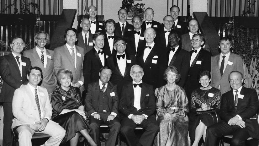
In 1985, 25 years after graduating, our class met for a reunion at the King Edward VII School’s Old Boys’ Club in Johannesburg. Our classmate Ephraim Dove, a physician in Johannesburg, was invited to help organise the event for the university.
So far, we have identified nearly all in the picture taken on the occasion – but nobody seems to remember very much about the event.
Back row: Kenneth (Roger) Edge, Ray Palmer, W (Bill) Wilson, Ivan McCusker
Third row: Leon Kahn, Gerald Lampert, Mike Plit, Tony Meyers, Irving Lissoos
Second row: Stan Zail, Theo Kretzmar, Mervyn Hurwitz, Anthony Crosley, Basil Kuming, Robert Otten, David B David Bengani, Dennis Roussow, David Paton
Front row: Ephraim Dove, Naomi Livni, Jack Kussell, Lawrence Willies, Jane Otten, Gloria Davis-Meyers, Michael Nissenbaum
The 40th reunion of the Wits Class of 1960 was organised by the Wits Alumni Relation Office and held in July 2000.
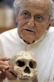
On a Saturday morning at the Wits Club, on the West Campus of the university, with a talk on A Philatelic History of Bio-Ethics by Professor Asher Dubb (who several of our class members listed as one of their favourite teachers). His hobby was stamp collecting and he illustrated many of his scientific papers with stamps. He was physician and head of the department of medicine at the Chris Hani-Baragwanath Hospital from 1964 to 2004. His talk was followed by morning tea in the garden of the club.
Next, Professor Udo Schuklenk, head of Bioethics at the Faculty of Health Sciences, spoke on the Ethics of the Doctor-Patient Relationship, after which the group was invited to take a bus tour of the Wits campus, which had expanded greatly into territory that used to house the Rand Easter Show and was now the Wits West Campus – the acquisition of which land in the 1980s was much attributed to the vision and hard work of our professor of surgery DJ du Plessis, when he became Vice Chancellor of the University.
The highlight of the reunion was a gala dinner held that evening in the elegant ballroom of the Hilton Hotel in Sandton. The guest speaker was Professor Phillip Vallentine Tobias, an alumnus of the class of 1950 – a man of international reputation for his exceptional scientific achievements. Tobias is also renowned for his vitality as a speaker and is remembered fondly by our class as a riveting anatomy lecturer in 1956.
This picture taken at the Jackson’s home when Avroy Fanaroff (Cleveland) visited London in 2002. From the left Avroy is with classmates based in London, Ronnie Auerbach, Allan Gottlieb, (Avroy Fanaroff), Gary Katz, and Rodwin Jackson.
Geoffrey Boner, who was present at the 45th reunion in 2005, wrote: “Three expats, Farol Sims, John Lee and I, joined our South African-based class members to celebrate the 45th anniversary of our graduation. The festivities started at a restaurant with an African theme located at the Zoo Lake.
The leading lecturer of the day was future Nobel prize-winner Sydney Brenner. Other lectures detailed the problems of asbestos and HIV in South Africa. The country had continued to mine and use asbestos for several years after most countries stopped its use. Asbestos was used in the construction of roads and playgrounds for children, especially in remote areas. Most of the workers in the mines came from outside of South Africa. If they developed pulmonary illnesses, only those who reported back to South Africa for examination were offered compensation.
Following the lectures and the visit to Baragwanath Hospital, we toured Soweto and had lunch in a local restaurant. In the afternoon, we visited the Apartheid Museum, the Bernard Price Institute of Palaeontology, and the Museum of the History of Medicine, where Phillip Tobias was on hand to sign his latest book, Into the Past: A Memoir. The festivities ended with an elegant dinner at a restaurant in Sandton.
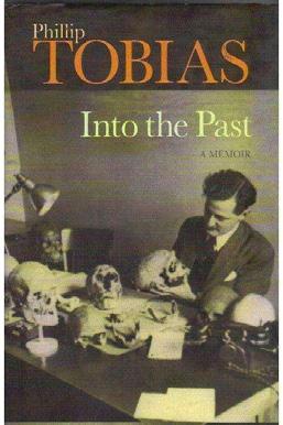
In 2020, I had taken a deep look at the picture of the 1960 graduating class on the steps of the University of the Witwatersrand, and I asked myself. “What has happened to those eager young people over the past sixty years?’ I could not have imagined how far we would get and how pleasurable it would be to reconnect with old friends and colleagues.
Close friends in the class, Avroy Fanaroff, Gary Katz, Ronnie Auerbach, Andre van As and others, joined me on this adventure. We all found that eliciting the fascinating stories of our colleagues thoroughly absorbed us during the forced stay-at-home of the worldwide COVID-19 pandemic. Learning about the success of the Class of 1960, as reported by folks in their mid-80s, has been among the most pleasing activities of my life. Being able to document (with the miracle of email and the internet) our time together at medical school and remember our special teachers and the libraries and hospitals that we worked in, and develop the profiles of over 50 of our colleagues and have them posted on a website for all to stumble upon, is something I could not dreamt of, nor would it have been possible 60 years ago.
Our contacts led to a strong desire to get together again and meet for a more social experience. Of course, COVID-19 and the almost universal lockdowns put paid to our travelling and meeting in person.
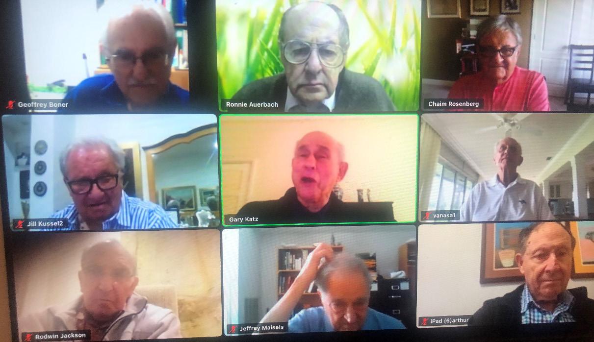
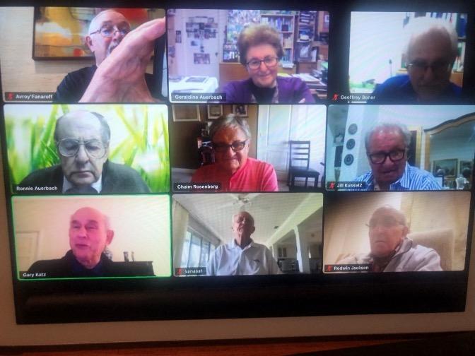
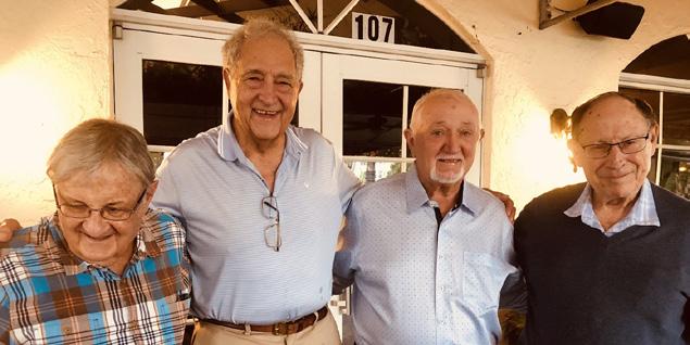
In 2022, when we were two years on from the diamond jubilee reunion of our graduation, we were delighted when, out of the blue, we received a long letter by email from a current medical student at Wits who had stumbled upon our website and took the time and trouble to contact us and give us his impressions.
This young man Taariq Hassim,
Good day Doctors
I am a current Wits Medical Student who stumbled across the website of the 1960 Wits MBBCh class. As of writing this email, I am in my final year with only a few weeks of clinical time left before (hopeful) graduation.
I would like to thank you all for the wonderful stories contained on the website. Reading through various newsletters offered a glimpse into Wits’ long and fascinating history. It allowed me to learn more about such historic figures such as Professors Raymond Dart, Phillip Tobias, DJ Du Plessis and Asher Dubb.
Professor Tobias sadly passed away in 2012, while I completed my 2nd year Anatomy course only in 2018. Professor Tobias’ (and Maurice Arnold’s) Man’s Anatomy is no longer used to teach students Anatomy; unfortunately, our dissection times have been cut down to once daily from what I believe to have been more frequent dissections in the past. Despite this, Professor Tobias’ legacy lives on as the main dissection hall in which most of the students dissect, bears his name, as does the main administration building for the Health Sciences Faculty, a pleasing and modern building in walking distance from the medical school in Parktown.
Many of the other mentors mentioned still have their names whispered along the corridors of the medical school and the associated hospitals. A senior Endocrinologist at Baragwanath told us the quality of his teachers, including Professors Shamroth and Dubb, while the former head of General Surgery at Helen Joseph Hospital told us how serious and frightening Professor Du Plessis was!
The former medical school in Hillbrow now serves as a base for Forensic Pathology if I’m not mistaken. Medical students would sit in for autopsies there, but this practice has stopped, possibly due to COVID. The main teaching hospital for students now includes Charlotte Maxeke Johannesburg Academic Hospital (CMJAH, formerly Johannesburg General) adjacent to the medical campus, as well as Helen Joseph Hospital, formerly JG Strydom, in addition to the mighty Baragwanath and Rahima Moosa Mother and Child Hospital, formerly Coronation. There are additional sites we visit during certain rotations, for example, Tara Psychiatric Hospital, an assortment of various clinics and community health centres (CHC) (including Alexandra CHC, as mentioned in one of the newsletters), and a 2-week rural public health rotation to Bushbuckridge.
The MBBCh curriculum has undergone significant change in the past two decades with the addition of the Graduate Entry Medical Programme (GEMP). Suitably qualified applicants with a Bachelor’s Degree or greater, and after sitting an exam, are allowed direct entrance into MBBCh III (now also called GEMP 1). Though I did not enter medicine through this pathway, many of my colleagues have. These students are often older, wiser, and bring fascinating stories and backgrounds with them - Masters Degrees, PhDs, decades of working in other environments, adult children, and the like.
One possible consequence of the GEMP is the unfortunate shortening of the clinical years; currently, only MBBCh V and MBBCh VI students inhabit the hospital, while the lower years deal more with preclinical
theory and some clinical skills lab-based practical teaching. A great deal of the curriculum has moved online due to the COVID-19 pandemic, which, together with the fire at Charlotte Maxeke Hospital, has severely affected academic activities. On a more hopeful note, the class now more closely resembles the population of the country and the patients we care for – a majority is female and of colour, which is a strong departure from an unfortunately racist past.
The Leech, the student-run Journal I had never heard of till your newsletters, is unfortunately no more. I have been unable to identify exactly when publication stopped, though I know of students from a decade ago who were still interested in continuing its legacy. The Wits Health Sciences Library (WHSL), the main library available to medical students on the Parktown campus, has an incomplete collection of Journal copies which I have enjoyed reading through. It seems a pity for such an excellent journal to have been discontinued.
I see the last newsletter entry is from January 2021. I know the gap has been considerable but do still hope for additional pieces to be added, especially so that future students can learn more about those who came before, their wonderful achievements, and the hardships we are no longer exposed to.
Kind regards,
Taariq Hassim, MBBCh VI (2022), Wits
It is thrilling to us that Taariq found the site and found so much interest in it that even 60 years later, it resonates with his life as a Wits medical student. It is very good to know that we can be so connected with the present generation of medical students. We appreciate that he has written to us, sharing his thoughts and opinions.
This is what our colleagues from the classes of ’61 to ’66 said
Almost one full year has passed since the 60th Anniversary Zoom Meeting celebrating the graduates of the Wits Medical School in my year, 1961. I was inspired by the example and grateful for the help of the 1960 team to reach out to graduates in my year. I was very pleased that we have captured the stories of more than 30 of those who graduated with me in 1961. It would be wonderful if the bios we received from the 1961 graduates could also be turned into a book alongside the digital format that already exists on our Class of ‘61 Website.
On the medical front, here in Ontario Canada, we are now in the 7th wave of the COVID-19 pandemic, the dominant virus now being Omicron and its recent variations. In addition, the ‘flu season is upon us, and there is a massive resurgence of RSV (respiratory syncytial virus) among the children in our population. As a result, our hospitals are running at total capacity, and children’s hospitals have opened up second Paediatric Intensive Care Units. These include the Toronto Sick Kids Hospital and the Children’s Hospital of Eastern Ontario (CHEO) in Ottawa, where one of our daughters is the Medical Chief of Staff. Masking in public places is now highly recommended, but not yet mandatory. (This latter state was the cause of a three-week debilitating occupation of Parliament Hill in Ottawa over the last Christmas and New Year Holidays). The anti-vaxxers also joined in with huge trucks, and brought the whole of downtown to a noisy standstill. Trucks honked their horns continuously day and night. A Federal order of State of Emergency broke up the stalemate.
On the personal side, my wife Toni and I moved to our present Condo in Guelph, Ontario, in September 2020 during the pandemic to be near our family. They responded by presenting us with our first greatgrandchild in October of this year. We look forward to teaching, playing with, loving, and spoiling him as he grows up.
Ivan Samson: Clinical, community and hospital-based paediatric practice, caring for at least three generations of patients is also trumpeter and tennis player.
We are extremely sad to report that Ivan Samson who did so well in connecting and collecting the stories of the class of ’61, passed away in April 2023.
Though the Wits Class of 1962 remains largely invisible today, Wulf Utian’s voice echoes with a heartfelt plea to reconnect and remember.
From: Wulf H. Utian, Class of 1962
Anyone reading this is likely to ask, did a class graduate in 1962? With the Alumni Office, I tried to contact class members to emulate the classes of 1960 and 1961…So, as we celebrate 60 years since our graduation, we largely remain invisible. It does not need to remain this way.
As for myself, I have loved my work and still claim it to be on the longest sabbatical in recorded history! Currently, I divide my time between South Africa and the USA. While still actively consulting, I value my increased free time, especially travelling with my wife, hiking with my grandsons, and attending their sporting events. I read a lot and am still a close observer of current politics. Currently, I divide my time between South Africa and the USA.
Wulf H. Utian is an Arthur Bill Professor Emeritus of Reproductive Biology at Case Western Reserve University. He has a blog and website where he writes inspiring essays and posts outstanding photos of the ‘incredible’ places he loves to visit. Wulf also expresses his views about the country and its politics.
From: Martin Colman, Class of 1964
Many of the 1960 class members were well known to our class as senior colleagues during our medical school rotations and as senior mentors during our internship and residency training years.
Many of the 1960 class members were well known to our class as senior colleagues during our medical school rotations and as senior mentors during our internship and residency training years. It is my hope that we can arrange in-person gatherings in countries where we have significant numbers and Zoom gatherings that are accessible to all.
Martin Colman is Emeritus Professor of Radiation Oncology at the University of California and Emeritus Chair of Radiation Oncology at the University of Texas Medical Branch in Texas.
From: Michael Eliastam, Class of 1966
The Class of 1966 celebrated its 55th reunion in 2021. We celebrated it at the beginning of this year. The class stays in touch via its ‘Class Creator’ website (maintained for us by Michael Belman), primarily through the birthday reminder function. Communication among members is diminishing, but we continue to look for opportunities to stay in touch.
We look for the answer to the perpetual question: is there any way (other than personal donations) we could be helpful to the Medical School? We are open to consider any serious suggestions. The University's celebration of its 100th anniversary did energise interest in alumni activity and many of us were able to meet Vice Chancellor Vilakazi to better understand the plans for the future of the University. The plans are very exciting, and everyone should look at the Wits University website to fully appreciate the proposed future.
I can concentrate on my twice weekly poetry classes from Israel but in English; my almost daily Lockdown University webinars from London; and various irregular webinars from Los Angeles, Israel, and South Africa. So, I am busy enough for sure. It is interesting for me to realise that at my current stage of
life, most of my friends are from my time at medical school. It is a most important part of my life. Your willingness to remind us of our past lives is much appreciated!
Michael Eliastam is Associate Dean Emeritus at Saba University Medical School in the Dutch Caribbean.
1960: In 1955, we entered medical school as the systemic apartheid policies further entrenched the privileges of the white minority at the expense of the majority of South Africans. Our 1960 graduation picture taken at the steps of the Great Hall of Wits University, shows we were predominately white males, in our early twenties. There were very few women, Black, Indian or Chinese students.
2019: South Africa's democratic rule in 1994 opened opportunities for all the citizens of the nation. How has it changed? I asked Poovy Govender, [Strategic Projects Coordinator, Dean’s Office, Faculty of Health Sciences, University of the Witwatersrand] to send us a photograph of the most recent Wits medical school graduating class. Here it is. The 2019 picture, taken on the same steps leading into the Great Hall, shows the tremendous changes in the number and composition of the medical class and displays the great promise of the Rainbow Nation. We were proud to be supporting the class of 2020 – on our 60th anniversary!
We are now in the ninth decade of our lives and still delight in sharing a common bond as members of the Wits
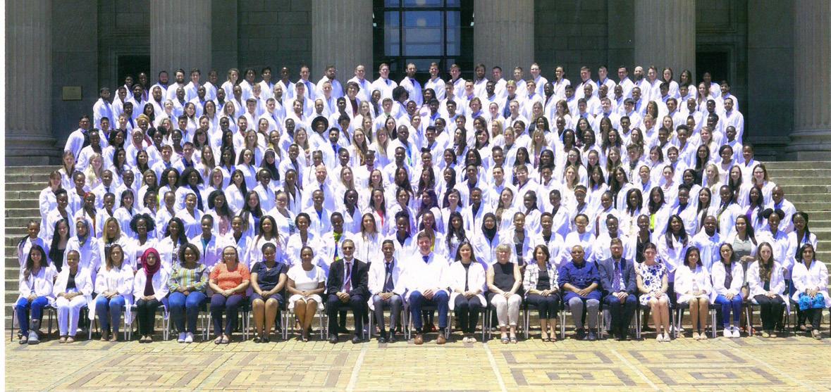
Class of 1960. We know that we were very privileged to have had this opportunity and I am humbled to have learned how seriously we took our responsibility to do good.
It is now a privilege for the Class of 1960 to help the medical students of today attain their MBBChs, in the hope and expectation that their medical careers will be as fulfilling as ours.
In November 1955, 115 first year medical students sat for the examinations in chemistry, physics, botany and zoology. Standards were high, with only nine obtaining first-class passes in all four subjects.
During our six years of study for MBBCh, a number of students dropped out of the class, and others came in. Our Second Year Anatomy Class of 1956 was larger, as it included dental students and others who went on to the degree of BSc before completing their medical studies a year or two later. In 1960, 55 members of the original class made up the 84 people who graduated MBBCh from the Medical School of the University of the Witwatersrand. (See the picture on page IV, with Dean Professor Eustace Cluver in the middle of the front row.)
Most members of the Class of 1960 were white men who started medical school in their late teens and were in their early 20s at the time of graduation. Women, Black, Chinese, and Indian graduates numbered fewer than ten. In our list of students below and in our celebration, we include those who studied with us for all or part of our medical studies.
By clicking their names (where they are links) you will be able to read their personal stories they have written and see pictures (and in some cases, obituaries that their families have submitted) for this diamond jubilee celebration and commemoration.
Where the name is not yet a link – we appeal to anyone who knows or knew them, to get in touch and tell us their story.
Agrotis, Ernest Nicholas
Arnold, Peter (BSc) (61)
Auerbach, Ronald Been, Hilary Bengani, David Dumiso Berger, Michael Benjamin Blankfield, Adele (61) Bobrow, Martin (BSc) (62) Boner, Geoffrey Bosman, Christopher Kay
Botha, Pierre Albert Cochrane, Raymond Ivan Cohen, Manley Cohen, Stanley Gordon Crosley, Anthony Ian
Damelin, Mervyn (61) Davis, Gloria Dawes, Marion Elizabeth
Dimopoulos, Panayous Ditira, Stadrach Seleboga Dove, Ephraim
Edge, Kenneth Roger Esrock, Jenny
Fanaroff, Avroy Arnold Feldman, Joan Diane Felgate, Edwin John Festenstein, John (61) Gazedis, Costa (BSc) (62) Geffen, Laurence (62) Geldenhuys, Andries Retief Gershater M Glazer, Harry Gordon DE
Gottlieb, Allan Michael Hall, Leonard Usher Heyns, Anthon Duplessis Hollis, Richard Roborg Howell, Michael Eric Hurwitz, Mervyn Bernard Jackson, Rodwin Albert Janse van Rensburg, Marthinus Phillipus Jassat, Essop Essak (BSc)
Kahn, Leonard Bernard Kahn, Leopold Kalell, Alec Katz, Gary
Kretzmar, Theodore David
Kuming, Basil Stanley
Kussel, Jack Josiah
Lampert, Gerald
Landsberg, Pieter Guillaume
Le Roux, J. Stephanus (Fanie) (61)
Lee, John Arthur
Lissoos, Irving
Livni, Naomi Margaret
Maass,Gerhard Friedrich
Maisels, Jeffrey (61)
Mauff, Alfred Carl
McCusker, Vivian Ivan
Meyers, Anthony Molyneux
Milunsky, Aubrey
Mjali, Sikose Julia
Mokgethi, Kegerpi Paul
Nafte, Joshua (61)
Nissenbaum, Michael Meyer
Otten, Jane Louise Featherstone (née Stuart)
Otten, Robert Bernard
Ou Tim, Seamus
Palmer, Raymond Ivor
Parry, David Graham
Paton, David Francis
Plit, Michael
Prentice, Bernard Ross
Pretorius, Jack
Price, Anthony Robin (61)
Rakusin, Wolfe (61)
Ravdel, Arnold
Rosenberg, Chaim Meir
Rosendorff, Clive (BSc) (62)
Rossouw, Dennis Pieter
Rubenstein, Arthur Harold
Schneider, Gerald
Shaff, Max (61)
Shapiro, Isaac Shulman, Gerald Silver, Hilton
Sims, Farrol Hyman
Urbani, Pietro
Van As, Andre
Van Druten LF
Van Veen, Evert Jan Willem. (Kleintjie)
Vinik, Aaron (Arthur)
Willies, Lawrence James Sears
Wilson, William Wood, Edward Roy
Zail, Solam Soloman (Stanley)
Zeilinga, Jan Abraham
Memorable Teachers
Arnold, Maurice (Toby)
Barlow, John Brereton
Dart, Professor Raymond
Elliott, Professor Guy Abercrombie
Levin, Solomon
Suzman, Moses Myer
Tobias, Professor Philip
Known Domicile Of Class Members:
South Africa 59
United States 17
United Kingdom 8
Israel 3
New Zealand 1
Australia 3
University Of The Witwatersrand Higher Degrees
Doctor of Science
Aubrey Milunsky 1982
Arthur Rubenstein 2001
Maisels, Jeffrey M 2007
Doctor of Medicine
Avroy Fanaroff 2001
Gary Katz 1971
Rodwin A. Jackson 1974
John A. Lee 1973
David Paton 1974
Chaim M. Rosenberg 1966
Clive Rosendorff 1977
Arthur I. Vinik 1970
Stanley S. Zail 1964
Doctor of Philosophy in Medicine
Andre van As 1976
Published in South Africa by: Wits Faculty of Health Sciences, © University of the Witwatersrand 7 York Road, Parktown, Johannesburg
First published in 2025 978-1-0370-7559-9(print)
978-1-0370-7560-5(Web PDF)
This book is freely available under a CC-BY-NC-ND 4.0 Creative Commons License. (https://creativecommons.org/licenses/by-nc-nd/4.0/).
Author: Wits Faculty of Health Sciences, © University of the Witwatersrand
Editor: Geraldine Auerbach
Sub-editors: Didi Fombad and Beth Amato
Project Manager: Didi Fombad
Cover design and artwork: Wow Tiger Graphic Design
Layout design: Wow Tiger Graphic Design and Rethabile Tlale
Printed in South Africa
Phone: +27 (0) 11 717 2025
Email: comms.health@wits.ac.za www.wits.ac.za/health/
Disclaimer
The views, opinions and/or anecdotes expressed by the authors in this e-book do not reflect the views of the University, publishers and/or associated sponsors.University, publishers and/or associated sponsors.