
T Th he eS Sc ci ie en nc ce eB Be eh hi in nd d
t th he e S Se en ns se es s


T Th he eS Sc ci ie en nc ce eB Be eh hi in nd d
t th he e S Se en ns se es s
Look at the art diagram below Close your left eye and stare with your right eye at the cross on the left side of the paper. Slowly bring the picture straight toward your face, and at a certain distance, the dot will suddenly vanish into your blind spot!
When you open both eyes, your brain combines the two images and fills in the missing picture. Even if you are using only one eye, your brain will automatically use surrounding visual information, such as colour, pattern, and texture, to extrapolate the missing part. Our brain performs this trick so that we “see” a full image without a black hole. If you are looking at a patterned wallpaper, your brain will fill the blind spot with a continuation of that pattern!
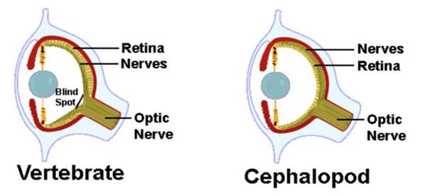
The blind spot exists as a result of the eye’s internal structure. The retina, which is like the digital sensor of a camera, is found at the back inner wall of the eyeball Light-sensitive cells, known as photoreceptors, line the back of the eye and convert light into electrical signals. These signals are then gathered by nerve fibres that bundle together to form the optic nerve This axon bundle must exit the back of the eye to deliver critical information to the brain, creating a small, circular gap in the retina called the optic disc. Because the optic disc is a passageway, it lacks the light-detecting photoreceptors like rods and cones that are responsible for vision Any light landing directly on this area cannot be detected, resulting in a blind spot. Despite this permanent hole in our visual field, we are rarely aware of it during daily life. This is because our two eyes have blind spots in different locations
This natural blind spot is a normal feature. However, the term “blind spot” in a medical context can also refer to areas of vision loss caused by damage along the visual pathway An enlarged or misshapen blind spot can suggest serious conditions like glaucoma, optic neuritis, or papilledema. These are pathological blind spots, or scotomas areas of partial or complete vision loss In these cases, the brain cannot fill in the gap because the neural signal from the affected area is dead or scrambled. Patients will experience the scotoma as a persistent dark grey or blurry patch in their visual field. This is fundamentally different from the natural blind spot, in which the brain continuously fills in the missing information


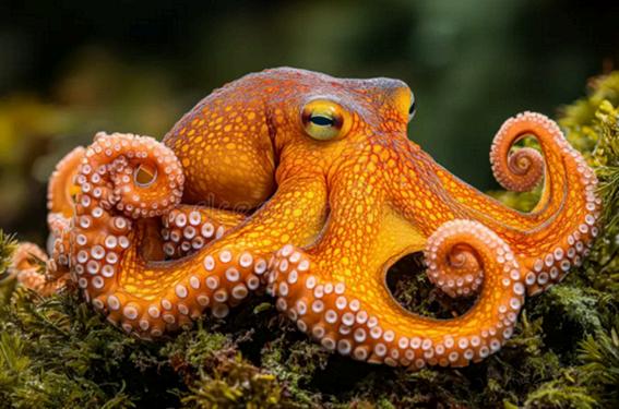
Most vertebrates, including humans, dogs, and horses, have a natural blind spot; however, not all animals have one Cephalopods, such as octopuses, do not! The octopus’s eye has gone through what is known as convergent evolution their eyes evolved separately. Their photoreceptor cells are found at the very front of the retinal layer, and the nerve fibres that carry the signal to the brain are attached to the back end of these photoreceptors. This means they can simply bundle together into the optic nerve and exit the eyeball from behind the light-sensing layer. There is no need for the nerve cable to tunnel through the retina The retina remains continuous, so all reflected light is detected, and no blind spot exists. Their eyes evolved more logically and effectively than ours. So why didn't human evolution rewire the eye to remove the blind spot?
We evolved to have what is known as the "insideout" retina, which creates the natural blind spot, but this flaw does not significantly affect our survival and reproduction Evolution doesn’t aim for theoretical perfection; its "goal" is for us to live, adapt, and produce offspring. So, the genes for this eye structure were passed on. Vertebrate eyes inherited this constraint, while octopus eyes evolved along a different pathway and avoided it entirely This does not mean evolution was aiming to make a better eye only that different starting points and constraints can lead to different outcomes.
The blind spot is proof that we do not perceive the world as it is, but rather as our brain's constructed simulation of reality We are constantly experiencing a neural simulation, a highly processed model of reality built by the brain. It continuously fills in gaps and extrapolates details, like an artist completing an artwork on canvas It’s remarkable how our senses respond to our constantly changing surroundings by not only detecting and reflecting but also creating and reforming the input. This challenges the assumption that what we see is what exists.

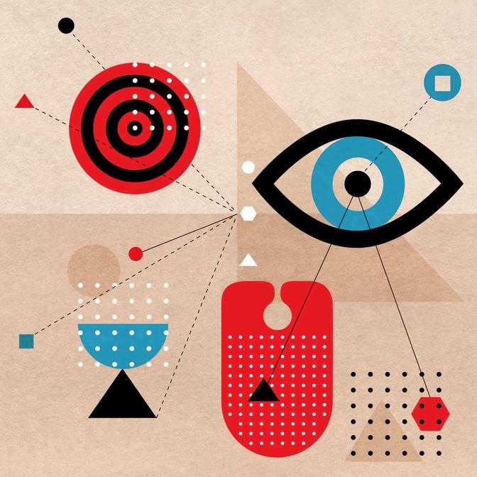
Eyes are often described as windows of our mind, and in turn, they reveal reality to us. It is widely assumed to provide an objective record of the external world However, a famous mind experiment has revealed that the so-called reality on which we have relied for the past 200 years of human history might be just an illusion. In this article, we are going to dive deeper into the human senses of light and see (philosophically, perhaps) how it changes how we see the world.
Proudly speaking, I have individually worked out the theory myself. It was when I played Minecraft with a feature called Metamorph, where we were allowed to transform into other creatures in the game. When I transformed into a creeper (a green explosive creature), my vision turned green – then I asked myself: what if this is a real person?
Now here’s the original version of the experiment. Imagine a boy named Peter, who conceives the colours blue and green reversely. That is, the grass seems blue to him while the sky is green.
Weird, isn’t it? But it isn’t the craziest part yet. Throughout the whole of Peter’s life, his parents have taught him that ‘the sky is blue and the grass is green’, so he practices this all the time Now let’s ask Peter, ‘What colour is the ocean?’ Think about it – the ocean is blue to us, so it looks green to Peter. However, green is also his colour of the sky. Since his parents said, ‘the sky is blue, he would answer ‘blue’!
Astonishing, isn’t it? A person with a completely wrong sense of colour gave the correct answer! With any kind of experiment, you’d never find out if he is colour-blind or not. (@spacechronicles07, 2025)
The real concerning part is that, after reading this amazing discovery, you start to doubt what you see. What if I see different colours to my best friend? What if my favourite colour is actually pink? What if all I have received from the world is not real?

At this point, this Paradox evolves from a mind experiment into a disaster of biology and philosophy. The problem is not if someone is ‘wrong’ but if right or wrong even exist here. If Peter’s colour system is internally consistent, totally functional, and indistinguishable from ours, then there is no external test that can prove his understanding of the world is false. And if that system works perfectly for Peter, it can work perfectly for us!

This shows a fundamental property of human senses: they do not aim to show you reality. Instead, they evolved to support survival and reproduction. From a scientific perspective, colour is not a property of objects at all. Objects reflect light beams of certain wavelengths. What we call “colour” is the brain’s feeling of these wavelengths, built up using signals from cone cells and processed through multiple layers of neural computation. In short, colours are just in our mind but not in reality.
Then philosophy quietly steps in. The Colour Blindness Paradox connects directly to the philosophical problem of qualia or the property of consciousness that it is non-transferable and cannot be shared with another person. No matter how advanced science becomes, there is no way to step into another person’s consciousness and experience how they live. The paradox does not prove that our perception is false but it indeed proves that certainty about perception is impossible.
So, the question remains: could you or me be Peter? Scientifically speaking, there is no evidence that you are. Philosophically speaking, there is no evidence that you are not. And perhaps that is the final lesson of the Colour Blindness Paradox reality is (not probably an illusion but) always, inevitably, personal.

References
A video by @spacechronicles07, 2025. Instagram. [Online] Available at: https://www.instagram.com/reels/DR2 VUhjQQm/ [Accessed 14 01 2026].
O'Rear, C., 1996. The Bliss. [Art].
Also see “Do we see reality as it is?”, a TED talk by Donald Hoffman in March 2015

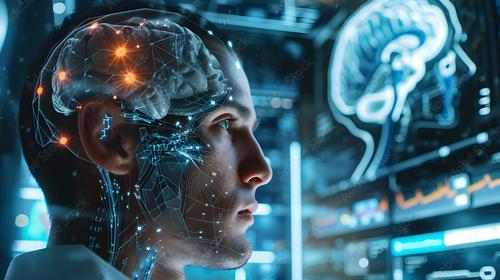
When you scroll on your phone, your brain translates light signals from what the screen is displaying into electrical commands to your fingers which in turn allow you to scroll. Braincomputer interfaces (BCIs) reform this process: with BCIs there is no need for physical movement (you wouldn’t need to move your finger to scroll), since they directly translate your thought into electrical signals. By translating subtle patterns of electrical activity into machine-readable code, they simulate human movement. Today, this technology is no longer experimental; it’s actively transforming lives: paralyzed users can type messages, browse the web, or control wheelchairs and prosthetic limbs purely through their thoughts By bridging the gap between computer science and neuroscience, BCIs unlock a new form of human-machine interaction, and their basic underlying mechanics are more accessible than many might imagine
Brains operate on action potentials; rapid, tiny electrical spikes that neurons fire when communicating with one another. Every thought, movement, or intent triggers distinct patterns of these spikes in specific regions of the brain. For instance, when you intend to swipe right on a screen, a cluster of neurons in your motor cortex (the area responsible for controlling voluntary movement) creates a unique, repeatable sequence. When you intend to swipe left, that same cluster produces a different pattern. The first critical task of any BCI is to capture these unique neural patterns accurately, as they hold the key to decoding human intent.
To capture neurological signals, BCIs use three main types of electrodes. Scalp-worn electrodes (like EEG headsets) are noninvasive, easy to use, and require no surgery, making them ideal for an average person or preliminary research use, but they pick up significantly less signals from the brain, and surrounding muscles Semi-invasive electrodes, implanted under just under the skull but slightly above the brain tissue, offer better signal clarity without the full risks of brain implantation. Implanted electrodes, placed directly into the brain’s cortex, capture the clearest, most precise signals, even detecting spikes from individual neurons, critical for users who need fine control over prosthetics or devices. Regardless of the type, the end product is a stream of messy voltage spikes: raw neural data that’s ready to be translated into actionable commands.
Neural data is naturally chaotic, contaminated by unwanted impulses from muscle twitches, eye movements, nearby electronic devices, and even other non-relevant neurons.
Machine learning (ML) algorithms are the backbone of decoding these signals data into meaningful intent. First, these algorithms separate the signal by applying advanced filtering techniques to isolate the meaningful neural spikes from background interference; similar to how noise-cancelling headphones block out ambient sound. Next, the ML model is trained on hundreds (or thousands) of labelled examples: a user repeatedly thinks a specific command (like "swipe right" or "select"), The system records the corresponding neural pattern. Over time, the model learns to map each unique spike pattern to a clear intent Once trained, the algorithm can translate a user’s thoughts into device commands in mere milliseconds, creating a seamless, almost instantaneous interaction.
Modern BCIs stand out for their ability to enable bidirectional communication, they don’t just read brain activity; they also write information back to the brain. This two-way dialogue opens the door to restoring lost senses. For example, a BCI designed for blind users can pair a small camera (to capture visual data from the environment) with a processor that converts that data into simplified electrical pulses. These pulses are then sent via implanted electrodes to the user’s visual cortex, and over time, the brain learns to interpret these artificial signals as basic shapes, light, or even recognizable objects. Similarly, BCIs for hearing loss convert sound waves into targeted electrical stimuli that are delivered to the auditory cortex, helping users with severe hearing impairment regain partial hearing, including the ability to understand speech.
A surprising insight from BCI research is the remarkable adaptability of the brain’s "code." We often think of our senses as fixed vision tied to eyes, hearing to ears, but BCIs prove the brain is a flexible interpreter. It doesn’t care how the sensory information arrives (whether via natural organs, electrodes, or even light); it only cares that the data comes in a pattern it can learn to decode
For example, researchers have used lightbased BCIs (via optogenetics) to help brains "hear" by converting sound into light pulses that stimulate the auditory cortex. Similarly, prosthetic limbs equipped with pressure sensors can send electrical signals to the somatosensory cortex, allowing users to "feel" touch, texture, and pressure, proving that our senses are not fixed biological functions, but interpretable signals that can be rerouted and redefined.
The future potential of BCIs is vast and transformative. For paralyzed individuals, they offer a path to greater independence, letting them engage with the world on their own terms without relying on others for basic tasks. For those with sensory loss such as blindness, deafness, or loss of touch, BCIs hold the promise of restoring not just function, but a more complete experience of the world Beyond restoration, BCIs hint at a future where human perception is no longer limited by biological constraints: imagine being able to "see" in infrared, "hear" ultrasound, or communicate with computers and other devices directly through thoughts, without needing keyboards or voice commands. At its core, BCI technology redefines the boundaries of human-machine communication, opening up possibilities that were once confined to science fiction.

How the muscle we taste with How the muscle we taste with gives blind people their sight back
Though crucial for people’s pleasure, taste has become the least useful of our five senses. Back when we were evolving it was a way to make sure we did not consume anything dangerous but now in a world where packages tell us all the ingredients we are eating it’s become simply an accessory, This was before we realised the tongue, yes the tongue can be used to make blind people see again.
s
Due to the immense number of sensory receptors scientists were able to convert one piece of sensory information into another through a sensory substitution device (SSD) Through the camera an image is able to be captured which each pixel corresponding to an electrode on the muscle. This seemingly small machine creates a low tactile image on the surface of the tongue. Although some may say this is not sight through the device users were gifted the ability to detect their surrounding as well as read letters and numbers. The previously mentioned Eric Weihenmayer is well known within the climbing world for his use of the device on his many expeditions.
Many devices have been created over the years to assist those who are less able yet nothing on this scale. Unlike previous devices where a user is assisted by something else, like a voice, Brainport allows for a new sense of independency as people are able to move, detect and see the world around them as never before. Though non-traditional sight in this format opens a new world to those who use it. Eric Weihenmayer, a blind American athlete, describes how Brainport allowed for him to reconnect with those around him he was able to see his son’s “whole face [turn] into like this giant smile”. Through this small device Dr Paul Bach-yRita was able to change the world of neuroscience.
Similarly as with any innovation limitations do exist. The tongue unlike the rest of our body does not react to cued information leading to slower reactions from users On top of that the hypersensitivity leads to quick overstimulation of the muscle as all receptors become overfilled by sensation resulting in the best use of Brainport to be when combined with another SSD. Finally wearers of the device describe it to be like a new language as the brain once again begins the difficult process of taking in information and translating, but like learning a language it is very rewarding.
Brainport changes the life of those suffering from visual impairments for the better, through giving a newfound sense of independence and control over their life Though simpler ways exist, nothing quite works in the same concept or with the same ability as the device described here, showing how it is truly a revolutionary item in the world of neuroscience

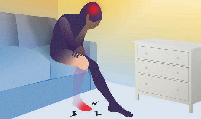
Have you ever walked into a room and completely forgotten why you’re there? Or you’ve felt your phone vibrating in your pocket, but realize it was never there at all? The human body is a complicated symphony of signals an ongoing dialogue between the periphery and the core. Imagine what happens when an instrument breaks while its melody lingers This is the agonizing reality of Phantom Limb Pain (PLP) PLP is not merely a psychological magic trick, but a neurological phenomenon. Clinical data indicates that it affects nearly 80%-90% of amputees, with about 70% reporting debilitating pain decades after their physical wounds have healed This is a profound glimpse into the brain’s marvelous capacity to reconstruct reality, challenging step by step our fundamental understanding of the human experience.
For centuries, PLP was dismissed as a psychiatric delusion or misconceptions that had misguided people for so long, that it is not until 1992 when V.S. Ramachandran had finally proved that it is caused by 'cortical remodeling' of the brain, even though early observations can trace back to 1550 when Ambroise Paré was doing suture surgery and his patients claimed they still feel their cut limb ache, ignoring the fact that the wound had already been treated.
But why? Try to imagine the brain’s somatosensory cortex as a meticulous partitioned map, which in specialized term, the Sensory Homunculus, where every body part owns specific real estate.
When a limb is lost, its neural territory does not go dormant, but instead razed and then becomes a vacant lot, where its rapidly colonized by the neighboring areas in a display of aggressive neural plasticity.
The most striking evidence of this "neural invasion" was discovered by Dr. V.S. Ramachandran in his study of Tom Sorensen, a teenager who lost his left arm in a car accident. Tom began experiencing an intense, localized "itch" in his missing fingers whenever Dr Ramachandran stroked Tom’s left cheek It was geometry On the homunculus map, the area representing the face is physically adjacent to the area representing the hand. Because Tom’s "hand" neurons were no longer receiving input, the neighboring "face" neurons sprouted into that vacant space Tom’s brain was cross-wiring sensations: it "saw" a touch on the face but "felt" it in a hand that no longer existed. This encroachment creates a state of sensory dissonance. The motor cortex continues to send commands such as rub your hands or clench the fist, but the expected feedback from body is missing. This neurological feedback loop is then interpreted by the mind as pain.
To combat this, scientists turned to the brain’s visual dominance through Mirror Therapy. By placing a mirror between the limbs, patients automatically imagined the reflection of their intact arm movement in a place of phantom In a landmark study published in the New England Journal of medicine, 100% patients using mirror therapy reported a significant decrease in physical pain. By deceiving the brain into seeing the missing limb move, the visual feedback had settled down the internal conflict

Today, thanks to modern technology, this visual deception had been taken into the 21st century with Virtual Reality(VR) and Augmented Reality (AR). While mirror therapy is limited by the symmetry of the body, VR allows for a fully immersive sensory override. In recent clinical studies and trials, patients wear VR headsets that render a highfidelity digital version of their missing limb Unlike outdated mirror, VR is able to simulate complex tasks, such as catching ball or even playing the piano. This Targeted Muscle Reinnervation (TMR), combined with VR allows the brain to re-integrate the phantom into a digital body schema
Data from recent pilot studies indicates that VR therapy can reduce pain scores by an additional 3040% in patients who were non-responsive to mirror therapy. By providing a digital prosthetic for the brain to link and attach onto, we are now closer to soothing a screaming neurons of the cortex, meaning we are “one step ahead” before the pain
80%-90% Prevalence & 70% Chronic Pain:
Ultimately, the phenomenon of the phantom limb forces us to confront a startling truth: our reality is a controlled illusion, a map created by a mind that refuses to be erased We inhabit a neurological representation so loyal that it persistently mourns what is gone through the language of pain. This phantom ache is the price we pay for neuroplasticity the same resilient force that enables children to learn,a musician to master a piece of instrument, and even a broken heart to eventually heal. In every phantom throb, there is a testament to the brain’s capacity to bridge the gap between who we were and who we are becoming. It is in this beautiful persistence that we truly define what it means to be human
Sherman, R. A., Sherman, C. J., & Parker, L. (1984). "Chronic phantom limb pain: experience with 5,000 samples." Pain, 18(1), 83-95. Flor, H. (2002). "Phantom limb pain: characteristics, causes, and treatment." The Lancet Neurology, 1(3), 182-189.
Ambroise Paré (1550s Observations)
Finger, S , & Hustwit, M P (2003) "Five early accounts of phantom limb in context: Paré, Descartes, Lemos, Bell, and Mitchell " Neurosurgery, 52(3), 675-686.
V.S. Ramachandran & Tom Sorensen (1992) Ramachandran, V S , Stewart, M , & Rogers-Ramachandran, N S (1992) "Perceptual correlates of massive cortical reorganization " Science, 258(5085), 1159-1160
Mirror Therapy (New England Journal of Medicine)
Chan, B L , Witt, R , Charrow, A P , et al (2007) "Mirror Therapy for Phantom Limb Pain " New England Journal of Medicine, 357(21), 22062207
VR Therapy & 30-40% Pain Reduction
Ortiz-Catalan, M., Sander, N., Kristoffersen, M. B., et al. (2014). "Phantom limb pain management via augmented reality and gaming." Frontiers in Neuroscience, 8, 411 Dunn, J , Yeo, E , Moghaddampour, P , et al (2017) "Virtual and augmented reality in the treatment of phantom limb pain: A systematic review " American Journal of Physical Medicine & Rehabilitation

In October, on the flight to JFK for a New York drama trip, I found myself crying to Paddington 3, having previously watched it and been very in control of my emotions This led me to asking some of my friends if they had had a similar experience, and the result was almost unanimously yes So, have you ever sobbed watching a movie on a long haul flight? Well, according to a Virgin Atlantic study, conducted in 2011, anyone who answers no would be in the minority In response to this, Virgin actually introduced “weepy warnings” before particularly sad movies on flights But why does this happen? Well, as of 2026 there is no one conclusive reason, but, there are three key assumptions: hypoxia, stress, and dehydration.
Flying puts the body under physical and psychological stress, albeit mild, which can understandably affect mood Cabin pressurization is a process in which conditioned air is pumped into the cabin of an aircraft or spacecraft in order to create a safe and comfortable environment for humans flying at high altitudes. This pressure, however, is slightly lower than at a normal altitude. This lower pressure reduces alveolar oxygen tension and, subsequently, oxygen availability in the brain. The brain is highly oxygen-dependent, so even small reductions in oxygen availability can have noticeable effects on how we feel and function. In response to hypoxic stress, the HPA axis gets rapidly activated. The HPA axis, or hypothalamic-pituitaryadrenal axis, is a complex set of interactions between the hypothalamus, pituitary gland, and adrenal glands. It plays a critical role in regulating stress responses, mood, digestion, immune function, and energy storage and expenditure in the body. These effects are temporary and mild, but they may subtly reduce emotional resilience, thus causing a lower resilience to sadness
Even relatively small fluid losses - similar to those experienced during everyday activities - have been associated with changes in mood and perceived mental effort It is also well known that alcohol has much faster effects on planes - on aeroplanes, dehydration reduces the body’s available water for diluting alcohol, while low oxygen and fatigue make the brain more sensitive to its effects - causing people to feel drunk more quickly In other words, when the body is dehydrated, emotions become harder to regulate and the tears harder to hold back.

much not to worry about But what impact does stress have on our ability to regulate our emotions? Stress doesn’t just make emotions stronger - it interferes with the brain systems that normally help regulate them Under stress, the brain relies more on fast, automatic emotional responses and less on deliberate emotional control. When this balance shifts, emotional regulation weakens and instinctive reactions take precedence. This can make sadness feel more intense, even in response to otherwise ordinary stimuli. Stress reduces effectiveness of prefrontal regulation, meaning that the amygdala responses become more dominant. As a result, negative emotions last longer and feel more intense.
Finally, dehydration – weird, right? Well, it’s fairly common knowledge that aeroplane cabins have low humidity – according to a study conducted by Ola Häggfeldt, Chief Commercial Officer at CTT Systems AB, the flight deck, first class, and business class cabins are dryer than any place on Earth, with an average cabin humidity of 2-3%, 5%, and 7%, respectively Whilst being good for the metal that planes are made from, this isn’t so good for humans breathing the air Research consistently shows that dehydration affects mood, even when measurable cognitive performance remains largely unchanged. People who are mildly dehydrated commonly report feeling less alert, more fatigued, and under greater mental strain. Importantly, these effects do not require severe dehydration.
To conclude, for whatever reason we do it, the increase in crying on planes cannot be denied Whilst there is not a conclusive answer, many combined factors are to blame So, next time you find yourself tearing up at Shrek on a plane, just know your neighbour probably isn’t judging you - they’ve likely been there too.

Sweetness has the ability to make food feel both pleasurable and comforting to humans. From fresh fruits to sweet desserts, sweet taste plays an important role in shaping human eating habits. Because of this idea, sweet taste is often regarded as an ordinary sensory experience that all mammals can feel. Cats, however, are unaware of sweet taste, despite their biological similarities to humans. This striking contrast shows how sensory perception is not universally shared, but is shaped by genetics, physiology, and evolutionary adaptation.
In humans, perception of sweetness depends on the specialized taste receptors located within the taste buds of the tongue Sweetness is mediated by receptors that are composed of two proteins, T1R2 and T1R3, which belong to the receptor family that is bound to the G protein. When sugar molecules bind to these receptors, the signaling pathway is activated, sending electrical signals to the brain,

comparison
From an evolutionary point of view, sweetness provides fast energy for foods rich in carbohydrates Individuals with a high sensitivity to sweetness are more likely to consume enough calories, increasing their chances of survival and reproduction. As a result, the ability to taste sweetness has been greatly preserved through natural selection.
Cats, unlike humans, are carnivorous animals that fill their nutrition with meat. Meat contains almost no carbohydrates, so they do not need to be biologically sweet for their survival. During evolution, the cat's T1R2 gene accumulates mutations, causing the cat to lose function, which is called pseudogenization. Without a functional T1R2 gene, sweet receptors cannot be formed, and sweet taste cannot be detected.
The physical structure of the tongue makes sensory adaptation more visible. The human tongue has numerous taste buds distributed throughout its surface. While ‘taste maps’ present distinct areas for each taste, modern research has shown that most areas of the tongue can detect all basic taste buds, with varying differences in receptor density.
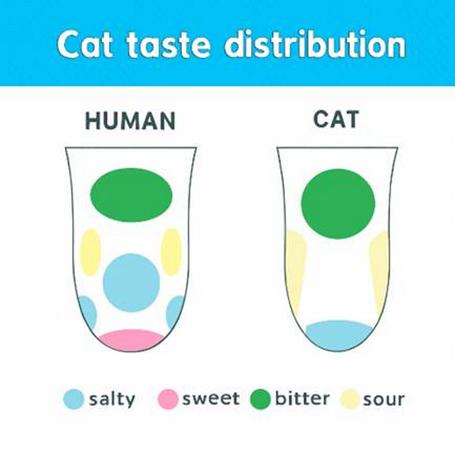
The differences in the taste distribution of cats and humans


Interestingly, cats are not the only animals with unique taste abilities Typically, birds have fewer taste receptors than humans, while cattle are more sensitive to certain taste sensitivities associated with plants. This shows that taste buds are more closely connected to diets and environments than to intelligence or complexity.
The fact that cats cannot taste sweetness is due to their evolutionary biological structure. Sweetness is not a food's inherent attribute, but a neurological flavor formed by genetics and a desire to survive. By comparing humans and cats, we can see how evolution tailors sensory systems to specific ecological needs. Understanding these differences will allow us to understand that all species experience the world through unique biological studies guided by the science behind their senses.

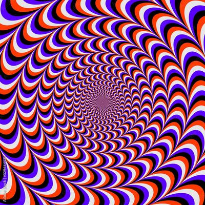
Furthermore, it is essential to understand why we experience optical illusions and why our brain does not just see the image for what it really is. The brains need for efficiency is one of the main reasons behind why optical illusions trick our brains. Our brain constantly strives to interpret electrical signals as quickly and efficiently as possible, using shortcuts based off prior knowledge and experience. However, optical illusions exploit these shortcuts and create situations in our brains where a combination of the brain’s predictions and assumptions do not match reality when forming an image. As a result of this, we perceive something that is technically incorrect, even though our brain is just simply operating as it is designed.
Moreover, contextual clues can heavily influence our perception when it comes to interpretation, demonstrating that vision is not just about the object itself but also its surroundings. The brain is constantly evaluating the relationship between lights, colours and shapes when it comes to creating a coherent picture of reality. For example, in the famous checker shadow illusion, there are two squares that are the exact same shade of grey appear drastically different because the brain accounts for the shadow cast by the object on the screen. This reliance on context demonstrates how making quick and practical visual judgements in every day life can often lead to misconceptions when images are intentionally designed to trick the brain.
Finally, the perception of optical illusions is different depending on the age category in question. By this, I am referring to differences in brain development and processing capabilities when it comes to interpretation.

The brain of a child is less developed in comparison to the brain of an adult; this means that children rely on raw sensory input instead of past experiences and information about context; they will therefore often interpret these illusions literally and may miss the themes that certain illusions exploit. Unlike children, adults, who have not only more experiences but also a deeper understanding of the world are more likely to have their brains tricked as they are more prone to ‘shortcuts.’ In addition to this, as people age, changes in both eyesight and neural processing often leads to a subtle alteration in the way optical illusions are received, making some appear weaker or slower to register.
In summary, optical illusions often depend on neural development, experiences and the strength of sensory input when it comes to forming a perception of an illusion.
Imagine walking by, staring at your feet every step, thinking "Now my right leg... bend 45 degrees... move forward!" How many years would that take for you to get from home to school? But how can you scroll your phone while walking? This leads to my topic: Proprioception. It basically allows you to know where your body parts are and what they are doing without paying attention to them. This includes the sense of movement, as the example provided above; position sense, the ability to touch your eyebrows when eyes are closed; force sense, deciding the amount of muscles used by the weight of the object, and all these actions are done unconsciously by the brain.
The proprioceptive system starts with sensory receptors, which are specialised structures embedded in your muscles, tendons and joint capsules These muscle afferent receptors detect limb position and movement through neural signalling of a change in muscle, skin or joint stretch Mechanoreceptors are the main type of receptors for proprioception, more specifically, it's the subset of mechanoreceptors called proprioceptors. Even more specifically, there are three types of them, each adapted with functions. Firstly, muscle spindles. They are embedded within skeletal muscles, they detect muscles being stretched and the speed of that change, and sense movement and position. Secondly, Golgi Tendon Organs (GTO), located at the junctions between muscles and tendons, detect and sense muscle tension or force, to prevent excessive force from being damaged. Lastly, joint receptors, located within the capsules and ligaments, detect joint angle, pressure and direction of movement, again also preventing injuries. Furthermore, signals from these proprioceptors travel through fast sensory nerve pathways up the spinal cord to the brain, carried parallel with other sensory data Several parts of your brain work together to process the sensory information
This controls movement and is the reason why you don't have to consciously think about every movement. There is also a strip across the top of your brain called the Somatosensory Cortex. It takes the signals and updates your internal sense of where your body parts are. Through this system, you are able to perform proprioception!
An example of impaired proprioception is a drunk person. Overconsumed alcohol impairs nerve function, which blunts proprioception. They will have symptoms like dizziness, balance issues and uncoordinated movements, which are hazardous. Another example is when a person has just experienced a severe ankle sprain, their proprioceptors are damaged, and the brain isn't receiving clear positional signals; their ankle may feel unstable and may easily get re-injured. Another example is when a ballet dancer is doing "Fouetté" (tough spins in which they swing one leg while the other leg is holding all of her weight and spinning on pointe shoes). Even though her eyes and inner ear are physically dizzy, the proprioceptors in her welltrained leg, ankles and torso with strong endurance will still know her body's exact position in space and maintain her balance, while the swinging leg motivates the spin

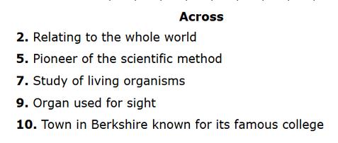
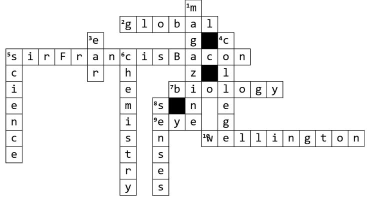
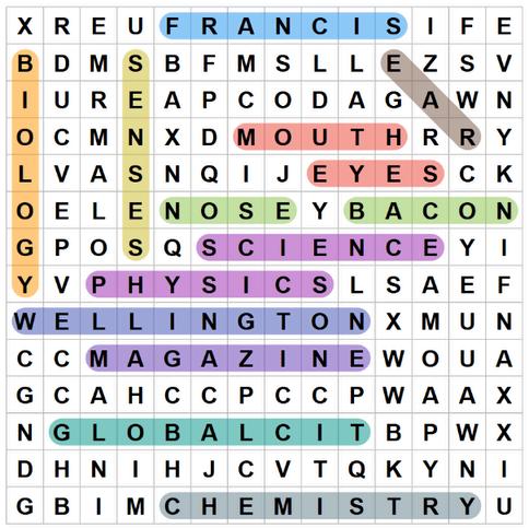
BACON, SCIENCE, EYES, MAGAZINE, FRANCIS, WELLINGTON, GLOBALCIT, SENSES, BIOLOGY, EAR, CHEMISTRY, NOSE, PHYSICS, MOUTH

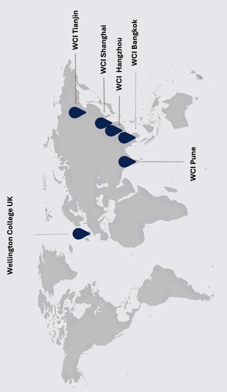
T H E W E LLI N G T O N C O L L E G E F A M I L Y T h e n u m b e r o f a r t i c l e s f r o m e a c h s c h o o l