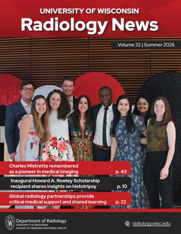
UNIVERSITY OF WISCONSIN
Radiology News Volume 32 | Summer 2026
Charles Mistretta remembered as a pioneer in medical imaging
p. 43
Inaugural Howard A. Rowley Scholarship recipient shares insights on histotripsy
p. 10
Global radiology partnerships provide critical medical support and shared learning
p. 22
radiology.wisc.edu
Upcoming Events
Contents Education Updates
4
Outreach 16 Research Report
17
Clinical News 22 Society Meetings
25
Faculty News 28 Honors and Awards
36
Retirements 38 Alumni Spotlight
41
In Memoriam 43 A Look Back
47
GE HealthCare MR Programming Workshop (EPIC and Orchestra) Sept. 8-Dec. 4, 2026
This workshop offers a unique opportunity to gain hands-on experience with GE HealthCare’s MRI pulse sequence and reconstruction tools. It’s tailored for all skill levels, providing foundational knowledge for newcomers and advanced insights for seasoned MR physicists. Learn more and apply online.
University of Wisconsin Radiology Summit Feb. 8-11, 2027
The UW Department of Radiology welcomes radiologists from across the globe for the annual University of Wisconsin Radiology Summit. The multi-day educational event offers opportunities to learn, connect and ski the beautiful mountains in Park City, Utah. Visit the webpage to learn more.
Zachary Clark Radiology Research Symposium Apr. 24, 2027
Join us in celebrating the high-caliber research conducted by our medical students, residents, and fellows. Visit the webpage to learn more.
Centennial Celebrations We will celebrate our centennial anniversary in 2027! To mark this monumental moment in our history, we will be hosting various events throughout 2027 and encourage alumni to join us in our celebrations. More details to come as 2027 draws near. Visit our centennial webpage for the most up-to-date information.
2
Letter from the Chair Honoring the past. Envisioning the future. In just a few months, we will celebrate our centennial year and the milestones and people that built our department’s reputation as a renowned leader in the field of radiology. That includes pioneers like Charles “Chuck” Mistretta, PhD, who recently passed away. We honor his contributions to radiology and medical physics and also his dedication to his many past mentees and colleagues in this issue. We also want to spotlight the future we are all building together. In this newsletter, you can read more about today’s accomplishments that will become tomorrow’s celebrated history. That includes the world’s first pediatric interventional radiology suite featuring the Allia Moveo, an AI-supported image-guiding solution that was developed by GE HealthCare. With this advanced technology, we will care better for our youngest patients and conduct cutting edge research. The issue also includes stories about how we invest in our people. Through our research and development funding program, an interdisciplinary team led by Diego Hernando, PhD and Alejandro Roldán-Alzate, PhD were able to evaluate how MRI can provide valuable information for liver transplant surgery. Through our new Howard A. Rowley Scholarship, MD-PhD student Katrina Falk, PhD presented at the Society of Abdominal Radiology meeting on the safety of histotripsy procedures after administration of ultrasound contrast. And through our Radiology Equity Program, Anand Naryan, MD, PhD and Lewis Jordan, MD, MPH, MS mentored medical students and guided their research on important topics such as rural versus urban hospital outpatient imaging efficiency. Together, we will honor the past and envision the future throughout 2027. I encourage everyone to stay connected by checking our centennial webpage and following us on LinkedIn and Instagram. On Wisconsin! Scott B. Reeder, MD, PhD
“Together, we will honor the past and envision the future.”
Scott Reeder, MD, PhD
3
Education Updates
Future plans for graduated fellows Abdominal Imaging and Intervention
Matthew Elissa, MD: Private Practice, Gundersen Health System, La Crosse, WI Sarah LaPierre, MD: Academic Practice, UW–Madison (Abdominal Imaging and Intervention) Alexander Moeller, MD: Academic Practice, UW–Madison (Community Division) Benjamin Prout, MD: Academic Practice, UW–Madison (Community Division) David Turner, MD: Private Practice, Radiology Associates of Indiana J. Erik Winterholler: Academic Practice, UW–Madison (Abdominal Imaging and Intervention)
Breast Imaging and Intervention
Jared Katchen, MD Amy Shah, MD: Academic Practice, University of Colorado Austin Webs, DO: Academic Practice, UW–Madison (Community Division, Breast Imaging and Intervention)
Cardiothoracic Imaging
Hannah Hodges, MD: Academic Practice, Vanderbilt, Nashville, TN Andrew Moore, MD: Academic Practice, University of Kansas Medical Center, Kansas City, KS, Kaitlin Robling, MD: Academic Practice Rush University Center, Chicago, IL
L to R: Program Director Jeffrey Kanne and faculty member Kevin McDonald pose with cardiothoracic imaging fellowhip graduates Andrew Moore, Hannah Hodges, and Kaitlin Robling.
4
Musculoskeletal Imaging and Intervention
Aaron Cronican, MD: Academic Practice, UW–Madison (Community Division) Brendan Franz, MD: Private Practice, Huron Valley Radiology, MI Brantley Grimball, MD: Private Practice, vRad, Charleston, SC Lisa Park, MD: Academic Practice, Northwell Health, Manhasset, NY Osvaldo Velez-Martinez, MD: Academic Practice, Vanderbilt, Nashville, TN
Neuroradiology Andrew Healey, DO: Private Practice, Guam Regional Medical City Cameron Kendall, MD: Second-year neuroradiology fellowship, UW–Madison Rasheed Nawaz, MD: Academic Practice, UW–Madison (Community Division) Luis Ortiz Figueroa, MD: Academic Practice, Penn State Hershey Medical Center Nolan Shen, MD: Private Practice, Alina Health, Minneapolis, MN Cory Pierson, MD: Private Practive, ARA Health Specialists, Asheville, NC
Neuroradiology fellow graduates, L to R: Andrew Healey, Cameron Kendall, Luis Ortiz Figueroa, Cory Pierson, Nolan Shen, and Rasheed Nawaz.
Education Updates
Resident graduation and future plans Diagnostic Radiology Residents
Diagnostic Radiology Residents, cont.
Cameron Fox, MD: Private Practice, Minneapolis Radiology Associates, Minneapolis, MN Madeline Jentink, DO: Breast Imaging and Intervention Fellowship, UW–Madison Samuel Koebe, MD: Interventional Radiology Independent Residency, UW–Madison Carolina McShane, MD: Thoracic Imaging Fellowship, UW–Madison Ruben Ngnitewe Massa’a: Private Practice, Synergy Radiology, LLC, Fort Wayne, IN Isha Pathak, MD: Neuroradiology Fellowship, UW–Madison
Jessica Perry, MD: Neuroradiology Fellowship, UW–Madison Vyshnavi Reddy, MD: Neuroradiology Fellowship, Emory University, Atlanta, GA Anna Sorensen, MD: Interventional Radiology Independent Residency, UW–Madison
Interventional Radiology Resident Allison Couillard, MD: Private Practice, Savannah Chatham Imaging, Savannah, GA
L to R: Madeline Jentink, Cameron Fox, Carolina McShane, Samuel Koebe, Anna Sorensen, Ruben Ngnitewe Massa’a, Isha Pathak, Vyshnavi Reddy, Jessica Perry.
5
Education Updates
New fellows Abdominal Imaging
Cardiothoracic Imaging
James Barr, MD Memorial Health University Medical Center
Shawn Leonard, MD Tulane University
Timothy Banks, MD Walter Reed Medical Center
Kelvin Diaz-Perez, MD Hartford Hospital
Carolina McShane, MD University of Wisconsin Hospitals and Clinics
Ravi Chintapalli, MD University of Chicago
Eve Goldstein, MD University of New Mexico Hospital
Blake Sarrazin University of Kansas Medical Center
Parker Howard, MD University of Cincinnati Medical Center Jacob Matulle, DO University of Illinois College of Medicine
Breast Imaging Madeline Jentink, DO University of Wisconsin Hospitals and Clinics
Chad Thompson, MD Saint Louis University School of Medicine
Madison Turner, MD University of MissouriKansas City
6
Not pictured: Laura Jorgensen, MD (Begins 10/1/26)
Musculoskeletal Imaging David Chen, MD Stony Brook Hospital
Liam du Preez, MD Norwalk Hospital Diagnostic Radiology
Breiona Hamilton, MD, MHS Vanderbilt University Medical Center Albert Hwang, MD Staten Island University Hospital
Robert Zaas, MD The Ohio State University
Neuroradiology
Yash Desai, MD Corewell Health East William Beaumont University Hospital Akram Khaja, MD Advocate Illinois Masonic Medical Center
Isha Pathak, MD University of Wisconsin Hospitals and Clinics
Anthony Peret Hôpital Erasme, Université Libre de Bruxelles Jessica Perry, MD University of Wisconsin Hospitals and Clinics
Education Updates
First-year residents Diagnostic Radiology Ibrahim Abdalla, MD University of Minnesota Medical School
Alex Thomas, MD University of Chicago Division of the Biological Sciences, The Pritzker School of Medicine
Ronnie AlRamahi, MD University of Wisconsin School of Medicine and Public Health
Onur Tuncer, MD Yeditepe Üniversitesi Tip Fakültesi
Annika Anderson, MD University of Illinois College of Medicine
Raphael do Vale Souza, MD Escola de Medicina Souza Marques, Fundação Tecnico Educação Souza Marques
Nathan Kauffman, MD, PhD Michigan State University College of Human Medicine - Grand Rapids
Vishwaarth Vijayakumar, MD Carle Illinois College of Medicine
Harsh Khilwani, MD University of Illinois College of Medicine
Annie Zlevor, MD University of Wisconsin School of Medicine and Public Health
Mark Lewis, MD University of Central Florida College of Medicine
Nuclear Medicine
Interventional Radiology
Lydienid Rodriguez Ortiz, MD American University of the Caribbean School of Medicine
Hassan Rizvi, MD University of Wisconsin School of Medicine and Public Health
Andrew Voter, MD, PhD University of Wisconsin School of Medicine and Public Health
Nathan Travers, MD University of Illinois College of Medicine - Rockford
7
Education Updates
Medical students gain firsthand research experience through summer programs
2026 summer research students pose with Matthew Lee, director of medical student research.
This summer, a dozen medical students gained valuable hands-on experience in radiology by participating in research projects through the Shapiro Summer Research Program, the Radiology Equity Program, and the Nth Dimensions Program. The Shapiro Program is part of a broader research initiative from the University of Wisconsin School of Medicine and Public Health, while the Radiology Equity Program is unique to the department. During the course of the programs, students are immersed in an active research environment, rather than observing. Working with faculty, trainees, and research staff, students learn valuable research skills like asking the right questions, evaluating evidence, and adapting to the unexpected. Students work closely with a radiology faculty mentor, participate in clinical shadowing opportunities, and learn about the day-to-day life as a radiologist. Director of Medical Student Research Matthew Lee, MD explained that the programs provide an opportunity to foster an early interest in radiology as a career path by engaging talented students in meaningful work. “Many students begin with relatively little exposure to radiology. It is rewarding to watch them discover the
8
breadth of the field, understand its impact on patient care, and develop a genuine interest in pursuing radiology themselves.” Ana Martinez Cavazos, a program participant, echoed this sentiment, sharing that gained a deeper understanding of the field. “I have most enjoyed the ‘day in the life’ presentations and clinical shadowing opportunities this program has offered” she said. “Through these activities I have learned a lot about the diversity of subspecialties and procedures done in radiology.” Helping students build community and make meaningful connections is one of Dr. Lee’s favorite aspects of working with the summer research programs. “Connecting students with faculty, projects, and opportunities that align with their interests can have an impact well beyond a single research project” he explained. “The right mentoring relationship can influence a student’s entire career trajectory, and it is rewarding to help create those connections.” Jeremiah Swanson highlighted that the mentorship he received was one of the top benefits from his experience this summer. “As a first-generation medical student and a veteran, stepping into an unfamiliar academic field can feel daunting. Having structured mentorship and
Education Updates hands-on procedural exposure gave me the confidence that I belong here and reinforced that radiology is where I want to be. The mentorship piece helped me build connections and gave me a clearer path toward radiology as a career.” Another program participant, Pouya Mirzaei, shared a similar sentiment, noting that the day-in-the-life presentations made him realize how valuable it is to form connections and community during this formative part of his career. Faculty mentor Andrew Wentland, MD, PhD noted how the summer research opportunities make a tangible impact in a relatively short span of time. “One of the most rewarding aspects is watching students gain confidence over the course of the summer. Many begin with little research experience, but by the end they are asking insightful questions, contributing ideas, and presenting their work with greater confidence.” He also shared how the research environment itself can help medical students gain a deeper understanding of translating research into clinical care. “Being immersed in that environment helps them appreciate how multidisciplinary teams collaborate, how challenging problems are approached, and how ideas evolve from initial concepts into solutions that can ultimately improve patient care. They also gain firsthand exposure to the life of a physician-scientist, seeing how I balance patient care, research, mentorship, and collaboration to translate new technologies into advances in clinical medicine.” Mirzaei appreciated the ability to branch out and learn about topics not covered in the medical student curriculum. “The biggest benefit of being in this program has been that it allowed me to explore and learn about artificial intelligence and medical physics; something that I could not do as a regular medical student.” There are a number of programs faculty engage medical students through. For instance, Andrew Ross, MD, MPH worked with Lawrence Baylin through the Nth Dimensions program this summer. Baylin shared “I came to Madison curious about radiology. I’m leaving certain about it. This summer cemented my decision to pursue the field, and that clarity is a gift I’ll carry through the rest of my training.”
Ana Marinez Cavazos presents her research to department members.
Lawrence Baylin presented his research with Andrew Ross at the NMA convention this summer.
2026 Summer Research Students Ana Martinez Cavazos Mentored by Meghan Lubner, MD Pouya Mirzaei Mentored by Ali Pirasteh, MD Jaskiran Sandhu Mentored by Andrew Wentland, MD, PhD Denis Alfin Mentored by Alison Gegios, MD Duaa Majeed Mentored by Alison Gegios, MD Liza Iqbal Mentored by Mark Kleedehn, MD Jeremiah Swanson Mentored by Mark Kleedehn, MD Tenzin Yuthok Mentored by Matt Lee, MD Steven Schomisch Mentored by Andrew Wentland, MD, PhD Emma Cepukenas Mentored by Wenhui Zhou, MD, PhD Dahlia Venny Mentored by Anand Narayan, MD, PhD Adiana Orama Otero Mentored by Lewis Jordan, MD, MS, MPH DaVarius Carter Mentored by Anand Narayan, MD, PhD Lawrence Bayin Mentored by Andrew Ross, MD
9
Education Updates
Katrina Falk receives inaugural Howard A. Rowley Scholarship to share insights on histotripsy Katrina Falk, PhD is the inaugural recipient of the Howard A. Rowley Scholarship Fund from the UW Department of Radiology. Established in recognition of former Neuroradiology Section Chief Howard Rowley, MD, the fund honors Dr. Rowley’s love of research, passion for travel, and mentorship by providing financial support for a student to present their research at a national or international conference. With interests at the intersection of research, engineering, and clinical care, Dr. Falk has contributed her expertise to many areas within the department for over a decade while pursuing her bachelor’s and master’s degrees in biomedical engineering; and now her medical degree and doctorate through the University of Wisconsin School of Medicine and Public Health. Most recently, she’s devoted research endeavors to histotripsy, an emerging non-thermal, non-invasive, and nonionizing focused ultrasound therapy for the treatment of liver tumors. Dr. Falk utilized the scholarship funds to present at the Society of Abdominal Radiology (SAR) annual meeting; sharing her findings on whether it is safe to perform a histotripsy procedure after administering ultrasound contrast. As the lead student, Dr. Falk conceived, designed, and led the research project in all phases, from image collection and analysis to assisting the pathologist reading the histology slides. Ultimately, she found that it is safe to administer ultrasound contrast prior to the procedure. The project has garnered multiple awards, including the Next Frontier Award at the SAR annual meeting and Best Oral Presentation by a Medical Student at the department’s Zachary Clark Radiology Research Symposium. While completing her master’s degree, two courses; Cancer Bioengineering and Energy-Tissue Interactions resonated with Dr. Falk, piquing her interest in device-based, directed tumor therapies. During her PhD studies, she joined the multidisciplinary Image Guided Interventions Lab led by Paul Laeseke, MD, PhD; Martin Wagner, Dr. sc. hum.; and Michael Speidel, PhD and collaborated with Timothy Ziemlewicz, MD and Fred Lee Jr., MD to improve image guidance for histotripsy.
10
Leslie Nelson, Katrina Falk, and Edwarda Golden at SAR 2026.
Katrina Falk performing a histotripsy procedure.
Education Updates Dr. Falk has made numerous breakthroughs to help target liver tumors for histotripsy treatment, as many can’t be targeted due to poor visualization with diagnostic ultrasound. By incorporating contrast enhanced ultrasound to improve guidance, implementing a calibration technique for cone-beam CT, developing new implantable targets, and using deep learning to accurately predict unwanted shifts in the targeted location during the procedure; she has narrowed the gap for clinical treatment. Dr. Falk reflected on an opportunity to assist Dr. Ziemlewicz in treating a patient by using contrastenhanced ultrasound to target a tumor for histotripsy: “This was a pivotal bench-to-bedside moment where I saw my research directly help a patient, which motivates me to continue translational research in radiology and engineering.” In addition to working in the Image Guided Interventions Lab, Dr. Falk has collaborated with the UW Health Ablation team in both clinical and research opportunities; including participating in the first kidney treatment with
histotripsy at UW through the HOPE4KIDNEY trial. Dr. Falk is looking forward to completing her MD-PhD program and continuing to improve medicine through innovation and research. “I am instinctively curious and enthusiastic and believe that we can find solutions to healthcare problems! There is a wealth of knowledge in this world, and I think the key is combining it through collaboration across disciplines, experiences, generations, and even campuses,” she explained. “As an engineer, I saw the tendency to overengineer things or improve them ‘just because you could.’ As a physician in training, I see great physicians identify problems to be solved; however, they don’t know what needs to be done to achieve this. This is the gap I am hoping to fill. Through scientific research, clinical trials, grant funding, collaboration with industry, and great patient care, I hope to continue doing meaningful translational research to impact patients.”
The team celebrates performing the first HOPE4KIDNEY histotripsy procedure.
11
Education Updates
Department celebrates trainee and student research at annual symposium
Symposium attendees pose with the Wisconsin ‘W’.
On Saturday, April 25, 2026, nearly 100 UW Department of Radiology members and alumni celebrated high-caliber radiology research projects conducted by trainees and medical students over the past year. Vice Chair of Research Christoph Lee, MD, MS, MBA welcomed attendees and turned the mic over to Department Chair Scott Reeder, MD, PhD for opening remarks. Dr. Reeder highlighted the importance of providing early opportunities for trainees and medical students to get involved with research. At its core, research is driven by curiosity, which he dubbed as one of the three key themes for the day, along with celebration and connection. Following the opening remarks, 10 residents, fellows, and medical students delivered oral presentations, covering topics from the safety of hepatic histotripsy after administration of ultrasound contrast to diagnostic yield of image-guided percutaneous biopsies of the bowel. Additionally, 13 individuals exhibited scientific posters and educational exhibits. After a brief break, during which attendees had an informal opportunity to connect with the presenters about their research, Christopher Hess, MD, PhD, the Alexander R. Margulis Distinguished Professor and Chair of the Department of Radiology and Biomedical Imaging at UC San Francisco, delivered the keynote speech, “The Innovator’s Mindset in Medical Imaging.”
12
“This being my first symposium as vice chair of research, I came away from the day fully inspired,” reflected Dr. Lee. “The keynote speech reminded us that innovation is the heartbeat of progress and highlighted how vital it is for us to continue fostering cutting edge research experiences for our trainees.”
Congratulations to the 2026 Research Symposium Winners: Best Oral Presentation by a Fellow: J. Erik Winterholler, MD Best Oral Presentation by a Resident: Sarah Daggett, MD Best Oral Presentation by a Medical Student: Katrina Falk, PhD Best Educational Exhibit by a Fellow: Cameron Kendall, MD Best Educational Exhibit by a Resident: John Caniglia, MD Best Educational Exhibit by a Medical Student: Joshua Verhagen Best Scientific Poster by a Resident: Anna Giarratana, MD, PhD Best Scientific Poster by a Medical Student: Abigail Keller
Education Updates
13
Education Updates
Kelly Capel reflects on first year as director of fellowships In July 2025, Kelly Capel, MD became the UW Department of Radiology’s director of fellowships. Dr. Capel brings first-hand experience to the role, having completed both her residency and neuroradiology fellowship with the department. Dr. Capel looks back on her first year in the role, her goals for the department’s fellowship programs moving forward, and how her past experiences shaped her interest in trainee education in the Q&A below.
How did you first get involved with trainee education?
My involvement in education began at a young age and has remained a central part of my life and career ever since. I first started tutoring fellow students in high school and continued teaching throughout college, where I worked with both classmates and student athletes. In medical school, I served as a peer tutor and discovered how rewarding it was to help others learn and grow. Teaching has always come naturally to me, and that passion has continued to develop throughout my training as an intern, radiology resident, fellow, and now as a faculty member. After starting in a hybrid academicprivate practice position, I ultimately decided to come back to academics since education remains one of the most meaningful and fulfilling aspects of my career.
What is your favorite aspect of being the director of fellowships?
My favorite aspect of serving as the director of fellowships is the opportunity to build strong relationships with both our fellowship program directors and our fellows. I enjoy getting to know fellows on a personal level and supporting them as they progress through an important stage of their professional development. During my time as chief resident, I was passionate about identifying opportunities for improvement and implementing meaningful changes that enhanced the trainee experience. In this role, I look forward to continuing that work on a broader scale and strengthening the fellowship experience across our department.
14
How did your experience with the UW Neuroradiology Fellowship program help shape your vision for fellowship programming?
My experience with the UW Neuroradiology Fellowship program has provided me with a unique perspective across multiple stages of training and career development. I first experienced the program as a resident who benefited from mentorship by former UW fellows, followed by my own training as a neuroradiology fellow. More recently, I served as a junior faculty member and associate program director for the Neuroradiology Fellowship. These experiences have allowed me to appreciate the full continuum from trainee to faculty member and have shaped my vision for fellowship education. My goal is to make the transition from fellowship to independent practice as seamless as possible by providing trainees with practical knowledge, professional guidance, and career insights that I wish I had received early in my own career. By addressing common challenges and knowledge gaps, I hope to better prepare fellows for success as they begin the next stage of their professional journey.
What key initiatives have you implemented in the past year?
Following in the footsteps of Greg Avey, MD was both an honor and a challenge, as he established a strong foundation for fellowship education within our department. One initiative I prioritized during my first year was increasing the visibility and transparency of fellowship policies and procedures. While still a work in progress, we are creating a centralized, easily accessible repository of information for fellows, fellowship directors, departmental leadership, and administrative staff. By streamlining access to important resources, we hope to improve communication, enhance consistency across programs, and make the fellowship experience more efficient for everyone involved.
Education Updates
Kelly Capel demonstrates how imaging technology is used in lumbar punctures during a workshop.
Are there any insights from your first year as fellowship director that you’d like to share? One of the most valuable lessons I have learned during my first year as fellowship director—particularly while transitioning back to academic practice from a community and private practice environment—is the importance of listening first. I have made it a priority to seek feedback from fellows, fellowship program directors, faculty, and administrative staff to better understand the strengths of our programs as well as areas for growth. These conversations have provided valuable insights that help guide decision-making and ensure that future improvements are informed by the experiences and needs of those directly involved in fellowship education.
What are some upcoming projects that you’d like to focus on in the next year? We have several successful educational initiatives already in place that we plan to continue expanding. Two of our most well-received programs have been the lumbar puncture and spine procedure workshop, which provides fellows with hands-on procedural training using simulation models, and our Transition to Practice seminar, which offers practical guidance as fellows prepare to begin their first attending positions. One of our major initiatives for the coming year is the implementation of a new oral boards curriculum in response to the American Board of Radiology’s (ABR)
transition to an oral examination format. This inaugural year will serve as a pilot program and will include a series of fall and winter educational sessions culminating in a mock oral examination in February. The curriculum has been developed collaboratively by David Kim, MD; Tess Chapman, MD; and myself; with the goal of helping fellows prepare for the upcoming examination while also establishing a framework that can support resident preparation as the ABR transitions the oral examination to the end of the R4 year. Looking ahead, we also hope to continue expanding professional development opportunities for fellows. As the radiology job market evolves and artificial intelligence becomes increasingly integrated into clinical practice, we are exploring additional programming focused on career development, job searching, contract negotiation, and the effective integration of emerging technologies into radiology practice.
Is there anything else you’d like to share?
I am deeply grateful for the opportunity to serve in this role and to contribute to the continued success of our fellowship programs. This first year has provided an invaluable opportunity to learn more about the unique strengths and needs of each fellowship while working closely with our program directors, faculty, and trainees. I look forward to building upon that foundation and continuing to enhance educational experiences, professional development opportunities, and mentorship for the next generation of radiologists.
15
Outreach
Wisconsin’s new ‘Gail’s Law’ a win for patients, department faculty say Wisconsin Radiological Society at a Wisconsin State Senate hearing on measure. Jennifer Bergin, MD, a former Breast Imaging & Intervention fellow and resident, also testified. Dr. Narayan said this measure will help Wisconsin women have access to the tools that can detect breast cancer early. Mai Elezaby
Anand Narayan
With the passage of a new mandate referred to as Gail’s Law, Wisconsin patients with dense breast tissue or an elevated risk of developing breast cancer have broader access to supplemental screening. Department faculty say this change, which expands insurance coverage and took effect for some residents on July 1, 2026 will help providers detect cancer earlier. In The Cap Times’ “Wisconsin’s new ‘Gail’s Law’ for breast cancer tests: What to know,” Mai Elezaby, MD explained that some patients with dense breast tissue, which makes it difficult to identify cancer in a standard mammogram, require additional scans. But these come at a cost that can be enough of a financial burden that some patients decline the extra tests.
“Unlike screening mammograms, which are covered by law without copays or deductibles, diagnostic exams can cost a lot of money, even if women have health insurance,” Dr. Narayan said during his testimony. “This often leaves women with a terrible choice: either pay hundreds or thousands of dollars out of pocket to pursue a test their doctors have recommended for them. Or skip the test and hope for the best.” Gov. Tony Evers signed the bipartisan law in March, and the expansion of benefits took effect July 1 for those on Medicaid. Residents with governmental self-insured health plans, small group health plans through their employers, and individual market health insurance plans will see the change on Jan. 1, 2027.
“This is a very common situation,” Dr. Elezaby said. “We’ve seen in real world experience that financial barriers exist for our patients accessing the tools that we know are helpful in detecting cancer early.” According to Dr. Elezaby, detecting cancer early means less aggressive treatment and a “very high cure rate.” The new law requires Wisconsin health plans to provide no-cost coverage for both diagnostic and supplemental breast imaging, like MRIs and ultrasounds, for patients who are at increased risk for breast cancer or whose dense breast tissue can make cancers more difficult to detect on a standard mammogram. Anand Narayan, MD, PhD has advocated for expanded coverage and testified in May 2025 on behalf of the
16
Anand Narayan, MD, PhD delivering his testimony at the Wisconsin State Senate Hearing on May 28, 2025.
Research Report
Grants overview Christoph Lee, MD, MS, MBA received a 5-year $3.8 million grant for “Evaluating the Impact of AI on Breast Cancer Screening Quality and Effectiveness: A Prospective RCT” from the NIH.
Oliver Wieben, PhD received a 5-year, $960K grant for “AI Driven End-to-End 4D flow MRI Pipeline for Hemodynamic Assessment in BAV Disease” from the NIH.
Alan McMillan, PhD received a 1-year $229K grant for “A Clinical Evaluation of a Dynamic WholeBody PET/CT Protocol for Quantitative Tumor Assessment” from GE HealthCare. He also received a 1-year $50K grant for “A Translational Pilot Assessing the Safety, Usability, and Feasibility of a Patient-Facing AI Chatbot for Adrenal Nodule Evaluation” from ICTR/NIH NCATS Clinical CTSA Program.
Dania Daye, MD, PhD received a 1-year $35K grant for “The CLOSE Framework: A MultiAgentic AI System for End-to-End Management of Incidental Findings in Diagnostic Imaging” from the National Academy of Medicine.
Diego Hernando, PhD received a $2.4M grant for “MRIbased Quantification of Perfusion and Microstructure” from the National Institutes of Health/ National Institute of Biomedical Imaging and Bioengineering.
Leonardo RiveraRivera, PhD received a 3-year $300K grant for “Unraveling pathways to cerebral small vessel disease in VCID and AD with Ferumoxytol-enhanced MRI” from the BrightFocus Foundation Alzheimer’s Disease Research Program.
Wenhui Zhou, MD, PhD received a $75K grant for “Deeplearning Based Preoperative Tumor Margin Assessment for Personalized Surgery in Lobular Breast Cancer” from RSNA.
Vivek Prabhakaran, MD, PhD received a 1-year $45K grant for “Synergistic Neuromodulation for Stroke Recovery: A Pilot Study of Translingual StimulationEnhanced Brain-Computer Interface Therapy (T-BCI)” from ICTR/NIH NCATS Clinical CTSA Program.
Andrew Wentland, MD, PhD received a 4-year $1.5M grant for the project “Technical Development and Validation of Synthetic Images in Multiphasic CT Examinations of the Kidneys” from the NIH/ National Institute of Biomedical Imaging and Bioengineering. He was also awarded a $50,000 catalyst pilot award for “MAP-RC: Multi-modal AI-Predictor of Renal Cancer” from the Institute for Clinical and Translational Research (ICTR).
Michael Veronesi, MD, PhD is Co-PI for “Development of new glioblastomatargeting theranostic radiopharmaceuticals,” awarded by the Institute for Clinical and Translational Research (ICTR) as part of their catalyst pilot awards. He also received a 1-year $60K grant for “Novel xCT ReceptorTargeted PET Imaging for Detection of Glioblastoma Stem Cell-Driven Invasion” from the UW Carbone Cancer Center.
Sarv Priya, MD, MBBS received a 4-year $26K grant for “Novel 3D Quantitative Dynamic MRI to Characterize Obstructive Sleep Apnea” from the NIH.
17
Research Report
Interdisciplinary team evaluates IVIM MRI using explanted human livers
A visual representation of the stages of the project.
An interdisciplinary team at UW–Madison, comprised of experts from the Departments of Radiology, Surgery, and Mechanical Engineering, are evaluating how MRI may provide valuable information for liver transplant surgery. Diego Hernando, PhD; Alejandro Roldán-Alzate, PhD; Jennifer Philip, MD: and Gregory Simchick, PhD recently published “Validation of Intravoxel Incoherent Motion MRI using Perfused Explanted Human Livers.” The initial research project was funded by the Radiology Department via a research and development (R&D) grant. By providing seed funding, the department enables investigators to obtain preliminary data in preparation for major grant applications. The researchers explore how Intravoxel Incoherent Motion (IVIM) MRI, which is a technique capable of evaluating tissue microstructure and microvascular blood flow, may assist in the evaluation of liver organ condition for transplantation. Currently, organs are evaluated for transplant based on visual appearance, the medical history of the donor, blood tests, biopsies, and CT imaging. By utilizing quantitative MRI techniques, including IVIM, a more comprehensive assessment of organ condition is possible, potentially enhancing the identification of suitable organs for transplantation and ultimately creating more efficient and life-saving workflows in transplantation clinics. The study validated quantitative IVIM, which previously proved challenging to substantiate in vivo due to unknown microvascular flow conditions. To validate these methods, the team acquired explanted human livers that could not be used for clinical transplantation and connected the major blood vessels to a flow pump. The flow pump allowed the researchers to perfuse the livers using highly controlled inflow and perform IVIM MRI
18
using inflows that mimic blood perfusion and circulation in the body. “Importantly, the perfused explanted livers served as biologically accurate systems with distinct microstructural and microvascular flow compartments, demonstrating their potential for validating quantitative IVIM techniques,” Dr. Hernando highlighted. “Further, this study uncovered different microcirculatory parameters in livers with no-to-mild fibrosis compared to those with moderate-to-advanced fibrosis.” The collaborative structure of the team allows members from each department to contribute their expertise; radiology members bring experience with cutting-edge diffusion MRI-based IVIM methods, transplant surgeons provide clinical and surgical expertise, and mechanical engineers oversee the setup and execution of the controlled-flow experiments. In addition to the expertise that the researchers themselves bring, Dr. Hernando credits the resources available at UW–Madison, such as MRI systems dedicated for research and the ability to create a sophisticated experimental setup, both of which were crucial components to the success of the initial research project. “The university has outstanding technical resources and a highly collaborative environment that supports innovation and exploring new methods.” The investigative team hopes to continue building on this work through a major NIH-funded project that is likely to be funded in spring 2026. According to Dr. Hernando, “the explanted liver data we obtained with support from Radiology R&D was critical to our ability to put together a strong NIH grant proposal that was well received by the study section. We hope this investment from the department will enable us to keep advancing novel imaging methods for years to come, with multiple exciting applications including in Radiology and Transplant Surgery.”
Research Report
Wenhui Zhou aims to personalize DCIS management with multimodal AI By combining imaging with complementary physician observations and test results, Wenhui Zhou, MD, PhD’s research aims to guide surgical decisions and preoperative planning. Ductal carcinoma in situ (DCIS) is an early-stage, non-invasive breast cancer that has variable potential for invasive progression. In Dr. Zhou’s 2023 R&E Foundation grant for “Prediction of Occult Invasive Breast Cancer in Women with Ductal Carcinoma In Situ Using Multitask Deep Learning,” he and his team hypothesized that imaging and clinicopathological data could improve the ability to differentiate between pure DCIS and DCIS with invasive breast cancer. “Our research aims to develop a personalized risk assessment using multiparametric MRI and clinicopathological data to better predict invasive disease,” Dr. Zhou said in the RSNA News article “Toward Personalized DCIS Care With Multimodal AI.” “This data-driven approach could revolutionize risk-adaptive treatment strategies and identify patients who can be safely managed with active surveillance instead of upfront surgery.”
Dr. Zhou continues to build on this research at UW–Madison. He and his team are using a multitask deep learning approach to develop the framework, data infrastructure, and methodology for bringing together and analyzing multidisciplinary data from routine diagnostic workup. The AI model can integrate quantitative MRI data and clinicopathological data for risk assessment of DCIS. This tool could change the shared decision-making between surgeons and patients when determining whether their DCIS diagnosis can be managed with a watchful waiting approach rather than immediate surgery. “Once we validate the model to automate DCIS characterization directly from breast MRI, the second goal of this project is to develop and validate a risk assessment score using multimodal data from preoperative assessment, for predicting invasive breast cancer in DCIS,” Dr. Zhou said. “The end product will be a user-friendly, clinically actionable, point-of-care tool for radiologists, surgeons and oncologists to estimate the likelihood of invasive breast cancer for individual patients diagnosed with DCIS.”
A visual representation of the project.
19
Research Report
Veena Nair studies biomarkers to assess post-stroke cognitive impairments
tau 217, or p-tau217, were emerging as promising indicators of Alzheimer’s disease-related pathology. Adding p-tau217 will allow the team to examine whether cognitive outcomes after stroke reflect vascular injury alone or, in some patients, a combination of vascular and Alzheimer’s disease related processes. Their project will also involve advanced machine earning methods to build predictive models that can identify changes in the brain associated with poststroke cognitive outcomes. This work is happening in collaboration with Pallavi Tiwari, PhD’s Integrated Diagnostics and Analytics (IDiA) Laboratory for Precision Medicine.
By studying imaging, clinical, cognitive, and blood-based biomarkers in stroke patients, Veena Nair, PhD hopes her research will inform a risk prediction model that could help triage patient care and better target interventions to prevent post-stroke cognitive impairment and dementia. Each year, more than 795,000 people in the United States have a stroke, and approximately one-third of the survivors go on to develop cognitive impairments and dementia later in life. Yet predicting which individuals are most vulnerable remains challenging. Dr. Nair is a principal investigator, together with Nagesh Adluru, PhD, senior scientist at the Waisman Center and honorary fellow in the UW Department of Radiology, on the National Institutes of Health R01 project, “Stroke Connectome MRI Biomarkers for VCID Risk Assessment.” The research team is working to identify early indicators of post-stroke cognitive outcomes. “The idea was to use advanced imaging tools to identify MRI biomarkers early on in the timeline after the stroke to see if those markers could help predict which patients are more likely to develop cognitive impairment and dementia,” Dr. Nair said. Dr. Nair said as their project evolved, they realized it would be helpful to analyze blood samples in addition to comprehensive MRI and cognitive assessments. Blood-based biomarkers, specifically phosphorylated
20
The team hopes that the results could be used in targeted interventions to prevent post-stroke cognitive impairments and dementia and for informing future clinical trial designs. Dr. Nair plans to continue her NIH R01 project that could ultimately help ensure better outcomes for stroke patients. To support the addition of blood biomarker analyses, Dr. Nair received research and development funding from the department. The R&D program supports new directions in imaging research, educational initiatives, and the development of promising early-stage projects. The departmental award was important in two ways. It provided initial resources to expand the scientific scope of the study, and it demonstrated the department’s commitment to the project. Evidence of institutional support can be valuable when investigators seek additional external funding because it shows that the institution is also investing in the success and development of the research. The R&D support complemented Dr. Nair’s successful application for an NIH administrative supplement, allowing the team to incorporate blood biomarkers into the larger R01 study. “The departmental funding helped us establish the importance and feasibility of adding this new component,” Dr. Nair said. “It also showed that the institution was committed to the direction of the work, which strengthened our ability to seek additional support from the NIH.”
Research Report Learn more about Dr. Nair’s research in this Q&A, which has been edited for clarity and brevity.
What inspired you to focus on this topic?
When I came here to UW–Madison as a postdoc, I worked with my mentor Vivek Prabhakaran, MD, PhD, who is a neuroradiologist, and our primary focus was stroke. I came here to do a postdoc with him to study neuroplasticity: How does the brain change after disease, after injury, even with healthy aging? My focus has always been functional MRI. You can use functional MRI to map areas of the brain that are engaged in cognitive or motor or visual functions. It’s also a great tool to study how the brain changes with aging or with disease. Stroke brings many of those questions together.
What informed your NIH R01 project?
In 2015 I had published a paper showing that even when stroke patients don’t show any apparent cognitive deficits in routine clinical interactions, they may have subclinical deficits. Somebody post-stroke could appear cognitively intact, but if you administer a neuropsych assessment, they will still perform below average compared to an age-matched healthy individual. Using resting-state functional MRI, we also observed changes in the organization of brain networks after stroke compared with healthy individuals. Some of these network changes appeared to evolve as patients recovered. Those findings helped provide the preliminary foundation for the R01. The broader question is compelling to me: How does the brain reorganize after an injury, and why do some people recover more successfully than others?
How is the NIH R01 project evolving?
With stroke, one of the challenges is the heterogeneity. Strokes differ in their location, size, severity, vascular territory, and effects on surrounding brain systems. Patients also differ in age, health history, cognitive reserve, and recovery trajectory. A cohort of 100 participants is substantial for an intensively characterized imaging study, but it can still be difficult to account for the many sources of variability. As we plan the next phase of the work, we hope to expand the cohort and obtain longer-term follow-ups. A larger sample will allow us to examine clinically meaningful subgroups and develop more reliable models of cognitive outcome.
What have you learned from the research so far?
Because we have required that patients be able to consent on their own and be able to participate in the cognitive testing, a fair amount of participants in our sample seem to be doing cognitively OK or have relatively mild impairments. In future work, we hope to develop approaches that will allow broader participation, including appropriate consent procedures and assessment methods for people with more substantial impairments. Our preliminary results have also encouraged us to look at the mechanistic aspects, not just why this is happening. We are thinking about it in terms of what Professor Emeritus Howard Rowley, MD’s described as the “Ps” of acute stroke imaging: parenchyma, pipes, perfusion, and penumbra. I am adapting that framework for our work in chronic stroke patients. I also have another “P” – performance – to describe how injury to the brain may ultimately affect cognitive function. That framework helps us connect what we observe on imaging with the outcomes that matter to patients in their daily lives.
How do you hope this study will advance the field of radiology and patient care?
We are getting these different features or markers including blood flow, brain white matter and gray matter integrity, network connectivity, as well as cognitive assessments. Can we combine them into a reliable risk prediction model? In the future, a patient might undergo a standardized imaging and clinical assessment after a stroke, and the resulting information could help identify whether that person is at relatively higher or lower risk for cognitive decline. Patients at higher risk could then be followed more closely and considered for appropriate preventive strategies, such as management of vascular risk factors, physical activity, cognitive interventions, or other individualized care. That’s hopefully where AI-based methods will help us because this modeling work, with large numbers of interacting variables, requires a lot of input data. We do have some data from the UW Hospitals and Clinics database that we are starting to work through, so we can develop a model that is clinically interpretable and then test it on our prospective data set as we collect it.
21
Clinical News
Partnerships promote shared learning in Department of Radiology Traditional Malaysian artwork hanging on the walls of Jeffrey Kanne, MD’s office nods to his close connection with fellow radiologists who have traveled from far to shadow him. It also symbolizes the exchange of knowledge that happens between the department and communities around the world. “There’s a two-way teaching,” said Dr. Kanne, professor of radiology at the University of Wisconsin School of Medicine and Public Health. “Malaysian doctors have a lot of experience with things like tuberculosis, which we don’t see a lot of here. Our observers partake in our daily clinical work just by observing, but they also participate in our daily teaching sessions – both providing the teaching but also as a learner.” Through programs including the department’s Global Health Elective residency rotation, Dr. Kanne, who is also chief of the Thoracic Imaging Section in the Department of Radiology, is among UW faculty physicians and trainees who have made a meaningful difference in communities across the globe while gaining valuable perspectives and experiences in return.
From Malaysia to Madison
Over the years, Dr. Kanne has welcomed radiologists from Malaysia, Italy and Australia who have traveled to Madison, Wisconsin for a unique opportunity that allows them to observe physicians in a clinical care setting. One such observer is Raja Adilla, a second-year fellow from Malaysia who arrived in May and will stay with the department through the end of August to experience the variety of patients who are evaluated by UW thoracic imaging radiologists. Thoracic radiology is a medical subspecialty that uses imaging technology to diagnose diseases affecting the chest. “It’s very good to see the daily work,” said Adilla, explaining similarities and differences in approaches to the diseases she has seen while caring for patients in Malaysia. “There are a lot of things we are not sure about in terms of how to approach patients. Dr. Kanne and the other fellows are very helpful, and I can see how to report and approach things that are quite different.”
22
Jeffrey Kanne holds an image of former international observers who have visited the department. Raja Adilla is a second-year fellow from Malaysia.
Dr. Kanne, who has also visited Malaysia twice to lecture, said he enjoys teaching trainees who come from a different background in medicine with varying resources and expertise. These experiences have shown him the importance of considering his perspective as a medical professional from the U.S when giving presentations to an international audience. “It’s good for our residents to work with somebody who not only has a different experience with disease but also a different teaching style. I think they learn more from each other than from me,” Kanne said.
Outreach renews love of sonography
Through RAD-AID International, ultrasonographer Christina Hendricks traveled to Guyana in August 2024 and Tanzania in August 2025 to support ultrasound programs and resident training. “With each outreach trip, I bring back a renewed sense of why I love being a clinical sonographer,” Hendricks said. “I remember why it is important to have patience as a health care professional, as an educator, and as a patient.” At the Georgetown Public Hospital in Guyana, residents perform all ultrasounds because the country lacks professional criteria for sonographers through the
Clinical News “By highlighting the similarities, differences, and challenges faced in diagnosing and treating patients in such different health care and country settings, we hoped to foster cross-border discussions and, ultimately, stimulate interest and research in global health radiology programs,” Dr. Hartung wrote in the RSNA 2022 Daily Bulletin.
Christina Hendricks takes a selfie with residents in Guyana.
ministry of health, and sonographer training programs do not exist. Among other responsibilities, Hendricks provided training in all areas of abdominal, vascular, and OB/GYN ultrasound as well as renal transplant ultrasound. The goal of Hendricks’ trip to Tanzania was to start a new class of sonography students at Kilimanjaro Christian Medical Center and continue training the existing radiology residents in ultrasound. “In my current work environment, I have the opportunity to learn, to teach others, to ease the stress on a patient, to build trust with patients and other health care professionals, and to be on the cutting edge of technology and patient care,” Hendricks said. “We are fortunate to have access to some of the best care and education at UW Health and being able to spread knowledge to others is an indescribable feeling.” While she provided knowledge and training, Hendricks also gained professional insights from these experiences, like learning which images the Georgetown Public Health transplant surgeon needed for dialysis fistula, a surgical procedure that makes it easier for the body to receive dialysis. “Dialysis fistula exams are not part of my daily scan skills, but I was able to learn from the surgeon and reflect back on my education to help the residents,” Hendricks said.
Through the elective program, residents spend one to two weeks providing virtual consultations on CT scans that are captured at the Tenwek Hospital in Bomet, Kenya and the Diospi-Suyana Hospital in Curahausi, Peru. There are also opportunities for quality improvement projects, education, research, and supporting local tumor boards — all without needing to pack a bag. The program has also inspired Dr. Hartung’s colleagues. In 2022, Richard Bruce, MD professor of radiology and vice chair for informatics in the radiology department, learned of a significant opportunity for improving documentation efficiency at Tenwek Hospital. Radiologists there were typing their reports, converting them to a PDF format and then manually attaching reports to each exam. Dr. Bruce contributed expertise in redesigning radiology workflows to support the doctors in Kenya. He connected the hospital with his collaborators at Microsoft, who agreed to donate a license to a speech recognition and radiology reporting platform called PowerScribe. Dr. Bruce traveled with his family to Kenya to implement the system, assisting with the hospital’s catheterization laboratory workflows and Picture Archiving and Communication System (PACS) connectivity. “We enabled digital imaging workflows just in time for the first STEMI, which is a very serious type of heart attack, treated in the cath lab at Tenwek,” Dr. Bruce said. “My own personal experience at Tenwek has given me a new perspective on global healthcare and the impact that radiology can have.”
Helping patients across borders
An associate professor of radiology, Michael Hartung, MD’s medical volunteer experience inspired him to establish the Global Health Elective program, which allows U.S. radiology residents to volunteer their support to underserved communities in Kenya and Peru by helping with CT scans remotely from their home institutions. The collaboration not only supports the universities they are partnering with but also offers residents valuable global health experience.
Richard Bruce (far right) poses for a photo with the Tenwek Hospital team immediately following the first STEMI cardiac cath at Tenwek.
23
Clinical News
UW Health Kids interventional radiology suite aims to enhance patient care
Eric Monroe (front center) and colleagues celebrate the opening of the suite.
A new, state-of-the-art imaging suite dedicated to interventional radiology at American Family Children’s Hospital will enhance patient care, increase the number of interventional radiology procedures annually, and be used for research. The UW Health Kids suite uses Allia Moveo, an imageguiding solution that was developed by GE HealthCare. Globally, it is the fourth suite of its kind being used for patient care and the first implementation for pediatric interventional radiology. “Our care team supports all kinds of patients, from those with vascular disorders to those with cancer, performing a wide range of procedures and interventions such as placing catheters, performing biopsies and more,” Eric Monroe, MD said. “This new space is going to enable us to perform all these vital services for so many more patients moving forward.” In an interview with Channel 3000 for the segment “New radiology suite introduced,” Dr. Monroe commented on how patients will benefit from the new room.
24
“It not only provides us the ability to take care of kids in the least invasive way possible, it also expands access, which means shorter wait times and faster return to recovery,” Dr. Monroe said. Additional media outlets covered the news, including the Wisconsin State Journal, Radiology Business, and In Business Madison. In the front-page article in the Wisconsin State Journal, Dr. Monroe described how the new imaging suite can be used to perform multiple image-guided procedures during the same session, which is ideal for children needing this type of care. “We can use it to look through the body in one projection, or we can make CT-like images, and then we can use those CT images to navigate ourselves through the vascular system,” Dr. Monroe said in the article called “Imaging suite provides kid-friendly environs.” Read more information about the full project in this UW Health news article.
Society Meetings
Scott Reeder receives Gold Medal from ISMRM On May 11, 2026, UW Department of Radiology Chair Scott Reeder, MD, PhD accepted the Gold Medal from the International Society for Magnetic Resonance in Medicine (ISMRM) during the organization’s annual meeting in Cape Town, South Africa. The first recipient from UW–Madison, Dr. Reeder received the honor for his contributions to the development and clinical translation of advanced MR imaging technologies that are focused on some of the most important health challenges facing humanity. Dr. Reeder has championed advancements in Magnetic Resonance Imaging (MRI), particularly through the UW Liver Imaging Research Program (LIRP) and his NIHfunded research applying MRI for improved detection and quantification of diseases affecting the abdomen. His interests include quantification of liver fat, liver iron overload and other features of diffuse liver disease, as well as quantitative strategies to quantify perfusion in
Scott Reeder accepts the ISMRM Gold Medal.
liver tumors, hemodynamics of portal hypertension, and the use of new contrast agents in liver and biliary diseases. Actively involved in national and international societies dedicated to imaging sciences, Dr. Reeder served as ISMRM president from 2022-2023. Bringing together the global community of 8,000 physicians, scientists, and engineers has always been a priority for Dr. Reeder as demonstrated by his role as program chair for the 2017 annual meeting and past service on the ISMRM Board of Trustees. Dr. Reeder is also a former president of the Society of Advanced Body Imaging (SABI). He is a fellow with ISMRM, SABI, the American Institute of Medical and Biological Engineering (AIMBE), the Society of Abdominal Radiology (SAR), and the American College of Radiology (ACR).
Scott Reeder and his wife, Jean Brittain, celebrate after the Gold Medal ceremony.
25
Society Meetings
Highlights from the conference season
The season of annual meetings brings together faculty, trainees, students and alumni from across the country. It also brings opportunities for our department members to shine!
L to R: David Kim, Megan Lubner, Sarah Daggett, J. Louis Hinshaw, Edwarda Golden, Erik Winterholler, Matthew Lee, Giuseppe Toia, Perry Pickhardt, Leslie Nelson.
Society of Abdominal Radiology names award after Elizabeth Sadowski The Society of Abdominal Radiology (SAR) has renamed its Lifetime Achievement Award in Diversity, Equity, and Inclusion as the Ramchandani–Sadowski Award, recognizing the enduring contributions of University of Pennsylvania’s Parvi Ramchandani, MD and UW–Madison’s Elizabeth Sadowski, MD to advancing a culture of belonging within academic radiology. “I am deeply humbled to be honored in this way by the SAR Board and Society,” said Dr. Sadowski. “Mentoring others in academic medicine has been my mission from day one. Being inclusive and actively promoting one another is how we all move forward in a positive direction, and it is an important part of academic radiology. To be recognized for this work makes me truly grateful—and hopeful that I’ve made a small but meaningful difference in the lives and careers of others.” Dr. Sadowski is a well-known advocate for social impact and belonging both at an institutional level and nationwide. In addition to being a founding
26
member of the department’s Women Professionals in Radiology (WPR) committee; she has led committees focused on social impact and belonging, including the American College of Radiology Committee on Women and Diversity as well as the SAR Diversity and Inclusion Committee.
Society Meetings Society of Skeletal Radiology Annual Meeting, March 7-11, 2026
Sandip Biswal, MD was a co-author on another featured abstract during the lecture, “The potential of FDG PET/ MRI to identify pain generators andguide targeted treatment in diagnostically challenging chronic low backpain.”
Sandip Biswal, MD received the SSR Outstanding Abstract Award for “FDG PET/MRI Transforms Management of Post-Surgical Hip Pain Refractory to Standard Care.” Kenneth Lee, MD, MBA and Ali Pirasteh, MD co-authored the abstract.
Tomas Romero, MD and nuclear medicine resident Chamil Jinadasa, MD were on the winning team for the Knowledge Bowl at the 2026 meeting.
Jade Anderson, MD received the ACR Education Award for “Tackling the Curveballs: Unique Solutions for MSK Radiology Procedural Challenges.”
Society of Abdominal Radiology Annual Meeting, March 15-20, 2026
Katrina Falk, PhD, received the Next Frontier Award for “Hepatic Histotripsy After Administration of Ultrasound Contrast: Is it Safe?” Matthew Lee, MD received the SAR Emerging Investigator Award for “AI-driven Abdominal CT Biomarkers Predict Survival in a Large Multicenter Adult Population.” John Garrett, PhD and Perry Pickhardt, MD co-authored the abstract.
Association of Academic Radiology Annual Meeting, March 17-20, 2026
Taylor Sellers, MD was selected for and participated in the AAR Faculty development program. Medical Student Arissa Milton received 2nd Place Paper Trainee Prize.
Wisconsin Radiological Society Annual Conference, April 10-11, 2026
Thomas Grist, MD received the Flaherty Lifetime Achievement Award.
American Roentgen Ray Society Annual Meeting, April 12-15, 2026
American Society of Neuroradiology Annual Meeting, May 17-20, 2026 Anthony Peret, MD received a Summa Cum Laude award for his exhibit “Treatment of Alzheimer’s Disease: From Trajectory Change to Amyloid-Related Imaging Abnormalities.” Faculty members Leonardo RiveraRivera, PhD; Kevin Johnson, PhD; Aaron Field, MD, PhD; and Laura Eisenmenger, MD were co-authors of the exhibit.
John Paul (JP) Yu, MD, PhD won the annual ASNR Alzheimer’s Association grant competition. He will receive funds for “Quantifying the dynamic cellular landscape of the brain in AD,” and notably secured funding for the purchase of 25 new NVIDIA DGX Sparks for departmental use to prototype and fine-tune AI models locally.
National Academy of Inventors Annual Conference, June 1-4, 2026
Reinier Hernandez, PhD was inducted into the NAI Class of 2026 Senior Members.
Society of Cardiovascular Computed Tomography Annual Scientific Meeting, July 9-12, 2026
Prashant Nagpal, MD was the “Last Imager Standing” at the annual meeting.
Alison Gegios, MD; Amy Fowler, MD, PhD; Lonie Salkowski, MD, PhD; Roberta Strigel, MD, MS; Thomas LoDuca, MD; Anand Narayan, MD, PhD; and Ryan Woods, MD, MPH received the ARRS 2026 Certificate of Merit for the educational exhibit “Anatomic Variants Encountered in Breast Imaging: A Multi-Modality Review.”
Society of Nuclear Medicine and Molecular Imaging Annual Meeting, May 30-June 2, 2026 Sandip Biswal, MD; Kenneth Lee, MD, MBA; and Ali Pirasteh, MD’s abstract “18F-FDG PET/MRI Identifies Previously Unrecognized Pain Generators In Patients With Persistent Pain After Hip Surgery” was featured during the 2026 SNMMI Annual Meeting’s Henry N. Wagner, Jr., MD Highlights Lecture.
Prashant Nagpal celebrates his victory as ‘Last Imager Standing.’
27
Faculty News
New faculty, promotions, and appointments Salman Islam, MD joined the Section of Abdominal Imaging and Intervention as an assistant professor (CHS). Dr. Islam completed his internship at Louis A. Weiss Memorial Hospital and his residency at the University of Chicago Medicine, during which he served as chief resident. He completed a body imaging fellowship at Massachusetts General Hospital.
Rasheed Nawaz, MD joined the Division of Community Radiology as a clinical assistant professor in August.Dr. Nawaz obtained his bachelor and medical degrees at Howard University through a six-year program and completed his internship at Loyola Medicine. He joined the department to complete his residency and fellowship in neuroradiology.
Promotions Associate Professors Kelly Capel, MD, Clinical Associate Professor Jeffrey Swick, MD, Clinical Associate Professor John Symanski, MD, Associate Professor (CHS) Giuseppe Toia, MD, MS, Associate Professor (CHS)
Professors Venkata (Vinny) Meduri, MD, Clinical Professor Alexey Samsonov, PhD, Professor (CHS)
Appointments Associate Professors
Austin Webs, DO, MBAjoined the Division of Community Radiology as a clinical assistant professor in July. He will also provide cross-section coverage to the Section of Breast imaging and Intervention. From Lincoln Memorial University in Harrogate, Tennessee, he earned both his master of business administration and his medical degree. Dr. Webs interned at Hennepin County Medical Center in Minneapolis and completed his diagnostic radiology residency at the University of Minnesota. Prior to joining the faculty, Webs was a fellow in the Section of Breast Imaging and Intervention.
Mohammed Parvez Bhuiyan, PhD, Associate Professor (CHS) Anu Brixey, MD, Associate Professor (CHS) Nicholas Burris, MD, Associate Professor Dania Daye, MD, PhD, Associate Professor (CHS) Stéphane Desouches, DO, Associate Professor (CHS) Jennifer Febbo, MD, Associate Professor (CHS) Monique Mogensen, MD, Associate Professor (CHS)
Professors Christoph Lee, MD, MS, MBA, Professor Veena Nair, PhD, Professor (CHS) Deepa Pai, MD, MHS, Clinical Professor
28
Faculty News
Leadership updates Christopher Brace, PhD was named Associate Dean for the Graduate School.
Monica Cooley, MD has been appointed as an Associate Program Director of Diagnostic Radiology Residency.
Sean Golden, MD has been appointed as Co-Director of Medical Student Education.
Erica Knavel Koepsel, MD has been elected as an At-Large member for the UW Hospitals and Clinics Medical Board.
Matthew Lee, MD was appointed as the Director of Medical Student Research.
Alan McMillan, PhD assumed the role of Section Chief of Imaging Sciences. The role was previously help by Frank Korosec, PhD.
Nandukumar Menon, MD was named Director of Cardiac PET.
Elliott Russell, MD has been appointed as an Associate Program Director of Interventional Radiology Residency.
Nathan Skinner, MD, PhD has been appointed as an Associate Program Director of Diagnostic Radiology Residency.
Giuseppe Toia, MD, MS assumed the role of Section Chief of Abdominal Imaging and Intervention. The role was previously held by J. Louis Hinshaw.
29
Faculty News
Frank Korosec steps down as section chief of Imaging Sciences In January 2026, Frank Korosec, PhD stepped down from his role as section chief of Imaging Sciences after 15 years of impactful leadership; passing the mantle to Alan McMillan, PhD. The Section of Imaging Sciences was formed in 2010 with Dr. Korosec serving as the inaugural section chief. Section members conduct research in a variety of areas and provide technical support for the imaging modalities within the department. Over the past decade, Dr. Korosec has grown the section to a robust 40 members, made countless improvements to support both researchers and research spaces, and played a key role in negotiating and overseeing the department’s research agreement with GE HealthCare (GEHC).
Frank Korosec, PhD What are some of the accomplishments you’re most proud of from your tenure as section chief?
I have always felt rewarded by helping others succeed. It has been a pleasure advocating for research faculty and working to build an environment that allows researchers to flourish. During my time in the role of chief of the Section of Imaging Sciences, I have participated in remodeling of research space, acquisition of cutting edge imaging and computer equipment, developing research processes and procedures, creating umbrella IRB protocols, hiring research support personnel (program managers, technologists, IRB specialists, grant administrators, study coordinators, etc.), organizing the support personnel into the Medical Imaging Research Support (MIRS) team, and facilitating many other innovations that have enhanced the research enterprise. I am very proud that I was able to lead the effort of creating faculty roles for clinical physicists. Many clinical physicists in the department previously were hired into scientist track positions despite performing tasks commensurate with faculty roles.
30
Prior to assuming his current role as section chief, Dr. McMillan worked closely with Dr. Korosec as the associate section chief. During this time, he facilitated projects related to external funding and faculty compensation, gaining insight into the administrative workings of the section and larger institution, and deeper understanding of the needs of the faculty and scientists. Through the Q&A below, discover more insights as to how Dr. Korosec’s leadership shaped the section as we know it today, and how Dr. McMillan plans to build upon the strong foundation and legacy to shape the future of the section.
How has the section evolved over the past decade?
The pace of change in research is rapid. Today, researchers are exploring topics that were barely concepts when I took this position. Researchers in the Imaging Sciences section have been pioneers in many areas of research over the years in areas such as quantitative imaging, theranostics, photon counting CT, and the overwhelming explosion of machine learning and artificial intelligence. Like other sections, the Section of Imaging Sciences has gone through a number of challenges over the years. The most notable was dealing with the complete cessation of research during the pandemic.
Dr. McMillan served as associate section chief before taking on the role. What are some of the projects or initiatives the two of you have collaborated on for the section? During the time that Alan served as associate section chief, he took over the lead role of working with the faculty in the section to define a framework of expectations for faculty to obtain funding from external sources and exploring meaningful and fair incentives for achieving these levels of funding. Alan and I also worked together to develop a compensation plan for PhDs in the department with the goal of providing salaries that are equitable and competitive with those of faculty at peer institutions.
Faculty News Alan McMillan, PhD How did your time serving as associate section chief help prepare you before taking on the role of section chief?
Working closely with Dr. Korosec as associate section chief gave me invaluable insight into the strategic and administrative operations of both the section and the department. It allowed me to look beyond my own research lab and understand the broader needs of our faculty. It was a hands-on apprenticeship in how to advocate effectively for our scientists within a large, dynamic clinical department as well as the greater institution.
What are some of your favorite things about the field of imaging sciences?
I love the multidisciplinary nature of imaging sciences. It is a field where physics, engineering, computer science, and biology all intersect. There is something deeply rewarding about developing a new algorithm, refining an imaging sequence, or testing new hardware, and knowing that this technology can improve how we diagnose or treat patients. You literally get to see the immediate clinical impact of your scientific curiosity.
Why do you think it’s important that UW has a Section of Imaging Sciences within the department of radiology?
Having a dedicated Section of Imaging Sciences within the Department of Radiology is what allows UW to be a global leader in medical imaging. It embeds scientists creating the technology right next to the physicians who are using it. This proximity drives clinical relevance and impact.
What are some of the initiatives or projects that Dr. Korosec established during his tenure as section chief that you think have helped the section grow and be successful?
Dr. Korosec’s legacy is immense. The Master Collaborative Research Agreement (MCRA) with GE HealthCare is perhaps the most transformative initiative, providing our researchers with unparalleled access to
state-of-the-art equipment and solidifies a long-standing and deep industry collaboration. Additionally, his work in establishing the Medical Imaging Research Support (MIRS) team provides the infrastructure that researchers in the department need to execute complex grants and clinical trials. Finally, his advocacy for all faculty has been crucial for recruiting and retaining top-tier talent in the department.
What are some of your ideas for projects or initiatives, or some of your goals, that you have in mind that you’d like to incorporate with the section? My goal is to build upon the strong foundation Dr. Korosec established. Given the current landscape of federal funding, I want to explore diversified funding streams and support early-career investigators in navigating these challenges. I am highly interested in creating frameworks to seamlessly integrate artificial Intelligence and data science tools into our research workflows, continuing to work to support clinical translation of our research outputs, and foster even stronger ties between our basic scientists and clinical subspecialties.
Imaging technology and radiology technology are evolving quickly, where do you see the technology headed in the next decade? How do you think that will shape or impact the section moving forward?
The next decade will be defined by a convergence of fundamental hardware breakthroughs and advanced computation. We are moving away from imaging as purely “pictures” and toward highly complex, quantitative data sets. We are seeing major leaps in core technology from ultra-high field and portable MRI systems to photon-counting CT and the rapid expansion of theranostics. Artificial intelligence will act as a force multiplier for these fundamental technologies. We are going to see AI enabling modalities in entirely new ways. For our section, staying at the cutting edge means we must be agile, blending traditional physics and engineering with data science to maximize the potential of these new tools. Fortunately, the environment that Dr. Korosec has helped build over this tenure puts our department in a great position to continue leadership in these areas.
31
Faculty News
Department welcomes colleagues from the Netherlands at second international symposium
Symposium attendees.
Strengthening a growing relationship rooted in shared values, faculty and researchers from the University of Wisconsin Department of Radiology and the Department of Radiology & Nuclear Medicine Erasmus University Medical Center (MC) gathered in Madison to connect at the second Radiology & Nuclear Medicine Symposium. The department welcomed the visitors from the Netherlands June 3-5, 2026, for presentations from UW, Erasmus MC and GE HealthCare colleagues on a broad range of topics including musculoskeletal imaging, AI, sports medicine, abdominal imaging, and the sustainability of radiology. Kenneth Lee, MD, MBA, one of the symposium’s planners, said expanding topics of interest was a goal of the event in addition to fostering connections and leveraging what was learned in 2025. “It worked. People were more engaged. People felt more comfortable with each other, and so they were brainstorming ideas and collaborations,” Dr. Lee said. Over 50 people attended the symposium, which was planned by Sandip Biswal, MD; Rianne van der Heijden, MD, PhD; and Edwin Oei, MD, PhD; in addition to Lee. In-person presenters from the department included Chair Scott Reeder, MD, PhD; Tyler Bradshaw, PhD; Thomas Grist, MD; Diego Hernando, PhD; Samuel Hurley, PhD; Alan McMillan, PhD; James Pipe, PhD; Ali Pirasteh, MD; and Pallavi Tiwari, PhD. Kendall Barrett, PhD, and Jason Mixdorf, PhD, researchers in the Cyclotron Research Group, also presented.
32
Bryan Heiderscheit, PT, PhD; Keith Knurr, DPT, PhD; and Jie Deng, MD, PhD from the UW Department of Orthopedics and Rehabilitation also shared their work on imaging for player health, identifying early signs of osteoarthritis in former college athletes after ACL surgery, and researching a tendon injury called “jumper’s knee.” Representatives from GE HealthCare also participated, as the Department of Radiology and Erasmus MC both operate the equipment from that manufacturer.
Shared Mission and Outcomes
While the two departments have similar infrastructure and equipment, they align in their mission and interests. UW–Madison’s Wisconsin Idea emphasizes that research, education, and resources should influence people’s lives beyond the boundaries of the classroom and campus. Similarly, a pillar of Erasmus MC’s purpose is Valorization, defined as “the social or economic use of knowledge gained through research.” Though the first symposium took place in 2025, faculty from both institutions have been connected for years. Dr. Thomas Grist, former chair of the Department of Radiology, and Gabriel Krestin, MD, PhD, former chair of Erasmus MC, embraced the idea of a joint symposium, which Dr. Reeder and Erasmus MC Department of Radiology & Nuclear Medicine Chair Aad van der Lugt, MD, PhD continue to support. Dr. van der Heijden and Dr. Biswal have worked closely on identifying musculoskeletal pain in patients, publishing papers on the same research topic and partnering on grant applications.
Faculty News Dr. van der Heijden was pivotal in creating the event and solidifying the relationship between UW and Erasmus. She joined UW–Madison as a Bracco Fellow in 2022, worked in Dr. Reeder’s lab, and cultivated connections with faculty members across sections. “This catapulted into this symposium, hoping we could encourage other people to do similar things – not necessarily in musculoskeletal but in other sub-specialty disciplines in radiology,” Biswal said. “The overall goal is to foster collaborations between the two groups.”
Coming Up
As a result of the investments both UW and Erasmus MC are making into this unique collaboration, Dr. Lee said the ideal outcome will be “shared papers, shared grants, and shared project development.” Both Dr. Van der Heijden and Dr. Biswal share a common passion of helping individuals suffering from chronic musculoskeletal pain and have teamed up to use positron emission tomography (PET) and MRI to more accurately identify the exact peripheral source of pain generation.
Presenters: Chair Aad van der Lugt, MD, PhD, Erasmus MC Chair Scott Reeder, MD, PhD, UW Department of Radiology Kendall Barrett, PhD, a researcher in the Cyclotron Research Group Sandip Biswal, MD, UW Department of Radiology Tyler Bradshaw, PhD, UW Department of Radiology Michael Carl, GE HealthCare Jie Deng, MD, PhD, UW Department of Orthopedics and Rehabilitation Thomas Grist, MD, UW Department of Radiology Bryan Heiderscheit, PT, PhD UW Department of Orthopedics and Rehabilitation Rianne van der Heijden, MD, PhD, Erasmus MC Diego Hernando, PhD, UW Department of Radiology Jukka Hirvasniemi, PhD, Erasmus MC Samuel Hurley, PhD, UW Department of Radiology
Their collaboration has led to both institutions using FDG PET-MRI – a hybrid imaging technique that uses fluorodeoxyglucose (FDG) – for low back pain. This technique is being used for persistent pain after hip arthroscopy at UW and hip prosthesis at Erasmus MC. A third collaborative project is radiotracer development at UW to translate to Erasmus MC.
Stefan Klein, PhD, Erasmus MC
Dr. Lee highlighted a coordinated effort to create a tendinopathy grading classification with ultrasound and MRI. The two groups are also looking at using Intravoxel Incoherent Motion (IVIM) MRI, a technique capable of evaluating tissue microstructure and microvascular blood flow, for sarcoma evaluation.
Daniel Litwiller, PhD, GE HealthCare
In the spirit of global collaboration, Dr. Grist left the two groups with a joint call to action to consider radiology’s environmental impact and implement solutions to reduce energy use.
Ali Pirasteh, MD, UW Department of Radiology
A vocal advocate of the cause, Dr. Grist co-authored “Sustainability in Radiology: Position Paper and Call to Action from ACR, AOSR, ASR, CAR, CIR, ESR, ESRNM, ISR, IS3R, RANZCR, and RSNA” in 2025.
Keith Knurr, DPT, PhD, UW Department of Orthopedics and Rehabilitation Gyula Kotek, PhD, Erasmus MC Kenneth Lee, MD, MBA, UW Department of Radiology Alan McMillan, PhD, UW Department of Radiology Jason Mixdorf, PhD, UW Department of Radiology Edwin Oei, MD, PhD, Erasmus MC James Pipe, PhD, UW Department of Radiology Yann Seimbelle, PhD, Erasmus MC J.A. Hernandez Tamames, PhD, Erasmus MC Maarten Thomeer, PhD, Erasmus MC Pallavi Tiwari, PhD, UW Department of Radiology Esther Warnert, PhD, Erasmus MC
33
Faculty News
Tyler Bradshaw shares what makes him passionate about medical physics on SNMMI podcast oversaw PET scanner testing, trained an imaging physics resident, and met with graduate students. “I did have a passion for being a resource to the clinic…I loved being part of the clinical team,” said Dr. Bradshaw, reflecting on his career track. But at the same time, he loves research, particularly machine learning. “I took a machine learning class when I was a graduate student … it opened my mind and I was so passionate about it and that’s been driving me ever since,” Dr. Bradshaw explained.
At most career fairs, there’s likely not a table for medical physicists – the specialized role that has a significant impact on the future of personalized medicine and treating diseases. A recent episode of the Society of Nuclear Medicine and Molecular Imaging’s (SNMMI) podcast called “So You Want to Be a Medical Physicist…” highlights the growing field of nuclear medicine and how three medical physicists, including Tyler Bradshaw, PhD, discovered their careers and what makes them passionate about their work. Dr. Bradshaw, associate professor of radiology and medical physics, represented hybrid clinical/research career track. He said his hybrid track can be challenging to balance and advised listeners to “do the work that you’re passionate about” and “carve out space in both the clinic and research.” Dr. Bradshaw outlined how his responsibilities vary each week as a faculty member with one foot in a clinical space and the other in an active research program. During the week the podcast was recorded, Dr. Bradshaw proctored a final exam for the machine learning course he teaches at the University of Wisconsin–Madison, attended meetings related to research and committees,
34
Now, Dr. Bradshaw’s Learning Algorithms in Radiology Lab focuses on automating the analysis and interpretation of medical images through machine learning. The lab also explores novel methods for combining medical image analysis and natural language processing (NLP) of medical text using multimodal learning. On the episode, they discuss the impact of artificial intelligence on nuclear medicine and how physicists can be prepared for changes to the field. For those considering the field, Dr. Bradshaw recommends reading the most recent research, reaching out to mentors, supporting trainees, and keeping in touch with leadership. Listen to the full podcast episode on the SNMMI’s website.
Faculty News
Faculty in the public eye Giuseppe Toia, MD, MS quoted by
Giuseppe Toia, MD, MS was quoted in GE HealthCare’s press release announcing that its Photonova Spectra photon-counting CT has received FDA clearance. The story has been picked up by multiple news sources.
Andrew Wentland, MD, PhD featured in
Andrew Wentland, MD, PhD co-authored the paper “Merlin: a computed tomography vision-language foundation model and dataset,” which was published in Nature.
Mai Elezaby, MD interviewed by
Mai Elezaby, MD was interviewed for the article “New tool in the doctor’s office: The AI assistant” by the Wisconsin State Journal that noted ways AI is being used at UW Health, including as part of the PRISM Trial. She was also nterviewed by The Cap Times for the article “Wisconsin’s new ‘Gail’s Law’ for breast cancer tests: What to know.”
Scott Reeder, MD, PhD featured in
Scott Reeder, MD, PhD’s article, “Elective MRI Screening of the General Public—Buyer Beware” in JAMA has been picked up by media outlets including ABC News and Radiology Business. He was also quoted in The Verge article “Something’s off with Midjourney’s pivot to body scanners.”
Ryan Woods, MD, MPH interviewed by
Eric Monroe, MD featured by
Ryan Woods, MD, MPH was interviewed by Spectrum News for the article “AI helps Wisconsin health experts see things that may not be perceptible to the human eye.”
Eric Monroe, MD was quoted in UW Health’s press release about the new interventional radiology suite at the American Family Children’s Hospital. He has been featured in the multiple media outlets that picked up the story, including a video segment for Channel 3000 and a frontpage article in the Wisconsin State Journal.
35
Honors and Awards
Honors and awards Roberta Strigel, MD, MS received the Clinical Practice Award from UW Health at their Physician Excellence Awards Ceremony in May. Matthew Lee, MD received the Rising Star Clinical Educator Award from UW Health at their Physician Excellence Awards Ceremony in May. Dania Daye, MD, PhD has been appointed to the Society of Interventional Radiology (SIR) Foundation Board as the research education and outreach division chair. Elizabeth Burnside, MD, MPH will receive the Outstanding Alumna award from the University of California San Francisco at their resident and fellow graduation this summer. Elizabeth Burnside, MD, MPH will receive the Outstanding Alumna award from the University of California San Francisco at their resident and fellow graduation this summer. Jade Anderson, MD has been selected as the secretary treasurer for the Wisconsin Radiological Society (WRS).
36
Kevin Johnson, PhD was awarded a H.I. Romnes Fellowship, which recognizes faculty with exceptional research contributions within their first six years from promotion to a tenured position. Beverly Aagaard Kienitz, MD, DDS was named as one of the 2026 Exceptional Women in Medicine by Castle Connolly in recognition of her outstanding leadership, expertise, and dedication. Kenneth Lee, MD, MBA; Monica Cooley, MD; Sandip Biswal, MD; Mitchell Daun, MD; Nicholas Laucis, MD; Jade Anderson, MD; and B. Keegan Markhardt, MD authored the newly published 6th edition of “Fundamentals of Skeletal Radiology.” The previous edition was the iconic ‘pink book’ by Clyde Helms. Pamela Propeck, MD has been appointed as vice chair of the American College of Radiology (ACR)’s Breast ultrasound accreditation program. John-Paul Yu, MD, PhD has been appointed to the Dean of Students Advisory Committee for a threeyear term beginning in fall 2026. Kevin Johnson, PhD was named as a 2026 fellow of ISMRM at the annual meeting.
John Swietlik, MD and Erica Knavel Koepsel, MD have opened the BOOMBOX clinical trial for histotripsy of liver tumors, and accrued their first patients.
Reinier Hernandez, PhD has been inducted as a senior member of the National Academy of Inventors (NAI).
Fred Lee, MD has been nominated as a joint recipient with Daniel Van Der Weide, PhD (Dept. of Electrical & Computer Engineering) for the 2026 UW–Madison Chancellor’s Entrepreneurial Achievement Award.
Weibo Cai, PhD was elected Member of Academia Europaea (MAE) The Academia Europaea was established in 1988 and is the Pan-European Academy of Humanities, Letters and Sciences. Membership is by invitation only.
Allison Grayev, MD was an invited faculty speaker and the SMPH’s Match Day event.
Dania Daye, MD, PhD was accepted into the 20262027 SCARD-GE Healthcare Leading Empowering and Disrupting (LEAD) Program
Leah Krasniqi, MBA and Claire Ly, MBA are part of a team that created SMPH’s Fiscal KnowledgeBase Platform, and they received UW–Madison’s 2026 Administrative Improvement Award for the project.
Nuclear medicine resident Anna Giarrantana, MD, PhD was elected as president of the Nuclear Medicine Resident Organization (NMRO) for the 2026-27 term.
Honors and Awards Timothy Szczykutowicz, PhD has been named to the Roll of Honor for the 2026 Outstanding Student Parent Support Award, presented each year to a UW-Madison faculty or staff member who has demonstrated exceptional dedication and commitment to supporting parenting students. Donna Blankenbaker, MD published the book “Diagnostic Imaging: Musculoskeletal Trauma” in May. Sanna Herwald, MD, PhD received a 2026 CERRAF Award from the Association of Academic Radiology (AAR) for “Quantifying Procedural Burden and Longitudinal Outcomes in Nephrostomy and Nephroureteral Tube (NNUT) Management. John Symanski, MD received an Editor’s Recognition Award for reviewing with Special Distinction from Radiographics. Amy Fowler, MD, PhD; David Kim, MD; and Perry Pickhardt, MD received Editor’s Recognition Awards for reviewing with Distinction. Pallavi Tiwari, PhD was featured in the SMPH article “How AI is advancing medical research.” Meghan Lubner, MD was a guest on the Science Fare podcast episode “Careers in Cancer Care, How Oncologists and Radiologists Can Best Communicate, and How AI is Helping in Cancer Diagnosis and Treatment.”
Jade Anderson, MD has been selected by the Wisconsin Medial Society Foundation as the 2026 recipient for the Kenneth M. Viste, Jr., MD Young Physician Leadership Award. Ivan Rosado Mendez, PhD was invited to lecture on quantitative ultrasound by AJR for their forum series. Elizabeth Burnside, MD, MPH was interviewed by the Northern Wisconsin Women magazine to share her expertise on breast cancer screening and prevention. Anand Narayan, MD, PhD was interviewed by RSNA News for the article “Novel Approaches Boost Students’ Exposure to Radiology.” Dania Daye, MD, PhD has been selected for the 2026-2027 National Academy of Medicine (NAM) Scholars in Diagnostic Excellence program. Andrew Ross, MD, MPH co-authored the paper “Portrayal of Medical Students in Artificial Intelligence-Generated Images” with former SMPH mentees Molinna Bui, BS; Paul H. Yi, MD; Ifeanyichukwu Onuh, BS; Ian Kuckelman, MD; which was featured in the Wisconsin Medical Journal.
Andrew Ross, MD, MPH was named as one of AJR’s Distinguished Viewers for 2025.
Richard Bruce, MD; John Garrett, PhD; and Alan McMillan, PhD received the Best Paper Award from Tomography for their paper “BAE-ViT: An Efficient Multimodal Vision Transformer for Bone Age Estimation.”
Perry Pickhardt, MD was a featured panelist for the AJR forum “Opportunistic Imaging: 2026 Status Update and Future Outlook”, hosted by AJR Editor in Chief Andrew Rosenkrantz, MD.
Dania Daye, MD, PhD was interviewed by Endovascular Today for the article “Mentors and Protégés: Conversations on Career and Craft With Dr. Dania Daye, MD, PhD.””
Alejandro Roldán-Alzate, PhD’s paper “Computational fluid dynamics of bladder voiding using 3D dynamic MRI” was a Top Cited Article in the International Journal for Numerical Methods in Biomedical Engineering.
Nuclear medicine resident Abhinav Patel, MBBS has been selected to serve on UW Health’s Graduate Medical Education Committee, a standing committee of the Medical Board that provides oversight and administration of the UWH ACGMEaccredited residency and fellowship programs.
Resident Benjamin Weber, MD joined the Radiology Advances RSNA Trainee Editor Board. Joshua Warner, MD, PhD received a Deputy Editor Commendation from Academic Radiology.
Perry Pickhardt, MD was featured in the SMPH news article “Eight researchers transforming colorectal cancer care,” highlighting his major contributions to CTC.
37
Retirements
Holly Jackson retires after nearly four decades with the department After nearly 40 years with the UW Department of Radiology, Medical Program Assistant Holly Jackson retired in April. Since she joined, the department has undergone tremendous expansion and technical innovation. Jackson shares highlights and insights from her career below. Plus, colleagues share messages about their time with her.
Interview with Holly Jackson
typewriters until we got our first “word processors”. We still transcribed letters, journal articles, etc. from dictaphone tapes. A lot has changed!
I actually started working for the UW in July of 1982 in Neurophysiology for about five years. I worked for PhDs who were doing auditory research. I worked on many grants and research articles. I started working in Radiology in April of 1988. At that time, the Interventional Radiology section was very small with three faculty and one fellow.
What are you most looking forward to in your retirement?
How long have you been with the department?
What are some of your fondest memories?
Some of the best memories working in Interventional Radiology were how close we became, much like family. I stayed in touch with many of the faculty and fellows that moved on or retired over the years. I also have fond memories working on both the John Juhl Professorship Fund with Dr. Sackett and the Andrew B. Crummy Professorship Fund with Dr. McDermott. I was able to attend some society meetings around the country and arrange dinners to promote and celebrate them. I also have fond memories of Dr. Juhl when he still came to teach residents during the summer months. He would always come into my office and tell me a story or a joke. He would also invite me to attend annual lunches with some of the other administrative staff. I loved having an office near all my faculty back then as I felt more part of the group and atmosphere.
Probably to spend more time with my husband Dan and family. Just having an open schedule will be nice where I’m not required to be someplace everyday. I’m hoping to do some traveling to see friends and family that live out of state as well.
Is there anything else you’d like to share?
I have truly loved working in Radiology for almost 40 years. So many memories and so many friendships over the years. I believe I’m leaving the section in good hands though with Dawn and Taylor as the new admins. Special thank you to Dr. John McDermott for being a great mentor and friend all these years!
Radiology is a field that evolves quickly – what are some of the most interesting changes from when you started to now?
38
When I first started in Interventional Radiology, we had one procedure room and one multipurpose room in the D4/3 module which also included the X-ray file room where all the hard copy films were kept. At that time, they were still reading “films” on the lightboards. The first MRI scanner was also in that module. So much has happened since I started. We didn’t have email or the internet at that time. I typed everything back then on electronic
John Juhl, Holly Jackson (far right) and other colleagues at one of the annual lunches.
Retirements Messages from Holly’s Colleagues John McDermott, MD; Former Faculty
Holly was the quintessential executive secretaryinsightful, caring, compassionate and complete. She was a 0.70 FTE who often managed 2-3 divisions at once as well as mentoring new secretary hires.
Camille Endres, MBA, Chief Administrative Officer
For nearly four decades, Holly has been a steady source of dedication, wisdom, and heart within our department. Her commitment has shaped our work, strengthened our community, and inspired all who’ve had the privilege of working alongside her. We celebrate her remarkable career and wish her a retirement filled with joy, adventure, and well‑earned rest
Erica Knavel Koepsel, MD; Interventional Radiology Section Chief
Holly’s dedication, hard work and steady presence have made an incredible impact on our section and the entire department. We are fortunate to have worked alongside her. She has been the person who keeps everything running smoothly, often behind the scenes, with grace, reliability, and a commitment that has never gone unnoticed. We are grateful for the countless ways she’s supported our entire team throughout the years.
Orhan Ozkan, MD; Former Interventional Radiology Section Chief
It is very difficult to explain Holly’s importance to the
Colleagues celebrate Holly at her retirement party.
IR section and do justice to her contribution to every aspect of it. She is one of those people who make it look so easy that one might miss how much skill, organization and diligence it actually takes to do the job she has been doing for decades. I have witnessed many times the surprise people showed when they learned that what they assumed to be a job done by several people was done by Holly single handedly, yet she was not even working full-time. She has been quietly and wisely bringing order into the chaotic world of interventional radiology. I will always be grateful for the opportunity to have worked with her. Holly has been a remarkable coworker. I hope she gets to enjoy every minute of her very well-deserved retirement.
Ashley Hinrichs, Administrative Supervisor
I feel incredibly fortunate to have worked with and learned from Holly over the years. With more than 35 years of dedicated service, she brings an extraordinary depth of departmental and institutional knowledge. She has always been generous in sharing her expertise with the team, and her kindness is truly unmatched. We wish Holly a joyful and well-deserved retirement—she will be greatly missed!
Tyler Gause, Administrative Supervisor
I have always thought of Holly as a bit of a Department of Radiology anthropologist. Person, place, or practice, Holly adds a deep layer of context to any topic, which has been invaluable to me and my understanding of the department. I count myself fortunate and honored to have spent the last six years working with her!
39
Retirements
Mark Schiebler retires from UW Radiology after 19 years
Mark Schiebler, MD is departing from the UW Department of Radiology after 19 years of service. During his time with the department, Dr. Schiebler held numerous leadership roles, including chief of the Section of Thoracic Imaging and program director of the MRI/MBA fellowship. Read the Q&A with Dr. Schiebler below to learn more about his time at UW–Madison.
Why did you decide to pursue a career in radiology? Was there a specific moment that sparked your interest?
I enjoyed working with Terry Hudson, MD and William F. Enneking, MD at the University of Florida on bone tumor Rad-Path correlation.
What initially drew you to UW–Madison?
The weather! Not really. The opportunity to work with the excellent team here at UW-Madison.
What are some of the significant changes you’ve seen in radiology during your career? Artificial intelligence is the most significant change. This will need to be carefully integrated into the reading paradigm we now use.
Looking back at your career, what are some of your proudest moments? Being able to teach and be around great people is the most singular item. Also, working with Pulmonary Medicine on a few research projects has been fun.
40
What are some of your fondest memories of working at UW?
The Chest and the Cardiac MRI reading rooms have been a wonderful place to work. This is the best job that I have ever had.
What advice would you give to young radiologists?
Play the long game. This is a terrific program and Radiology is a fantastic specialty. Have fun!
Is there anything else you’d like to share?
I wish to thank all of you. Susan and I have been so lucky to be in a great city and academic environment for these 19 years. Wishing that you all have the wind be at your backs and smooth sailing in the future.
Alumni Spotlight
Alumni Spotlight: Nicholas Stabo Nicholas Stabo, MD completed his diagnostic radiology residency at the UW Hospital and Clinics in 2021, and stayed the following year for his neuroradiology fellowship. Currently, Dr. Stabo is a diagnostic radiologist with Aspirus Rhinelander in northern Wisconsin.
What’s your favorite aspect of being a radiologist?
My favorite aspect of radiology is being the first to discover a diagnosis or identify an acute problem. I remember even in medical school that the most rewarding part of seeing patients was figuring out what was causing their symptoms. As a radiologist, I get to spend much of my day solving those puzzles, and every case presents a unique opportunity to make a difference in patient care.
What career milestones have you achieved since your time at UW–Madison?
Early in my career, I became a partner in a private practice group in Iowa. During that time, I served as Chair of Radiology at a community hospital, participated on the breast leadership committee and stroke team, and served on the local practice board, helping shape the practice and its future direction. These leadership opportunities allowed me to grow professionally and better appreciate the importance of collaboration and stewardship within a radiology group.
Nicholas Stabo and his family.
How would you say your training at UW–Madison has helped you in your career? The training I received at UW–Madison was exceptional. In particular, the independent overnight call experience in a busy academic environment gave me the confidence to hit the ground running in private practice. My breast imaging training provided me with the skills and confidence to manage complex breast cases and perform the full spectrum of breast procedures. Likewise, my neuroradiology fellowship allowed me to establish a niche within my practice and develop strong relationships with referring physicians. Overall, I felt extremely well prepared for independent practice upon graduating from the UW Radiology program.
Nicholas Stabo and his son on a fishing trip.
41
Alumni Spotlight
Nicholas Stabo and his family.
Tabby Kennedy and Nicholas Stabo at the 2026 Zachary Clark Radiology Research Symposium.
What’s your favorite memory from your time at UW–Madison?
Is there anything else from either your time at UW or your career after that you’d like to share?
My favorite memories are the daily morning and noon conferences shared with my fellow residents. We developed great friendships, and I always looked forward to learning alongside my residency class. Looking back, those years felt like being in a foxhole with your comrades—working hard together, growing in knowledge, and building friendships that have endured long after training.
What advice would you share with prospective and current trainees at UW–Madison?
Take the time to get to know your fellow residents and faculty, not only as colleagues but as people. There is tremendous wisdom to be gained not just about radiology, but about how to navigate practice and become a good physician and colleague. UW– Madison Radiology has a wonderful culture and is filled with outstanding people. Also, don’t let the weather discourage you—Madison has something to offer in every season. Even during the busy years of training, taking time to enjoy the city and all it has to offer is well worth it.
42
I would simply like to express my gratitude for the education and mentorship I received at UW–Madison. It is a tremendous privilege to practice medicine, and I am grateful for the faculty who invested so much in training the next generation of radiologists. I don’t think I ever appreciated my professors and training more than during my first few months in independent practice, when I realized just how thoroughly and thoughtfully they had prepared me and wished they were there for one last staff read-out! I will always be thankful for my time at UW and proud to be one of its alumni.
In Memoriam
Charles Mistretta remembered as a pioneer in medical imaging Charles “Chuck” Mistretta, PhD, a pioneering researcher who transformed the field of medical imaging, passed away on June 9, 2026. The University of Wisconsin–Madison professor emeritus of medical physics, radiology, and biomedical engineering had a profound impact on both the field as well as his students and collaborators. “Chuck was an inspiring colleague whose spirit of innovation knew no limits,” UW Department of Radiology Chair Scott Reeder, MD, PhD, said. “He encouraged students to make the world a better place, and his work transformed medical imaging and human health.” “Chuck shaped the Department of Medical Physics in fundamental ways, from its intellectual direction to its culture of innovation,” said UW Department of Medical Physics Interim Chair Michael Speidel, PhD. “He showed that when deep physical insight is combined with close collaboration with physicians, it can lead to technologies that transform clinical practice, from digital subtraction angiography to later advances in MRI and time-resolved imaging. I most appreciated his creativity and enthusiasm for new ideas, his ability to inspire colleagues, and his kind and generous nature. He will be deeply missed.”
Pathway to Medical Physics
Dr. Mistretta’s path to medical physics was anything but ordinary. He trained as a high-energy physicist earning a PhD from Harvard University in 1968, and came to the University of Wisconsin–Madison for postdoctoral work in physics. At that time, he could not have known that Wisconsin would become not only his academic home, but also the place where he would help transform the future of medical physics and medical imaging. Dr. Mistretta often said that Wisconsin held a special place in his heart, in part because of childhood memories and his lifelong love of fishing. But it was his special gift as a teacher that helped anchor him at UW–Madison. As a young physicist, he taught large introductory physics courses and quickly became beloved by his students. When no tenure-track position was available for him in high-energy physics and he began looking for opportunities elsewhere, his students made their feelings
known. As Dr. Mistretta later recalled, several hundred students marched to the dean’s office demanding that the university find a way to keep him at UW–Madison. That moment became a turning point. UW–Madison did find a way. Through the vision of Professor John Cameron and the openness of the Department of Radiology, Dr. Mistretta was appointed assistant professor of physics and radiology, with the opportunity to teach physics and begin research in medical imaging in the radiology department. What might have seemed like an unexpected detour became the beginning of an extraordinary career.
43
In Memoriam This transition from high-energy physics to medical physics revealed one of Dr. Mistretta’s defining qualities: his ability to bring deep physical insight into unfamiliar territory and see possibilities that others had not yet imagined. He entered medical imaging without conventional training in the field, but with an uncommon combination of mathematical and physical intuition, experimental courage, engineering instinct, and clinical curiosity. Over the next five decades, that combination would help shape modern angiographic imaging, inspire generations of students and collaborators, and establish Dr. Mistretta as one of the most original and influential figures in medical physics.
film-based toward real-time digital imaging. The result was a new imaging platform that became central to the diagnosis and treatment of vascular disease and helped enable the growth of minimally invasive image-guided interventions. DSA reflected the essence of Dr. Mistretta’s scientific style. He understood that a true imaging breakthrough required more than a clever algorithm or a new device. It required physics, engineering, computation, clinical insight, and practical translation to come together. Under his leadership, concepts that began in the laboratory were transformed into instruments that could be used in the clinic, licensed to industry, and disseminated around the world. This commitment to clinical translation became a hallmark of his career. After helping revolutionize X-ray angiography, Dr. Mistretta turned his imagination toward magnetic resonance angiography (MRA). In collaboration with colleagues, including Thomas Grist, MD, Frank Korosec, PhD and others, he helped develop Time-Resolved Imaging of Contrast Kinetics (TRICKS), a major technological advance in time-resolved contrastenhanced MR angiography. TRICKS improved the ability to capture the passage of contrast through the vascular system, reducing uncertainty in scan timing and giving physicians a clearer view of vascular dynamics.
Charles Mistretta (L) and Andrew Crummy (R) collaborated to create Digital Subtraction Angiography, with the first commercial prototype unveiled in 1981 at the annual RSNA meeting.
A Career of Groundbreaking Research
Dr. Mistretta’s scientific career was defined by a rare ability to imagine technologies before they existed, build them with extraordinary collaborators, and carry them all the way from physical principle to clinical practice. His work did not simply improve medical imaging; rather, he repeatedly changed what medical imaging could do in radiology practice. His most celebrated contribution was the development of Digital Subtraction Angiography (DSA), a technology that transformed vascular imaging by allowing physicians to visualize blood vessels in real time with greatly improved clarity. Working with a multidisciplinary team at UW–Madison, including key clinical collaborators such as Dr. Andrew Crummy and Dr. Charles Strother, Dr. Mistretta helped move angiography from analog
44
Dr. Mistretta’s research continued to push imaging beyond conventional speed limits. His later work on Vastly Undersampled Isotropic Projection Reconstruction (VIPR), and Highly Constrained Back Projection (HYPR), opened new pathways to accelerate image acquisition and reconstruction in MRI and other fields. These methods reflected a bold philosophy that characterized Dr. Mistretta’s thinking throughout his career: do not simply accept the limits imposed by conventional imaging, but rethink the problem from the ground up. That same spirit carried into 4D DSA and other timeresolved imaging methods, where his work continued to expand the ability to visualize anatomy and flow dynamically rather than as static images alone. Across X-ray, CT, and MR imaging, Dr. Mistretta’s career was united by a central vision: medical imaging should not merely show structure, it should reveal function, motion, flow, and other clinically actionable medical information of patients.
In Memoriam The impact of this work was extraordinary. Dr. Mistretta’s inventions and discoveries contributed to technologies used worldwide, influenced multiple imaging modalities, and helped shape modern image-guided diagnosis and therapy. He has a career that not only records his scientific productivity, but also his lifetime of imagination that made it practical. His work not only produced patents, publications, commercial technologies, and clinical tools, but more importantly, it changed the lives of patients and the careers of the many scientists, physicians, engineers, and students who had the privilege of working with him. Once noted by the Radiological Society of North America (RSNA) as “one of the primary architects of modern time-resolved angiography,” Dr. Misettra’s contributions to medical physics, radiology, and medical imaging are significant. His work resulted in over 40 US patents, illustrating the usefulness of his inventions and attentiveness to clinical needs. “Chuck Mistretta had a profound impact on the field of radiology through his innovations in x-ray angiographic imaging, MRI, and computed tomography. The clinical impact of his scientific contributions is evident by publications, commercial products, and countless patients who have benefited from the imaging technologies he helped create,” longtime collaborator and former Department of Radiology Chair Thomas Grist, MD said. “What may be less apparent is the extraordinary legacy he leaves through his commitment to his medical imaging “family.” Chuck’s kindness, humor, generosity, and mentorship inspired the careers of generations of medical physicists and radiologists, many of whom have gone on to make important contributions of their own to improving human health through advances in medical imaging,” Dr. Grist added. “I know I share the sentiments of many others when I say that it was the meaningful personal connection I felt with Chuck that brought me to the University of Wisconsin and shaped the course of my career. For that, and for his friendship and mentorship, I will be forever grateful.”
Recognition and Accolades
Dr. Mistretta’s career stands as one of the greatest examples of how imagination, physics, engineering, and medicine can come together to transform human health. His work reshaped the field of medical imaging, advanced technologies that changed clinical practice, and inspired generations of scientists, physicians, and
inventors. The magnitude of his contributions was recognized at the highest levels by international and national scientific, engineering, medical, and academic organizations, as well as by the University of Wisconsin– Madison. Among his many honors were: • • • •
• •
•
• •
Member, National Academy of Engineering, 2014 Fellow, National Academy of Inventors, 2020 Gray Medal, International Commission on Radiation Units and Measurements (ICRU), 2017 IEEE (426,000 members) Medal for Innovations in Healthcare Technology, the Institute of Electrical and Electronic Engineers, 2016 RSNA (54,000 members) Outstanding Researcher Award, in 2010 Edith Quimby Award for Lifetime Achievement from the American Association of Physicists in Medicine (AAPM), in 2012 Marie Curie Skłodowska Award, from the International Organization for Medical Physics (IOMP), in 2012 WARF Named Professor, University of WisconsinMadison, 1985 Hilldale Award, University of Wisconsin-Madison, 2012
A Lasting Influence
Deeply committed to supporting future generations of imaging scientists, Dr. Mistretta mentored 36 PhD students and 25 postdoctoral fellows, many of whom went on to become leaders in medical physics, radiology, biomedical imaging, and industry. His mentorship extended far beyond technical training. He challenged his students and fellows to pursue work that was bold, clinically meaningful, and capable of improving human lives. When asked what advice he gave his students, Dr. Mistretta said, “Pick a career that makes a difference in the world and hopefully helps people. When you get old someday and start becoming aware of your mortality, it really helps to look back and say, ‘I did my best, and I helped make the world a little better place.’” Dr. Mistretta’s influence as a mentor was also reflected in the way he honored those who had shaped his own life. When he received a WARF Named Professorship in 1985,
45
In Memoriam he chose to name it the John R. Cameron Professorship in honor of John Cameron, the mentor who brought him into medical physics. That gesture revealed something central to his character: he understood science not only as discovery and invention, but also as a lineage of mentorship, gratitude, and responsibility. Decades later, that same spirit continued through those he mentored, including Guang-Hong Chen, PhD, one of Dr. Mistretta’s former postdoctoral fellows, who named his own WARF professorship the Charles A. Mistretta Professorship of Medical Physics and Radiology in recognition of his profound influence on his career. “It is deeply meaningful to me to hold a professorship named in honor of Chuck,” said Dr. Chen. “I was not trained as a medical physicist, but as a theoretical physicist working at the intersection of physics and mathematics. It was Chuck who reached out to me and invited both me and my wife, an MRI physicist, to join
L to R: Guang-Hong Chen, Charles Mistretta, and Thomas Grist
46
the University of Wisconsin–Madison. He gave me the freedom and encouragement to apply my talent to X-ray CT, a field that became the center of my career. That invitation was a true turning point. Without Chuck, my career would not have unfolded as it did. Chuck taught me to think boldly and unconventionally, to “build the pyramid upside down,” as he would say. He challenged me not to pursue only incremental improvements, but to imagine technologies that could be ten times better than what already existed. He also taught me to work with people smarter than myself, and to work closely with radiologists and industry partners so that my research could lead to real clinical impact. Naming this professorship after Chuck is my small way of thanking Chuck for his mentorship, his vision, and his profound impact on my career and life. Even so, Chuck left very big shoes to fill, and I continue to feel both the responsibility and the privilege of trying to follow in his footsteps.”
A Look Back
47
radiology.wisc.edu @UWiscRadiology @University of Wisconsin Department of Radiology