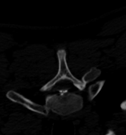
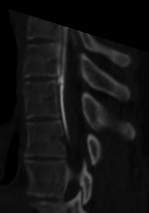
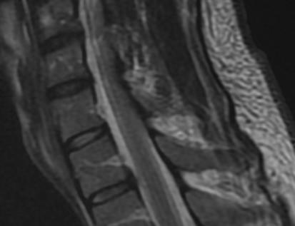
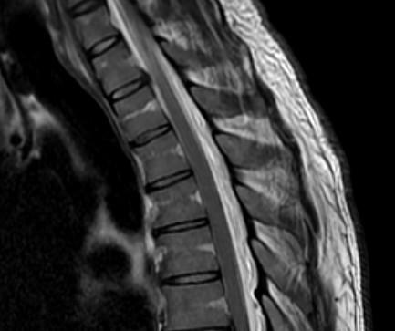
• Clinical syndrome with a wide variety of clinical presentations related to the spontaneous leakage of cerebrospinal fluid (CSF) in the spine leading to CSF hypovolemia
• Related to decreased CSF volume, not pressure
• Estimated annual incidence of 5 per 100,000
• Approximately half as common as aneurysmal subarachnoid hemorrhage
• Mean age 42.5 years, 63% female
Clinical Presentation
Most common clinical manifestation is orthostatic headache.* Headache may begin as orthostatic and become non-orthostatic.
Location and quality are highly variable. Some are exertional or second-half-of-the-day.
May worsen with Valsalva.
Other non-headache signs and symptoms:
Neck and interscapular pain
Tinnitus, dizziness, change in hearing
Nausea and emesis
Cognitive and behavioral abnormalities, including a dementia-like presentation
Encephalopathy, coma, stupor
Bibrachial amyotrophy (lower motor neuron disorder confined to the upper limbs)
Headache thought to be due to traction on meningeal layers and venous engorgement.
*Not all patients with orthostatic headache are related to CSF leakage.
Etiologies
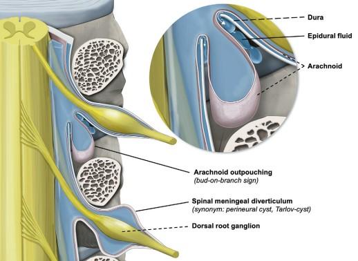
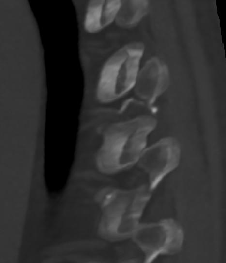
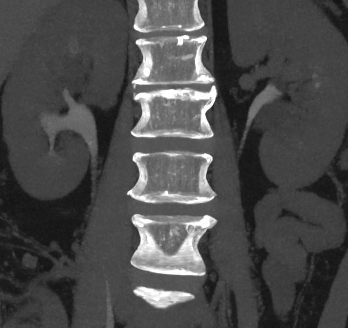
Arachnoid herniation through dural defect near nerve root
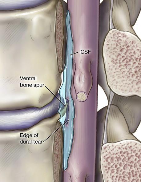
Ventral Dural Tear
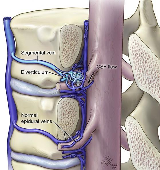
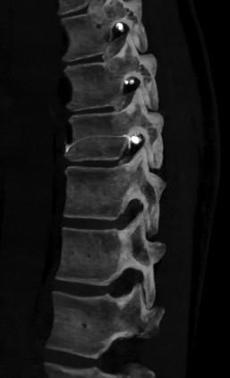
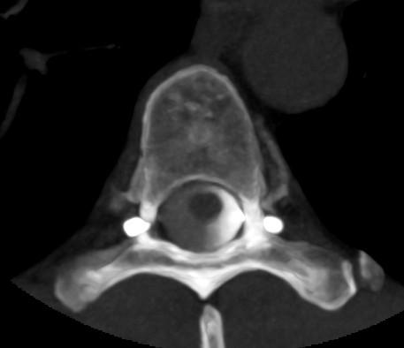
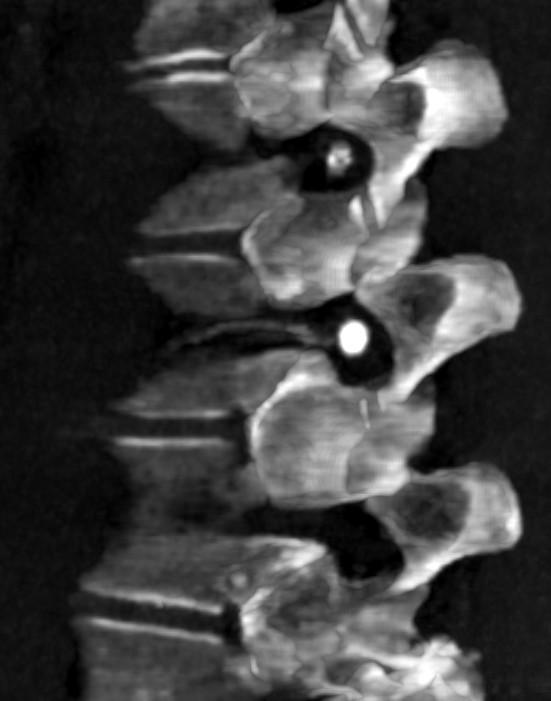
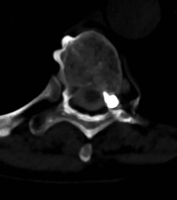
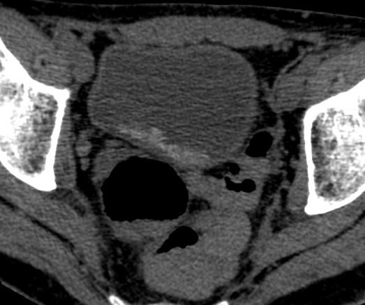
CSF-to-Venous Fistula
Adapted from Fig. 1. Kranz et al. 2019. Neuroimag Clin N Am.
Fig. 5. Lutzen et al. 2025. Radiology.
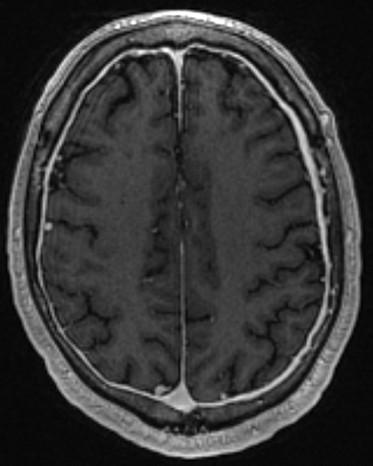
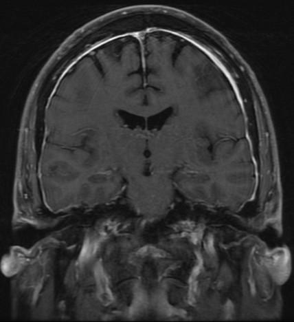
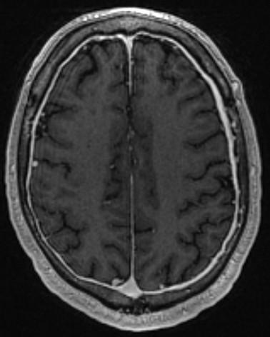
Imaging - Brain
SEEPS
• Subdural Fluid Collections
• Enhancement of the Pachymeninges
• Engorgement of the Venous Structures
• Pituitary Hyperemia
• Sagging of the Brain
• Important to note negative brain imaging does NOT exclude SIH.
• Up to 20% of SIH patients have normal brain imaging.
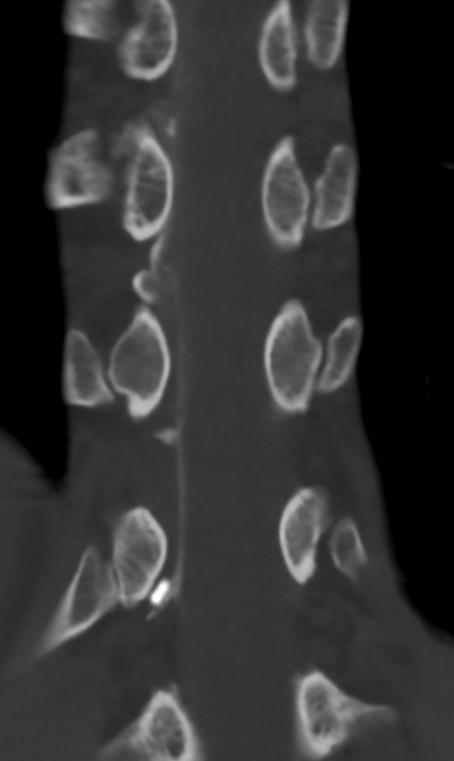
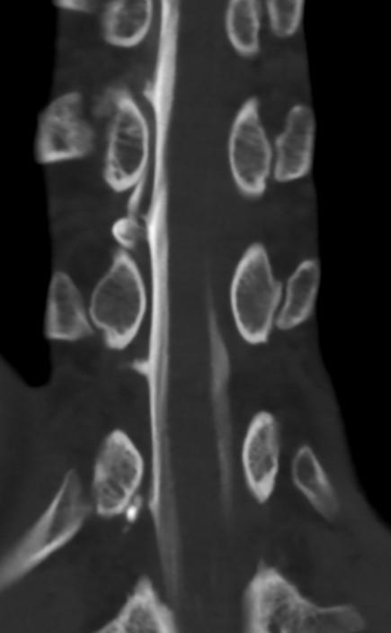
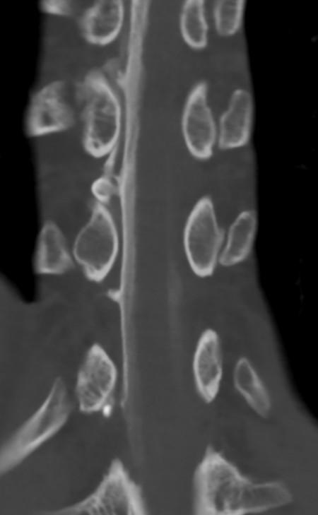
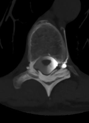
Dynamic Myelograms
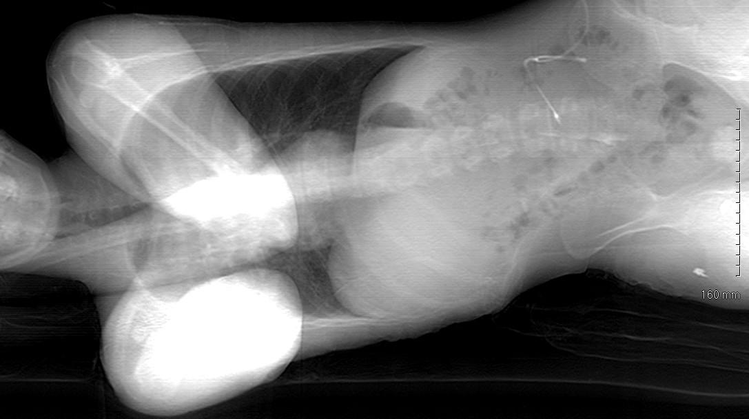
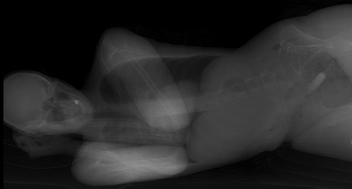
• Performed prone (ventral dural tears) or decubitus (lateral dural tear and CSF venous fistulas)
• Total spine MR reviewed prior to dynamic myelogram with attending to decide positioning.
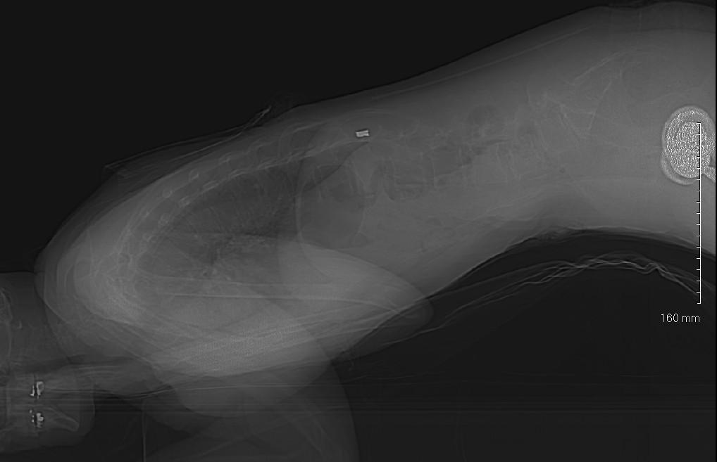
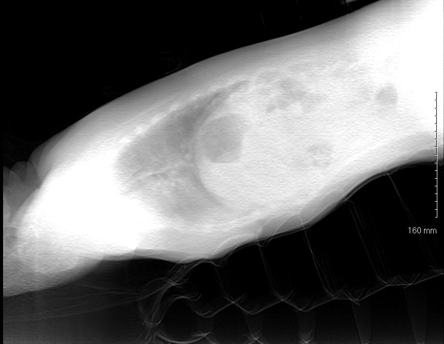
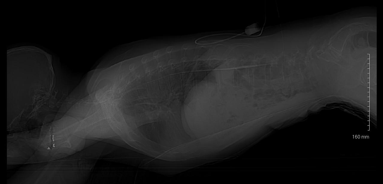
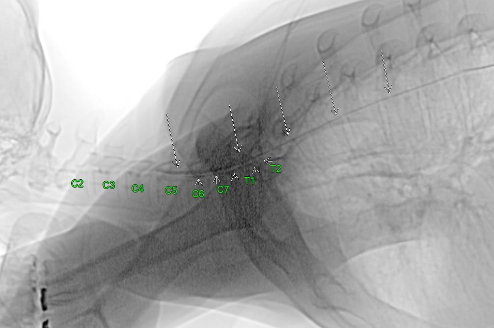
Dynamic CT Myelogram – Prone –
Steps:
1. Patient positioned prone with hips elevated using wedge air mattress.
• Ensure appropriate slope to overcome thoracic kyphosis
2. Perform planning scan, pick LP site
3. Mark, prep and drape patient
4. Perform CT fluoro-guided LP
5. Confirm intrathecal access and prepare scanner for diagnostic scans
6. Amount and frequency of contrast injected determined by attending during procedure
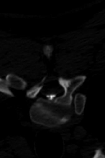
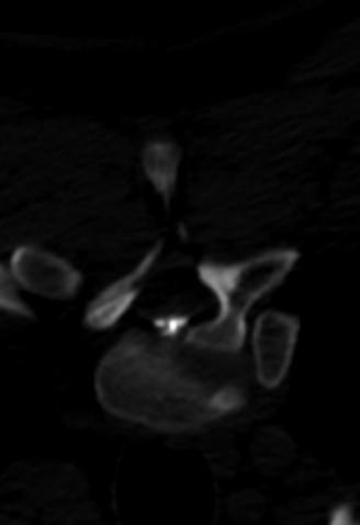
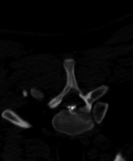
Dynamic CT Myelography – Prone
Air Mattress
Wedge pillow
Dynamic CT Myelography - Prone
Dynamic CT Myelography - Prone
Dynamic CT Myelogram – Decubitus –
Steps:
1. Patient positioned decubitus with hips elevated using pillows
• Ensure appropriate slope
• Beginning side based on review of total spine MR with attending
2. Perform planning scan, pick LP site
3. Mark, prep and drape patient
4. Perform CT fluoro-guided LP
5. Confirm intrathecal access and prepare scanner for diagnostic scans
6. Amount and frequency of contrast injected determined by attending during procedure