VIR FOCUSES ON GROWTH, COLLABORATION AND WORK-LIFE BALANCE
Fueled by collaboration and expansion of its geographic footprint, the Division of Vascular & Interventional Radiology (VIR) is experiencing a significant surge in clinical growth. Patient volume has increased by about 150 percent over the past four years, and that figure is expected to continue to rise. Based on division data, there were 1,775 independent clinic visits in fiscal year 2022 compared to 4,413 patient visits spread across six separate clinic spaces in Ann Arbor in the fiscal year ending in March of 2026.
VIR has grown from its original single clinical space to multiple sites and a broad spectrum of treatments across University Hospital, the Samuel & Jean Frankel Cardiovascular Center, C. S. Mott Children and Women’s Hospital, and the Ann Arbor Veteran’s Medical Center — as well as the Neurointerventional Radiology Clinic at the Taubman Center and a VIR clinic at Domino’s Farms.
According to William Sherk, M.D., Division Director for VIR, the growth reflects not only the expanded physical space, but also greater collaboration with referring providers. “We’re seeing a lot of direct patient referrals, and we’re seeing external providers seeking out our care, not just regionally but also nationally,” Dr. Sherk said. “More patients are seeking our care directly too.
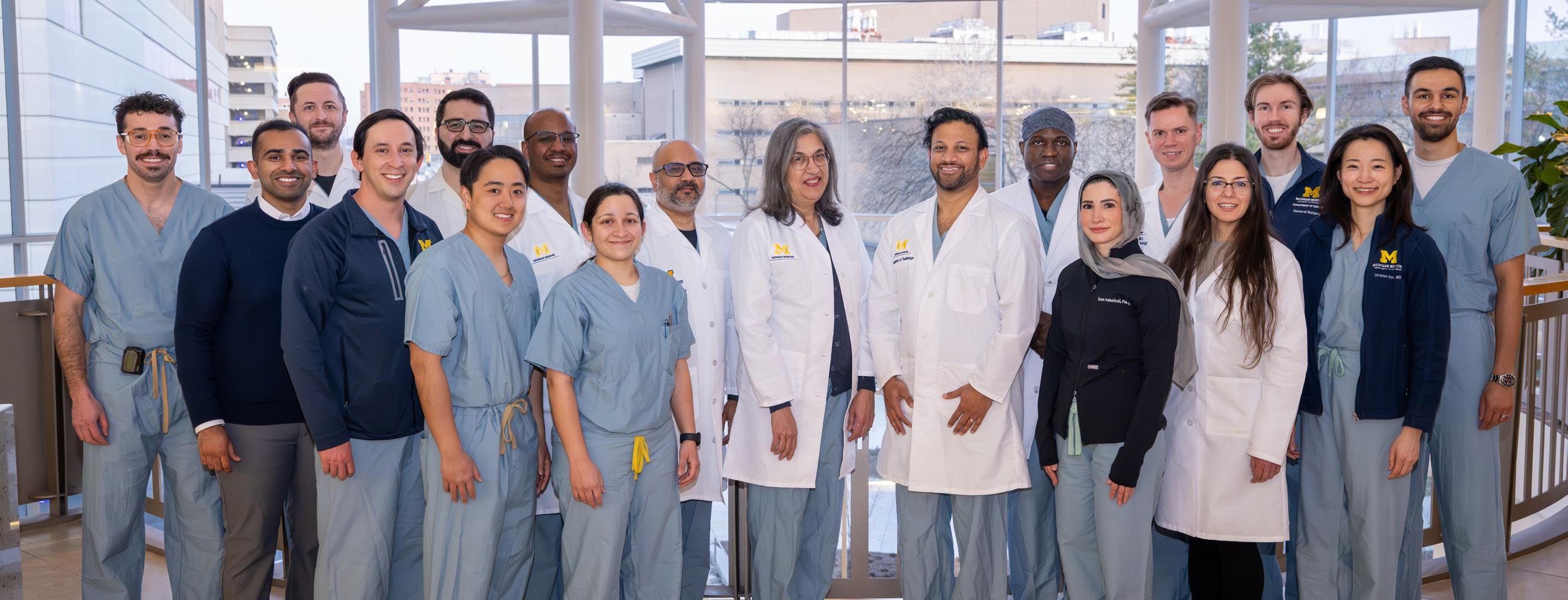
VIR Faculty, Trainees, and Advanced Practice Providers gather in the Cardiovascular Center
LETTER FROM THE CHAIR
Dear Michigan Radiology Alumni and Colleagues:
Welcome to the Spring 2026 edition of our Radiology Newsletter. This is the season when we say goodbye to departing residents and fellows, although it feels like they just joined us last week. Speaking as a former resident and fellow myself, I know that time spent at Michigan Radiology is foundational and has a lasting impact on its trainees. But what they may not know is how much their curiosity, energy, and engagement in learning means to us as their faculty. We wish them every success and thank them for trusting us with their education.
This newsletter edition also includes tributes to several retiring faculty members, who have contributed tremendously both to our department and the field of Radiology. We thank them for their many years of service, their friendship and mentorship, and we hope that they will keep in touch and enjoy a healthy, fulfilling, and relaxing retirement.
We bring you feature articles touching on all key elements of our academic mission: Research, Education, and Clinical Radiology. These missions are inherently linked—each supports and contributes to the others, and we succeed together. Please take a few minutes to read about the outstanding work happening in this department, of which I am extremely proud.
Finally, we include the sad news of the loss of two former faculty members: Chuck Meyers, PhD, and David Woodbury, MD, and are honored to share their obituaries.
Thank you for being part of the U-M Radiology Community, and have a wonderful summer!
Sincerely, Vikas
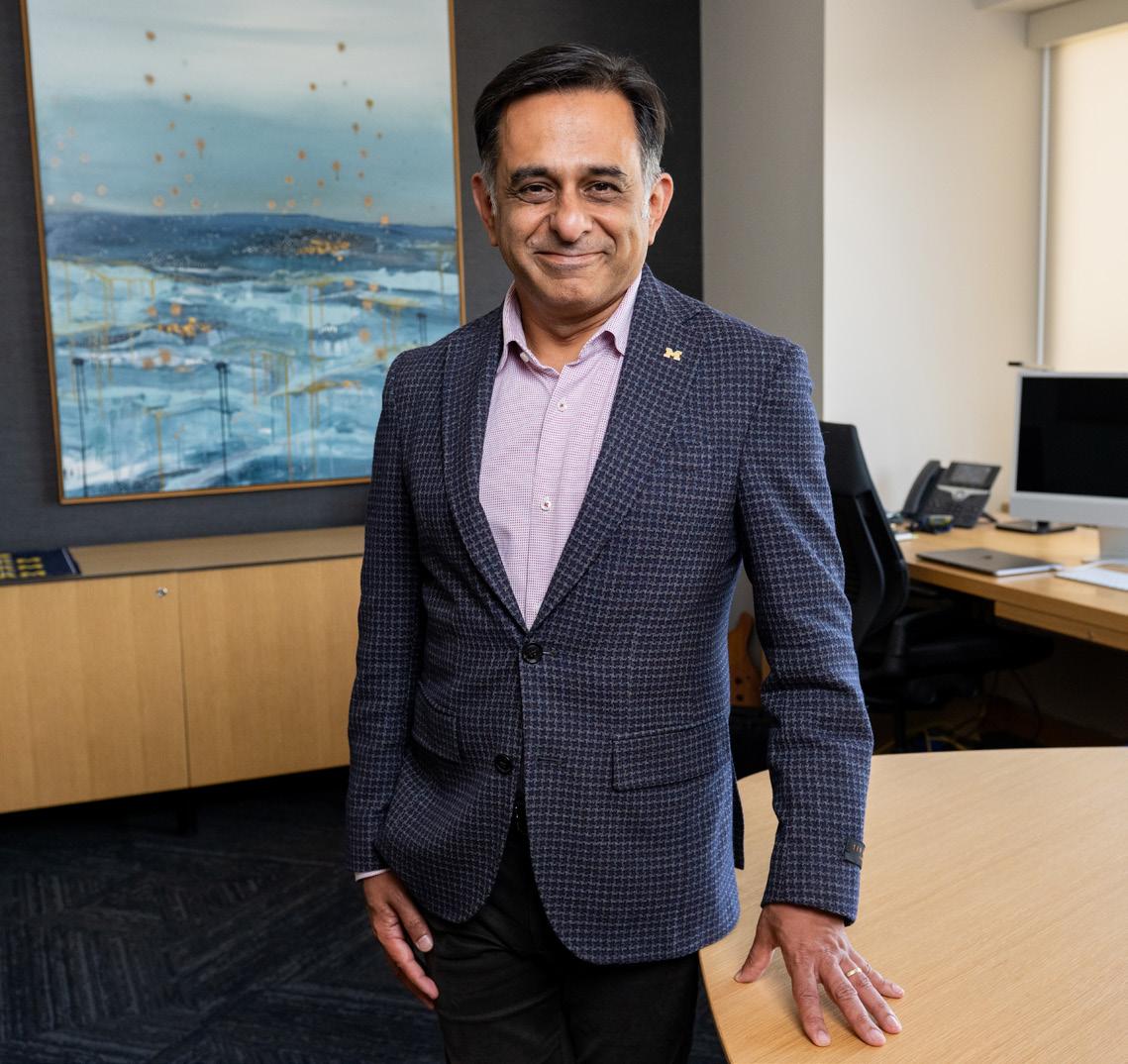
Vikas Gulani, MD, PhD
ACOUSTIC DROPLET VAPORIZATION LAB CELEBRATES A MILESTONE YEAR
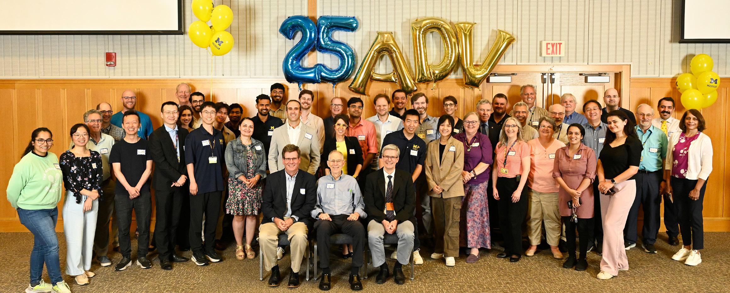
It’s been nearly two years since the group of dedicated U-M ultrasound scientists studying Acoustic Droplet Vaporization (ADV) gathered to commemorate their 25th anniversary. The event that took place on campus in July 2024 gave the team the opportunity to reflect on their accomplishments and look forward to future goals.
“The evolving research is amazing to watch,” says Oliver Kripfgans, PhD, one of the early developers of the technique. “The people involved in the research and development of ADV are a true blessing.” He delivered a heartfelt speech to the gathering of 60 current and former lab members, alumni and colleagues. Other speakers included key radiology faculty members and mentors including Drs. Paul Carson, Douglas Miller, J. Brian Fowlkes, and Mario Fabiilli.
One of the mutual goals of the research group is to share this technology with other medical schools, health systems and laboratories through its peer reviewed publications and national organizations, both medical and engineering. Another goal is to pursue patents on ADV-based technologies, which is important for clinical translation.
“ADV can be used in many different types of applications,” says Mario Fabiilli, PhD, Associate Professor of Radiology and Biomedical Engineering. For those less familiar with this technique, “ADV is the process by which ultrasound is used to turn perfluorocarbon liquid droplets into gas bubbles.” Once the droplets are injected into the bloodstream, an ultrasound transducer device converts the
bioinert liquid droplets into microbubbles, which are the size of red blood cells.
“Because they’re so small, they don’t cause tissue infarcts,” says Dr. Kripfgans. “We intended to create bubbles in situ, that is, directly inside the arteries. These microbubbles are also known as ultrasound contrast agents, which circulate freely through blood stream. These bubbles are called transpulmonary contrast agents because they pass through the capillaries, including the pulmonary bed. Rather than making bubbles out of nothing, which is called cavitation, a fascinating effect to discuss another time, we’re making bubbles out of droplet emulsions,” he says, explaining how bubbles are gas filled objects and droplets are composed of fluid. The ADV process is a phase change, with ultrasound converting perfluorocarbon droplets into perfluorocarbon bubbles.
“The reason these tiny droplets are vaporized into gas bubbles is because they are triggered by acoustic pulses,” adds Brian Fowlkes, PhD, the director of the ultrasound group, who has investigated diagnostic and therapeutic ultrasound applications for decades. As two of the initial developers of the process, Kripfgans and Fowlkes, with appointments in Radiology and Biomedical Engineering, determined that ADV could have valuable clinical applications.
When Dr. Kripfgans, now Associate Professor, joined U-M as a graduate student in 1996, he began to consider the physics associated with these vaporization events, such as: what
Ultrasound ADV team gathers to celebrate 25 years
continued from page 3
RESEARCH SPOTLIGHT
causes these bubbles to vaporize? And how rapidly is the vaporization progressing? “He began doing ultra-highspeed photography of vaporization events to learn how the acoustic field in and around these droplets were affecting the vaporization process,” shared Dr. Fowlkes. “One needs to understand that a single ADV event lasts just a few millions of a second only. To put this time scale into perspective, a typical lightning strike lasts for 0.2 to 0.3 seconds, a period during which we could observe 50,000 ADV events,” adds Dr. Kripfgans.
Moving from bench to pre-clinical research, they discovered that ADV could not only be utilized as a drug delivery system, but it could also be used to locally halt blood flow and occlude a target tissue, potentially allowing clinicians to starve cancer cells. “By tailoring the size of droplets and resulting bubbles, we can control how blood flows into different tissues, even in the brain,” says Dr. Fowlkes. Before ADV, this type of intervention could only be accomplished surgically or by catheterization, now it could be done with this minimally invasive technique or transcutaneously.
Along with Dr. Fabiilli, the ongoing ADV research at U-M is being conducted in the Ultrasonic Cavitation and Calibration Lab by Mitra Aliabouzar, PhD, Research Assistant Professor. “I use ADV as a tool to characterize tissue properties,” says Dr. Aliabouzar, whose research specializes in integrating acoustics, mechanics, and material science to advance the biomedical applications of ultrasound. “ADV is also used to estimate the age of a blood clot by how soft or stiff it is, which can help with identifying the best treatment options. Detecting changes to the tissue can lead to early detection in diseases like cancer.” In addition to Dr. Kripfgans’
ultra-high-speed camera, which allows us to visualize ADV at these rapid time scales across frequencies relevant to medical ultrasound, the lab also employs a range of highly sensitive hydrophones to capture the acoustic emissions generated by ADV bubbles, providing further insight into bubble dynamics and tissue interactions. These acoustic signals can serve as real-time, noninvasive indicators of bubble activity, enabling monitoring and control of ADV processes for improved diagnostic accuracy and therapeutic precision. As the birthplace of ADV, U-M has played a central role in both developing and expanding the technique, growing its applications from foundational research into diverse diagnostic and therapeutic uses.
While much of Dr. Aliabouzar’s ADV research centers around its potential diagnostic usages, Dr. Fabiilli focuses on its strengths for medical therapy. He loads drugs inside the droplets, which allows him to use ultrasound to control the release of drugs locally. “Another application that is very unique involves loading droplets into implants that then get placed into the body.” This process was recently used for an NIH funded study to develop a treatment for peripheral artery disease by stimulating blood vessel formation. Additional applications include regrowing bone as well as using ADV for treating Type I diabetes.
After more than 25 years of research, the ADV lab scientists continue to work towards real life clinical applications, so that this unique process can be used to benefit the lives of patients.
by Nikolas Charles
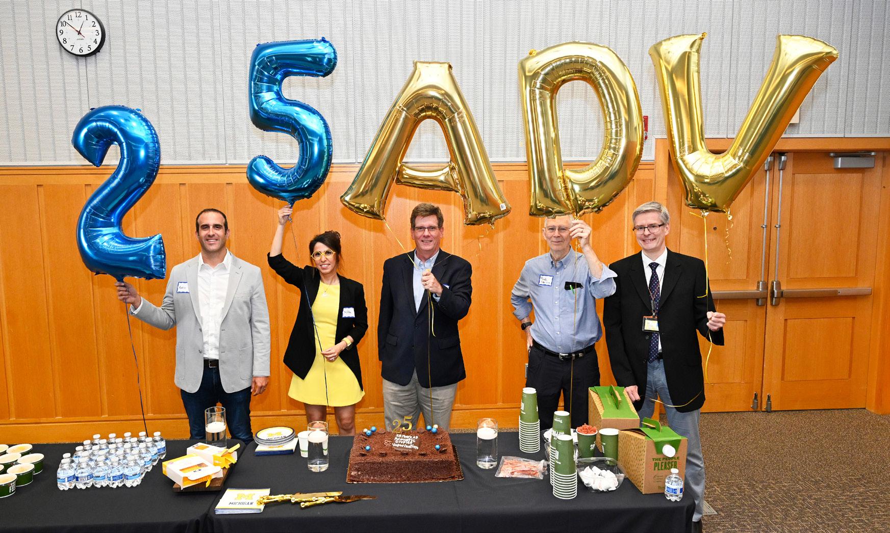
Mario Fabiilli, PhD; Mitra Aliabouzar, PhD; Brian Fowlkes, PhD; Paul Carson, PhD; Oliver Kripfgans, PhD
Retirees
We reached out to our recent retirees to hear their reflections on time spent at U-M Radiology, and to learn what they are doing now. The responses illustrated the depth, creativity and commitment of our faculty, and the respect and admiration of their peers and trainees. Profiles follow for all who chose to participate.
Kenneth Buckwalter, MD
Clinical Professor
Musculoskeletal Radiology and ACMIO
Reshmi Mediratta, MD
Clinical Assistant Professor VA Service Chief
Perry Pernicano, MD
Clinical Associate Professor, Emeritus Cardiothoracic Radiology
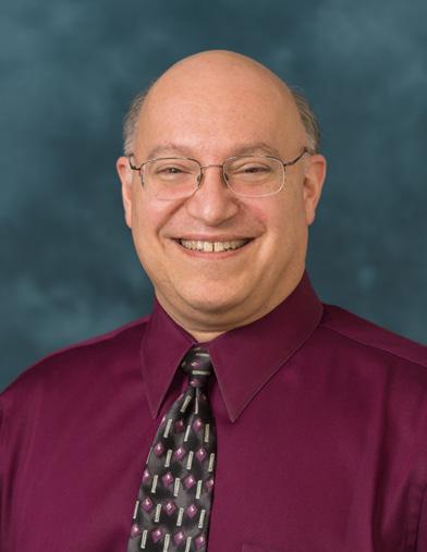
Perry Pernicano, MD
Clinical Associate Professor, Emeritus Cardiothoracic Radiology
Years of service: 1989 - 2025
Marilyn Roubidoux, MD
Professor Emerita
Breast Imaging
Xia (Shannon) Shao, PhD
Research Scientist Emerita
Nuclear Medicine
James Shields, MD
Clinical Assistant Professor
Vascular Interventional Radiology
Favorite aspect of working in department: The best part of any job is the people you get to work with. I have been blessed throughout my career to work with tremendous colleagues, fantastic residents, and terrific medical students. I also had the great privilege and honor of caring for the brave veterans of our country for 32 years at the VA from 1990 until 2022.
Memorable roles during your career: I served as Chief of the Radiology Service at the VA for two years from 2006 until 2008. I became a Fellow of the American College of Radiology in 2009. I was elected and served as President of the Michigan Radiological Society for the 2019-2020 term. I was awarded the Gold Medal of the Michigan Radiological Society in 2024. The Regents of the University conferred me with Emeritus status in 2025.
Advice for our trainees: Never lose the wonderment of what we do. Everyone gets to see people on the outside, only a select few of us get to see people on the inside. It is truly amazing that we noninvasively get to peer inside our patients every day.
Pleasures of retirement: I really enjoy being able to sleep in every day. There is a lot less stress. I am also hoping to do more traveling, particularly internationally.
Reflections from colleagues: Dr. Pernicano is not only known for his unwavering love of Michigan sports and all things maize and blue, but also for the exceptional care he brings to his work and his deep commitment to teaching medical students. His attention to detail on radiographs is truly remarkable. No subtle finding escapes his keen eye (not even a faint “Go Blue” on a shirt)! More than that, his dedication sets a standard that inspires everyone around him, leaving a lasting impression as both a clinician and a colleague. His MRS Gold Medal in 2024 is a proud and well-deserved milestone, and a fitting recognition of his impact.
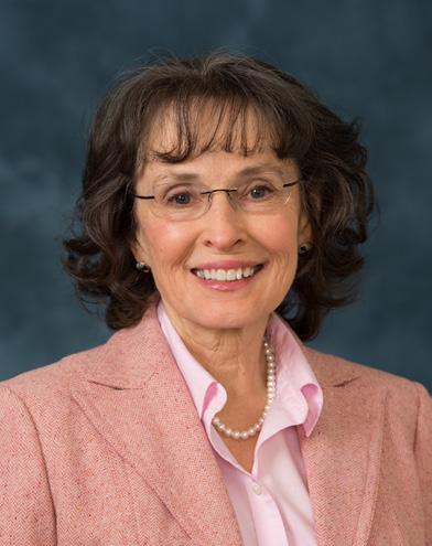
Marilyn Roubidoux, MD
Professor Emerita
Breast Imaging
Years of service: 1992 - 2025
Favorite aspect of working in department: I enjoyed taking care of patients. Mammography is especially rewarding, as we find breast cancers early, when they are usually curable. I once did a biopsy on a famous musician. She was quite anxious and sang during the procedure. (She had a great voice, especially for someone in the supine position.) I also enjoyed showing residents how to do procedures.
As a faculty member, I could get any book on any subject from the University Library. If they didn’t have it, which was unusual, they would get it. They provided great service.
Memorable roles during your career: I initiated cryoablation at Michigan and am still regarded as one of the pioneers. As recently as the last Society of Breast Imaging, I served as the cryoablation moderator – and actually had to give a short talk on it.
Probably my most impactful role was the mobile mammography project serving the Northern Plains Indians. This was quite involved, getting the van, driver, technologist, tribal permission and mammography accreditation from the ACR. The mobile mammo unit drove to small clinics where the women would have a screening study. Since travel was so difficult for the women, we read the studies (sent via satellite) in real time. While they waited to see if additional views were needed, the women were given educational materials to read or watch. This program lasted over 10 years.
Advice for our trainees: Subspecialize. Radiology is too large for anyone to really master it all. Even if you want to go into private practice, a fellowship is expected. I liked academics, because you interacted with learners at all levels. At Michigan, we were expected to participate in research and I was happy working to advance the field.
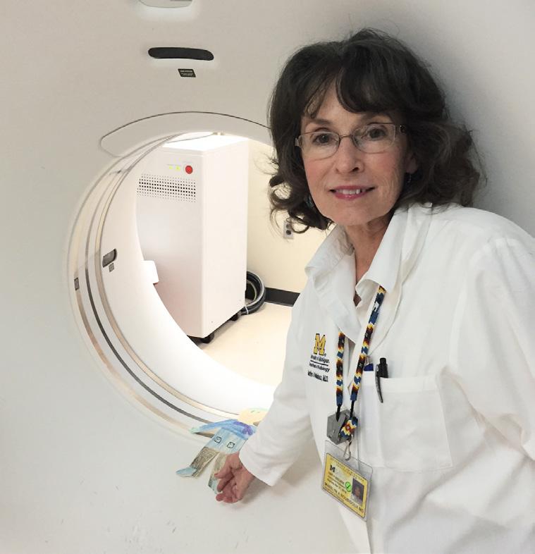
Pleasures of retirement: Since I enjoyed working, I have mixed feelings about retirement. The best things about it include not needing an alarm clock and being able to travel without worrying about scheduling.
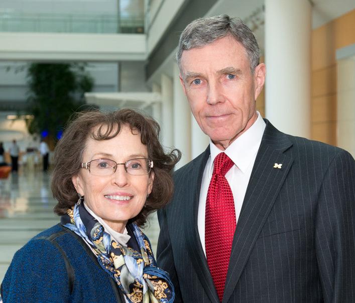
Reflections from colleagues: Marilyn Roubidoux retired this year after 34 years at Michigan Medicine. In those years she was an abdominal radiologist at the VA, covered general imaging call at UH, and throughout was a faculty member in Breast Imaging. Marilyn was known for being a prolific researcher, with numerous publications and presentations, and for her humanitarian efforts, especially directed toward the American Indian community. She single-handedly launched a mobile mammography screening program serving Native American women in the Dakotas, not an easy feat to say the least. A wonderful teacher, Marilyn trained hundreds of residents, fellows, medical students, and undergraduates. What we will miss most though is our loving, inspiring, brilliant, creative, hilarious friend in the reading room. Thanks for all the wonderful years, Marilyn, and best of luck in your welldeserved retirement.
Marilyn Roubidoux, MD; N. Reed Dunnick, MD
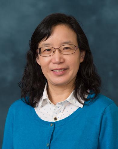
Xia (Shannon) Shao, PhD
Research Scientist Emerita Nuclear Medicine
Years of service: 2000 - 2025
Favorite aspects of working in department: We have supportive leaders and collaborative teams within the department. I enjoy continuously learning new knowledge, developing new techniques, and contributing to the advancement of science.
Memorable roles during your career: I have served in a variety of roles throughout my career. The most rewarding part is being an educator, where I have had the opportunity to watch students grow into independent researchers and become leaders in the field.
Advice for our trainees: Stay curious and never stop learning. As a researcher, be prepared for failure — positive results often come after many negative ones.
Pleasures of retirement: Retirement gives me more free time to travel and see the world.
Thoughts from colleagues: Dr. Shao is a world-renowned carbon-11 radiochemist, a nurturing teacher and a generous colleague. The GMP production methods she developed during her career are now used by PET drug production facilities all around the world, while the trainees she has mentored now hold leadership positions throughout industry and academia, and her candy drawer is NEVER empty! Best of luck with retirement and don’t be a stranger!
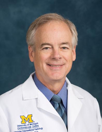
James Shields, MD
Clinical Assistant Professor Vascular Interventional Radiology
Years of service: 2007 - 2025
Favorite aspect of working in department: I had the privilege of working in two settings: “Private Practice “ and “Academic Practice”, and I spent about half of my time in each. I enjoyed both about equally well and was very fortunate to experience very high quality radiology in each setting.
Advice for our trainees: Keep in mind that you will be entering into a culture set by all of your fellow radiologists, and we spend many hours living in a culture that will foster long-term relationships. Our success and happiness will be largely determined by how effective is the match between our own goals and those of the group we are joining.
Pleasures of retirement: I am now “retired” after spending almost 50 years working as a radiologist. Retirement planning is important for trying to ensure happiness in these later years. As far as advice goes, I would say have some hobbies and have activities you enjoy that require interaction with people. I think that success depends on developing these hobbies and activities before actually retiring.
Thoughts from colleagues: Jim Shields has been far more than an outstanding interventional radiologist – he’s been the kind of colleague, teacher, and mentor that can turn any conversation into something memorable, whether about medicine, music, or life. Many of us who trained with Jim remember his unflappable composure and his ability to infuse this sense of calm to his patients and others around him. He is truly multi-talented, both in and outside of the IR suite.
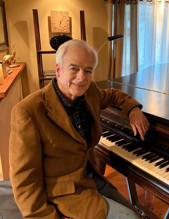
In Memoriam
The University of Michigan Department of Radiology mourns the passing of Charles “Chuck” Raymond Meyer, PhD, age 81, of Ann Arbor, who died on Wednesday, March 25, 2026, with his wife and daughter by his side. For colleagues, trainees, and collaborators, Chuck was a steady presence in the department and a pioneering force in computational imaging—bringing an engineer’s precision, a collaborator’s instincts, and a commitment to methods that could ultimately improve patient care.
Chuck was born January 7, 1945, in Delta, Colorado, to the late Raymond and Josephine Meyer. He served in the United States Air Force and earned his BSEE (1967) and MSEE (1969) degrees from Colorado State University, followed by a PhD from Iowa State University (1972). His early academic career bridged engineering and medicine: he was Assistant Professor of Electrical Engineering at Carnegie Mellon University (1976–1979) and then Assistant Professor of Radiology at the University of Colorado (1979–1981).
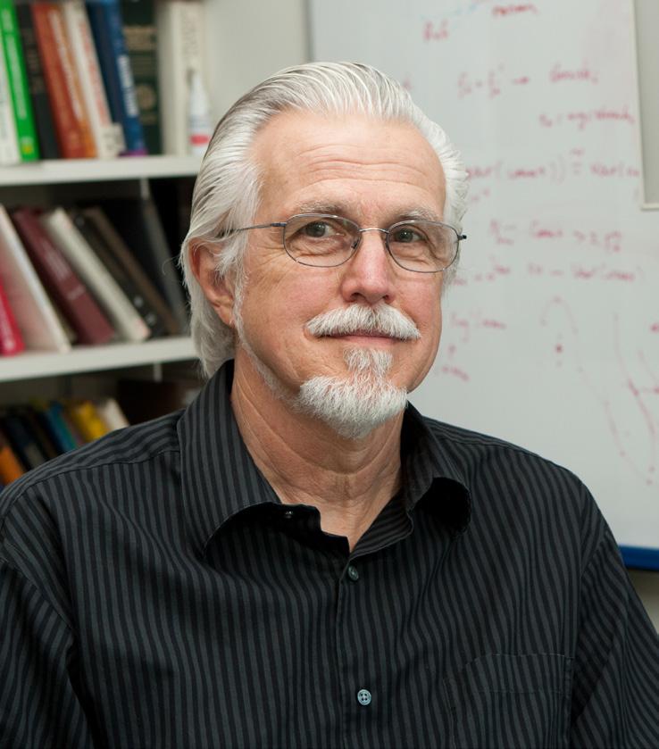
Chuck Meyer, PhD 1945 – 2026
In 1981, Chuck joined the University of Michigan as an Assistant Professor of Radiology, among the first faculty members in the Division of Basic Radiological Sciences. He was promoted to Associate Professor in 1988 and Professor in 1996, and in 1998 was appointed Professor of Biomedical Engineering, reflecting the cross-disciplinary reach of his work. Chuck’s contributions were further recognized in 2011, when the Medical School named him a founding inductee in its League of Research Excellence. The Regents named him Professor Emeritus of Radiology and Professor Emeritus of Biomedical Engineering in December 2012.
A defining milestone for the department came in 1985, when Chuck founded the Digital Image Processing Laboratory (DIPL) within Basic Radiological Sciences. Radiology Chair William Martel, MD, appointed Chuck as the lab’s first director. From the beginning, Chuck built DIPL around the idea that the hardest imaging problems require diverse expertise and a culture of shared credit. Early DIPL efforts addressed foundational problems in medical image analysis and visualization, including interactive visualization, edge detection and linking, liver atlas construction, MRI B1 field inhomogeneity correction, and the development of methods for registration of volumetric datasets. As the field advanced, mutual-information approaches introduced in the mid-1990s accelerated automatic 3D registration and helped shape DIPL’s evolving focus.
Across his career at the University of Michigan, Chuck’s research centered on image registration, particularly in three-dimensional imaging. He developed techniques to optimize comparison of images over time within the same modality and to compare tissue across modalities—capabilities essential to monitoring disease progression and evaluating response to therapy. These methods proved especially valuable in oncologic imaging, where precise alignment of interval exams can reveal subtle but clinically meaningful change. He authored more than 121 peer-reviewed publications and maintained continuous research support, much of it from the National Cancer Institute (NCI).
DIPL’s success also reflected Chuck’s emphasis on team science and longrunning collaborations. Shortly after launching the lab, he asked Peyton Bland, PhD, to help build and run it. Early on, DIPL collaborated with the Artificial Intelligence Lab in Electrical Engineering and Computer Science, applying Bayesian statistical methods to construct a liver atlas and use it for liver boundary definition in CT datasets. The student who performed this initial work, Jennifer Boes, PhD, later joined the lab as a co-investigator and major developer of registration code. In 1995, Boklye Kim, PhD, joined DIPL to advance registration methods, including later work on retrospective correction of head motion during fMRI. Gary Laderach joined in 2001 as senior computing systems manager, and Bing Ma, PhD, came on board in 2004, further strengthening DIPL’s computational and scientific capacity. This collaborative model was also evident in DIPL’s participation in an NIH/NCI Program Project Grant focused on “Automatic 3D Registration for Enhanced Cancer Management.” Co-investigators included Jeff Fessler, PhD, Al Hero, PhD, Tim Johnson, PhD, Fred L. Bookstein, PhD, Paul L. Carson, PhD, and Veronica Berrocal, PhD.
He married Kathleen (Alloway) on September 21, 1984, and their daughter, Kelly Meyer, was born in 1988. He is survived by Kathleen and Kelly, and by the many colleagues, collaborators, and trainees who benefited from his intellect, rigor, and generosity. The Department of Radiology extends heartfelt condolences to his family and honors Chuck Meyer’s enduring legacy in imaging science at Michigan and beyond.
by Oliver Kripfgans, PhD
“That means more imaging studies, more consultants involved, and it reflects the complex and effective care that we can provide in VIR. We just find that to be an incredible opportunity to increase our presence in the hospital and to generate new patients for the health system. We’re not ones to sell our ‘block M’, though marketing is something that we will bring to the table as we continue to grow our clinic space in the future. But the growth has happened very naturally just through word of mouth.”
About two-thirds of VIR procedures are generated from outpatient clinic visits, while a third result from urgent or emergent procedures that arise from inpatient care at the main hospital.
Dr. Sherk also attributes VIR’s clinical growth to internal collaboration. VIR works with University of Michigan colleagues in about 20 different multidisciplinary teams spanning vascular and non-vascular interventions. These include oncology, pediatrics, liver and kidney transplant, and many more.
“These developments happen both naturally and intentionally,” he said. “An example of a natural growth has been through the Prostate Artery Embolization service line, which grew out of our faculty’s involvement with urology.”
“And then providing time for our providers to be involved in these multidisciplinary groups, emphasizing that as part of our faculty development, is intentional,” Dr. Sherk continued. “Our team members are active in the multidisciplinary work groups but also co-lead many of them, such as our Venous Health Program and the Pulmonary Embolism Response Team. We’ve generated a lot of interest in what we have to offer from multidisciplinary care.”
From an educational standpoint, VIR continues to reach out to medical students and prospective resident applicants to reinforce the division’s educational mission. Dr. Sherk has been with Vascular Interventional Radiology at the University of Michigan for about a dozen years, from residency training to a member of the faculty, before assuming the role of division leader in the fall of 2025.
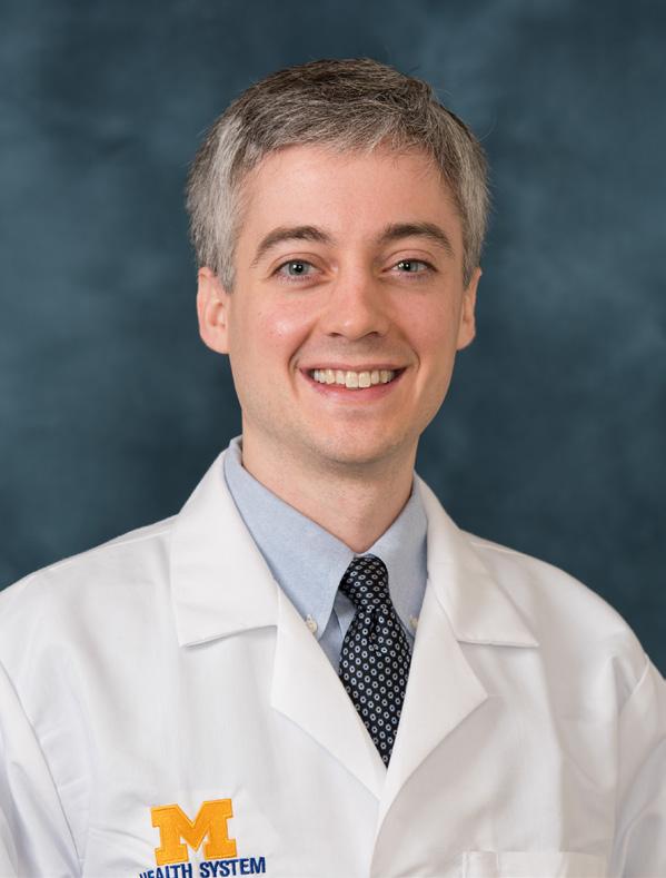
On the research front, VIR is building momentum in establishing new grants and new foundation and industry relationships. In addition to clinical, educational and research objectives, Dr. Sherk says he wants to focus on the well-being of providers and staff by building a “culture of balance”.
For example, the division has introduced the concept of “narrative medicine”, which involves sharing short stories, poems, etc., to integrate more broad liberal arts concepts into the practice of medicine.
“I’m just continuously impressed by the people we have here and the amazing opportunities that we have for growth,” he said. “But one of the challenges with our VIR faculty and staff, is they’re very invested in clinical care of patients. We need to provide time for them to recover, frankly, from what they do. Interventional radiologists always operate in kind of ‘survival mode’ so it’s hard to disconnect from that sympathetic nerve system drive. That’s why an emphasis on recovery, reflection and rejuvenation can really help them maintain a sustainable lifestyle that’s also beneficial for their clinical care and academic pursuits.”
by Lynn Waldsmith
“We are truly a top five program for VIR training through our residency program,” he said.
William Sherk, MD
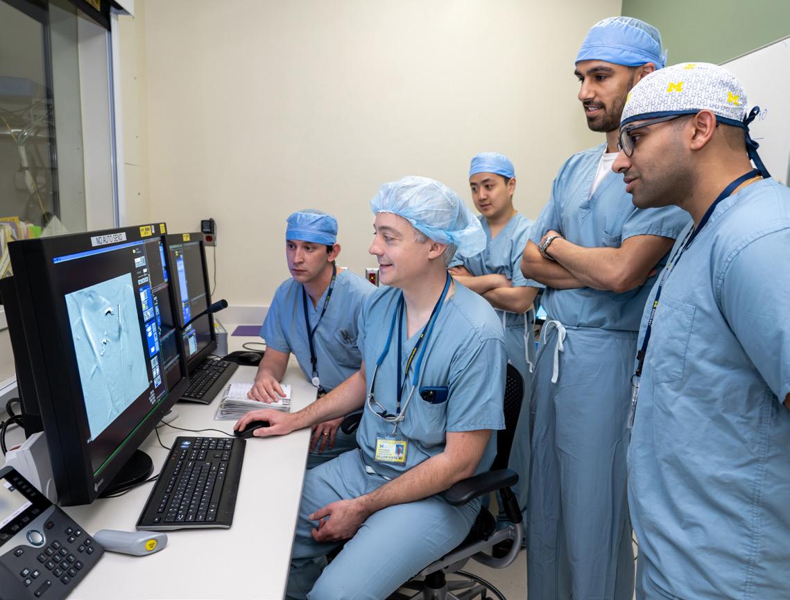
Dr. Sherk and trainees review a case
U-M RADIOLOGY RESIDENTS “ON TRACK”
Graduates of our residencies often become leaders in their practices, health systems, and communities. Beginning in July of 2027, new training pathways will enhance radiology residency education at U-M. All diagnostic radiology (DR) and interventional radiology (IR) residents will choose to enroll in one of four national leadership tracks: Health System Administration, Research, Education, or Information Technology.
The four-year educational tracks were established in partnership with the Michigan Ross School of Business and are designed to create national leaders in healthcare. Pilot versions of the courses are already underway, with resident participation optional for the 2025-26 and 2026-27 academic years.
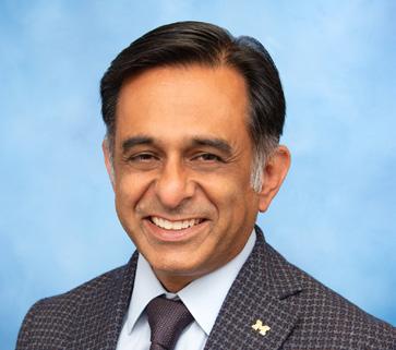
“Our goal is to transform the curriculum of the residency at Michigan Medicine to create national leaders in health care and radiology,” says Dr. Davenport. “Each one of these pathways includes structured education and training elements, such as the research track, which is a National Institutes of Health (NIH) T32 funded service award designed to create physician scientists.” Research track Directors, Drs. Nicole Seiberlich, Vikas Gulani, and Peter Scott, will guide residents to identify appropriate mentors and apply for NIH and Michigan Translational Imaging programs.
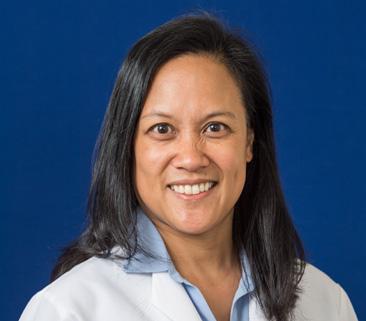
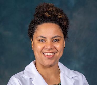
Trainees who choose the Health System Leadership track will have their own set of outcomes. Associate Residency Program Director and Health System Leadership track Director Elaine Caoili, MD, MS, will work with Dr. Davenport to advise residents as they complete the Radiology Leadership Institute (RLI) Essentials Program in the R2 year and the RLI Health Care Economics Milestones Program in the R3 year, both sponsored by the American College of Radiology (ACR). One R2 will also be able to attend the optional Association of Academic Radiology Resident Academic Leadership Development (ARRALD) Program.
No matter which track residents choose, core leadership curriculum elements will be constant, including negotiation, teamwork, crucial conversations, and change management. There will also be a designated U-M program director on-hand to ensure each person’s journey provides them with the proper knowledge and tools. “In addition to teaching students how to be radiologists, we will teach them how to think and act like leaders that create positive changes, not just in their local circle but nationally. A huge component of leadership is people skills,” adds Dr. Davenport. “This includes gaining influence, consensus and knowing how to engage with colleagues to solve problems.”
All four Resident National Leadership Tracks include on-campus training and lectures for the first three years supplemented by national involvement in radiology societies and parallel training experiences. The R4 year will culminate with a capstone project that proposes
RESEARCH LEADERSHIP (NIH T32)


EDUCATION
(COMET-ALIGNED)
HEALTH SYSTEM (RLI-ALIGNED) IT LEADERSHIP (WITH SIEMENS
Dr. Dr. Gulani
Dr. Caoili
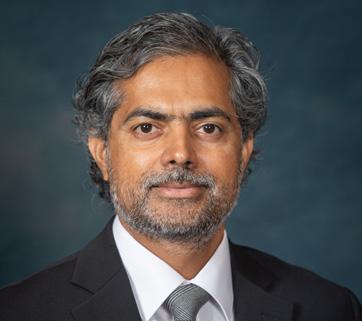
Dr. Sharma
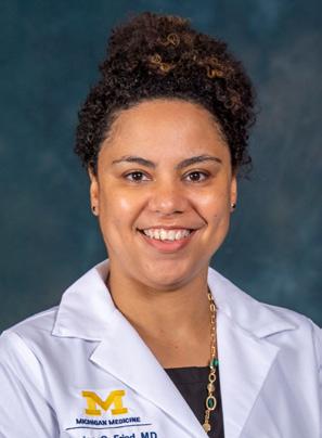
Dr. Fried
Dr.
TRACK” TO BECOME LEADERS IN THE FIELD
significant national impact. “We envision expanding the tracks in the future based on the residents’ interests,” says Kara Udager, MD, Diagnostic Radiology Program Director. “Some potential new tracks would include Quality Improvement/Patient Safety and National and Global Outreach.”
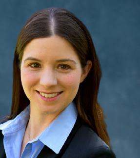
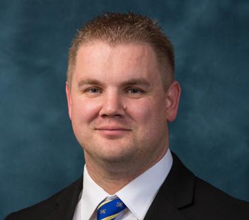
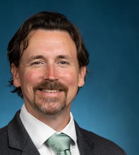
During the formal launch of the program in July 2027, all incoming R1’s will select the track that most closely aligns with their future career goals as a radiologist. Associate Residency Program Director and Education Track Leader, Aseem Sharma, MD, says “we want these future radiologists to feel empowered to be leaders in their chosen area. In addition to educating them to be excellent general or subspecialist radiologists, we want to give them the wings to fly as high as their aspirations will take them.”
IR/DR Residency Program Director, Shantanu Warhadpande, MD, agrees that the new tracks are aligned with the department’s goals for IR trainees now and in the future. “It’s the missing piece of the puzzle for building strong leaders in our medical specialty,” he says. And while he won’t be leading any of the new tracks, he will still actively participate in the development of courses that allow residents to reach their full potential. “I see my role as encouraging our residents to immerse themselves in both the clinical and leadership training.”
“For an institution like University Michigan, it’s ideal for us to be able to graduate residents that have skill sets beyond diagnostic radiology,” adds Jessica Fried, MD, Associate Chief Medical Information Officer (ACMIO) of Radiology and Radiology Information Technology Track Leader. “There are many things they can do with their career as a leader in radiology. We have designed the IT track to be substantive, but digestible. They will learn about administrative, operational, research, and innovation aspects of informatics, in addition to principles related to quality improvement and quality assurance.”
The track directors expressed their gratitude for the dedication and support of their whole team that includes Erica Stein, MD, Associate Residency Program Director, Janet Bailey MD, Associate Chair for Education, Amy Spencer, Program Coordinator, Jessica Meyers, Fellowship and Medical Student Coordinator, and Sarah Goldie, Assistant Program Coordinator. “The whole team contributes to the success of the program,” says Dr. Davenport. “There is a tremendous need for national leadership. Michigan Radiology will take a more authoritative role in creating long-lasting favorable outcomes. One of our mantras at U-M is to be the number one residency program in the country. We feel strongly that this formal leadership curriculum will complement our medical training and help us achieve that goal.”
by Nikolas Charles
Congratulations Graduating Residents
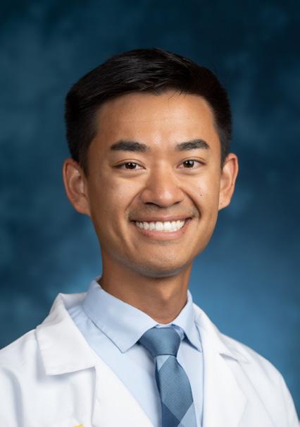
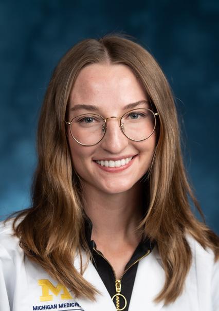
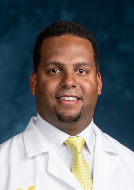
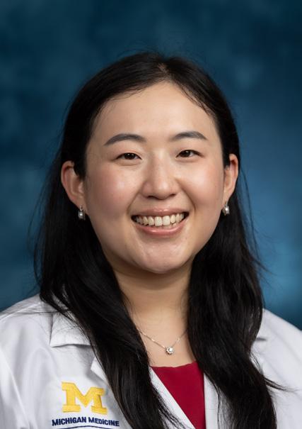
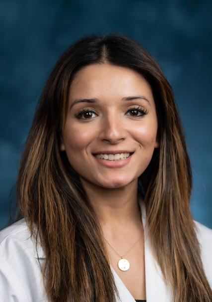
Michael Bui, MD
Future plans: Body Imaging Fellowship, UC San Diego
Favorite memory: Traveling to Arizona for SAR 2025, to help screen radiologist for color vision deficiencies. I was able to spend time with my co-residents Dave Mortensen and AJ Winkelman, as well as many body faculty.
Carolyn Dishuck, MD
Future plans: Breast Imaging Fellowship, U-M
Favorite memory: When my class held our own soda blind taste test as R1s during a summertime lunch break.
Jordan Ellis, MD, MBA
Future plans: IR Integrated Resident, U-M
Favorite memory: Working closely with my coresidents while on-call. Building relationships through shared experiences as a team was really meaningful.
Ran Jing, MD
Future plans: IR Integrated Resident, U-M
Favorite memory: First time at the Big House with radiology friends for a U-M football game—in the rain!
Sakina Kazmi, MD, MPH
Future plans: Breast Imaging Fellowship, U-M
Favorite memory: Spending time with my coresidents during call nights. Every last night of the call week, which is always on a Thursday night, my coresidents and I celebrate surviving call by either ordering food late at night or by having a small potluck. This always made completing a week of call feel even more special!
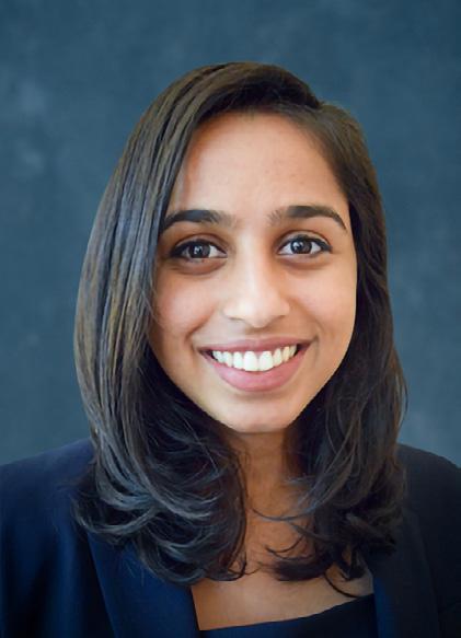
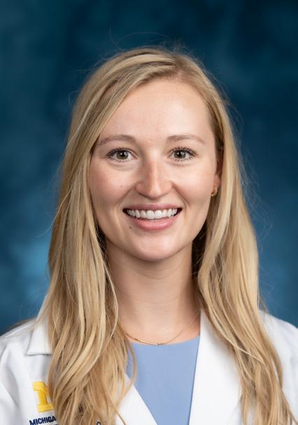
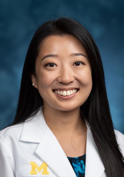
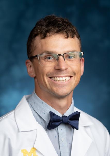
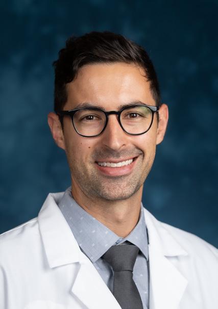
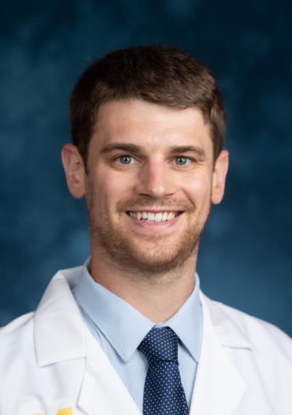
Preethi Kesavan, MD, MPHS
Future plans: MSK Fellowship, U-M
Favorite memory: Taking walks in the Arb with my class - especially in the spring when the peonies are blooming!
Hannah Lamberg, MD
Future plans: MSK Fellowship, U-M
Favorite memory: Hosting my residency classmates at our home for gettogethers or going to others’ homes for game-nights/hang outs! A favorite one of those times was when a group of us ladies got together after one of the call exams and intended to exercise together, but ended up sitting on the couch chatting and then going out for ice cream instead! In general, the camaraderie and friendship among my class has made training here feel special.
Joy Li, MD
Future plans: Neuroradiology Fellowship, Johns Hopkins
Favorite memory: Eating lunch and walking around the Arb with my class in the summer when the peonies were in bloom.
Grant MacKinnon, MD
Future plans: Neuroradiology Fellowship, U-M
Favorite memory: Board game nights with my classmates!
Alexander Oserowsky, MD
Future plans: Abdominal Fellowship, U-M
Favorite memory: Having over half my residency class over to my house as R1s multiple weeks in a row to watch the Bachelorette. Was a great way to build camaraderie early on!
Christian Sleeper, MD
Future plans: IR Integrated Resident, U-M
Favorite memory: I have really enjoyed working with all the faculty at U-M! I couldn’t imagine a better group to learn from over the past 5 years.
Congratulations Graduating Fellows
Learn about our fellows’ future plans below
Anjelica Abate, MD
Breast Imaging
MD Anderson Cancer Center Houston, TX
Alexa Arvidson, MD Musculoskeletal
Faculty for Musculoskeletal and Community Divisions
Michigan Medicine Ann Arbor, MI
Michael Bair, DO Musculoskeletal
Pinehurst Radiology Pinehurst, NC
L. Jordan Breyfogle, MD
Interventional Radiology (Integrated)
Mount Auburn Hospital Boston, MA
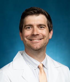
Hussein Chahrour, MD
Interventional Radiology (Independent)
Henry Ford St. John Hospital Detroit, MI
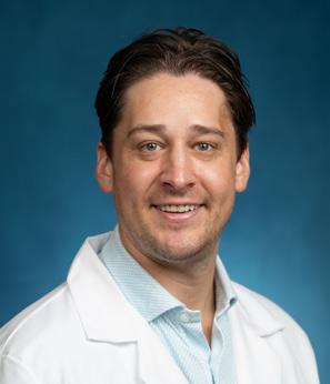
Mark Cooks, MD
Abdominal
Huron Valley Radiology Ann Arbor, MI
Mary Creedon, MD Musculoskeletal
Faculty for Musculoskeletal and Community Divisions
Michigan Medicine Ann Arbor, MI
Zachary Gaughan, MD
Interventional Radiology (Independent)
University Health Truman Medical Center Kansas City, MO
Jennifer Hayes, MD, PhD Neuroradiology
Faculty for Neuroradiology and Pediatric Radiology
Michigan Medicine Ann Arbor, MI
James Iordanou, DO Neuroradiology
McLaren Medical Group Flint, MI
Alex Jessmore, DO Abdominal
Charlotte Radiology Charlotte, NC
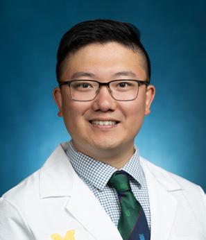
Albert Jiao, MD Neuroradiology
Duke University Durham, NC
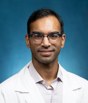
Kevin John, MD
Neurointerventional
Second Year Fellow for Neurointerventional Michigan Medicine
Ann Arbor, MI
Nathan Loudon, MD
Interventional Radiology (Integrated)
Radiology & Imaging Consults
Colorado Springs, CO
Steven Machusko, MD
Abdominal
Bay Imaging Consultants San Francisco, CA
Mark Manganaro, DO
Neuroradiology
Triad Radiology Associates Winston-Salem, NC
Antigone Mitchell, MD
Abdominal
Advanced Radiology Services (Remote Practice) Grand Rapids, MI
Luke Odisho, MD
Neuroradiology
Corewell Health William Beaumont University Hospital
Royal Oak, MI
Laura O’Donohue, MD
Pediatric Radiology
Kaiser Permanente Oakland, CA
Lisa Philips, MD
Cardiothoracic
Faculty for Cardiothoracic Division
Michigan Medicine
Ann Arbor, MI
Michael Ramalia, DO
Abdominal
Lumexa Imaging (Remote Practice) Raleigh, NC
Nicholas Rasmussen, MD
Musculoskeletal
Regional Medical Imaging
Novi, MI
Madhusudan Reddy, MD
Nuclear Medicine
Fellow for Cardiothoracic Division
Michigan Medicine
Ann Arbor, MI
Mariam Salman, MD
Breast Imaging
Henry Ford Hospital Detroit, MI
Orlin Sergev, MD
Neuroradiology
Roper Radiologists Charleston, SC
RaeLynn Shanker, MD
Abdominal
Cleveland Clinic (Remote Practice) Cleveland, OH
FEATURED ALUMNA
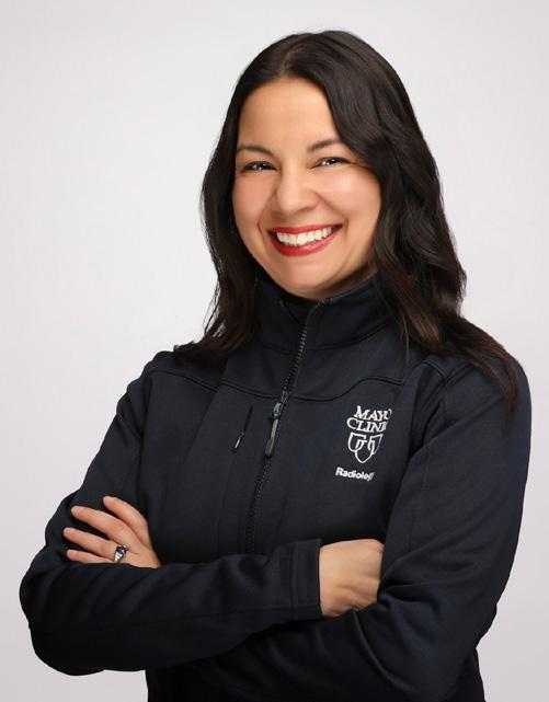
Tanya J Rath, MD
Division Chair of Neuroradiology
Associate Professor
of Radiology
Consultant in Radiology, Otolaryngology and Neurosurgery (secondary appts)
Mayo Clinic, Scottsdale AZ
U-M Resident 2002–2006
U-M Neuroradiology Fellow 2006–2007
What are some favorite U-M memories?
Going to RSNA every year with fellow residents and enjoying Chicago. Our group of residents were both extremely talented and fun. I learned so much from them and some have become lifelong friends.
For me, it was a privilege to train as a resident at U-M. It was a progressive, highly influential department. The faculty were clinically and academically strong. We had exceptional leadership amongst the division chiefs and the department chair. The faculty were outstanding teachers and approachable, often treating residents as colleagues. This fostered a culture of mutual respect. I recall Dr. Cohan encouraging me to call him “Rich”—a notable shift from my more formal upbringing and medical school training. That informality felt progressive for its time and stood in contrast to the hierarchical cultures often criticized in medicine. It was truly unique.
What is something you wish you knew/understood when you were in training?
It’s important to prioritize your family and your own personal health. Radiology residency is demanding, both physically and mentally. It is easy to neglect your life outside of work. Make a conscious effort to care for your physical and mental health, give yourself grace, and set clear boundaries. Doing so will make you more effective in all your roles—spouse, parent, educator, physician, and researcher.
What do you do in your current role that you never learned in school/training?
I have learned how to assemble and lead a high performing team. Leaderhsip was not something we learned in medical training! Several leadership roles have taught me that a critical part of effective leadership is identifying talent and recruiting team members with high EQ and a growth mindset that aligns with your institutional values. I never had any training in how to lead a team!
Do you have any advice for our residents and fellows?
Regardless of your radiology subspecialty, if you want to be diagnostically great and impactful, you need to understand the natural history of the diseases that you are managing, the treatment paradigms for those diseases and treatment related effects. Because these are constantly evolving, you must be adaptable and continually update your knowledge.
Anything else you’d like to add?
One of my daughters started singing in a city youth choir in Pittsburgh when she was in kindergarten. The girls were tiny and their voices were very soft, sweet, and breathy. Trying to make them sing louder turned into screaming. The choir director reminded the parents “their voices are unique to their age, and you just have to enjoy it for what it is at each moment.” This is a good analogy for life—each stage and moment has its own sound and feeling and deserves to be appreciated for its unique beauty. Be open to hearing the special sound of each time in your life, because it won’t sound the same again.
U-M will always hold a special place in my heart. I am truly grateful for the time I spent at Michigan. Go Blue!
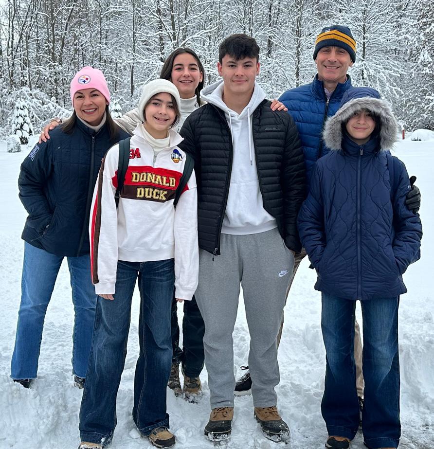
Tanya with her husband Aaron and kids Alex (18), Meera (15), Salena (13), and Tara (13)
BUILDING THE FUTURE OF RADIOLOGY THROUGH COLLABORATIVE RESEARCH
An old joke about one of the largest and most prestigious public research universities in the nation goes something like this: Investigators from the University of Michigan attend national and international meetings to meet new collaborators from the University of Michigan.
There’s definitely some truth behind the ironic humor, according to Gary Luker, M.D., Professor of Radiology and Associate Chair for Clinical Research. Along similar lines, Dr. Luker recalls how he once formed a research collaboration with two faculty members from the College of Engineering whom he had never met before, while waiting for a graduation ceremony to begin at Rackham Auditorium.
“We were standing in line for a long time and we finally started saying, ‘What is it you do?’ And from those conversations, we actually developed a collaboration and got an NIH (National Institutes of Health) grant,” he said.
“So, building bridges from the clinic to basic science and vice versa, not only within the department, but across departments, and then across schools and colleges, is something that needs to be leveraged. We need more opportunities to get people in the same space for informal discussions. And I know that’s hard because time is precious but developing opportunities for people to talk about ideas in a low pressure, low-stress environment can have great returns.”
The simple act of getting people together in one room to discuss research problems isn’t so simple. Yet Radiology is making tremendous progress in fostering greater collaboration -- one of the main drivers behind its research mission. The department continually searches for innovative strategies for aligning the three pillars of academic radiology:
Patient care — treating our patients today; Education — building our faculty and leaders for tomorrow; and Research – building the future of radiology through new technologies.
“When I think about the research that we do every day, the driving factor has always been the translational nature of the work,” said Peter Scott, PhD, Professor of Radiology and Associate Chair for Basic Science Research. “That focuses our research across the whole department. So, we’re being more intentional in making sure that the right bridges are being built between our basic scientists and our clinical faculty, such that the innovation that’s happening in our labs ultimately will end up in the clinic and benefit our patients.”
Radiology Resea Rch suppoR ts all stages of R esea Rch t R anslation
Three associate chairs oversee five key thematic areas in radiology research. Cellular and Molecular Imaging has a state-of-the-art facility and three research labs focusing on cancer drug development and its intersection with state-of-the-art imaging. MRI houses the Michigan Institute for Imaging Technology and Translation (MIITT), Functional MRI Laboratory and 3T Research MRI. Nuclear Medicine focuses on image analysis, physics, dosimetry, theranostics and the development of novel radiopharmaceuticals. Additional teams are dedicated to Ultrasound and AI/CAD/ML (artificial intelligence, computer aided diagnosis and machine learning).
“There are lots of researchers who focus on the early stages of technology development, or just on clinical trials,”
continued on page 18
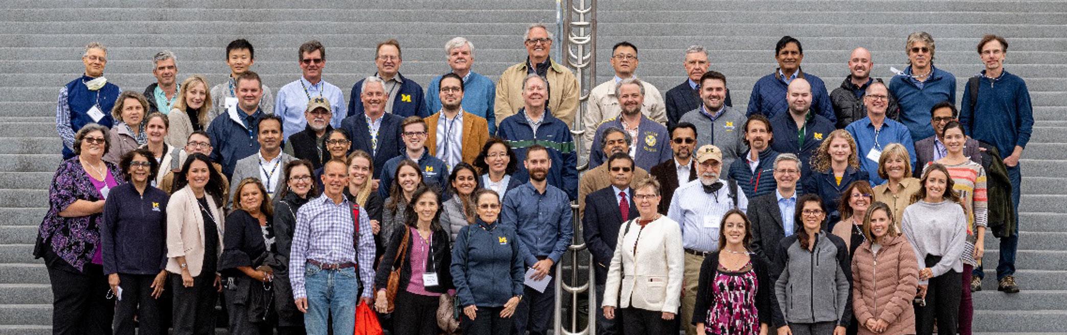
Radiology Researchers gather at 2025 Retreat
RADIOLOGY RESEARCH
AREAS & LABS
Cellular and Molecular Imaging
• Center for Molecular Imaging
• Brock Humphries Lab
• Gary Luker Lab
• Stefanie Galban Lab
MRI
• Michigan Institute for Imaging Technology and Translation (MIITT)
• Functional MRI Laboratory (fMRI)
• 3T Research MRI Resource
Nuclear Medicine
• PET Center
• Functional NeuroImaging, Cognitive and Mobility Lab (FNiCoMo)
AI/CAD/ML
• Computer Aided Diagnosis - Artificial Intelligence Research Lab (CAD-AI)
• Craig Galban Lab
Ultrasound
• Ultrasound Lab
The Department of Radiology also supports research across Michigan Medicine and beyond by providing preclinical and clinical imaging resources that include:
• PET/CT
• Interventional Radiology
• Neuroradiology
• Nuclear Medicine
• Mammography
• MRI
• Ultrasound
• X-ray
continued from page 17
said Katie Wright, PhD, Radiology Research Operations Manager. “But you can see across our portfolio that we have faculty at all stages of research translation. It’s unique to have a department that has such a range from bench science through clinical trials, and it presents unique challenges but also wonderful opportunities to have every stage of research going on. We get an amazing cross-section of how an academic institution can support building the future of medicine.”
What’s more, Radiology offers imaging expertise and resources to support the entire research portfolio of all investigators at Michigan Medicine.
i ntegR ating scientists and clinicians
Nicole Seiberlich, Ph.D., is a founder and co-director of the Michigan Institute of Imaging Technology and Translation (MIITT). The purpose of the group is to bring together experts in clinical radiology and MRI research to develop and deploy novel MRI techniques for clinical applications. She has grown the group from an initial eight members in 2019 to about 40 trainees and scientists and has developed the research infrastructure required for translational research. To build true partnerships, she insisted that MIITT be located on the medical campus, which is no small accomplishment since finding space can be so challenging.
“What makes MIITT different than almost any other MRI research group is that we really have our radiologists sitting with us,” said Dr. Seiberlich. “What happens a lot in medical imaging research, is that researchers want to solve a ‘problem’ because it seems interesting or relevant. But our radiologists might ask, ‘Why? What problem are you actually trying to solve?’ Unless we have buy-in from clinical radiologists, we might be solving a problem that doesn’t even exist. Being able to coordinate with the people who will eventually be using these technologies helps us a lot.”
Dr. Luker agrees. “We have amazing scientists, but in many cases, they have hammers looking for nails, so to speak. So, part of my job on the clinical side is to provide a meaningful context for that hammer. What should we really focus on trying to hit? And then build those bridges and teams to be able to do so.”
Through MIITT, all MD and PhD faculty are invited to weekly meetings that are often built around the clinical schedules of radiologists to make it easier for them to attend.
“We really have to be quite open and not afraid to look stupid in front of each other,” Dr. Seiberlich explained. “The more basic questions we ask, the more we can get to the heart of the science that we’re trying to do together.”
While the scientists’ closest collaborators are clearly radiologists because they are the ones who work with the technology, MIITT has also formed new collaborations with urology and cardiology.
“And through some of these growing collaborations, we were lucky that the NIH awarded us a T32 training program, which is designed to co-train MD and PhD scientists who are interested in medical imaging -- not just MRI, but all kinds of medical imaging,” Dr. Seiberlich said. “We see that as a way of growing what we’ve done in MIITT to hopefully expand it beyond MRI, and beyond radiology, to capture everyone who might be interested in medical imaging here on campus. And as those trainees go out into the world, we also see it as a way to keep that mindset of true collaboration between MDs and PhDs to drive the technology forward.”
f unding the futu R e of R adiology
Historically, federal funding has been the benchmark for research and academic medical centers, but investigators must now navigate a hyper-competitive funding environment with ambiguous rules that often change. Both Dr. Luker and Dr. Scott said the department and the university are adopting a broader view that any source of external funding to support research is good. They argue that researchers can and should take greater advantage of philanthropic or industry funding, partnerships with tech, and more bridge funding opportunities from the university. (Bridge funding supports researchers facing federal funding cuts or helps to span gaps between major grant funding cycles.)
Along with research collaborations, Dr. Luker would like to see more personal connections built to promote the work being done and develop new sources of funding. He points to colleagues in oncology, for example, with grateful patients who donate to support their research, or research collaborators who engage with donors on a regular basis to demonstrate their work, answer questions and show the impact their contribution can make.
“Social media is nice. You get your name out there,” he said. “But the people who really want to connect and support the mission want a more personal interaction. Many of them want to see what’s going on. They want to see an outcome that is going to help themselves or loved ones. If you’re generating funding, you need to build relationships. U of M has one of the largest alumni bases in the world. We need to connect with them more.”
by Lynn Waldsmith
ALUMNI INFORMATION UPDATE
We would love to hear what you are up to and potentially feature you in a future newsletter. Please fill out the Alumni Update Form to send us your news and update your contact information.
AWARDS & RECOGNITION
Atefeh Abdolmanafi, PhD
Received U-M Frankel Cardiovascular Center Innovation Challenge Award
Elaine Caoili, MD, MS
Named Associate Program Director, Radiology Residency Program, U-M Radiology
Matt Davenport, MD, MBA
Received the Early Distinguished Career Award from the Michigan Medicine Alumni Society (MMAS)
J. Brian Fowlkes, PhD
Received U-M Empowering Research with AI Award, Third Prize for Multiparametric Ultrasound Imaging for Urethral Functional Assessment and Therapeutic Guidance
Jessica Fried, MD
Named Associate Chief Medical Information Officer (ACMIO), U-M Radiology
Craig Galban, PhD
Received U-M Empowering Research with AI Award, Third Prize for AI-Based Mucus Plug Detection on Chest CT and Third Prize for Real-time AI-based Health Monitoring of ICU Patients Using STREAM
Stefanie Galban, PhD
Received U-M Empowering Research with AI Award, Third Prize for Realtime AI-based Health Monitoring of ICU Patients Using STREAM
Ansel Hillmer, PhD, MS
Received Society of Nuclear Medicine and Molecular Imaging (SNMMI) Mars Shot Research Fund grant for Molecular Imaging of Brain Inflammation in Alzheimer’s Disease: Targeting the Colony Stimulating Factor 1 Receptor
Jon Jacobson, MD
Published “Fundamentals of Musculoskeletal Ultrasound” as sole author, (Elsevier, 4th edition)
John Jonides, PhD
Named Edward E. Smith Distinguished University Professor of Psychology and Neuroscience
Aparna Joshi, MD
Named to Academic Radiology Editorial Board, Associate Editor
Prabesh Kanel, MS, PhD
Awarded grant from the Michael J. Fox Foundation, Striatal and extra-striatal Cholinergic Terminal Density in LRRK2-PD mutation
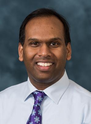
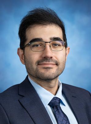
Kate Klein, MD
Served as President-Elect and Program Chair for Association of Academic Radiologists (AAR) annual meeting in Atlanta, GA (March 2026)
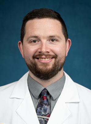
Gary Luker, MD
Elected to membership in the Association of American Physicians
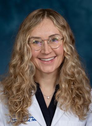
Kate Maturen, MD, MS
Served as President 2025-26 and hosted Society of Abdominal Radiology (SAR) annual meeting in Fajardo, Puerto Rico (March 2026)
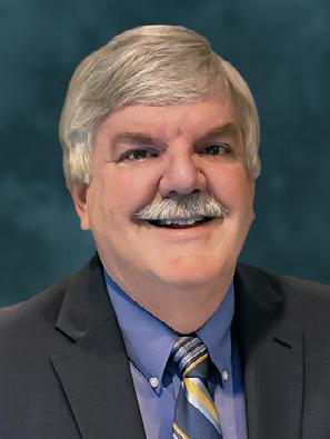
Benjamin Mervak, MD
Named Associate Chair for Information Technology, U-M Radiology
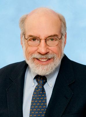
Venkatesh L. Murthy, MD, PhD
Received U-M Empowering Research with AI Award, First Prize for A Foundation Vision Model for Electrocardiography: Reducing Data Demands to Democratize Diagnosis of Microvascular Dysfunction
Ali Namvar, PhD
Received U-M Empowering Research with AI Award, Third Prize for AI-Based Mucus Plug Detection on Chest CT
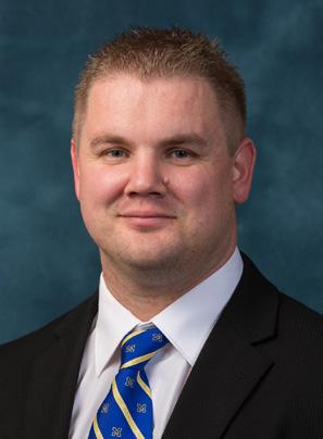
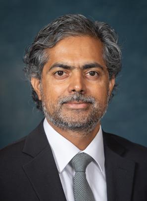
Andrew Navarro, MD
Appointed as Training Committee Chair to Society of Interventional Radiology Resident and Fellow Section Governing Council
Molly Roseland, MD
Named Associate Program Director, Nuclear Medicine Fellowship and Nuclear Medicine Representative to the U-M Radiology Residency Clinical Competency Committee
Brian Ross, PhD
Honored with Technology & Innovation Award; 2025 Dean’s Awards
Jonathan Rubin, MD, PhD
Received Distinguished Alumni Award from the University of Chicago Medical Biological Sciences Alumni Association
Peter Scott, PhD
Received Society of Nuclear Medicine and Molecular Imaging (SNMMI) Radiopharmaceutical Sciences Council’s 2026 Michael J. Welch Award for outstanding contributions to the field of radiopharmaceutical sciences
Aseem Sharma, MD
Named Associate Program Director, Radiology Residency Program, U-M Radiology
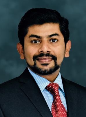
AWARDS & RECOGNITION
Steven Soliman, DO
Received U-M Empowering Research with AI Award, Third Prize for AI-Driven Muscle Ultrasound and Multimodal Data Fusion for Early Detection and Personalized Risk Stratification of Metabolic Dysfunction
Peter Strouse, MD
Published Kleinman’s Diagnostic Imaging of Child Abuse, Cambridge University Press, 4th edition. (Served as editor, 68 contributing authors), and received the Society of Pediatric Radiology Gold Medal, 2026
Kara Udager, MD
Named to Academic Radiology Editorial Board, Deputy Editor
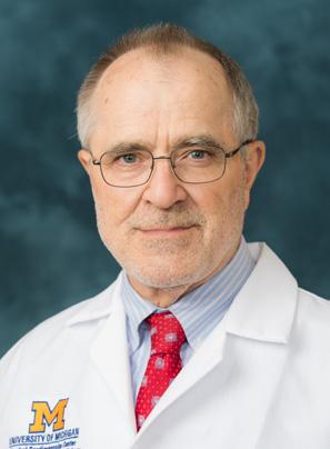
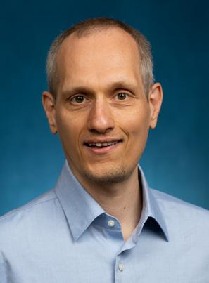
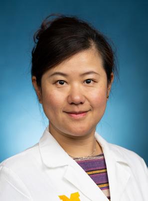
Benjamin Viglianti, MD, PhD
Named co-chair for the Multi-Tumor Experimental Therapeutics Clinical Team (CRT) in the U-M Rogel Cancer Center and named to the Academic Radiology Editorial Board, Associate Editor
Ashish Wasnik, MD
Named to Academic Radiology Editorial Board, Associate Editor
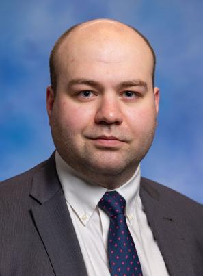
Eric Webb, PhD
Received Society of Nuclear Medicine and Molecular Imaging (SNMMI) Mars
Shot Research Fund grant for Molecular Imaging of Brain Inflammation in Alzheimer’s Disease: Targeting the Colony Stimulating Factor 1 Receptor
David Williams, MD
Honored with the Lifetime Achievement in Clinical Care Award; 2025 Dean’s Awards
Matthias Wilms, PhD
Received U-M Empowering Research with AI Award, First Prize for Beyond the Black Box: Generative AI and Synthetic Data for Trustworthy Medical Image Analysis
Maggie Zhang, MD, PhD
Named to Academic Radiology Editorial Board, Associate Editor
Celebrating a Remarkable Career
Cheryl Kucharski
honoR ing 37 y ea R s of l eade R ship, s e Rvice, and h ea R t
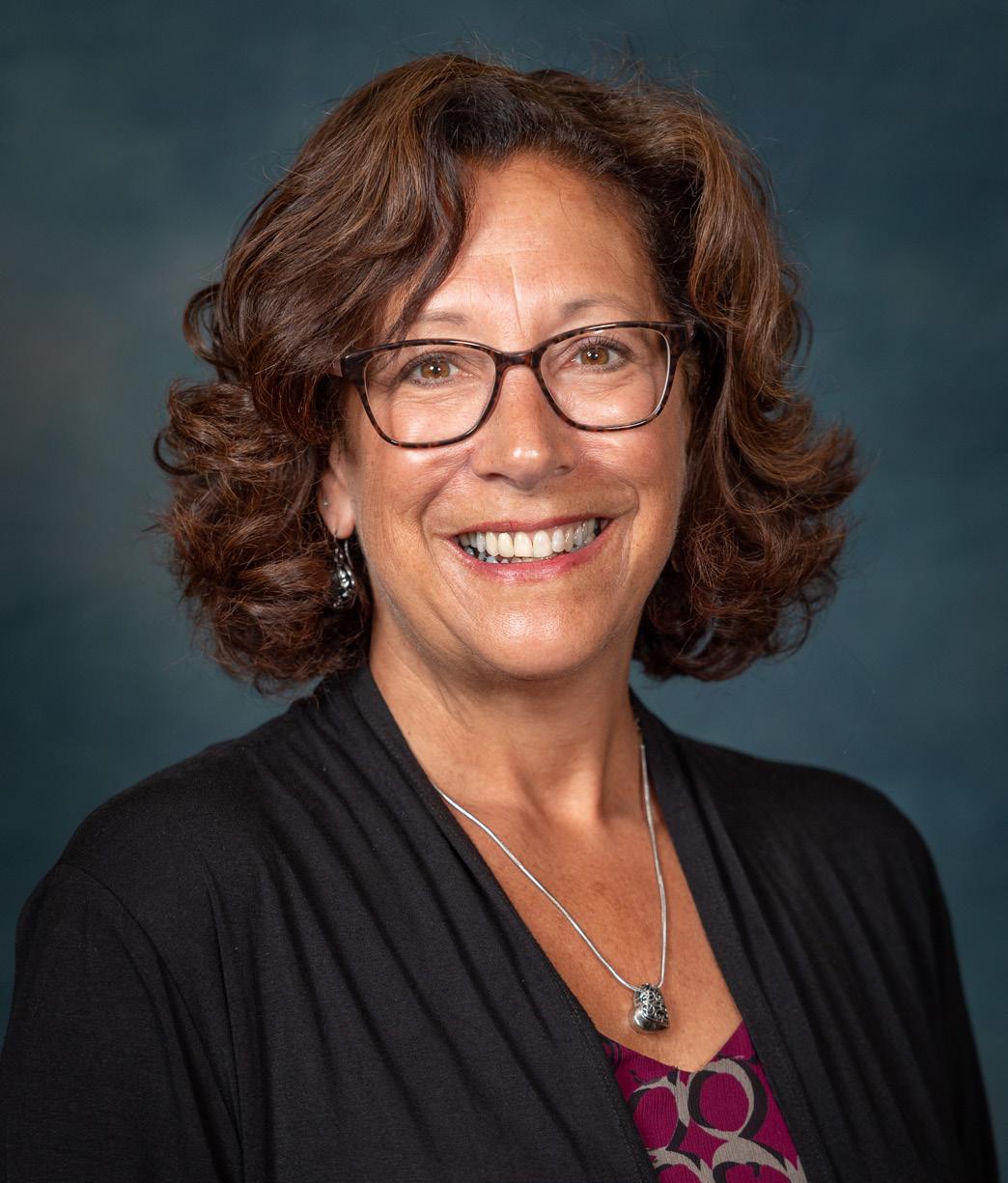
On March 6, 2026, our Radiology Department celebrated an extraordinary milestone: Cheryl Kucharski officially retired after an exceptional 37-year career marked by dedication, expertise, and genuine care for those around her.
For nearly four decades, Cheryl has been a cornerstone of our department—especially within CT—leaving a mark on both the people and the programs that make our work possible. Anyone who has crossed her path knows the experience well: her warm smile, unmistakable laughter, and the sincere way she greets everyone are impossible to forget. Many have expressed the same sentiment over the years: “I want to be like her when I grow up.”
a c a R ee R Built on e xcellence
Cheryl began her career in 1989 as an X-ray technologist and quickly distinguished herself as an exceptional clinician and colleague. Her leadership potential was unmistakable, and
for the past 20 years she has served as CT Manager, helping shape the department into what it is today.
Throughout her tenure, Cheryl led the CT team through numerous expansions, renovations, and periods of significant transformation—many happening simultaneously. Whether opening new scanners, designing clinical spaces, reworking workflows, or adapting to technological advancements and evolving staffing models, Cheryl was the steady hand guiding the way. Her ability to lead through change with clarity, collaboration, and unwavering calm earned her deep respect across the department.
a l egacy t hat l ives on
Many of the systems, processes, and achievements CT relies on today are the result of Cheryl’s vision, resilience, and commitment. Her investment in people has been just as impactful. Cheryl has mentored countless staff members, offering support through challenges, celebrating their successes, and fostering a culture grounded in teamwork and trust.
a f ond fa R ewell—and deep gR atitude
Cheryl’s retirement leaves a meaningful void—professionally, operationally, and personally. While we will miss her leadership and daily presence, we celebrate with immense gratitude everything she has contributed over her remarkable 37 years.
When asked how Cheryl plans to spend her retirement, she said, “I will be visiting my daughter, traveling, and improving my golf game.”
by Shelby Beard
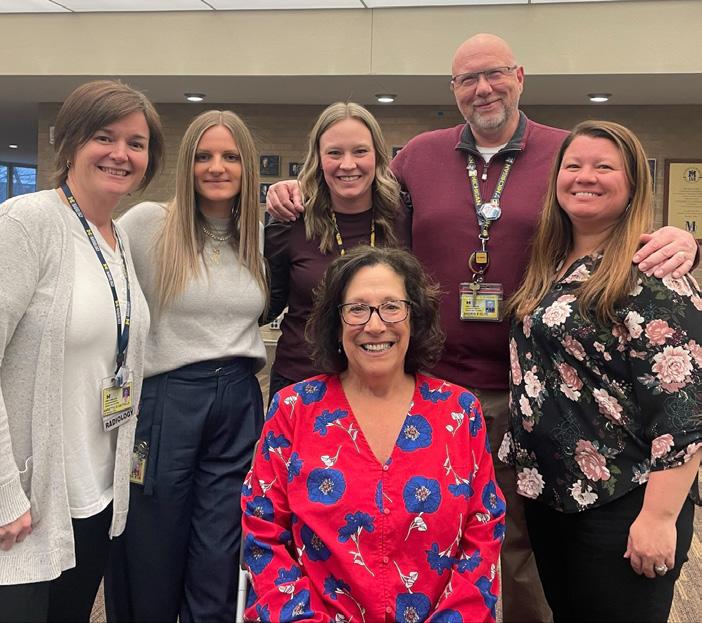
Cheryl Kucharski
Retirement Party
David H. Woodbury, Jr. (M.D. 1961) died on September 17, 2025, at age 95. He was a devoted husband of over 56 years and father to a blended family of six children.
Woodbury graduated with distinction from Johnson C. Smith University. He received a master’s degree in biology from Virginia State University and a medical degree from the University of Michigan. Woodbury was an associate professor of radiology at the Medical School and a senior nuclear medicine physician at the VA Ann Arbor Healthcare System.
In Memoriam
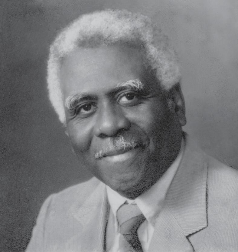
David Woodbury, MD
1930 – 2025
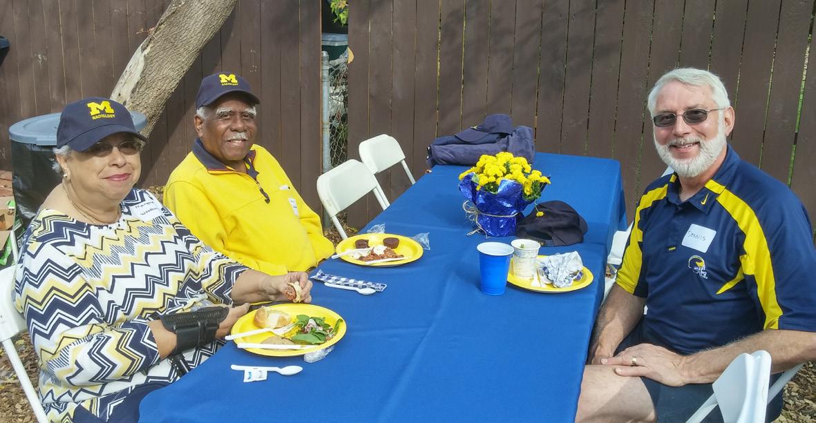
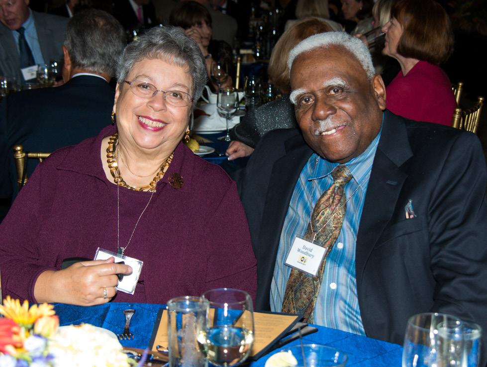
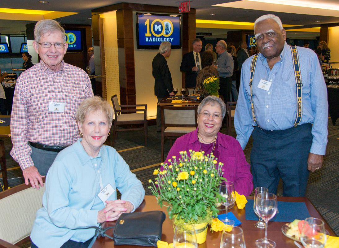
He was a member of the Omega Psi Phi Fraternity, Incorporated and the Sigma Pi Phi Fraternity, also known as the Boulé. He was known for his mentorship and keen wit. Woodbury was a longtime supporter of the Medical School, in particular the Fitzbutler Jones Society Scholarship. He was a dedicated supporter and attendee of many U-M events and activities. Woodbury is survived by his wife Margaret C. Woodbury, M.D., Assistant Professor Emerita of Internal Medicine at the Medical School.
Michigan Medicine, based on an Ann Arbor News obituary.
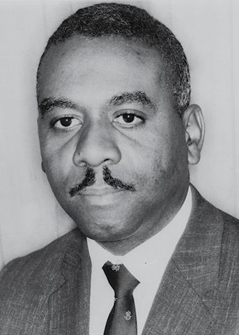
1966
David Woodbury, MD with wife Margaret Woodbury, MD
2013 Centennial Celebration
2015 Radiology Tailgate
GIVING BACK…
The Department of Radiology at the University of Michigan is most grateful to our family of alumni who have supported our programs in education, training and research. Over a century’s worth of excellence has been enhanced through your generosity. We hope you will continue being a part of this legacy by considering a gift.
JAMES H. ELLIS, MD AND JEAN A. LAWTON, MD EARLY CAREER PROFESSORSHIP IN RADIOLOGY
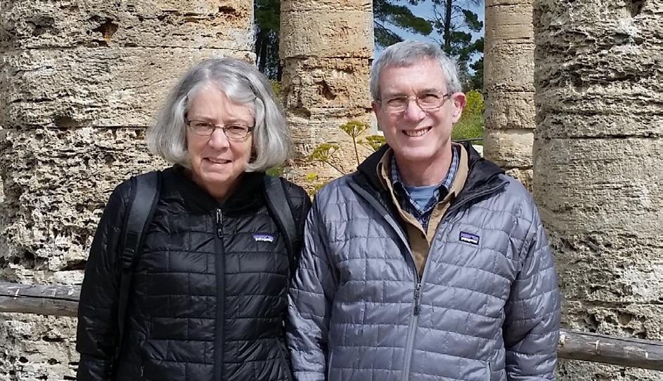
We invite you to support the establishment of the James H. Ellis, M.D., and Jean A. Lawton, M.D. Early Career Professorship in Radiology. This professorship honors Dr. James H. Ellis for his exceptional career in medical imaging and his steadfast dedication to education and mentorship in the radiology community. Once fully funded, this will recognize and support emerging leaders in the field of radiology.
RESIDENT EDUCATION FUND
Support the education, training and enrichment of residents.