UIC College of Dentistry Clinic & Research Day Booklet 2026
39TH ANNUAL
UNIVERSITY OF ILLINOIS CHICAGO
COLLEGE OF DENTISTRY
March 5, 2026
Program
Wednesday, March 4, 2026
Location: College of Dentistry
12:30–4:30 p.m. Badge Pick-up 5th Floor Common Area for Student Presenters
Thursday, March 5, 2026
Location: Student Center West
7:30–9:30 a.m. Check-in for Judges, Sponsors, 1st Floor Foyer and Guests
8:00–8:30 a.m. Poster A Set-up Poster Area
8:00–10:00 a.m. Coffee & Muffins 2nd Floor Foyer
8:30–10 a.m. Poster Session A Presentations & Judging
10:00–10:30 a.m. Poster A Takedown & Poster B Set-up
10:30 a.m.–noon Poster Session B Presentations & Judging
12:15–1:15 p.m. Keynote Lecture Thompson Hall “Stem Cells in Dental PulpTissue Regeneration”
Jacques E. Nör DDS, MS, PhD
Dean & Donald A Kerr Professor of Dentistry University of Michigan
1:15–2:00 p.m. Lunch Pick-up 2nd Floor Foyer
2:00–3:00 p.m. Faculty Talks Thompson Hall
2:00–2:20 p.m “Two Degrees, One Unexpected Path:Dentistry, Research, and the Mentors Who Made It Possible"
Luisa DiPietro, DDS, PhD Professor, Department of Periodontics Director, Center for Wound Healing & Tissue Regeneration
2:20–2:40 p.m. “Big Data and Artificial Intelligence in Dentistry: Applications in Patient Care and Dental Education”
Sath Allareddy, BDS, MBA, MHA, MMSc, PhD Head & Professor, Department of Orthodontics
2:40–3:00 p.m. "Bridging Clinical Practice and Research in Endodontics”
Shlomo Elbahary, DMD, PhD Visiting Clinical Associate Professor, Department of Endodontics
3:00–3:15 p.m. Poster B Takedown Poster Area
3:15–3:45 p.m. Awards Ceremony Thompson Hall
4:00 p.m.
Event Adjourns
Susan Rowan
Welcome to the 39th Annual Clinic and Research Day of the University of Illinois Chicago College of Dentistry, held at UIC’s Student Center West.
The UIC College of Dentistry is a vibrant community of learners, educators, clinicians, and researchers, proudly rooted in the dynamic city of Chicago. Like our city, the College thrives on energy, creativity, and collaboration and continually advances oral health through discovery and innovation.
Today’s research environment is shaped by collaboration across disciplines and perspectives. At UIC, we actively support this culture by investing in team science, encouraging curiosity, and building connections across campus, within Chicago’s diverse communities, and beyond.
Our students and postgraduates are at the heart of this research mission. Working alongside faculty mentors, they engage in a wide range of scholarly activities—from basic and translational research to public health initiatives, evidence-based clinical practice, educational innovation, and emerging areas such as diagnostics, artificial intelligence, biomaterials, and regenerative dentistry and medicine.
Through these collaborative efforts, the College of Dentistry continues to rank among the leading recipients of National Institutes of Health research funding, supported by the National Institute of Dental and Craniofacial Research, other NIH institutes, federal agencies, foundations, and corporate and philanthropic partners.
Clinic and Research Day is a celebration of your work. It brings our community together and provides a platform for students and postgraduates to share their research, exchange ideas, and learn from one another, as well as from faculty, alumni, and the broader research community.
As you take part in today’s activities, we invite you to take pride in your achievements and to experience how the UIC College of Dentistry is advancing Better Oral Health Through Transformative Innovation—together.
Thank you for your participation!
Susan Rowan, DDS, MS Dean and Clinical Professor of Restorative Dentistry
Ana Bedran-Russo, DDS, MS, PhD
Associate Dean for Research; Professor and Head, Department of Oral Biology; and Lead, 2026 Clinic & Research Day Committee
Dr.
Dr. Ana Bedran-Russo
Welcome Message from the Scientific Committee
Dr. Sobia Bilal
Co-Chair Scientific Committee
Dr. Mateusz S. Wietecha
Co-Chair Scientific Committee
On behalf of the Scientific Committee, we are delighted to welcome you to the 39th Annual Clinic and Research Day at the University of Illinois Chicago College of Dentistry. This longstanding event brings our academic community together to recognize innovation, scholarship, and collaboration across dental and craniofacial science, education, and clinical care.
Clinic and Research Day reflects the collective efforts of learners and researchers at every stage of professional development. From undergraduate volunteers and dental students to residents, graduate students in our MS and PhD programs, postdoctoral fellows, and research associates, this event celebrates the depth and diversity of inquiry taking place within our institution. These accomplishments are made possible through the mentorship, leadership, and dedication of our exceptional faculty.
We are especially honored this year to welcome our keynote speaker, Jacques Nör, DDS, MS, PhD, Dean of the University of Michigan School of Dentistry and Professor of Otolaryngology - Head and Neck Surgery at the University of Michigan. A nationally and internationally recognized leader in academic dentistry and translational research, Dr. Nör’s work has shaped the future of dental education and oral health research. His perspective as a clinician-scientist and dean will offer valuable insight and inspiration to our community.
In addition to the keynote address, we are proud to feature presentations by three distinguished members of the UIC College of Dentistry faculty. Their work highlights the impactful and innovative research being conducted within our own institution and underscores our commitment to advancing oral health through discovery and scholarship.
This year’s program also provides opportunities to engage with industry partners who are showcasing emerging products and technologies relevant to clinical care and research. We extend our sincere appreciation to all of our sponsors, whose generous support is essential to the success of this event.
As we come together to exchange ideas, recognize achievement, and strengthen collaboration, Clinic and Research Day reminds us of the vital role research and innovation play in shaping the future of dental profession aiming at improving oral health and patient care.
Thank you for joining us. We look forward to a meaningful and inspiring day.
Sobia Bilal, BDS, MSc, PhD, and Mateusz S. Wietecha, DMD, PhD
Co-Chairs, Scientific Committee
UIC College of Dentistry Awards
The UIC College of Dentistry will present awards of recognition to deserving undergraduate students, dental students, residents, graduate students, and post-docs/research staff across the following scientific categories: Basic/Basic Translational Sciences (BTS), Clinical/Clinical Translational Sciences (CTS), Educational Research (EdR), Population/Public Health Sciences (PHS), Case Reports (CaseR), and Literature Reviews (LitR).
AADOCR/Dentsply Sirona Student Competition for Advancing Dental Research and its Application (SCADA)
One student (dental student, dual doctoral student, or PhD student at the UIC College of Dentistry), who has at least one year of program remaining, will be chosen to represent the College in the 2026 American Association for Dental, Oral, and Craniofacial Research/Dentsply Sirona SCADA Program at the American Association for Dental, Oral, and Craniofacial Research (AADOCR) Annual Session in March 2026 in San Diego, CA.
Hinman Student Research Symposium
One student will be chosen to represent the UIC College of Dentistry at the Hinman Student Research Symposium sponsored by the University of Tennessee Health Science Center in Memphis at the Peabody Hotel, Fall 2026.
The Illinois State Dental Society Foundation Award
The premier dental student will be chosen by a panel of Illinois State Dental Association Foundation dentists in basic sciences, clinical sciences, and population/public health sciences, to receive an Illinois State Dental Society Foundation Award. The Foundation is the society’s philanthropic wing; it provides scholarship money to qualified students, encourages and promotes student research, and sponsors continuing education for dentists throughout the State of Illinois.
Omicron Kappa Upsilon Awards
In 2026, the Sigma Chapter of Omicron Kappa Upsilon, the National Dental Honor Society, will provide awards to three dental students for Superior Achievement, Outstanding Achievement, and Achievement in abstract writing and presentation in educational research.
Dr. Christine Da-Ruh Dental Student Research Scholarship
Dr. Christine Da-Ruh Wu, professor of pediatric dentistry, has established a fund that will support scholarships for dental students in the College of Dentistry who demonstrate excellence in research. Funding may also be used for travel to attend or present research at professional conferences.
Awards
Dental Student Research Group Peer Award
The Dental Student Research Group (DSRG) is a student-led initiative dedicated to fostering a vibrant research community within the dental school. The DSRG’s goal is to connect fellow students with research opportunities that align with their academic and professional interests. The DSRG provides guidance and support at every stage of the research process—whether you're starting a new project, refining a poster, or preparing a paper for publication. The DSRG team is here to help you succeed and excel in your research endeavors. The DSRG looks forward to collaborating with you! At each Clinic and Research Day, the DSRG presents the "Peer Award" to students—from undergraduates to residents—who demonstrate excellence with the best scientific poster. Posters are evaluated by student researchers based on the clarity of content, quality of research, and effective communication of key findings. In recognition of their achievement, recipients are proudly awarded unique, 3D-printed trophies, a distinctive symbol of their outstanding contributions.
Dr. Isaac Schour Memorial Dentistry Student Research Award
This award stimulates student interest in research, giving students an opportunity to work creatively, develop excellence, and be inspired by the researchers with whom they work. We thank Dr. Seymour Gottlieb, ’58, whose generosity funds the Schour Award.
George-Evans Student Research Award
New this year, the Anne George-Carla Evans Student Research Award is given to a dental student who demonstrated outstanding research. The recipient will be selected and announced during the Clinic and Research Day.
Clinic and Research Day Steering Committee
Ana Bedran-Russo (Lead), Alexandria Bell, Sobia Bilal, Meghan Dietrich, Ellie Healy, Dina Kutinsky, Darryl Langston, Chris Menden, Elsa Mejia, Amsaveni Ramachandran, Cassandra Villani, and Mateusz S. Wietecha.
Scientific Committee
Co-chairs: Sobia Bilal, Mateusz S. Wietecha
Basic Sciences Subcommittee
Lead: Miya Kang
Member: Yutong Li
Clinical Sciences Subcommittee
Lead: Camila Zamperini
Members: Russell Pesavento, Qian Xie
Educational Research Subcommittee
Lead: Kaleb Sellers
Members: Betti Shahin, Robert Bara, Aniruddh Narvekar
Population Health Sciences Subcommittee
Lead: Bhakti Desai
Members: Rebecca van Horn, Khatija Noorullah
Case Reports Subcommittee
Lead: Evelina Kratunova
Members: Majd Alsaleh, Georgia Lymberopoulos, Lisa Rawle, Flavia Lamberghini
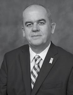
Dr. Jacques E. Nör, DDS, MS, PhD, is the Donald A. Kerr Endowed Professor of Dentistry and serves as the Dean of the University of Michigan School of Dentistry. He has a joint appointment as Professor of Otolaryngology (Medical School) and as Affiliate Faculty of Biomedical Engineering at the University of Michigan. He is PastPresident of the American Association for Dental, Oral and Craniofacial Research (AADOCR). Dr. Nör served as Chair of the Oral Dental and Craniofacial Sciences (ODCS) NIH study section and is a member of the National Advisory Dental and Craniofacial Research Council (NADRC). He has published 245 peer-reviewed papers, 15 book chapters, and has been continuously funded by the NIH since 2002. He has served as Co-Director of the University of Michigan Head Neck SPORE funded by the National Cancer Institute (NCI). Dr. Nör is the past-Chair of the Section on Dentistry & Oral Health Sciences of the American Association for the Advancement of Sciences (AAAS), Fellow of the American Association for Dental Research (AADR) and Fellow of the AAAS. He received the Distinguished Scientist Award, the William J. Gies Award, and the Hatton Award from the International Association for Dental Research (IADR). Dr. Nör is Associate Editor of the Journal of Dental Research and member of several editorial boards. His research interests are in the study of the pathobiology of cancer stem cells in head neck squamous cell carcinoma and salivary gland cancer, as well the studies of mechanisms regulating dental pulp stem cell fate.
Dr. Jacques E. Nör
Faculty Presentations
Dr. Luisa DiPietro, DDS, PhD
Dr. Sath Allareddy, BDS, MBA, MHA, MMSc, PhD
Dr. Shlomo Elbahary, DMD, PhD
Dr. Luisa DiPietro, Professor, Department of Periodontics; Director of the Center for Wound Healing & Tissue Regeneration presents “Two Degrees, One Unexpected Path: Dentistry, Research, and the Mentors Who Made It Possible.”
Dr. Sath Allareddy, Department Head and Professor, Department of Orthodontics presents “Big Data and Artificial Intelligence in Dentistry: Applications in Patient Care and Dental Education.”
Dr. Shlomo Elbahary, Visiting Clinical Associate Professor, Department of Endodontics presents “Bridging Clinical Practice and Research in Endodontics.”
Registered Judges
Dr. Orlando Abreu
Dr. Guy Adami
Dr. Zabihulla Ahmadi
Dr. Azza Ahmed
Dr. Seema S Ashrafi
Dr. Phimon Atsawasuwan
Dr. Velavan Bakthavachalam
Dr. Robert M Bara
Dr. Matt Bernard
Dr. Susan Bishop
Dr. Alan A Boghosian
Dr. Dina A Casey
Dr. Wendy L Cerny
Dr. Lin Chen
Dr. Yinghua Chen
Dr. Andrew Chung
Dr. Seung Chung
Dr. Asha Eapen
Dr. Shlomo Elbahary
Dr. Shahrbanoo Fadavi
Dr. Jim Fieldhouse
Dr. Amudha Ganapathy
Dr. Feng Gao
Dr. Mary Claire Garcia
Dr. Anne George
Dr. Joseph Gluck
Dr. Jimmy Gonzalez-Vicker
Dr. Colin Haley
Dr. Rainah Hanif
Dr. Jianjun Hao
Dr. Mark Heiss
Dr. Nathaniel Henning
Dr. Brittaney Hill
Dr. Chun-Chieh Huang
Dr. Raza A Hussain
Dr. Miya Kang
Dr. Linda M Kaste
Dr. Farhana Khan
Dr. Yukiko Koizumi
Dr. Qiang Ma
Dr. Michael Mabborang
Dr. David Maclin
Dr. Carla Massignan
Dr. Steve Miller
Dr. Ronald M Milnarik
Dr. Leda Mugayar
Dr. Raza Ali Naqvi
Dr. Afsar Naqvi
Dr. Philip Patston
Ms. Valerie S Prater
Dr. David Reed
Dr. Mariana Reis-Havlat
Dr. Peggy Richardson
Dr. Alexandra Rodriguez
Dr. Reem Sabbagh
Dr. Linda Sangalli
Dr. Michael L Schmerman
Dr. Betti Shahin
Dr. David Shonberg
Dr. Walter B Stanford
Miss Darlistene Stevens
Dr. Sadiq Umar
Dr. Christine D Wu
Dr. Susan K Zelazo-Smith
History of Clinic and Research Day
In 1988, the University of Illinois at Chicago College of Dentistry began its student-based Clinic Day, later to be renamed Clinic and Research Day, providing a forum for students to showcase their cutting-edge research projects and for students, alumni, and faculty to receive educational and professional enrichment information from a keynote speaker. The first Clinic Day, held April 6, 1988, was proposed and organized by students. Its success and the growth of the event prompted faculty and administration to make it an official annual activity at the College.
In 1996, the modern Clinic and Research Day was initiated through a formal gathering of faculty and interested students, to re-invigorate the existing student-based Clinic Day. It provided an opportunity for students and faculty to interact in advancing oral healthcare and research while embracing the entire local community. Clinic and Research Day has grown into an incredibly diverse and rich gathering that reflects on all of the clinical and research activities going on at the College.
Clinic and Research Day in the past included comedic “game show” quizzes, dexterity challenges such as wax tooth carving, table clinics, technology demonstrations, and tours of the College. While some of these offerings have been eliminated, the event has become more focused and fine-tuned and is similar to a national dental meeting with oral and poster presentations on clinical and research subjects, dental products vendors and service providers’ exhibits, a keynote speaker, continuing education, awards for excellence, and prizes.
For many years, Department Heads were in charge of the faculty component of Clinic and Research Day, but since the creation of the College’s Office of Research Services in 2003, the Associate Dean for Research, has overseen the Clinic and Research Day Committee. Dr. Ana Bedran-Russo, Associate Dean for Research and Professor and Head, Department of Oral Biology, is this year’s lead. Throughout Clinic and Research Day’s history, however—it’s the students’ day. With every Clinic and Research Day, the College builds on the experience, expands the event, and displays the increases in the diversity of research along with the diversity of the COD community. This year’s event features some faculty posters and short talks, and enhanced involvement by the external research community; thus providing more research role modeling for the students.
Abstracts
BASIC SCIENCES
1. Transport of Mature miRNA Into Mesenchymal Stem Cell Extracellular Vesicles
Niloufar Abedi, Chun-Chieh Huang, Yu Lu, Sriram Ravindran Department of Oral Biology, UIC College of Dentistry, Chicago, IL, USA
Objectives: Mesenchymal stem cell extracellular vesicles (MSC EVs) have emerged as critical paracrine mediators of cellular function and serve as an alternative to stem cell therapy in regenerative medicine applications. MSC EV resident miRNAs significantly contribute towards EV function. EV resident miRNAs are selectively transported into EVs via RNA-binding proteins(RBP). While multiple proteins have been shown to possess this function in various mammalian cells, the proteins involved in miRNA transport into MSC EVs remain unclear. Our objectives are to: 1) Determine what shuttle proteins are highly expressed in MSCs. 2) Identify which proteins are effective in MSC EV miRNA transport. 3) Identify the best tag/s (recognition sequences for shuttle proteins) to effectively transport therapeutic miRNAs into MSC EVs.
Methods: miR148a-3p, a salivary EV miRNA with wound-healing function, was chosen as a candidate for experiments due to its low expression in MSCs and MSC EVs. q-RTPCR screened RBPs in MSCs. We custom-modified a miRNA expression vector to express miR148a-3p with/without sorting tags in MSCs for these highly expressed proteins. We cloned the pre-miRNA148a sequences into this vector with different tags at the 3p strand and the control group without a tag. We then generated a lentivirus with these vectors and transfected MSCs, followed by twice Puromycin selection for stable expression and q-RTPCR to verify miRNA expression. Also, q-RTPCR and western blot were applied to evaluate the effect of the tag on three highly expressed protein levels in cells. Immunoprecipitation (IP) of the candidate RBPs in control and miR148a-3p overexpressed cells followed by q-RTPCR for miR148a-3p expression was performed to evaluate the efficacy of the tags as well as to identify the dominant RBPs involved in MSC EV miRNA transport.
Results: Three highly expressed shuttle proteins in MSCs were RNPA2B1, ANAX2, and YBX1. Of these three candidates, miR-148a-3p presence in the MSCs EVs was significantly enhanced with YBX1 tag (ACCAGCCU) and ANAX2 tag (AACAG) compared to RNPA2B1 tag(GGAG) and control groups. The basal expression levels of RBPs were unchanged after constitutively expressing miR-148a-3p with and without tags, indicating that increased miRNA presence in the EVs with the YBX/ANAX tags is due to the transportation activity of the RBPs and not due to increased presence of RBPs. IP results revealed the interaction of YBX and ANAX2 with their respective tags present in miR-148a-3p. In contrast, RNPA2B1’s interaction with its tag was on par with controls, confirming that this protein may not play an active role in MSC EV miRNA transport.
Conclusions: Identifying MSC-specific shuttle proteins for EV miRNA transport opens an interesting avenue for miRNA-based EV engineering. Future studies will focus on confirming the role of and identifying specific RBP and tag combination/s to for maximal transport of mature miRNAs into MSC EVs.
Jonathan Banks, Dakshina Acharya, Mateusz Wietecha, Anne George, David Reed
Department of Oral Biology, UIC College of Dentistry, Chicago, IL, USA
Objectives: Traumatic mandibular fractures cause worsened quality of life, loss of mobility and function, and poor healing outcomes, yet their prevalence increases annually. The modern standard of care often incorporates surgical bone grafts, but this approach can result in tissue death, poor vascularization, and suboptimal tissue integration. Therefore, there remains a clinically relevant need to advance therapeutic approaches for unstable mandibular fractures. These fractures heal via endochondral ossification, when osteoprogenitors migrate and differentiate into chondrocytes and osteoblasts to repair damaged bone. Neuron-glial antigen 2 (NG2/CSPG4) is a type VI collagen receptor implicated in tissue mineralization and cell signaling in the temporomandibular joint during mechanical loading and injury. However, the role of NG2/CSPG4 as a transcriptional regulator of cell recruitment and differentiation during fracture healing remains unresolved. This study aims to characterize NG2/ CSPG4-mediated ligand-receptor interactions to elucidate its role in bone growth and remodeling during fracture healing.
Methods: Surgical osteotomies were conducted on the mandibular condylar neck of male and female skeletally-mature (16wk) C57BL/6J wild-type (WT) and NG2/ CSPG4 knockout (NG2ko) mice. Bone formation was quantified in 0-, 7-, and 21-day fracture calluses using µCT and histology. Single-cell RNA sequencing (scRNAseq) was used to characterize transcriptional heterogeneity within the 7-day callus via the 10X Genomics pipeline. Sequencing data were bioinformatically analyzed using Seurat (RStudio and CellChat) for clustering, cell marker identification, and cell-typing of individual clusters using genetic signatures. Furthermore, integrated datasets were analyzed using differential gene set enrichment analysis, module scoring, and ligandreceptor interaction analysis.
Results: NG2ko fracture calluses displayed impaired bone quality metrics, with lower bone density, volume, and trabecular thickness compared to the WT calluses. scRNAseq analysis identified 6 cell clusters corresponding to populations including fibroblasts, osteoclasts, macrophages, and chondrocyte/osteoblasts. NG2/CSPG4 expression was highest in WT fibroblasts, featuring upregulated gene sets for biomineralization and chondrocyte differentiation. Differential module scoring revealed NG2ko cells from the osteoblast/chondrocyte cluster were associated with decreased mineralization and extracellular matrix (ECM) organization. NG2ko osteoclasts exhibited decreased ‘sender’ and ‘receiver’ involvement for fibronectin and laminin pathways, indicating impaired ECM deposition and self-assembly.
Conclusions: NG2/CSPG4 significantly modulates fibroblast and osteoclast cell-cell signaling and alters the cell-cell and cell-matrix signaling that regulates bone growth and remodeling during endochondral ossification and fracture healing, resulting in NG2ko mice displaying impaired fracture healing potential.
Approval: ACC Protocol Number 24-089
Funding: NIH F30 DE033287
Abstracts
3. Silver Diamine Fluoride Alters Mineral Properties of Hypomineralized Enamel
Kathy Bedasee 1, Camila A Zamperini 2, Evelina Kratunova 1, Sahar Alrayyes 1 , Majd Alsaleh 1, Christine Wu 1, Giuliana CC Lamarque 2 , Azza Ahmed 1
1 Department of Pediatric Dentistry, UIC College of Dentistry, Chicago, IL, USA;
2 Department of Restorative Dentistry, UIC College of Dentistry, Chicago, IL, USA
Objectives: The aim of this study was to compare the effect of silver diamine fluoride (SDF) on mineral density (MD), color change, and chemical composition of MolarIncisor Hypomineralization (MIH) and sound enamel.
Methods: Sound and MIH permanent first molars were collected and examined by an experienced investigator. Sound (n=5) and MIH (n=7) enamel specimens were prepared and coated with nail polish to expose standardized enamel windows (3 x 3 mm) where SDF was applied, according to manufacturer’s instructions, followed by storage in artificial saliva (37°C; 7 days). MD and color data were obtained before and after SDF by using micro-computed tomography and digital spectrophotometer, respectively. For chemical composition, sound and MIH surfaces treated or untreated with SDF were analyzed using Raman spectroscopy, following a pH cycling protocol for 4 days. Data were analyzed by Shapiro-Wilk and Levene’s tests for normality and homogeneity of variance, respectively, followed by t-tests and ANOVA t-tests and ANOVA (α = 0.05).
Results: While there was a significant difference in MD between sound and MIH enamel before SDF treatment (p = 0.039), no difference was detected after treatment (p = 0.28), indicating a change in tendency induced by SDF treatment. For MD gain, both groups (sound: -275 mmHA/cm3; and MIH: +23 mmHA/cm3) differed significantly (p = 0.018). Statistically significant differences (P<.05) in color changes and chemical composition were also observed among groups following SDF application. Conclusions: The findings demonstrate that SDF has the potential to interact and protect hypomineralized enamel from demineralization.
Approval: IRB Protocol 2024-0741
Funding: Not applicable
4. Col6a1 Regulates Subchondral Bone Remodeling During TMJ PTOA
Tyrese Bentford, Dakshina Acharya, Santa Nawwara, David Reed Department of Oral Biology, UIC College of Dentistry, Chicago, IL, USA
Objectives: Col6a1 is a pericellular matrix molecule in temporomandibular joint (TMJ). During the progression of posttraumatic osteoarthritis (TMJ PTOA), Col6 is remodeled to change the microenvironment of the cell. Col6 is implicated into normal physiological bone growth in the mandible, with Col6ko mice having less subchondral bone than wild-type controls. There is a gap in knowledge related to the role of Col6 in the allostatic remodel that occurs during PTOA progression. We hypothesize that Col6a1ko mice will have impaired subchondral bone remodeling during the progression of PTOA.
Methods: Male and female Col6 alpha1 chain knockout mice (Col6a1KO) were grown
Abstracts
to skeletal maturity. PTOA was induced through surgical destabilization of the TMJ by unilateral partial discectomy. Tissues were collected 4-weeks after PTOA induction and analyzed by µCT and histology. All data were compared to wildtype, c57 BL/6 J mice. Mean values for bone variables and Mankin Scores were compared using a t-test with post-hoc Tukey corrections (α = 0.05). A sample size of 6 was used for all experimental groups.
Results: The µCT analysis illustrates that the trends in allostatic bone remodeling during PTOA were similar in the control and Col6a1ko mice. In the PTOA samples, significant differences by genotype were identified in trabecular number (both sexes; p < 0.05), trabecular spacing (male; p < 0.001), bone volume/total volume (female; p < 0.05), and tissue mineral density (male; p < 0.01). This bone phenotype was confirmed with histological analysis. Proteoglycans were found to be concentrated in the pericellular region of many cells in the mandibular condylar cartilage of Col6a1ko mice, lacking the interterritorial distribution common in wildtype controls.
Conclusions: Our findings implicate Col6a1 in the allostatic bone remodeling associated with the progression of TMJ PTOA and underscore the importance of the Col6 signaling axis as a potential target for the therapeutic modulation of subchondral bone for regenerative or tissue engineering strategies.
Approval: ACC Protocol Number 23-042
Funding: NIH R01 DE029835
5. Single-cell Analysis of Structural and Immune Cell Interactions in Periodontitis
Jessica Castillo 1, Jonathan Haller 1 , Mateusz S. Wietecha 2
1 Department of Periodontics, UIC College of Dentistry, Chicago, IL, USA;
2 Department of Oral Biology, UIC College of Dentistry, Chicago, IL, USA
Objectives: Periodontitis is a chronic inflammatory disease characterized by progressive destruction of tooth-supporting tissues. While bulk transcriptomic studies provide insight into the inflammatory pathways involved, single-cell RNA sequencing (scRNA-seq) enables detailed characterization of cell-type-specific responses and interactions in periodontal tissues. This study aimed to compare the transcriptional landscapes of structural and immune cell populations in periodontitis versus healthy gingival tissues using single-cell analysis, focusing on fibroblasts, endothelial cells, macrophages, and neutrophils.
Methods: ScRNA-seq datasets of human periodontitis-associated and control gingival samples (GSE152042, GSE161267, GSE164241) were obtained from the NCBI Gene Expression Omnibus (GEO) database. R (v4.5.1) in RStudio (2025.05.1+513) was used to re-analyze datasets. Seurat (v5.1.0) was utilized for quality control, integration, and analysis of the datasets, including batch correction and data visualization. Cell clustering, cell type annotation, and differentially-expressed gene (DEG) analyses were performed to identify unique transcriptional signatures in structural and immune cells from periodontitis-associated samples. Pathway enrichment (using EnrichR) and ligand-receptor interaction (using CellChat) analyses were conducted to explore functional alterations and intercellular communications between structural and immune cells. STRING was utilized to identify, integrate and predict protein-protein interaction
Abstracts
(PPI) networks between structural and immune cells in periodontitis versus control samples.
Results: From a total of 105,574 cells in the dataset, we subset 17,533 fibroblasts, 22,947 endothelial cells, 2,129 macrophages, and 1,836 neutrophils to individually analyze and identify upregulated genes in periodontitis. Nine genes were upregulated across the four selected stromal cell types, and they formed a well-connected PPI network of common interactions that included increases in oxidative stress (e.g. SOD2, HSPAIA) and pro-inflammatory IL-17 signaling (e.g. JUND, TNFAIP3). In structural cells (fibroblasts and endothelial cells), 30 common genes formed a PPI network conveying increases in vascular proliferation (e.g. LAMB1), evidence of epigenetic regulation (e.g. KDM6B, NR4A1), and communications with immune regulators (e.g. KLF6, NDRG1). In immune cells (macrophages and neutrophils), 34 upregulated genes formed a PPI network showing increases in the NF-KB pathway (e.g. REL), IL1B signaling, and oxidative stress (e.g. HIF1A, TNFAIP3).
Conclusions: Single-cell transcriptomic profiling revealed highly coordinated alterations among structural and immune cells in periodontitis-affected compared to healthy tissues. The findings highlight key intercellular interactions driving chronic inflammation and tissue damage, and they may inform the development of cell-targeted therapeutic approaches for periodontal disease.
1 Weinberg College of Arts and Sciences, Northwestern University, Evanston, IL, USA; 2 Department of Biomedical Engineering, UIC Colleges of Engineering and Medicine, Chicago, IL, USA; 3 Department of Restorative Dentistry, UIC College of Dentistry, Chicago, IL, USA
Objectives: Curcumin treatment of commercially pure titanium (cp-Ti) dental implants may reduce corrosion under peri-implantitis-like acidic conditions and improve implant longevity. To determine the effects of curcumin on the electrochemical behavior and surface characteristics of cp-Ti using electrochemical impedance spectroscopy (EIS) and surface characterization techniques.
Methods: Corrosion testing of commercially pure titanium (cp-Ti IV) was performed using a three-electrode electrochemical system consisting of a titanium working electrode, a graphite auxiliary electrode, and a saturated calomel reference electrode (SCE), controlled by a Gamry potentiostat. Experiments were conducted in 10 mL of artificial saliva at 37 ± 1°C, with pH adjusted to 3 or 6.5 using lactic acid. Curcumin was applied at two concentrations (0.5 µg/mL and 1 µg/mL), and corrosion was quantified using a sequence of open-circuit potential (OCP), potentiostatic testing, electrochemical impedance spectroscopy (EIS), and cyclic polarization. Each condition was repeated in triplicate (n = 18). Corrosion rate (Icorr), corrosion tendency (Ecorr), and corrosion kinetics (Rp) were analyzed using electrochemical data, complemented by surface characterization via scanning electron microscopy (SEM) and 3D
profilometry.
Results: Under acidic conditions (pH 3), a sharp decrease in corrosion current (Icorr) was observed when curcumin concentration increased from 0.5 µg/mL (3.649 x 10^-7 A/cm²) to 1 µg/mL (1.173 x 10^-7 A/cm²), suggesting enhanced resistance to corrosion. At pH 6.5, Icorr increased with curcumin concentration. Polarization resistance (Rp) decreased with increasing curcumin concentration at pH 3, whereas Rp increased at pH 6.5 with minimal changes in double-layer capacitance (Cdl). Surface analyses showed reduced roughness and localized oxide formation at pH 3 with 1 µg/ mL curcumin, while increased pitting and surface roughness were observed at pH 6.5, particularly at higher curcumin concentrations. Curcumin exhibited pH-dependent effects on the corrosion behavior of commercially pure titanium. A reduction in corrosion rate (Icorr) was observed under acidic conditions, while corrosion kinetics (Rp) showed differing trends, highlighting the distinct and non-equivalent nature of these corrosion parameters.
Conclusions: These findings demonstrate that curcumin may slow corrosion rate without necessarily improving overall corrosion kinetics, emphasizing the complex interaction between inhibitor adsorption, passive layer stability, and electrochemical behavior. Further studies are required to understand the corrosion inhibition mechanisms under simulated oral environments.
Approval: Not applicable
Funding: Northwestern Summer Undergraduate Research Grant (Summer 2025)
7. Microhardness Effects of Non-Invasive Caries Treatments
Bilyana Daskalova 1, Camila A Zamperini 2, Majd Alsaleh 1, Christine D Wu 1 , Azza Tagelsir Ahmed 1 , Sahar Alrayyes 1
1 Department of Pediatric Dentistry, UIC College of Dentistry, Chicago, IL, USA;
2 Department of Restorative Dentistry, UIC College of Dentistry, Chicago, IL, USA
Objectives: To evaluate the effectiveness of resin infiltration, 38% silver diamine fluoride (SDF), and 5% sodium fluoride varnish (FV) on demineralized primary enamel using microhardness as the outcome measure.
Methods: Forty-eight extracted primary molars were sectioned into standardized enamel specimens, coated with nail varnish leaving a 3 x 3 mm window, and demineralized using a Streptococcus mutans biofilm model for 7 days to create incipient lesions. Specimens were randomly assigned to Control, SDF, FV, or resin infiltration (Icon). Treatments were applied per manufacturers’ instructions and stored 24 hours at 37 °C in 100% humidity. Specimens then underwent pH-cycling with 30 minutes of demineralization (pH 4.5) followed by 10 minutes of remineralization (pH 7.2), repeated six times daily, with overnight storage in remineralizing solution. Specimens were sectioned, polished, and cross-sectional enamel microhardness was measured using a Knoop indenter from 25 to 225 µm in 25-µm intervals. Data were analyzed using linear mixed-effects models with treatment and depth as fixed effects and specimen as a random effect. Pairwise comparisons were Tukey-adjusted with significance at p < 0.05.
Results: Across all depths, resin infiltration demonstrated significantly higher mean
Abstracts
microhardness than Control (difference=74.1, p = 0.0001), FV (difference=57.0, p = 0.0028), and SDF (difference=44.2, p = 0.0314). Estimated marginal means were highest for resin infiltration (299), followed by SDF (255), FV (242), and Control (225). At superficial depths (25–75 µm), resin infiltration consistently outperformed all other groups (p < 0.001). SDF showed limited improvement over Control at shallow depths, while FV did not differ significantly from Control across most depths.
Conclusions: Resin infiltration produced significantly greater enamel microhardness than SDF, fluoride varnish, or no treatment, particularly in superficial enamel. These findings suggest resin infiltration is more effective at improving hardness of incipient lesions in primary teeth than the other non-invasive treatments evaluated, supporting its use as a minimally invasive treatment option.
Approval: IRB Protocol 2024-0741
Funding: Not applicable
8. TGF-β Induces Stress Fiber Formation in Mandibular Fibrochondrocytes
Syed Farhan, Priti Mulimani, David Reed
Department of Oral Biology, UIC College of Dentistry, Chicago, IL, USA
Objectives: Degenerative changes in the condyle of the TMJ include changes in extracellular matrix (ECM) stiffness. In response to ECM changes, TMJ cells alter their cytoskeletal structure which initiates and propagates inflammatory changes associated with painful TMJ disorders like osteoarthritis (OA). Such cytoskeletal changes are mediated by alterations in stress fiber shape, length, thickness, and number. Furthermore, cytokines like TGF-β have been implicated in causing cartilage degradation. However, its effect on substrate stiffness mediated cytoskeletal changes in initiating TMJ OA remains unknown. Hence, the objective of this study is to investigate the combined effects of TGF-β and ECM stiffness on TMJ fibrochondrocyte cells’ cytoskeletal structure, in order to elucidate their mechanistic roles in OA etiopathogenesis.
Methods: Immortalized mouse mandibular fibrochondrocytes were seeded for 24 hrs onto 5ug/mL fibronectin coated substrates. Substrates were fabricated by coating glass coverslips with different proportions of Sylgard-184 and Sylgard-527, which are polydimethyl siloxane (PDMS) elastomers, to mimic a low ECM stiffness value (3 kPa). The experimental group was treated with 10 ng /mL TGF-β for 24 hours and the control group with regular culture media. After 24 hours, cells were fixed in 4% paraformaldehyde and permeabilized in 0.1% Triton-X, to prepare the cells for fluorescent antibody staining. They were then stained with Phalloidin-488 (green/Factin), Alexa Fluor 561 (red/alpha-smooth muscle actin), and DAPI (blue/nuclei).
Confocal microscopic imaging of the cells with a 20x objective was carried out. Images were analyzed using Fiji Image analysis software. Stress fiber analysis was conducted using Filament Sensor v2.0 which generated data on the number, length, thickness, and number of stress fibers per unit cell area. Statistical analysis was carried out using unpaired T-test in Biorender and significance value was p < 0.05.
Results: Under low ECM stiffness conditions, a statistically significant difference between the length of stress fibers for the control group and the experimental group
was observed. However, no differences were found between TGF-β and control groups for F-actin stress fiber thickness, amount per cell area, and total number.
Conclusions: Since TGF-β resulted in longer stress fibers under low ECM stiffness conditions, we can infer that the pathological mechanisms leading to TGF-B mediated joint degeneration involve the cytoskeletal machinery. Further studies on how cytoskeletal alterations influence downstream gene expression mediated cell phenotype and behaviors would be valuable in understanding TMJ OA and developing therapies.
Approval: ACC Protocol Number 23-042
Funding: NIH R01 DE029835
9. Cell Accelerated Corrosion Can Be Assessed Using Electrochemical Biosensor
Karam M Fattouhi 1, Sujoy P Ghosh 1, Mostafa Alhamad 1, Anne George 2 , Yinghua Chen 2 , Mathew Thoppil Mathew 1
1 Department of Restorative Dentistry, UIC College of Dentistry, Chicago, IL, USA; 2 Department of Oral Biology, UIC College of Dentistry, Chicago, IL, USA
Objectives: Corrosion at the modular junctions of dental implants remains a significant concern. Multiple types of damage modes have been identified at modular junctions, correlated with different corrosion characteristics that may eventually lead to implant failure. Among the primary challenges to dental implant treatment success is peri-implantitis, a condition increasingly linked to titanium (Ti) corrosion products and undetected residual dental cement. Despite this, the impact of Ti corrosion remains poorly understood. This study aims to evaluate the electrochemical behavior of macrophages and fibroblasts in response to metal ion leaching caused by Ti corrosion. Specifically, it seeks to investigate whether Ti corrosion influences phagocytic activity and contributes to implant-related complications. The null hypothesis posits that Ti corrosion has no effect on phagocytic activity.
Methods: Both macrophages and fibroblasts were cultured separately. Macrophages and Fibroblasts were cultured under identical conditions to ensure a consistent experimental environment. The cells were exposed to five distinct media groups to assess their response. Groups used 1) control media (PBS solution) 2) spent media (PBS solution when dental implant was submerged 3) Ti Ions (PBS with implant and Ti Ions) 4 ) Ti partials (PBS with implant and Ti particles) 5) Ti Ions+ Particles (PBS with implant and Ti Ions + particles). Electrochemical analysis procedure: 100 µl was pipetted onto a biosensor (Dropsens-250A) connected to a Gamry-made Potentiostat to study the electrochemical activity of collected samples. Open circuit potential-(OCP), electrochemical impedance spectroscopy-(EIS), and cyclic voltammetry-(CV) were analyzed.
Results: Macrophages showed significant OCP and impedance variations, indicating immune responses to titanium ions and particles, with the combination having the strongest effect. Almost no changes seen with fibroblast with OCP. Macrophages demonstrated higher sensitivity, as evidenced by significant shifts in Nyquist and Bode plots, as well as changes in EIS modeling. Fibroblasts exhibited lesser sensitivity, as evidenced in Nyquist and Bode plots, and EIS modeling. Interestingly The combination of titanium ions and particles produced synergistic effects,
Abstracts
amplifying electrochemical perturbations in both cell types. Increased cellular stress in macrophages, is reflected by changes in charge transfer resistance and capacitance. CV analysis further highlighted these differences, showing that macrophages had a much higher level of charge transfer and interaction with the media compared to fibroblasts.
Conclusions: This suggests that macrophages are more responsive to titanium ions and particles, while fibroblasts maintain a more stable electrochemical profile. This study demonstrates that macrophages and fibroblasts react differently to environmental changes. It is an ongoing study and requires further research.
Approval: Not applicable
Funding: Not applicable
10. NG2/CSPG4 Regulates Alveolar Bone and PDL Microstructure in a Sex-Dependent Manner
Polina Gubareva 1 , David Reed 2
1 Department of Periodontics, UIC College of Dentistry, Chicago, IL, USA; 2 Department of Oral Biology, UIC College of Dentistry, Chicago, IL, USA
Objectives: Periodontal disease causes alveolar bone loss in a complex, multifactorial etiology driven by chronic inflammation. Understanding the molecular mechanism controlling the anabolic and catabolic regulation of bone in alveolar tissues can help identify new therapeutic targets for treating periodontitis. Recent findings have demonstrated that the loss of type VI collagen leads to reduced levels of alveolar bone. NG2/CSPG4 is a type I transmembrane proteoglycan that binds with type VI collagen and is found in mineralizing tissues. There is an important gap in knowledge related to if NG2/CSPG4 regulates the density and organization of alveolar bone. The objective of this study is to define the role of NG2/CSPG4 in the growth and development of alveolar bone and PDL.
Methods: To characterize alveolar bone in vivo, skulls from 16-week old male and female, control and NG2/CSPG4 knockout mice were collected, fixed overnight in 4% paraformaldehyde, and µCT scanned at 70 kV at a resolution of 12 µm voxel size. A region of interest (ROI) was contoured that included the alveolar bone measuring 140 slices centered between the roots of the first molar, and the same was repeated, measuring the PDL space, with the ROI around the middle third of the mesial root of the first molar. These skulls will then be decalcified, paraffin embedded, and sectioned at 8 µm. The PDL will be assessed using picro Sirius red technique, H&E stain, and Herovici Stain to assess collagen organization. The ROI was analyzed using the Trabecular Analysis package (Scanco Medical). Means comparing sex and genetic differences were compared using a one-way ANOVA with post-hoc Tukey corrections (α = 0.05).
Results: When comparing male and female mice, there was a sex dependent decrease in the ratio of bone volume to total volume and bone mineral density in both control and NG2ko mice (p < 0.01), a sex dependent decrease in trabecular thickness in only the NG2ko mice (p < 0.05; n=5), and a sex-dependent decrease in trabecular number in only control mice (p < 0.01). When comparing control and NG2ko mice, there was an NG2- dependent decrease in trabecular number only in female mice (p < 0.05). Female NG2ko mice showed wider PDL widths compared to control mice (p < 0.01). Histological findings validated the µCT findings.
Conclusions: The outcomes from this study demonstrate that NG2/CSPG4 plays a role in maintaining alveolar bone microstructural homeostasis in female mice. These findings align with parallel studies showing that NG2/CSPG4 regulates subchondral bone quality in the temporomandibular joint. Additionally, the observed outcomes are consistent with data from Col6a2 knockout mice, suggesting convergent ligand–receptor signaling pathways. This is the first report to implicate NG2/CSPG4 in alveolar bone homeostasis, highlighting its potential as a novel therapeutic target and biomarker for bone resorption in conditions such as periodontitis.
Department of Oral Biology, UIC College of Dentistry, Chicago, IL, USA
Objectives: Wound healing typically results in the formation of a scar. Compared to uninjured tissues, scars exhibit denser extracellular matrix, reduced elasticity, and tensile strength. These abnormalities reflect the broader process of fibrosis, which can occur in every organ and ultimately lead to irreversible tissue damage and failure. Fibroblasts are key regulators of tissue repair, and their persistent activation during wound resolution leads to pathological scarring. Compared to biochemical regulation, biomechanical cues in wound healing are poorly understood. Therefore, we used single-cell RNA sequencing (scRNA-seq) and spatial transcriptomics (ST) to study wound fibroblast activation under various tensile forces.
Methods: We integrated several publicly available scRNA-seq and ST datasets of murine incisional scars from mechanically-strained (excessive stretch) and nonstrained wounds and murine excisional wounds (including small and large fullthickness wounds). All quality control (QC) and downstream analysis steps were performed using Seurat in RStudio. Clustree was used for subclustering the fibroblasts, Monocle3 for trajectory analysis, and EnrichR for functional enrichment.
Results: QC and filtering yielded 20,396 cells, which clustered into eight wound fibroblast subtypes expressing canonical fibroblast genes (Col1a1, Dcn, Pdgfra).
Trajectory analyses suggest divergent paths of fibroblast activation along a biomechanical stress pseudotime. Spatial analysis showed higher enrichment of Cluster_7 signature at wound tension sites. Overall, mechanical strain expanded contractile and migratory fibroblast states while reducing pro-angiogenic and immunemodulatory subtypes.
Conclusions: Our results indicate that wound fibroblasts are activated by increasing stretch forces along a spatio-temporal path from quiescent to contractile/myo-
Abstracts
fibroblastic to migratory signatures at the expense of pro-resolution functions. This data may lead to new insights into mechanically-induced scarring and novel wound management strategies.
Approval: Not applicable
Funding: NIH R35 GM154921
12. Effects of Aging on Human Cementum Morphology and Composition
1 Department of Periodontics, UIC College of Dentistry, Chicago, IL, USA; 2 Department of Oral Biology, UIC College of Dentistry, Chicago, IL, USA
Objectives: Cementum is a mineralized tissue essential for tooth stability, anchoring periodontal ligament to bone. Aging is a risk factor for periodontal disease, but little is known about its impact on acellular and cellular cementum. The objective of this study was to evaluate the ultrastructure of human cementum across different age groups. Methods: Human teeth were extracted and selected by age [young (18-25 y), adult (35-45 y), middle-age (45-55 y) and older adults (65-70 y)] after assessing morphological integrity and oral health status (n = 5). Samples were sectioned and randomly assigned to morphological and ultrastructural analysis. For morphology, specimens were fixed in 10% neutral buffered formalin, then demineralized in 10% ethylenediaminetetraacetic acid (EDTA) for 5-6 weeks until decalcified. Decalcified specimens were dehydrated, xylene-cleared, infiltrated and embedded with paraffin, and sectioned at 5 µm. Cementum morphology was assessed with hematoxylin and eosin (H&E) staining, while collagen content and organization were evaluated using picrosirius red (PSR) staining. Cementum thickness, collagen composition, and cementocyte density were evaluated using ImageJ. Microstructure and mineral density of native cementum were analyzed using micro-computed tomography. Data were analyzed using ANOVA and post-hoc tests (α = 0.05).
Results: Differences in fiber structure and alignment between acellular (AC) and cellular (CC) cementum were identified with H&E staining in all age groups. AC formed a continuous cervical layer, lacked embedded cells, and had a dense, homogeneous extracellular matrix with a clear dentin interface and basophilic appearance. CC was observed in the mid- to apical root regions, with cementocytes embedded in lacunae, arranged in a layered pattern. In middle-age and older adults, the CC matrix was less homogeneous and showed more layering of acellular extrinsic and cellular intrinsic fibers than AC. PSR staining under polarized light showed tightly packed, organized collagen fibers with strong birefringence, indicating increased maturity. CC showed a more evident heterogeneous collagen population, with red/ orange and green/yellow birefringence representing mature and immature fibers. Cementum thickness increased with age for AC and CC. No differences were found in the cementocyte density with aging. Mineral density increased in CC with age (p < 0.001), where adult, middle and older adults displayed higher density than young CC. No differences were observed in AC across age groups (p > 0.05).
Conclusions: This study characterizes age-related morphological features of cementum and shows that aging is associated with distinct structural and compositional
changes that differ between acellular and cellular regions. These findings highlight the dynamic nature of cementum across the lifespan and emphasize the importance of regional considerations when evaluating age-related changes in human dental tissues.
Approval: IRB Protocol 2023-1002
Funding: NIH R00 DE032049
13. Commercial Saliva Substitute Chemically Enhanced with a Surface-Stable Nanohybrid Aggregate
Nadeen Hammad 1, Kassapa Ellepola 1, Shoaib Masood 2, Heidi Yuan 3, Lin Chen 3, Mohamad A. Alhadlaq 4, Robert F. Klie 2 , Russell P. Pesavento 1
1 Department of Oral Biology, UIC College of Dentistry, Chicago, IL, USA; 2 Department of Physics, UIC College of Liberal Arts and Sciences, Chicago, IL, USA; 3 Center for Wound Healing and Tissue Regeneration, UIC College of Dentistry, Chicago, IL, USA; 4 Department of Oral Biology, UIC College of Dentistry, Chicago, IL, USA; Department of Pediatric Dentistry and Orthodontics, College of Dentistry, King Saud University, Riyadh, Saudi Arabia.
Objectives: Hyposalivation is a prevalent chronic condition contributing to substantial oral pathology, including both hard and soft tissue lesions. Commercial salivary substitutes provide patient comfort by assisting in swallowing/speaking but are often limited in their capacity to prevent oral pathology. Our goals herein were to increase the chemical and biological function of a salivary substitute (Biotene®) by formulating it with a cost effective, ‘protective’ salivary protein biomimetic for improved disease prevention in patients with chronic hyposalivation. The ‘enhanced’ formulations were evaluated in a series of in vitro and ex vivo assays relevant to oral administration and disease prevention.
Methods: Both single nanoceria (CeO2-NP) and a nanohybrid aggregate containing nanoceria and chondroitin sulfate A (CeO2-NP-CSA-B) prepared in our lab were dispersed in Biotene® (Haleon) and evaluated for chemical stability at room temperature. Following this storage period, we screened the enhanced formulations for their ability to increase the antioxidant activity of Biotene with known commercial assays. Further, a series of literature based bioactivity assays of the enhanced formulation were carried out, including efficacy studies against Streptococcus mutans biofilms and toxicity against human gingival fibroblasts (HGF). Characterization studies (e.g. STEM, EELS) were carried out to provide chemical and mechanistic support for the observed activities of nanoparticle-dispersed Biotene® solutions.
Results: CeO2-NP-CSA-B (10 mM Ce) remained dispersible in Biotene® (67%, v/v) for 30 days at room temperature compared to single nanoceria, while also increasing the antioxidant activity. Further, the addition of whole human saliva (WHS) from healthy adults to dispersions containing CeO2-NP-CSA-B rapidly increased both the agglutination and biofilm inhibition of S. mutans compared to untreated Biotene®. STEM and EELS studies supported retention of the nanohybrid aggregate structure and its principal oxidation state in Biotene® formulations compared to single nanoceria which produced non-tractable mixtures, with a significant change in the oxidation state unsuitable for storage. Non-significant changes to formulations containing CeO2-NPCSA-B in Biotene® were found when compared to untreated Biotene® solutions.
Abstracts
Conclusions: The formulation of Biotene® with CeO2-NP-CSA-B enhanced its antioxidant and agglutination/antibiofilm function under conditions relevant to storage and administration in the oral cavity. The increased activities can be in part attributed to the ability of CeO2-NP-CSA-B to resist surface chemical changes. Future studies are aimed at the oral administration of CeO2-NP-CSA-B-dispersed Biotene® in rodent models with chronic hyposalivation.
Approval: IRB Protocol 2023-0562
Funding: NIH K08 DE028009, IADR Innovation in Oral Care Award, University of IL Start-Up Fund
14. Morphometric Evaluation of Craniofacial Development in SNED1 Knockout Mice
Abdalrahman M Hasan 1, Alexandra Naba 2, Steven Miller 3 , Christina Nicholas 1
1 Department of Orthodontics, UIC College of Dentistry, Chicago, IL, USA; 2 Department of Physiology and Biophysics, UIC College of Medicine, Chicago, IL, USA; 3 Department of Oral Medicine and Diagnostic Sciences, UIC College of Dentistry, Chicago, IL, USA
Objectives: Craniofacial growth and skeletal patterning are central to orthodontic diagnosis and treatment planning. SNED1 is an extracellular matrix protein implicated in craniofacial development, yet its contribution to postnatal craniofacial morphology remains poorly defined. The objective of this study was to evaluate the effects of neural crest specific SNED1 knockout on craniofacial dimensions and palatal morphology using quantitative morphometric analysis.
Methods: A cross-sectional morphometric study was performed using high resolution micro computed tomography scans from wild type and SNED1 knockout mice. 3D craniofacial reconstructions were generated, and standardized anatomical landmarks were placed following established craniofacial morphometric protocols to ensure measurement consistency. Linear morphometric measurements were taken (e.g., interorbital distance, nasal suture length, maxillary width). Landmark placement reliability was confirmed through repeated measurements of an initial subset of specimens.Palatal arch shape was analyzed using geometric morphometric approaches. Standard statistical analyses were used to compare craniofacial parameters between genotypes.
Results: SNED1 knockout mice demonstrated a significantly increased interorbital distance compared with wild type controls (p = 0.0166) and a significantly reduced nasal suture length (p = 0.00123). In contrast, geometric morphometric analysis revealed no significant differences in palatal arch shape between groups (PC1 p = 0.791; PC2 p = 0.494). These findings indicate that while SNED1 deletion affects select craniofacial dimensions, overall palatal curvature is preserved.
Conclusions: Neural crest specific deletion of SNED1 results in selective alterations of midfacial and nasal craniofacial dimensions without affecting palatal arch morphology. Further studies with larger sample sizes and inclusion of additional linear and angular craniofacial measurements, 3D shape analyses of the maxilla and mandible, assessment of cranial base relationships, and evaluation of transverse, sagittal, and vertical skeletal
parameters are needed to clarify the role of SNED1 in craniofacial development and its potential relevance to orthodontic craniofacial variation.
Approval: ACC Protocol Number 16-149 (AN)
Funding: UIC College of Dentistry Office of Research Fund
15. Fibroblast Heterogeneity During Post-traumatic Injury In The Craniofacial Musculoskeletal System
Khushroop Kaur, Jonathan Banks, Priti Mulimani, Mateusz Wietecha, David Reed
Department of Oral Biology, UIC College of Dentistry, Chicago, IL, USA
Objectives: Post-traumatic injury to the craniofacial musculoskeletal system (CFMS) initiates a healing response that involves complex interactions among heterogeneous cell populations. Transcriptomic analysis of pre-clinical mandibular injury models of induced temporomandibular joint post-traumatic osteoarthritis (TMJ-PTOA) and osteotomy (OSTEO) identified a craniofacial musculoskeletal fibroblast (CMFB) population that participated in crosstalk with chondrocytes through the bone morphogenetic protein (BMP) signaling pathway. We hypothesize that CMFBs orchestrate divergent repair and regeneration in mandibular injury models via the BMP signaling pathway.
Methods: The R coding environment RStudio was used to analyze single-cell RNA-sequencing (scRNAseq) data from two murine mandibular injury models and nonsurgical mandibular tissue as control (NSC). Seurat was used to integrate datasets and classify shared cell populations. Gene ontology (GO) analysis was used to identify pathways enriched by differentially expressed genes. CellChat was used to assess cellto-cell and ligand-receptor interactions.
Results: An integrated scRNAseq dataset of NSC, TMJ-PTOA, and OSTEO tissues resulted in 28,051 cells that passed quality control and filtering. Chondrocytes were defined by Col11a1, Col12a1 expression, and a progenitor cell population was defined by Sox5, Prg4. A heterogenous CMFB population defined by Vim, Fap was identified, with a higher abundance in OSTEO (25% of cells) compared to TMJ-PTOA (22% of cells). GO analyses showed an enrichment for BMP signaling in both injury models. CellChat analyses revealed directional BMP signaling in the injury models, with CMFB-to-chondrocyte signaling in TMJ-PTOA compared to chondrocyte-to-CMFB signaling in OSTEO. Differential analysis of ligand-receptor interactions illustrates that BMP2-to-Bmpr1a+Bmpr2 signaling occurs between CMFB cells in the TMJPTOA model but between chondrocytes in the OSTEO model. In TMJOA, progenitor cell and CMFB populations utilize the BMP6 pathway. In OSTEO, the BMP6 path is utilized by endothelial cells.
Conclusions: CMFBs serve as choreographers of mandibular injury responses by coordinating divergent CMFB and chondrocyte signaling through the BMP pathway. During endochondral ossification following fracture healing in the appendicular skeleton, BMP2 and BMP6 work synergistically to promote bone formation. This cell-signaling axis crosstalk regulates transitions between fibroblasts and chondrocytes,
Abstracts
with implications for understanding bone remodeling and regeneration across various injury types.
Approval: ACC Protocol Number 23-043, 24-050
Funding: NIH F30 DE033287, NIH R01 DE029835
16. Gingival and Dermal Keratinocyte-Fibroblast Interactions in 3D Co-Culture
Shalyn Keiser 1,2, Heidi Yuan 2, Lin Chen 3, Luisa DiPietro 3 , Mateusz Wietecha1 1 Department of Oral Biology, UIC College of Dentistry, Chicago, IL, USA; 2 Department of Integrative and Translation Physiology, UIC College of Medicine, Chicago, IL, USA; 3 Center for Wound Healing and Tissue Regeneration, UIC College of Dentistry, Chicago, IL, USA
Objectives: Compared to skin wounds, oral wounds heal more quickly, undergo faster re-epithelialization, and generally do not result in scar formation. Fibroblasts (FBs), the main cellular component of scar formation, and keratinocytes (KCs), the cells that constitute the epithelial barrier, demonstrate tissue-specific phenotypes in skin and oral wounds. It remains unclear, however, if KC signaling through secreted products influences FB function, and if this KC-FB signaling differs between skin and oral cells. Thus, the aim of this study was to investigate if oral and skin KCs induce differential FB healing phenotypes in vitro.
Methods: To determine if KCs from the oral mucosa influence FB function differently than KCs from skin, four co-culture groups were compared: 1) Oral KCs-Oral FBs, 2) Skin KCs-Skin FBs, 3) Oral KCs-Skin FBs, and 4) Skin KCs-Oral FBs. For these assays, we used the following cell types: human skin KC (HaCaT), human gingival KC (TIGK), human dermal FB (HDF), and human gingival FB (HGF). To more accurately recapitulate the 3D wound environment while maintaining the ability to study FBs in isolation, we used a 3D co-culture system that allows for paracrine signaling from KCs, grown on a permeable membrane, to FBs, grown on the bottom plate. The migration and proliferation of HDF and HGF after exposure to HaCaT, TIGK, or no KCs were assessed by an in vitro scratch wound and MTS assay, respectively. To determine if TIGK and HaCaT secreted factors influence FB expression of genes relevant to healing, we extracted the RNA from HDF and HGF after 24 hours of exposure to TIGK and HaCaT. We then performed RT-qPCR on extracellular matrix, matrix metalloproteinase, and contraction genes. We then grew the co-cultures for 14 days to enable the FBs to deposit collagen-1 and -3 and analyzed the relative abundance of the collagens using immunofluorescence (IF) microscopy.
Results: 24 hours post scratch injury, there was a significant difference in the migration between HDF and HGF exposed to HaCaT. Our preliminary qPCR results suggest that HGF express higher baseline COL3A1 and MMP1 than HDF and lower baseline ACTA2 and MMP9, and that exposure to KC secreted products mostly decreased the expression of COL1A1, COL3A1, ACTA2 and MMP1 in HDF, but only ACTA2 and MMP1 in HGF. Consistent with our gene expression data, our IF microscopy results show that HDF grows more densely than HGF, and that exposure to both TIGK and HaCaT secreted products decreases collagen-3 production in HDF but not in HGF. Based on these findings, we anticipate that TIGK and HaCaT will have
distinct influences on HGF and HDF proliferation, and that subsequent IF analyses of collagen and ECM deposition will demonstrate a greater effect of KC-signaling on HDF than HGF.
Conclusions: Our studies suggest that KC activation of FBs is tissue-specific, multifactorial, and may contribute to the observed differences in oral vs skin scar formation after injury.
Approval: Not applicable
Funding: NIH R35 GM154921
17. Endogenous Protease-Responsive Delivery of Engineered Immunomodulatory Extracellular Vesicles
Department of Oral Biology, UIC College of Dentistry, Chicago, IL, USA
Objectives: Mesenchymal stem cell-derived extracellular vesicles (MSC-EVs) can be engineered to exhibit targeted therapeutic properties. Optimizing their spatial and temporal release enhances therapeutic efficacy. This study aimed to (1) engineer MSC-EVs enriched with miR-22-3p to inhibit the NLRP3 inflammasome and promote anti-inflammatory activity, and (2) develop a hydrogel-based system for proteaseresponsive EV release during the inflammatory phase of healing.
Methods: A lentiviral system was used to selectively load mature miR-22-3p (antiinflammatory miRNA targeting NLRP3) into MSC-EVs. miRNA enrichment was confirmed by RT-qPCR, and anti-inflammatory effects of the MSC-EVs were evaluated in mouse macrophages at the gene and protein levels. For the hydrogel-based system, a MMP2-RGD cleavable fusion peptide was conjugated to alginate to enable inflammation-responsive EV release, as MMP2 enzyme is present during inflammation. EV release kinetics in the presence of MMP2 was quantified using a protein-based assay (n = 3). Retained bioactivity of hydrogel-released EVs was assessed in mouse macrophages using RT-qPCR and ELISA (n=3). In vivo efficacy of the system was evaluated in a rat calvarial defect model with H&E and IHC staining. Statistical analyses used were Student’s t-test or ANOVA followed by Tukey’s test (CI = 95%).
Results: Engineered EVs exhibited enhanced miR-22-3p expression and antiinflammatory activity compared to controls. The MMP2-RGD fusion peptide was successfully conjugated to alginate. MMP2 enzyme exposure to the peptide-conjugated hydrogel increased EV release and decreased t 1/2 values relative to controls, demonstrating that the peptide-conjugated hydrogel is MMP2 responsive. EVs released from the peptide-conjugated hydrogel retained anti-inflammatory function. In vivo, EV-containing hydrogels demonstrated cellular infiltration into the hydrogel and reduced cellular iNOS expression, indicating attenuated inflammation.
Conclusions: MSC-EVs enriched with miR-22-3p exhibit enhanced anti-inflammatory activity. Incorporating these EVs into MMP2-responsive alginate hydrogels enables controlled, localized EV release in response to inflammation. This system provides a promising platform for targeted, temporally regulated immunomodulation to improve tissue repair outcomes.
18. Remineralizing Incipient Lesions in Primary Teeth: DepthDependent Microhardness Changes
Yi-Ling Akiho Loo 1, Azza Tagelsir Ahmed 1, Majd Alsaleh 1, Christine Da-Ruh Wu 1, Camila A Zamperini 2 , Evelina H Kratunova 1
1 Department of Pediatric Dentistry, UIC College of Dentistry, Chicago, IL, USA;
2 Department of Restorative Dentistry, UIC College of Dentistry, Chicago, IL, USA
Objectives: Dental caries remains one of the most prevalent chronic diseases in children. Caries management has increasingly emphasized minimally invasive approaches, particularly for early, non-cavitated lesions, alongside established topical agents such as fluoride varnish (FV) and silver diamine fluoride (SDF) and newer biomimetic products such as self-assembling peptides (Curodont® Repair). This study aimed to assess and compare, in vitro, the ability of SDF, 5% sodium fluoride varnish (FV; 3M™ Vanish™ 5% Sodium Fluoride White Varnish), and Curodont® Repair to remineralize incipient enamel caries lesions in primary teeth.
Methods: Caries-free primary molars were sectioned into enamel specimens (n = 32), inoculated with Streptococcus mutans, and subjected to repetitive sucrose challenges for 7 days to create white spot lesions. Specimens were divided into four treatment groups (n = 8 per group): SDF, FV, Curodont®, and a non-treatment control. Following treatment, all samples underwent pH cycling for 7 days to simulate a high-caries-risk oral environment. Specimens were then sectioned, and microhardness values were measured and calculated using the Knoop microhardness test (25gf; 10 sec), beginning 25 µm below the outer enamel surface and extending in 25 µm intervals to a depth of 225 µm. Quantitative data was statistically analyzed using R software, and a linear mixed-effects model was used to evaluate the effects of the different treatments (α=0.05).
Results: SDF-treated specimens demonstrated the highest microhardness values from 25–100 µm, while microhardness values were similar among all groups from 125–225 µm. SDF specimens were significantly harder than the control at 25 µm (p < 0.0023) and significantly harder than FV at 25 µm (p < 0.0002) and 50 µm (p < 0.0056). No significant differences were observed among the experimental groups or compared with the control at other depths.
Conclusions: Under these in vitro conditions, SDF demonstrated the greatest superficial hardness recovery, supporting its use for early enamel caries management, particularly in high-caries-risk patients.
Approval: IRB Protocol 2024-0741
Funding: Not applicable
19. Effect of Aging on the Remineralization of Root Dentin
Lana Mallah, José Guilherme Neves, Ana Bedran-Russo Department of Oral Biology, UIC College of Dentistry, Chicago, IL, USA
Objectives: The aim of this in vitro study was to assess whether chronological aging affects tooth remineralization. This preliminary investigation selected two different age brackets and employed a chemically induced cariogenic model to create lesions on root dentin surfaces.
Methods: Human teeth with sound root dentin were extracted and selected based on two age brackets (n = 8): young adult (18-24 years) and older adult (60+ years). The mesial-cervical portion of the root was sectioned, and the surface was polished to remove cementum. A 3-4 mm window was left exposed using acid-resistant polish. Chemical-induced carious lesions were created by placing the specimens in a 50 mM acetate buffer (pH 4.6) for 48 hours. The lesions were subjected to a 2-week remineralization protocol using a 50 mM Tris buffer containing calcium and phosphate (Ca/P = 2.1, pH 7.4) at 37 °C. The specimens were treated on days 1 and 7 with a 1000 ppm NaF solution for 10 minutes. The specimens were prepared for micro-computed tomography (µCT, Scanco Medical) to estimate lesion depth (µm) and mineral density (mg HA/ccm). Data was statistically analyzed using Levene’s test and one-way ANOVA (α=0.05).
Results: The average values of lesion depth (µm) were 77.45 (young) and 82.79 (old). The mineral density (mg HA/ccm) was 1184.2 (young) and 1210.94 (old), and after remineralization, the values were 600.59 (young) and 635.89 (old). The mineral recovery (%) was 50.65 (young) and 52.50 (old). All data met the assumption of homogeneous distribution (p > 0.05). There were no statistically significant differences between the young and old groups for lesion depth (p = 0.385), mineral density (p=0.098), post-remineralization (p = 0.347), and overall mineral recovery (p = 0.527).
Conclusions: This preliminary study indicates that there are no statistically significant differences in mineral density, lesion depth formation, and remineralization of root dentin surfaces.
Approval: IRB Protocol 2018-0346
Funding: UIC Bruce Douglas Award for Aging Research
20. Calvarial and Femoral Bone Phenotypes of MicroRNA-29 Sponge Transgenic Mice
Department of Orthodontics, UIC College of Dentistry, Chicago, IL, USA
Objectives: This study is to evaluate the effects of microRNA-29 underexpression on craniofacial and femoral bones by comparing these bones of miR-29 SPONGE transgenic mice to their control littermates. We hypothesize that the underexpression of miR-29 results in dysmorphogenic calvarial and femoral bones compared to controls in a sex- and age-matched manner.
Methods: MicroRNA-29 sponge transgenic mice (miR-29 SPONGE), a functional loss-of-function model in which miRNA-29 expression is suppressed, and their control littermates (SPONGE–) were used in this study. Mice were bred under NIH standard laboratory conditions. A total of 26 mice were included and analyzed in sex- and agematched manners. Animals were euthanized at 4 weeks of age (n = 15; 4 SPONGE–and 11 SPONGE+) and at 8 weeks of age (n = 11; 3 SPONGE– and 7 SPONGE+). Following euthanasia, calvarial and femoral bones were harvested and prepared for morphological evaluation. Bone specimens were scanned using micro-computed tomography (micro-CT) and quantitative assessment was performed. Data distribution was assessed for normality using the Shapiro–Wilk test. Student’s independent
Abstracts
t-tests were used to compare mean differences between genotypes and between developmental timepoints. Statistical significance was set at 0.05.
Results: Femoral micro-CT analysis demonstrated comparable anatomy between SPONGE– and SPONGE+ mice at both ages. Comparing between genotypes, trabecular bone parameters, including total volume (TV), bone volume (BV), BV/TV, trabecular number (TbN), trabecular thickness (TbTh), trabecular separation (TbSp), bone mineral density (BMD), and tissue mineral density (TMD), as well as cortical parameters (cortical thickness [CtTh], cortical area [CtAr], total tissue area [TtAr], and TMD), showed significant increases from 4 to 8 weeks of age (P < 0.035). When sexes were considered for analysis, a significant genotype-related difference was observed only in TbN at 4 weeks, with SPONGE+ mice exhibiting altered TbN compared with SPONGE– controls (P = 0.044). No significant differences were detected between genotypes for any parameters at 8 weeks. Calvarial analysis revealed strong agedependent increases in BV, TbTh and BMD (p < 0.001). Significant genotype effects were observed for TbSp and BMD with SPONGE+ mice. When analysed with consideration of sex and genotypes, significant differences were identified in BV, BV/TV, TbN, and TbTh, indicating sex-specific modulation of miR-29 effects during cranial bone maturation.
Conclusions: MiR-29 plays an important role in skeletal development, particularly in craniofacial bone architecture. The genotype-specific effects were modest in the femur but pronounced in the calvaria and were influenced by sex and developmental stages.
Approval: ACC Protocol Number 22-197
Funding: American Association of Orthodontists Foundation, Dr. Alan G Brodie Research Fund
21. Exosomal Cargo of PDLSCs Stimulated with DMP1 Contain Osteo-immunomodulatory Factors
Ashlin Monish, Cassandra Villani, Anne George
Department of Oral Biology, UIC College of Dentistry, Chicago, IL, USA
Objectives: MicroRNAs are the predominant exosomal cargo that can regulate a variety of cellular processes. Previously, we have shown that Dentin Matric Protein 1 (DMP1) stimulated periodontal ligament stem cells (PDLSCs) release exosomes containing miRNA cargo that promoted osteogenic differentiation. In this study, we demonstrate that DMP1-stimulated PDLSC- exosomes carry miRNA-rich cargo that facilitates cytokine signaling.
Methods: Exosomes from PDLSCs cultured with and without DMP1 were isolated using ultracentrifugation. A Qiagen miRNome PCR array was used to identify miRNA differentially expressed in exosomes of DMP1 treated PDLSCs. Predicted gene targets of differentially expressed miRNAs were evaluated using Gene Ontology to identify pathways related to cell differentiation, immune and inflammatory signaling. qRT-PCR was performed on the total RNA of PDLSCs to identify the expression of miRNA targets.
Results: Nanosight analysis of the isolated exosomes showed sizes in the 50-150nm range. Western blot analysis confirmed the presence of the exosomal marker CD63. The miRNA array showed 45 differentially expressed miRNAs. 5 miRNA were further
analyzed including miR-106a-5p (fc = 0.33), miR-145-5p (fc = 0.22), miR-27a-3p (fc = 0.4), miR-92a-3p (fc = 0.32), and let-7a-5p (fc = 0.21). Gene Ontology analysis of these miRNA targets, with a predication score >90, demonstrated enrichment of MAPK and TNF-beta signaling pathways, cell differentiation and cytoskeletal alterations. qRT-PCR results of gene targets indicate a positive correlation between miRNA and gene target expression.
Conclusions: Exosomal miRNAs enriched in DMP1 stimulated PDLSCs may function in cellular differentiation and paracrine immunomodulation via cytokine signaling. Future study on miR-106a-5p and its role in regulating targets, such as CTSK and DUSP10, could provide insight into the downstream effects of DMP1 on miRNA regulated immunomodulatory signaling in PDLSCs.
Approval: Not applicable
Funding: NIH R01 DE031737, NIH R01 DE028531, Brodie Endowment Fund
22. Comparative Morphology of Human and Mouse Cementum: An Exploratory Study
Claire Moon 1, Ana Bedran-Russo 2 , Mariana Reis-Havlat 1 1 Department of Periodontics, UIC College of Dentistry, Chicago, IL, USA; 2 Department of Oral Biology, UIC College of Dentistry, Chicago, IL, USA
Objectives: Animal models are invaluable for mechanistic studies offering a controlled platform to investigate changes in human dental tissues. Mouse dentition has been widely used to explore features of human cementum, a mineralized tissue essential for periodontal integrity. However, the effects of aging on cementum remain poorly understood. This study aims to compare age-related changes in cementum morphology in both humans and mice.
Methods: Extracted human premolars and molars from young (18–25 y/o) and older adults (65–70 y/o) were cleaned and sectioned to expose cementum (n=5). Wildtype, specific pathogen-free male and female C57BL/6J mice aged 4 and 18 months were used as comparable young and older cohorts (n = 2); mice were euthanized, and mandibles were hemisected with molars intact. Specimens were fixed for 48–72 h, demineralized in 10% ethylenediaminetetraacetic acid for 5–6 weeks, paraffinembedded, and sectioned at 5 µm. Cementum morphology was assessed using H&E staining, and collagen organization and composition using picrosirius red. Cementum thickness, collagen maturity, and cementocyte density were quantified using Image J, with cellular cementum ultrastructure and density further analyzed via micro-computed tomography. Statistical comparisons were performed using ANOVA with post-hoc tests (alpha=0.05).
Results: H&E staining revealed spatial-structural differences in cementum matrix with age across species and cementum types. With age, acellular cementum (AC) showed increased hematoxylin affinity, whereas cellular cementum (CC) exhibited greater eosinophilia across species. Incremental lines in CC were more pronounced in younger samples across species. Cementocyte density did not differ with age in human teeth; analysis of mice samples is ongoing. Collagen birefringence was observed in both species and cementum, in which results suggest increased collagen disorder and compositional shifts with age. Cementum thickness increased significantly with age in
Abstracts
humans for both AC and CC (p<0.05). Mineral density could not be reliably measured in mice AC due to resolution limitations and tissue size.
Conclusions: These findings show that cementum structure, composition, and mineral density change with age in both humans and mice. While shared trends were observed across species, significant differences indicate important considerations when using animal models to study human cementum aging.
23. Bioengineering in vitro Temporomandibular Joint (TMJ) Models
For Mechanobiology Investigations
Priti Mulimani, Dakshina Acharya, David Reed
Department of Oral Biology, UIC College of Dentistry, Chicago, IL, USA
Objectives: TMJ overloading either due to its reduced load-bearing capacity or excessive mechanical forces or trauma causes remodeling and degeneration of the extracellular matrix (ECM), leading to arthropathies such as osteoarthritis (OA). ECM stiffness-mediated alteration in actin cytoskeleton and subsequent transcriptomic responses have been implicated as one of the critical pathways controlling cell behaviours deterministic of TMJ pathobiology. Despite affecting 9.8% of the population and causing osteoproliferative changes, joint hardening, pain and dysfunction in patients, details of the ECM-cytoskeleton-gene expression axis remain unclear to develop effective disease modifying therapeutic targets. Hence, the objective of our research was to develop in vitro TMJ model systems to understand how cellmatrix interactions are regulated by the ECM’s biomechanical properties to regulate cellular cytoskeletal architecture, transcription of key differentiation markers and cell behaviors.
Methods: For our project, we developed complementary 2-dimensional (2D) and 3-dimensional (3D) in vitro model systems with tuneable substrate stiffness and inbuilt mechanical loading functionality. For our 2D system, we fabricated 2D substrates with varying stiffnesses by coating glass coverslips with different proportions of Sylgard-184 and Sylgard-527, which are polydimethyl siloxane (PDMS) elastomers. Substrates were then coated with 5ug/mL fibronectin and seeded with immortalized mouse mandibular fibrochondrocytes (iMFCs) for 24 hours, following which cells were fixed, stained for F-actin/DAPI, and imaged with immunofluorescent (Leica DMI6000 B, 40X). F-actin intensity and cytoskeletal fiber properties were quantified using FIJI image analysis software. For the 3D in vitro culture system, tissue constructs were fabricated containing iMFCs mixed with 4 mg/ml type I rat tail using the Mantarray system (Curi Bio). The resultant 3D constructs were then cultured with and without recombinant TGF-beta1 at 10 ng/mL for 24 hours. The constructs were then fixed, cryosectioned, stained with Alcian blue, Herovici polychrome, and Picrosirius Red, and imaged (Leica DMI2000; 5X and 20X).
Results: The 2D in vitro cell culture system demonstrated that the iMFCs on the 7.7 kPa substrate had elevated F-actin intensity and upregulated TGF-beta1 gene compared to the 0.5 kPa or 16 kPa stiffness substrates (p < 0.05). In the 3D in vitro model TGF-beta1 treatment altered ECM proteoglycan content as measured by Alcian
blue, collagen remodeling as measured through the Herovici Polychrome, and matrix organization as measured by picrosirius red circular polarizing microscopy.
Conclusions: Thus, together our two complementary preclinical in vitro systems permit mechanistic interrogation of the ECM-cytoskeleton-function axis in TMJ cells, thus advancing our understanding of TMJ pathophysiology to provide new targets for therapeutic interventions.
Approval: Not applicable
Funding: NIH R01 DE029835, UIC Ignite Team Science Fund
24. Carious Cementum Physiochemical Properties in Periodontitis vs Non-Periodontitis Subjects
Ibrahim Munshi 1, Camila Zamperini 2, Mariana Reis-Havlat 3 , Russell Pesavento 1
1 Department of Oral Biology, UIC College of Dentistry, Chicago, IL, USA; 2 Department of Restorative Dentistry, UIC College of Dentistry, Chicago, IL, USA;
3 Department of Periodontics, UIC College of Dentistry, Chicago, IL, USA
Objectives: Root caries is highly prevalent in older adults and has been associated with chronic periodontal disease. The objective of this study was to investigate potential differences in physiochemical properties in the mineral and organic composition of carious cementum in teeth extracted from periodontally diagnosed patients and those without. We hypothesized that cementum from periodontally diseased teeth would exhibit greater demineralization and collagen degradation than cementum from extracted from patients without chronic periodontitis.
Methods: Extracted human teeth (n = 6) presenting root caries were collected from patients with and without a diagnosis of chronic periodontitis under IRB approval. Teeth were sectioned at 1 mm and 5 mm apical to the cemento-enamel junction, bisected, and labeled accordingly. Micro-computed tomography (Micro-CT) was performed to quantify the mineral density of cementum. Raman spectroscopy was employed to analyze the mineral and organic matrix composition, including phosphate and carbonate mineral peaks, as well as collagen-related amide bands. Spectral ratios reflecting mineral-to-matrix ratio, mineral quality, collagen maturity, and collagen post-translational modifications were analyzed and compared between groups.
Results: Micro-CT analysis demonstrated a lower average cementum mineral density in periodontally diagnosed, carious specimens compared to similar teeth from nonperiodontitis specimens. Raman spectroscopy revealed reduced intensity of phosphate and carbonate peaks in diseased cementum, indicating mineral loss. Samples also showed alterations in collagen-associated spectral bands, suggesting degradation of the organic matrix. Specimens from non-chronic periodontitis patients exhibited higher mineral-to-matrix ratios and more intact collagen signatures relative to periodontally diseased samples. Although trends were consistent across samples, further studies with increased sample size are required to confirm statistical significance.
Conclusions: Carious cementum from periodontally diseased teeth exhibits preliminary structural and chemical changes consistent with increased demineralization and collagen degradation compared to non carious teeth. These findings suggest that years of chronic periodontitis may compromise the integrity of cementum, potentially
increasing susceptibility to root caries. Early preventative strategies in periodontally diseased patients may help reduce the risk of root caries development. However, this study uses preliminary data due to the limited a sample size. Future studies will utilize a more rigorous inclusion/criteria as well as higher enrollments.
Approval: IRB Protocol 2023-1002
Funding: UIC College of Dentistry Office of Research Fund
25. NG2/CSPG4 and ColVI Dual Knockout Mouse Effects TMJ Development
Santa Nawwara, Dakshina Acharya, Tyrese Bentford, David Reed
Department of Oral Biology, UIC College of Dentistry, Chicago, IL, USA
Objectives: NG2/CSPG4 and ColVI interactions are important for the homeostasis of bone and cartilage in the temporomandibular joint (TMJ). The NG2/CSPG4 knockout mouse has a slight cartilage phenotype and more subchondral bone. The ColVI knockout mouse has no cartilage phenotype and less subchondral bone. To define how this ligand-receptor interaction regulates TMJ homeostasis, we generated a NG2/CSPG4 and ColVI dual knockout (dKO) mouse. We hypothesize that the dKO mouse will have severe impairment of osteochondral differentiation in the mandibular condylar cartilage.
Methods: To generate the dKO mouse, homozygous NG2/CSPG4ko and ColVIko mice were cross bred. The homozygous dKO mice are embryologically viable and grow skeletal maturity with no severe phenotype. To characterize changes in subchondral bone, male and female dKO mice were grown to skeletal maturity and euthanized at 16-weeks postnatal. Tissues were fixed overnight in 4% PFA and then µCT scanned. The trabecular bone was quantified using the Scanco Trabecular Bone analysis. These samples were then decalcified, paraffin embedded and sectioned at 8 µm. Slides were stained using alcian blue.
Results: The µCT analysis illustrates a significant reduction in BV/TV, trabecular thickness, number, and spacing and an increase in total mineral density (p < 0.01 m; p < 0.001 f) in the dKO compared with wildtype controls. Histological analysis illustrates dysfunctional organization of the mandibular condylar cartilage, lower cell number, and matrix defects.
Conclusions: The dKO mouse model share a phenotype with both the NG2/CSPG4 and ColVI knockout mouse models, with both a cartilage and bone phenotype. However, this phenotype is dramatically more severe, indicating that NG2/CSPG4 and ColVI have overlapping roles regulating cartilage osteochondral differentiation in the TMJ.
José Guilherme Neves 1, Walleska Feijó Liberato 1, Shuxi Jing 2, Chenglei Wang 2, Shao-Nong Chen 2, Guido Pauli 2 , Ana Bedran-Russo 1
1 Department of Oral Biology, UIC College of Dentistry, Chicago, IL, USA; 2 Department of Pharmaceutical Sciences, UIC College of Pharmacy, Chicago, IL, USA
Objectives: Proanthocyanidins (PACs) are flavonoids investigated as multifunctional biomaterials for dental applications due to their beneficial effects on tissue and dentinresin interfaces. This study evaluated the long-term biomechanical properties of dentinresin interfaces formed using ligand-based PACs formulated as acidic primers.
Methods: Four acidic primers (Tetramer ABA, Trimer AB, Trimer AA, and Trimer BB) were produced using a DESIGNER/chemical conversion approach and glycolic acid. Dentin-resin interfaces (n = 8) were created using a 2-step etch-and-rinse bonding protocol consisting of application of acidic primers for 1 min, rising, and coating with two-drop of a methacrylate adhesive. Restoration was completed with build-up of resin composite. After 24 h, specimens were sectioned and either assessed immediately or after stored in simulated body solution for 12-months. The micro-tensile bond strength (TBS) was assessed using a universal testing machine. The viscoelastic properties, storage modulus (E’) and damping capacity (tan delta) of the adhesive layer, hybrid layer (HL), and underlying dentin were determined using a triboindenter in modulus mapping mode. Test parameters included a frequency of 200 Hz, a load amplitude of 2 µN, and a 7.5 ms time constant. Data were analyzed using two-way ANOVA followed by post hoc tests (alpha = 0.05).
Results: PAC-DESIGNER acidic primers significantly increase the TBS (means 49-61 MPa) when compared to control (32-19 MPa) (p < 0.05), regardless of timepoints. ABtype PAC-DESIGNER acidic primers promoted higher E’ values in the adhesive layer when compared to control (p < 0.05). The HL and underlying dentin layers of PACDESIGNER acidic primers groups also exhibit higher E’ when compared to control (p < 0.05). PAC-DESIGNER acidic primers exhibited effects on the damping capacity of the dentin-resin components (p<0.05).
Conclusions: PAC-DESIGNER acidic primers offer a promising application by promoting remarkably stable TBS over time and enhancing the viscoelastic properties of dentin-resin components.
Approval: IRB Protocol Number 2023-0717
Funding: NIH R01 DE028194
27. EV-Specific Expression of Therapeutic miRNA in Mesenchymal Stem Cells
Shreeya Patel, Sriram Ravindran, Niloufar Abedi Department of Oral Biology, UIC College of Dentistry,
Chicago, IL, USA
Objectives: Mesenchymal stem cell-derived extracellular vesicles (MSC-EVs) are a promising candidates for cell-free molecular therapeutics. They exert their influence primarily through their microRNA (miRNA) cargo. Engineering MSC EVs with specific miRNA can be a powerful tool to exert pathway-specific therapeutic
effect. While RNA binding shuttle proteins involved in this function are broadly characterized, the precise mechanism in involved in MSCs is poorly understood. This study aims to understand this mechanism of transport using a unique miRNA nonnative to MSCs (miR-148a-3p) but one that has important immunomodulatory role in salivary EVs. My role was to generate an MSC cell line that constitutively expresses miR-148a-3p with a genetic tag specific to an RNA binding shuttle protein YBX.
Methods: Lentiviral particles containing a plasmid that encodes for miR-148a-3p with a specific genetic tag that is expected to bind the RNA binding protein YBX were generated to transduce primary bone marrow-derived MSCs. To attest for efficiency and aid purity of selection, green fluorescence protein (GFP) was also expressed by the plasmid. Infected MSCs were subjected to puromycin antibiotic treatments to eliminate the cells with failed transduction. To confirm overexpression of miR-148a-3p, quantitative reverse transcription polymerase (qRT-PCR) was used and then compared to the control MSCs.
Results: The lentiviral particles successfully transduced MSCs, as evident by GFP expression following the transfection process and antibiotic selection. The puromycin treatment ensured stable miR-148a-3p were presented and the non-expressing cells were eliminated. qRT-PCR analysis (2^-delta delta CT method) revealed that there was an increase in the miR-148a-3p in the engineered MSCs compared to their respective control groups, confirming stable and successful overexpression of the targeted miRNA.
Conclusions: The results concluded that engineered MSCs were able to overexpress miR-148a-3p using a lentiviral delivery system. This study establishes the fundamental basis for future studies that will evaluate selective miRNA loading into MSC-derived EVs and identify the RNA-binding protein-mediated transport mechanism. Overall, identification of this pathway can allow future development of miRNA-based MSC EV therapeutics for regenerative medicine applications.
Approval: Not applicable
Funding: NIH R01 DE030495
28. Calvaria Bone Phenotype Evaluation of MicroRNA-145 Deficient Transgenic Mouse Model
Natnicha Praneetpong 1, Toleen Mazloum 1, Claire Noh 1, Sullivan Lown 1, Zujian Chen 1, David Reed 2, Christina Nicholas 1, Grace Viana 1 , Phimon Atsawasuwan 1
1 Department of Orthodontics, UIC College of Dentistry, Chicago, IL, USA; 2 Department of Oral Biology, UIC College of Dentistry, Chicago, IL, USA
Objectives: microRNA-145 (miR-145) is a potential loading-mediated Oocyte signaling miRNA but has never been studied for its bone phenotypes. This study aims to investigate the effects of miR-145 deficiency in osteocytes on their calvarial bone phenotypes.
Methods: Four and eight-week-old miR-145 floxed mice (miR-145 floxed) from Jackson Laboratories (Bar Harbor, ME), transgenic mice carrying Sost-Cre promoter (Sost-cre), and miR-145 floxed mice carrying Sost promoter (miR-145/Sost KO) (an osteocyte-specific miR-145-deficient transgenic mouse model) (n = 64 with 10-12 samples per group) were compared for their calvarial bone phenotypes. All mice
are vital and genotyped in Dr. Atsawasuwan’s laboratory and housed at the UIC COD animal facility. Calvarial specimens from the studied mice were harvested and analyzed by quantitative real-time PCR and microcomputed tomography (mCT) in an age- and sex-matched fashion. The bone matrix and thickness will be compared in both the control and experimental groups, as well as among the genotypes of the mice. The data were analyzed by descriptive statistics and nonparametric statistical analyses with a significance level of 0.05.
Results: No statistically significant differences in microarchitecture trends and mineralization and density were observed among miR-145/Sost KO group, Sost-cre group and floxed miR-145 group in both time points.
Conclusions: The progression in bone development is primarily driven by aging rather than experimental variables. While most parameters remained consistent across genotypes at both time points, the influence of sex became significantly more pronounced by week 8.
Approval: ACC Protocol Number 23-030
Funding: American Association of Orthodontists Foundation, Alan G. Brodie Craniofacial Endowed Scholars Award
29. Disrupting Streptococcus mutans Adherence By GlucanDependent ‘Sticky’ Nanohybrid Aggregate
Interactions
Anna
M. Remiasz, Kassapa Ellepola, Mohamad Alhadlaq, Russell P. Pesavento Department of Oral Biology, UIC College of Dentistry, Chicago, IL, USA
Objectives: Streptococcus mutans contributes to dental caries through biofilm formation and the production of extracellular glucans derived by glucosyltransferase (Gtf) enzymes. These glucans are integral to the development of adherent biofilms. Our lab developed a non-toxic, nanoceria–chondroitin sulfate A aggregate formulation (CeO2-NP-CSA-B) that was found to agglutinate S. mutans cells and reduce both in vitro biofilm and in vivo plaque buildup in a rodent model. This study aimed to explore a potential second and novel mechanism of mature biofilm inhibition involving CeO2NP-CSA-B mediated disruption of glucan dependent adherence.
Methods: CeO2-NP-CSA-B was prepared following published procedures. Gtfs were isolated from S. mutans and their activity was validated using a zymogram assay. The experiments included the S. mutans wild-type, LT11 and its delta gtfBCD deletion mutant. The effects of CeO2-NP-CSA-B on bacterial growth were evaluated using optical density measurements. Mature (24 hour) in vitro biofilms were developed using polystyrene 96 well plates grown in biofilm media supplemented with sucrose, in the presence of CeO2-NP-CSA-B. Exogenous Gtfs isolated from the S. mutans cell culture supernatant were added to the reaction mixture when specified to evaluate their contribution to biofilm formation and sensitivity to CeO2-NP-CSA-B treatment.
Results: The zymogram assay confirmed the in vitro enzyme activity of the isolated Gtfs. CeO2-NP-CSA-B treatment had no effect on the growth of both the wild-type, LT11 and its delta gtfBCD mutant. Consistent with previously published reports showing no inhibition of Gtf enzyme activity, our research shows a mechanism involving the disruption of the glucan network that facilitates cellular attachment. CeO2-NP-CSA-B treatment did not change single-species biofilm formation by the
Abstracts
S. mutans delta gtfBCD mutant after 24 hours of growth in biofilm media compared to the untreated control. Exogenous Gtfs supplementation increased overall biofilm formation of the mutant, however, CeO2-NP-CSA-B treatment significantly decreased the biofilm formation.
Conclusions: The disruption of mature S. mutans biofilm adherence is primarily due to proposed strong interaction(s) between CeO2-NP-CSA-B and glucans. To the best of our knowledge, this interaction has not been previously reported and is postulated to reduce more complex, cariogenic biofilms (multispecies) comprised of similar glucans.
Approval: Not applicable
Funding: NIH K08 DE028009
30. Impact of Diabetes and Sexual Dimorphism on Mouse Femur Microarchitecture
Timothy Sedlacek, Chun-Chieh Huang, Miya Kang Department of Oral Biology, UIC College of Dentistry, Chicago, IL, USA
Objectives: Osteoporosis is the most common bone disease in adults in the United States, and diabetes has emerged as a major risk factor in both men and women. Understanding this relationship is critical, as potential osteoporosis treatments should account for their effects on bone quality and structural integrity. The objective of this study is to evaluate the combined influence of diabetes and biological sex on femoral microarchitecture in skeletally mature wild-type and diabetic mouse models. Methods: Male and female wild-type (C57BL/6J, WT) and diabetic (BKS.Cg-Dock7m +/+ Leprdb/J, DB) mice were obtained from Jackson Laboratory and aged to 16 weeks. Right femurs (n=3) were extracted and scanned by µCT (SCANO Medical AG) at 55kV intensity, 10µm voxel resolution, and 300ms integration time. Cortical volumes of interest (VOI) were standardized as 50 slices towards proximal and distal ends from mid-diaphysis. Trabecular VOI were standardized as 20% of total bone length from the distal epiphyseal growth plate towards the proximal epiphysis. From the cortical region, six parameters were analyzed: cortical thickness (Ct.Th), cortical area (Ct.Ar), total area (Tt.Ar.), cortical area fraction (Ct.Ar/Tt.Ar), bone mineral density (Ct.TMD), and polar moment of inertia (J). From the trabecular region: trabecular thickness (Tb. Th), trabecular separation (Tb.Sp), trabecular number (Tb.N), bone volume fraction (BV/TV), bone mineral density (Tb.BMD), and connectivity density (Conn.D). Data was analyzed by two-way ANOVA and post-hoc mean comparison tests (alpha = 0.05). Results: Compared to the DB samples, male WT femurs had significantly higher Ct.Th, Tt.Ar, Ct.Ar, Ct.Ar/Tt.Ar, J, Tb.N, BV/TV, Conn.D, and Tb.BMD (p < 0.001).
Female WT femurs had the same trends compared to DB female with the exceptions of lower significance for Tt.Ar, J, and Tb.BMD (p < 0.01) and no significance for Tb.Th, Tb.Sp, BV/TV, or Conn.D (p > 0.05). No significant differences between WT and DB of either sex for Ct.TMD or Tb.Sp were found. Male WT femurs had significantly higher values than female WT for all parameters except Ct.TMD, whereas the only significant differences observed between male and female DB femurs were Tb.Sp and Cr.Ar/Tt.Ar.
Conclusions: Differences between WT and DB microarchitecture suggest increased vulnerability to compressive (Ct.Th, BV/TV) and torsional (J) stress and lower
bone quality (Tb.BMD). Some sex differentiation was noted: WT female femurs have been observed with lower cortical and trabecular parameters than male femurs. This difference may dilute comparisons of WT vs DB in female samples. Future experiments can complement these results with mechanical testing or analysis of additional bone regions, as well as utilize these parameters to indicate efficacy of osteoporosis treatments on bone quality.
Approval: ACC Protocol Number IPROTO202500000563
Funding: NIH K08 DE030634
31. Bone Characterization of Dicer Deficiency Transgenic Mouse Driven by Dmp1 Promotor
Zainah Shaker 1, Grace Viana 1, Sullivan Lown 1, Mallek Nahhas 1, Anne George 2, David Reed 2, Christina Nicholas 1 , Phimon Atsawasuwan 1 1 Department of Orthodontics, UIC College of Dentistry, Chicago, IL, USA; 2 Department of Oral Biology, UIC College of Dentistry, Chicago, IL, USA
Objectives: This study aims to evaluate the calvarial and femoral bone phenotypes of transgenic mice that lack functional miRNAs in the early stage of osteocyte differentiation (Dicer/DMP1cre KO) compared to their control mice, namely Dmp1 cre recombinase (DMP1cre) and Dicer flox (Dicer flox) mice.
Methods: The calvarial and femoral bones of a sex- and age-matched Dicer-deficient mice under the DMP1 promoter (Dicer/DMP1cre KO) and their controls (Dmp1 cre and Dicer flox mice) were characterized using microcomputed tomography, linear morphometric analysis, generalized procrustes analysis and H&E histological sections. The calvarial and femoral bone from a total of 75 mice at 4 and 8 weeks old were harvested and dissected (20 Dicer/DMP1cre KO, 21 Dicer floxxed, and 34 DMP1cre) and subjected to evaluation of each bone parameter in a sex- and age-matched fashion. Proper statistical analyses were used for the average mean difference. The significance level was set at 0.05.
Results: The results suggested that the lack of functional miRNAs at early stages of osteocytes affected the animals’ bone phenotypes, especially in femurs as higher cortical bone porosity of femurs (p < 0.03) and lower all bone parameters (p < 0.034). The effect in the calvaria was exhibited at an early age and then faded over time. Moreover, the effect of Dicer deficiency in early osteocytes exhibited sexual dimorphism. The overall anatomy of the femurs and calvaria exhibited some dysmorphological phenotypes in Dicer/DMP1cre KO mice compared to the controls, although these were minimal.
Conclusions: This study concluded that functional miRNAs in early osteocytes are critical for both intramembranous and endochondral bone development. The effects of the lack of functional miRNAs in early osteocytes exhibited sexual dimorphism phenotypes.
Approval: ACC Protocol Number 23-183
Funding: American Association of Orthodontists Foundation, Alan G. Brodie Craniofacial Endowed Scholars Award
Abstracts
32. C5L2-Engneered DPSCs Enhance In Vivo Dentin Regeneration
Sreelekshmi Sree Kumar, Ji Hyun Kim, Yinghua Chen, Anne George, Seung Chung
Department
of
Oral Biology, UIC College of Dentistry, Chicago, IL, USA
Objectives: Dental caries emerges as a prominent global health concern, arising from the complex interplay of bacteria. A beneficial consequence of detrimental stimuli to a tooth is the formation of tertiary dentin, a protective reparative process that generates new hard tissue. Dentin regeneration is a multifaceted process involving coordinated odontogenic differentiation, extracellular matrix remodeling, vascular and neuronal integration.
Methods: In this study, we employed dental pulp stem cell (DPSC) engineering with the complement C5a receptor, C5L2 modification through CRISPR/Cas9 gene editing, and examined their in vivo dentin regeneration potential.
Results: Our data demonstrate that C5L2 Knockout enhances odontogenic DPSC differentiation in vitro as evidenced by increased alkaline phosphatase activity, mineralization, and upregulation of dentin-specific markers DMP1 and DSPP. Additionally, we confirmed that C5L2 directly represses DMP1 promoter. In vivo transplantation of C5L2 knockout DPSCs into a mouse pulp capping model demonstrated enhanced reparative dentin deposition, accompanied by significant increase in dentin volume/total volume (BV/TV%), dentin mineral density (DMD), and total-mineral density (TMD) parameters. To further elucidate the mechanism during this process, transcriptomic analysis was conducted. Results revealed the activation of several key biological processes, including extracellular matrix remodeling, angiogenesis, and the expression of integrins. Notably, the downregulation of transcription factor NFATC2 indicates a lineage-specific induction of odontoblast differentiation. These findings suggest that C5L2 play a significant role in reparative dentinogenesis.
Conclusions: Our findings provide the potential of combining CRISPR gene editing technique with stem cells as a novel therapeutic approach for promoting dental tissue regeneration, which could have future clinical applications.
Approval: ACC Protocol Number 24-106
Funding: NIH R01 DE029816
33. Corrosion Risk Estimation of Titanium Implant Under Different Simulated Oral Conditions
Nicki Ta 1, Avirup Sinha 2, Remya Ampadi Ramachandran 2, Sujoy P Ghosh 1 , David Shonberg 1, Rand F. Harlow 1, Qiao Fang 1, Luis Mezzomo 1 , Mathew T. Mathew 1
1 Department of Restorative Dentistry, UIC College of Dentistry, Chicago, IL, USA; 2 Department of Biomedical Engineering, UIC Colleges of Engineering and Medicine, Chicago, IL, USA
Objectives: Titanium metal has been widely used in various biomedical applications, including hip, knee, and dental implants, making them highly essential in orthopedics and dentistry. However, a major concern with these implants is their long-term corrosion resistance, as corrosion can harm patient health. When titanium corrodes, it
releases ions and particles in the bloodstream that may trigger inflammation, promote osteolysis, cause tissue damage, and lead to numerous adverse effects on the body. Corrosion-associateddegradation may contribute to implant failure, increasing the risk of infection. Corrosion resistance is influenced by a variety of factors, including environmental conditions that can affect the release of metal ions and their interactions with surrounding cells and tissues. This study investigated how different physiological environments within the body affect dental implants when titanium is used as the implant material.
Methods: In this study, titanium is treated under three different oral conditions: normal, inflammatory, and infectious. To stimulate these environments, artificial saliva with a pH of 6.5 was used for normal conditions, artificial saliva containing 0.5 mM hydrogen peroxide for inflammation, and artificial saliva containing 15 µg/ mL LPS for infectious conditions. To test the long-term effects of these conditions on titanium metal, 24-hour corrosion experiments were performed using a three-electrode setup and a Gamry-made potentiostat (Interface 1000) with titanium samples at room temperature.
Results: Titanium samples were polished to have a surface roughness of >50 nm. The experiments were run at a constant potential of -0.75 V (expected corrosion potential of Ti in oral conditions). The electrochemical testing protocol includes the sequence of open-circuit potential (OCP) and a potentiostatic run for 24 hours. From the data, the measured current (Amps) was used to determine which condition resulted in the greatest alloy loss, which corresponds to the greatest corrosion response.
Conclusions: Of the three conditions, inflammatory conditions exhibited the greatest corrosion, with material loss of 45.38 ± 7 µg, while infectious conditions resulted in a loss of 43.20 ± 6.5 µg, and normal conditions had a loss of 8.91 ± 2 µg. The increased material loss observed under inflammatory conditions suggests that such environments are unfavorable for the long-term stability of titanium implants. Furthermore, using these values, the long-term material loss of titanium implants can be projected over a 10-year period for each of the three conditions. In the future, long-term experiments will be conducted under more biologically simulated conditions.
Katherine Takla, Chao Wang, Miya Kang Department of Oral Biology, UIC College of Dentistry, Chicago, IL, USA
Objectives: Hypoxia-inducible factor 1alpha (HIF-1alpha) is a master transcriptional regulator that promotes angiogenesis and directs the osteogenic commitment of mesenchymal stem cells (MSCs) during the early hypoxic phase of bone healing. Human adipose-derived stem cells (hADSCs) represent an abundant and easily accessible cell source with strong regenerative potential, making them highly suitable for bone tissue engineering. This study investigates whether activation of HIF-1alpha through hypoxic culture conditions and knockdown of EGLN1, which encodes the HIF-1alpha inhibitor prolyl 4-hydroxylase 2 (PHD2), can enhance osteogenic differentiation of hADSCs.
Abstracts
Methods: Human adipose-derived stem cells (hADSCs) were cultured under normoxic (21% O2) and hypoxic (2% O2) conditions. Cell proliferation was quantified on days 1, 3, and 5 using the CellTiter96® MTS assay. HIF-1alpha expression after 3 days was examined by immunostaining and confocal microscopy. Osteogenic differentiation under hypoxia was evaluated by RT-qPCR for osteogenic markers (BMP2, RUNX2, OSX, ALP, VEGF) and mineralization assessed via Alizarin Red S staining, which was performed after 3 and 10 days in hypoxia, respectively. For EGLN1 knockdown, hADSCs were transfected with siEGLN1 under normoxia, and knockdown efficiency was confirmed by RT-qPCR ddCt analysis after 3 days in hypoxia. Expression of osteogenic markers in siEGLN1-transfected cells was compared to scramble siRNA controls after 7 days in hypoxia. Statistical significance was determined using pairwise t-tests between experimental and control groups.
Results: The proliferation rate of hADSC was reduced under hypoxic conditions compared to normoxia. Hypoxia triggered HIF-1alpha activation, which was confirmed by nuclear enrichment. This activation was associated with significant upregulation of osteogenic genes and increased calcium deposition, indicating enhanced osteogenic differentiation. EGLN1 knockdown using siRNA effectively reduced EGLN1 expression and elevated HIF-1alpha levels. This change led to further upregulation of osteogenic genes, with a statically significant difference compared to the scrambled siRNA tranfected-hADSC expression. Overall, these findings demonstrate that EGLN1 suppression mimics hypoxic signaling and promotes osteogenesis.
Conclusions: Both hypoxic conditions and EGLN1 downregulation enhance HIF1alpha stabilization and increase the osteogenic potential of hADSCs. These findings indicate that constitutive activation of HIF-1alpha through EGLN1 knockout may represent a promising strategy to induce osteogenesis under normoxic conditions, thereby improving the therapeutic potential of hADSCs for bone and periodontal regeneration.
Approval: Not applicable
Funding: NIH R01 DE033167, Matula Oral Biochem Fund
Michaela Tamamidis 1, Anthony Lopez 1, Wei Li 1, Christine Wu 2 , Guy Adami 1 1 Department of Oral Medicine and Diagnostic Sciences, UIC College of Dentistry, Chicago, IL, USA; 2 Department of Pediatric Dentistry, UIC College of Dentistry, Chicago, IL, USA
Objectives: Networks of different bacteria taxa contribute to oral health. Antibacterials ideally would selectively target a subset of those bacteria that contribute to pathology. Numerous biomolecules made by plants have been shown to inhibit various monocultures of oral bacteria tested in liquid culture or in simple biofilms. However, the cost of clinical studies makes testing individual polyphenols for their effect overall on oral health prohibitive. For this reason, little is known on whether these putative antibacterials will target bacteria in the complex oral biofilms made up of scores of bacteria found in people.
Methods: Oral biofilms from donors were created in vitro. They were then incubated with various polyphenols to test effects on individual components of the oral
microbiome. 16S rRNA was measured using NGS DNA sequencing to determine inhibited bacterial targets.
Results: 3 out of 5 polyphenols tested at physiological levels showed inhibition of select oral bacteria that was reproducible across 3 independent time points. Some targets were person specific. Biofilms reconstructed from an additional 5 individuals revealed several taxa that were targeted consistently across individuals. We hope to report on tests across 10 individuals to verify the generalizability of bacterial targeting.
Conclusions: 3 purified polyphenols used at physiological level were shown to exhibit inhibition of select oral bacterial taxa. These polyphenols have the potential to be used to target these bacteria in those with poor oral hygiene.
Approval: IRB Protocol 2021-0947
Funding: Not applicable
36. A Dual-Domain Peptide Strategy for Targeted Regenerative Repair
Li-Wei Teng 1, Miya Kang
2 ,
Chun-Chieh Huang
2
1 Department of Biomedical Engineering, UIC Colleges of Engineering and Medicine, Chicago, IL, USA; 2 Department of Oral Biology, UIC College of Dentistry, Chicago, IL, USA
Objectives: This project aims to develop and evaluate a modular synthetic peptide designed to enhance tissue regeneration through coordinated extracellular matrix targeting and immune modulation. The peptide construct incorporates a collagenbinding domain (CBD) to spatially localize bioactivity within matrix-rich tissues. Two alternative CBDs are investigated: a DMP1-derived acidic motif (CBD1: DSESSEEDR) and a collagen-interactive sequence derived from a collagenaseassociated domain (CBD2: TKKTLRT), enabling tunable collagen affinity and sustained local retention. This matrix-targeting module is coupled to an antiinflammatory kinase inhibitory region derived from suppressor of cytokine signaling 1 (SOCS1; KIR), fused to a TAT cell-penetrating peptide (KIR-TAT) to promote intracellular delivery and attenuation of pro-inflammatory signaling pathways. Collectively, this dual-domain design seeks to confine therapeutic activity to the target tissue while modulating inflammation, thereby establishing a microenvironment favorable for regenerative repair.
Methods: To validate the functionality of the dual-domain peptide platform, both extracellular matrix targeting and intracellular anti-inflammatory activity were systematically evaluated. Collagen-binding performance of CBD1 and CBD2 was assessed using collagen-coated substrates, with quantitative analysis of binding affinity, matrix retention, and peptide release kinetics. Controlled release studies were conducted to characterize peptide dissociation profiles from collagen surfaces over time. The immunomodulatory module, consisting of SOCS1-derived KIR fused to a TAT cell-penetrating peptide, was evaluated for cellular uptake and biological activity. Intracellular delivery was visualized using confocal microscopy, while anti-inflammatory efficacy was quantified by qPCR analysis of pro-inflammatory gene expression. Together, these studies establish the structure–function relationships of each peptide component and assess their combined performance in creating a regenerative microenvironment.
Abstracts
Results: The results demonstrate effective and complementary functionality of the two modular components of the synthetic peptide platform. Comparative collagenbinding assays showed that CBD2 (TKKTLRT) exhibited significantly stronger and more sustained binding to collagen-coated surfaces than the DMP1-derived CBD1 (DSESSEEDR). This enhanced binding translated to improved matrix retention and slower peptide release kinetics, indicating superior suitability of CBD2 for applications requiring prolonged local bioactivity. In parallel, cellular studies confirmed that the KIR-TAT module functioned as intended, exhibiting efficient intracellular delivery as observed by confocal microscopy and robust suppression of pro-inflammatory gene expression as measured by qPCR. These findings validate the dual-domain design strategy by demonstrating enhanced matrix anchoring via CBD2 and effective intracellular immune modulation via KIR-TAT.
Conclusions: This study establishes the feasibility of a modular dual-domain peptide approach for regenerative applications. CBD2 provides superior collagen binding and sustained matrix retention compared to CBD1, while the KIR-TAT module effectively mediates intracellular anti-inflammatory signaling. Together, these results support a rational peptide design strategy that integrates extracellular matrix targeting with immune modulation to create a localized, pro-regenerative microenvironment.
Approval: Not applicable
Funding: NIH R01 DE033167
37. Comparative Enamel Remineralization Using SDF, Fluoride Varnish, and Curodont Repair
Sydni L Thomas 1, Evelina H Kratunova 1, Camila A Zamperini 2, Christine D Wu 1, Sahar Alrayyes 1, Azza T Ahmed 1 , Majd Alsaleh 1
1 Department of Pediatric Dentistry, UIC College of Dentistry, Chicago, IL, USA;
2 Department of Restorative Dentistry, UIC College of Dentistry, Chicago, IL, USA
Objectives: To compare the remineralization efficacy of 5% sodium fluoride varnish (FV), 38% silver diamine fluoride (SDF), and Curodont® Repair on incipient enamel caries lesions in permanent teeth.
Methods: Thirty-two enamel blocks were prepared from eight sound human permanent teeth. Artificial white spot lesions were induced using a Streptococcus mutans biofilm model over seven days with repetitive sucrose challenges. Specimens were randomly assigned to four groups (n=8): 38% silver diamine fluoride (SDF), Curodont® Repair, 5% NaF fluoride varnish, and negative control (no caries; sound enamel). Following treatment application with SDF, Curodont®, and FV, specimens underwent a pH cycling for seven days to simulate a cariogenic environment. The treated specimens were sectioned and evaluation included cross-sectional Knoop microhardness (CSMH) testing at depths of 25–225 µm to assess mechanical properties. Additionally, micro-computed tomography (micro-CT) was performed to assess lesion depth and evaluate the mineral density of each treatment group. CSMH data and mineral density (MD) were analyzed using one-way ANOVA tests to evaluate the effect of the different treatments (α = 0.05).
Results: At superficial depths (25-5050 µm), Curodont® exhibited the highest
microhardness values (219.02 ± 68.35) compared with SDF (210.22 ± 86.81) and FV (179.80 ± 83.80). In contrast, at deeper depths (75–225 µm), SDF demonstrated a greater remineralization, achieving a maximum microhardness of 279.70 ± 47.65, approaching values of sound enamel (323.12 ± 50.27). SDF was the most successful at 225µm compared to FV (263.18 ± 61.09) and Curodont® (226.75 ± 98.30). Curodont® reached peak hardness at 125 µm (230.86 ± 89.44) followed by a progressive decline, whereas both SDF and FV exhibited increases in microhardness with increased depth. No statistically significant differences were observed among the three treatment groups for microhardness (p > 0.05). Micro-CT analysis also demonstrated no statistically significant differences in either BMD (p = 0.129) or TMD (p = 0.426) among the treatment groups and the control.
Conclusions: SDF showed the highest microhardness and greatest rate of remineralization, particularly at deeper lesion levels. Curodont® was more effective at superficial depths, and fluoride varnish (FV) also contributed to deep-layer remineralization. Although SDF appeared most effective in this in vitro model, the overall findings indicate that all three minimally invasive treatments demonstrate comparable remineralization potential, supporting their use in the management of early carious lesions.
Approval: IRB Protocol 2024-0741
Funding: Not applicable
38. Targeting Cross-Kingdom Biofilms Relevant to ECC with a Glucan-Disrupting Agent
Nell Tso, Mohamad Alhadlaq, Kassapa Jayo Bandara Ellepola, Russell P. Pesavento
Department of Oral Biology, UIC College of Dentistry, Chicago, IL, USA
Objectives: Streptococcus mutans (S. mutans) and Candida albicans (C. albicans) are major species associated with robust plaque and early childhood caries (ECC), often in low SES populations. We developed cost-effective nanohybrid aggregates comprised of cerium oxide nanoparticles (CeO2-NP), chondroitin sulfate A (CSA), and sodium bicarbonate (CeO2-NP-CSA-B) to prevent biofilm formation. Published data indicate that these nanohybrid aggregates exhibit strong in vitro and in vivo activity against S. mutans-based biofilms, with no inherent toxicity. Herein, we aim to examine 1) the efficacy/selectivity of CeO2-NP-CSA-B against S. mutans-C. albicans dual species biofilms and 2) glucans (produced by S. mutans glucosyltransferases enzymes) as the potential target for the CeO2-NP-CSA-B-mediated cross-kingdom activity in mature biofilms.
Methods: Ultra-Filtered Tryptone Yeast Extract (UFTYE) media, supplemented with 1% sucrose, was prepared according to the literature and used to grow in vitro biofilms. Single (S. mutans or C. albicans) and dual-species (S. mutans-C. albicans) biofilms were incubated in 96-well plates for 24 h at 37 °C with 5% CO2. Biofilm was assessed using two methods: biomass reduction with Crystal Violet (CV) staining, as well as species-specific colony-forming units (CFU). Samples were divided into a treatment group, which received 1 mMCeO2-NP-CSA-B, and a control group, which received Milli-Q water. In all experiments, we used S. mutans LT11 as the wild type (wt) and
Abstracts
its delta gtfBCD mutant (lacking glucan production), alone and in combination with C. albicans for the dual-species experiments.
Results: In vitro biomass assays demonstrated a significant reduction in biofilm formation in S. mutans and dual-species (with C. albicans) groups following treatment with 1 mM CeO2-NP-CSA-B, compared to the control group. However, no significant reduction was seen in single-species C. albicans biofilms under similar conditions. To investigate our agent’s specificity toward S. mutans, CFU assays were performed. A significant reduction in C. albicans CFU counts was only seen in the dual-species biofilms (S. mutans-C. albicans) treated with 1 mM CeO2-NP-CSA-B, compared to the control group. On the other hand, a significant reduction in S. mutans CFU, both by itself and in the dual-species biofilms, was seen following treatment with 1 mM CeO2NP-CSA-B, compared to the control group. However, this strong antibiofilm activity against S. mutans and dual-species biofilms was significantly diminished in the delta gtfBCD S. mutans groups treated with 1 mM CeO2-NP-CSA-B, and the results were similar to the untreated group.
Conclusion: CeO2-NP-CSA-B is selective towards S. mutans over C. albicans in both single and cross-kingdom biofilms. The reduction in C. albicans is dependent on the presence of S. mutans. Mutant studies suggest that glucans from S. mutans are a major target in reducing mature cross-kingdom biofilm adherence by nanohybrid aggregates.
Approval: Not applicable
Funding: Not applicable
39. Role of DMP1 in Regulation of Phosphate Signaling in PDLSCs
Cassandra Villani, Ava Robbins, Anne George Department of Oral Biology, UIC College of Dentistry, Chicago, IL, USA
Objectives: Biomineralization of bone and dentin involves the deposition of a proteinaceous matrix containing predominantly collagen and non-collagenous proteins. Dentin matrix protein 1 (DMP1) is highly expressed in bone and dentin and plays a regulatory role in biomineralization. It’s known that DMP1 functions as a phosphate regulator that promotes phosphate homeostasis in odontoblasts and osteocytes. Periodontal ligament stem cells (PDLSCs) are post-natal stem cells that have osteogenic properties and are currently being explored for use in regenerative medicine. As DMP1 has been implicated in phosphate homeostasis, we examined if DMP1 is a specific signal for induction of phosphate regulated targets in PDLSCs. Methods: PDLSCs were cultured under growth conditions with or without DMP1 for 48hrs. Total RNA was isolated for bulk RNA-sequencing and transcriptomic analysis. The edgeR package was used to generate differential gene expression profiles for Gene Ontology pathway enrichment analysis. Real-time reverse transcriptase polymerase chain reaction (qRT-PCR) was used to validate differentially expressed genes (DEGs) with an adjusted p-value < 0.05. PDLSC were also treated with inorganic phosphate and beta-glycerophosphate at 2.5mM and 5mM for 0-24 hours prior. Gene and protein expression of phosphate signaling molecules were further evaluated using qRT-PCR and western blotting.
Results: Differential analysis resulted in a profile of 7,427 DEGs with 3,531 genes upregulated and 3,896 genes downregulated in PDLSCs stimulated with DMP1. Gene
Ontology pathway enrichment of DEG profiles in PDLSCs with and without DMP1 indicated an upregulation of phosphate regulators including: ICAM1 (indirect role in phosphate homeostasis), FBLN1 (required for morphogenesis of neural crest derived cells), BMP4, FAM20C (a kinase that phosphorylates FGF23 and periostin), TRIB3 (a pseudokinase that regulates other kinases), PDGFD, FAM20A (a pseudokinase that binds and regulates Fam20C localization), and SLC20A1 (a sodium/phosphate transport protein). RT-PCR results in PDLSCs confirmed that with DMP1 stimulation ALPL increased steadily from 0, 6, 24, 48 hrs. FAM20A expression increased to 10fold at 48hrs. TRIB3 increased 5-fold at 24 hrs while FBLN1 increased expression at 48 hrs. Among the matrix molecules, osteopontin, an inhibitor of mineralization, had decreased expression from 0-48hrs, while osteocalcin increased at 24hrs. Periostin expression increased to 3.5-fold at 24 hrs. Expression of KLF4 a transcription factor that plays a role in phosphate homeostasis exhibited 6-fold increase at 6 hrs.
Expression of SLC20A1/SLC20A2 showed changes under different DMP1 and phosphate treatments in a time dependent manner.
Conclusions: Altered enrichment of phosphate metabolism related genes in PDLSCs with DMP1 and phosphate stimulation suggests a complex regulatory role for DMP1 in osteogenesis through phosphate-controlled targets.
Approval: ACC Protocol Number 25-076
Funding: NIH R01 DE031737, NIH R01028531, Brodie Endowment Fund
40. Functionalized Beads Enhance Osteogenic Differentiation of Human Mesenchymal Stem Cells
Chao Wang, Chun-Chieh Huang, Miya Kang
Department of Oral Biology, UIC College of Dentistry, Chicago, IL, USA
Objectives: This study aims to develop a bioactive scaffold by encapsulating osteoinductive extracellular vesicles (EVs) within functionalized alginate beads to enhance osteogenic differentiation of human mesenchymal stem cells (HMSCs) and address challenges in large-scale bone defect repair.
Methods: Osteoinductive EVs were isolated from BMP2-overexpressing HMSCs (BMP2 EVs). These EVs were encapsulated in 3% (w/v) alginate hydrogel functionalized with an RGD peptide motif (A-RGD) to enhance EV retention, stability, and sustained release, as well as cell adhesion. Alginate beads ranging from 200 to 300 nm in diameter were fabricated using a microfluidic system to ensure consistency in size and delivery properties. In vitro assays were performed to assess the effects A-RGD encapsulating BMP2 EVs (A-RGD@BMP2EV) on HMSC proliferation, osteogenic differentiation and mineralization.
Results: A-RGD beads exhibited a sustained release profile of encapsulated EVs and significantly enhanced cellular uptake compared to control alginate beads lacking the RGD motif. In vitro studies demonstrated that BMP2 EVs released from A-RGD beads significantly enhanced osteogenic differentiation of HMSCs, as evidenced by elevated alkaline phosphatase (ALP) activity and increased expression of osteogenic genes including RUNX2, Sp7(Osterix), DMP1, and ALPL. Furthermore, Alizarin Red S staining revealed substantial mineralized nodule formation, indicating robust matrix mineralization.
Abstracts
Conclusions: This alginate bead system synergistically provides sustained osteoinductive cues while supporting cell attachment and uptake. These combined features make it a promising platform for large-scale bone defects, where prolonged signaling are essential for effective regeneration.
Approval: Not applicable
Funding: NIH R01 DE033167
CLINICAL SCIENCES
41. Poor Oral Health Associates with Mental Disorders and Immune Dysregulation
Luai Akhras 1, Raza Ali Naqvi 1, Seema Ashrafi 1, Afsar Naqvi 2 , Joel Schwartz 3
1 Department of Periodontics, UIC College of Dentistry, Chicago, IL; 2 Department of Microbiology and Immunology, UIC College of Medicine, Chicago, IL, 3 Department of Oral Medicine and Diagnostic Sciences, UIC College of Dentistry, Chicago, IL
Objectives: Oral mucosal inflammation and brain disorders may be linked through a bidirectional oral–brain inflammatory axis, yet the mechanisms remain poorly defined. We hypothesize that oral disease drives anxiety and depression by disrupting mucosal integrity and propagating inflammatory signals to the brain.
Methods: A retrospective cross-sectional analysis was performed using the AxiUm dataset (N = 5,890). Demographics and clinical variables were summarized using percentages, means, and standard deviations to characterize oral health status and the presence of neurodegenerative/neuropsychiatric conditions. We evaluated 12 neural disorder–related diagnoses/terms, assessed exposure to 18 psychotropic medications and calcium channel blockers, and captured additional oral pathologies including gingival inflammation. Oral health was classified using six indicators to define acceptable versus poor oral health. Chi-square testing and multivariable logistic regression identified a significant association between brain disorders and oral health status (p < 0.01). Total RNA and leukocytes were isolated from saliva to assess bacterial burden and perform immune profiling from subjects with PD and DP pre- and post-NSPT. Transcript levels were measured by RT-qPCR, and immune cell populations were quantified by flow cytometry.
Results: Individuals with mental disorders showed the strongest association with poor oral health, supported by both a chi-squared test (p = 0.004) and logistic regression (p = 0.005). Overall, poor oral health was linked to 34.8% higher odds of having a mental disorder, and this association increased to 46.2% higher odds after adjustment for demographic factors. Medication use further strengthened this relationship, with calcium channel blockers associated with 90.1% higher odds of mental disorders and other medications associated with 84.1% higher odds. Among mental health conditions observed in those with poor oral health, depression (22.89%), anxiety (18.14%), and bipolar disorder (4.34%) were most prevalent. Interestingly, pulpitis was associated with a 41.7% higher likelihood of mental disorders compared with individuals without pulpitis.
Conclusions: Poor oral health was significantly associated with mental disorders, and this association was stronger among individuals using calcium channel blockers. These findings support the need for prospective patient follow-up, including collection of saliva samples, to determine whether the oral microbiome contributes to this oral–mental health link.
Approval: IRB Protocol 2024-1051 and 2016-0696
Funding: Not applicable
42. Global Trends in Intentional Replantation: Frequency and Strategies Among Endodontists
Rama Alzein, Neils Badia, Christopher Lee Greenwaldt, Shlomo Elbahary Department of Endodontics, UIC College of Dentistry, Chicago, IL, USA
Objectives: Intentional replantation is a conservative endodontic treatment option that has declined in routine clinical use due to concerns regarding its predictability and the increasing availability of alternative therapeutic approaches. Despite this decline, the procedure has demonstrated favorable outcomes in managing trauma-related cases and situations where conventional root canal therapy, surgical access, or alternative options are not feasible. The objective of this global survey is to evaluate current trends in endodontic practice regarding intentional replantation, including clinicians’ comfort levels, frequency of use, and exposure during postgraduate training, with the aim of identifying opportunities to reintroduce this preservation technique into contemporary endodontic practice.
Methods: A global cross-sectional Qualtrics survey was distributed to endodontists and residents to assess training, confidence, and clinical use of intentional replantation (IR). Descriptive statistics summarized practitioner demographics and treatment patterns. Chi-square testing evaluated the relationship between training and confidence, the Kruskal-Wallis test compared experience level with IR perception, and Spearman’s Correlation assessed the association between case volume and follow-up consistency. All tests were two-tailed with significance set at P<0.05. Other qualitative responses were analyzed via thematic coding to identify anatomical trends.
Results: A total of 189 clinicians participated. Most respondents were practicing endodontists (84.1%), with experience levels evenly distributed between early career (0-5 years) and >20 years. Only 27.5% reported extensive formal training in intentional replantation (IR), and this strongly correlated with clinician confidence (p < 0.001).
Perception of IR as a last-resort treatment remained consistent across experience levels and was not statistically significant (p = 0.157). Case volume was predominantly low, with 40.7% having performed no IR cases; however, higher lifetime case volume showed a significant positive correlation with follow-up consistency (p < 0.01). Clinical trends showed variability in indications, contraindications, and material preferences.
Conclusions: The findings of this study are significant for endodontic practice as they highlight substantial inconsistencies in the understanding and utilization of intentional replantation among clinicians and residents. By identifying gaps in clinician confidence, clinical application, and procedural standardization, this research underscores the need for refined education, clearer guidelines, and greater emphasis on intentional replantation as a viable preservation option. Addressing these disparities may support more informed clinical decision-making and help ensure that intentional replantation is appropriately considered in cases where conventional treatment options are limited.
Approval: IRB Protocol 2025-0869
Funding: Not applicable
43. Molar–Incisor Hypomineralization and Developmental Dental Anomalies in U.S. Children
Susej M Bandres-Rivas 1, Michelle Kagei 1, Juno Park 1, Majd Alsaleh 1, Sahar Alrayyes 1, Evelina Kratunova 1, Helen Rodd 2, Hani Nazzal 2, Yousef Khader 3 , Azza Tagelsir Ahmed 1
1 Department of Pediatric Dentistry, UIC College of Dentistry, Chicago, IL, USA; 2 School of Clinical Dentistry, University of Sheffield, UK; 3 School of Medicine, Jordan University of Science and Technology
Objectives: To evaluate the association between Molar Incisor Hypomineralization (MIH) and co-existing developmental dental anomalies (DDA). Also, to determine if ethnic disparities exist between children with MIH/DDA and controls.
Methods: Children aged 6–13 years were examined for the presence of MIH and co-existing DDA by calibrated examiners using standardized diagnostic criteria. Demographic data were obtained via structured questionnaires. DDA was identified through clinical and radiographic assessment. Statistical analyses included descriptive statistics, Chi-square and Fisher’s exact tests, and logistic regression.
Results: A total of 156 children (MIH n=83, controls n=73 subjects) were enrolled. Mean age was 9.32 ± 1.89 and 8.24 ± 1.46 years in the case and control groups, respectively. Cases and controls had no differences in gender, birthplace, or ethnicity (P>.05). The overall prevalence of any DDA (23%) was significantly higher in the MIH group (χ²= 5.84, P = .016). Short root anomaly was the most prevalent DDA (9%), followed by hypodontia (5%). Hispanic/Latino children with MIH showed significantly higher prevalence of short root anomaly compared to non-MIH children (χ²= 4.36, P =.037). Children with DDA showed increased severity of MIH compared to children without DDA (P < .001).
Conclusions: The data demonstrate a significant association between MIH and DDA. The increased prevalence of short root anomaly among Hispanic/Latino children with MIH suggests possible ethnicity-related developmental correlations and the need for increased surveillance in affected populations.
Approval: IRB Protocol 2023-1293
Funding: Not Applicable
44. Esthetic Benefit of Orthodontic Treatments in Patients of Different Racial Backgrounds
Jenna Berry 1, Michael Han 2, Maria Therese Galang-Boquiren 1, Phimon Atsawasuwan 1 , Mohammed Elnagar 1
1 Department of Orthodontics, UIC College of Dentistry, Chicago, IL, USA; 2 Department of Oral and Maxillofacial Surgery, UIC College of Dentistry, IL, USA
Objectives: (1) Understand how an individual’s own race may influence their perception of women’s profiles following orthodontic treatment. (2) Understand how the race of the person being viewed may influence how they are perceived by others following orthodontic treatment.
Methods: This questionnaire-based study included 303 lay participants recruited from UIC College of Dentistry waiting areas. Eight female cases were selected (two from each racial group: White, Black, Hispanic, and Asian), all presenting with
Abstracts
skeletal Class I bimaxillary protrusion. Pre-treatment profile images were converted into extraction and non-extraction profiles using Dolphin Virtual Treatment Outcome software. Each case was presented in silhouette and color. Questionnaire participants ranked the three profile variations from most to least attractive. Pearson chi-square tests were used to examine associations between race of cases, race of questionnaire participants, and esthetic preference.
Results: In 11/16 scenarios, (68.8%) extraction treatment was ranked more attractive than pre-treatment or non-extraction simulations. This suggests a general esthetic preference for more retrusive profiles for Class I bimaxillary protrusive patients. Preferences varied significantly by the race of the individual being viewed. For Black, White, and Hispanic cases, extraction profiles were most often selected as most attractive. This was most pronounced in Black cases, where 66.9% of responses favored the most retrusive profile. Conversely, Asian cases showed the opposite pattern, with observers preferring the pre-treatment profile most often. These findings suggest that esthetic standards may not apply uniformly across cases of all racial groups. Race of the viewer was also a significant factor in esthetic perception. Observers across all racial groups showed substantial agreement in their overall preferences. White, Asian, and Hispanic viewers selected the extraction profile as their top choice for 75% (6/8) of cases, while Black viewers selected it for 50% (4/8) of cases. Esthetic preference did differ by viewer race in specific case scenarios. These specific scenarios do not show consistent patterns of a shared racial group, actual treatment type, or image format. This implies that other factors present in the case images that are not accounted for in this study may be responsible. It is also possible that differences based on viewer race are highly case dependent. Frequency analysis and Pearson chi-square tests revealed significant associations between race and esthetic preferences for the viewer and person being viewed (p < .05), leading to the rejection of both null hypotheses.
Conclusions: These findings support that the race of both the observer and the individual being viewed may influence perceptions of facial attractiveness, though the degree of this influence is unclear. Further research is needed to clarify the relationship between race and esthetic perception.
Approval: IRB Protocol 2024-0060
Funding: American Association of Orthodontics Foundation
45.
Is There a Difference in Outcomes of Mandibular Resection Between Connected and Separated Cutting Guides
Afriti Chinoy 1, Ashleigh Weyh 1, S. Loren Moles 2 , Nicholas Callahan 1 1 Oral and Maxillofacial Surgery, UIC College of Dentistry, Chicago, IL, USA; 2 Oral and Maxillofacial Surgery, University of Texas Houston, Houston, TX, USA
Objectives: Cutting guides are routinely used with ablative mandibular surgery, but there is little to no evidence about the efficacy of using a single connected guide versus two separate cutting guides. To assess if there is a difference between these two different types of cutting guides in both accuracy relative to the planned resections and clinical outcomes.
Methods: A retrospective review of 14 patients who underwent a mandibular resection
with computer assisted titanium guides was completed, this group evenly split between connected and separate guides. Post operative CT scans were compared to the planned resected mandible and a volumetric analysis was undertaken.
Results: The connected guides cohort showed an average difference of 0.93 cc (1.69%) compared to the planned resection, while the separative guide cohort showed an average difference of -1.17 cc (-2.38%). There was a statistical difference between both the net difference relative to volume (p=0.0349) and net difference percentage (p=0.0327). There was no difference (p=0.8119) when an absolute difference percentage was used.
Conclusions: Despite the small sample size, in this study there was a statically significant difference between connected and separate cutting guides. While there was no difference when comparing the absolute difference, the connected guides showed that they tended to very slightly undercut the planned resections while the separate guides tended to slightly overcut the planned resection.
Approval: IRB Protocol 2019-0836
Funding: KLS IPS
46. Remineralization Potential of EGCG and EDC on Irradiated Root Caries
Helena de Assis 1, José Guilherme Neves 1, Fabiane Carneiro Lopes Olhê 2 , Ana Bedran-Russo 1
1 Department of Oral Biology, UIC College of Dentistry, Chicago, IL, USA;
2 Department of Restorative Dentistry, School of Dentistry of Ribeirão Preto, University of São Paulo, Ribeirão Preto, São Paulo, Brazil
Objectives: Radiotherapy for head and neck cancer can lead to numerous acute and chronic sequelae, including hyposalivation and xerostomia, which increase the risk of dentin demineralization and dental caries. Irradiated root caries progresses rapidly and can lead to significant dental loss. This study investigated the effects of epigallocatechin-3-gallate (EGCG) and carbodiimide (EDC) on the remineralization of root caries induced on irradiated and non-irradiated dentin.
Methods: Thirty-two extracted human single-rooted teeth were randomly assigned to non-irradiated (control) and irradiated groups (n= 16 per group). Irradiation was performed using a computer-assisted linear accelerator (6 MV X-rays) with a cumulative dose of 60 Gy, fractionated into 30 sessions (2 Gy per fraction). Root caries were chemical-induced (pH 4.6, 48 h, 37°C) to obtain lesions of 100 µm. Then, lesions were treated with two 10-minute applications of 1% (w/v) EGCG or 0.5 mol/L EDC or left untreated (control). Remineralization proceeded in Tris-buffer (pH 7.4, Ca/P ratio = 2.1) for 14 days at 37°C. Remineralization of lesions was determined by estimation of the mineral density - MD (n = 5) (mgHA/cm³) using micro-CT. Data were analyzed using 2-way ANOVA and post-hoc tests (α = 0.05).
Results: There was no statistically significant difference in the MD of chemically induced caries lesion between dentin types (p=0.069). No significant interaction (dentin types vs. treatment, p = 0.874) in MD was found after remineralization, nor statistical differences between dentin types (p=0.154) and among treatments (p=0.592). The mineral recovery of non-irradiated and irradiated dentin for EGCG varied between
Abstracts
56.55-74.71%, for EDC between 41.48-64.14%, and for untreated dentin varied between 22.19-62.50%, respectively.
Conclusions: EGCG and EDC resulted in similar mineral density and mineral recovery compared to untreated dentin, indicating that these treatments did not improve the remineralization of root caries in either irradiated or non-irradiated dentin.
Approval: IRB Protocol 68126123.2.0000.5419
Funding: FAPESP/Brazil (award # 2024/13223-2)
47. Comparison of Horizontal Versus Vertical Tracheostomy Incision on Scar Cosmesis
Emilie Dosoudil, Nicholas Callahan
Department of Oral and Maxillofacial Surgery, UIC College of Dentistry, IL, USA
Objectives: Tracheostomy is a commonly performed procedure amongst oral and maxillofacial surgeons, particularly for patients undergoing surgical resection of benign and malignant disease or complex trauma reconstruction. Postoperative scarring is one of the unfortunate sequelae of the procedure, which can have implications not only on patient satisfaction but also quality of life. It is important to consider the cosmetic satisfaction of patients who have undergone tracheostomy and evaluate subjective symptoms that could impact patient quality of life. At present, literature lacks any well-defined analysis of cosmetic outcomes between horizontal versus vertical tracheostomy creation. The goal of this study is to evaluate scar satisfaction, including both subjective cosmesis and symptomatology of patients in addition to objective scar evaluation between vertical and horizontal tracheostomy incision to help guide surgeons and improve patient outcomes.
Methods: A retrospective cohort study was designed to compare patients who underwent tracheostomy with either a vertical or horizontal skin incision at the University of IL Hospital and Health Sciences System, Department of Oral and Maxillofacial Surgery. Subjects included in the dataset underwent surgery between December 2020 and December 2014. The primary outcome was patient cosmetic opinion based on the POSAS 2.0 survey. Secondary outcomes were patient postoperative symptomatology and objective cosmetic score. The POSAS 2.0 survey, a reliable and validated scar assessment scale that measures scar quality and symptoms was collected for the patient perspective, completed at ≥ 6 months post operatively. The observer assessment was performed using the SCAR scale, a validated photographic scar assessment scale, by a single board certified facial cosmetic surgeon who was blinded to incision type. Note, in both surveys, a lower score represents a superior cosmetic outcome.
Results: After application of inclusion and exclusion criteria, seven subjects were included in this study. The dataset included 6 (86%) males and 1 (14%) female, with a mean age of 48.7 (range 17-76). There were 4 subjects in the horizontal incision group and 3 subjects in the vertical incision group. Of the seven subjects, the primary outcome, patient satisfaction measured by POSAS 2.0 scale was 7.2 ± 4.2 for horizontal and 5.6 ± 3.2 for vertical (p=0.602 ). Results of the SCAR survey performed by the cosmetic surgeon who rated the scars resulted in a mean score of 4.7 ± 4.5 for horizontal and 4.6 ± 3.5 for vertical (p = 0.978). Secondary outcomes of
symptomology of pain or itching was not reported by any subject in either group.
Conclusions: Data suggests that horizontal and vertical tracheostomy incisions result in equal cosmetic and quality of life outcomes, from both the patient and observer perspective. Neither incision type was associated with symptoms of itching nor pain at ≥ 6 months post operatively.
Approval: IRB Protocol 2025-0542
Funding: Not applicable
48. Intentional Replantation: Outcomes and Predictive Factors
Meryem Goren, Shlomo Elbahary Department of Endodontics, UIC College of Dentistry,
Chicago, IL, USA
Objectives: The objective of this study is to evaluate patient- and procedurerelated prognostic factors associated with treatment success or failure of intentional replantation procedures at a university dental clinic and to evaluate both clinical and radiographic outcomes.
Methods: This retrospective observational study included patients who had previously undergone intentional replantation at the University of IL Chicago College of Dentistry. Electronic clinical records and periapical radiographs were examined. Data gathered include patient demographics, medical status, tooth type and morphology, preoperative pulp and periapical diagnoses, periodontal status, and procedural factors like extra-oral time, root surface treatment, retrofilling material, splinting, grafting, and postoperative complications. The main outcome is overall treatment success, which is defined as the absence of clinical symptoms, normal function, and radiographic evidence of periapical healing. Secondary outcomes include tooth survival and the frequency of complications, including ankylosis, external inflammatory root resorption, periodontal attachment loss, and need for further intervention. Associations between prognostic variables and treatment outcomes will be assessed using statistical analyses.
Results: Data are being extracted, and statistical analyses are carried out. We will summarize patient, tooth, and procedural characteristics based on descriptive analyses. Relationships between prognostic variables and clinical and radiographic outcomes will be explored with inferential analyses.
Conclusions: A major hope of this study is to bring forward clinically relevant knowledge about the outcomes of intentional replantation and for establishing prognostic markers that can be associated with treatment success. The results might help clinicians in more efficient case selection and process tuning and could underpin intentional replantation as an effective remedy for complicated endodontic cases.
Approval: IRB Protocol 2025-1361
Funding: Not applicable
Abstracts
49. Does Maxillomandibular Fixation Technique Affect Occlusion Quality in Segmental LeFort I Osteotomy?
Sterling Gray, Michael Han Department of Oral and Maxillofacial Surgery, UIC College of Dentistry, IL, USA
Objectives: Segmental maxillary osteotomies require precise occlusal control due to variability in individual segment positioning. The role of maxillomandibular fixation (MMF) technique on occlusal control has not been validated. The purpose is to measure and compare the accuracy of occlusal positioning among MMF techniques. Methods: This was a double-blinded in vitro study on experiment models to simulate a 3-piece LeFort I osteotomy. The models were constricted posteriorly and expanded using 3 different MMF techniques and compared to the unaltered baseline occlusion. Based on sample size calculation, 32 separate attempts were made for each MMF technique. Predictor Variable: The predictor variable was MMF technique (brackets, MMF screws, and embrasure wires). Main Outcome Variables: The primary outcome variable was the visual occlusal analysis score, a 1.00 to 4.00 continuous scale measuring the similarity of the achieved occlusion to the planned (control) occlusion assessed by an oral and maxillofacial surgeon and an orthodontist. High visual occlusal analysis score indicated greater occlusal accuracy, with 3.50 defined as the threshold for accuracy. The secondary outcome variable was the linear error of the achieved occlusion at the canine and first molar teeth, with lower error indicating greater accuracy. An a priori accuracy threshold of 0.5 mm was set for this variable. Covariates: None. Analyses: Kruskal-Wallis test with post hoc testing was used to analyze the difference in the outcome variables of interest. P value < .05 was considered statistically significant.
Results: Thirty-two attempts for each technique showed that brackets had higher VAOS than MMF screws and embrasure wires (median differences 1.49 and 0.48, P < .001), and had lower linear occlusal error (median differences 0.35 to 0.99 mm, P < .001).
Conclusions: MMF technique influences the quality of occlusal control, with greater visual rating scores and lower linear errors seen with brackets than with embrasure wires or MMF screws.
Approval: Not applicable
Funding: Not applicable
50. Monitoring Parkinson’s Disease in the Dental Chair
Vivian Ha 1, Lena Dibenedetto 2, Phillip A. Engen 2, Bryan Killinger 2, Stefan J. Green 2, Mohhamed Abdel-Mohsen 2, Ankur Naqib 2, Maliha Shaikh 2, Kristi Lawrence 2, Michelle Villanueva 2 , Joel Schwartz 3
1 Department of Periodontics, UIC College of Dentistry, Chicago, IL, USA; 2 Rush Center Medical Center, Chicago, IL, USA; 3 Department of Oral Medicine and Diagnostic Sciences, UIC College of Dentistry; Chicago, IL, USA
Objectives: Understanding mechanisms underlying Parkinson’s disease (PD) is essential to developing therapies, prevention strategies, and predicting disease progression. Yet, non-invasive approaches to studying the pathogenesis of PD remain elusive. Multiple reports demonstrate that alpha-synuclein (ASYN) aggregating in
PD can spread from the gut to the oral cavity and brain via the microbiota-gut-oralbrain-axis (MGOBA), with particular interest in ASYN deposition in the oral cavity. Pro-inflammatory microbial dysbiosis has also been documented in the intestinal, oral, and nasal cavities of PD patients, which may trigger neuroinflammation, ASYN aggregation, and neurodegeneration. The objective of this case-control, observational, proof-of-concept study is to fill the mechanistic knowledge gap in the role of MGOBA in PD pathogenesis by studying the association between PD severity and gingival status.
Methods: A multidisciplinary team from the University of IL College of Dentistry and Rush University Medical Center conducted a case-control study of including patients with PD (n=16) and matched household controls (n=16). To minimize confounding effects of motor disability on oral hygiene, we recruited case patients with mild (non-medicated) or moderate (medicated) PD. PD participants had similar, highlevel motor function (Hoehn and Yahr stages I/II). All participants met the minimal American Dental Association oral hygiene standards. Each participant completed an oral screening for periodontal attachment levels, gingival indices, oral pathology signs potentially promoting ASYN pathology. Oral mucosal brush cytology, saliva, and gingival crevicular fluid samples were collected. Dental examiners and laboratory staff were blinded to case-control status.
Results: PD patients showed statistically significantly increased inflammatory cytokine levels in GCF than household controls for IL-1beta (p=0.01) and IL-8 (p=0.01). IL-6 (p=0.07) and TNF-alpha (p=0.057) were increased for PD patients but results were not significant. PD patients had significantly higher rates of gingival inflammation manifested as bleeding index (BI) (p=0.04), plaque index (PI) (p=0.006), gingival index (GI)(p=0.0008), and attachment loss (AL) (p=0.01). Oral microbial communities formed distinct niches based on PD and on PD disease severity. No significant differences were noted in dexterity between PD and controls.
Conclusions: Oral proinflammatory cytokine levels are elevated in PD. Increased PD severity is associated with higher gingival indices. Progression from mild to moderate PD parallels worsening neurodegeneration and gingival condition. Additionally, PD progression shows a dissociated profile of oral bacterial dysbiosis from household controls in mild and moderate PD.
Approval: IRB Protocol 2016-0696
Funding: NIH Grant 1R21AG094055-01
51. Brush Biopsy and miRNA to Detect Oral Squamous Cell Carcinoma
Papatsa Hameyotin 1, Dhruvi Bhayani 1, Samira Cheruku 1, Madeleine Fine 1 , Ashleigh Weyh 2, Nicholas Callahan 2, Joel Schwartz 1, Antonia Kolokythas 3 , Beth Miloro 1 , Guy Adami 1
1 Oral Medicine and Diagnostic Sciences, UIC College of Dentistry, Chicago, IL, USA; 2 Oral and Maxillofacial Surgery, UIC College of Dentistry, Chicago, IL, USA; 3 Oral and Maxillofacial Surgery, Vanderbilt University Medical Center, Nashville, Tennessee, USA
Objectives: Late stage diagnosis of oral squamous cell carcinoma (OSCC) occurs in about half of cases and results in poor outcomes. RNA-based diagnostic methods for
Abstracts
oral squamous cell carcinoma (OSCC) are simpler and faster than current methods to detect this disease. Current diagnostic approaches often require referral for invasive scalpel biopsy, and the overlapping clinical appearance of benign and malignant oral lesions complicates accurate diagnosis by primary care clinicians. The objective of this study is to evaluate the accuracy and reproducibility of a miRNA-based brush biopsy classifier for differentiating OSCC from benign oral lichen planus (OLP) using samples collected by multiple clinicians, with the goal of supporting a noninvasive diagnostic approach for early OSCC detection. A secondary goal is to determine the minimum number of mRNA features required to accurately identify OSCC.
Methods: Brush biopsy samples were collected from 29 patients with confirmed oral lichen planus (OLP) and 34 oral squamous cell carcinoma (OSCC). Total RNA was extracted from brush biopsy specimens and analyzed for miRNA expression using polyadenylation RT-PCR across a panel of 360 miRNAs (Qiagen). To correct for technical variation between RT-PCR panel versions, batch correction was performed using COMBAT. Differential expression analysis identified miRNAs associated with OSCC and OLP. An miRNA-based classifier was developed and validated using Random Forest modeling with 10-fold cross-validation in BRB-ArrayTools.
Results: Random Forest Machine Learning Algorithm used miRNA levels in the different samples to predict lesion class. Class prediction was performed using three supervised models with varying p-value. This approach was used to confirm that classifier performance was consistent across varying levels of feature selection thresholds. At p = 0.001, the classifier achieved a sensitivity of 0.94 and a specificity of 0.87. At p = 0.0001, sensitivity was 0.88 and specificity was 0.80. At the most stringent threshold, p = 0.00001, classifier performance remained strong with a sensitivity of 0.91 and a specificity of 0.90. Across all three models, the classifier achieved an average sensitivity of 91% and an average specificity of 86%, demonstrating stable performance across selection thresholds.
Conclusions: Brush biopsy of suspicious oral lesions can be used to collect miRNA that can be used to differentiate OSCC from OLP. Classifiers based on 13 to 47 miRNAs showed on average sensitivity of 91% and specificity of 86%. These values are the same or superior to current methods. Next step: Acquire and test an additional 36 samples to allow usage of more sophisticated machine learning analysis.
Approval: IRB Protocol 2007-0873
Funding: Pilot Grant 2023-03, Center for Clinical and Translational Sciences, University of IL Chicago. Wach Research Fund.
52. Influence of Bracket System on Smile Esthetics
Adrienne Hoffman 1, Mohammed H. Elnagar 1, Grace Viana 1, Fatemeh S. Afshari 2 , Maria Therese Sabater Galang-Boquiren 1
1 Department of Orthodontics, UIC College of Dentistry, Chicago, IL, USA; 2 Department of Restorative Dentistry, UIC College of Dentistry, Chicago, IL, USA
Objectives: This study aimed to determine which bracket system produces the most esthetic outcome when used as prescribed.
Methods: A survey compared Pitts®, Damon®, and 3M MBT® prescription brackets. Photos of 18 treated cases (3 male and 3 female per system) were shown to laypeople
and orthodontists, who rated each smile from 1 (least esthetic) to 10 (most esthetic).
Results: Statistical analysis showed participants rated the Pitts® system higher than MBT® and Damon® (P < .001). Female cases received higher scores than males. Among females, Damon® and MBT® systems showed no significant difference. Laypeople consistently rated smiles higher than orthodontists.
Conclusions: Bracket systems with different prescriptions and mechanics yield varied esthetic results. While mean score differences were minimal, results revealed a statistically significant preference for the Pitts® system.
Approval: IRB Protocol 2024-0750
Funding: Not applicable
53. Validation of rASUDAS for Ancestry Estimation in a Contemporary Population
Sara Akili Khawaji 1, Jordan Karsten 2 , Christina Lynne Nicholas 1 1 Department of Orthodontics, UIC College of Dentistry, Chicago, IL, USA; 2 Department of Anthropology, University of Wisconsin–Oshkosh, Wisconsin,
Objectives: The purpose of this study was to evaluate the accuracy and validity of the modified Arizona State University Dental Anthropology System (rASUDAS) and rASUDAS-2 for biogeographical ancestry estimation in a contemporary population with known age, sex, and self-reported ancestry. The null hypothesis was that rASUDAS and rASUDAS-2 would accurately estimate ancestry in a modern, clinically derived population.
Methods: This retrospective, cross-sectional study included approximately 300 individuals with permanent dentition. Study data were obtained from the ongoing DENTAGE research project at the University of IL Chicago. Data sources included intra-oral digital scans and detailed questionnaires documenting age, sex, and self-identified ancestry. Participants were orthodontic patients recruited from the University of IL Chicago and the Temple University School of Dentistry as part of the DENTAGE study. Inclusion criteria consisted of complete intra-oral digital scans and fully completed demographic questionnaires. Exclusion criteria included incomplete demographic data, intra-oral scans of insufficient quality for dental trait assessment, or missing permanent teeth, excluding third molars. Dental morphological traits were scored using rASUDAS and rASUDAS-2 according to published protocols. Ancestry predictions generated through the rASUDAS platform were compared with selfreported ancestry to assess concordance.
Results: In our sample, we see an approximately 45% concordance between ancestry predictions generated using the rASUDAS web-based application and self-reported ancestry within the study population. Ongoing analyses are evaluating trait-specific performance and reliability across ancestry groups.
Conclusions: Preliminary findings indicate moderate agreement between rASUDASbased ancestry estimates and self-reported ancestry in a contemporary clinical population. These results highlight both the potential utility and current limitations of applying dental morphological ancestry estimation methods to modern, admixed
Abstracts
populations. Continued analysis will further assess the validity and reliability of rASUDAS and rASUDAS-2 for use in contemporary clinical and research contexts.
Approval: IRB Protocol 2025-0848
Funding: Not applicable
54. Assessment of AI Landmark Identification and Measurements on Posteroanterior Cephalograms
Mallory Kocher 1, Mohammed H Elnagar 1, Budi Kusnoto 1, David Avenetti 2 , Christina Nicholas 1, Grace Viana 1, Nawal Dairi 3 , Flavio Sanchez 1
1 Department of Orthodontics, UIC College of Dentistry, Chicago, IL, USA; 2 Department of Pediatric Dentistry, UIC College of Dentistry, Chicago, IL, USA; 3 Dentistry, University of Pennsylvania, Philadelphia, PA, USA
Objectives: Keys to success in orthodontic cases are good diagnosis, accurate treatment planning, and prognosis of treatment. In orthodontics, frontal (posteroanterior) cephalograms can improve accuracy of diagnosis and efficiency, when traced and measured correctly by providers. Modern technology can now allow clinicians to extract frontal cephalograms quickly from CBCTs without extra radiation. Artificial Intelligence programs have been introduced and found to be accurate in tracing different types of radiographs. With artificial intelligence, we are hoping the software can accurately identify points and diagnostic measurements on a frontal cephalogram derived from CBCTs. This particular study is using a novel AI program called DR ANNIE® (Data Research Artificial Neural Network Intelligence Expert) a courtesy of FMBO (Research Foundation of Modern Bioprogressive Orthodontic), to evaluate the accuracy of landmark identification and diagnostic measurements on CBCT derived frontal cephalograms compared to manual tracing by experienced orthodontists.
Methods: A total of 101 CBCT derived frontal cephalograms were analyzed and digitized automatically using auto calibration by DR. ANNIE® as well as manually and calibrated by human using Dolphin Imaging. Clinically acceptable differences in measurements and landmark coordinates were set at 1.5 mm for linear measurements and 2 degrees for angular measurements.
Results: Statistically significant mean differences (p < 0.05) were found between the Human and AI measurements for a substantial portion of the study variables. Most of these statistically significant mean differences fall within the established clinically acceptable interval: X coordinates at 90%, Y coordinates at 87% and Measurements at 77%.
Conclusions: Overall, the differences of landmark coordinates and measurements between Dr. ANNIE® and human tracings fell within the clinically acceptable ranges. The points outside of these ranges were mostly Type 2 and 3 landmarks, making them hard for even experienced trained orthodontists. The AI model demonstrated in this study exhibits promising accuracy and reproducibility with proper calibration at edge points and dental landmarks.
Sharanpreet Kaur Kooner 1, Phaedra S. Silva-Bermúdez 2, Sandra E. Rodil 3 , Argelia Almaguer Flores 4, Gina Prado-Prone 4, Lorena Reyes Carmona 4 , Camila A. Zamperini 1 1 Department of Restorative Dentistry, UIC College of Dentistry, Chicago, IL, USA; 2 Unidad de Ingeniería de Tejidos, Terapia Celular y Medicina Regenerativa; Instituto Nacional de Rehabilitación Luis Guillermo Ibarra Ibarra, Ciudad de México, México; 3 Instituto de Investigaciones en Materiales, Universidad Nacional Autónoma de México, Ciudad de México, México; 4 Laboratorio de Biointerfases, DEPeI, Facultad de Odontología, Universidad Nacional Autónoma de México, Ciudad de México, México
Objectives: The high prevalence of dental caries, along with the increasing reluctance to use fluoride, underscores the urgent need for innovative strategies to manage this dental disease. As the maintenance of dysbiotic (cariogenic) biofilms is considered a major factor of the caries etiology, the use of antibiofilm agents is an alternative to conventional treatments to avoid caries development. Our central hypothesis is that experimental membranes, loaded with antimicrobial microparticles, could potentially be used to prevent enamel carious lesions formation. The goal of this study was to evaluate the anti-caries properties of these membranes in preventing the development of in-vitro carious lesions on human enamel.
Methods: For this purpose, specimens of human permanent enamel (~5×4×2mm) were obtained. Streptococcus mutans (UA 159) grown for 16 h in Brain Heart Infusion (BHI) médium was used as inoculum (1 x 10^6 cells/ml) to create biofilms on enamel specimens in the presence of the experimental membranes, as follows (n=6): PCL (Polycaprolactone+Gelatin); PCL+BSS (Bismuth subsalicylate) microparticles; PCL+1%ZnP (Zinc phosphate); PCL+2%ZnP; PCL+5%ZnP; and control (caries+no membrane). After 24h of biofilm growth on specimens, cariogenic challenges were repeated twice daily (10% sucrose BHI). After 7 days of caries formation, crosssectional microhardness (CSMH) and mineral density (MD) were measured to compare caries development among the experimental groups.
Results: For CSMH, data were evaluated using the Kruskal-Wallis test, which revealed no statistically significant differences among groups, regardless of depth (p>0.05).
Analyses of Variance showed no statistically significant differences among groups for MD (p=0.849).
Conclusions: Despite their known antibiofilm properties, the experimental membranes failed to show in vitro anti-caries properties against the development of carious lesions on human enamel.
Approval: IRB Protocol 2020-1557
Funding: University of IL (UI) System and the Universidad Nacional Autónoma de México (UNAM) Seed Research Grant
Abstracts
56. The Effect of Childhood Obesity on Dental Crowding and Arch Shape
Megan Y Lee, Christina Nicholas Department of Orthodontics, UIC College of Dentistry, Chicago, IL. USA
Objectives: To quantitatively examine the associations among dental arch shape and size, pediatric body mass index, dental age, and the presence of tooth crowding using three-dimensional landmark analysis of orthodontic patient dental arches. This study seeks to clarify the effects of childhood obesity on dental arch shape. The hypothesis is that higher body mass index is associated with arch shape differences and an increased prevalence of tooth crowding.
Methods: Child and adolescent patients with dental treatments at the Orthodontic Clinic at the UIC College of Dentistry were invited to participate as a part of the DENTAGE project. Inclusion criteria: Child/adolescent (5-17.9 years); patient at UIC with medically necessary need for a panoramic, cephalometric, or CBCT, and intraoral scans; speaks English or Spanish; does not have craniofacial abnormalities, extracted teeth, prior jaw surgery, craniofacial trauma involving teeth or bones, documented prior orthodontic treatment, or known metabolic, endocrine, or other growth disorders, including endogenous obesity. Subjects (n=124) were categorized into 2 groups based on their body mass index (BMI): ‘normal’ BMI and ‘high’ BMI. Checkpoint software was used to generate 3D images of each patient’s dental arch and landmarking the interdental papilla from the distal of the first molar (M1) to the distal of the opposite first molar, creating 13 data points per arch.
Results: Sex and BMI category are both associated with arch size (p<0.05), with males and those with a high BMI having larger dental arches. High BMI also appears to potentially be associated with a wider distal mandibular arch.
Conclusions: Sex was a significant predictor of centroid size, with males exhibiting larger maxillary arches than females. Both BMI category and sex were significant predictors of mandibular arch size. Individuals classified as high BMI demonstrated larger mandibular dimensions compared with those in the normal BMI category.
Consistent with these findings, males had larger mandibles than females.
Approval: IRB Protocol 2022-1374
Funding: 15PNIJ-22-GG-04433-RESS
57. Herpesvirus-Associated Dysbiosis in Periodontal Disease
Eric Moon 1, Joseph Gluck 1, Sonia Thakkar 1, Azim Kazmi 1, Kristofer Ceredon 1, Raza Ali Naqvi 1, Matthew Tokarski 1, Sarah Elshourbagy 1, Joel Schwartz 2 , Afsar Naqvi 1
1 Department of Periodontics, UIC College of Dentistry, Chicago, IL, USA; 2 Department of Oral Medicine and Diagnostic Sciences, UIC College of Dentistry, Chicago, IL, USA
Objectives: Viruses are key members of the microbiome that can shape microbial ecology, modulate host immunity, and influence disease. However, our knowledge of viral diversity and their role in oral health and disease remains understudied. This study investigated the role of herpesviruses in periodontal health, periodontal disease (PD) and post-therapy.
Methods: Public metatranscriptomic reads were downloaded using the SRA Toolkit, quality-filtered with fastp v1.0.1 (--cut_front --cut_tail --cut_mean_quality 20 --n_ base_limit 0), and decontaminated by removing human-aligned reads (GRCh38) using Bowtie2 v2.5.4 (--sensitive-local); metatranscriptomes also underwent rRNA removal using the SILVA SSU+LSU database. Reads were classified using Kraken2 (default settings) with a custom viral database (NCBI viral RefSeq + NCBI-Virus), and classlevel abundances were estimated with Bracken. Read counts were normalized to total reads per sample, then aggregated in Python and filtered to retain only viral classes present in more than n samples. Clinical parameters and saliva were collected from periodontally healthy, diseased and post-therapy subjects. Viral genome of EBV and HSV-1 was quantified by RT-qPCR.
Results: Phages (Caudoviricetes) dominated plaque, saliva, and tongue samples, while gingival biopsies showed proportionally fewer phage reads. Herpesviricetes (alpha-, beta-, and gammaherpesvirinae) were more frequently detected in PD than healthy controls, though inference was limited by low viral RNA signal and small sample size. RT-qPCR validated higher salivary EBV and HSV-1 in PD, with viral burden decreasing in most patients after therapy, supporting a potential role for viruses in periodontal disease and treatment response.
Conclusions: Diverse viruses are detected in periodontally healthy and diseased subjects. Increased burden of herpesvirus, especially elevated salivary EBV and HSV-1, suggest a potential role for viruses in periodontal pathobiology and treatment response.
Funding: NIDCR/NIH: R01DE027980 (ARN) and R56DE033249 (ARN, and JS); NEI/NIH R01EY033622 (ARN).
58.
Salivary Profiling Reveals Distinct Immune Activity in Periodontitis and Peri-Implantitis
Laura Sofia Popa 1, Raza Ali Naqvi 1, Kristofer Ceredon 1, Matthew Tokarski 1, Sarah Elshourbagy 1, Joseph Gluck 1, Joel Schwartz 2, Salvador Nares 1 , Afsar Naqvi 3
1 Department of Periodontics, UIC College of Dentistry, Chicago, IL, USA; 2 Department of Oral Medicine and Diagnostic Sciences, UIC College of Dentistry, Chicago, IL, USA; 3 Department of Microbiology and Immunology, College of Medicine, University of IL Chicago, Chicago, IL, USA
Objectives: This study aimed to compare salivary inflammatory biomarker profiles among patients with peri-implantitis (PI), periodontitis (PD), and periodontal health (PH). Given the more rapid and aggressive disease progression typically observed in peri-implantitis, we hypothesized that salivary samples from peri-implantitis patients would demonstrate higher levels of M1-dominant pro-inflammatory biomarkers compared with those from periodontitis patients.
Methods: This ex vivo case–control study evaluated salivary immune profiles from human subjects diagnosed with peri-implantitis (PI; n = 5; age range 31–60 years; mean age 50.2 years), periodontitis (PD; n = 5; age range 30–55 years; mean age 42.4 years), and periodontal health (PH; n = 5; age range 25–47 years). Individuals with a
Abstracts
history of immune disorders, smoking, or cancer were excluded. Clinical parameters, including probing depth (PD), bleeding on probing (BOP), and plaque index (PI), were recorded at the time of saliva collection. Salivary immune cell populations were characterized using flow cytometry following staining with a panel of cellspecific antibodies, including markers for CD4+ T cells and CD19+ B cells. Clinical parameters were compared among groups using one-way ANOVA with appropriate post-hoc testing.
Results: Mean BOP was significantly higher in both the PD and PI groups compared with healthy controls (p < 0.0001), with no significant difference between PD and PI (p = 0.7057). Plaque index differed significantly among groups (p < 0.0001), with PD patients demonstrating higher plaque levels than healthy controls (p < 0.0001) and significantly greater plaque accumulation than PI patients (p = 0.0010). Probing depth increased stepwise across groups, with significantly greater depths observed in PD compared with PH (p = 0.0247) and in PI compared with PD (p < 0.0001). Both CD4+ T cells and CD19+ B cells were markedly elevated in PD and PI relative to healthy controls. Notably, PI demonstrated higher salivary abundance of pro-inflammatory CD4+IFN-γ (Th1) and CD4+IL-17 (Th17) subsets compared with PD.
Conclusions: Peri-implantitis was associated with significantly greater probing depths than periodontitis despite comparable plaque levels and bleeding on probing. Elevated pro-inflammatory immune subsets suggest distinct disease-specific inflammatory mechanisms that may contribute to the more severe clinical presentation of periimplantitis.
Approval: IRB Protocol 2016-0696
Funding: NIDCR/NIH R01DE027980 (ARN) and R56DE033249 (ARN, and JS); NEI/NIH R01EY033622 (ARN).
59. CBCT-Based Risk Analysis in Implant Placement: Retrospective Validation Study
Amanda Beatriz Rodriguez Betancourt 1, Tolga Fikret Tozum 1, Allen Yu 1 , Mohammed H. Elnagar 2, Grace Viana 2, Michael Schmerman 1, Salvador Nares 1 , Aniruddh Narvekar 1
1 Department of Periodontics, UIC College of Dentistry, Chicago, IL, USA; 2 Department of Orthodontics, UIC College of Dentistry, Chicago, IL, USA
Objectives: This retrospective observational study analyzed anterior maxillary and mandibular skeletal changes using 500 CBCT scans. The aims were to quantify longitudinal buccal/lingual and vertical bone changes around dental implants and to develop a predictive model for implant-related risk.
Methods: Clinical records included implant-specific, demographic, and 2D/3D radiographic data. Baseline and follow-up CBCT scans were evaluated using axial and sagittal slices aligned to the ANS–PNS plane (maxilla) or Go–Me plane (mandible). Variables assessed included skeletal growth, bone volume, graft type and procedure, implant position and dimensions, and buccal/lingual bone thickness measured at 0, 1, 3, and 5 mm. CBCT scans were superimposed by a blinded, calibrated examiner. Descriptive statistics were calculated, and inferential analyses were performed using chi-square tests and generalized estimating equations (GEE) to account for repeated measures (α = 0.05, Bonferroni-adjusted).
Results: A total of 125 implants in 52 subjects met inclusion criteria. Most implants were placed in the maxilla (57.6%) and in bicuspid sites (38.4%), with implant location significantly associated with tooth position (p = 0.015). Mean patient age was 69.5 years, with a mean follow-up of 55.7 months (mean CBCT follow-up: 32 months). Progressive bone loss over time was significant (p < 0.001). Common systemic conditions were not significantly associated with bone changes; however, periodontitis (76%) and peri-implant disease (56%) were highly prevalent. Narrow (< 3.5 mm) and long (> 10 mm) implants demonstrated greater buccal and lingual bone loss. Restoration type influenced remodeling patterns, particularly fixed complete dentures and implant overdentures. Socket preservation significantly increased bone thickness (p < 0.001), whereas guided bone regeneration showed no significant effect, and non-resorbable membranes were associated with increased lingual bone loss. Buccal bone thickness ranged from 0.54–2.22 mm and was consistently thinner than lingual bone. Mean bone loss was 0.5–0.6 mm and was significant at the 5 mm level (p < 0.001). Skeletal growth was correlated with buccal and lingual bone changes at select measurement levels. Mean probing depth was 3.7 mm, bleeding on probing occurred in 53% of sites, and radiographic horizontal bone loss was present in 66%. Periodontitis (OR = 6.68, p < 0.001) and prior implant failure (OR = 4.73, p = 0.007) were the strongest predictors of risk. The IRIS-Dx model integrates 11 clinical and imaging variables into a 0–10 risk score (low: 0–3; moderate: 4–7; high: 8–10), validated using ROC analysis and logistic regression, and visualized with a hendecagonal diagram.
Conclusions: Integration of the IRIS-Dx model into digital implant planning platforms or EHR may enable automated identification of high-risk cases, supporting standardized risk assessment, earlier intervention, and improved implant outcomes.
Approval: IRB Protocol 2022-1465
Funding: Not applicable
60. Presence of Air in Odontogenic Infections, Implications, and Outcomes
Allison Renella Salmon 1, Jack Maddalozzo 1, S. Loren Moles 2, Ashleigh Weyh 3 , Nicholas Callahan 3
1 College of Dentistry, University of IL, Chicago, IL, USA; 2 Department of Oral and Maxillofacial Surgery, University of Texas Health Science Center, Houston, TX, USA; 3 Department of Oral and Maxillofacial Surgery, UIC College of Dentistry, Chicago, IL, USA
Objectives: With early surgical intervention and targeted antibiotics, most head and neck odontogenic infections resolve with low morbidity and mortality. However, these infections can be life-threatening due to airway obstruction, necrotizing fasciitis, mediastinitis, cerebral abscess, and sepsis among other severe complications. The study purpose was to measure the association of gas within an odontogenic infection on computed tomography (CT) with patient outcomes.
Methods: A retrospective cohort study of patients at the University of IL Health Hospital Emergency Department, with confirmed odontogenic abscesses, from 20052024. Those included required incision and drainage (I&D) in the operating room. Subjects were excluded for incomplete records, if they had already undergone incision
Abstracts
and drainage elsewhere, or if they were diagnosed with necrotizing fasciitis. The predictor variable was radiographic evidence of gas on CT, coded as present or absent. The main outcome variable was patient outcome measured as total length of hospital stay, in days, after initial index incision and drainage procedure. Secondary outcome variables were need for intensive care unit (ICU) admission, need for secondary I&D procedures, need for intubation, and duration of intubation. Covariates: The covariates were age, sex, alcohol use, tobacco use, uninsured proportion, admission white blood cell count (WBC), ASA status, and diabetes status. The data were analyzed using the chi-square test, t-test, and regression analysis, with statistical significance set at P <0.05.
Results: The sample included 321 subjects with a mean age of 41.1 +/- 18.6 (range 6-89) and 176 (54.8%) were male. Of the 321 subjects, 86 (26.8%) had gas on CT imaging. The mean length of hospital stay (LOS) was higher in the gas cohort, 5.9 +/- 7.8 days compared to 3.3 +/- 3.1 days (p=0.003). There was a higher rate of ICU admission, with 16.3% of the gas cohort admitted compared to 10.2% (p=0.135). The need for multiple I&D procedures was higher in the gas cohort, 16.3% compared to 8.1% (p=0.033).
Conclusions: Presence of gas within an odontogenic infection on CT imaging correlates to increased severity based on length of stay and the need for multiple incision and drainage procedures. Gas within an infection should be recognized as a negative prognostic clinical sign and when present it needs to be suspected that these patients could have rapidly progressing severe odontogenic infections.
Approval: IRB Protocol 2024-1387
Funding: Not applicable
61. What Is the Relationship Between Maxillary Movement and Incisal Display in Orthognathic Surgery?
Andy Shimchuk, Emilie Dosoudil, Michael Han
Department of Oral and Maxillofacial Surgery, UIC College of Dentistry, Chicago, IL, USA
Objectives: Maxillary incisal display (MID) is a key factor in orthodontic and orthognathic surgical planning, influencing facial esthetics and guiding vertical maxillary positioning. However, limited evidence exists regarding how maxillary movements affect MID. This study aimed to evaluate the relationship between maxillary movement and changes in MID following maxillary orthognathic surgery. Methods: A retrospective cohort study was performed using patients treated at the University of IL Hospital and Health Sciences System, Department of Oral and Maxillofacial Surgery, who underwent maxillary orthognathic surgery between July 2017 and January 2019. Inclusion criteria were: (1) full-field cone-beam computed tomography (CBCT) scans at preoperative and long-term postoperative timepoints, (2) age 12–64 years, and (3) presence of maxillary incisors. Exclusion criteria included perioral soft tissue distortion on CBCT, postoperative CBCT obtained <6 months after surgery, unrelated upper lip scarring, and cleft or syndromic craniofacial deformities. Predictor variables were maxillary impaction (IMP; vertical movement of the right maxillary central incisor [U1]) and maxillary advancement (ADV; horizontal
movement of U1) from preoperative to long-term postoperative assessments. The outcome variable was change in MID (ΔMID). Covariates included age, sex, and use of V-Y closure. Measurements were obtained from CBCT using regional voxelbased registration, and multiple linear regression and partial correlation analyses were performed (P < 0.05).
Results: Nineteen subjects met inclusion criteria (21% male, 79% female; mean age 20.36 ± 2.9 years). Mean IMP was 0.90 mm (range −3.70 to 10.80 mm) and mean ADV was 1.98 mm (range −1.60 to 6.30 mm). Mean ΔMID was 1.17 mm (range −3.80 to 12.20 mm). Regression analysis demonstrated a significant association between IMP and ΔMID (P < 0.001), while ADV was not significantly correlated with ΔMID (P = 0.574). The regression model was significant (P < 0.001) with R² = 0.906: ΔMID = 0.915(IMP) − 0.808(ADV) + 0.508, with IMP as the only significant predictor.
Controlling for V-Y closure did not alter the observed relationships.
Conclusions: Changes in maxillary incisal display after maxillary orthognathic surgery are strongly associated with vertical maxillary movement (impaction), whereas horizontal advancement does not significantly predict ΔMID. These findings support the use of impaction magnitude as a primary determinant when planning MID changes in maxillary repositioning.
Approval: STUDY2025-0323
Funding: Not applicable
62. Nanoengineered Biodegradable Membranes with Anti-Biofilm Activity for Root Caries Remineralization
Henrique Xavier Silva Cruz 1, Luan dos Santos Menezes 2, Phaedra S. SilvaBermúdez 3, Sandra E. Rodil 4, Argelia Almaguer-Flores 5, Gina Prado-Prone 5 , Lorena Reyes-Carmona 5 , Camila A. Zamperini 1
1 Department of Restorative Dentistry, University of IL at Chicago, Chicago, IL, USA, College of Dentistry, University of IL Chicago, USA.; 2 Department of Restorative Dentistry, College of Dentistry, State University of Campinas, Campinas, Brazil; 3 Unidad de Ingeniería de Tejidos, Terapia Celular y Medicina Regenerativa, Instituto Nacional de Rehabilitación Luis Guillermo Ibarra Ibarra, Ciudad de México, Mexico; 4 Instituto de Investigaciones en Materiales, Universidad Nacional Autónoma de México, Ciudad Universitaria, Ciudad de México, Mexico; 5 Laboratorio de Biointerfases, División de Estudios de Posgrado e Investigación, Facultad de Odontología, Universidad Nacional Autónoma de México, Ciudad Universitaria, México
Objectives: Considering that caries lesions initiation and progression depend on demineralization of hard dental tissues by cariogenic biofilm-derived acids, its treatments very commonly aim at remineralizing agents. The goal of this study was to evaluate the adjunct effect of these membranes in supporting fluoride-mediated remineralization of root caries lesions. Our central hypothesis is that nanoengineered and biodegradable membranes, loaded with functional microparticles, could potentially be used to facilitate dentin caries remineralization.
Methods: For this purpose, specimens of human root dentin (~5×4×2mm) were obtained. Root caries-like lesions were created on the exposed dentin surfaces (3 × 3
Abstracts
mm) using an acidic model (pH 4.6; 48 hours; 37 °C). After root caries formation, the specimens were remineralized in the presence of diferent experimental membranes, as follows (n=6): PCL (Polycaprolactone+Gelatin); PCL+BSS (Bismuth subsalicylate) microparticles; PCL+1%ZnP (Zinc phosphate); PCL+2%ZnP; PCL+5%ZnP; and control (caries+no membrane). The 4-day remineralization cycling consisted of 2 hour-demineralization, followed by 22 hour-remineralization and 3 min-NaF (5000 ppm fluoride) exposure daily. Following, caries depth and porosity among groups were compared through rhodamine infiltration (confocal laser scanning microcospy) and mineral density (MD; computed microtomography). Shapiro-Wilk and Levene’s tests, followed by Analyses of Variance (caries depth and porosity) or Kruskal-Wallis (MD) were conducted at α=0.05.
Results: For caries depth, there were no differences among experimental groups (p=0.511). The two-way Anova revealed no statisticaly significant effect of the experimental groups (p=0.078) and a significant impact of depth (p=0.031) on caries porosity. For MD data, there were statistically significant differences of the PCL, PCL+1%ZnP and PCL+5%ZnP groups compared to control group, indicating higher rates of remineralization.
Conclusions: The adjunct use of the PCL, PCL+1%ZnP and PCL+5%ZnP membranes resulted in enhanced fluoride-induced remineralization.
Approval: IRB Protocol 2020-1557
Funding: University of IL (UI) System and the Universidad Nacional Autónoma de México (UNAM) Seed Research Grant.
63. Saliva Contamination Effects on Dentin Interfaces Using Bioactive Hybrid Materials
Joao Levi de Lima Silva, José Guilherme Neves, Evgenia Stoyanov, Helena C. de Assis, Ana Bedran-Russo
Department of Oral Biology, College of Dentistry, University of IL Chicago, Chicago, IL, USA.
Objectives: Dental substrates contaminated by saliva film, generate a low surface tension, preventing suitable interaction between dentin surface and biomaterials. This study investigated the influence of saliva contamination and air-drying decontamination on the mechanical properties and interfacial micropermeability of dentin–biomaterial interfaces.
Methods: Dentin-biomaterial interfaces were produced on polished (320 and 600 grit) dentin surfaces under manufacture instructions and stored in simulated body fluid for 48 h. Two bioactive hybrid restorative materials (ACTIVA BioACTIVE, PULPDENT and STELA, SDI), were applied under three dentin surface conditions (Control; Saliva contamination (20 sec); and Saliva contamination (20 sec) + Air dry (10 sec). Shear bond strength (SBS) (n=10) was assessed using a universal testing machine under shear loading until interfacial failure, and results expressed in MPa. For the interfacial micropermeability assessment, specimens (n=8) were embedded in epoxy resin, polished and immersed in freshly prepared 0.1 mM rhodamine-B dissolved in 0.01 M PBS (pH = 7.0) for 1 h and rinsed for 60 s. Fluorescence emission intensity (FEI) was analyzed under a fluorescence microscope (Leica DMI 6000B) at 40x magnification,
and measured by quantification of infiltrated fluorescent dye using ImageJ. Data were statistically analyzed using Two-way ANOVA, followed by Tukey (α = 0.05).
Results: No significant interaction between biomaterials and conditions was found for both assessments (p > 0.05). Regarding SBS, ACTIVA exhibited higher values compared to STELA (p < 0.05). Saliva contamination significantly affected the mechanical properties of both biomaterials (p < 0.05), and decontamination by airdrying did not lead to recovery of these properties (p = 0.932). For micropermeability, no significant difference was found between biomaterials (p = 0.119). Saliva contamination increased the interfacial micropermeability (p < 0.05); however, the airdry decontamination effectively restored the micropermeability values to the control (p = 0.998).
Conclusions: Saliva contamination affected dentin-biomaterials interfaces by decreasing mechanical properties and increasing micropermeability. Air-drying decontamination did not restore mechanical properties; however, this strategy exhibited a beneficial effect on interface infiltration.
Approval: ACC Protocol Number 2023-0717
Funding: Not Applicable
64. AI-Based Detection Agreement of Marginal Bone Changes Around Dental Implants
Joslynn Smith 1, Rangel Lidani 2, Beatriz Souza 2, Analucia Philippi 2, Gabriela Sabatini 3 , Luis Mezzomo 1
1 Department of Restorative Dentistry, University of IL Chicago, Chicago, IL, USA; 2 Department of Dentistry, Federal University of Santa Catarina, Santa Catarina, Brazil; 3 Department of Reconstructive Dentistry and Gerontology, University of Bern, Bern, Switzerland
Objectives: To assess the diagnostic agreement of a YOLOv8-based artificial intelligence (AI) model for the automated detection of Marginal Bone Level (MBL) changes around dental implants, in comparison with human examiners. Methods: This study assessed the diagnostic agreement of 130 periapical radiographs from 65 Straumann Tissue Level implants in 21 patients. Radiographs were acquired using the paralleling technique and were standardized longitudinally at two time points: baseline (T1) and five years (T2). Three calibrated examiners independently performed linear measurements of marginal bone level (MBL) using ImageJ® software, which served as the gold standard. A YOLOv8-based artificial intelligence (AI) model was trained on an independent dataset comprising 64 periapical radiographs. Rectangular annotations were created to identify the implant body and the mesial and distal bone levels. Changes in MBL were calculated as T2 – T1. Comparative analyses between the AI model and the human examiners were performed using the Wilcoxon signed-rank test. Agreement and reliability were assessed using the intraclass correlation coefficient (ICC).
Results: There was no statistically significant difference between the mean marginal bone level (MBL) values obtained by the human examiners and those generated by the AI model for both mesial (p = 0.852) and distal (p = 0.173) sites. However, individuallevel agreement between the two methods was weak, with intraclass correlation
Abstracts
coefficients (ICCs) of 0.33 for the mesial and 0.22 for the distal measurements, indicating low correspondence between the AI model and the gold standard measurements at the implant level.
Conclusions: Although no statistically significant difference was observed in the overall mean Marginal Bone Level (MBL) changes compared with human evaluation, the artificial intelligence (AI) model demonstrated low agreement at the individual assessment level. Despite its promising potential, clinical application of AI for MBL evaluation still requires further validation and should be used as a complement to, rather than a replacement for, human assessment.
Approval: IRB Protocol 1.452.492
Funding: International Team for Implantology (ITI), National Council for Scientific and Technological Development (CNPq, Brazilian Ministry of Education), CAPES (Coordination for the Improvement of Higher Education Personnel, Ministry of Education, Brazil), and the Straumann® Dental Implant System (Institute Straumann, Switzerland)
65. Optimizing Clear Aligners Efficiency with Remote Dental Monitoring® and Artificial Intelligence Algorithm
Monika Soliman 1, Analia Tahir 1, Veerasathpurush Allareddy 1, Phimon Atsawasuwan 1, Budi Kusnoto 1, Grace Viana 1, Tolga Tozum 2, Deemah Tarabichi 1, Adel Alkhadra 1, Gloria Ahn 1 , Mohammed H. Elnagar 1 1 Department of Orthodontics, UIC College of Dentistry, Chicago, IL, USA; 2 Department of Periodontics, UIC College of Dentistry, Chicago, IL, USA
Objectives: Teledentistry offers remote dental care, focusing on treatment optimization rather than solely reducing office visits. In orthodontics, tools such as Dental Monitoring® (DM®) use artificial intelligence, metaheuristics, and computer vision to track patient progress. This study evaluates the use of AI-Driven Dental Monitoring® (DM), an innovative remote tooth-movement monitoring technology, to individualize aligner change intervals in orthodontic treatment and to determine whether AIpowered remote monitoring can optimize treatment efficiency, effectiveness, and patient experience while reducing in-office visits without compromising outcomes. Conventional aligner protocols rely on fixed time intervals and clinician judgment, which may not adequately reflect individual biological responses and can lead to inefficient aligner progression. Although the in vitro accuracy of DM has been demonstrated, its clinical effectiveness remains limited. Therefore, this study aims to compare orthodontic treatment outcomes between AI-guided remote monitoring and conventional in-office protocols, assess the ability of AI-based monitoring to optimize aligner replacement timing based on individual biological response, evaluate its impact on treatment duration and the number of refinements, and determine its effect on the frequency of in-office visits and overall cost-effectiveness of orthodontic care. The study hypothesizes that implementing Dental Monitoring® in clear aligner therapy is an effective approach for guiding aligner progression without compromising treatment outcomes. DM is expected to provide reliable and accurate assessment of tooth movement and treatment progress throughout treatment.
Methods: A total of 106 healthy subjects from the University of IL Chicago were
divided into three groups: one experimental group (DM® with customized aligner change intervals - Group 1) and two control groups with 7-day (Group 2) and 14day (Group 3) aligner change protocols. DM® guided aligner changes, monitored oral hygiene, and tracked broken attachments. Treatment outcomes, refinement requirements, appointment frequency, and patient satisfaction were evaluated.
Results: Descriptive statistics and Shapiro–Wilk tests were used to assess data distribution, and paired t-tests evaluated changes over time. Preliminary results from a three-arm parallel randomized clinical trial with balanced allocation demonstrated improvement in ABO index scores across all three groups; however, the experimental group did not demonstrate the greatest improvement. Results for completed treatment are still pending.
Conclusions: Dental Monitoring® is a viable clinical tool for guiding clear aligner treatment; however, clinicians must continue to rely on professional judgment for appropriate case selection and carefully consider patient compliance.
Approval: IRB Protocol 2021-0644
Funding: American Association of Orthodontists Foundation (AAOF)
66. Properties of Thermoformed and 3D Direct-Printed Retainers After Various Cleaning Methods
Thy Tran 1, Grace Viana 1, Vivian Kusnoto 2, Alison Chung 3, Toleen Mazloum 4, Budi Kusnoto 1, Mohammed Elnagar 1, José Neves Guilherme 5, Ana BedranRusso 5 , Phimon Atsawasuwan 1
1 Department of Orthodontics, UIC College of Dentistry, Chicago, IL, USA; 2 St. Ignatius College Prep, Chicago, IL, USA; 3 Glenbrook North High School, Northbrook, IL, USA; 4 UIC College of Dentistry, Chicago, IL, USA; 5 Department of Oral Biology, UIC College of Dentistry, Chicago, IL, USA
Objectives: The purpose of this study is to evaluate the physical and biocompatible properties of thermoformed and 3D direct-printed retainers after exposure to various cleaning methods.
Methods: Four different materials: 2 single-layered thermoformed and 2 directprinted retainer materials. Essix ACE (polyethylene terephthalate Glycol-modified, PETG) and Zendura A (thermoformed Polyurethane, TPU), and Shining 3D Dental retainer material, and Class IIb direct-printed materials from Sprint Ray® company were utilized in the study (N=160). All specimens were prepared to the dimensions of 50.8 mm x 12.7 mm x 1.0 mm which is proposed by the American National Standard/American Dental Association (ANSI/ADA) standard no. 188 (materials used to produce orthodontic sequential aligners). The specimens in each group of materials were exposed to various cleaning methods namely, Retainer Brite, Hydrogen Peroxide (H2O2), Tooth Brushing and Tooth Brushing with Detergent. All specimens were washed thoroughly with distilled water and left to air-dry in a clean biological cabinet. A set of specimens were sterilized with ethylene oxide and subjected to Mass Spectrometry for leaching study of BPA and UDMA according to ISO 10993-13 and 18: 2005 standard protocols at the UIC Mass Spectrometry core facility. The other set were used for 3-point mechanical testing at the Bedran-Russo laboratory. Results: No BPA was detected in all tested specimens; however, the UMDA
Abstracts
were detected in thermoformed materials after mechanical cleaning methods. The mechanical testing exhibited a lower level of flexural strength in the direct 3D printed materials compared to the thermoformed material group. The cleaning methods did not change the physical properties of the studied materials.
Conclusions: The biocompatibility of retainer materials, but not the flexural strength of the materials, is affected by the cleaning methods. However, the flexural strength is lower in the 3D-directed printed materials compared to thermoformed materials. Further investigation is needed to determine the level of leached chemicals in relation to the safety recommendations for each chemical found in the study.
Approval: Not applicable
Funding: American Association of Orthodontists Foundation, Dr. Alan G Brodie Scholarship funds.
EDUCATIONAL RESEARCH
67. Small Sessions, Big Impact: Weekly Anatomy “Microteaching” Improves Learning, Confidence
Jonathan Banks, Noah Almaui, Itzel Ochoa, Colin Haley, Asha Eapen Department of Oral Medicine and Diagnostic Sciences, UIC College of Dentistry, Chicago, IL, USA
Objectives: First-year dental students face numerous educational challenges during their Anatomy course due to the substantial amount of knowledge and skill acquisition required within a short timeframe. The purpose of this study is to evaluate the impact of weekly anatomy “microteaching” sessions on first-year dental students’ confidence, course satisfaction, and academic performance.
Methods: This educational research study evaluated the impact of weekly anatomy “microteaching” sessions on first-year dental students enrolled in the gross anatomy course (DBCS 311) at the University of IL Chicago College of Dentistry. Microteaching sessions were designed as brief, targeted, student-centered learning activities to emphasize high-yield anatomical concepts, spatial understanding, and clinical relevance. Sessions were offered weekly throughout the course and were delivered using a multimodal format that included in-person instruction in the anatomy laboratory, live virtual sessions, and asynchronous learning materials to maximize accessibility. Participation in microteaching sessions was voluntary and open to all students in the class. Attendance was tracked for each session. Student perceptions of microteaching effectiveness were collected via an anonymous Qualtrics survey (IRB #2025-0839) that assessed self-reported confidence, course satisfaction, perceived preparedness for laboratory activities and examinations, and overall impact on academic performance using Likert-scale items. Academic performance data were obtained from deidentified course examination scores. Relationships between tutoring attendance, student-reported confidence, course satisfaction, and exam performance were analyzed using Spearman’s rank correlation coefficient to account for nonparametric data. Statistical analyses were conducted in RStudio using the Tidyverse. Results: Peak attendance reached 68 in-person and 31 online participants out of a class of 72 students. Attendees (n = 45) reported microteaching was effective for exam preparation (96%) and in-class dissections (84%). Most students agreed that microteaching increased confidence (98%) and course satisfaction (73%), with 100% recommending the sessions to future students. Spearman’s rank correlation revealed a moderate, statistically significant association between student-reported confidence and course satisfaction (p = 0.000015). Exam performance was positively associated with confidence (p = 0.0028) and course satisfaction (p = 0.0063), while tutoring attendance alone showed no positive correlation with exam scores.
Conclusions: Weekly microteaching sessions support continuous learning, enhance student confidence and course satisfaction, and are positively associated with academic performance. These findings suggest that accessible, multimodal microteaching is an effective educational strategy for improving the first-year dental anatomy learning experience.
Approval: IRB Protocol #2025-0839
Funding: UIC ADEA Academic Dental Careers Fellowship Program
Abstracts
68. Comparison of OSCE Performance and Preparation in DMD and DMDAS Students
Tala Elfaki 1, Judy Chia-Chun Yuan 2, Angelica Alvarez 3, Michael Spector 4 , Cortino Sukotjo 5, Adriana Semprum- Clavier 1, Fatemeh S Afshari 1 , Zabihulla Ahmadi 1
1 Department of Restorative Dentistry, UIC College of Dentistry, Chicago, IL; 2 Clinical Affairs, UIC College of Dentistry, Chicago, IL; 3 Academic Affairs, UIC College of Dentistry, Chicago, IL; 4 Department of Periodontics, University of Iowa, College of Dentistry; 5 Department of Prosthodontics, University of Pittsburgh, College of Dentistry
Objectives: This study looked at how foreign-trained dentists enrolled in a 28-month international dental program (DMDAS) compared with traditional domestic dental students (DMD) in preparing for and performing on the Objective Structured Clinical Examination (OSCE). OSCE is a performance-based exam designed to evaluate students’ knowledge, clinical skills, and professional behaviors through standardized real or simulated clinical scenarios.
Methods: De-identified OSCE data from 337 DMD students and 249 DMDAS students graduating between 2021 and 2025 were analyzed. Student perspectives on the exam and their preparation methods were collected through an anonymous electronic survey sent to the class of 2025 four months after the OSCE.
Results: Overall, DMD students scored statistically higher than DMDAS students at several OSCE stations, with notable performance gaps seen in Medical Referral, Lifelong Learning, and Evidence-Based Dentistry. Despite these differences, both groups felt the exam was fair, objective, realistic, and reflective of the curriculum and clinical practice. Factors such as the exam platform and English proficiency were perceived to have minimal impact.
Conclusions: While performance varied between the two student groups across certain disciplines, their overall perceptions of the OSCE were largely similar.
Approval: IRB Protocol #2024-1441
Funding: Not applicable
69. Conductive 3D-Printed Tooth Model for Preclinical Electronic Working Length Determination
Rachel Habbal 1, Steven Faouaz 1, Alexandra Rodriguez 2, Will Lee 2 , Shlomo Elbahary 1
1 Department of Endodontics, UIC College of Dentistry, Chicago IL, USA; 2 Department of Restorative Dentistry, UIC College of Dentistry, Chicago IL, USA
Objectives: In modern day endodontic treatment, electronic apex locators(EALs) have become crucial in obtaining and confirming the location of the apical constriction and providing successful endodontic outcomes. Currently, DMD students have minimal to no pre-clinical experience with EAL despite its clinical significance. Research has been completed confirming the accuracy of different EAL as well as the usage of 3D printed teeth in endodontic education; however, no studies document the exposure and importance of pre-clinical education using the EAL. The objective of this study is to first determine if using the resources of this college, we can successfully 3D print a
replica of an extracted human tooth. This includes the surface anatomy as well as the intra-canal anatomy. The second objective is to determine if these 3D printed replicas within a EAL simulation model are beneficial to the pre-clinical endodontic education.
Methods: This study is an in vitro, educational study. A de-identified extracted human tooth was replicated by taking a CBCT, using an open-source image computing software (3D Slicer) software, and 3D printing the segmented tooth utilizing SprintRay 55S 3D printer and clear resin. This 3D printed tooth was mounted on the typodont used for pre-clinical trainings, incorporating a conductive medium to allow for EALs to be used. Second year DMD students at UIC COD were asked to participate in a voluntary, pre-clinical endodontic session where the usage of EAL was introduced using the pre-accessed 3D printed tooth, integrated into a conductive, stimulation model. Students were asked to determine and record working length of the replica. The student’s measurements, as well as a pre- and post-active survey were recorded to observe the influence this type of pre-clinical session had on the students.
Results: The data collection and analysis is in process.
Conclusions: The results and outcomes of this study will allow us to determine how influential simulation models, such as this one, are at preparing students and increasing confidence entering clinics. In addition, we can assess the reliability of the simulation model that we have created. Future students can explore ways to make simulations such as this even more realistic and reproducible.
Approval: IRB Protocol #2025-1392
Funding: Not applicable
70. Artistic Training: Impact on Student Confidence in Preclinical Waxing
Abby Huang, Priscilla Chang, Lee Jameson Department of Restorative Dentistry, UIC College of Dentistry, Chicago,
IL, US
Objectives: The development of manual dexterity and visual perception is a critical objective of the preclinical dental curriculum that typically presents a steep learning curve for first-year dental students. The purpose of this study is to assess whether participation in photorealism-based Preclinical Artistic Training (PAT) workshops is associated with changes in first-year dental students’ confidence in their hand and eye skills during preclinical waxing. We hypothesized that first-year dental students who participated in PAT would report increased confidence in these skills.
Methods: First-year dental students were invited to participate in voluntary Preclinical Artistic Training (PAT) workshops that focused on observational accuracy, interpretation of light behavior, and relative positioning. Participants completed preand post-PAT surveys that used Likert-scale items to assess self-reported confidence in manual dexterity, visual perception, and the ability to accurately recreate dental anatomy. Paired pre- and post-PAT responses from five students (n=5) were included in the primary analysis. Changes in confidence scores were analyzed using the Wilcoxon signed-rank test due to small sample size and non-normal data distribution. Analyses were conducted on de-identified data and reported in aggregate. Open-ended responses were analyzed using thematic analysis.
Results: Median self-reported confidence scores increased across all measured skills
following participation in PAT workshops. Although statistical significance was limited by the small paired sample size, Wilcoxon signed-rank testing demonstrated trends toward increased confidence in manual dexterity (p=0.077), visual perception (p=0.064), and ability to accurately recreate anatomy (p=0.051). Qualitative thematic analysis revealed five overarching themes related to challenges in visual perception, executing corrections, and preclinical skill development.
Conclusions: This pilot study suggests that participation in photorealism-based artistic training may be associated with increased confidence in fundamental hand and eye skills among first-year dental students. While statistical significance was not achieved, qualitative findings and observed trends toward significance support the educational relevance of PAT. Further studies that include larger sample sizes are needed to increase statistical power and to more accurately evaluate the impact of artistic training on preclinical dental education.
Approval: IRB Protocol #2025-0920
Funding: ADEA ADCFP
71. Clinical Transition Pathways: Insights From a Dental Mentorship Program
Hannah Kowalkowski 1, Judy Chia-Chun Yuan 1, Fatemeh S. Afshari 1 , Danny Hanna 2
1 Department of Restorative Dentistry, UIC College of Dentistry, Chicago, IL, USA; 2 Urgent Care Patient Services, UIC College of Dentistry, Chicago, IL, USA
Objectives: The Clinical Mentorship Program (CMP) at the University of IL Chicago, College of Dentistry (UIC-COD) was designed to support dental students’ transition from preclinical coursework to clinical patient care. Peer mentorship programs in healthcare education boost confidence, strengthen practical skills, and provide psychosocial support. This study explored the CMP across two educational tracks, the traditional four-year DMD program and the 28-month Doctor of Dental Medicine Advanced Standing (DMDAS) program for foreign-trained dentists. While both tracks share competencies in oral health promotion and patient-centered care, they differ in program length, curricular sequencing, and prior clinical experience. The objective was to highlight common strengths of the CMP while examining differences in student perceptions from the two pathways.
Methods: Two faculty-facilitated focus groups were conducted, one with ten DMD students (five mentees and five mentors) and another with ten DMDAS students (five mentees and five mentors). Sessions followed a pre-written script, were audio recorded, transcribed, and analyzed for recurring themes.
Results: Students across both pathways perceived the program as a valuable support in adapting to clinical care. Mentees highlighted greater comfort with patient interactions, operatory set up, and clinical documentation. DMDAS students generally reported larger gains in confidence across procedural and clinical skills compared with DMD students. Mentors from both groups valued the opportunity to reflect on their own learning and progress. Areas identified for improvement from both pathways included standardizing mentor responsibilities and reducing scheduling conflicts with rotations.
Conclusions: The CMP is widely perceived as a beneficial initiative that fosters
professional growth, confidence and skill development for both mentees and mentors from both pathways. Differences in the perceived benefits were noted between the two educational tracks – with DMDAS students emphasizing procedural skill development while DMD students valued progressive integration into clinic workflow.
Approval: IRB Protocol #2024-1181
Funding: Not applicable
72. Dental Students’ Perception of Education Regarding Patients With Special Needs
1 Department of Population Oral Health, UIC College of Dentistry, Chicago, IL, USA; 2 Department of Pediatric Dentistry, UIC College of Dentistry, Chicago, IL, USA
Objectives: Patients with special needs often do not have access to equitable or comprehensive dental care due to factors such as facility accessibility, high risk needs, high cost of care, and low confidence levels of providers in treating this patient population. This study aimed to evaluate whether dental students who had clinical training to provide care for patients with special needs would be more satisfied with their dental education and have improved attitudes and perceptions working with this patient population.
Methods: This study followed a pre-test post-test design. Data was collected through an online Qualtrics survey administered to UIC D3, D4, AS3, AS4 students during the spring of 2024, prior to the opening of UIC’s Inclusive Care Clinic. The same survey was administered to D4 and AS4 students after rotating through the clinic in the spring of 2025. The survey was based on a 5-point Likert scale and categories from each survey pool were compared using a Chi-Square test.
Results: Results showed that after students rotated through the Inclusive Care Clinic, they had improved perceptions toward their dental education and the school climate surrounding patients with special needs. Students had increased satisfaction toward their experience with the clinic, faculty, and patient pool regarding special health care needs patients. Additionally, students responses demonstrated improved comfort treating this patient population and having them as part of their future practice.
Conclusions: These findings help support that increased exposure and training to provide care to patients with special needs during predoctoral years will improve graduates’ readiness for providing more equitable and comprehensive care to this patient population in the future. Furthermore, understanding how the current curriculum in the Inclusive Care Clinic is impacting student confidence levels can help further improve student educational experiences.
Approval: IRB Protocol #2023-0584
Funding: Keith and Jill Alley Student Research Fund
Abstracts
73. Assessing Pediatric Dental Urgent Care Utilization and SDOH
John Kraynik 1, Veerasathpurush Allareddy 2, Brittaney Hill 1, David Avenetti 2 , Bhakti Desai 1
1 Department of Pediatric Dentistry, University of IL-Chicago, Chicago, IL, USA;
2 Department of Orthodontics, University of IL-Chicago, Chicago, IL, USA
Objectives: Pediatric patients affected by adverse social determinants of health are more likely to utilize emergency dental services. Social and structural barriers contribute to preventable acute dental conditions requiring urgent care. The aim of this observational study is to understand how a child’s social determinants of health may predict urgent care utilization patterns in an urban university-based pediatric dental clinic.
Methods: Caregivers of children, who were not patients of record, presenting to the pediatric dental urgent care clinic at the University of IL-Chicago were recruited. Caregivers were asked to complete a survey that included the Accountable Health
Communities Health-Related Social Needs assessment, demographic information, child’s pain assessment using the Wong-Bake Faces Pain Scale, and the dental diagnosis at the urgent care visit.
Results: Twenty-nine patients were included in the study. Chronic dental caries (24.1%) was the most frequent reason for seeking urgent dental care followed by trauma in the permanent dentition (13.8%). Approximately 45% of the participants reported experiencing barriers related to at least one SDOH. There was no significant difference in likelihood of experiencing difficulties in accessing emergency dental care between children receiving regular dental care and those who did not. About half of the study participants reported concerns with the ability to pay for treatment as a barrier to seeking care. Additional analyses are pending.
Conclusions: Pediatric patients presenting for urgent dental care may also be impacted by other health determinants that can influence overall health outcomes.
Approval: IRB Protocol #2025-0385
Funding: Not applicable
74. Self-Perceived Anxiety Among Dental Students Treating Pediatric Dental Patients
Sullivan A Lown 1, Amir Vakili 1, Luma Odeh 2, Grace Viana 3 , Majd Alsaleh 1
1 Department of Pediatric Dentistry, UIC College of Dentistry, Chicago, IL, USA; 2 Department of Oral Medicine and Diagnostic Sciences, UIC College of Dentistry, Chicago, IL, USA; 3 Department of Orthodontic Dentistry, UIC College of Dentistry, Chicago, IL, USA
Objectives: To assess differences in self-perceived confidence between DMD and DMD-AS students when performing pediatric dental procedures. To determine whether student confidence varies when treating younger versus older pediatric age groups. To assess the level of confidence across pediatric procedures, identifying areas of highest and lowest comfort.
Methods: A cross-sectional survey was conducted among fourth-year DMD and DMD-AS students at the University of IL Chicago College of Dentistry following completion of their pediatric dentistry rotation. IRB approval was obtained prior to
study initiation. Eligible students (n=125) were recruited via email and completed an anonymous Qualtrics survey between September 2024 and January 2025. Electronic informed consent was integrated into the survey. The questionnaire assessed selfperceived anxiety and confidence in performing pediatric dental procedures across various age groups, as well as comfort using behavioral guidance techniques. Survey responses were exported into a secure database for analysis. Descriptive statistics summarized demographics, confidence, and anxiety levels. Comparative analyses were performed between student cohorts and procedure types using Fisher’s exact test. Statistical significance was established at p-value<0.05. All statistical analyses were performed using SPSS version 29.0.
Results: A total of 71 students completed the survey (38% male, 62% female); 53.5% were DMD and 46.5% DMD-AS. Most respondents preferred treating adults (59.2%) and did not plan to pursue pediatric dentistry (66.2%); however, 80.3% anticipated treating children in general practice, and 76.1% felt somewhat confident managing a child in the dental chair. DMD-AS students consistently reported lower anxiety than DMD students across multiple procedures and age groups, and female students were more likely to consider pediatric dentistry while reporting lower anxiety in select procedures. Anxiety was highest for invasive procedures such as local anesthesia, restorative treatment, and extractions, particularly in younger children (ages 3–5), and steadily decreased as patient age increased. In contrast, anxiety was lowest for oral examinations and dental sealants.
Conclusions: Anxiety was highest for invasive procedures, especially with younger children, and decreased with patient age. DMD-AS students reported lower anxiety than DMD students, reflecting the benefits of prior clinical experience. A gender difference was found only for local anesthesia, with female students showing lower anxiety for 10–12-year-olds. Low anxiety levels for non-invasive procedures indicate strong confidence in basic pediatric skills. Findings support the need for targeted training, enhanced simulation, and guided clinical exposure to improve competence and reduce anxiety in pediatric dentistry.
Approval: IRB Protocol #2024-0734
Funding: Not applicable
75. Assessing Predoctoral Dental Students’ Knowledge and Attitudes Toward Social Determinants of Health
Anais Krista Mancini, Anne George
Department of Behavioral Science, University of IL Chicago, Chicago, IL, United Stated of America
Objectives: Social Determinants of Health (SDOH) play a critical role in shaping oral health outcomes and contributing to health disparities. As SDOH instruction becomes more integrated into predoctoral dental curricula, limited empirical data assess how well students are prepared to operationalize these concepts in clinical settings. This study evaluated predoctoral dental students’ knowledge, attitudes, and perceived preparedness regarding SDOH within the College of Dentistry at the University of IL Chicago.
Methods: A cross-sectional survey design was used. A survey was administered to
Abstracts
46 predoctoral dental students who received didactic instruction related to SDOH. The participants completed the survey before and after a didactic session on SDOH. Respondents were mostly 22–30 years old (82.6%), and included more females (73.9%) than males. Respondents identified themselves as Asian (43.5%), African American (6.5%), Hispanic/Latino (13%), Middle Eastern/North African (13%), and White (23.9%). Half of the respondents reported prior education in public health related subjects, and most (63%) had participated in at least 1-2 community-based learning experiences. The survey assessed conceptual knowledge of SDOH, attitudes toward equity-oriented dental practice, perceived relevance to patient care, and confidence in addressing social factors in clinical settings. Quantitative data were analyzed using descriptive statistics, correlational analyses, and pre–post comparisons following a didactic SDOH session.
Results: Significant positive correlations were observed between SDOH knowledge and attitudes (r = .62, p < .001), as well as between prior public health education and both knowledge (r = .39, p = .01) and attitudes (r = .41, p = .01). Greater SDOH knowledge was also associated with increased concern about patient discomfort when discussing social factors (r = .60, p < .001). Following didactic instruction, students demonstrated significant improvements in recognizing educational access, social and community context, and geographic location as SDOH. Students reported increased preparedness to address SDOH in clinical settings and greater confidence in screening patients for SDOH. Despite these gains, students identified ongoing gaps in readiness, including the need for more hands on, community-based experiences, additional training to support patients with SDOH-related barriers, and stronger preparation for interprofessional collaboration.
Conclusions: While didactic instruction improves SDOH knowledge and attitudes, gaps remain in students’ perceived ability to apply these concepts clinically. Longitudinal, experiential, and clinically integrated SDOH education may betterprepare dentists to deliver equitable, patient-centered care.
Approval: IRB Protocol #2025-0975
Funding: Not applicable
76. Implementation of CAD/CAM Technologies in RPD Pre-Patient Care Courses
Itzel
Ochoa, Camila Zamperini, Alexandra Rodriguez, Reem Sabbagh, Betti
Shahin
Department of Restorative Dentistry, UIC College of Dentistry, Chicago, IL, USA
Objectives: To evaluate the potential use of intraoral scanners and computer-based software in Removable Partial Dentures (RPD) pre-patient care courses as an adjunct tool to improve students’ learning experiences in assessing clinically acceptable RPD preparations.
Methods: An optional CAD/CAM session was incorporated into the D2 fall semester RPD pre-patient care course. This session utilized 3Shape Trios Core scanners and 3Shape Unite software, specifically the patient monitoring app. Ten stations were set up, each containing a scanned cast with near-ideal guide planes and rest seats as a control model. Students had prepared the rest seats and guide-planes on their own
casts, which were then randomly distributed in each station. Students scanned the student-prepped cast and assessed undercuts and guide planes in the software’s analysis page. Then they superimposed the student cast onto the control model and used the patient monitoring app to compare and measure the differences in depth and width of the rest seats. Following the session, students and faculty completed a survey to evaluate their perception of the experience.
Results: Both faculty and students were very engaged during the session. Students reported that using the software was very easy to navigate. The visual feedback from the software was valuable for understanding preparation accuracy. Students expressed a strong interest in utilizing and incorporating this more frequently in the future.
Conclusions: The integration of CAD/CAM technologies into RPD pre-patient care courses enhances visualization for assessment and providing feedback on RPD tooth preparations. These digital tools are effective in supporting student learning during tooth preparation exercises.
Approval: Not applicable
Funding: Not applicable
77. Digital vs. Traditional Assessment of Endodontic Access: A Comparative Pilot Study
Maryam Othman, Ruchi Naik, Shlomo Elbahary Department of Endodontics, UIC College of Dentistry, Chicago, IL, USA
Objectives: Endodontic training is a cornerstone of dental education, with access preparation crucial for clinical competence. Traditional assessment of performance is examiner-dependent and subject to variability. Advances in digital dentistry, particularly intraoral scanning, provide objective and reproducible evaluation opportunities. This study investigated the validity of three-dimensional intraoral scanning (3Shape) for evaluating students’ endodontic access preparations on anterior and posterior teeth.
Methods: 60 plastic teeth (20 anterior, 20 premolar and 20 molar) were prepared by undergraduate students following preclinical instruction in access design. 3Shape intraoral scanner was used to scan preparations, producing digital models. Two calibrated endodontic specialists examined parameters, including access dimensions, pulp horn removal, internal wall confluence/thickness, taper and straight-line access. Each examiner independently evaluated with digital scans and microscopes twice to allow determination of both interrater reliability (Cohen’s kappa) and intrarater consistency.
Results: Preliminary results of the digital evaluation demonstrated higher interrater agreement (κ=0.86) than microscopic grading (κ=0.72, p=0.018). Intrarater consistency (ICC) was significantly greater (p< 0.05) for digital grading across all groups, maxillary anteriors (0.91 vs 0.80), premolars (0.87 vs 0.75), molars (0.83 vs 0.70), and mandibular premolars (0.85 vs 0.78), molars (0.81 vs 0.90) except mandibular anteriors (0.65 vs 0.83, p>0.05). Dimensions, pulp horn removal, and dentin thickness had higher ICC and kappa (p<0.05) with digital assessment, while taper and straightline access showed no significant difference between grading methods (p>0.05).
Conclusions: Digital scanning has potential to introduce standardized, objective
Abstracts
and reproducible methods for evaluating student performance in endodontic access preparation, providing students with immediate digital feedback.
Approval: IRB Protocol #2025-1155
Funding: Not applicable
78. Using Graded Projects to Improve Student Performance
William Alan Range, Colin Michael Haley
Department of Oral Medicine and Diagnostic Sciences, UIC College of Dentistry, Chicago, IL, USA
Objectives: Students often struggle to interpret expectations and grading criteria of projects, which can hinder both academic performance, skill development, and student confidence. While it is common for instructors or tutors to use prior examples to help students succeed, this study will be different, pairing past examples with the associated graded rubric. A presentation will also be provided before each project’s due date explaining the terminology and concepts of each criterion, with the aim of helping students understand what is required of them for the project as well as improving understanding of what is considered “excellent” in a pre-clinical setting. This pilot study evaluates whether providing students with previously graded waxing projects paired with their corresponding rubrics, along with a structured instructional session reviewing evaluation criteria, improves student understanding, performance, and perceived preparedness for first year dental students.
Methods: Students participated in tutoring sessions that included a presentation detailing explanations of the rubric, review of previous graded projects, as well as one-on-one tutoring comparing their work with the previous work. At the end of the Fall 2025 semester, participants completed surveys collecting both quantitative and qualitative data. Quantitative outcomes were measured using a 5-point Likert-scale items (Strongly Disagree, Disagree, Neutral, Agree, Strongly Agree) while qualitative feedback was obtained through open-ended questions to assess perceived usefulness and areas for improvement. Quantitative data is currently being analyzed via bivariate analysis and qualitative data is undergoing thematic analysis to identify overall student perception of usefulness of sessions.
Results: Statistical analysis is ongoing with preliminary observations suggesting improved student understanding of project expectations, enhanced academic performance and reduced stress related to project completion.
Conclusions: This pilot study suggests that pairing graded examples with explicit rubric-based instruction may enhance student learning and confidence in preclinical coursework. Results so far have been extremely positive, and the study will continue into this Spring semester. With continued success, this study can become a key tool for tutoring, supporting the success of first-year dental students. If supported by final analysis, this approach could be sustained not only within the peer tutoring program, but also adapted for broader use in dental education beyond the UIC College of Dentistry.
Approval: IRB Protocol #2025-0997
Funding: Not applicable
79. Digital Denture Workflows in Predoctoral Clinics:
Faculty and Student Perspectives
Joshua Charles Scharm, Maryam Gheisarifar, Fatemeh Afshari, Judy Chia-Chun Yuan, Swee Tan
Department
of Restorative Dentistry,
UIC College of Dentistry, Chicago, IL, USA
Objectives: This study compared faculty and student perspectives of two distinct digital complete denture workflows. The objective was to evaluate clinical efficiency, technical difficulty, and final denture outcomes based on participant feedback. Additionally, the study aimed to identify workflow-specific advantages and limitations that affect the implementation of digital denture workflows in predoctoral clinics.
Methods: Six student-faculty pairs were formed to provide two separate sets of digital complete dentures for the same edentulous patient. Participants followed two distinct protocols that differed in clinical and laboratory steps. Inclusion criteria required patients to be completely edentulous with no need for pre-prosthetic surgical modifications. Faculty and student perspectives were collected through de-identified structured surveys by the Office of Clinical Affairs at the University of IL Chicago College of Dentistry following completion of both workflows. Data analysis focused on descriptive and comparative metrics, including the total number of patient visits, the number of trial dentures fabricated, the number of post-insertion follow-up visits, perceived technical difficulty, and the quality of the final prostheses.
Results: Results are currently pending the final distribution of surveys and subsequent data analysis of the faculty and student responses. Preliminary data collection will focus on identifying any significant differences in efficiency and provider satisfaction between the two digital workflow protocols.
Conclusions: Conclusions will be formulated upon completion of data analysis to compare workflow efficiency and clinical performance. The findings are expected to provide evidence-based insights into which digital workflow offers superior efficiency and outcomes. Conclusions will compare digital protocols and identify the most effective digital denture strategy for implementation in predoctoral clinics.
Approval: IRB Protocol #2026-0029
Funding: Not applicable
Abstracts
POPULATION HEALTH SCIENCES
80. Socioeconomic Status and BMI in a Chicago Child/Adolescent Population
Keyla Estrada, Ben Wigley, Ingrid Harb, Sunny Ren, Andres Martinez, Christina L. Nicholas
Department of Orthodontics, University of IL Chicago, Chicago, IL, USA
Objectives: Socioeconomic status (SES) is a significant predictor of pediatric health outcomes, including body mass index (BMI), which is in turn linked to long-term morbidity and mortality risks. Despite these associations, there is limited information regarding the relationship between BMI and socioeconomic status.
Methods: A retrospective cross-sectional study was conducted. Records from 213 Pediatric and Orthodontic patients were surveyed to assess BMI and self-reported income and education. BMI was divided into five groups: underweight, healthy weight, overweight, obese, and severely obese. SES was defined as low, middle, or high status based on annual income and educational attainment. Descriptive analyses were used to determine the distribution of BMI for each SES group.
Results: It was found that 77.9% of the patients were low SES, 19.3% were middle SES, and 2.8% were high SES. Of the low SES patients, 3.0% were underweight, 48.8% healthy weight, 18.1% overweight, 20.5% obese, and 9.6% were severely obese. Among middle SES patients, 2.4% were underweight, 70.7% healthy weight, 12.2% overweight, 9.8% obese, and 4.9% severely obese. In the high SES group, 16.7% of patients were underweight, 50% were at a healthy weight, and 33.3% were overweight (i.e., no reported cases of obesity or severe obesity).
Conclusions: Results demonstrate a socioeconomic gradient in BMI among pediatric populations. Low SES patients have the highest levels of obesity and severe obesity. This pattern highlights the importance of addressing socioeconomic barriers to nutrition and preventative services that reduce childhood obesity and the long-term health consequences.
Approval: IRB Protocol 2022-1374
Funding: 15PNIJ-22-GG-04433-RESS
81. Caregiver Health Literacy and Fluoride Hesitancy
Amber Hanson Dansby, Bhatki Desai, Brittaney Hill, Scott Tomar, David Avenetti, Lisa Rawle
Department of Pediatric Dentistry, UIC College of Dentistry, Chicago, IL, USA
Objectives: Widespread misinformation regarding fluoride safety has contributed to increasing hesitancy towards fluoride-based preventive interventions in pediatric dentistry. The purpose of this study is to assess the relationship between fluoride hesitancy and caregiver health literacy at an urban, university-based dental pediatric dental clinic.
Methods: Caregivers of infant children presenting for their child’s first dental appointment at the University of IL-Chicago were recruited in this cross-sectional study (IRB #2023-0612). Caregivers were asked to complete a survey assessing health
literacy using the HLS19-Q12 instrument, fluoride use and/or hesitancy, and caregiver demographic information.
Results: Thirty-two participants were included in this pilot study. Approximately half of caregivers reported some degree of opposition to using topical fluoride for their infants. Average HLS-Q12 score for caregivers was 89.4 (SD=14). The largest uncertainty in health literacy stemmed from caregivers not knowing where to go to get help when ill (26.7%) followed by not knowing how to use mass media to find information they need to protect them from illness (23.4%). Caregivers reported no difficulty in understanding advice from their doctor or from friends and family. There was no significant different in opposition to use of topical fluoride based on caregiver education level (p=.142). Additional bivariate and multivariate analyses are pending. Conclusions: Caregivers may best understand health advice that is presented from their healthcare providers or trusted peers, which may be an effective way to combat fluoride misinformation.
Approval: IRB Protocol #2023-0612
Funding: Not applicable
82. Impact of Childhood Obesity on Dental Crowding, Arch Shape, and Arch Width
Ingrid Harb 1, Megan Lee 2, Ben R. Wigley 1, Flavio Sanchez 1, Sahar Alrayyes 3 , Christina L. Nicholas 1
1 Department of Orthodontics, UIC College of Dentistry, Chicago, IL, USA;
2 Department of Oral Biology, UIC College of Dentistry, Chicago, IL, USA; 3 Department of Pediatric Dentistry, UIC College of Dentistry, Chicago, IL, USA
Objectives: There is mixed data on whether high Body Mass Index (BMI) in children is associated with dental arch form. We explore the association of obesity with arch shape and dimensions.
Methods: Subjects 7-17.9 years were recruited from the UIC Orthodontic Clinic (n=124). OrthoCAD and Stratovan Checkpoint were used to measure arch shape and size.
Results: Between-group age differences were not significant (p=0.964). There was a significant difference in maxillary intercanine breadth (p=0.003) driven by greater intercanine breadths for the “overweight/obese” group. There was a significant difference in maxillary intermolar breadth (p=0.030); comparisons showed greater intermolar breath for the “overweight/obese” group when contrasted to the “underweight” (p=0.038).
Conclusions: We see significant differences in the maxillary arch which appear to be unrelated to normal, age-related development. Differences may indicate precocious maxillary growth in children with high BMI.
Approval: IRB Protocol #2022-1374
Funding: NIJ FY22 Forensic Sciences R&D Grant 15PNIJ-22-GG-04433-RESS
Abstracts
83. Water Fluoridation and Social Media: Crosscurrents of Pre-Dental Students’ Understanding
Sarah Ibrahim 1, Georgia Lymberopoulos 2 , Linda M Kaste 1
1 Department of Oral Biology, UIC College of Dentistry, Chicago, IL, USA; 2 Department of Restorative Dentistry, UIC College of Dentistry, Chicago, IL, USA
Objectives: This cross-sectional study examined the knowledge and perceptions of fluoride and community water fluoridation (CWF) among pre-dental students at the University of IL Chicago (UIC), along with the influence of social media (SM) on these students’ knowledge and opinions of CWF.
Methods: UIC Pre-Dental Students were recruited to participate in this study by an email invitation sent via the UIC Pre-Dental Club (PDC) ListServ (n=400). The email contained a description of the research and a link to a Qualtrics Survey with 19 questions. GroupMe was used to remind students to check their emails for the survey information throughout the one-month period that the survey was open. Consenting students were asked to complete the voluntary and anonymous survey which included questions about demographics, knowledge of CWF (recoded as a knowledge score), opinions on CWF, and the type of CWF-related content (if any) they have seen on SM. SPSS was used for data analysis.
Results: Overall knowledge of participating pre-dental students (n=151) was low: 57% of students correctly identified that the purpose of CWF is to prevent caries and 31% knew that Chicago tap water is fluoridated. The average knowledge score was 1.56 out of 3.00. Physical/life science majors had a significantly greater knowledge score compared to behavioral/social science majors (p=0.042). Students’ undergraduate year was not associated with knowledge (p=0.917). Those exposed to CWF content on SM (n=58) were more likely to get above average knowledge scores (p=0.048), even though more students reported seeing negative content than positive content (76% vs 69%). Students’ opinions swayed depending on the type of content seen. Those who saw positive content were more likely to say SM made them more supportive of CWF (p=0.046) while those who saw negative content were more likely to say it increased their opposition (p=0.023). Students who do not fact check sources on SM had significantly lower knowledge scores (p=0.017).
Conclusions: CWF knowledge among pre-dental students was low and knowledge score levels were constant across the undergraduate years. Surprisingly, SM increased knowledge, suggesting that students seem to be learning more about CWF on SM than at the University, despite the amount of misinformation online. A possible explanation is that students with greater SM experience are better at judging its validity and reliability. This study can be used to encourage universities to better educate predental students for their future careers. Unfortunately, the general public may not be as intuitive about SM and thus be more likely to believe misinformed claims. If pre-dental students have low knowledge of CWF, what can be expected for the general public? More fact-checking rules on SM are needed to prevent the spread of misinformation and to help assure future oral healthcare providers are well-founded in understanding CWF.
Approval: IRB Protocol STUDY2025-0764
Funding: Not applicable
84. National Hospitalization Outcomes Among Newborns with Cleft Lip and Palate
Bushra Kanwal 1, Veerasathpurush Allareddy 1, Michael D Han 2, Mohammed H Elnagar 1, Gloria Ahn 1, Sofie Vogel 1, Shankar R Venugopalan 3, Maysaa Oubaidin 1 , Min Kyeong Lee 1
1 Department of Orthodontics, University of IL Chicago College of Dentistry, Chicago, IL, USA; 2 Department of Oral and Maxillofacial Surgery, University of IL Chicago College of Dentistry, Chicago, IL, USA; 3 Department of Orthodontics, Tufts University School of Dental Medicine, Boston, Massachusetts, USA
Objectives: To estimate the incidence of Cleft Lip and/or Palate (CLP) in newborns and examine the association between occurrence of CLP and in-hospital outcomes such as mortality, length of stay (LOS) and hospital charges.
Methods: Newborns born with a Cleft Lip and/or Palate were identified using a National Inpatient Sample database for years 2020 to 2022.The primary outcomes of interest in our study included: disposition status of newborn (died in hospital versus other disposition status), LOS, and hospital charges. Multivariable regression models were used to examine the association between presence of CLP at birth and the outcomes of interest.The primary independent variable of interest was diagnosis of CLP at birth. Other independent variables whose effects were adjusted in the regression models included: birth indicator (diagnosis reported as born in a hospital versus others), year of birth, sex, race, type of admission (elective versus emergency/urgent), geographic region of hospital, and hospital location/teaching status.Confounding effects of several variables were adjusted in the models.
Results: Of 10,422,242 newborn hospitalizations in 2020 through 2022, 15,300 were diagnosed with CLP. During this period, the in-hospital mortality rate was more than ten times higher among newborns with CLP (3.9%) than those without CLP (0.03%).
Hospitalization charges for newborns with CLP were substantially higher than those without CLP (mean charges of $106,617 versus $22,671). The average LOS in those with CLP was 10.5 days compared to 3.4 days in those without CLP. In-hospital mortality, LOS and hospital charges were significantly higher in newborns with CLP compared to those without CLP after controlling for potential confounders (p<0.001). Other significant predictors of LOS were birth indicator (place of birth), sex, type of admission, race, geographic region of hospital, and hospital location/teaching status of hospital.
Conclusions: The overall incidence rate of CLP amongst newborns was 1.468 per 1,000 newborns during the study period. After adjusting for a variety of patient and hospital related variables, in-hospital mortality rate, LOS and hospital charges were significantly higher among newborns with CLP compared to those without CLP.
Approval: IRB Protocol STUDY2024-1486
Funding: American Association of Orthodontists Foundation Award, Dr. Allan G. Brodie Chair Endowment, and Dr. Robert and Donna Litowitz Term Gift.
Abstracts
85. Impact of Dental General Anesthesia Wait Times on OHRQoL
Marrisa
Lee, Patrick Smith, Bhakti Desai, Lisa Rawle, Brittaney Hill
Department of Pediatric Dentistry, UIC College of Dentistry, Chicago, IL, USA
Objectives: The purpose of this study was to examine the impact of general anesthesia (GA) wait times on the oral health–related quality of life (OHRQoL) of pediatric patients.
Methods: A cross-sectional survey was administered to caregivers of children scheduled for complete oral rehabilitation under GA at a hospital-based pediatric dental clinic. Surveys were available in English and Spanish and collected via REDCap. Measures included demographics, dental pain history, urgent and emergency care utilization, antibiotic and analgesic use, and OHRQoL, assessed using the Early Childhood Oral Health Impact Scale (ECOHIS). Descriptive and bivariate statistics were generated via SPSS.
Results: Thirty-five surveys were completed, with 89% completed in English. Fifty-one percent of patients waited 3-6 months for their GA appointment. Dental pain was reported in 73% of children, with 50% of them experiencing symptoms lasting longer than three months. Analgesic use was common, and 43% of caregivers reported antibiotic use. Sixty percent reported at least one urgent care visit, and one child required hospitalization. The mean ECOHIS scores was 14 (SD 9.9) with scores ranging from 0 to 38. ECOHIS responses demonstrated negative impacts on sleep, eating, behavior, and school attendance. Caregivers reported emotional strain, including worry, guilt, and missed work. Caregivers of patients with longer wait times reported lower OHRQoL (p=.021).
Conclusions: Children awaiting dental treatment under GA experience sustained symptoms and functional impairment, while families face significant emotional and practical burden, ultimately decreasing their OHRQoL. These findings underscore the clinical and systems-level consequences of prolonged GA wait times and support the need for strategies that reduce delays and strengthen interim management for affected children.
Approval: IRB Protocol 2025-0413
Funding: Not applicable
86. Exploring State-Level SDOH and Water Fluoridation Continuation
Jennifer N Magana 1, Chuxian (Shirley) Tang 2, Anne George 3 , Linda M Kaste 1
1 Department of Oral Biology, UIC College of Dentistry, Chicago, IL, USA; 2 Department of Medicine, Division of Endocrinology, Diabetes and Metabolism, UIC College of Medicine, Chicago, IL, USA; 3 Department of Oral Medicine and Diagnostic Sciences, UIC College of Dentistry, Chicago, IL, USA
Objectives: Community water fluoridation is a premier and highly effective means of equitably preventing dental caries. Recent discontinuation of fluoridation in Utah and Florida raises concerns that other states may follow. This study applies the Social Determinants of Health (SDOH) framework, encompassing five domains (economic stability, education access, healthcare access, neighborhood environment, and social context) to explore state-level vulnerability to water fluoridation discontinuation. The
primary objective is to identify U.S. states and Washington, DC with SDOH profiles most similar to those of Utah and Florida, indicating potential risk of fluoridation discontinuation.
Methods: State-level data were collected from national sources (e.g., U.S. Census Bureau, CDC) for demographics (mapped) and to provide datapoints for each SDOH domain (mapped), each categorized into binary levels, with “0” representing low or similar prevalence, and “1” representing high prevalence, as compared to the values of Utah and Florida. Each of the five SDOH domains had a composite total score ranging from 0–4 for each state for a possible 20 (mapped stratified on the mean). A Euclidean Distance–based classification analysis used Florida and Utah as reference states to identify the five most similar states based on their ranked distances of each state’s SDOH data.
Results: Composite SDOH total scores ranged from 1-16, with no state demonstrating high performance across all five domains. Florida and Utah scored 1 and 3, respectively. Twenty-seven states scored less than 7 and were concentrated in the southern and parts of the northcentral and western regions. The Euclidean Distance analysis identified Minnesota, Idaho, Nebraska, Kansas, and Montana as states with smaller distance to Utah using SDOH data while Texas, South Carolina, North Carolina, Georgia, and Alabama were closest to Florida.
Conclusions: SDOH total scores range across states. Utah and Florida show low SDOH scores. Euclidean Distance identify states with similarities to Utah and Florida by SDOH numeric data. With uncertain future direction of fluoridation policy, continued monitoring of future policy is necessary to determine if accuracy of SDOH-based profiling relates to fluoridation discontinuation. SDOH profiling offers a valuable framework for predicting vulnerability to fluoridation discontinuance, raising the prospect of enabling proactively targeted public health actions.
Approval: Not applicable
Funding: 2025 Summer Research Fellowship Program was supported by UIC Dentistry Office of Research Fund
87. How Limited Access to Food Retailers Impacts Children’s BMI
Andres Rudolfo Martinez 1, Katharine Howard 2, Ben Wigley 2, Keyla Estrada 3 , Bhakti Desai 1, Patrick Smith 1, Sahar Alrayyes 1 , Christina Nicholas 2
1 Department of Pediatric Dentistry, UIC College of Dentistry, Chicago, IL, USA;
2 Department of Orthodontics, UIC College of Dentistry, Chicago, IL, USA; 3 Department of Medicine, UIC College of Medicine, Chicago, IL, USA
Objectives: The aim of this study was to examine relationships between food insecurity, childhood Body Mass Index (BMI), and geographic proximity to food retailers, with a focus on the Chicago metropolitan area.
Methods: This prospective observational cross-sectional study utilized participant surveys and dental records collected from the Pediatric Dental Clinic at the University of IL Chicago. Eighty-three participants were recruited based on predefined inclusion and exclusion criteria. BMI was calculated using patient height and weight. To investigate food insecurity, parents or guardians provided information about the availability of food in the previous 12 months. Participants were divided into three
groups: those who had never experienced food insecurity, those who had some experience of it, and those who often faced food insecurity. Patient zip codes were also entered into the Chicago Atlas, and nearby supermarket locations were obtained from the Close Cite website. This geographic data was merged using Canva to determine the number and proximity of supermarkets within each zip code. Statistical analyses were performed using standard univariate tests.
Results: No statistically significant link was found between BMI and food insecurity (p=0.757). Subject ZIP code revealed a wide geographic dispersion and range of access to supermarket locations within walking distance.
Conclusions: In this cohort, food insecurity was not associated with BMI. Geospatial mapping is a powerful tool for understanding the lived experience of patients. Further research with larger samples and additional social determinants of health is warranted.
Approval: IRB Protocol #2022-1374
Funding: National Institute of Justice (NIJ 15PNIJ-22-GG-04433-RESS).
88. Panoramic Radiographic Diagnosis of Molar-Incisor Hypomineralization in Permanent Dentition
Ana Aparecida Perucio Camargo Morschel 1, Shlomo Elbahary 2, Kelly
1 Department of Pediatric Dentistry, UIC College of Dentistry, Chicago, IL, USA;
2 Department of Endodontics, UIC College of Dentistry, Chicago, IL, USA; 3
Department of Pediatric Dentistry, University of São Paulo, São Paulo, Brazil; 4
Department of Orthodontics, UIC College of Dentistry, Chicago, IL, USA
Objectives: To evaluate the diagnostic potential of panoramic radiographs for identifying MIH in the permanent dentition of children and to compare the diagnostic performance of pediatric dental specialists with that of other dental professionals.
Methods: This cross-sectional, observer-based study was conducted in two phases. First, a retrospective analysis of 1,026 panoramic radiographs of children aged 5–12 years, obtained between 2016 and 2024, was performed to identify radiographic features suggestive of MIH, including enamel opacity, post-eruptive enamel breakdown, atypical caries, and atypical restorations. A total of 292 radiographs met the MIH inclusion criteria. In the second phase, 50 MIH-positive panoramic radiographs were randomly selected and independently evaluated by 20 calibrated dental professionals (11 pediatric dentists and 9 general dentists/other specialists).
Diagnostic sensitivity, specificity, predictive values, total agreement, and interobserver agreement were analyzed.
Results: Radiographic signs suggestive of MIH were more frequently identified in erupted teeth at advanced developmental stages and in lesions involving multiple tooth surfaces. Significant associations were observed between lesion type, lesion extent, eruption stage, and dental development (P < .001). Pediatric dentists demonstrated significantly higher total agreement (76.12% vs. 72.61%; P = .0346), sensitivity (20.97% vs. 13.80%; P = .0439), and negative predictive value (78.58% vs. 76.66%; P = .0185) compared with other dental professionals. No statistically significant differences were observed for specificity or positive predictive value (P > .05). Overall diagnostic sensitivity was low across all evaluators, particularly for mild MIH presentations.
Conclusions: Panoramic radiography may serve as a useful adjunctive tool for detecting moderate to severe MIH but demonstrates limited sensitivity and is insufficient as a standalone diagnostic method. Although pediatric dental specialists showed modestly superior diagnostic performance, the clinical advantage was limited. Future studies should explore integrated diagnostic approaches combining radiographic assessment, clinical examination, and artificial intelligence–based tools to improve MIH detection and diagnostic accuracy.
Approval: IRB Protocol STUDY2024-0837
Funding: Not applicable
89. Dietary Risk Assessment in Special Needs Dental Patients
1 Department of Population Oral Health, UIC College of Dentistry, Chicago, IL, USA; 2 Inclusive Care Clinic Director, UIC College of Dentistry, Chicago, IL, USA
Objectives: Patients with special health care needs frequently exhibit unique dietary behaviors that may increase their risk for oral disease. The purpose of this study was to evaluate the dietary habits, nutritional risks, and cariogenic potential of patients presenting for dental treatment. We hypothesize that medical comorbidities and sensory processing disorders are associated with increased consumption of cariogenic foods and reliance on high-sugar diets.
Methods: This retrospective chart review included 285 patients who completed a standardized nutrition assessment between September 2024 and September 2025 in the Inclusive Care Clinic at the UIC College of Dentistry. Medical history, feeding skills, and specific dietary habits (frequency of sugary beverages/snacks, texture modifications) were collected. Descriptive statistics were used to determine the prevalence of high-risk dietary behaviors and common medical comorbidities within the population.
Results: The medical comorbidities that were being analyzed were Autism Spectrum Disorder and Neurodevelopmental disorders like cerebral palsy. Approximately 63% of patients reported daily consumption of acidic or sugar-sweetened beverages, such as soda or juice. Approximately 25% of the patients, particularly those with neurodevelopmental disorders, required texture-modified diets (soft or pureed) due to dysphagia. Sensory aversions and caregiver dependence were frequently noted as barriers to dietary modification.
Conclusions: A significant majority of the study population exhibited high-risk cariogenic behaviors, particularly the frequent consumption of sugary beverages. Texture modifications required for many patients often lead to increased intake of fermentable carbohydrates. These findings underscore the necessity for targeted nutritional counseling that addresses specific medical and sensory barriers to improve oral health outcomes. This study further allows us to target nutritional counseling based on the dental and medical conditions the patient presents with.
Approval: IRB Protocol STUDY2025-0677
Funding: Not applicable
90. Cost Comparison of Obstructive Sleep Apnea Treatments
Garima Sinha, Maria Therese Sabater Galang-Boquiren Department of Orthodontics, UIC College of Dentistry, Chicago, IL, USA
Objectives: Obstructive sleep apnea (OSA) is a highly prevalent condition associated with adverse cardiovascular, metabolic and neurocognitive outcomes, reduced quality of life, and increased healthcare burden. Traditional treatment includes Continuous Positive Airway Pressure (CPAP), Mandibular Advancement Devices (MAD), MaxilloMandibular Advancement Surgery (MMA), and Hypoglossal Nerve Stimulation (HNS). Recently, pharmacological management has expanded with the approval of a glucagon-like peptide-1 (GLP-1) receptor agonist for OSA. However, limited data exists comparing the costs of these treatment options. This study aims to conduct a cost comparison of CPAP, MAD, MMA, HNS, and GLP-1 therapy for the management of OSA, to assess their financial impact on patients and the healthcare system.
Methods: A cost comparison will be performed using data from durable medical equipment suppliers, dental laboratories, device manufacturers, insurer claims databases, and the Centers for Medicare & Medicaid Services (CMS). Cost components will include initial acquisition expenses, maintenance and replacement costs, drug acquisition costs (for GLP-1 therapy), follow-up care, and patient out-ofpocket expenditures. Descriptive and comparative statistical methods will summarize costs and evaluate sensitivity to key variables such as device longevity and medication dosing duration.
Results: We expect to generate cost estimates for each OSA treatment option, including the newly FDA-approved GLP-1 therapy. We hypothesize that although GLP-1 therapy involves ongoing medication costs, its ability to reduce OSA severity through weight loss may affect long-term costs compared with other treatments.
Conclusions: This study will provide the first cost comparison of major treatment strategies for obstructive sleep apnea, including GLP-1 pharmacotherapy. By clarifying the economic implications of each treatment option, the findings will support clinical decision making and promote more effective and accessible management of OSA. The detailed cost data generated will serve as a foundation for future cost-effectiveness research incorporating treatment adherence and quality-of-life outcomes.
Approval: Not applicable
Funding: Dr. Allan G. Brodie Chair Endowment Scholarship
91. Parental Knowledge and Acceptance of Human Papillomavirus Vaccination in Dentistry
Sofia Testai 1, Adriana Nestorova 2, Flavia Lamberghini 3, Brittaney Hill 3, Scott Tomar 4 , Evelina Kratunova 3
1 University of IL Urbana-Champaign, Urbana, IL; 2 Walter Payton College Preparatory High School, Chicago, IL; 3 Department of Pediatric Dentistry, UIC College of Dentistry, Chicago, IL; 4 Division of Prevention and Public Health Sciences, UIC College of Dentistry, Chicago, IL, USA
Objectives: To assess parent/guardian knowledge, attitudes, and perceptions regarding Human Papillomavirus (HPV) vaccination among families attending pediatric dental settings, with emphasis on how parents perceive the role of dental providers in HPV
vaccine education, recommendation, and potential administration.
Methods: Adults who were parents/legal guardians accompanying a child to a pediatric dental appointment and able to read English or Spanish completed a brief, confidential, single time-point questionnaire during their child’s dental visit. No identifiers were collected and there were no procedures or follow-up. Data collection is ongoing across university-based and community (Apple Dental Care) pediatric dental clinics (target N=1,000). The primary endpoint is accurate HPV/HPV vaccine knowledge (HPV-associated cancers; vaccine purpose and target age). Secondary endpoints include comfort with dental professionals discussing and administering the vaccine and perceived barriers and facilitators to HPV vaccine uptake.
Results: A preliminary sample (n=30) collected at the Apple Dental Care clinic was analyzed descriptively. Completion was high across most domains, supporting feasibility of clinic-based questionnaire administration and data capture. Among participants with complete responses to the six core knowledge items, 28.6% demonstrated fully accurate HPV/HPV vaccine knowledge. Item-level knowledge was uneven: 45.8% correctly stated that HPV can cause oral and throat cancer and 57.1% correctly identified the optimal vaccination age range (9–12 years). HPV vaccination status indicated that 37.5% reported their child had received at least one HPV vaccine dose, while 45.8% reported no HPV vaccination; medical provider recommendation was reported by 48.3%. Only 18.5% reported knowing that dentists can help prevent HPV-related oral cancers, and 3.3% reported ever discussing HPV or the HPV vaccine with a dentist. The most frequently reported barriers were concerns about side effects (34.5%) and insufficient information (34.5%). Comfort was higher for dental team education (mean 3.31/5) than dentist-administered vaccination (mean 2.71/5).
Conclusions: Preliminary findings indicate feasible implementation of clinic-based data collection and suggest gaps in HPV-related knowledge, particularly regarding HPV-associated oral cancer, along with limited dental-provider engagement in HPV vaccine discussions. Parents expressed greater acceptability for dental professionals providing education and written information than administering the vaccine, supporting future dental-based efforts centered on counseling and referral.
Approval: IRB Protocol Approval: STUDY2025-0888
Funding: Not applicable
92. Barriers and Facilitators to Flossing in Families with Young Children
Malik Noel Walters, Brittaney Hill, Lisa Rawle, Bhakti Desai, Geisel Collazo, David Avenetti
Department of Pediatric Dentistry, UIC College of Dentistry, Chicago, IL, USA
Objectives: Regular flossing is empirically recommended to improve periodontal health and prevent caries in children and adults. However, more than 70% of children under six years of age never use dental floss, underscoring the need to better understand barriers and facilitators to flossing behaviors. This cross-sectional survey aimed to: 1) evaluate demographic and socioeconomic factors associated with lower flossing frequency in young children, and 2) assess caregivers’ perceived barriers and facilitators to flossing their children’s teeth.
Abstracts
Methods: This cross-sectional survey included adult legal guardians of children aged 3 to 7 years who were able to read English. Participants were recruited from three dental clinics serving a mix of privately and publicly insured patients, including one university-based clinic and two private practices. Caregivers completed an electronic survey administered via Qualtrics. The primary outcome was child flossing frequency. Primary exposures included demographic and socioeconomic characteristics. Secondary outcomes included caregiver-reported barriers and facilitators to flossing. Additional covariates included self-reported oral health status and other oral health behaviors of both children and caregivers. Univariate analyses described distributions of participant characteristics and perceived barriers and facilitators. Bivariate analyses examined associations between flossing frequency and covariates. Multivariable regression was used to identify factors independently associated with higher flossing frequency after adjustment for potential confounders.
Results: Pending.
Conclusions: This study will improve understanding of demographic, socioeconomic, and oral health factors associated with lower flossing frequency in young children. Identifying familial barriers and facilitators to flossing may inform targeted, caregivercentered interventions aimed at increasing floss use and potentially reducing caries in pediatric populations.
Approval: IRB Protocol #2026-0044
Funding: Not applicable
CASE REPORTS
93. Titanium Mesh-Assisted Guided Bone Regeneration in an Atrophic Alveolar Ridge
Mohammad Aref, Polina Gubareva, Michael Cunningham Department of Periodontics, UIC College of Dentistry, Chicago, IL, USA
Objectives: Severe alveolar bone defects pose a considerable challenge in implant dentistry, as adequate bone volume is crucial for reliable implant placement in optimal positions. Guided bone regeneration (GBR) procedures commonly use resorbable membranes; however, these membranes may lack space-maintaining properties in complex defects. Titanium mesh has become a dependable, non-resorbable barrier due to its outstanding biocompatibility, mechanical stability, and ability to inhibit softtissue ingrowth while allowing the use of particulate graft materials. The emphasis of this case study is the use of titanium mesh in guided bone regeneration (GBR) to reconstruct an atrophic alveolar ridge for subsequent dental implant placement.
Methods: This case report describes the surgical management of a patient displaying a notable alveolar bone defect in the posterior maxilla using GBR with titanium mesh. An FTMPG was elevated, and the defect was prepared. A mixture of autologous bone, allograft, and xenograft material was grafted into the defect site along with plateletrich fibrin. A custom titanium mesh was constructed using a digitally aided workflow, including 3D printing a model of the ridge to facilitate preoperative construction of the mesh scaffold, and was fixed with screws to maintain space and stabilize the graft. The site was covered with a collagen and PRF membrane, and primary closure was achieved. Healing was monitored clinically and radiographically, with follow-up for up to 2 months.
Results: The initial stages of healing were uneventful, with soft-tissue closure maintained over the titanium mesh without evidence of infection or significant inflammation. The clinical examination demonstrated that the mesh was stable and that the space was preserved without collapse. The initial radiograph showed early signs of graft integration and new bone growth in the defect. To ensure the stability and adequacy of the augmented bone, extended follow-up involving additional imaging and re-entry for implant placement will be necessary. During this first phase of healing, there were no complications.
Conclusions: Titanium mesh in guided bone regeneration is a reliable and effective approach for treating complex alveolar bone defects, particularly when vertical augmentation is required. Given its rigidity, it maintains space better than traditional membranes, resulting in consistent bone volume gains and high implant success rates. When used with appropriate grafting materials and careful surgical techniques, titanium mesh results in more predictable implant site development over time. This is a favorable alternative for patients with atrophic ridges who want effective longterm rehabilitation. Further studies with larger cohorts are necessary to support these conclusions across a wide range of defect types.
Approval: Not applicable
Funding: Not applicable
Abstracts
94. Subperiosteal Implant Perforated Flap for Maxillary Reconstruction and Dental Rehabilitation
1 Department of Oral and Maxillofacial Surgery, UIC College of Dentistry, Chicago, IL, USA; 2 Charleston Area Medical Center, Charleston, WV USA; 3 Department of Restorative Dentistry, UIC College of Dentistry, Chicago, IL, USA
Objectives: The purpose of this study is to propose a new approach for ablative or atrophic maxillary reconstruction, using patient specific subperiosteal implants combined with a perforated microvascular soft tissue free flap.
Methods: Four consecutive patients were treated and followed for at least six months postoperatively. The sample consisted of patients with any low-level maxillary defect, Brown class I or II. All patients received a patient specific implant (PSI) designed with abutments to retain a fixed dental prosthesis, with a soft tissue free flap harvested from the forearm within the same operation to provide soft tissue coverage. The sample was composed of four patients (mean age 53, range 37 to 63yr), undergoing a reconstruction of the maxilla with a patient specific implant and soft tissue free flap. Patients were followed for a mean of 29 weeks post-operatively (range 9 – 72). Three cases utilized radial flaps and one utilized ulnar forearm free flap. The mean operating time was 447 minutes (range 430 – 462 minutes).
Results: All patients had successful insertion of the subperiosteal implant, perforated through a soft tissue free flap, with immediate prosthetic temporization on the day of index surgery. There were no instances of free flap or prosthetic failure over the time of the study.
Conclusions: This technique offers a simple and reliable reconstruction method to provide patients with concurrent maxillary reconstruction and immediate dental rehabilitation of low-level maxillary defects.
Approval: Not applicable
Funding: Not applicable
95. Fully Digital Guided Single Molar Implant Workflow
Ashley Choi, Qiao Fang Department of Restorative Dentistry, UIC College of Dentistry
Chicago, IL
Objectives: To describe a case of fully guided dental implant placement at a healed site #30 following extraction of a hopeless tooth. This case highlights a fully digital workflow incorporating CBCT-based planning, digital planning and design, CAD/ CAM surgical guide fabrication, and post-placement alveoloplasty to optimize the alveolar ridge contour and restorative outcomes.
Methods: A medically stable 70 year-old Caucasian male presented with a hopeless non-restorable mandibular molar at site #30. Three months after extraction, preoperative cone-beam computed tomography (CBCT) imaging was obtained to evaluate bone volume and confirm adequate distance from adjacent anatomic structures, including the inferior alveolar nerve. A fully digital CAD/CAM workflow was used to plan the implant placement, including surgical guide design and fabrication. A tooth supported 3D printed stereolithographic surgical guide was printed in the UG Digital Lab to facilitate precise osteotomy and implant placement. The
implant was placed using the Straumann BLT implant system with the aid of a fullyguided surgical kit. A Straumann BLT 4.8 x 10 mm implant was placed, achieving primary stability of 35 Ncm and optimal prosthetically driven implant position. Post implant placement alveoloplasty was performed to refine the buccal alveolar ridge contour and optimize prosthetic space. The surgical site was irrigated and sutured closed. The patient was given standard post-operative care and follow-up instructions. Results: The implant at site #30 achieved good primary stability and optimal position at placement and demonstrated good postoperative healing without complications. At a 1-month follow-up, clinical and radiographic evaluation showed proper healing and initial osseointegration with no adverse findings.
Conclusions: A fully digital workflow combining CBCT-based virtual planning with a tooth supported surgical guide, supplemented by post-placement alveoloplasty, may support accurate posterior mandibular implant placement while optimizing ridge form and prosthetically driven restorative space. Continued follow-up through definitive restoration is needed to assess longer-term functional and prosthetic outcomes.
Approval: Not applicable
Funding: Not applicable
96. Supraclavicular Flap Reconstruction for Cervical Necrotizing Fasciitis: A Case Report
Joshua Robert Habermann, Nicholas Ford Callahan
Department of Oral and Maxillofacial Surgery, UIC College of Dentistry, Chicago, IL, USA
Objectives: Necrotizing soft tissue infections (NSTIs) of the head and neck are rare, but life-threatening conditions requiring prompt surgical debridement and multidisciplinary management. Extensive tissue loss following infection often results in complex cervical defects that pose reconstructive challenges. There are several viable options for reconstruction of these defects, such as local tissue transfer, free flaps, and skin grafts. In this case report, a case of NSTIs requiring multiple debridements and delayed reconstruction using a supraclavicular artery flap (SCAF) is presented.
Methods: This case report outlines the surgical management of patient with large right cervical neck defect following serial debridement of NSTI. The patient is a 59 year old male who originally presented in June of 2020, 2 weeks after assault with CT scan showing left subcondylar and right mandibular body fracture with concern for overlying right submandibular infection. Patient was taken to operating room for open reduction and internal fixation of mandible fractures as well as incision and drainage of infection with evidence of necrotizing soft tissue infection noted at right mandible. Patient underwent a total of three wound debridements with extensive tissue loss and residual cervical defect. The final debridement occurred with reconstruction with supraclavicular artery flap.
Results: Following debridement of NSTI, there was large cervical defect which measured 8x5cm at the right mandible with exposed mandibular hardware. This defect was reconstructed with a SCAF which provided reliable coverage with satisfactory functional and aesthetic outcome. During the procedure the SCAF was outlined,
Abstracts
dissected free from surrounding tissues and was rotated into its final position with good approximation to the cervical defect and primary closure of the right shoulder donor site. Patient progressed well with good cosmesis and functionality at the follow up appointments.
Conclusions: The supraclavicular artery flap is an effective and reliable reconstructive option for cervical defects following necrotizing soft tissue infection. It provides satisfactory functional and aesthetic outcomes with minimal donor-site morbidity and should be considered in reconstructive options for post-infective neck defects.
Department of Oral and Maxillofacial Surgery, UIC College of Dentistry, Chicago, IL, USA
Objectives: Odontogenic clear cell carcinoma represents a rare and heterogeneous group of malignant neoplasms characterized by the presence of tumor cells with histologically clear cytoplasm. These tumors arise from salivary glands posing significant diagnostic challenges in early detection due to their overlapping of clinical and histopathological features of benign and malignant clear cell lesions. Clinically, odontogenic clear cell carcinomas often present slow growing aggressive lesions, and most commonly present in the oral cavity, minor salivary glands and oropharynx. Management typically involves wide surgical excision. Given their rarity and diagnostic complexity, increased awareness and documentation of oral clear cell carcinomas are essential to improve diagnostic accuracy, guide appropriate treatment, and enhance patient outcomes.
Methods: This case report outlines the surgical methodology used to diagnose and render definitive treatment for a 59 year old male patient with biopsy proven invasive clear cell carcinoma of the anterior maxilla.
Results: Incisional biopsy was submitted and final pathology report revealed “Invasive carcinoma with clear cell features, stromal hyalinization and squamoid immunophenotype”. Patient was taken to surgery for hemi-maxillectomy.
Reconstruction with left radial free flap using left lingual, left common facial vein, left internal jugular vein for anastomosis forearm vessels.
Conclusions: Oral clear cell carcinoma represents a rare and heterogeneous group of malignant neoplasms that are slow growing and locally aggressive. Definitive treatment should include radial excision of the lesion, and long term follow up with periodic imaging is crucial in monitoring for reoccurrence.
Approval: Not applicable
Funding: Not applicable
98. Preservation Over Extraction: Case Report on Quality of Life Post-RPD
Saghar Kargar, Georgia Lymberopoulos, Betti Shahin Department of Restorative Dentistry, UIC College of Dentistry, Chicago, IL, USA
Objectives: Tooth preservation plays a crucial role in restorative dentistry, significantly impacting patient confidence, comfort, and overall quality of life. The decision between full-arch extraction and partial preservation is often guided by clinical findings, esthetic priorities, and the patient’s emotional readiness for edentulism. This case report presents the management of a 60-year-old female with a few remaining periodontally compromised maxillary teeth. The objective was to demonstrate how selective tooth preservation and fabrication of a removable partial denture, instead of complete extraction and an immediate complete denture, resulted in increased patient satisfaction and oral health-related quality of life (OHRQoL).
Methods: The patient presented with mobility and bone loss in the remaining upper teeth and expressed distress over the possibility of losing her entire maxillary dentition. The initial treatment plan recommended complete maxillary extraction with an immediate complete denture. After consulting with the supervising faculty, the treatment plan was modified to preserve the restorable teeth and fabricate an immediate, removable partial denture. Two hopeless teeth were extracted, followed by scaling, root planning, and delivery of the maxillary partial denture. The patient was recalled for adjustments, oral hygiene reinforcement, and evaluation of periodontal health, adaptation, and function.
Results: At follow-up, the patient exhibited improved oral hygiene and healthier gingival tissues. Functionally, the partial denture restored masticatory efficiency and phonetics. Psychologically, the patient reported an improvement in confidence, comfort, and motivation to maintain oral health. These findings align with existing evidence that demonstrates that partial denture wearers experience superior oral health–related quality of life compared to complete denture wearers.
Conclusions: A conservative, patient-centered treatment plan focusing on tooth preservation rather than full extraction resulted in a favorable clinical and psychosocial outcome. The combination of evidence-based decision-making and empathetic communication enhanced both function and emotional well-being. This case reinforces the importance of individualized care and highlights that preserving natural teeth, when feasible, can significantly improve a patient’s quality of life and satisfaction with treatment.
Approval: Not applicable
Funding: Not applicable
99. Utilizing Digital Workflow to Address Denture Lab Reline Adverse Outcomes
Jenny Liang, Hajar Mchabcheb, William Lee, Swee Tan Department of Restorative Dentistry, UIC College of Dentistry, Chicago, IL, USA
Objectives: The conventional workflow is the most widely used method for fabricating complete dentures. Following denture delivery, patients may require a laboratory reline to address any issues with fit and retention. These can include complications
Abstracts
beyond the patient’s control, such as residual ridge resorption and deficiencies during prosthesis fabrication. While lab relines are often successful, adverse outcomes may occur. These can include an excessively bulky base that may lead to protruded teeth, increased vertical dimension of occlusion (VDO), anterior open bite, and overall poor esthetics and function. The primary objective is to utilize a digital workflow to address an adverse outcome as a result of a laboratory reline.
Methods: A 65-year-old female patient presented for follow up of her conventional complete dentures. During the appointment, a superficial crack was observed on the labial flange of the maxillary denture at the site of #9. A reline was decided, and a wash impression was made before the denture was sent to the lab. After trying in the relined dentures, the patient was not satisfied with the results. The patient’s occlusion had become class II, with the maxillary teeth protruded, resulting in an anterior open bite. Excessive VDO was also observed. The patient was unable to use the relined dentures, and a decision was made to remake them using a digital denture workflow. The patient’s dentures and bite registration were scanned using a 3Shape e4 desktop scanner. Using the obtained data, the denture was designed using 3Shape software and fabricated with computer-aided manufacturing. A try-in of the 3D printed trial dentures was completed and the patient’s occlusion, maxillary anterior teeth position, and VDO were evaluated.
Results: The outcome of the digital trial dentures was favorable and the previously noted issues with the relined maxillary denture were corrected. The trial dentures were returned to the lab, and the final dentures were milled and subsequently delivered. The patient reported satisfaction with the esthetics, comfort, and fit of the final milled dentures and was pleased with the final outcome.
Conclusions: Adverse outcomes following lab relines are not uncommon and may necessitate remaking the prosthesis – in this case, new complete dentures. A digital denture workflow can be chosen to improve the efficiency of the fabrication process, as well as provide ease of modifications with the design. Fewer appointments are needed, computer-aided design is less labor intensive compared to manually setting teeth in wax, and the use of a trial denture allows for the patient to confirm the design prior to the final milling. The digital scan provides a readily available record in case modifications or a remake becomes necessary in the future. By utilizing a digital workflow, a new prosthesis was successfully made in response to the adverse outcome and the patient was ultimately satisfied with the result.
Approval: Not applicable
Funding: Not applicable
100. Intraosseous Myofibroma of the Mandible: A Rare Case Report
Minh N Luong, Nicholas Callahan
Department of Oral and Maxillofacial Surgery, UIC College of Dentistry, Chicago, IL, USA
Objectives: Myofibroma is a benign myofibroblastic tumor that predominantly affects the head and neck region, typically in infants and young children. Intraosseous involvement of the jaws is rare and represents a diagnostic and therapeutic challenge due to its nonspecific clinical and radiographic presentation, often mimicking other
odontogenic and non-odontogenic lesions. Although myofibroma of the mandible and maxilla is more frequently reported in younger age groups, intraosseous mandibular myofibroma remains uncommon, with only fewer than 50 cases reported in the English-language literature and merely six cases involving adult patients. The objective of this report is to present a rare case of intraosseous myofibroma of the mandible in a young adult and to review the pertinent clinical, radiographic, and diagnostic considerations associated with this entity.
Methods: This case report describes a 19-year-old female who presented with an intraosseous lesion in the left posterior mandible. The patient had no prior history of trauma, surgery, or pathology in the affected region. Clinical examination and radiographic assessment revealed a mandibular lesion with features suggestive of a benign intraosseous process. Patient underwent the left segmental mandilectomy and reconstruction with left free fibula flap. The specimen was submitted for histopathological examination to establish a definitive diagnosis.
Results: Histological evaluation of the incisional biopsy demonstrated features consistent with myofibroma, characterized by spindle-shaped cells arranged in storiform pattern with focal condensation around vascular channels. The diagnosis of intraosseous myofibroma was confirmed, allowing for appropriate treatment planning. The clinical and radiographic findings, combined with histopathological analysis, were essential in distinguishing this rare lesion from other jaw pathologies.
Conclusions: Intraosseous myofibroma of the mandible is a rare entity, particularly in adolescent and adult patients, and should be considered in the differential diagnosis of radiolucent mandibular lesions. Accurate diagnosis relies on a combination of clinical, radiographic, and histopathological findings. Awareness of this condition is critical to avoid misdiagnosis and overtreatment, and to guide appropriate management strategies for favorable clinical outcomes.
Approval: Not applicable
Funding: Not applicable
101. Improving Retention of Maxillary Wax Rim Without Denture Adhesive
Kseniia Melnyk, Swee Tan
Department of Restorative Dentistry, UIC College of Dentistry, Chicago, IL, USA
Objectives: The proper retention of the record base is essential for accurate bite registration in complete denture fabrication. Achieving adequate retention necessitates good final impression. A distorted final impression will negatively impact denture retention and fit. The retention of the maxillary record base depends on the presence of an adequate seal in the Posterior Palatal Seal (PPS) area. This case study aims to demonstrate how a distorted final impression in the PPS area results in inadequate retention and how the retention can be improved without the use of denture adhesive. Methods: A 44 y/o male presented for a wax rim try-in appointment for maxillary and mandibular complete dentures. The record bases exhibited proper fit on the master cast. During the appointment, it was noted that the maxillary record base had poor retention, and a slight gap between the record base and PPS area was noticed.
Abstracts
The patient has a strong gag reflex, and it was suspected that the impression in the PPS area was likely displaced. Further evaluation revealed proper extension of the record base in the hamular notch area. Initially, rope wax was placed on the intaglio surface of the record base at the PPS area to determine whether this would resolve the retention issue. The wax rim exhibited improved retention, indicating the need for a more permanent solution. The PPS area was then carved on the master cast, and Triad material was added to the intaglio surface of the maxillary record base to correspond to the carved PPS area. The newly added Triad was light-cured, and excess material was subsequently removed to ensure a smooth transition between the PPS area and the palate.
Results: The retention of the modified maxillary wax rim was evaluated at the subsequent bite registration appointment. The modification of the PPS area of the master cast eliminated the space on the intaglio surface and improved tissue contact. This resulted in improved retention of the modified maxillary wax rim, which allowed for the successful completion of the procedure.
Conclusions: This case report provides the clinician a technique to increase retention of the maxillary wax rim, the compromise of which creates challenges in bite registration, phonetic evaluation, and verification of vertical dimension of occlusion. The modification of the PPS area of the record base eliminates the need for the use of denture adhesive to improve retention, thereby contributing to a more comfortable clinical experience for the patient and enhancing the efficiency of the appointment for the provider.
Approval: Not applicable
Funding: Not applicable
102. Open Proximal Contact and Implant Crown Loosening: A Case Report
Jinkle Privesh Modi, Fatima Moeen, Swee Tan Department of Restorative Dentistry, UIC College of Dentistry, Chicago, IL, USA
Objectives: The success of an implant depends on both the implant and restoration being able to meet the aesthetic and functional requirements without any complications such as mobility, pain, peri-implant bone loss, loss of function, and patient dissatisfaction. Loosening of implant crown over time is a major factor in patient dissatisfaction. The objective of this case report is to present open proximal contact as a possible factor leading to implant crown loosening, and to identify ways for clinicians to better assess the restorations during delivery to increase the success rate. Methods: This report identifies a case of a 57-year-old female who received a screw-retained implant restoration in 2020. The patient presented with a complaint of repeated implant crown loosening, which has occurred six times over the course of five years prompting us to investigate the cause. Some common causes of abutment screw loosening include occlusal overload, connection design, insufficient/ excessive preload, and type of restoration. The patient denied any parafunctional habits. Clinical examination did not reveal any heavy centric contacts or unfavorable lateral excursive contacts. However, an open mesial contact was noted leading us to suspect that the lack
of proper proximal contact may be associated with the crown loosening. Two other cases have been identified recently at UIC with similar findings.
Results: The implant crown is planned to be re-delivered after additional porcelain is added to the mesial surface to ensure proper contact with the adjacent tooth. A new abutment screw will be used during the delivery of the modified crown. The anticipated outcome is that this would prevent crown loosening from occurring in the upcoming years. Due to the frequency of recurrence of her crown loosening, an occlusal night guard is recommended to the patient.
Conclusions: Further research needs to be done to understand the relationship between open proximal contact and implant crown loosening. A proper understanding of this relationship will help clinicians in assessing the crown more thoroughly during the delivery appointment, thereby preventing post-delivery complications that would decrease patient satisfaction and increase the long term success of the implant restoration.
Approval: Not applicable
Funding: Not applicable
103. Digitally Fabricated Mandibular IOD With Metal-Reinforcement for Fracture Prevention
Dhara
Parikh, Swee Tan
Department of Restorative Dentistry, UIC College of Dentistry, Chicago, IL, USA
Objectives: Mandibular implant-retained overdentures (IOD) are widely used to improve retention and quality of life in edentulous patients. However, fracture of the denture base remains a common clinical problem, particularly in the mandible where prosthesis thickness is limited, and functional stresses are concentrated around implant attachments. This presentation aims to describe the fabrication of a new set of digitally milled dentures and to strengthen the mandibular overdenture by incorporating a metal reinforcement.
Methods: A 70-year-old male patient presented with a fractured mandibular IOD supported by two endosteal dental implants. The patient had a history of previous prosthetic fracture, while the maxillary complete denture was clinically acceptable. Conventional clinical steps, including final impressions, wax rims and jaw relation records were completed using traditional techniques. The wax rims and bite registration records were scanned and transferred into a digital workflow using 3Shape software. A new mandibular implant overdenture was designed with an embedded lingual metal bar positioned to reinforce areas of high flexural stress. The overdenture was fabricated digitally and retained using implant attachments.
Results: The digitally designed metal-reinforced overdenture demonstrated improved rigidity and uniform acrylic thickness, particularly around the implant attachment sites where fractures commonly occur. At insertion and follow-up visits, the prosthesis showed excellent fit, retention, and stability, with no signs of denture base fracture or peri-implant tissue complications. The patient reported improved comfort, confidence during function, and satisfaction with the new prosthesis.
Conclusions: This case highlights how understanding the biomechanics of mandibular
Abstracts
implant overdentures—specifically flexural stress concentration and reduced acrylic thickness—can guide prosthetic design to prevent fracture. The combination of a digital workflow with strategic metal reinforcement provides a predictable and practical solution for managing mechanically compromised overdentures. This approach may be especially beneficial for patients with a history of prosthetic failure and should be considered during treatment planning to improve long-term clinical outcomes.
Approval: Not applicable
Funding: Not applicable
104. Managing a Fractured Implant Abutment Screw: A Case Report
Darshvinder K Pawa, Qiao Fang Department of Restorative Dentistry, UIC College of Dentistry,
Chicago, IL, USA
Objectives: Implant-supported restorations often face mechanical complications, with screw loosening or fracture being among the most common complications aside from biological issues. Excessive occlusal forces from trauma or parafunctional habits (e.g., bruxism) over years may contribute to such screw fractures. This case report aims to illustrate the conservative management of a custom abutment screw fracture (with a stripped head) in a Straumann implant at #19, using a specialized retrieval technique to preserve the implant and restore function.
Methods: A 51-year-old female patient presented with a fractured implant crown on tooth #19. Clinical examination revealed the custom abutment was still attached to the implant, but the abutment screw had fractured and its head was stripped, preventing removal with a standard driver. A screw retrieval kit (Salvin) was utilized to create a new purchase point/thread within the broken screw fragment, enabling engagement with a special extraction driver to rotate the fragment out counter-clockwise without harming the implant’s internal threads. This complication management procedure was performed under faculty supervision as part of the Advanced Predoctoral Implant Program (APIP), providing structured clinical exposure for APIP students in managing implant mechanical complications.
Results: Using the retrieval kit, the fractured screw fragment was successfully removed and the implant’s internal threads remained intact. The custom abutment was then removed, and a new abutment screw was placed in the implant. A new custom abutment and crown will be fabricated to restore the implant, reestablishing function and esthetics.
Conclusions: This case demonstrates that even when an implant abutment screw fractures, the implant can often be salvaged. Utilizing specialized screw retrieval instruments and a minimally invasive approach helps preserve the implant’s osseointegration and internal thread integrity. Recognition of contributing factors like occlusal trauma and bruxism is essential for prevention, while prompt intervention with an appropriate retrieval kit allows for successful removal of broken screws and continuation of implant therapy.
Approval: Not applicable
Funding: Not applicable
105. Floor of Mouth Lowering: A Novel Approach Using 3D Printing
Cameron Phen, Emilie Dosoudil, Kervin Chong, Nicholas Callahan
Department of Oral and Maxillofacial Surgery, UIC College of Dentistry, Chicago, IL, USA
Objectives: Floor of mouth lowering and vestibuloplasty are pre-prosthetic surgeries that increase vestibular depth to improve prosthetic outcomes for edentulous patients with severely atrophic mandibles. The procedures entail repositioning soft tissue and fixing it to its new location in order to prevent relapse. Various techniques have been described for these surgeries. This innovative approach utilizes 3D printed custom stents as an adjunct for tissue fixation.
Methods: This report outlines the process for custom stent fabrication and implementation in two floor of mouth lowering cases. Both patients presented with ill-fitting dentures due to severe ridge resorption for which floor of mouth lowering was indicated. Digital scans of the patients’ existing dentures were used to design and 3D print personalized stents. Each patient underwent floor of mouth lowering using the stents for tissue fixation.
Results: The use of custom splints were effective in preventing soft tissue relapse thereby helping to maintain the newly achieved vestibular depths from floor of mouth lowering surgery.
Conclusions: Postoperative splints are effective devices for preventing muscle reattachment in floor of mouth lowering procedures. This novel approach provides an effective way to create and implement 3D printed custom stents for personalized treatment and predictable outcomes.
Approval: Not applicable
Funding: Not applicable
106. Bilateral Coronoid Hyperplasia as a Cause of Severe Trismus
Jordan
Phen, Michael Miloro
Department of Oral and Maxillofacial Surgery, UIC College of Dentistry, Chicago, IL, USA
Objectives: Coronoid hyperplasia is an uncommon etiology of trismus that can significantly impair speech and mastication. Since symptoms often manifest gradually, diagnosis and treatment is often delayed. This case report focuses on the evaluation, diagnosis, and management of coronoid hyperplasia and how treatment can significantly impact trismus in a young adult patient.
Methods: A 25-year-old otherwise healthy male presented with a chief complaint of difficulty with chewing, talking, and tolerating dental treatment. He first noticed these symptoms due to limited mouth opening around four years ago. Clinical examination demonstrated a maximal interincisal opening (MIO) of 15mm. An intraoral examination revealed partial edentulism and impaired oral hygiene. The patient had previously undergone extraction of tooth #31 due to caries that were difficult to access and restore, with subsequent supraeruption of tooth #2. A CBCT revealed flattening of the coronoid process bilaterally with normally seated condyles. A diagnosis of bilateral coronoid hyperplasia was established. Surgical treatment included bilateral coronoidotomy/coronoidectomy under general anesthesia via an intraoral approach.
Abstracts
Results: Intraoperative mandibular opening measured 15 mm under general anesthesia prior to intervention. Following surgical incisions and bilateral coronoid osteotomies, immediate improvement in incisal opening and mandibular range of motion was achieved. Due to limited access, superior displacement, and temporalis attachment, the coronoid segments were left unremoved. Intraoperative mandibular opening measured 36 mm after intervention. Post-operatively, the patient was admitted under the OMFS service for 1 night and was monitored for concerns of swelling and dysphagia and overall recovery was uneventful. The patient demonstrated improved opening postoperatively and began daily jaw physiotherapy to maintain surgical outcome.
Conclusions: Bilateral coronoid hyperplasia should be considered in the differential diagnosis of painless, progressive trismus, particularly in young male adults with functional limitations. Advanced imaging is essential for diagnosis. Bilateral coronoidotomy or coronoidectomy can provide significant improvement in maximal interincisal opening. Postoperative physiotherapy and patient compliance is critical to maintaining long-term functional outcomes.
Approval: Not applicable
Funding: Not applicable
107. Digital Dentures: Techniques, Clinical Outcomes, and Workflow Challenges
Hussein Salem, Swee Tan, William Lee, Maryam Gheisarifar Department of Restorative Dentistry, UIC College
of Dentistry, Chicago, IL, USA
Objectives: This case report outlines two established digital denture workflows’ methodologies, challenges, and clinical outcomes. The goal is to evaluate differences in efficiency, learning and clinical experience in an academic setting.
Methods: In this case report, six completely edentulous patients were treatment planned for Maxillary/ Mandibular complete dentures. Workflow B utilized prefabricated record bases and combined border molding, final impression, and wax rim try-in into a single clinical appointment. In contrast, Workflow A required separate appointments for final impression and wax rim try-in. A unique technique was employed to digitally design the record base for Workflow A, allowing the accuracy of the final impression to be preserved after merging the scans ( both workflows are illustrated in the flowchart). Computer-aided design (CAD) and computer-aided manufacturing (CAM) technology was successfully used to design and fabricate 3D-printed trial dentures (Apex Denture Resin, SprintRay) and milled definitive dentures (Ivotion, Ivoclar).
Results: Some challenges for both workflows included short border extensions, which were corrected by border molding the trial denture, and inadequate retention of the maxillary trial denture, which was addressed by taking a wash impression. In certain cases, occlusion was adjusted on the trial denture. After all corrections, a new scan was taken, and the design was modified accordingly. Workflow B consolidated multiple clinical steps into a single appointment, although total chair time was comparable to Workflow A. Overall, time savings were observed across multiple steps, particularly in wax-rim adjustments, and trial denture fabrication including setting teeth. The
digital workflow’s precision resulted in superior prostheses fit, as evidenced by fewer post-delivery adjustments compared to the conventional method. When compared with conventional denture fabrication, both digital workflows were favored for their improved efficiency, reproducibility, and chairside time, with Workflow A demonstrating superior acceptance. Please refer to a separate poster presenting faculty and student survey results and feedback on the workflows.
Conclusions: Within the limitations of this case report, digital workflows in edentulous cases management improved efficiency, precision, and positive clinical outcomes. Workflow A was preferred due to more structured educational experience, reduced stress and better workflow pacing. The continued advancement of digital technologies highlights their value in predoctoral clinical education. The integration of digital workflows offers substantial educational value by enhancing student learning, clinical competency, and overall training experiences. Future investigations should assess long-term clinical outcomes, scalability, and cost-effectiveness to inform the sustainable implementation of digital denture workflows in educational settings.
Approval: Not applicable
Funding: Not applicable
108. Optimizing Outcomes and Reducing Complications in Full-Arch Zygomatic Implant Rehabilitation
Dillan Michael Smith, Dima Ghunaim Department of Restorative Dentistry,
UIC College of Dentistry, Chicago, IL, USA
Objectives: To present a staged surgical and restorative approach focused on improving outcomes and minimizing complications in full-arch rehabilitation utilizing quad zygomatic implants.
Methods: A 55-year-old male presented partially dentate with terminal dentition, severe maxillary atrophy, and poor bone quality, precluding conventional implant placement. A staged full-arch treatment protocol was developed to optimize periimplant soft-tissue architecture and alveolar bone conditions before zygomatic implant placement. All remaining maxillary teeth were extracted, and free gingival grafts (FGGs) were harvested from the patient’s palate and sutured over exposed periosteum along the buccal and lingual aspects near sites #4, #5, and #13 to increase the width of keratinized mucosa. Interim complete dentures were secured to the palate with three self-tapping screws to stabilize the grafts and maintain adaptation during the initial two-week healing period. Soft-tissue maturation was uneventful, and graft incorporation was confirmed at eight weeks. Four Straumann ZAGA Round/Flat zygomatic implants were then placed following extra-sinus trajectories consistent with the ZAGA concept to minimize sinus involvement, reduce palatal emergence, and achieve favorable prosthetic emergence profiles. Each implant was restored using the Screw-Retained Abutment for Zygomatic Implants (Ø 4 mm External Hex, straight 0°, Ø 4.6 mm, titanium), the dedicated restorative interface for this system. The buccal fat pad was gently mobilized and adapted over the mid-shaft of each implant to enhance soft-tissue coverage and reduce postoperative dehiscence risk. All zygomatic implants achieved excellent primary stability, allowing immediate loading with a screw-retained
Abstracts
provisional prosthesis. The definitive restoration was digitally designed, verified for passive fit, and fabricated as a monolithic zirconia prosthesis supported by a CAD/ CAM-milled titanium framework. Occlusion was refined to provide bilateral, evenly distributed contacts in centric and light group function in excursions with shallow guidance angles, consistent with recommendations by Bidra et al. (2019) and Aparicio et al. (2018) to minimize lateral stress and framework fatigue. The opposing arch was restored with a mandibular All-on-4 fixed complete denture, fabricated as a monolithic zirconia prosthesis supported by a CAD/CAM-milled titanium framework, completed under a delayed loading protocol.
Results: At 12-month follow-up, the zygomatic-supported prosthesis demonstrated excellent stability, healthy peri-implant tissues, and no biological or mechanical complications. The patient exhibited improved hygiene and periimplant tissue health.
Conclusions: Zygomatic implant rehabilitation utilizing soft-tissue grafting enhances predictability of tissue healing and reduced risk of periimplant mucositis risk.
Approval: Not applicable
Funding: Not applicable
109. Hybrid Denture Workflow for the Tori Patient: A Case Report
Katarina
Surjancev, William Lee, Swee Tan Department of Restorative Dentistry, UIC College of Dentistry, Chicago, IL, USA
Objectives: Transitioning the dentate patient to edentulism with an interim immediate denture poses numerous challenges to both patient and clinician. Presence of excess bone, such as mandibular tori and buccal exostoses, present an additional consideration when treatment planning for these patients. For patients requiring significant preprosthetic surgery and full mouth extractions, a hybrid workflow incorporating both conventional and digital methods can potentially improve patient outcomes in two ways: firstly, by reducing chair-time, and secondly, by allowing flexibility in fabricating subsequent prostheses to accommodate changes to the healing edentulous ridge.
Methods: This case report outlines the hybrid immediate denture workflow utilized for a 60-year-old male patient, who presented with notably large bilateral mandibular tori, maxillary exostoses, and a periodontally and restoratively hopeless dentition. Initially, an intra-oral scan was attempted using the Trios-3 Intraoral Scanner; however, the soft tissues were not adequately captured at the depth of the vestibule. Ultimately, hybrid analog-digital workflow was utilized to accommodate the limitations of the Trios-3 Intraoral Scanner. A border-molded final impression was obtained following posterior extractions and tori removal. From this, a master cast and wax-rims were fabricated, and the mounted master-cast with interocclusal records were given to the lab and scanned. A digital denture was then designed using the 3Shape Software. Once designed, maxillary and mandibular trial dentures were 3D printed for a posterior tryin. The immediate interim prostheses were then 3D-printed and delivered following extractions of the remaining anterior teeth and recontouring of exostoses.
Results: During the one-month follow up, a wash impression was made of the maxillary denture to allow for a second interim denture to be printed to improve comfort and retention.
Conclusions: Despite the limitations of the intra-oral scanner (due to the mobility of the soft tissue), a hybrid workflow was utilized to modify the denture workflow to decrease chair-time and improve efficiency. The ability to retain a digital copy of the denture to modify and improve upon for the patient at subsequent appointments may also prove to be a valuable tool for patients with less predictable outcomes.
Approval: Not applicable
Funding: Not applicable
110. Shaping Peri-Implant Tissues in Immediate Implant Therapy
Using 3D-Printed Healing Abutment
Amanda Vicari 1, Flavia Lakschevitz 2, Laura Sofia Popa 1 , Aniruddh Narvekar 1
1 Department of Periodontics, UIC College of Dentistry, Chicago, IL, USA; 2 Department of Periodontics, University of Minnesota, Minneapolis, USA
Objectives: Three-dimensional (3D)–printed custom healing abutments enable precise control of peri-implant soft-tissue architecture and support prosthetically driven implant therapy, particularly in immediate implant placement. This case report describes a fully digital, fully guided static surgery workflow integrating intraoral scanning (IOS), CBCT imaging, and prosthetically driven implant planning performed in 3Shape Implant Studio by Straumann. This workflow enables pre-surgical digital design and 3D printing of both the surgical guide and a custom healing abutment. Integration of IOS and CBCT datasets allows three-dimensional visualization of hard- and soft-tissue relationships, facilitating accurate implant positioning and early modulation of peri-implant tissues. By replacing chairside improvisation with digitally planned tissue shaping, this approach highlights the evolving role of periodontics beyond surgical placement, positioning the periodontist as an active contributor to restorative outcomes through biologically driven soft and hard-tissue shaping.
Methods: A 48-year-old female presented with a hopeless mandibular molar (#30) due to extensive caries and Grade III furcation involvement. A fully digital workflow using intraoral scanning (Trios 4) and CBCT imaging was performed. The datasets were merged in 3Shape Implant Studio by Straumann for prosthetically driven implant planning and digital design of a fully guided static surgical guide and custom healing abutment. Both components were fabricated pre-surgically using in-office 3D printing with a SprintRay Pro 95S printer and biocompatible resins. The custom healing abutment was designed to reproduce ideal peri-implant soft-tissue dimensions and emergence profile using a ceramic-filled photopolymer crown resin (A1 shade). Following atraumatic extraction, guided osteotomies were performed, and a Straumann BLT implant (4.8 × 10 mm) was placed with >45 Ncm insertion torque. Residual peri-implant gaps were grafted with freeze-dried bone allograft and a collagen membrane using a poncho technique, and the 3D-printed custom healing abutment was immediately delivered.
Results: The digital workflow enabled accurate transfer of the planned implant position and peri-implant soft-tissue contours to the clinical setting. High primary stability was achieved, and immediate delivery of the custom healing abutment facilitated early stabilization and controlled maturation of peri-implant hard and soft tissues, resulting in an ideal emergence profile while reducing chairside time and increasing precision.
Conclusions: Digitally planned and 3D-printed custom healing abutments allow proactive peri-implant tissue management during immediate implant placement. Fully guided workflows enhance precision, predictability, and interdisciplinary coordination, reinforcing the role of periodontics in biologically and prosthetically driven implant therapy.
Approval: Not applicable
Funding: Not applicable
111. Adverse Reaction to 3D-Printed Trial Denture
Adam Wahby, Swee Tan
Department of Restorative Dentistry, UIC College of Dentistry, Chicago,
IL, USA
Objectives: The use of three-dimensional (3D) printed trial dentures has increased due to their efficiency, accuracy, and ability to predict how final dentures will appear. There is limited evidence published regarding adverse oral tissue reactions associated with 3D-printed trial dentures. This case report aims to document an adverse response of oral tissue related to prolonged (eight hours) intraoral use of a 3D-printed trial complete denture.
Methods: A 77-year-old edentulous female patient was provided with a 3D-printed trial dentures made of “SprintRay Try-In” material during the try-in stage to help her decide on the final fit, esthetics, and support of the denture before the final dentures are milled. Following prolonged wear of about 8 hours, the patient reported oral discomfort, pain, a burning sensation, and a mild elevation in body temperature. No symptoms were observed when the denture was worn for shorter durations ranging from minutes to three hours. The patient does not appear to have an allergy to polymethylmethacrylate (PMMA) material, as she did not recall having any issues with her conventional dentures as evidenced by her previous denture wearing history. The patient was instructed to discontinue wearing the trial denture immediately and was called in for an urgent follow-up appointment within the same week. Upon clinical examination, the right and left buccal oral mucosa presented with a large, irregular, white, yellowish desquamative pseudomembranous coating and an erythematous base. Patient is allergic to Amlodipine, Nifedipine, Atorvastatin, Pravastatin, Carvedilol, Hydrochlorothiazide, and Trandolapril. Patient mentioned that she stored the 3D-printed trial dentures in solutions of Polident and StainAway Plus.
Results: Once the patient stopped wearing the 3D-printed trial denture, her symptoms gradually reduced and completely resolved within seven days without any additional medical intervention. A clinical examination conducted during a follow-up appointment after two weeks revealed no persistent oral tissue lesions, and the oral mucosa had returned to normal.
Conclusions: This case highlights the need for further investigation and research into the reason for the adverse reaction to a 3D-printed trial denture, which may be due to a previously unknown allergy, interaction of the acrylic oligomers in the trial denture to the denture cleaning products, or reaction of acrylic oligomers to a food item or beverage that the patient night have consumed. Increased awareness of such adverse reactions is essential to ensure patient safety as digital denture workflows continue to expand.
Approval: Not applicable
Funding: Not applicable
LITERATURE REVIEWS
112. Analysis of Student-Focused Pediatric Dentistry Research in the United States
Sadia Ahmed, Azza Tagelsir Ahmed
Department
of Pediatric Dentistry, UIC College of Dentistry,
Chicago, IL, USA
Objective: To evaluate trends in the publication of student-focused pediatric dentistry research and examine differences in research output and impact by training program characteristics, geographic region, and time.
Methods: A retrospective bibliometric study was conducted reviewing original research articles published over the past 10 years in a major U.S. pediatric dentistry journal (Pediatric Dentistry), one of the official publications of the American Academy of Pediatric Dentistry. Articles were screened for resident and student authorships. Extracted variables included year of publication, study design, area of research, student authorship role, program type, geographic region, and approximate program size. Descriptive statistics summarized publication trends, and comparative analyses evaluated differences across programs, regions, and publication periods.
Results: Between 2020-2025, a total of 55 publications were included, of which 75% involved trainee authorship and 56% were trainee-first-authored. More than half of the studies (58%) involved multi-institutional collaborations. Observational epidemiologic studies accounted for over 50% of publications, while in vitro research represented more than 25%. Regional differences were observed, with the Midwest and Northeast accounting for the largest share of trainee-involved publications. Across sequential intervals, trainee-involved publications increased by more than 60%, with corresponding growth in trainee first authorship and multi-institutional collaboration over time.
Conclusions: Over the past decade, pediatric dentistry residents and students have made substantial contributions to scholarly literature. Collaborative environments appear to influence research productivity, emphasizing opportunities to strengthen resident research training and mentorship nationally.
Approval: Not applicable
113. The Digital Shift in Pediatric Behavior Guidance
Aya
Alshboul, Majd Alsaleh
Department of Pediatric Dentistry, UIC College of Dentistry, Chicago, IL, USA
Objectives: The primary objective of this review is to evaluate the clinical effectiveness of modern technological distractions including virtual Reality (VR), interactive digital technology (apps/games), and audiovisual (AV) distraction in mitigating dental anxiety among healthy pediatric patients. This study also assesses the impact of these interventions on pain perception during invasive procedures, such as local anesthesia and extractions. The review compares “Active Distraction” (interactive engagement) against traditional behavior management such as Tell-Show-Do.
Methods: A search was performed across PubMed, Scopus, Embase, and DOSS for literature published between December 2021 and December 2025. Using a PICO framework, the study targeted healthy children and adolescents undergoing digital, VR,
Abstracts
or AV distraction compared to traditional Tell-Show-Do protocols to measure anxiety and pain. Inclusion was limited to Randomized Controlled Trials (RCTs), Systematic Reviews, and Meta-analyses in English. Studies involving Special Health Care Needs (SHCN), sedation, or traditional methods (music/magic) were excluded. Out of 268 identified records, 55 high-quality, full-text articles were finalized for synthesis following deduplication and screening.
Results: Synthesis of the 55 included articles reveals that immersive VR has emerged as a powerful tool for acute pain distraction, demonstrating significant reductions in pain scores during extractions. Data from 2021–2025 favors “Active Distraction” (interactive games/gamification), which often results in higher patient compliance and better behavior on the Frankl Scale than “Passive Distraction” like watching cartoons. While digital tablets and AV glasses effectively lowered baseline anxiety, VR proved most impactful during high-distress phases, specifically local anesthesia delivery.
Conclusions: Digital distraction, particularly Virtual Reality (VR), is a highly effective, non-invasive support tool for the healthy pediatric dental patient. While VR produces strong results for acute pain by capturing a child’s mental focus and sensory attention, interactive technology offers better outcomes for general anxiety and longterm cooperation. Integrating these tools significantly reduces the need for medications to manage mild-to-moderate anxiety and improves the patient experience, representing a major technological shift in behavior guidance.
Approval: Not applicable
Funding: Not applicable
114. Icon Resin Infiltration as a Microinvasive Approach for Caries Management
Serena Simone Brown 1, Camila A. Zamperini 2, Azza Ahmed 1, Evelina H Kratunova 1 , Sahar Alrayyes 1
1 Department of Pediatric Dentistry, UIC College of Dentistry, Chicago, IL, USA; 2 Department of Restorative Dentistry, UIC College of Dentistry, Chicago, IL, USA
Objectives: ICON resin infiltration is a minimally invasive dental treatment in which a low-viscosity resin containing triethylene glycol dimethacrylate, initiators, and stabilizers is used to enhance resin penetration into enamel lesions. This technique aims to arrest white spot lesions and prevent further progression of enamel caries by occluding subsurface microporosities and forming a diffusion barrier within the lesion body rather than on the lesion surface. This review aimed to evaluate the effectiveness of ICON resin infiltrant in managing caries in permanent teeth and to identify gaps for future research.
Methods: A literature search was conducted, including in vitro studies published within the past 10 years. PubMed databases were searched using the MeSH terms “ICON resin infiltration”, “permanent teeth”, “dental caries,” and “tooth remineralization.” Predefined inclusion criteria included in vitro studies involving permanent teeth and comparisons between ICON resin infiltration and conventional preventive therapies. The search yielded eight articles for this review.
Results: The reviewed literature consistently demonstrated that ICON resin infiltration
effectively slows or arrests the progression of non-cavitated enamel and proximal caries lesions. In vitro analyses revealed significantly greater penetration depths of ICON resin infiltrant into white spot lesions compared with resin-modified glass ionomer varnish (Edunoori et al., 2022). Additionally, the ICON application was associated with increased surface microhardness and altered surface roughness, suggesting mechanical reinforcement of previously weakened tooth structure (El Meligy, O.A.E.S et al., 2021). Several studies demonstrated that ICON-treated lesions showed reduced depth progression and improved resistance to further demineralization when subjected to multiple cariogenic challenges compared with non-infiltrated specimens (Freitas et Al., 2018). However, surface roughness outcomes varied depending on lesion activity. Thus, suggesting that while the subsurface morphology of lesions may be stabilized, the surface characteristics may appear unchanged. Overall, ICON infiltration consistently improved the mechanical and morphological properties of enamel lesions across these studies.
Conclusions: Current evidence supports ICON resin infiltration as a promising microinvasive approach for arresting non-cavitated enamel caries in permanent teeth. However, the existing literature is limited by small sample sizes, short or absent follow-up periods, and reliance on in vitro models that do not fully replicate the oral environment. Further well-designed in vitro studies are warranted to evaluate the effectiveness of ICON resin infiltration in lesions of greater caries depth and in high caries risk populations.
Approval: Not applicable
Funding: Not applicable
115. Does Canal Taper Drive Fracture Resistance in Endodontically Treated Teeth: A Systematic Review
Lianna M Chacon
, Edilberto Castillo, Lauren Allegretti Department of Endodontics, UIC College of Dentistry, Chicago, IL, USA
Objectives: To systematically evaluate the effects of minimally invasive endodontic access cavity designs and conservative root canal instrumentation on fracture resistance and dentinal crack formation in endodontically treated teeth.
Methods: A comprehensive literature search was conducted using PubMed, Scopus, and Google Scholar databases. Search terms included combinations related to minimally invasive endodontics, access cavity design, canal taper, fracture resistance, vertical root fracture, and dentinal defects. Predefined inclusion and exclusion criteria were applied. The initial search identified 103 articles. After screening titles and abstracts and completing full-text assessment, 20 in vitro and ex vivo experimental studies using extracted human teeth were included for qualitative synthesis. Eligible studies evaluated fracture resistance and/or dentinal crack formation following endodontic access preparation and canal instrumentation. Data were extracted for tooth type, access cavity design, instrumentation system, canal taper, restorative protocol, loading method, and outcome measures.
Results: Heterogeneity was observed among studies in tooth type, access cavity design, instrumentation technique, taper magnitude, restorative approach, and mechanical testing protocols. Across studies, increasing canal taper and greater
Abstracts
dentin removal were consistently associated with reduced fracture resistance and higher incidence of dentinal crack formation. Larger tapers and motor-driven rotary or reciprocating systems were more frequently linked to microcrack formation, particularly in apical and cervical regions. Evidence supporting a consistent biomechanical advantage of minimally invasive or contracted access cavities was limited and appeared to be tooth-type dependent. Studies incorporating mechanical aging or cyclic fatigue loading demonstrated that fracture resistance benefits observed under static testing were reduced or eliminated after fatigue simulation. Intact teeth consistently exhibited higher fracture resistance and more favorable fracture patterns than endodontically treated teeth, regardless of access design.
Conclusions: Available evidence indicates that preservation of radicular dentin through conservative canal instrumentation has a greater influence on fracture resistance than access cavity design alone. Minimally invasive access cavities do not consistently enhance fracture resistance, particularly after canal instrumentation, restoration, and simulated functional loading. Larger canal tapers and aggressive shaping techniques are more strongly associated with dentinal crack formation and structural weakening. Future studies using standardized experimental designs and clinically relevant fatigue loading are needed to better define the biomechanical implications of minimally invasive endodontic strategies.
Approval: Not applicable
Funding: Not applicable
116. The 21st-Century Orthodontist: Residency Training and Practice Readiness
Chris Clarke, Maysaa Oubaidin
Department of Orthodontics, UIC College of Dentistry, Chicago, IL, USA
Objectives: Orthodontic residency programs aim to develop the clinical and professional competencies required for independent practice. However, variability in curriculum structure, clinical exposure, and mentorship—combined with evolving practice demands—may influence graduates’ perceived preparedness. This review synthesizes evidence on orthodontic residents’ and graduates’ perceptions of training sufficiency, identifies key strengths and deficiencies in postgraduate orthodontic education, and examines how educational priorities have evolved in response to contemporary practice.
Methods: A narrative review was conducted using PubMed and Google Scholar databases (2000–2025), with search terms including “orthodontic residency training,” “postgraduate orthodontic education,” “clinical orthodontic exposure,” “practice management,” and “preparedness for practice.” Peer-reviewed, English-language studies examining resident, graduate, or educator perspectives on orthodontic training were included. Approximately 20 studies met inclusion criteria and were thematically analyzed to identify recurrent trends in educational content, program characteristics, and evolving curricular priorities. Articles focused solely on undergraduate education, clinical outcomes, or non–peer-reviewed commentary were excluded.
Results: Residents and graduates consistently reported high satisfaction with training in core clinical competencies such as fixed appliance therapy, diagnosis, and routine
treatment planning. In contrast, commonly reported deficiencies included exposure to temporary anchorage devices, aligner and lingual orthodontics, adult orthodontics, interdisciplinary care, and practice management. These gaps were often more apparent after entry into independent practice. Program-level analyses associated stronger training experiences with broader exposure to treatment modalities, adequate clinical case variety and volume, and relevant exposure to appliance systems. Substantial variability in curricular emphasis and clinical exposure persisted across residency programs.
Conclusions: Orthodontic residency programs continue to provide strong foundational training but face ongoing challenges in aligning curricula with real-world practice demands. Curricular evolution has been incremental and uneven, particularly regarding digital technologies, interdisciplinary collaboration, and business management skills. Incorporating structured feedback from recently graduating orthodontists may help narrow the gap between academic training and clinical reality, supporting residency education that is both relevant and effective for the 21st-century orthodontist.
Approval: Not applicable
Funding: Not applicable
117. Has A Secular Change In Dental Development Occurred?
Ian Creaser, Tara Tadayon, Christina Nicholas Department of Orthodontics, UIC College of Dentistry, Chicago, IL, USA
Objectives: To review the literature for studies evaluating dental development and possible secular changes through time. To identify if a trend of advanced or delayed dental development exists today, when compared to populations of the past.
Methods: A search using Google Scholar, NCBI and PubMed databases was conducted using the MeSH terms “dental development”, “dental maturation”, “tooth development”, “tooth growth”, “secular changes”, and “longitudinal”. Specifically defined inclusion and exclusion criteria were applied, and 10 articles were selected for the final detailed review.
Results: Dental development is often calculated through radiographic interpretation of calcification and/or root formation. Thus, studies looking at the longitudinal relationship of dental development are constrained mainly to after the time panoramic radiographs became more prevalent in practice. However, some recent studies were able to evaluate dental development pediatric patients from previous generations, before the advent of the panoramic radiograph. Most studies show a general trend of accelerated dental development, with little to no differences between males and females. This trend is consistent across different populations. However, variations exist within the literature, indicating that this pattern is not definitive.
Conclusions: There is evidence that dental development has accelerated across time, regardless of race or ethnicity. Thus, today’s population is undergoing more rapid dental development with implications for timing of orthodontic treatment –earlier intervention may be warranted. Proposed mechanisms for this change include precocious puberty, increased BMI, environmental factors, increased sanitization, decrease disease burden, or possible genetic changes. More research is needed to understand the exact cause of this advanced development.
Approval: Not applicable
Funding: Not applicable
Abstracts
118. Remineralization Efficacy
of Fluoride Varnishes on Incipient Permanent Enamel Lesions
Emily Fromel, Carla Massignan, Evelina Kratunova, Azza Ahmed, Majd Alsaleh
Department of Pediatric Dentistry, UIC College of Dentistry, Chicago, IL, USA
Objectives: Dental caries remains one of the most prevalent chronic conditions globally. The early stage of enamel demineralization, incipient caries, provides a critical window for non-invasive intervention. This literature review aims to evaluate the effectiveness of topical fluoride-based varnishes at various concentrations, including traditional fluoride varnish, nano-silver fluoride (NSF), and fluoride varnish containing calcium and phosphate additives in remineralizing incipient enamel lesions in permanent teeth.
Methods: A structured literature search was performed using PubMed, Scopus, and Google Scholar. Search terms included “Tooth Remineralization,” “White Spot Lesions,” “Sodium Fluoride,” “Permanent,” “3M Varnish,” and “In Vitro.” Inclusion criteria included full-text in vitro studies on human permanent enamel with artificially induced incipient lesions that evaluated one or more fluoride agents. Studies involving primary or bovine enamel, dentin, combination therapies, or non-English publications were excluded. Seven eligible studies were included. Primary outcome measures included surface microhardness recovery and mineral gain, assessed through methods such as the Vickers hardness test or SEM.
Results: Across all reviewed studies, conventional fluoride varnish of different concentrations consistently demonstrated effective remineralization through increased microhardness in the permanent dentition when compared with control conditions.
Fluoride varnishes containing silver nanoparticles and additives such as calcium and phosphate exhibited increased remineralization efficacy to conventional fluoride varnish. Differences in outcomes between studies were attributed to variations in enamel type, mineral content, and experimental protocols. These findings highlight fluoride varnish as a clinically relevant remineralization strategy while underscoring the need for standardized methodologies and long-term clinical studies to clarify the role of silver, calcium, and phosphate0based adjuncts in caries management.
Conclusions: The reviewed evidence supports the effectiveness of fluoride varnish as a reliable, non-invasive agent for enhancing enamel remineralization, as measured by enamel microhardness. The incorporation of silver nanoparticles and additives such as calcium and phosphate appears to confer a clear advantage over conventional fluoride varnish in permanent enamel under in vitro conditions. Future research with standardized protocols is recommended to validate these findings and optimize early caries management strategies.
Approval: Not applicable
Funding: Not applicable
119. Artificial Intelligence and Dental Education: Global Trends, Pedagogical Impact, Ethics
Department of Restorative Dentistry, UIC College of Dentistry, Chicago, IL, USA
Objective: Artificial intelligence (AI) is revolutionizing many fields by enabling machines to accomplish tasks that previously required human intelligence, such as learning, decision-making, and problem-solving. In dentistry, AI is increasingly applied to diagnostics, treatment planning, and clinical training. Its integration enhances educational experiences for students while supporting more accurate and efficient patient care. This review synthesizes recent research on AI in dental education, highlighting global adoption trends and key pedagogical, ethical, and practical considerations.
Methods: A narrative review of contemporary qualitative, quantitative, and mixedmethods studies was conducted to analyze trends in AI use within dental education, including diagnostic support systems, virtual simulation, adaptive learning platforms, and clinical decision-support tools.
Results: The literature indicates a steady global increase in the use of AI-enabled technologies in dental education, particularly in radiographic interpretation, virtual simulation, and preclinical skill development. AI integration was associated with improved diagnostic accuracy, enhanced student engagement, and greater confidence in clinical training. However, adoption remains inconsistent due to disparities in institutional infrastructure, faculty AI literacy, and access to training resources. Faculty acceptance was strongly influenced by leadership support, perceived pedagogical value, and opportunities for professional development. In contrast, students demonstrated high enthusiasm but limited awareness of ethical and data governance issues. Common challenges included concerns related to algorithmic bias, data privacy, academic integrity, and the potential erosion of human judgment. The Technology Acceptance Model, Diffusion of Innovations theory, and ethics of care emerged as key frameworks guiding responsible AI adoption.
Conclusions: AI has significant potential to enhance dental education through innovative teaching, clinical training, and data-driven decision-making. Effective integration depends on institutional readiness, ethical oversight, and stakeholder engagement. Evidence-based strategies, guided by theoretical frameworks, are crucial for supporting curriculum development, faculty training, and the responsible adoption of AI globally.
Approval: Not applicable
Funding: Not applicable
Abstracts
120. How Reflection Supports Learning and Mastery of Dental Procedural Skills
Gavin Hage, Priscilla Chang Department of Restorative Dentistry, UIC College of Dentistry, Chicago, IL, USA
Objectives: To examine the existing evidence on the role of reflection for developing dental procedural skills in preclinical and clinical education. Specifically, the aims of the literature review are to: 1) Identify how reflection is defined in dental education related to procedural learning. 2) Identify and describe the types of reflective strategies used learning dental procedural skills and timing in the curriculum. 3) Evaluate reported outcomes of reflection on procedural skill development (accuracy, efficiency, error recognition, trouble-shooting, and self-assessment, learner perception). 4) Identify gaps and limitations in the current dental education literature regarding reflection and dental procedural skill acquisition.
Methods: A search using Google Scholar, NCBI and PubMed databases was conducted using the MeSH terms. To broaden the search, key word search terms were combined using Boolean operators and adapted for each database. Key terms included: MeSH term “cognitive reflection” OR reflection OR reflective practice OR self-reflection AND MeSH term “education, dental” OR dental education OR dentistry OR preclinical dentistry OR clinical dentistry AND procedural skills OR psychomotor skills OR technical skills OR clinical skills. Reference lists of included articles were also used to identify additional relevant studies. Specifically defined inclusion and exclusion criteria were applied and articles were selected for the final detailed review. Titles and abstracts were screened for relevance. Full texts of potentially eligible articles were reviewed and then inclusion criteria confirmed. For each included study, the following data were extracted: 1) Study design and setting, 2) Learner level (preclinical, clinical, postgraduate), 3) Type of reflection used, 4) Dental procedure(s) involved, 5) Outcomes measured (performance, accuracy, efficiency, transfer, learner perception), 6) Key findings related to procedural learning.
Results: Reflection is a key component of healthcare and is commonly used in all levels of dental education. It helps learners apply their experiences into deeper learning and motivates learners to consider changes in their practice to improve outcomes. Deep reflection allows learners to explore both cognitive and affective aspects of challenging clinical or learning experiences, which fosters insight and behavioral change. Most forms of reflection reported were written-based, examples included guided prompts, completing logbooks, journal entries, portfolios, and essays. Creative and novel methods included video blogging, storyboards and poetry.
Conclusions: Reflection can deepen learning and encourage learners to explore improvement. Despite the benefits of reflection, evidence on the most effective formats and methods for facilitating procedural reflection in dental education remains limited, indicating a need for further rigorous research and documentation of outcomes.
Approval: Not applicable
Funding: Not applicable
121. Parent Perspectives on Recurrent General Anesthesia for Pediatric Dental Care
Mariam Getachew Hawaz 1, Brittaney Hill 1, Bhakti Desai 1, Helen Lee 2 , Carmen Simion 2 , David Avenetti 1
1 Department of Pediatric Dentistry, UIC College of Dentistry, Chicago, IL, USA; 2 Department of Anesthesiology, UIC College of Medicine, Chicago, IL, USA
Objectives: Early childhood caries (ECC) remains one of the most prevalent chronic conditions in children and frequently necessitates dental treatment under general anesthesia (GA) when children are young, anxious, or require extensive care. Despite the seriousness of GA, caries recurrence remains common, suggesting that clinical intervention alone does not sufficiently address underlying behavioral and environmental risk factors. The behavioral impact of GA on families, particularly whether GA serves as a cue to action for sustained oral health behavior change, remains poorly understood. This literature review examined how pediatric dental treatment under GA influences parental/guardian oral health attitudes and behaviors, with particular attention to families with multiple children requiring GA or children who undergo GA more than once.
Methods: A narrative literature review was conducted in Nov-Dec 2025 using PubMed and Dentistry & Oral Sciences Source (DOSS). English-language studies published within the past 30 years were identified using anesthesia-, pediatric dentistry-, caregiver attitude-, and health behavior–related MeSH terms. Titles and abstracts were screened using predefined inclusion and exclusion criteria, yielding 10 studies for final inclusion. Studies, primarily qualitative or mixed-methods in design, were thematically analyzed using constructs of the Health Belief Model to synthesize parental perceptions and behavioral responses following GA.
Results: Across studies, parental interpretations of GA clustered into several recurring patterns. Feelings of guilt and perceived severity were common, with many parents expressing remorse that their child’s disease progressed to require GA and reporting limited preventive guidance. Fatalistic beliefs and low perceived control also emerged, particularly among families with repeated GA exposure, contributing to normalization of GA as an expected outcome rather than a sentinel event. Importantly, GA did not consistently function as a cue to action for sustained oral health behavior change. Social and psychological factors, including peer norms and perceived inevitability of disease, appeared to influence parental decision-making and contribute to recurrent caries.
Conclusions: Parental responses to pediatric dental treatment under GA align closely with constructs of the Health Belief Model, supporting its relevance for preventive intervention design. While GA often evokes guilt and concern, these responses do not reliably translate into sustained behavior change, particularly when GA is normalized within families. This review underscores the need to reframe GA as an opportunity for prevention to reduce recurrent disease and repeated GA exposure and can provide meaningful guidance for interventions aimed to address major ECC risk factors.
Approval: Not applicable
Funding: Not applicable
Abstracts
122. Early-Life Risk Determinants for Molar–Incisor Hypomineralization
Michelle Mariko Kagei 1, Sahar Alrayyes 1, Majd Alsaleh 1, Evelina Kratunova 1, Christina Nicholas 2 , Azza Tagelsir Ahmed 1
1 Department of Pediatric Dentistry, UIC College of Dentistry, Chicago, IL, USA; 2 Department of Orthodontics, UIC College of Dentistry, Chicago, IL, USA
Objectives: To review the literature for primary studies evaluating risk determinants associated with molar–incisor hypomineralization (MIH) in children. Also to identify gaps in current knowledge for future study.
Methods: This literature review used PubMed and the UIC Library databases to identify English-language primary studies. Studies published since 2015 that examined affected children and used standardized MIH diagnostic criteria, calibrated examiners, and validated questionnaires were included.
Results: Eight studies conducted across multiple countries—including Spain, Poland, India, Colombia, Korea, and Saudi Arabia—were included in this review. Risk factors associated with MIH were categorized into four domains: maternal/ prenatal, perinatal, postnatal, and environmental determinants. The association between teratogens and MIH demonstrated conflicting evidence. Alcohol intake was associated with a significant increase in the odds of MIH in one European study, while smoking had no impact. A similar study demonstrated that neither of these had an impact. Maternal systemic conditions showed more consistent associations. Gestational diabetes was repeatedly identified as a risk factor, with a 1.29-fold increase in odds of MIH reported in two studies. Evidence for perinatal factors includes birth weight and gestation time. The majority of the studies found a positive relationship between preterm birth and MIH, with increased odds ranging from 1.29 to 2.88. Pre-term and low birth weight children had 3.02 times higher odds of MIH. When examining birth weight by itself, contradictory results were reported by two studies. Illness in the first three years of life disrupts ameloblast function. General childhood illness was associated with a 2.64 times increased odds of MIH. Otitis media was positively associated with MIH with an odds ratio of 2.5 in a Polish study. However, high fever has not been associated with MIH in any of these studies. Environmental risk determinants like medications and nutrition are thought to play a role. However, several studies have demonstrated that breastfeeding beyond six months, intake of analgesics, antibiotics, or steroids did not significantly increase the risk of MIH.
Conclusions: MIH is a serious oral health condition with significant aesthetic, orthodontic, and restorative complications for young dental patients. Though geographical variations in MIH burden are established, there is currently a lack of consistent data on MIH risk determinants from the United States.
Approval: Not applicable
Funding: Not applicable
123. Associations Between Oral Viruses and Periodontal Disease
Azim Ali Kazmi, Eric Moon, Sonia Thakkar, Afsar Naqvi Department of Periodontics, UIC College of Dentistry, Chicago,
IL, USA
Objectives: To review the literature for studies evaluating the role of oral viruses in periodontal disease. To identify trends in viral prevalence between healthy and diseased cohorts and determine areas for further research.
Methods: A literature search was conducted using NCBI and PubMed databases. Search terms included “oral virome”, “oral viruses”, “periodontal disease”, and “herpesvirus”. Articles were selected based on relevance to human periodontal health. Studies that examined viral presence or abundance in healthy and diseased periodontal tissues were prioritized.
Results: Although much of periodontal research is focused on bacterial pathogens, multiple studies report the detection of both viral and bacterial species in diseased periodontal tissues. In particular, oral viruses, especially herpesviruses, are detected more frequently and/or at higher levels in individuals with periodontal disease compared to healthy controls. Viral and bacterial involvement in periodontal disease may occur through modulation of distinct or overlapping host immune and inflammatory pathways, contributing to disease pathology. However, findings vary due to differences in study populations, sampling sites, and microbial detection methodologies.
Conclusions: Oral viruses appear to be associated with periodontal disease and may play a contributing role in disease progression alongside bacterial pathogens. There is currently considerable variability in methodologies used to examine viral involvement in periodontal pathology. Current literature suggests that further analysis of the oral virome and its interactions with bacterial communities may improve understanding of periodontal disease mechanisms and inform future diagnostic strategies.
Approval: Not applicable
Funding: Not applicable
124. Clinical Effectiveness of Custom Orthodontic Appliance Systems Compared with Conventional Systems
Revati Kelusakar, Patrick Brady Department of Orthodontics, UIC College of Dentistry, Chicago, IL, USA
Objectives: To evaluate peer-reviewed evidence comparing treatment efficiency and clinical outcomes between custom orthodontic appliance systems and conventional orthodontic systems.
Methods: A comprehensive review of published comparative studies evaluating custom versus conventional orthodontic systems was conducted using Google Scholar and PubMed databases. Studies measuring treatment duration and quality outcomes using validated assessment tools including the American Board of Orthodontics
Objective Grading System, Cast Radiographic Evaluation, and PAR indices were included. Evidence was analyzed according to study design hierarchy, with attention to randomized controlled trials, prospective studies, and retrospective comparative investigations. Methodological quality and potential conflicts of interest were considered in the evaluation.
Abstracts
Results: Evidence comparing custom and conventional orthodontic systems presents varied findings across different technologies. The studies showed mixed results, with one retrospective study demonstrating significantly better ABO scores and reduced treatment time compared to conventional treatment, while another found shorter treatment time but no significant quality differences. Another retrospective analysis showed significantly reduced treatment duration compared to both direct bonding and indirect bonding, with no differences in treatment effectiveness. However, a prospective randomized controlled trial found no significant differences in treatment duration or quality, with the customized group experiencing more bracket failures and patient complaints. Recent studies reported substantial treatment time reductions, though interpretation should consider disclosed research relationships. Several newer custom bracket systems have entered the market but lack peer-reviewed research representing significant evidence gaps.
Conclusions: Available evidence shows inconsistent results regarding treatment efficiency and clinical outcomes of custom versus conventional systems. While retrospective studies suggest potential advantages in reducing treatment duration, the randomized controlled trial found no significant differences in treatment time or quality. The discrepancy between retrospective and prospective findings suggests possible selection bias in observational research. Both systems appear capable of achieving satisfactory outcomes. The absence of peer-reviewed data for newer custom bracket systems represents critical knowledge gaps requiring independent clinical evaluation to inform evidence-based decision-making. The studies in this field have methodological limitations including small sample sizes and inadequate conflict of interest disclosure. Further research with standardized outcome measures, larger sample sizes and control for confounding variables is needed to provide reliable evidence for clinical practice.
Approval: Not applicable
Funding: Not applicable
125. Fluoride Varnish Concentration for Remineralizing PrimaryEnamel White Spot Lesions
Elissa Kim 1, Camila A. Zamperini 2, Majd Alsaleh 1, Azza Tagelsir Ahmed 1 , Reem Sabbagh 2 , Evelina H. Kratunova 1 1 Department of Pediatric Dentistry, UIC College of Dentistry, Chicago, IL, USA; 2 Department of Restorative Dentistry, UIC College of Dentistry, Chicago, IL, USA
Objectives: Fluoride varnish is widely used to prevent and manage early enamel caries, yet the fluoride concentration needed to remineralize primary-enamel white spot lesions (WSL) remains unclear. This review aimed to summarize in vitro evidence in human primary enamel evaluating fluoride varnish effects on artificial WSL, and to identify future research opportunities supporting in vitro evaluation of newer fluoride varnish formulations, such as 3M™ Clinpro™ Clear Fluoride Treatment (2.1% NaF; 9,500 ppm fluoride ion), in relation to traditional 5% NaF varnishes (22,500 ppm fluoride ion).
Methods: PubMed and Google Scholar were searched using MeSH terms and
keywords for in vitro studies using human primary (deciduous) enamel that created artificial enamel WSL and applied fluoride varnish. Outcomes included lesion depth and enamel microhardness. Studies using non-primary substrates only (e.g., bovine) or lacking a fluoride varnish intervention were excluded. Titles, abstracts, and full texts were screened by one reviewer. Five studies met eligibility criteria and were assessed in this review.
Results: All included studies reported improved lesion outcomes for fluoride varnish versus no-fluoride controls. Lesion-depth studies showed reduced demineralization after pH cycling, with Duraphat (5% NaF) demonstrating the greatest reduction among varnishes in one 14-day model (Santos et al., 2009). Microhardness studies similarly showed higher post-treatment hardness in fluoride groups as in a 10-day cycling study, hardness was highest for a 5% NaF varnish containing functionalized tricalcium phosphate (Alamoudi et al., 2013). Conventional and nano-NaF varnishes produced similar microhardness outcomes while remaining superior to control (Jahanimoghadam et al., 2024). In a study comparing three fluoride varnishes (Clinpro White, Duraphat, and MI Varnish), microhardness increased in all groups over 14 days, with the largest reported increase in the MI Varnish group (Alazmah, 2024).
Conclusions: Available in vitro evidence in primary enamel supports fluoride varnish as beneficial for artificial WSL, but heterogeneity in varnish formulations, cycling protocols, and outcome measures limits conclusions about the independent role of fluoride concentration. Future research should include standardized primary-enamel WSL models that assess newer, lower-fluoride varnish formulations, including Clinpro™ Clear (2.1% NaF).
Approval: Not applicable
Funding: Not applicable
126. Association Between BMI and Dental Anomalies in Children
Isaac Lee 1, Sahar Alrayyes 1, Azza Ahmed 1 , Christina Nicholas 2
1 Department of Pediatric Dentistry, UIC College of Dentistry, Chicago, IL, USA; 2 Department of Orthodontics, UIC College of Dentistry, Chicago, IL, USA
Objective: Body mass index (BMI) has been used as an indicator of nutritional status and linked to variation in developmental and health outcomes in pediatric populations. Nutritional factors are known to influence dental development, but the relationship between BMI and dental anomalies in children remains poorly understood. The aim of this study is to critically review the existing literature on the associations between BMI and dental developmental outcomes, and to identify gaps that justify further investigation within pediatric dental populations.
Methods: PubMed and Google Scholar databases were searched. Medical Subject Headings (MeSH) terms included “Body Mass Index,” “Pediatric Dentistry,” “Tooth Eruption” and “Developmental Dental Anomalies,” “Dental Anomalies,” “Enamel Defects,” “Enamel Hypoplasia,” and “Enamel Hypomineralization”. Studies were considered if they involved pediatric populations and evaluated BMI or nutritional status in relation to dental developmental outcomes.
Results: Seven articles met the inclusion criteria for review. Available evidence suggests that abnormal BMI, including both underweight and overweight categories,
Abstracts
may be associated with altered dental development, such as the formation of enamel defects and variation in tooth eruption timing. However, few studies directly assessed dental anomalies as primary outcomes. Variability in study design, outcome measures, and population characteristics of past work limits comparability.
Conclusion: While current evidence indicates a potential relationship between BMI and dental developmental outcomes, there is a notable gap in the literature regarding the association between BMI and dental anomalies in children. Existing studies largely focus on dental caries or eruption timing, with limited attention to developmental anomalies (e.g., molar-incisor hypomineralization, hypodontia, taurodontism). This highlights the need for further research within pediatric dentistry to better understand how BMI and nutritional status influence dental development and to inform early screening and preventive care strategies.
Approval: Not applicable
Funding: Not applicable
127. Lingual Frenotomy for Breastfeeding Difficulties in Newborns with Ankyloglossia
Danielle M Leyva, Evelina Kratunova, Claire Garcia, Justin Welke, Brittaney Hill, Bhakti Desai
Department of Pediatric Dentistry, UIC College of Dentistry, Chicago, IL, USA
Objectives: Ankyloglossia is commonly reported in newborns (around 3–11%) and has been associated with breastfeeding difficulties. Diagnostic definitions and assessment approaches vary and the clinical benefit of lingual frenotomy remains debated. This review aimed to summarize high-quality evidence from randomized controlled trials (RCTs) on whether lingual frenotomy improves breastfeeding outcomes and to identify gaps for future research.
Methods: PubMed and Google Scholar were searched for the past 15 years using MeSH terms and keywords including ankyloglossia, tongue-tie, lingual frenotomy, breastfeeding, and infant. Eligibility criteria were English-language randomized controlled trials that enrolled infants with ankyloglossia and breastfeeding concerns and randomized participants to lingual frenotomy versus sham or breastfeeding support and usual care. One reviewer screened titles, abstracts and full texts. Six RCTs met the inclusion criteria.
Results: Across the six RCTs, lingual frenotomy consistently demonstrated the greatest benefit for short-term, maternal-reported outcomes, specifically regarding the reduction of nipple pain. Buryk et al. (2011) randomized 58 infants and reported immediate improvements in both nipple pain and breastfeeding scores following the procedure. Similarly, Berry et al. (2012) analyzed 57 infants and found that 78% of mothers reported feeding improvements immediately after division compared to only 47% in the non-division group. While Ghaheri et al. (2022) noted improvements in objective bottle-feeding parameters and reduced pain in a group of 47 infants, other studies reported more neutral findings regarding objective measures. For instance, Emond et al. (2014) randomized 107 infants and found no significant difference in LATCH scores at five days. More recently, Dinh et al. (2025) randomized 112 dyads and reported no significant differences between frenotomy and sham groups for immediate
pain or LATCH scores. Long-term breastfeeding outcomes remain difficult to clarify due to significant participant crossover; in the FROSTTIE trial (Knight et al., 2023), which randomized 169 infants, substantial crossover in the breastfeeding-support arm resulted in similar breastmilk feeding rates between groups at the three-month mark.
Conclusions: Evidence from RCTs supports frenotomy for short-term maternal symptom relief, particularly nipple pain. Effects on objective feeding measures and longer-term breastfeeding remain uncertain because findings are mixed and many trials are limited by crossover and variable outcome measures. Future studies should use consistent diagnostic definitions, standardized outcomes, objective feeding measures, and trial designs that reduce crossover to better assess longer-term benefit.
Approval: Not applicable
Funding: Not applicable
128. Salivary Polymicrobial Aggregation in Children
1 Department of Pediatric Dentistry, UIC College of Dentistry, Chicago, IL, USA; 2 Department of Oral Biology, UIC College of Dentistry, Chicago, IL, USA; 3 Department of Oral Biology, University of Florida College of Dentistry, Gainesville, FL, USA
Objectives: This literature review examines the prevalence of salivary polymicrobial aggregation in children, focusing on Streptococcus mutans–associated communities and the potential role of Streptococcus sobrinus. The goal is to evaluate how bacterial co-aggregation in pediatric saliva influences early biofilm formation, antimicrobial tolerance, and caries risk.
Methods: A literature search was conducted in Google Scholar, PubMed, and SCOPUS using terms including “pediatric saliva,” “polymicrobial aggregates,” “oral biofilm,” “Streptococcus mutans,” “Streptococcus sobrinus,” “early childhood caries,” and “antimicrobial susceptibility.” English-language, peer-reviewed studies examining salivary microbial organization, biofilm development, and antimicrobial response in pediatric or early-life contexts were reviewed and synthesized. Specifically defined inclusion and exclusion criteria were applied, and 2 articles were selected for the final detailed review.
Results: A study by Simon-Soro et al. (2022) revealed that microorganisms in pediatric saliva predominantly exist as polymicrobial aggregates rather than as single planktonic cells. These aggregates rapidly attach to tooth-like surfaces and act as early nuclei for biofilm formation. Aggregated communities demonstrate enhanced growth, greater structural complexity, and increased tolerance to mechanical stress and antimicrobial agents. A study by Ren et al. (2022) reported that Streptococcus mutans within salivary aggregates exhibits increased antimicrobial tolerance, supporting its persistence in the developing oral environment. Although less studied, Streptococcus sobrinus is associated with higher caries severity and often co-occurs with Streptococcus mutans, suggesting a potential synergistic role in pathogenicity.
Conclusions: Polymicrobial aggregation in pediatric saliva is a key factor in early biofilm development and antimicrobial tolerance, contributing to high caries risk.
While Streptococcus mutans has been well characterized, limited data exist on Streptococcus sobrinus–Streptococcus mutans interactions in children. Further studies using whole pediatric saliva are needed to better define species-specific interactions and to guide improved strategies for preventing early childhood caries.
Approval: Not applicable
Funding: Colgate Award for Research Excellence (CARE)
129. The Impact of GLP-1 Receptor Agonist Therapy on Periodontal Inflammation and Host Response: A Literature Review
Sofija
Mitevska, Michelle Morris, Silvia M Villalobos Sancho Department of Periodontics, UIC College of Dentistry, Chicago, IL, USA
Objectives: To review the current literature evaluating the effects of glucagon-like peptide-1 receptor agonist (GLP-1RA) therapy on periodontal inflammation, clinical periodontal outcomes, and host immune response. To identify gaps in existing evidence and opportunities for future periodontal research involving GLP-1RA therapies.
Methods: A narrative literature review was conducted to identify peer-reviewed studies examining the systemic anti-inflammatory effects of glucagon-like peptide-1 receptor agonists (GLP-1RAs) and their potential relevance to periodontal disease.
Relevant articles were identified through searches of PubMed and Google Scholar using keywords related to GLP-1 receptor agonists, periodontal inflammation, cytokines, diabetes, and obesity. Clinical, translational, and mechanistic studies published in English were reviewed and synthesized qualitatively.
Results: GLP-1 receptor agonists demonstrate well-documented systemic antiinflammatory effects, including reductions in pro-inflammatory cytokines such as TNF-alpha and IL-6. Emerging evidence suggests potential benefits of GLP1RA therapy on bone metabolism, immune modulation, and inflammatory burden, mechanisms that are relevant to periodontal disease pathogenesis. However, direct clinical studies evaluating periodontal outcomes in GLP-1RA users remain limited, and available data are largely indirect or extrapolated from systemic findings.
Conclusions: Current literature suggests that GLP-1RA therapy may positively influence inflammatory pathways relevant to periodontal disease. Despite biologic plausibility, there is a lack of focused clinical studies directly assessing periodontal parameters and oral inflammatory biomarkers in patients receiving GLP-1RAs. Further clinical and translational research is warranted to clarify the role of GLP-1RA therapy in periodontal health and treatment response.
Approval: Not applicable
Funding: COD Faculty Seed Grant - Clinical Research
130. Traditional Versus Digital Complete Dentures: A Literature Review
Fatima Moeen, Swee Tan
Department of Restorative Dentistry, UIC College of Dentistry, Chicago, IL, USA
Objectives: The purpose of this literature review was to evaluate and compare traditional complete dentures with digitally fabricated complete dentures, including CAD/CAM-milled and three-dimensionally printed dentures. This review aimed to assess differences in patient-reported outcomes, clinical performance, material properties, cost effectiveness, and treatment efficiency.
Methods: A comprehensive literature search was conducted using PubMed, NCBI, and Google Scholar databases. Boolean search strategies were applied using combinations of the terms “traditional complete dentures,” “conventional dentures,” “digital dentures,” “CAD/CAM dentures,” and “3D printed dentures.” Articles published that directly compared digital and conventional complete denture fabrication methods were considered. Inclusion criteria included systematic reviews, meta-analyses, randomized controlled trials, and comparative clinical studies evaluating patient satisfaction, clinical performance, cost, or material properties. Following title and abstract screening and full-text review, 6 articles were selected for final qualitative analysis. Mesh terms used were Complete Dentures, dental prosthesis, computer aided design. computer aided manufacturing.
Results: Overall, the literature demonstrated that digitally fabricated complete dentures provide clinical outcomes that are comparable to, and in some cases superior to, conventional dentures. Multiple studies reported improved adaptation to denture-bearing tissues with CAD/CAM-milled dentures, attributed to reduced polymerization shrinkage and standardized manufacturing processes. Patient-reported outcomes, including comfort, retention, esthetics, phonetics, and overall satisfaction, were generally similar between digital and conventional dentures. Digital workflows consistently require fewer clinical appointments, which may benefit elderly or medically compromised patients. Cost analyses suggested that initial laboratory and equipment expenses were higher for digital dentures, overall treatment costs were often reduced due to decreased clinical time, fewer remakes, and improved efficiency. Material property evaluations indicated that CAD/CAM denture base resins demonstrated improved mechanical properties. Despite these advantages, some studies reported no significant differences in specific functional domains, highlighting variability in outcomes across study designs.
Conclusions: Digitally fabricated complete dentures represent a predictable and clinically acceptable alternative to traditional dentures. However, limitations in the existing literature include heterogeneity of study designs, small sample sizes, and a lack of long-term follow-up data. Further high-quality randomized controlled trials with standardized outcome measures are needed to establish definitive clinical guidelines and determine the long-term cost effectiveness.
Approval: Not applicable
Funding: Not applicable
Abstracts
131. Perinatal Maternal Oral Health Education and Pediatric Dental Outcomes
Nithya K Murugan, David Avenetti, Leda Mugayar, Lisa Rawle Department of Pediatric Dentistry, UIC College of Dentistry, Chicago, IL USA
Objectives: Early childhood caries (ECC) remains the most common chronic disease of childhood. ECC is strongly associated with caregiver knowledge and behaviors related to oral hygiene practices, diet, and timely establishment of a dental home. This review aimed to synthesize the literature evaluating prenatal and early postpartum, mother-focused oral health education interventions on pediatric oral health behaviors and caries outcomes, and to identify gaps that may guide future research.
Methods: A PubMed search of the past 10 years was conducted using keywords and MeSH terms related to perinatal maternal oral health education and pediatric outcomes (e.g., “dental caries,” “child,” “health education”) along with text words including maternal, pregnancy, postpartum, oral health education. Eligible studies included pregnant women or mothers ?12 months postpartum, incorporated maternal oral health education or counseling, and reported pediatric behavioral and/or caries outcomes. Studies lacking an oral health education component or enrolling mothers >12 months postpartum were excluded. Titles/abstracts and full texts were screened by one reviewer. Three studies met the eligibility criteria and were included.
Results: Ju et al. (2025) followed 367 children to age 9 (immediate intervention [II] n=180; delayed [DI] n=187). Mean decayed, missing, and filled primary teeth (dmft) was higher in the delayed group (3.61 vs 3.20), with an adjusted relative risk (RR) of 1.13 (95% CI 1.01–1.26). Engh et al. (2022) evaluated 357 of 511 children (70% follow-up; intervention n=200, control n=157), reporting lower caries prevalence in the intervention (41% vs 60%; OR 0.46, 95% CI 0.24–0.86), higher toothbrush use (66% vs 38%; OR 3.39, 95% CI 1.54–7.45), and fewer reports of toothache (10% vs 19%).
Yu et al. (2022) recruited 580 families during pregnancy and followed 436 toddlers to age 3 (test n=228; control n=208; 75.2% follow-up). Compared with pamphlet-only education, counseling was associated with reduced cariogenic feeding behaviors, improved brushing and fluoride toothpaste use, and fewer plaque, white spot, and cavitated lesions (all statistically significant).
Conclusions: Across three studies, perinatal maternal oral health education and counseling-often combined with preventive services-were associated with improved caregiver practices and more favorable outcomes for pediatric dental caries. However, heterogeneity in intervention components and outcome measures limits the ability to isolate the effects of education alone. Future studies should clarify optimal timing and intensity of interventions, identify key active components, and evaluate effects on caregiver beliefs and long-term behavior change.
Approval: Not applicable
Funding: Not applicable
132. Evaluating Alveolar Graft Success in Individuals with Alveolar Clefts
Tesse Okunseri, Ben Wigley, Christina Nicholas Department of Orthodontics, UIC College of Dentistry, Chicago, IL, USA
Objectives: Conduct a literature review to assess outcome metrics for alveolar graft success in patients with alveolar clefts.
Methods: A PubMed search was conducted using the MeSH terms “cleft lip,” “cleft palate,” and “bone transplantation,” along with keywords “alveolar cleft” and “alveolus.” Specific inclusion and exclusion criteria were applied, as well as indicators of quality research. Sixteen articles met the inclusion criteria and were selected for the critical review.
Results: All outcome metrics targeted at least one of three areas: alveolar cleft, alveolar graft, or changes in bone quantity. Studies primarily used quantitative and physiological metrics derived from radiographic analysis (e.g. cleft and graft volume).
Qualitative or non-physiological metrics include cost and duration of hospital stay, as well as patients’ self-reported postoperative pain. Using these metrics, alveolar grafts were considered successful when the treatment group either outperformed or performed equally to the control group.
Conclusions: Alveolar graft success should be defined with reference to quantitative and qualitative criteria. This includes the procedure’s ability to outperform conventional grafting methods with the same or fewer resources. Patient experience during research activities and clinical treatment should also be considered important.
Approval: Not applicable
Funding: Not applicable
133. Patterns of Maxillofacial Fractures in Female Victims of Intimate Partner Violence: A Systematic Review
Sonali Patel, Asha Eapen
Department of Oral Medicine and Diagnostic Sciences, UIC College of Dentistry, Chicago, IL, USA
Objectives: This systematic review evaluates the patterns, anatomical distribution, and severity of maxillofacial fractures among female victims of intimate partner violence (IPV), a clinically significant yet underrepresented area within dental and craniofacial research. Dentists, particularly oral and maxillofacial surgeons, are frequently among the first healthcare professionals to encounter facial trauma, therefore improved recognition of IPV-associated fracture patterns may enhance early identification, screening, and trauma-informed clinical care. Characterizing these injury patterns supports more accurate differential diagnosis, informed treatment planning, and timely referral within interdisciplinary care pathways.
Methods: A systematic literature search was conducted using PubMed, Scopus, and Embase databases with Medical Subject Headings (MeSH) and keyword combinations including maxillofacial fractures, facial fractures, zygomatic fractures, orbital fractures, intimate partner violence, and domestic violence. Predefined inclusion and exclusion criteria were applied. 12 peer-reviewed studies out of 77 were selected for qualitative analysis.
Results: Across the included studies, IPV-related maxillofacial trauma in women demonstrated a consistent pattern of unilateral midfacial fractures, most commonly involving the left side of the head and neck. Nasal bone fractures were the most frequently reported injury, followed by mandibular fractures, orbital floor fractures, and zygomaticomaxillary complex (ZMC) fractures. Concomitant soft-tissue injuries to the scalp, face, arms, and hands were also commonly observed, reflecting patterns of defensive and repetitive trauma.
Conclusions: This review synthesizes existing evidence to enhance awareness of IPV-associated maxillofacial injury patterns among dental professionals. Improved recognition of these characteristic findings may facilitate earlier detection of IPV, support patient safety, and reinforce the critical role of dental providers in interdisciplinary responses to domestic violence-related trauma.
Approval: Not applicable
Funding: ADEA-ADCFP
134. Association Between Childhood BMI and Facial Skeletal Growth Patterns
Huize Ren 1, Sahar Alrayyes 2 , Christina Nicholas 1
1 Department of Orthodontics, UIC College of Dentistry, Chicago, IL, USA; 2 Department of Pediatric Dentistry, UIC College of Dentistry, Chicago, IL, USA
Objectives: This poster reviews recent literature evaluating variations in facial skeletal growth with reference to Body Mass Index (BMI), sex, and ancestry to identify areas warranting further research.
Methods: A PubMed search was conducted, using the search words “childhood obesity”, “cephalometric” and “skeletal development”. After limiting the search to English-language articles published between 2016 and 2026, four articles were critically evaluated.
Results: The review revealed a suite of associations between BMI and facial growth. One article found that overweight/obese children and adolescents exhibited a more accentuated bimaxillary prognathic pattern and larger craniofacial dimensions. Other studies highlighted specific regions of increased growth in overweight and obese adolescents, affecting mandibular length (articulare-gnathion), maxillary length (condylion-anterior nasal spine, and posterior nasal spine-anterior nasal spine], and the anterior lower face height [anterior nasal spine-menton), both at the beginning and the end of dental interventions. In obese individuals, there also appears to be significant enlargement of anterior cranial base length, increased height and length of mandibular features, greater facial projection and prognathism, and substantial clockwise rotation of the cranial base. Regarding sex differences, one article reported no significant differences in linear and angular measurements across the BMI groups in the male sample but found significant differences in one linear and two angular measurements among females. A correlation between facial shape and ancestry was not detected in the articles reviewed here.
Conclusions: Childhood obesity has been on the rise for several decades. Current literature suggests a relationship between childhood obesity and the timing of skeletal development. Many studies imply that obesity accelerates the timing of skeletal and
dental maturation in children and adolescents. However, there is a lack of unanimous data regarding the specific skeletal growth patterns affected by variation in BMI, suggesting larger-scale investigations would be beneficial.
Approval: Not applicable
Funding: National Institute for Justice (NIJ 15PNIJ-22-GG-04433-RESS)
135. Chemo-Mechanical Caries Removal: Effects on Pain and Pediatric Behavior
Samah Shafi, Majd Alsaleh
Department of
Pediatric Dentistry,
UIC College of Dentistry, Chicago, IL, USA
Objectives: This literature review aimed to assess whether chemo-mechanical caries removal (CMCR), compared to conventional rotary methods, is associated with reduced treatment-related pain and improved behavioral outcomes in pediatric dental patients with carious primary teeth.
Methods: A comprehensive literature search was conducted using PubMed, Google Scholar, and Embase. Relevant Medical Subject Headings (MeSH) and keywords—including “chemo-mechanical caries removal,” “Papacarie,” “Carie-Care,” “BRIX3000,” “pain perception,” and “pediatric behavior”—were used to identify appropriate studies. Inclusion and exclusion criteria were applied, and five eligible full-text articles were selected.
Results: Across the five reviewed studies, CMCR techniques such as Papacarie®, Carie-Care®, BRIX3000®, and Carisolv® consistently demonstrated reduced pain perception compared to conventional rotary caries removal methods. Pain was evaluated using validated scales, including the Wong-Baker FACES, FLACC, and SEM. CMCR approaches were also associated with a lower need for local anesthesia and enhanced patient cooperation, particularly in younger or anxious children. While these methods generally required longer treatment times, this did not negatively affect patient behavior or acceptance. Clinical effectiveness of caries removal was comparable to conventional methods, and some studies reported additional benefits such as microbial reduction and patient preference for CMCR.
Conclusions: The current literature supports the use of chemo-mechanical caries removal as a minimally invasive, child-friendly alternative to conventional rotary techniques in pediatric dentistry. CMCR methods are effective in reducing procedural pain and improving patient behavior and cooperation, making them especially suitable for anxious or pre-cooperative children. Although treatment times are longer, they do not compromise patient outcomes. Further high-quality, standardized clinical trials are needed to confirm these findings and evaluate long-term restoration success.
Approval: Not applicable
Funding: Not applicable
Abstracts
136. Euclidean Distance in Dental Public Health: A Literature Review
Chuxian
Tang 1, Jennifer N Magana 2, Anne George 3 , Linda M Kaste 2
1 Department of Medicine, Division of Endocrinology, Diabetes and Metabolism, UIC College of Medicine, Chicago, IL, USA; 2 Department of Oral Biology, UIC College of Dentistry, Chicago, IL, USA; 3 Department of Oral Medicine and Diagnostic Sciences, UIC College of Dentistry, Chicago, IL, USA
Objectives: Quantitative methods are widely used in dental public health to assess trends, evaluate interventions, and identify risk factors to inform planning and policy decisions. Euclidean Distance is a mathematical metric that measures the straight-line distance between observations in a multidimensional space. It is commonly used in health research for clustering analysis, distance-matrix comparisons, and distancebased classification of multivariate profiles. The purpose of this literature review is to consider Euclidean Distance applications across health and dental research for its use as a distance-based approach to compare population-level similarities in dental public health.
Methods: A structured literature search was conducted in PubMed using keywords related to Euclidean Distance, dental research, public health, healthcare, and dental/ oral epidemiology. Eligible studies included those that applied Euclidean Distance directly or incorporated it into broader analytic methods (e.g., clustering, distancematrix analysis, or similarity-based classification) in healthcare, public health or dental/oral epidemiology related research. Ten of 47 articles were selected for the final review as examples of applications.
Results: In dental research, Euclidean Distance was used to quantify 3D scan deviations, classify dental caries patterns, and compare metabolomic profiles across oral health conditions. In public health research, it was applied in clustering and stratification analyses to compare groups and determine similarities or differences in multivariable profiles, and it was also used as a distance measure to estimate geographic access to resources.
Conclusions: The reviewed studies demonstrate that Euclidean Distance is a practical quantitative approach for multivariable comparisons using numerical data across both dental and population health applications. It is commonly applied by calculating each observation’s distance to a reference profile to quantify similarity and differentiate groups. These findings support its proposed use in dental public health to compare population-level profiles, identify groups with similar risk patterns, and targeted prevention planning.
Approval: Not applicable
Funding: Not applicable
137. The Exploration of Oral Viruses and their Disease Manifestations
Sonia Thakkar 1 , Afsar Naqvi 2
1 University of Illinois Urbana-Champaign, Champaign, IL, USA; 2 Department of Periodontics, UIC College of Dentistry, Chicago, IL, USA
Objectives: To review current literature on viruses of the oral cavity and their role in disease development and clinical presentation. To examine how viral persistence, host immune responses, and interactions with the oral microbiome contribute to oral and systemic disease presentations. To identify areas for future research.
Methods: A comprehensive literature review was conducted utilizing databases such as PubMed and Google Scholar to gather data on oral viruses. Articles were included based on their relevance to oral disease presentation and clinical outcomes. Data was synthesized to highlight common presentations and complications arising from these viral infections.
Results: Viruses affecting the oral cavity include herpesviruses (HSV, EBV, CMV), human papillomavirus (HPV), HIV, and SARS-CoV-2, among others, potentially causing acute, latent, persistent, or oncogenic infections. Rather than viral cytotoxicity alone, disease presentation is often driven by the host’s inflammatory response. Because the oral region acts as an important viral reservoir, where persistence and reactivation can occur especially under conditions of immune suppression or chronic inflammation such as periodontitis, viral–bacterial interactions within the oral microbiome can worsen inflammation and tissue damage; however, the mechanisms underlying this synergy warrant further research. Additionally, oral viral infections can be linked to systemic conditions such as cardiovascular disease, diabetes, neurological disorders, COVID-19, and long COVID. Clinical manifestations range from oral ulcers, warts, and taste disturbances to malignancies and systemic inflammatory syndromes.
Conclusions: Viral infections in the oral cavity play a key role in both localized oral disease and broader systemic pathology. Disease presentation is highly influenced by viral latency, and interactions with host and microbial factors. While it is established that viral-bacterial interactions can affect severity of various oral diseases, the mechanisms underlying this synergy remain to be deciphered. A deeper understanding of these interactions is critical to the improvement of the diagnosis, prevention, and management of virus-associated diseases.
Approval: Not applicable
Funding: Not applicable
138. Work-Related Traumatic Dental Injuries, and Their Detrimental Effects on Dentition and Oral Health
Marija Tomevska Jovanovski, Nadia Kawar Department of Periodontics, UIC College of Dentistry, Chicago, IL, USA
Objectives: To analyze and summarize current research on occupational hazards that can affect oral and dental health. Specifically, we aim to examine the different types of dental wear associated with professions that expose individuals to specific occupational risks based on work environment and job requirements.
Abstracts
Methods: An internet search was conducted using PubMed, and Google Scholar was used to explore the current literature on occupations that may predispose workers to damage and loss in the dento-alveolar system, with a focus on construction workers.
Results: Exposure of workers to high-frequency industrial noise, accumulation of dust, and presence of acid fumes from chemical processes, outdoor or underground work sites, also operating heavy machinery can lead to a number of orofacial disorders, including traumatic dental injuries and other systemic diseases.
Conclusions: Based on the gathered information, certain jobs, if performed without appropriate safety measures, can pose an occupational hazard. This could be through physical or chemical repetitive exposure. Teeth trauma and trauma to the orofacial structure can be a result of this long-term exposure. Working in construction, where a combination of many potential hazards can put workers in a position to lose their healthy dentition and orofacial structures if protective equipment is not used. While the effects of acids, dust, and noise on dental health are well-documented, the influence of whole-body or hand-transmitted vibration on teeth remains insufficiently investigated. Although the systemic consequences of vibration on the body are acknowledged, further research needs to be done to understand its involvement in dental health.
Approval: Not applicable
Funding: Not applicable
139. Success of Minimally Invasive Dentistry in Primary Teeth: A Literature Review
Amir Reza Vakili, Majd Alsaleh
Department of Pediatric Dentistry, UIC College of Dentistry, Chicago, IL, USA
Objectives: The purpose of this systematic literature review was to evaluate the clinical efficacy and longevity of minimally invasive dentistry (MID) techniques compared to conventional restorative methods in the primary dentition. The study focused on success rates for caries arrest and restoration survival over the last five years.
Methods: A systematic search was conducted in PubMed, Embase, and Scopus for articles published between January 2021 and January 2026. Inclusion criteria were randomized controlled trials (RCTs), systematic reviews, and meta-analyses focusing on silver diamine fluoride (SDF), the Hall Technique, and selective caries removal in primary teeth with at least a 12-month follow-up.
Results: 15 articles met the eligibility criteria and were selected for qualitative synthesis. Results from the literature review indicate that from a 6-12 month study the Hall Technique demonstrated the higher levels of success when compared to resin composites. Results from a study with 5 month and 1 year follow-up show there is a 92% rate of surfaces with caries being arrested when SDF and NaF are being used. Results from a 6, 9, 12 month study show SDF is more effective at caries arrest than fluoride varnish, and that there is no difference in caries arrest rate when using SDF vs NaF in terms of effectiveness when looking at caries arrest in a 1 month study. Results from a 6 month study also show that when these minimally invasive therapeutics are used in shorter intervals there are higher rates of caries arrest.
Conclusions: Minimally invasive dentistry provides highly effective, evidence-based alternatives to conventional restorative care in primary teeth. The Hall Technique and SDF are the most reliable methods for improving clinical outcomes and patient cooperation.
Approval: Not applicable
Funding: Not applicable
140. Manual Dexterity and Toothbrushing Effectiveness in Children: A Systematic Review
Rodrigo Jose Vega, Samah Shafi, Aya Alshboul, Brittaney Hill, Azza Tagelsir Ahmed, Majd Alsaleh, Carolina Pfeifer, Carla Massignan
Department of Pediatric Dentistry, UIC College of Dentistry, Chicago, IL, USA
Methods: A comprehensive search was conducted across five databases, without language/data restrictions in October/2025. Observational studies that evaluated manual dexterity using validated assessment tools and measured toothbrushing effectiveness through standardized plaque indices were included. Studies were excluded if they involved electric toothbrush or children with special health care needs. Risk of bias was assessed with JBI checklist for cross-sectional studies. Two independent reviewers performed study screening, selection, data extraction, and risk of bias appraisal using Covidence. A third reviewer resolved disagreements. Metaanalysis was planned, but due to high clinical heterogeneity, data were tabulated following Synthesis Without Meta-analysis (SWIM).
Results: A total of 480 citations were screened. The four included studies with a sample of 450 children (5-12 years old, from India, Singapore, Spain and United States) reported a positive association between higher levels of manual dexterity and improved toothbrushing performance, including greater plaque removal and overall better oral hygiene outcomes. Children with lower fine motor coordination, especially at younger ages, tended to demonstrate reduced brushing effectiveness. The included studies also suggested that neuromuscular development and age-related improvements in motor control influences brushing outcomes. Only one of the included studies achieved all the requirements in the risk of bias assessment. Lacking of confounding factors assessment was the main flaw. Variability in dexterity assessment tools, plaque indices, and study designs limited direct comparisons across studies and precluded meta-analysis.
Conclusions: Manual dexterity appears to be an important determinant of effective toothbrushing in children. Recognized fine motor skill limitations may allow clinicians to tailor oral hygiene. The findings highlight the need for individualized preventative approaches in pediatric dentistry. Future research should focus on standardized measurement tools and robust study designs to strengthen the evidence base and better understand the relationship between manual dexterity and oral hygiene outcomes. PROSPERO registration 420251174664.
Approval: Not applicable
Funding: Not applicable
Abstracts
141. Evaluating Pre-Operative Audiovisual Tools in Pediatric General Anesthesia Care
Katherine Winebrake 1, Brittaney Hill 1, Bhakti Desai 1, Helen Lee 2, Carmen Simion 2 , David Avenetti 1
1 Department of Pediatric Dentistry, UIC College of Dentistry, Chicago, IL, USA; 2 Department of Anesthesiology, UIC College of Medicine, Chicago, IL, USA
Objectives: Children may require dental treatment under general anesthesia (GA) due to treatment complexity, special health care needs, young age, or severe anxiety; however, caregiver (CG) acceptance of GA is often influenced by limited knowledge and heightened procedural anxiety. While pre-operative audiovisual (AV) educational tools have been increasingly studied in pediatric medicine, their role in pediatric dentistry remains less well defined. This narrative literature review aimed to: 1) synthesize existing evidence on the use and effectiveness of pre-operative AV educational tools in pediatric medicine and dentistry; 2) evaluate their impact on CG anxiety and GA-related knowledge; and 3) assess whether these tools function as health literacy interventions during the informed consent process.
Methods: A narrative literature review was conducted in December 2025 using PubMed, Google Scholar, SCOPUS, and Dentistry & Oral Sciences Source. Searches included anesthesia-, anxiety-, audiovisual education-, and health literacy–related terms. Inclusion criteria were English-language, full-text articles published within the past 30 years. Of 915 articles initially identified, titles were screened for relevance (n = 250), abstracts were reviewed, and 14 studies met criteria for final inclusion. Results: High levels of pre-operative CG anxiety and limited GA knowledge were consistently reported across studies. AV educational tools were more effective than written or verbal information alone in reducing CG anxiety, though magnitude of effect varied. In pediatric medicine, AV tools were associated with improved CG knowledge, enhanced understanding of procedural risks and benefits, and reduced anxiety during informed consent.
Results: There was significant heterogeneity in tools used to assess knowledge and anxiety. These tools also demonstrated potential to improve health literacy and support decision-making. In contrast, pediatric dental studies were limited in number and primarily focused on anxiety reduction and comprehension of treatment options (e.g., moderate sedation versus GA), with inconsistent findings and limited evaluation of health literacy or consent quality as outcomes.
Conclusions: Pre-operative AV educational tools appear effective in reducing CG anxiety and improving perioperative understanding, particularly in pediatric medical settings. However, significant gaps remain in the pediatric dental literature. This review highlights the need for targeted development and evaluation of AV tools in pediatric dentistry, with emphasis on caregiver-centered outcomes, health literacy, and informed consent quality, particularly for GA procedures. AV tools may also have the benefit of providing clear pre-operative guidance to increase adherence to preoperative instructions that would increase procedural safety and decrease the risk of adverse events.
Approval: Not applicable
Funding: Not applicable
Numbers after names refer to abstract numbers, not page numbers.