






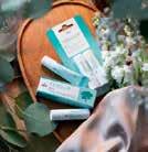








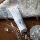





TEBODONT ®
TEBODONT ®
TEBODONT ®
TEBODONT ®
Gel
Gel
Gel
Gel
In case of irritated gums and oral mucosa, contains tea tree oil
In case of irritated gums and oral mucosa, contains tea tree oil
In case of irritated gums and oral mucosa, contains tea tree oil
In case of irritated gums and oral mucosa, contains tea tree oil
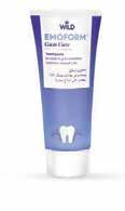
Gum Care
Gum Care
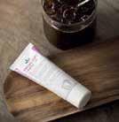
EMOFORM® Gum Care
Gum Care
Toothpaste
Toothpaste
Toothpaste In case of gum problems, contains mineral salts
Toothpaste
In case of gum problems, contains mineral salts
In case of gum problems, contains mineral salts
In case of gum problems, contains mineral salts
TEBODONT Spray
TEBODONT ®

TEBODONT ® Spray
TEBODONT ®
Spray
Spray
In case of irritated gums and oral mucosa, inhibits the formation of plaque, cares for and regenerates, contains tea tree oil
In case of irritated gums and oral mucosa, inhibits the formation of plaque, cares for and regenerates, contains tea tree oil
In case of irritated gums and oral mucosa, inhibits the formation of plaque, cares for and regenerates, contains tea tree oil
In case of irritated gums and oral mucosa, inhibits the formation of plaque, cares for and regenerates, contains tea tree oil
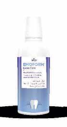
Gum Care
EMOFORM Gum Care
EMOFORM® Gum Care
Gum Care
Mouthbath concentrate In case of gum problems, contains mineral salts
Mouthbath concentrate In case of gum problems, contains mineral salts
Mouthbath concentrate In case of gum problems, contains mineral salts
Mouthbath concentrate In case of gum problems, contains mineral salts
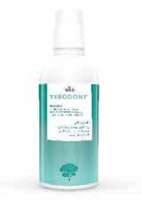
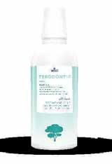
TEBODONT ®
TEBODONT ®
Mouthrinse
Mouthrinse
Mouthrinse
Mouthrinse
Inhibits the formation of plaque, cares for and strengthens the gums, contains tea tree oil, without fluoride
Inhibits the formation of plaque, cares for and strengthens the gums, contains tea tree oil, without fluoride
Inhibits the formation of plaque, cares for and strengthens the gums, contains tea tree oil, without fluoride
Inhibits the formation of plaque, cares for and strengthens the gums, contains tea tree oil, without fluoride
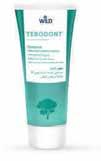
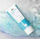
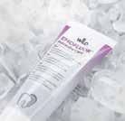
TEBODONT ®
TEBODONT ®
TEBODONT ®
TEBODONT ®
Toothpaste
Toothpaste
Toothpaste
Toothpaste
Inhibits the formation of plaque, strengthens the gums, contains tea tree oil, without fluoride
Inhibits the formation of plaque, strengthens the gums, contains tea tree oil, without fluoride
Inhibits the formation of plaque, strengthens the gums, contains tea tree oil, without fluoride
Inhibits the formation of plaque, strengthens the gums, contains tea tree oil, without fluoride
TEBODONT ®-F
TEBODONT ®-F
Mouthrinse
Mouthrinse
Mouthrinse Inhibits the formation of plaque, for the prophylaxis of caries, cares for and strengthens the gums, contains tea tree oil and sodium fluoride
Inhibits the formation of plaque, for the prophylaxis of caries, cares for and strengthens the gums, contains tea tree oil and sodium fluoride
Mouthrinse Inhibits the formation of plaque, for the prophylaxis of caries, cares for and strengthens the gums, contains tea tree oil and sodium fluoride
Inhibits the formation of plaque, for the prophylaxis of caries, cares for and strengthens the gums, contains tea tree oil and sodium fluoride

Sensitive
Sensitive
EMOFORM® Sensitive
Sensitive
Toothpaste
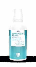
Sensitive
Sensitive
EMOFORM® Sensitive
Sensitive
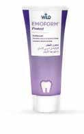
In case of sensitive gums, contains mineral salts
Toothpaste In case of sensitive gums, contains mineral salts
Toothpaste In case of sensitive gums, contains mineral salts
Toothpaste In case of sensitive gums, contains mineral salts
Mouthbath concentrate
Mouthbath concentrate In case of sensitive gums, contains mineral salts
Mouthbath concentrate In case of sensitive gums, contains mineral salts
Mouthbath concentrate In case of sensitive gums, contains mineral salts
In case of sensitive gums, contains mineral salts Protect
EMOFORM
Protect
Protect
Protect
Toothpaste For prevention of caries, hardens dental enamel
Toothpaste For prevention of caries, hardens dental enamel
Toothpaste For prevention of caries, hardens dental enamel
Toothpaste For prevention of caries, hardens dental enamel
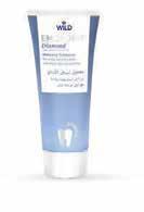
Diamond
Diamond
EMOFORM Diamond
Diamond
Whitening toothpaste
Whitening toothpaste For white and shiny teeth, contains finest diamond particles
Whitening toothpaste For white and shiny teeth, contains finest diamond particles
For white and shiny teeth, contains finest diamond particles
Whitening toothpaste For white and shiny teeth, contains finest diamond particles
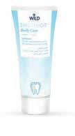
EMOFLUOR® Daily Care
EMOFLUOR® Daily Care
EMOFLUOR® Daily Care
® Daily Care
Toothpaste
Toothpaste
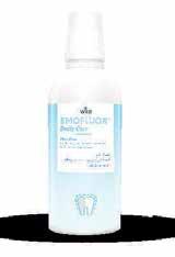
EMOFLUOR® Daily Care
EMOFLUOR® Daily Care
EMOFLUOR® Daily Care
EMOFLUOR® Daily Care
Mouthrinse
Mouthrinse
Toothpaste For daily care of sensitive teeth, contains stabilised stannous fluoride
For daily care of sensitive teeth, contains stabilised stannous fluoride
For daily care of sensitive teeth, contains stabilised stannous fluoride
Toothpaste For daily care of sensitive teeth, contains stabilised stannous fluoride
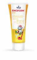
EMOFORM® Kids
EMOFORM® Kids
Mouthrinse
For the daily care of sensitive teeth and for the prophylaxis of caries
For the daily care of sensitive teeth and for the prophylaxis of caries
Mouthrinse For the daily care of sensitive teeth and for the prophylaxis of caries
For the daily care of sensitive teeth and for the prophylaxis of caries
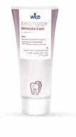
EMOFLUOR® Intensive Care
EMOFLUOR® Intensive Care
EMOFLUOR® Intensive Care
Intensive Care
Gel
Gel
Gel
Gel
For targeted protection against sensitive teeth and erosions, contains stabilised stannous fluoride
For targeted protection against sensitive teeth and erosions, contains stabilised stannous fluoride
For targeted protection against sensitive teeth and erosions, contains stabilised stannous fluoride
For targeted protection against sensitive teeth and erosions, contains stabilised stannous fluoride
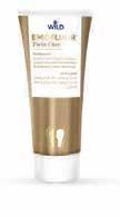
EMOFLUOR® Twin Care
EMOFLUOR® Twin Care
EMOFLUOR® Twin Care
Twin Care
Toothpaste
Toothpaste
Toothpaste
Toothpaste
Dual protection against erosions and sensitive teeth with stannous fluoride and vVARDIS technology
Dual protection against erosions and sensitive teeth with stannous fluoride and vVARDIS technology
Dual protection against erosions and sensitive teeth with stannous fluoride and vVARDIS technology
Dual protection against erosions and sensitive teeth with stannous fluoride and vVARDIS technology
EMOFORM Kids
EMOFORM® Kids
Toothpaste for children
Toothpaste for children
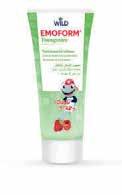
EMOFORM® Youngstars
EMOFORM® Youngstars
EMOFORM® Youngstars
EMOFORM Youngstars
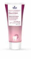
DEPURDENT® Clean & Polish
DEPURDENT® Clean & Polish
DEPURDENT® Clean & Polish
Clean & Polish

DEPURDENT® Clean & Polish
DEPURDENT® Clean & Polish
DEPURDENT®
DEPURDENT® Clean & Polish
Clean & Polish
Toothpaste for children
Toothpaste for children
From the first milk tooth, up to and including 5 years
From the first milk tooth, up to and including 5 years
From the first milk tooth, up to and including 5 years
Toothpaste for children
From the first milk tooth, up to and including 5 years
Toothpaste for children
Toothpaste for children Comprehensive protection for the teeth, as of 6 years
Paste for teeth cleaning
Paste for teeth cleaning
Mouthrinse
Mouthrinse
Toothpaste for children
Comprehensive protection for the teeth, as of 6 years
Comprehensive protection for the teeth, as of 6 years
Comprehensive protection for the teeth, as of 6 years
Paste for teeth cleaning
Removes dental plaque and teeth discoloration, with polishing eect
Removes dental plaque and teeth discoloration, with polishing eect
Paste for teeth cleaning
Removes dental plaque and teeth discoloration, with polishing eect
Removes dental plaque and teeth discoloration, with polishing eect
Mouthrinse
Mouthrinse
Helps preserve the natural whiteness of your teeth, helps prevent caries
Helps preserve the natural whiteness of your teeth, helps prevent caries
Helps preserve the natural whiteness of your teeth, helps prevent caries
Helps preserve the natural whiteness of your teeth, helps prevent caries
Al Rabee
Volume XXXII, Number 3, 2025
EDITORIAL TEAM
Alfred Naaman, Nada Naaman, Khalil Aleisa, Jihad Fakhoury, Dona Raad, Antoine Saadé, Lina Chamseddine, Tarek Kotob, Mohammed Rifai, Bilal Koleilat, Mohammad H. Al-Jammaz
Suha Nader
Marc Salloum
Micheline Assaf, Nariman Nehmeh
Josiane Younes
Albert Saykali
Gisèle Wakim
Tony Dib 1026-261X
DENTAL NEWS IS A QUARTERLY MAGAZINE DISTRIBUTED MAINLY IN THE MIDDLE EAST & NORTH AFRICA IN COLLABORATION WITH THE COUNCIL OF DENTAL SOCIETIES FOR THE GCC.
Statements and opinions expressed in the articles and communications herein are those of the author(s) and not necessarily those of the Editor(s) or publisher. No part of this magazine may be reproduced in any form, either electronic or mechanical, without the express written permission of the publisher.
DENTAL NEWS – Sami Solh Ave., G. Younis Bldg. POB: 116-5515 Beirut, Lebanon.
Tel: 961-3-30 30 48
Email: info@dentalnews.com Website: www.dentalnews.com

39th CAPP Int’l Dental ConfEx
November 14 – 15, 2025 Madinat Jumeirah | Dubai, U.A.E www.cappmea.com

ADF Association Dentaire Française

SIDC Jeddah 2025

GNYDM Greater New York Dental Meeting

www.instagram.com/dentalnews

Egyptian Orthodontic Society 2026
November 25 – 28, 2025 Palais des Congrès Paris Porte Maillot, FRANCE www.adf.asso.fr
November 27 – 29, 2025 Jeddah Hilton | Jeddah, K.S.A. www.sidc.org.sa
November 27 - December 3, 2025 Jacob Javits Convention Center New York, USA www.gnydm.com
January 15 – 17, 2026
Jolie Ville Kings Island | Luxor, EGYPT www.egyptortho.org
AEEDC 2026

SIDC Riyadh 2026

FDI World Dental Congress 2026
January 19 – 21, 2026 World Trade Center | Dubai, U.A.E. www.aeedc.com
February 5 – 7, 2026
Riyadh Front Exhibition & Conference Center | Riyadh, K.S.A. www.sidc.org.sa
September 4 – 7, 2026
Prague | Czech Republic www.2026.world-dental-congress.org
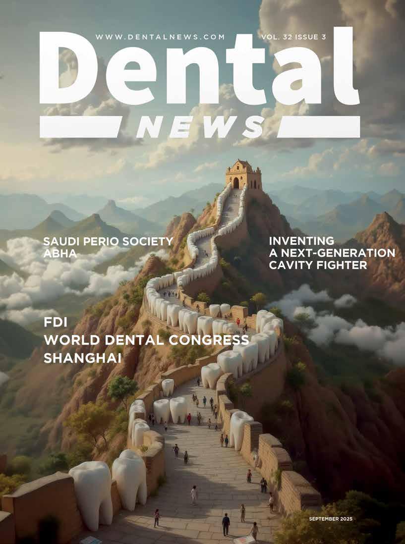
Inventing a Next-Generation Cavity Fighter
Posterior Tooth Replacement with Dental Implants
Oleg O. Yanushevich, Igor V. Maev, Natella I. Krikheli, Dmitrii N. Andreev , Svetlana V.
Lyamina ,Filipp S. Sokolov, Marina N. Bychkova, Petr A.
Beliy and Kira Y. Zaslavskaya
Dentsply Sirona honors talented dental students for their outstanding restorations

36
Exocad Announces Insights 2026 In Palma De Mallorca

500 2
9
the attachsockets instrumentawere was hemostatic replacethe augmentwith a the returned revealed ex11). harexisted at representative prerevealed bone” eviet al tismodeling was described as fast, the authors found the remodeling of the newly formed bone to be what they called “seemingly slow.” The trephine core presented in this particular case demonstrated this type of healing, as described by Trombelli et al.8 A high degree of woven bone as well as a cell and fiber-abundant provisional matrix was present. Two 4.8-mm x 10-mm implants were placed using standard protocol. These implants achieved primary stabilization and facilitated transmucosal healing. At just under 20 weeks, the two implants were restored with two individual cement-retained crowns (Figure 13).
1
15
25
72
21
29
3D 27
11
13
23
40
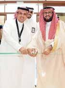
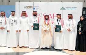
Saudi Periodontics and Implantology Forum SAPI Abha - Saudi Arabia 4-5 September, 2025

46
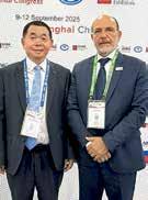
FDI World Dental Congress, NECC Shanghai-China 9-12 September, 2025
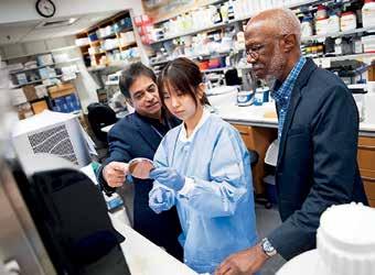
Tooth decay is the most common health condition worldwide. While it is preventable and treatable, billions of people are living with cavities and the pain that accompanies them. Given the massive scale of the problem, there’s a growing movement in dentistry to treat cavities without drilling and filling them. One such approach is applying a clear liquid called silver diamine fluoride to the surface of teeth. Silver diamine fluoride is already FDA approved to treat tooth sensitivity, and recent NYU research shows that the compound’s antimicrobial properties also make it effective at preventing cavities and stopping small cavities from progressing into larger ones. Because it’s inexpensive and easy to administer, it can be given in schools, in rural areas lacking dentists, or to patients who may have difficulty with dental care, including those with disabilities.
But treatment with silver diamine fluoride comes with one notable drawback: when the silver in it interacts with tooth decay, it turns the treated surface black. While this is not a significant issue for molars at the back of the mouth or baby teeth that fall out, it’s not a great option for teeth seen in a smile.
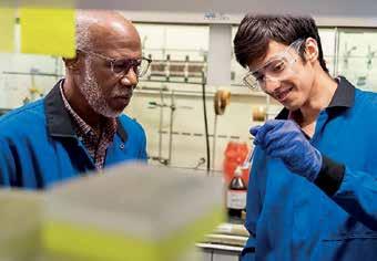
“Once your teeth are treated with silver diamine fluoride, that stain is permanent, which is a barrier for many people wanting to use the product,” explains Marc Walters, professor of chemistry at NYU.
Walters has long studied silver and other elements used in medicine to carry drugs and
imaging contrast agents. Several years ago, he was approached by researchers at NYU College of Dentistry seeking to better understand how silver stains teeth in order to avoid that outcome.
Walters had an idea. What if another mineral could be used that was also colorless and antimicrobial but didn’t turn teeth black? This question led him to zinc, an important nutrient found in foods like oysters and beef, as well as in over-the-counter products intended to shorten the duration of colds. Zinc is also used in dentistry, including in toothpaste and mouthwash to fight bacteria and bad breath, as well as in some denture adhesives and cementing agents to affix crowns or temporary fillings.
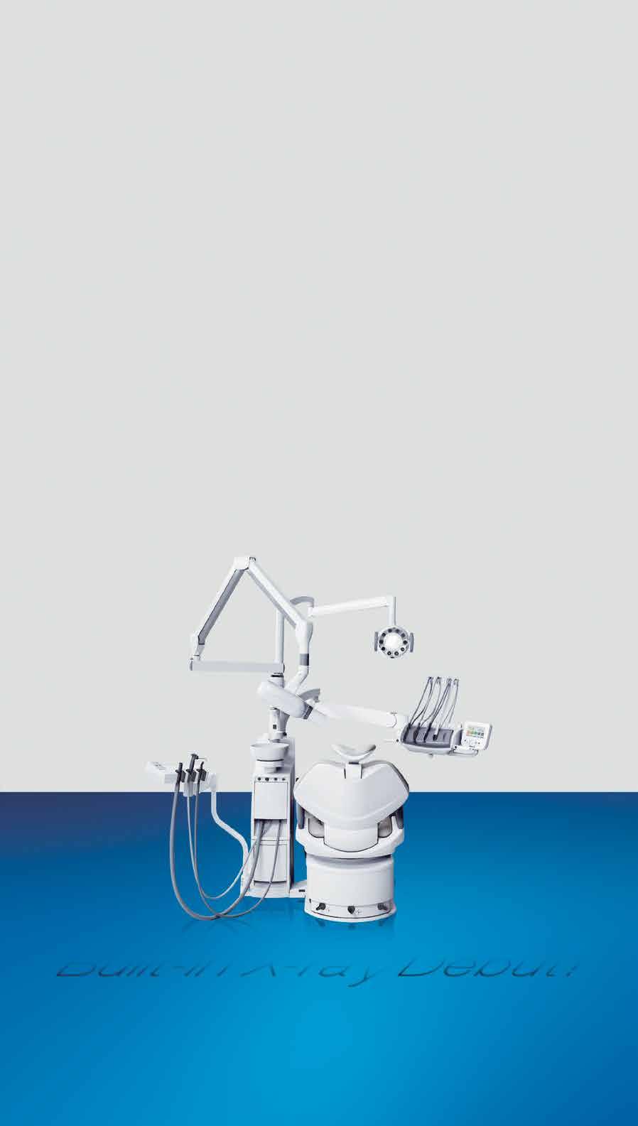
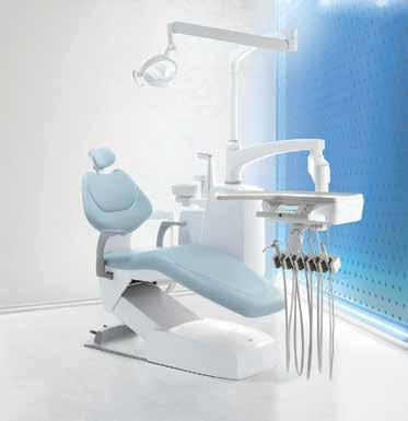
Embodies our belief of bringing highly reliable Japan-quality products to dentists in the world.

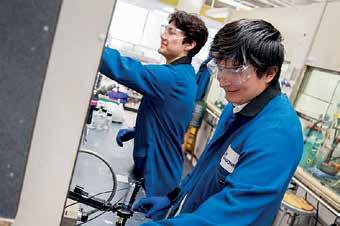
Walters began studying a zinc phosphate compound to see how it interacts with cavities, and crucially, to determine whether it can permeate deep into teeth. In order to address pain and hypersensitivity, the compound would need to reach the tooth’s dentin, the porous material sandwiched between the hard enamel outer layer and the nerves within. Dentin contains an abundance of microscopic, hollow channels— in fact, 40,000 of these tubules are packed into each square millimeter of dentin.
“We had to develop a solution to give dentists that will be taken up in these very small openings and go deep enough in the tubules so that the material will be retained,” Walters explains.
Walters applied phosphate followed by zinc to slices of a human tooth. Under the microscope, he saw deposits of the compound deep inside the dentin tubules. But while the zinc phosphate successfully permeated the teeth, he knew that a simpler approach that didn’t require applying two treatments would be easier for dentists. “Two steps is one too many,” says Walters.
Drawing inspiration from silver diamine fluoride, Walters developed another zinc-based molecule called zinc tetramine difluoride, which forms a colorless zinc oxide deep inside dentin tubules. The agent starts out as a liquid that is sensitive to concentration and pH. When painted onto a tooth and absorbed, the conditions within dentin tubules prompt a chemical change that quickly turns it into a solid, blocking the tubules and slowly releasing the antimicrobial zinc into the tooth.
His team is continuing to develop several related compounds for the treatment of cavities and has applied for patents of these zinc-based materials in several countries.
Having both fact-acting and long-lasting properties would offer an ideal combination for fighting cavities and tooth sensitivity, given that many current treatments for sensitive teeth require multiple applications and can take days or weeks to work.
“In one of our studies, two minutes after treatment with our agent, we can see using the electron
microscope that the zinc forms long cylinders of mineral that occupy the tubules,” says Walters. “Blocking the dentin tubules cuts off access to the nerves that are much deeper in dentin. It’s like putting a cork in place that shuts off the lower portion of the tubule from the outside environment—and this happens within a minute or two.”
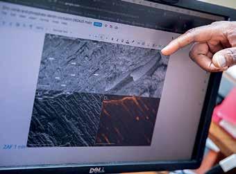
In additional tests, Walters found that zinc oxide persisted in tooth samples for at least one to two months. The goal is to develop a product that lasts for months or even years inside of teeth, stopping hypersensitivity and fighting bacteria on an ongoing basis.
“Not only do you have the analgesic result of having tubules blocked, but you also have a very low solubility agent that can slowly release the zinc into the tubule to prevent the growth of Streptococcus mutans and other bacteria,” Walters adds.
With a promising zinc nanocrystal agent in hand, Walters sought out other experts at NYU and beyond. His work caught the attention of Southern Dental Industries (SDI), an Australian company that makes restorative dental materials, including silver diamine fluoride. The company purchased the license for the zinc technology and NYU is working with them to develop it.
Closer to home, Walters began collaborating
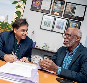
with Deepak Saxena, professor of molecular pathobiology and director of research innovation and entrepreneurship at NYU College of Dentistry, who has a strong track record of developing oral health products through Periomics Care, the startup company he co-founded with Xin Li at NYU Dentistry.
“As soon as I met Marc and I saw his enthusiasm, I decided that we should work together and try to make this as a commercial product,” Saxena recalls.
As a result of bringing together this diverse expertise—dentistry, chemistry, microbiology, and entrepreneurship—Saxena and Walters received a Technology Acceleration and Commercialization award from NYU, and last month, secured a nearly $300,000 grant from the National Institutes of Health under its Small Business Technology Transfer program. The NIH grant will fund feasibility studies for Walters’s team to further develop the formulation and confirm its ability to block tubules in a range of dentin samples. It will also fund research through Periomics Care in which Saxena’s team will study the agent’s antimicrobial properties. Specifically, they will look to see if the zinc creates a “zone of inhibition”—preventing the growth of decaycausing bacteria in the vicinity of it or even
killing bacteria that comes in contact with it.
“The mouth is full of bacteria. A compound needs to have good antimicrobial activity, which can occur from ionic imbalance, the properties of the zinc, or by the fluoride,” Saxena says. “If a compound does not stain, has good antimicrobial activity, plus it blocks the tubules, then it should be successful in stopping tooth decay and be aesthetically accepted.”
Saxena and Walters are already planning for the next phase of their research, which will include additional studies on the compound’s formulation, effectiveness, toxicity, and shelf life. Ultimately, if these studies go well, the researchers and SDI will approach the FDA for permission to do a clinic trial.
One factor working in their favor: because zinc phosphate has long been used as a dental adhesive, it’s known to be safe and the FDA has already approved it in other forms. These existing products may pave the way for faster research and development of a cavity treatment compared to untested elements, which can take many years to develop.
A new non-invasive treatment for cavities could help be a gamechanger in oral health. “We know that there’s a need—and a market—for a product that stops tooth decay that is effective, cheap, easy to use, and non-staining, given the rise in global numbers of untreated cavities,” Saxena says.
Dentists could use it to treat cavities without needing to scrape or drill out the cavity in preparation. Squirmy kids would need less time in the dentist’s chair. Older adults who get cavities near the roots of their teeth as their gums recede could have a new option for stopping sensitivity and decay in difficult-totreat areas. If safe and effective, perhaps small quantities could even be available on drugstore shelves and sold directly to consumers.
For Walters and Saxena, their goal is a future with less tooth decay and pain—and if their studies of zinc confirm its potential, silverstained teeth may be a thing of the past.
Rachel Harrison rachel.harrison@nyu.edu
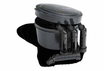
Transcend universal composite provides unprecedented shade matching with just one Universal Body shade due to its patented Resin Particle Match™ technology that eliminates the need for a blocker.
If you prefer a layering technique Transcend composite also includes four dentin shades and two enamel shades.
Before After
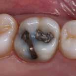
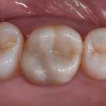
Deep amalgam staining presents one of the most difficult restoration situations to clinicians. In this case only the Transcend composite Universal Body shade was used to replace the amalgam no blocker needed. Note the excellent color blending of the preserved oblique ridge.

Scan the QR code to learn more about Transcend Universal Composite or go to ultradent.eu/transcend
Barry P. Levin, DMD; and Peter Tawil, DDS
● discuss the augmentation of molar extraction sockets with rhBMP-2/ACS
● analyze the outcome of a case series of consecutively treated patients who received grafting of the alveolus at time of molar extraction
● explain the fate of implants placed under functional, occlusal load in sites augmented with any bone graft
This case series demonstrates seven molarsite implants placed in six consecutively treated patients. All sites were augmented with rhBMP-2 (1.50 mg/cc)/ACS (recombinant human Bone Morphogenetic Protein-2/Absorbable Collagen Sponge) at extraction to regenerate bonefacilitating implant placement. In four patients, osteotomies were initiated with trephines to evaluate qualitatively for native bone and for the absence of residual ACS. All sites facilitated implant placement after augmentation. All seven implants achieved primary stabilization and were functionally loaded. No implants were lost or developed complications. It can be concluded that augmenting molar extraction sockets with rhBMP-2/ACS can allow standard implant placement in the posterior dentition that is capable of withstanding a functional load.
with the evolution of dental implant therapy, the treatment of extraction sockets has progressed from a simple matter of wound healing to what is often times a complex surgical procedure aimed at minimizing bone resorption. The consequences of physiologic wound healing from extractions often include both vertical and, more prominently, lateral reductions of the local alveolar process. The healing of extraction sockets is accompanied by marked ridge resorption within the first 3 to 4 months.1 Schropp demonstrated approximately
50% horizontal bone loss 12 months after extractions.2 Nevins et al demonstrated that when extraction of teeth with prominent roots are augmented with grafts, when compared to ungrafted sites the grafted sites facilitated favorable implant placement. Significantly fewer of the grafted sites required additional grafting procedures at the time of implant placement.3 Although many “ridge augmentation” techniques exist, it is certainly more efficacious for both surgeons and patients to prevent bone modeling that results in physiologic resorption and necessitates more involved modalities. The term “socket preservation” usually refers to the placement of various bone replacement grafts that are often covered with a barrier membrane. The bone graft materials are used to maintain space and serve as an osteoconductive scaffold to support passive osteogenesis within the “pores” both between and within the graft particulate. Iasella et al demonstrated significantly greater 3-dimensional (3-D) ridge preservation for extraction sockets augmented with allograft bone and collagen membranes compared to ungrafted controls.4 Araújo et al demonstrated in the canine model that the pores of tricalcium phosphate particulate can be invaded by erythrocytes, and that later these pores would become the locus of new bone formation. These same authors also noted that a degree of delayed healing and minimal bone formation occurred between the second and fourth weeks of recovery. These authors speculated that the β -TCP (beta-tri-calcium phosphate) graft may have retarded bone formation. 5
In a review article, Darby et al concluded that ridge preservation is an effective procedure in limiting both horizontal and vertical ridge alterations in post-extraction sites, and that there is no technique superior to another.6


The barrier membrane provides a soft-tissue exclusionary function, blocking the ingrowth of epithelial and fibroblastic cells and favoring the repopulation of osteoblast cells for bone replacement of the graft material. Investigators such as Carmagnola et al reported excellent clinical results when particulate xenograft coverage, without soft-tissue closure, was achieved when a collagen membrane was adapted over the bone graft and beneath the mucoperiosteal flap.7 The majority of these procedures provide 3-D bone volume, facilitating prosthetically driven implant placement. The caveat of these treatments is that osseointegration occurs to support longterm tooth replacement that may involve permanent inclusion of graft material.
At the inception of dental implantology, the phenomenon of osseointegration was investigated through histologic animal studies. Titanium implants were inserted into healed alveolar ridges, composed of “native” bone. Most long-term (over 10 to 15 years) studies followed these types of clinical situations. It would seem logical that ideal clinical situations would support the possibility of implant placement into sites composed of native bone, excluding bone graft materials occupying spaces of potential bony trabeculae. The challenge that still exists today, when teeth require extraction and site preparation is chosen to facilitate future implant placement, is to regenerate de novo bone and maintain osseous morphology favorable for restoratively driven implant placement. Recombinant technology has given surgeons the ability to stimulate wound healing and cellular differentiation, leading to tissue regeneration. The stimulatory properties of these peptides require vehicles for sustained delivery. Some of these growth factors are commercially available in combined packaging, with bone grafting particulate as the delivery
vehicle. These materials possess some degree of osteoconductivity and occupy a physical space, preventing maximum osseous fill of the desired space. Some of these growth factors are not specific for osteoblastic differentiation.
One of the few commercially available recombinant growth factors proven to be selectively osteoinductive is recombinant human Bone Morphogenetic Protein-2 (rhBMP-2) (INFUSE® Bone Graft, Medtronic, Inc. [FDA PMA submission number for INFUSE OMF indication is P050053.]). rhBMP2 is a differentiation factor that changes the phenotype of precursor cells (mesenchymal stem cells) into osteoblasts and chondroblasts. The standard dose of the protein is 1.50 mg per 1 cc of the solution. The lyophilized rhBMP-2 is combined with sterile water chairside. Once the solution is mixed, it is uniformly dispensed onto an absorbable collagen sponge (ACS). Following a minimal saturation time of 15 minutes, this collagen sponge can be cut into various size strips to be delivered to the site of desired regeneration. The release of the growth factor is sustained over an approximately 2-week time period. Because the ACS is not treated by crosslinking to delay degradation, it is resorbed quickly, leaving no remnants of graft material at the placement site. This facili- tates a maximum potential for bone-fill of the grafted defect. The stimulatory regenerative and vascular invasion effects of rhBMP-2 also accelerates bone formation. This is significant because it can shorten the overall treatment time for patients.
of implant placement, with the exception of the initiation of osteotomies with a small (2.7-mm outer and 2-mm inner diameter) trephine. The cores harvested were submitted for qualitative histologic evaluation to confirm the presence of vital bone. All implants subjectively achieved primary mechanical stability and were restored 3 to 8 months after placement.
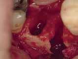
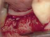
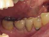
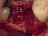
The patient was a 71-year-old man with significant caries and subsequent bone loss associated with tooth No. 30. The septal bone was lost, with the exception of the coronal aspect, resulting in a “bridge of bone” connecting the buccal and lingual cortices of the site (Figure 1). After reflection of the full-thickness buccal and lingual flaps, extraction, and manual and ultrasonic debridement of the socket to remove all visible soft-tissue remnants, the defect was obturated with the rhBMP-2/ACS material (Figure 2).
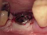
Fifteen weeks after the first procedure, the site was reopened to perform implant placement. Flap reflection revealed excellent and complete bone reformation (Figure 3). The implant osteotomy preparation was initialized with the harvest of an approximate 5-mm trephine core. The trephine had an internal diameter of 2 mm and an outer diameter of 2.7 mm. The completion of implant placement was performed according to the manufacturer’s guidelines, resulting in the delivery of a 5-mm x 11-mm implant with primary stability. Because of excellent subjective stability, a transmucosal healing was chosen, with placement of a healing cap and a nonsubmerged closure (Figure 4). Qualitative histology demonstrated lamellar bone without evidence of the ACS carrier. Restorative therapy commenced approximately 4 months after implant placement. Delivery of the definitive prosthesis, consisting of a gold custom abutment and cement-retained crown, occurred at 5 months following implant placement surgery and 8.5 months after extraction and augmentation (Figure 5).
The purpose of this article is to present a case series of consecutively treated patients (Table 1) who at the time of molar tooth extractions received grafting of the alveolus with the rhBMP-2/ACS material alone. This retrospective analysis fully complies with the Helsinki Accords and Ethical Guideline for Clinical Research. All patients included in this case series signed written consent forms that explained the nature of the procedure undertaken, stating that they agreed to undergo the prescribed therapy. These patients were also informed that the material used for augmentation was a recently FDA-approved material that was indicated for grafting of extraction sockets.
The purpose of this article is to present a case series of consecutively treated patients (Table 1) who at the time of molar tooth extractions received grafting of the alveolus with the rhBMP-2/ACS material alone. This retrospective analysis fully complies with the Helsinki Accords and Ethical Guideline for Clinical Research. All patients included in this case series signed written consent forms that explained the nature of the procedure undertaken, stating that they agreed to undergo the prescribed therapy. These patients were also informed that the material used for augmentation was a recently FDA-approved material that was indicated for grafting of extraction sockets.
All grafted sites received dental implants within a 3- to 6-month time period following extraction. Surgical procedures were not altered in terms of underpreparation, bone condensing, or additional grafting at the time of implant placement, with the exception of the initiation of osteotomies with a small (2.7-mm outer and 2-mm inner diameter) trephine. The cores harvested were submitted for qualitative histologic evaluation to confirm the presence of vital bone. All implants subjectively achieved primary mechanical stability and were restored 3 to 8 months after placement.
All grafted sites received dental implants within a 3- to 6-month time period following extraction. Surgical procedures were not altered in terms of underpreparation, bone condensing, or additional grafting at the time
The patient was a 71-year-old man with significant caries and subsequent bone loss associated with tooth No. 30. The septal bone was lost, with the exception of the coronal aspect, resulting in a “bridge of bone” connecting the buccal and lingual cortices of the site (Figure 1). After reflection of the fullthickness buccal and lingual flaps, extraction, and manual and ultrasonic debridement of the socket to remove all visible soft-tissue remnants, the defect was obturated with the rhBMP-2/ACS material (Figure 2). Fifteen weeks after the first procedure, the site was reopened to perform implant placement. Flap reflection revealed excellent and complete bone reformation (Figure 3). The implant osteotomy preparation was initialized with the harvest of an approximate 5-mm trephine core. The trephine had an internal diameter of 2 mm and an outer diameter of 2.7 mm. The completion of implant placement was performed according to the manufacturer’s guidelines, resulting in the delivery of a 5-mm x 11-mm implant with primary stability. Because of excellent subjective stability, a transmucosal healing was chosen, with placement of a healing cap and a nonsubmerged closure (Figure 4). Qualitative histology demonstrated lamellar bone without evidence of the ACS carrier. Restorative therapy commenced approximately 4 months after implant placement. Delivery of the definitive prosthesis, consisting of a gold custom abutment and cement-retained
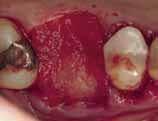
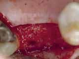
The second patient was a 75-year-old woman who presented with a chronic infection associated with a fracture of the distobuccal root of tooth No. 3. Following flap reflection, complete buccal bone loss was associated with the root fracture. Tooth No. 3 was extracted, with all remaining bony walls of the extraction socket being preserved (Figure 6). Debridement was followed by obturation of the defect with the rhBMP-2/ACS (Figure 7). A subepithelial, connective tissue pedicle graft was rotated to provide partial coverage of the grafting material. The graft was then closed with a monofilament polytetrafluoroethylene (PTFE) suture.
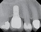
Approximately 18 weeks following extraction and grafting, full-thickness flaps were reflected, revealing complete osseous regeneration of the original defect (Figure 8). The osteotomy was initiated with the same trephine bur to harvest a core of representative bone present at the site of implant insertion. Implant placement proceeded without alteration from the manufacturer’s protocol by inserting a 4.8-mm x 8-mm fixture with primary, tactile, stability, and transmucosal healing properties. At about 8 months postplacement, the implant was restored with a custom abutment and cement-retained crown (Figure 9).
crown, occurred at 5 months following implant placement surgery and 8.5 months after extraction and augmentation (Figure 5).
The second patient was a 75-year-old woman who presented with a chronic infection associated with a fracture of the disto-buccal root of tooth No. 3. Following flap reflection, complete buccal bone loss was associated with the root fracture. Tooth No. 3 was extracted, with all remaining bony walls of the extraction socket being preserved (Figure 6). Debridement was followed by obturation of the defect with the rhBMP-2/ACS (Figure 7). A subepithelial, connective tissue pedicle graft was rotated to provide partial coverage of the grafting material. The graft was then closed with a monofilament polytetrafluoroethylene (PTFE) suture.
Approximately 18 weeks following extraction and grafting, full-thickness flaps were reflected, revealing complete osseous regeneration of the original defect (Figure 8). The osteotomy was initiated with the same trephine bur to harvest a core of representative bone present at the site of implant insertion. Implant placement proceeded without alteration from the manufacturer’s protocol by inserting a 4.8-mm x 8-mm fixture with primary, tactile, stability, and transmucosal healing properties. At about 8 months postplacement, the implant was restored with a custom abutment and cement retained crown (Figure 9).
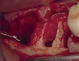
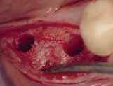
The patient was a 66-year-old man who required removal of the three mandibular right molars due to rampant caries and attachment loss. Following flap reflection and extractions, the sockets were debrided with both ultrasonic and manual instrumentation (Figure 10). The sockets of the first and second molars were
augmented with rhBMP-2/ACS. The site of the third molar was obturated with a noncrosslinked, collagen plug for hemostatic purposes only.
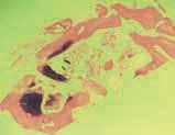
The restorative treatment plan encompassed tooth replacement in the first and second molar positions only, negating the need for the patient to incur the greater expense of augmenting the third molar site. Primary closure was achieved with a monofilament PTFE suture. Approximately 6 months after the extractions and augmentation procedure, the patient returned for implant placement surgery. Surgical reopening revealed excellent visual regeneration and ridge preservation (Figure 11). The site of the tooth No. 31 osteotomy was chosen for biopsy harvesting, because this is where the most severe bone loss existed at the time of extraction, and this site would be most representative of new bone formation, as opposed to possibly harvesting pre- existing native bone. This trephine core qualitatively revealed what was diagnosed by the histopathologist as “normal bone” without any foreign body or inflammatory responses being evident (Figure 12). Serving as a historic control, Trombelli et al reported on histomorphometric measurements of various tissues present at different time intervals. These authors describe great variability in human trephine cores taken from extraction sites. In relation to the present case series, Trombelli et al described the presence of a provisional matrix and woven bone dominating what they described as late-phase healing taken at 12 to 24 weeks after extractions. Although tissue modeling was described as fast, the authors found the remodeling
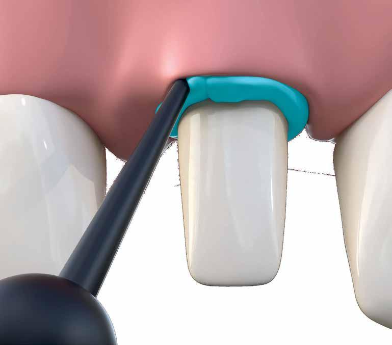
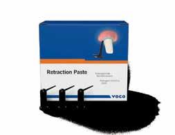
• Thin cannula with flexible tip – easy and pinpoint application into the sulcus
• Viscosity Change – paste consistency varies during application and sulcus widening
• Good visibility – contrasty to the gingiva
• A clean product – quick and easy to spray off
For more information please contact VOCO‘s Business Development in Middle East/Northern Africa, Mohamad El Fil (+961-3-805758 / m.elfil@voco.com).
augmented with rhBMP-2/ACS. The site of the third molar was obturated with a noncrosslinked, collagen plug for hemostatic purposes only.
The patient was a 66-year-old man who required removal of the three mandibular right molars due to rampant caries and attachment loss. Following flap reflection and extractions, the sockets were debrided with both ultrasonic and manual instrumentation (Figure 10). The sockets of the first and second molars were augmented with rhBMP-2/ACS. The site of the third molar was obturated with a noncrosslinked, collagen plug for hemostatic purposes only.
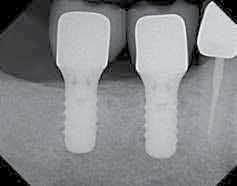
of the newly formed bone to be what they called “seemingly slow.” The trephine core presented in this particular case demonstrated this type of healing, as described by Trombelli et al.8 A high degree of woven bone as well as a cell and fiber-abundant provisional matrix was present. Two 4.8-mm x 10-mm implants were placed using standard protocol. These implants achieved primary stabilization and facilitated transmucosal healing. At just under 20 weeks, the two implants were restored with two individual cement-retained crowns (Figure 13).
The restorative treatment plan encompassed tooth replacement in the first and second molar positions only, negating the need for the patient to incur the greater expense of augmenting the third molar site. Primary closure was achieved with a monofilament PTFE suture. Approximately 6 months after the extractions and augmentation procedure, the patient returned for implant placement surgery. Surgical reopening revealed excellent visual regeneration and ridge preservation (Figure 11). The site of the tooth No. 31 osteotomy was chosen for biopsy harvesting, because this is where the most severe bone loss existed at the time of extraction, and this site would be most representative of new bone formation, as opposed to possibly harvesting preexisting native bone. This trephine core qualitatively revealed what was diagnosed by the histopathologist as “normal bone” without any foreign body or inflammatory responses being evident (Figure 12). Serving as a historic control, Trombelli et al reported on histomorphometric measurements of various tissues present at di erent time intervals. These authors describe great variability in human trephine cores taken from extraction sites. In relation to the present case series, Trombelli et al described the presence of a provisional matrix and woven bone dominating what they described as late-phase healing taken at 12 to 24 weeks after extractions. Although tissue

All of the consecutively treated patients underwent extraction of molar teeth,
The restorative treatment plan encompassed tooth replacement in the first and second molar positions only, negating the need for the patient to incur the greater expense of augmenting the third molar site. Primary closure was achieved with a monofilament PTFE suture. Approximately 6 months after the extractions and augmentation procedure, the patient returned for implant placement surgery. Surgical reopening revealed excellent visual regeneration and ridge preservation (Figure 11). The site of the tooth No. 31 osteotomy was chosen for biopsy harvesting, because this is where the most severe bone loss existed at the time of extraction, and this site would be most representative of new bone formation, as opposed to possibly harvesting preexisting native bone. This trephine core qualitatively revealed what was diagnosed by the histopathologist as “normal bone” without any foreign body or inflammatory responses being evident (Figure 12). Serving as a historic control, Trombelli et al reported on histomorphometric measurements of various tissues present at di erent time intervals. These authors describe great variability in human trephine cores taken from extraction sites. In relation to the present case series, Trombelli et al described the presence of a provisional matrix and woven bone dominating what they described as late-phase healing taken at 12 to 24 weeks after extractions. Although tissue
modeling was described as fast, the authors found the remodeling of the newly formed bone to be what they called “seemingly slow.” The trephine core presented in this particular case demonstrated this type of healing, as described by Trombelli et al.8 A high degree of woven bone as well as a cell and fiber-abundant provisional matrix was present. Two 4.8-mm x 10-mm implants were placed using standard protocol. These implants achieved primary stabilization and facilitated transmucosal healing. At just under 20 weeks, the two implants were restored with two individual cement-retained crowns (Figure 13).
RELATED CONTENT:
dentalaegis.com/go/cced58
modeling was described as fast, the authors found the remodeling of the newly formed bone to be what they called “seemingly slow.” The trephine core presented in this particular case demonstrated this type of healing, as described by Trombelli et al.8 A high degree of woven bone as well as a cell and fiber-abundant provisional matrix was present. Two 4.8-mm x 10-mm implants were placed using standard protocol. These implants achieved primary stabilization and facilitated transmucosal healing. At just under 20 weeks, the two implants were restored with two individual cement-retained crowns (Figure 13).
simultaneous bone augmentation with rhBMP-2/ ACS, and implant placement approximately 4 to 6 months after the first procedure. It is important to point out several findings: First, all of the augmented sites facilitated restoratively driven implant placement that was not possible at the time of extraction because of bony insufficiency. Second, all implants subjectively achieved primary stability. No mobility or rotation of the implants occurred upon tightening of the healing abutments. Third, no additional bone augmentation was necessary at the time of implant placement in any of
these cases. Implant placement resulted in circumferential bony coverage of the implant surfaces either to the collar of the implants, or to the rough–smooth titanium border, depending on the implant type used in each individual situation. The most common adverse event or morbidity was mild to moderate postoperative edema that was noted intra- and extraorally. This reaction to oral grafting with rhBMP-2/ ACS has also been reported by Boyne et al.9 These same investigators detected antibody production to rhBMP-2 in a small percentage (12%) of patients treated with a therapeutic
All of the consecutively treated patients underwent extraction of molar teeth, simultaneous bone augmentation with rhBMP-2/ACS, and implant placement approximately 4 to 6 months after the first procedure. It is important to point out several findings: First, all of the augmented sites facilitated restoratively driven implant placement that was not possible at the time of extraction because of bony insufficiency. Second, all implants subjectively achieved primary stability. No mobility or rotation of the implants occurred upon tightening of the healing abutments. Third, no additional bone augmentation was necessary at the time of implant placement in any of these cases. Implant placement resulted in circumferential bony coverage of the implant surfaces either to the collar of the implants, or to the rough–smooth titanium border, depending on the implant type used in each individual situation. The most common adverse event or morbidity was mild to moderate postoperative edema that was noted intra- and extraorally. This reaction to oral grafting with rhBMP-2/ACS has also been reported by Boyne et al.9 These same investigators detected antibody production to rhBMP-2 in a small percentage (12%) of patients treated with a therapeutic dosage of 1.50 mg/mL. This was a transient finding that did not a ect treatment outcomes or require further treatment.
bone, lamellar bone, and soft tissue within the cores were not evaluated. The purpose of the histologic component of this report is to demonstrate the presence of viable native bone, without undesired inflammatory processes or residual graft materials, in the locations of implant insertions.
dosage of 1.50 mg/mL. This was a transient finding that did not affect treatment outcomes or require further treatment.
Most importantly, from a clinical perspective, all consecutively placed implants in this case series achieved osseointegration and were functionally loaded in a standard period of time (3 to 8 months). Delayed loading due to poor subjective bone quality or suboptimal implant stability was not found with any of the implants in this case series.
stability was not found with any of the implants in this case series.
It is also worth mentioning that although the retrieved trephine cores revealed qualitative evidence of healthy bone, without evidence of persisting graft material or adverse cellular reactions,
It is also worth mentioning that although the retrieved trephine cores revealed qualitative evidence of healthy bone, without evidence of persisting graft material or adverse cellular reactions, histomorphometry was not performed. The percentages of woven bone, lamellar bone, and soft tissue within the cores were not evaluated. The purpose of the histologic component of this report is to demonstrate the presence of viable native bone,
without undesired inflammatory processes or residual graft materials, in the locations of implant insertions.
Most importantly, from a clinical perspective, all consecutively placed implants in this case series achieved osseointegration and were functionally loaded in a standard period of time (3 to 8 months). Delayed loading due to poor subjective bone quality or suboptimal implant
Ridge preservation is a frequently investigated subject. Numerous combinations of bone replacement grafts, barrier membranes, and the addition of various growth factors have been evaluated. The primary goal of these procedures is to preserve and/or regenerate alveolar bone associated with extraction sockets and prevent the anticipated, physiologic bone resorption that follows tooth loss. Araújo et al found that when canine extraction sockets are augmented with Bio-Oss Collagen® (Osteohealth, www. osteohealth. com), some of the expected dimensional alterations could be offset. The collagen portion of the graft was readily eliminated, whereas the xenograft portion of the graft persisted, although bone formation occurred on the surface of the graft particles.10 The presence of newly formed bone onto the nonresorbable graft surface demonstrates the passive process of osteoconduction. Graft particles are placed into the site and the repopulation of the bone-forming cells occurs over time and is dependent on the individual defect’s and patient’s ability to heal and regenerate lost or damaged tissue. This process lacks a stimulatory cellular component. According to Lane et al,11 the tissue engineering model is composed of three elements. First, each site of regeneration requires that cells be capable of differentiating into the desired cell line needed to regenerate the desired lost tissue. Second, a signaling molecule is required to provide chemotactic, morphogenic, and differentiation messages to these cells. Third, the signaling molecule and migrating cells require a scaffold or matrix to provide the physical space necessary to carry the message to the site and facilitate cellular migration. The fate of implants placed under functional,
Ridge preservation is a frequently investigated subject. Numerous combinations of bone replacement grafts, barrier membranes, and the addition of various growth factors have been evaluated. The primary goal of these procedures is to preserve and/or regenerate alveolar bone associated with extraction sockets and prevent the anticipated, physiologic bone resorption that follows tooth loss. Araújo et al found that when canine extraction sockets are augmented with Bio-Oss Collagen® (Osteohealth, www.osteohealth. com), some of the expected dimensional alterations could be o set. The collagen portion of the graft was readily eliminated, whereas the xenograft portion of the graft persisted, although bone formation occurred on the surface of the graft particles.10 The presence of newly formed bone onto the nonresorbable graft surface demonstrates the passive process of osteoconduction. Graft particles are placed into the site and the repopulation of the bone-forming cells occurs over time and is dependent on the individual defect’s and patient’s ability to heal and regenerate lost or damaged tissue. This process lacks a stimulatory cellular component.
According to Lane et al,11 the tissue engineering model is composed of three elements. First, each site of regeneration requires that cells be capable of di erentiating into the desired cell line needed to regenerate the desired lost tissue. Second, a signaling molecule is required to provide chemotactic, morphogenic, and di erentiation messages to these cells. Third, the signaling molecule and migrating cells require a sca old or matrix to provide the physical space necessary to carry the message to the site and facilitate cellular migration.
The fate of implants placed under functional, occlusal load in sites augmented with any bone graft is a primary concern for clinicians and patients. The possibility of placing implants is the first step in a multistep process of tooth replacement. Initial stability, followed by secondary stability or osseointegration, is the specialty of the restorative dentist. Treatment is considered a failure if these implants do not function in a healthy state, under normal occlusal conditions. In an animal model, Jovanovic et al demonstrated that machined titanium implants can function for 12 months after
teeth can give your patients the confidence to smile more.

Opalescence whitening is on a mission to help give your patients brighter, whiter smiles so they can look their best and feel their best, turning good days into better ones. As the global leader in professional whitening,1 Opalescence has brightened over 100 million smiles.1 That’s a lot of better days.
occlusal load in sites augmented with any bone graft is a primary concern for clinicians and patients. The possibility of placing implants is the first step in a multistep process of tooth replacement. Initial stability, followed by secondary stability or osseointegration, is the specialty of the restorative dentist. Treatment is considered a failure if these implants do not function in a healthy state, under normal occlusal conditions. In an animal model, Jovanovic et al demonstrated that machined titanium implants can function for 12 months after placement in sites of experimental defects augmented with rh- BMP-2/ACS 3 months prior to placement. Not only were these implants successfully loaded for 1 year, but the authors also noted that the bone-to-implant contact (BIC) for fixtures inserted into rh- BMP-2 grafted sites was comparable to implants placed into native bone.12 In a pilot study conducted in humans, Cochran et al evaluated a subtherapeutic dose of rhBMP-2 (0.43 mg/cc) in extraction sockets or ridge augmentations. The 3-year results demonstrated safety and long-term effcacy of this growth factor for site development facilitating implant placement.13 In a randomized controlled study, Fiorellini et al evaluated rhBMP-2/ACS delivered at 1.50 mg/ cc, 0.75 mg/cc, ACS alone, and ungrafted controls in the treatment of maxillary extraction sockets with buccal wall defects.14 Significant ridge height was preserved, and bone width was regenerated when the sites were augmented with rhBMP-2/ACS. Ungrafted and ACS- only grafted sites demonstrated little bone regeneration. The sites grafted with the commercially available dosage of 1.50 mg/cc of rhBMP-2 outperformed the sockets grafted with the subtherapeutic dose of 0.75 mg/cc in terms of dimensional bone maintenance and regeneration. These investigators evaluated anterior sites for socket augmentation and
preservation.
The present case series follows consecutively treated molar sites. This may be of significance because most human studies evaluating rhBMP-2/ACS for bone regeneration have focused on maxillary sinus grafts14,15 or maxillary anterior extraction sites. When comparing the findings of the present case series with ungrafted extraction sockets, it can be concluded that regeneration of native bone-not unlike normal bone remodeling—occurred. Serving as another historic control, Evian et al studied histologic cores of ungrafted extraction sites at varying time intervals. These investigators noted two distinct regenerative phases. From 4 to 8 weeks, a “progressive osteogenic phase” was described. From 8 weeks on, the “osteogenesis slows down” and maturation of bony trabeculae increases in bone volume. Bone that was present in the 16-week study specimen was described as mature, with fewer cellular elements compared to earlier specimens. The bone found in this case series was qualitatively similar to the later specimens in the Evian study of ungrafted extraction sites.16
It can be concluded that augmentation of molar extraction sockets with rhBMP-2/ACS results in the regeneration of de novo bone, capable of accepting timely implant placement, without altering manufacturer-specified osteotomy preparation, and functional loading in a standard time period. Because the implants were inserted and loaded prosthetically into native bone, without the presence of residual graft materials, these implants can be expected to achieve optimal long-term success, comparable to implants placed into unmanipulated, edentulous bone.


1. Amler MH. The time sequence of tissue regeneration in human extraction wounds. Oral Surg Oral Med Oral Pathol. 1969;27(3):3O9-318.
2. Schropp L, Wenzel A, Kostopoulos L, Karring T. Bone healing and soft tissue contour changes following single-tooth extraction: a clini- cal and radiographic 12-month prospective study. Int J Periodontics Restorative Dent. 2OO3;23(4):313-323.
3. Nevins M, Camelo M, De Paoli S, et al. A study of the fate of the buccal wall of extraction sockets of teeth with prominent roots. Int J Periodontics Restorative Dent. 2OO6;26(1):1929.
4. Iasella JM, Greenwell H, Miller RL, et al. Ridge preservation with freeze-dried bone allograft and a collagen membrane compared to extraction alone for implant site development: a clinical and histologic study in humans. J Periodontol. 2OO3;74(7):99O-999.
5. Araújo MG, Liljenberg B, Lindhe J. betaTricalcium phosphate in the early phase of socket healing: an experimental study in the dog. Clin Oral Implants Res. 2O1O;21(4):445454.
6. Darby I, Chen ST, Buser D. Ridge preservation techniques for implant therapy. Int J Oral Maxillofac Implants. 2OO9;24(suppl):26O-271.
7. Carmagnola D, Adriaens P, Berglundh T. Healing of human extraction sockets filled with Bio-Oss. Clin Oral Implants Res. 2OO3;14(2):137-143.
8. Trombelli L, Farina R, Marzola A, et al. Modeling and remodeling of human extraction sockets. J Clin Periodontol. 2OO8;35(7):63O-639.
9. Boyne PJ, Lilly LC, Marx RE, et al. De novo bone induction by recombinant human bone morphogenetic protein-2 (rhBMP-2) in maxillary sinus floor augmentation. J Oral Maxillofac Surg. 2OO5;63(12):1693-17O7.
10. Araújo M, Linder E, Wennström J, Lindhe J. The influence of Bio-Oss Collagen on healing
of an extraction socket: an experimental study in the dog. Int J Periodontics Restorative Dent. 2OO8;28(2):123-135.
11. Lane JM, Yasko AW, Tomin E, et al. Bone marrow and recombinant human bone morphogenetic protein-2 in osseous repair. Clin Orthop Relat Res. 1999;361:216-227.
12. Jovanovic SA, Hunt DR, Bernard GW, et al. Long-term functional loading of dental implants in rhBMP-2 induced bone. A histologic study in the canine ridge augmentation model. Clin Oral Implants Res. 2OO3;14(6):793-8O3.
13. Cochran DL, Jones AA, Lilly LC, et al. Evaluation of recombinant human bone morphogenetic protein-2 in oral applications including the use of endosseous implants: 3-year results of a pilot study in humans. J Periodontol. 2OOO;71(8):1241-1257.
14. Fiorellini JP, Howell TH, Cochran D, et al. Randomized study evaluating recombinant human bone morphogenetic protein-2 for extraction socket augmentation. J Periodontol. 2OO5;76(4):6O5-613.
15. Triplett RG, Nevins M, Marx RE, et al. Pivotal, randomized, parallel evaluation of recombinant human bone morphogenetic protein-2/ absorbable collagen sponge and autogenous bone graft for maxillary sinus floor augmentation. J Oral Maxillofac Surg. 2OO9;67(9):1947-196O.
16. Evian CI, Rosenberg ES, Coslet JG, Corn H. The osteogenic activity of bone removed from healing extraction sockets in humans. J Periodontol. 1982;53(2):81-85.
ABOUT THE AUTHORS
Barry P. Levin, DThD
Clinical Associate Professor, University of Pennsylvania, Philadelphia, Pennsylvania
Peter Tawil, DDS
Former Resident in Periodontics, University of Pennsylvania, Philadelphia, Pennsylvania; Private Practice, Beirut, Lebanon
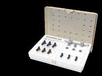
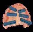
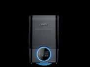
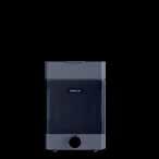
IPG - Intraoral Photogrammetry
Two-in-One System
ScanDesignDelivery






Barry P. Levin, DMD; and Peter Tawil, DDS
This article provides 2 hours of CE credit from AEGIS Publications, LLC. Record your answers on the enclosed answer form or submit them on a separate sheet of paper. You may also phone your answers in to 877-423-4471 or fax them to (215) 504-1502 or log on to www.dentalaegis.com/cced and click on “Continuing Education.” Be sure to include your name, address, telephone number, and last 4 digits of your Social Security number.
PLEASE COMPLETE ANSWER FORM ON PAGE 114, INCLUDING YOUR NAME AND PAYMENT INFORMATION.
1. Schropp demonstrated approximately what percentage of horizontal bone loss 12 months after extractions?
2. What term usually refers to the placement of various bone replacement grafts that are often covered with a barrier membrane?
3. Darby et al concluded that ridge preservation is an e ective procedure in limiting both horizontal and vertical ridge alterations in:
6. Trombelli et al reported on histomorphometric measurements of various tissues present at di erent time intervals and described great variability in:
4. Recombinant technology has given surgeons the ability to stimulate wound healing and cellular di erentiation, leading to:
7. All of the augmented sites facilitated restoratively driven implant placement that was not possible at the time of extraction because of:
5. Lyophilized rhBMP-2 is combined with sterile water chairside. Once the solution is mixed, it is uniformly dispensed onto:
8. In an animal model, Jovanovic et al demonstrated that machined titanium implants can function for how long after placement in sites of experimental defects augmented with rhBMP-2/ACS?
9. When comparing the findings of the present case series with ungrafted extraction sockets, it can be concluded that:
Course is valid from 2/1/2012 to 2/28/2015. Participants must attain a score of 70% on each quiz to receive credit. Participants receiving a failing grade on any exam will be notified and permitted to take one re-examination. Participants will receive an annual report documenting their accumulated credits, and are urged to contact their own state registry boards for special CE requirements.
10. Because the implants were inserted and loaded prosthetically into native bone, without the presence of residual graft materials, these implants can be expected to:
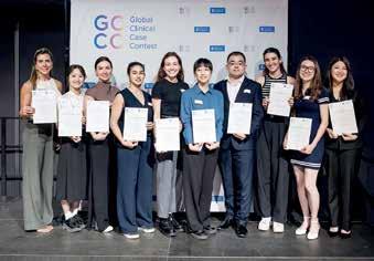
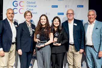
This year, a special competition for young dentists celebrated an important milestone: the final round of the 20th Global Clinical Case Contest (GCCC), a renowned event for dental students, took place in Constance, Germany. Ten finalists presented their challenging restorations, for which they used products from Dentsply Sirona. For the first time in this anniversary year, prizes were awarded in two categories. Six winners were honored in a festive awards ceremony.
Charlotte. The final of the 20th Global Clinical Case Contest (GCCC) – the international Dentsply Sirona student competition acknowledging dental students for achieving outstanding clinical results – was a day packed with events and, for the first time, six proud winners. Around 300 student case studies were submitted this year, about one-eighth on indirect restorations with the majority on direct restorations. This year, for the first time, the two categories were evaluated and awarded separately. Dental students from a total of 108 medical faculties took part.
Ten finalists from eight countries – Brazil, China, Estonia, France, Russia, Taiwan, Thailand, and Germany – made it to the final round at Lake Constance. They presented their esthetic dental restorations to the distinguished international GCCC jury, consisting of Dr. med. dent. Britta Hahn, University Hospital Würzburg (Germany), Polyclinic for Conservative Dentistry and Periodontology, Dr. Ian Cline, dentist at King’s College (Great Britain), and Prof. Gaetano Paolone from the University of Milan (Italy). All submitted cases were complex restorations in the anterior
or posterior region, implemented with equipment and materials from Dentsply Sirona. The finalists of the Global Clinical Case Contest were awarded attractive prizes.
To allow for a better comparison between the candidates’ work, the submissions were divided into two categories for the first time: “direct restorations” and “indirect restorations” with three winners selected in each category.
First prize in the “direct restorations” category went to Sofiia Grindenko from the Russian University of Medicine in Moscow, who presented the case of a 32-years-old male patient who received a direct composite restoration with full covering veneers (Neo Spectra® ST composites, Esthet-X® HD). Second place went to Yiting Chen from the Capital Medical University in Beijing (China) with the case of a female patient who suffered from a fractured central incisor that was treated with a direct composite restoration (Neo Spectra®, Prime&Bond Universal). Third place was awarded to Ying Cheng Lu from the Nation Cheng Kung University in Taiwan (R.O.C.) for a patient case in which a fractured upper central incisor was restored by a direct composite restoration (Spectra® ST resind, Prime&Bond Universal, Enhance Finishing).
In the “indirect restorations” category using CAD/ CAM-Technology, Chuttaporn Dejdumrongwood from the Chiang Mai University of Thailand won the competition with a case study in which a
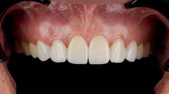
male patient with concerns about an unesthetic smile and spacing in the maxillary anterior region got a new smile with digital manufactured veneers (CEREC TesseraTM, Calibra® veneer, Prime&Bond® Universal). Second place went to Beatrice Julia Popescu from the Johannes Gutenberg University of Mainz (Germany) who impressed the panel with the case of a 19-yearold patient with peg-shaped front teeth and missing mandibular central incisors. He got crowns for 11 and 21 and veneers for 12 and 22, composite build-ups on teeth 13 and 23 and two single-wing adhesive bridges in the lower jaw (veneers: CEREC TesseraTM, bridge: CEREC MTLTM Zirconia). Third place went to Chang Guo from the Fourth Military Medical University of Xi’an, Shaanxi (China). He presented the case of a female patient with discolored teeth compromised by secondary caries and bruxism. She received a restoration with veneers from 12 to 22 (Spectra® ST, Celtra, Prime&Bond Universal, Calibra® Veneer).
The jury praised the high standard of the entries. “It’s remarkable how enthusiastically and precisely the students implemented their cases,” summarized jury member Dr. Britta Hahn. “Each finalist impressively demonstrated how CAD/ CAM workflows and modern materials can be used to benefit patients—both esthetically and functionally—at a very high level.”
The competition also demonstrated continuity among its participants and patrons. Several tutors who had successfully guided their students to the GCCC finals in previous years returned this year with new young talent. “I really appreciate
the support I receive from my university, from Dentsply Sirona, and through this competition,” said Sofiia Grindenko. “Winning is obviously a huge compliment for my work. But it’s also a great motivation to continue striving to achieve the best possible esthetic outcomes for my patients in the future.”
“The Global Clinical Case Contest is an important part of our efforts in supporting young dental professionals early in their careers,” explains Prof. Rainer Seemann, Vice President Chief Clinical Officer at Dentsply Sirona. “We aim to give future dentists confidence in using modern technology and enable them to deliver the best possible care right from the start of their clinical journey. We collaborate with universities around the world to develop innovative training programs to ensure the next generation remains at the forefront of research and clinical practice. This dental student case contest emphasizes Dentsply Sirona’s commitment to transforming dentistry and helping dental professionals move forward with confidence.”
The Dentsply Sirona Global Clinical Case Contest has been held since 2004/2005 and was open to dental students enrolled in a college or university at the time of entering the contest. Participants document their case with direct or indirect restorations including photographic evidence with the support of a tutor from their university.
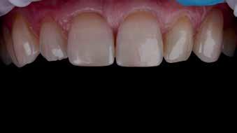
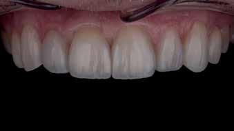
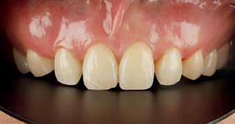
Learn more about Global Clinical Case Contest: GCCC Global Clinical Case Contest I Dentsply Sirona Global

Company name: VOCO GmbH
Company name: VOCO GmbH
Country of origin: Germany
Country of origin: Germany
Website: www.voco.com
Website: www.voco.com
Company name: VOCO GmbH Country of origin: Germany Website: www.voco.com
Company name: VOCO GmbH
Country of origin: Germany Website: www.voco.com
The family-run dental company VOCO located in Cuxhaven (Germany) is one of the leading manufacturers in the industry, both nationally and internationally.
The family-run dental company VOCO located in Cuxhaven (Germany) is one of the leading manufacturers in the industry, both nationally and internationally.
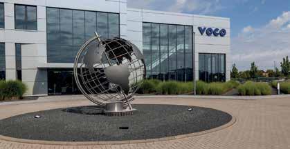
The family-run dental company VOCO located in Cuxhaven (Germany) is one of the leading manufacturers in the industry, both nationally and internationally.
The family-run dental company VOCO located in Cuxhaven (Germany) is one of the leading manufacturers in the industry, both nationally and internationally.
The product portfolio comprises more than 100 preparations, with a focus on preventive, restorative, prosthetic and digital dentistry.
The product portfolio comprises more than 100 preparations, with a focus on preventive, restorative, prosthetic and digital dentistry.
Another 400 employees are responsible for sales worldwide and take care of dentists and depots on site.
Another 400 employees are responsible for sales worldwide and take care of dentists and depots on site.
The product portfolio comprises more than 100 preparations, with a focus on preventive, restorative, prosthetic and digital dentistry.
The product portfolio comprises more than 100 preparations, with a focus on preventive, restorative, prosthetic and digital dentistry.
The products are manufactured at the headquarters and are therefore 100 percent “Made in Germany”. 440 people are employed in the departments of research, production and administration in Germany.
The products are manufactured at the headquarters and are therefore 100 percent “Made in Germany”. 440 people are employed in the departments of research, production and administration in Germany.
Another 400 employees are responsible for sales worldwide and take care of dentists and depots on site.
Another 400 employees are responsible for sales worldwide and take care of dentists and depots on site.
In addition to user-friendliness, the highest safety standards and product quality have top priority at VOCO: Since September 2021, VOCO has been one of the first German dental companies to be fully certified under the European Medical Device Regulation (MDR) in accordance with (EU) 2017/745.
In addition to user-friendliness, the highest safety standards and product quality have top priority at VOCO: Since September 2021, VOCO has been one of the first German dental companies to be fully certified under the European Medical Device Regulation (MDR) in accordance with (EU) 2017/745.
The products are manufactured at the headquarters and are therefore 100 percent “Made in Germany”. 440 people are employed in the departments of research, production and administration in Germany.
The products are manufactured at the headquarters and are therefore 100 percent “Made in Germany”. 440 people are employed in the departments of research, production and administration in Germany.
www.voco.com
www.voco.com
Flowable – and yet packable
Flowable – and

IonoStar Plus is a glass ionomer restorative material with numerous special features. Very easy to extract from the capsule, the material at first has excellent wetting characteristics resulting in optimal marginal adaptation. Its viscosity then changes within a few seconds, making the material malleable for at least one minute without sticking. It thus provides ideal viscosity at every stage of application.
IonoStar Plus is a glass ionomer restorative material with numerous special features. Very easy to extract from the capsule, the material at first has excellent wetting characteristics resulting in optimal marginal adaptation. Its viscosity then changes within a few seconds, making the material malleable for at least one minute without sticking. It thus provides ideal viscosity at every stage of application.
Moreover, IonoStar Plus has a curing time of just two minutes, after which work can continue immediately. This is a valuable advantage, particularly in the treatment of patients with low compliance, such as children.
Moreover, IonoStar Plus has a curing time of just two minutes, after which work can continue immediately. This is a valuable advantage, particularly in the treatment of patients with low compliance, such as children.
In addition to user-friendliness, the highest safety standards and product quality have top priority at VOCO: Since tember 2021, VOCO has been one of the first German dental companies to be fully certified under the European Medical Device Regulation (MDR) in accordance with (EU) 2017/745.
In addition to user-friendliness, the highest safety standards and product quality have top priority at VOCO: Since tember 2021, VOCO has been one of the first German companies to be fully certified under the European Medical Device Regulation (MDR) in accordance with (EU) 2017/745.
Meaning that medical products of class I and Ila manufactured at the Cuxhaven site are always produced and placed on the market in accordance with valid European guidelines.
Meaning that medical products of class I and Ila manufactured at the Cuxhaven site are always produced and placed on the market in accordance with valid European guidelines.
Meaning that medical products of class I and Ila man tured at the Cuxhaven site are always produced and on the market in accordance with valid European guidelines.
Meaning that medical products of class I and Ila man tured at the Cuxhaven site are always produced and placed on the market in accordance with valid European guidelines.
For all inquiries please contact us: m.elfil@voco.com
Simplified shade system throug Cluster-Shades
IonoStar Plus is a glass ionomer restorative material with numerous special features. Very easy to extract from the capsule, the material at first has excellent wetting characteristics resulting in optimal marginal adaptation. Its viscosity then changes within a few seconds, making the material malleable for at least one minute without sticking. It thus provides ideal viscosity at every stage of application.
IonoStar Plus is a glass ionomer restorative material with numerous special features. Very easy to extract from the capsule, the material at first has excellent wetting characteristics resulting in optimal marginal adaptation. Its viscosity then changes within a few seconds, making the material malleable for at least one minute without sticking. It thus provides ideal viscosity at every stage of application.
• Perfect marginal adaptation and packability in one product, thanks to a change in viscosity during application
• Perfect marginal adaptation and packability in one product, thanks to a change in viscosity during application

Admira Fusion 5 uses five different Cluster-Shades, each bundling several VITA® classical shades. This provides adaptation to the natural colour gradient of the tooth as well as more efficient and easy shade selection.
Admira Fusion 5 uses five different Cluster-Shades, each bundling several VITA® classical shades. This provides adaptation to the natural colour gradient of the tooth as well as more efficient and easy shade selection.
• The first glass ionomer material tooth-like fluorescence
• The first glass ionomer material with tooth-like fluorescence
• High level of fluoride release
• High level of fluoride release
• The new capsule design reaches smaller cavities and difficult-to-access areas of the mouth
• The new capsule design reaches smaller cavities and difficult-to-access areas of the mouth
Thanks to the simplified shade system, all 16 VITA® classical shades can be covered with only five Cluster-Shades Multi-shade layering is not necessary.
Thanks to the simplified shade system, all 16 VITA® classical shades can be covered with only five Cluster-Shades Multi-shade layering is not necessary.
Moreover, IonoStar Plus has a curing time of just two minutes, after which work can continue immediately. This is a valuable advantage, particularly in the treatment of patients with low compliance, such as children.
Moreover, IonoStar Plus has a curing time of just two minutes, after which work can continue immediately. This is a valuable advantage, particularly in the treatment of patients with low compliance, such as children.
• Fast setting time of only 2 minutes from placement of the filling
• Fast setting time of only 2 minutes from placement of the filling
• The first glass ionomer material with tooth-like fluorescence
• The first glass ionomer material with tooth-like fluorescence
• High compressive strength and abrasion resistance
• High compressive strength and abrasion resistance
This means that you are optimally positioned for your everyday practice with only five shades and can thus considerably streamline your inventory. This enables a better overview, and incorrect stocks are a thing of the past
This means that you are optimally positioned for your everyday practice with only five shades and can thus considerably streamline your inventory. This enables a better overview, and incorrect stocks are a thing of the past
• all VITA classical shades can be covered with just 5 Cluster-Shades – there is no need for multiple layering
• all VITA classical shades can be covered with just 5 Cluster-Shades – there is no need for multiple layering
• Perfect marginal adaptation and packability in one product, thanks to a change in viscosity during application
• Perfect marginal adaptation and packability in one product, thanks to a change in viscosity during application
• High level of fluoride release
• High level of fluoride release
• The new capsule design reaches smaller cavities and difficult-to-access areas of the mouth
• The new capsule design reaches smaller cavities and difficult-to-access areas of the mouth
• significantly reducing your inventory
• significantly reducing your inventory
• new resin matrix enables enhanced chameleon effect through optimised light scattering
• Fast setting time of only 2 minutes from placement of the filling
• Fast setting time of only 2 minutes from placement of the filling
• High compressive strength and abrasion resistance
• High compressive strength and abrasion resistance
• new resin matrix enables enhanced chameleon effect through optimised light scattering
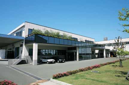
Company name: Takara Belmont Corporation
Country of origin: Japan
Website: https://dental.takarabelmont.co.jp/
Takara Belmont has been contributing to people's wish to stay healthy and beautiful through a century of our history. Founded in Japan in 1921, and we drew on our hydraulic technology and began manufacturing dental chairs in 1960s. Committing to the healthy life not only for patients, but also for dentists & staff, our products are designed and developed to support dentist, forging a bond of trust with their patients. Based on the spirit of Japanese manufacturing, each Belmont chair is the result of more than 50 years of product research and development, meticulous engineering expertise and innovative production methods. Through our worldwide network of offices and subsidiaries, Belmont continually monitors current industry and market needs as we strive to design and introduce high quality, creative products that help increase work efficiency and productivity.
BAHRAIN
MEDICAL & PHARMACEUTICAL
SERVICES CO. W.L.L. Road 13, Block 711 – Building 111 Phone: +973 17715785 bashirco@bashirco.com.sa
EGYPT
SAFWAN EGYPT
41 Lebanon St., Mohandessen, Giza
Phone: +20 2 33031796/ +20 2 33040208 info@safwanegypt.com
IRAN
ARDIN CO.
2nd Floor, No 86, Darya Blvd, Saadat Abad, Tehran
Phone: +98 21 8873004 ardincommercial2020@gmail.com
IRAQ
TECHNICAL MEDICAL CORNER
Bab Al-Moatham St. Erbil-40st Near Aya Moll, Bagdad
Phone: +964 7708386000 +964 770175573 info@tmc-iraq.com
JORDAN
NEW STAGE CO.
Mecca Street, Building No. 147, First Floor Ammam
Phone: +962 6 553 3270
basil.abuissa@newstagejo.com
KENYA DENTMED KENYA LTD.
Woodvale Grove,Westlands, P.O.Box 43873
Phone: +254 2 04 44 53 07 info@dentmedkenya.com
KUWAIT
AL-SAYAFE MEDICAL & PHARMACEUTICAL SUPPLIES CO. W.L.L Al-Arabia Tower, 18th Floor Ahmed Al-Jaber Street Phone: +965 69028580 alsayafe@alsayafemedical.com
OMAN
MEDICAL& PHARMACEUTICAL SERVICES CO. L.L.C 209 Postal Code 116 Muscat Phone: + 968 24567561 info@bashirco.com.sa
AL FARSI NATIONAL ENTERPRISES LLC
(government sector only) Flat No:4, Way No:3521, BLDG No:1992,Block No:235. Post Box:158, Al Khuwair Street Al Khuwair, Muscat Phone: + 968 24485625 director@alfarsi.me
QATAR
BASHIRCO TRADING MEDICAL SERVICES W.L.L.
Alkholaifi Building, RoomNo.4, Second Floor, Salwa Road Building, No.361, Street 340, Zone 56, Doha Phone: + 974 66419549 bashirco@bashirco.com.sa

SAUDI ARABIA
MEDICAL & PHARMACEUTICAL SERVICES BASHIR SHAKIB AL JABRI & CO. LTD P.O.BOX 9584 - Jeddah 21423
Phone: +966 1 26 70 04 30 bashirco@bashirco.com.sa
SUDAN
RASD PHARMA MULTI ACTIVITIES CO. LTD. No.761, Block 22, Altaif, Khartoum Phone: +249 155 141414 Info@rasdtec.com
SYRIA BH CO. DENTAL SUPPLIES
Syria-Damascus Al-Jusser Al-Abeead next to France Embassy Phone: +963 11 3333879 basel.hashem.co@gmail.com
TUNISIA MED PRO TUNISIA 19 Rue Tarek Ben Moussa Khaznadar 2017 Tunis
Phone: +216 21 65 16 32 medpro@laposte.net
TURKEY
LIDER DIS ITH. IHR. SAN. VE TIC. A.S. Fevzi Cakmak Sokak No:11/5 Kizilay Ankara
Phone: +90 31 22 31 64 85 lider@liderdis.com.tr
UNITED ARAB EMIRATES CITY PHARMACY CO. Office No. 901, 9th Floor, Al Otiba Tower,Shaik Hamdan Street, P.O.Box 2098, Abu Dhabi Phone: +971 2 6323016 headoffice@citypharmacyco.ae
Takara Belmont opened a representative office in Dubai, UAE in May to strengthen its business development in the Middle East region. Dubai is a hub for logistics, finance, and information, and is also a city that actively attracts global companies. Through this new sales base, we aim to strengthen our sales and marketing capabilities in the dental market in the Middle East and achieve the highest level of customer satisfaction in order to further expand its overseas business.
Outline of new office
Name: TAKARA BELMONT MIDDLE EAST(BRANCH)
Location: Office # 6WA 625, 6th Floor, West Wing, Dubai Airport Free Zone, Dubai, UAE
Branch Manager: Takahiro Horiguchi
Opening Date: May 1, 2024
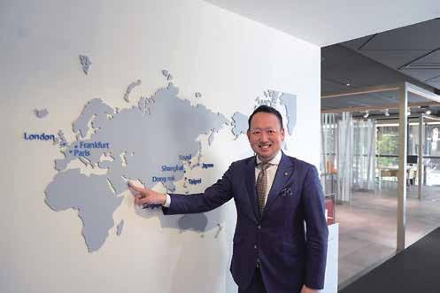

The hydraulic mechanism of the chair ensures smooth and quiet movement of the joints and provides a comfortable feeling for patients. One of the characteristics, touch screen interface has been designed to improve efficiency and reduce stress during treatment. When a handpiece is chosen and picked up, the screen displays only necessary information, helping the dentist concentrate on the treatment. EURUS Unit with a new option, "Built-in X-ray" is shown in AEEDC Dubai 2025, so we welcome your visit at our Belmont booth.
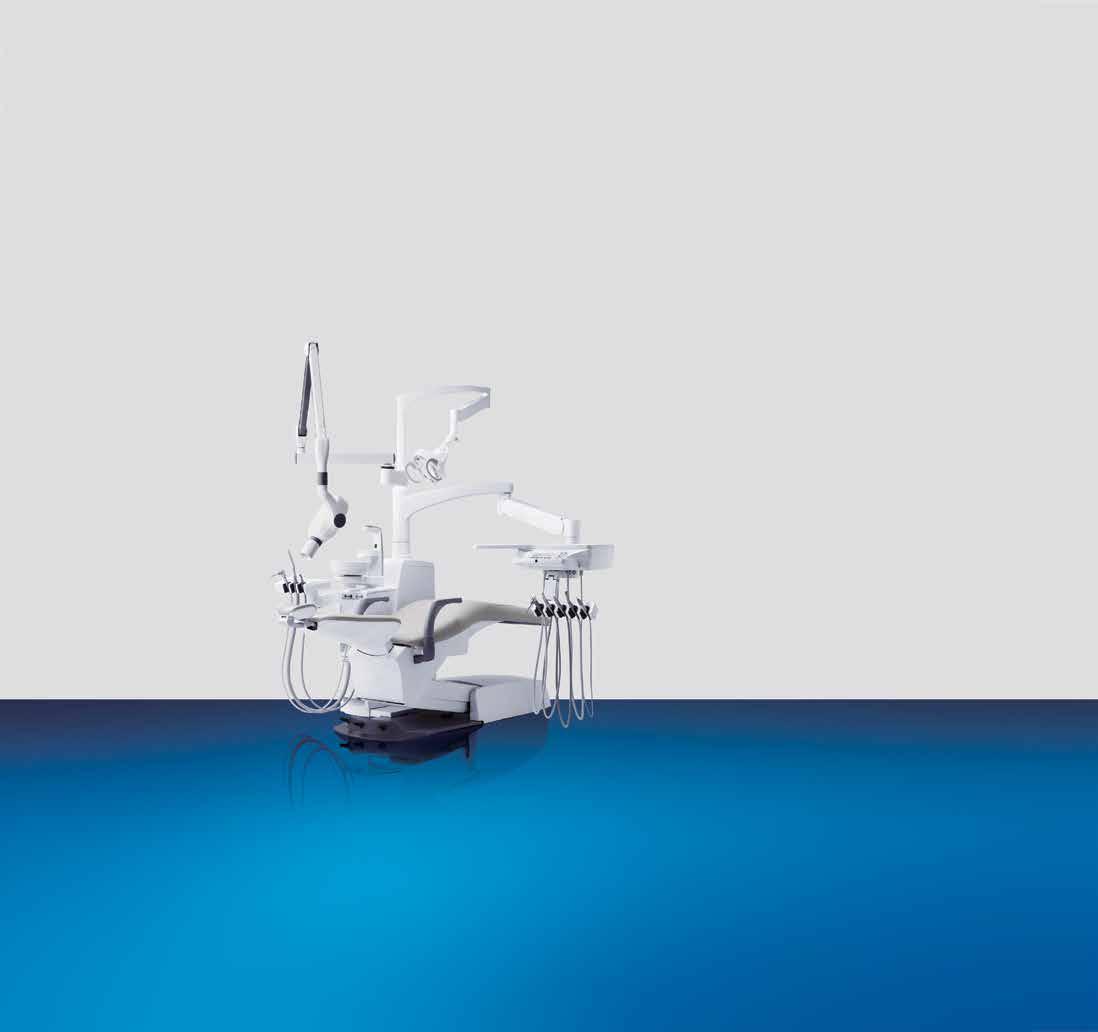
Two-day global event to spotlight multidisciplinary digital dentistry and foster international collaboration
DARMSTADT, Germany – August 15, 2025 – exocad, an Align Technology, Inc. company and a leading dental CAD/CAM software provider, today announced its upcoming global event, Insights 2026, to be held April 30–May 1, 2026, in Palma de Mallorca, Spain. With the inspiring motto “Calling all heroes,” the event will spotlight multidisciplinary digital dentistry, bringing together dental professionals from around the world to explore the latest innovations and collaborative workflows in digital dentistry.
The two-day program will offer a high-quality educational experience, featuring lectures from renowned international speakers and exocad experts, as well as indepth breakout sessions tailored to the needs of dental technicians and clinicians. Attendees will have the opportunity to explore a vibrant Innovation Expo showcase with solutions and product innovations from more than 50 companies in the dental industry.
“Insights 2026 is about bringing the global dental community together in one incredible event,” said Novica Savic, CCO and managing director at exocad. “With many thoughtleading speakers and the vast array of partners on board, this event will be more diverse and inspiring than ever. We’re expecting more than 850 digital dentistry heroes
to join us in Palma for two unforgettable days of learning, networking, and dental excellence.”
At Insights 2026, a dynamic lineup of global experts will deliver practical, futurefocused perspectives across every facet of digital dentistry. Caroline Kirkpatrick, Scotland, a clinical dental technician, will challenge traditional thinking as she invites crown and bridge technicians to reconsider the potential of removables. Digital designer and Independent Certified Trainer for exocad, Seth Potter, Canada, will reveal how smile design can be a powerful driver of business growth. Dr. Zhiqiang Luo, China/USA, dentist and clinical instructor at the University of Michigan School of Dentistry, will highlight the transformative advantages of the digital patient, while Prof. Marco Tallarico, Italy, dentist and assistant professor in dental prosthesis at the University of Sassari, will present an interdisciplinary workflow integrating exocad ART and exoplan. Lukas Wichnalek, Germany/ Mexico, dental technician and founder of HIGHFIELD.DESIGN, will introduce a standardized method for patient data acquisition, emphasizing esthetic precision and seamless collaboration between the lab and clinic through TruSmile™ tools. Rounding out the program, Dr. Alexis Ioannidis, Switzerland, head of fixed reconstructive dentistry at the University of Zurich, and dentist Michaela Sehnert, Germany, will explore how communication-centered workflows and the fully integrated digital platform
from exocad can elevate both patient outcomes and practice profitability.
For the first time, exocad is introducing a partner country for Insights 2026. exocad will spotlight a different country each Insights, fostering international exchange, celebrating diversity and highlighting unique regional perspectives in digital dentistry. China has been selected as the partner country for Insights 2026.
exocad will start ticket sales on September 1, 2025, and offer a limited number of super early bird tickets at a discount of 299 euros plus VAT until October 31, 2025, and will have special deals for dental teams who purchase together, as well as for students. Tickets will include access to all congress activities and the evening celebration, themed “exoGlam night” offering a unique networking experience.
Further information and event updates are available at exocad.com/insights2026.
exocad GmbH
Natalia Gonsior
Marketing & PR Manager
Rosa-Parks-Straße 2 64295 Darmstadt, Germany
Phone +49 (0)6151 6294132 ngonsior@exocad.com exocad.com
exocad announced its upcoming global event, Insights 2026, to be held again on April 30 and May 1, 2026, in Palma de Mallorca, Spain. (Source: exocad)

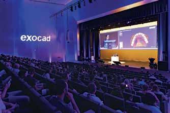
About exocad
exocad GmbH, an Align Technology, Inc. company, is a leading dental CAD/CAM software provider. exocad vigorously pushes the boundaries of digital dentistry, providing flexible, reliable, and easy-touse CAD/CAM software for dental labs and dental practices worldwide. More than 70,000 valued customers plan implants and create functional and refined restorations with exocad’s DentalCAD, ChairsideCAD and exoplan software. exocad and DentalCAD, among others, are trademarks of exocad GmbH or one of its subsidiaries or affiliated companies, and are registered in the U.S. and other countries. For more information and a list of exocad reseller partners, please visit exocad.com.
I-View Gold Intraoral sensor
Thanks to the advanced calibration tool, the I-View Gold captures high-definition images without any filters. Calibration files are used to remove any imperfections during acquisition, reducing noise and enhancing detail. As a result, the images captured by the I-View Gold require no further processing, ensuring reliable images, accurate diagnosis and time efficiency.
- DICOM compatibility
- TWAIN option
- Multiuser connection
Bonascan Bite Air is a scannable medium viscosity vinyl polysiloxane (VPS) material for bite registrations and is ideal for dentists who prefer traditional impression methods but who want to take advantage of the benefits of digital technology. Bonascan Bite Air is characterized by its mousse-like consistency, making it very light on extrusion. Due to its high thixotropy (non-drip), it ensures accurate placement and enhanced patient comfort. It is available in three setting times and retains enough flexibility for easy trimming or cutting.
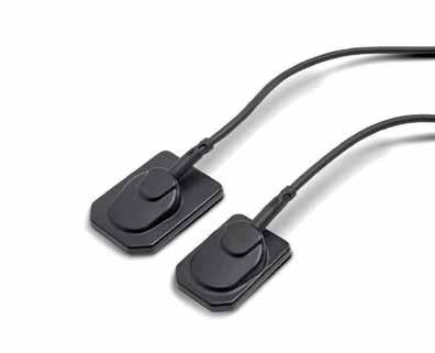
- Bridge feature for connecting to other dental management software.
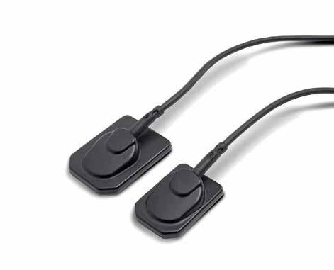
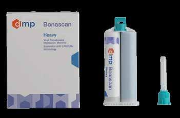
trident-dental.com
www.dmpdental.com
Etch, prime and bond in one step!
The trend in the development of adhesive systems is to improve adhesion while simplifying clinical manipulation steps. Along with that, the use of universal adhesives has been increasing and its performance has been reported to be clinically sufficient.
Nanoceram-Bright Flow is a light curing flowable version of the Nanoceram-Bright composite using the same nanohybrid technology to achieve optimum results. It is characterized by its superior aesthetics through its chameleon effect and high gloss polishability. This highly thixotropic product guarantees easy handling and precise application.
Indications
Anterior and limited posterior restorations
Liner for class I and II restorations
Sealing larger pits and fissures
Repair of composite restorations and ceramic veneers
Splinting of mobile teeth
Blocking out of undercuts
Characteristics - Advantages
Excellent handling characteristics
Excellent sculptability
Low shrinkage-Very good marginal adaptation
Extremely high polishability
High resistance to wear
Superior aesthetics
Strongly radiopaque
www.dmpdental.com
With BeautiBond Xtreme, SHOFU continues to evolve the latest generation of a true universal adhesive. By introducing a new type of Acid Resistant Silane coupling agent (ARS), a chemical composition with excellent stability has been designed to allow the surface treatment of a wide range of adherends with an easy-to-use system. BeautiBond Xtreme is a light-curing, self-etching all-in-one universal adhesive for bonding direct and indirect restorations. It has “inherited” the excellent properties of the former BeautiBond Universal bonding system, such as bond strength, low technique sensitivity, convenient application and simplified one-step application procedure.
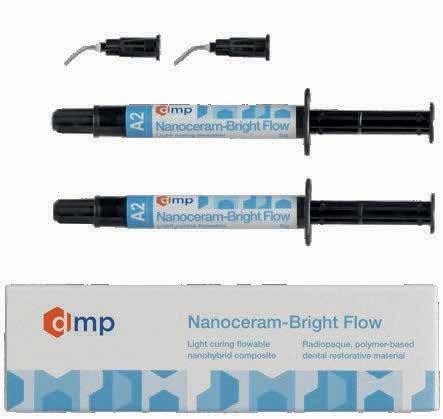
Thanks to the new silane bonding agent ARS, BeautiBond Xtreme bonds to enamel, dentine and various indirect restorative materials (composite, precious and non-precious alloys, glass-ceramics, alumina and zirconia). An additional primer is not
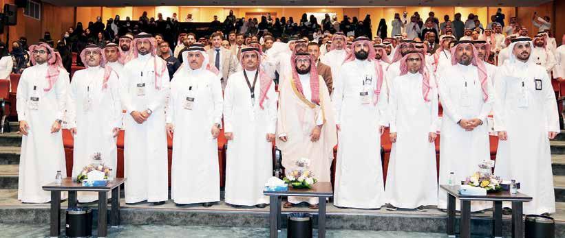
Sep 4-5 2025
Convention and Exhibition Center, King Khalid University, Abha, Saudi Arabia
The Future of Dentistry at the SAPI Abha Expert Forum.
Distinguished international experts participated in a prestigious forum promoted by the Saudi Society of Periodontology and Implantology (SSPI), where SEPA served this year as scientific partner. The Saudi Advanced Periodontics & Implantology (SAPI) Expert Forum, held at the Convention and Exhibition Center in Abha, Saudi Arabia, featured a keynote lecture by Dr. Erik Regidor Correa.
Under the title “Saudi Advanced Periodontics & Implantology: Expert Forum on the Adoption of Digital Implantology: The Future of Dentistry,” this gathering in Abha placed SEPA in a leading role, with Dr. Regidor as its principal representative. The contributions of digital applications in periodontics and implantology were the central theme of the event. In his lecture, Dr. Regidor introduced new and more versatile resources and tools in implantology, focusing on the history of autotransplantation and the analog workflow, while underscoring the benefits of digitalization and digital autotransplantation.
This forum of experts brought together professionals from a wide range of disciplines, including dental technology, oral surgery, general dentistry, periodontics, and implantology. It also highlighted the fruitful alliance and strong collaboration between SSPI and SEPA.
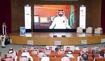
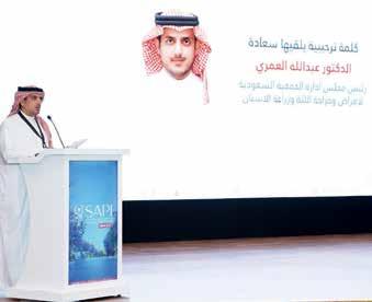
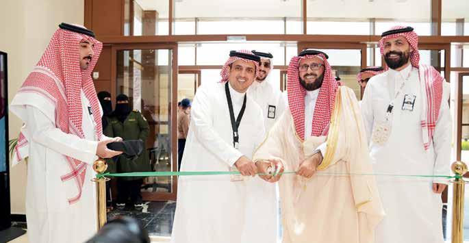
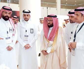
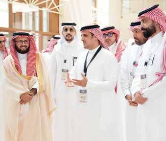
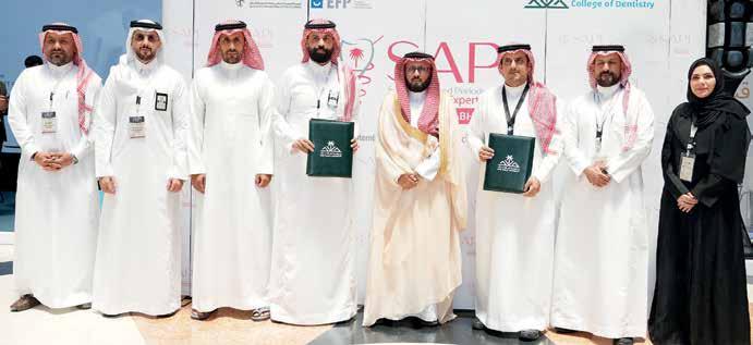
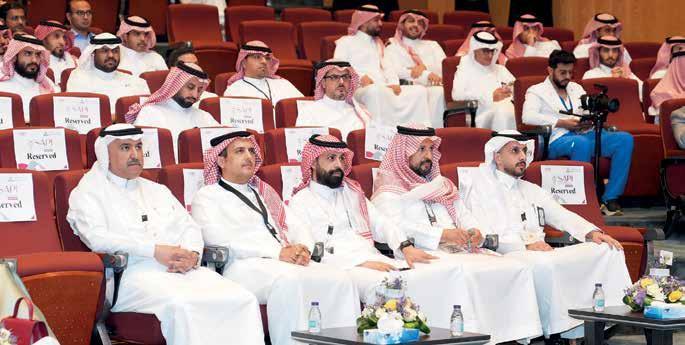

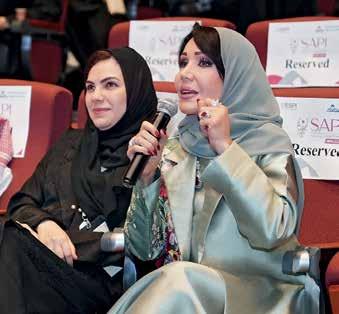
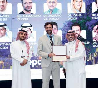
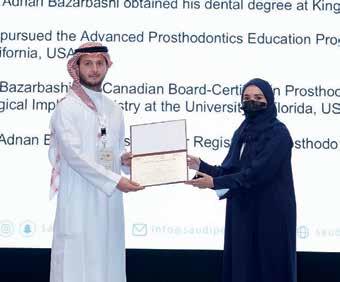
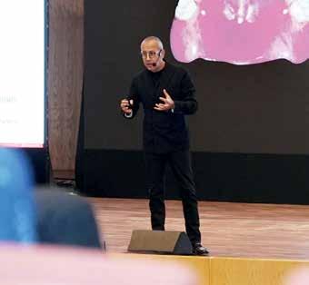
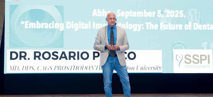
National Exhibition and Convention Center
Shanghai China 9-12 September, 2025

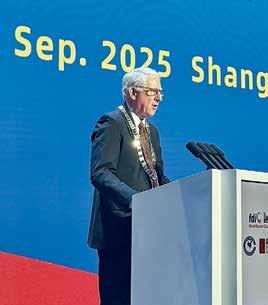
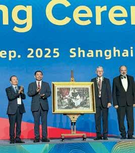

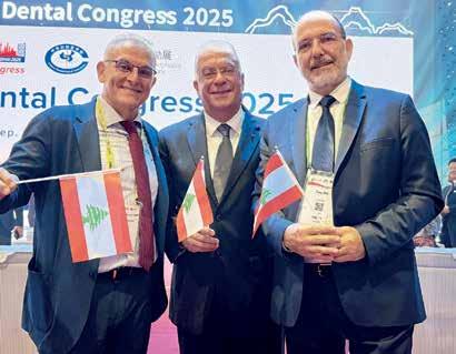
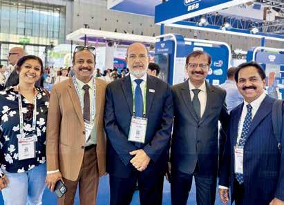
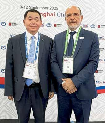
Dr. Guo, we are in ShanGhai at the national Convention anD exhibition Center. DurinG the openinG Ceremony, you mentioneD the viSion 2030 for healthCare in China. what about the Dental fielD?
we have a five-year Plan DeviseD by the Government we have a meDium- anD lonG-term Plan for the Prevention anD treatment of ChroniC Diseases in China from 2017 to 2025. it was issueD by the state CounCil in 2017. the Plan Promotes a healthy lifestyle anD foCuses on Prevention we refer to the “three reDuCtions” that imPaCt Caries anD the “three healthy aCtions.” the three reDuCtions are: reDuCtion of salt, reDuCtion of fat anD oil, anD reDuCtion of suGar. suGar is very Closely relateD to Caries. the three healthy aCtions are: a healthy mouth, healthy boDy weiGht, healthy bones, inCluDinG the maxilla, manDible, anD the whole boDy bones. so, the three reDuCtions anD three healthy aCtions are all about health Promotion. there is also a national Plan CalleD the national
interview with Dr. Chuanbin guO, presiDent Of the Chinese stOmatOlOgiCal assOCiatiOn (Csa)
nutrition Plan (2017–2030), issueD by the state CounCil in 2017. it Promotes nutritional transformation anD uPGraDes fooD ProCessinG anD the CaterinG inDustry. it emPhasizes suGar intake Control
aDDitionally, there is a Government PoliCy CalleD the healthy China initiative (2019–2030), issueD by the state CounCil in 2019. it tarGets major Diseases anD foCuses on key PoPulations. altoGether, 15 major initiatives have been imPlementeD, inCluDinG the healthy Diet initiative, whiCh ProviDes Dietary GuiDanCe anD reCommenDations this is the Plan you may have mentioneD: China’s oral health aCtion. the first aCtion is to strenGthen oral health eDuCation, whiCh is very imPortant to raise PubliC awareness about oral health. eDuCation for the PubliC is CruCial.
reGarDinG oral health eDuCation, what iS the role of the DentiSt?
first, sChools Play a very imPortant role. this is CalleD institutional resPonsibility. Professional institutions ComPile anD Promote materials, train the workforCe, anD Carry out a life-CyCle health eDuCation ProGram for stuDents, esPeCially ChilDren i Can tell you there are two very imPortant Dates one is marCh 20th—an fDi aDvoCaCy Day. in China, sinCe 1989, the Csa anD the Government have establisheD a “love teeth Day” on sePtember 20th. these Days are very imPortant for oral health eDuCation for the PubliC. but aCtually, eDuCation is not limiteD to just one Day, it extenDs to an entire week. our stuDents anD Csa members visit kinDerGartens anD sChools to eDuCate ChilDren.
what about teaCherS at SChool?
we train teaChers as well. teaChers shoulD first know how to Preserve health. it’s not a one-Day teaChinG; it is a Continuous effort beCause ChilDren Choose habits for GooD anD maintain a healthy
lifestyle. so, we teaCh teaChers how to maintain oral health ProPerly, anD we also teaCh Parents the Csa eDuCates fathers anD mothers. our Dental sChools anD Dental hosPitals also ConsiDer this their resPonsibility.
So, aS a patient, when i Go to the hoSpital, will the DentiSt teaCh me?
yes, but for a short time. when Patients arrive at the hosPital, there are eDuCational viDeos in the waitinG rooms that they Can watCh to learn how to stay healthy.
as for me, i’m a maxillofaCial surGeon sPeCializinG in onColoGy. oral CanCer is very imPortant in my fielD. i visit tv stations anD meDia outlets like yours, Dental news, to sPeak about how to Prevent CanCer, DeteCt early CanCer, anD DiaGnose it early with new teChnoloGies like Cameras, oral sCanners, anD artifiCial intelliGenCe, breakthrouGhs helP Dentists DeteCt early CanCer anD other Diseases.
are theSe teChnoloGieS available now?
yes, some are available, but i think they are still at a Primary staGe. the more imPortant thinG is that Dentists are not onColoGists they neeD to learn how to iDentify susPiCious lesions at sChool however, after GraDuation, many Dentists Do not see these Patients often anD may forGet their traininG. that’s why ContinuinG eDuCation is CruCial. DurinG reCent sessions, we haD a session DeDiCateD to oral CanCer.
reGarDinG toolS from inDuStry, iS there any new DeviCe for early oral CanCer DeteCtion?
some DeteCtors use ai teChnoloGy to analyze imaGes. the Camera senDs PiCtures to the ClouD, where DiaGnostiC aPPliCations analyze anD DistinGuish lesions
iS thiS wiDely available toDay?
not yet. it’s still in the early staGes, not extensively useD, but we exPeCt the use of these teChnoloGies will GraDually inCrease. returninG to oral health Prevention anD eDuCation, you tarGet ChilDren anD teaChers anD imPlement meDia CamPaiGns in hosPitals.
even the Private seCtor PartiCiPates; they also ProviDe eDuCational materials for Patients.
what about the partiCipation of CompanieS that manufaCture toothpaSte anD mouthwaSh?
they also PartiCiPate in the CamPaiGns. ComPanies that ProDuCe toothPaste anD brushes join us. the Private seCtor Collaborates with Government initiatives in oral health
what about fluoriDe uSe in China?
fluoriDe is useD in toothPaste anD mouthwash is there water fluoriDation?
we imPlement toPiCal fluoriDe aPPliCations.
who iS reSponSible for that? hoSpitalS or private CliniCS?
we have a Government-suPPorteD ProjeCt that Gives us finanCial suPPort. the ProjeCt is ConDuCteD by the Csa anD has been aCtive for more than 18 years sinCe 2008. it’s a larGe ProjeCt CalleD the national oral health ComPrehensive Prevention ProGram for Chinese ChilDren. it Covers over 16 million ChilDren annually in more than twothirDs of the Counties in China. it inCluDes health eDuCation for Parents, teaChers, anD ChilDren, reGular examinations, toPiCal fluoriDe aPPliCations, anD fissure sealants. at the hosPital level, ChilDren must be brouGht to hosPitals for treatment. also, our Private seCtor suPPorts this ProGram. some use Portable DeviCes to visit sChools anD kinDerGartens. the ChilDren Don’t neeD to Pay. it is Government-suPPorteD. our DoCtors anD nurses Perform this work as volunteers. this ProjeCt is manaGeD by the Csa, anD every ProvinCial Dental assoCiation has an offiCe to manaGe the ProGram. all funDinG Comes from the Government.
reGarDinG thiS fDi meetinG, Do you know the number of loCal partiCipantS?
we have international attenDees, but many Come to visit anD buy at the exhibition. we estimate arounD 40,000 visitors. Professionals attenD leCtures anD visit CommerCial exhibitions
thank yOu very muCh, Dr. guO.
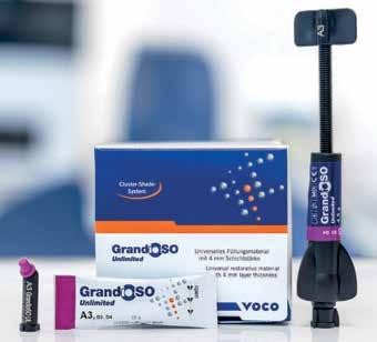
While aesthetic universal composites have to be layered and polymerised in 2 mm increments, which is complicated, bulk-fill composites offer the advantage of application into the cavity in increments of up to 4 mm. However, due to their insufficient aesthetics, bulk-fill composites are normally only used for the posterior region. VOCO has now resolved this issue with the new universal composite GrandioSO Unlimited. The aesthetic material can be applied into the cavity in 4 mm increments and is indicated both for the anterior and posterior regions. As a result, dental practices now only need one packable universal composite for their day-today work! With this extension to the GrandioSO product family, tried and tested for more than a decade, VOCO is setting a new standard for universal composites.
The universal composite without layers thanks to visual transformation technology (VTT)
In an uncured state, the material is so translucent that it enables a reliable depth of
cure of 4 mm. During light polymerisation, a visual transformation (visual transformation technology) takes place, whereby the translucency of the material visibly decreases and the desired shade is achieved at a level of opacity that matches the natural tooth. The result is an aesthetic material for the anterior and posterior regions, which can be applied into the cavity in layers of 4 mm thanks to this special technology.
cluster shades
In addition to universal use and the usual simple handling, GrandioSO Unlimited offers further attractive advantages: thanks to the simplified shade system, the 5 cluster shades are able to cover all 16 classic VITA shades. The optimised light scattering leads to a targeted and intensified chameleon effect, ensuring perfect adaptation of the restorative material to the natural tooth shade. As a result, dental practices are ideally equipped for their day-today work with just five shades and can reduce their inventories. The cluster shades A1-A3.5 that are frequently used require a polymerisation time of just 10 seconds. With an impressive filler content of 91%, GrandioSO Unlimited is setting new standards in the high-performance composites class when it comes to stability and strength. The low volume shrinkage of 1.44% and the resulting low shrinkage stress also reduce the risk of poor marginal integrity and marginal discolouration, and contribute to topquality restorations.
GrandioSO Unlimited is compatible with all conventional bonding agents and is available in both syringes and caps.
www.voco.dental
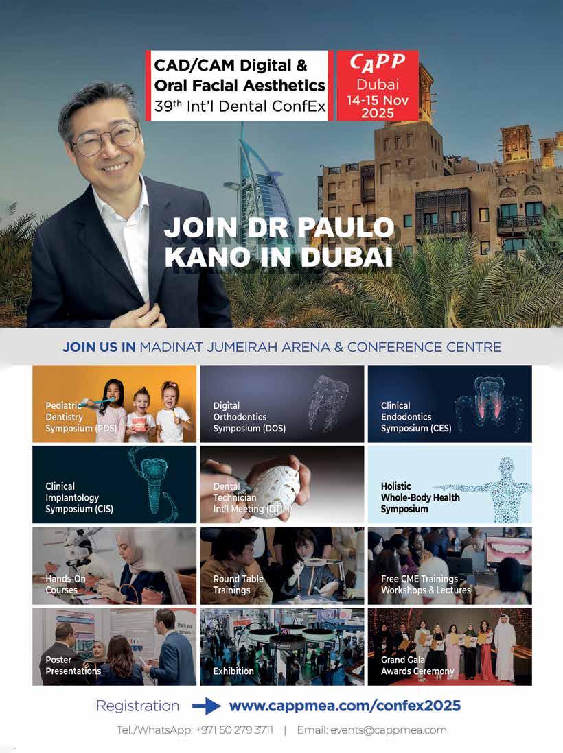
World Dental Congress, NECC
Shanghai China 9-12 September, 2025

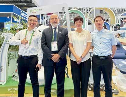
interview with Dr. nikOlai sharkOv, presiDent, fDi wOrlD Dental feDeratiOn, fOr Dental news
Dr. Sharkov, ConGratulationS on your eleCtion. what oral health poliCieS Do you believe are paramount for implementation Globally?
thank you. my tenure with fDi sPans over 23 years, ProGressinG from DeleGate for bulGaria throuGh roles as Committee member, viCe-Chair, Chair, CounCilor, anD treasurer. it is an immense Professional anD Personal honor to serve as PresiDent, PartiCularly rePresentinG bulGaria on the Global staGe
in oral health PoliCy, the foremost Priority is the inteGration of oral health within General health systems. oral anD systemiC health are intimately ConneCteDinteGrative PoliCy is therefore essential. historiCally, oral health was siloeD within health PoliCy; however, Current eviDenCe Demonstrates unequivoCally that oral Diseases share Common risk faCtors, unDerlyinG meChanisms, anD imPaCts with other non-CommuniCable Diseases (nCDs). henCe, all 134 fDi member Countries anD over 200 national Dental assoCiations must aDvoCate that oral health reCeive Parity with other health ConDitions within national strateGies
our DisCiPline has shifteD lanGuaGe-from “Dental” to “oral” health-refleCtinG a more ComPrehensive unDerstanDinG. the oral Cavity ComPrises Diverse anatomiCal struCtures beyonD teeth, inCluDinG the GinGiva, muCosa, tonGue, salivary GlanDs, anD osseous tissues, all of whiCh Contribute to funCtions of mastiCation, sPeeCh, swallowinG, anD overall well-beinG. the fDi’s exPanDeD Definition unDersCores this ComPlexity
oral health’s reCoGnition as an nCD within the frameworks of the who anD uniteD nations has aDvanCeD multi-seCtoral aPProaChes to Prevention, health Promotion, anD PoliCy DeveloPment. the inClusion of oral health in the banGkok DeClaration anD Global aCtion Plans Creates new avenues for interProfessional Collaboration, surveillanCe, anD resourCe alloCation
how DoeS fDi’S Global aDvoCaCy enGaGe poliCy makerS anD aliGn with who’S viSion 2030?
effeCtive health PoliCy is PreDiCateD on PolitiCal will. as PresiDent, i Consistently emPhasize that it is inCumbent uPon all Dental Professionals anD leaDers to enGaGe DireCtly
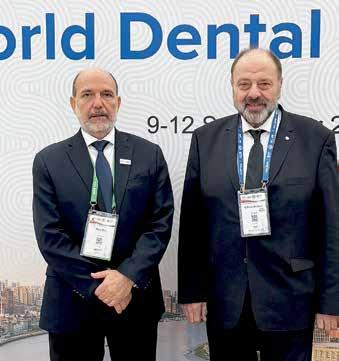
with PoliCymakers, leGislators, anD funDinG boDies translatinG sCientifiC eviDenCe anD burDen-of-Disease
Data into aCtionable PoliCy DemanDs sustaineD aDvoCaCy by national assoCiations anD the fDi at suPranational levels suCh as the un General assembly, the worlD health assembly, anD who teChniCal Committees
fDi’s vision 2030 suPPorts the who’s Global oral health aCtion Plan, aDvoCatinG for inteGrateD, PersonCentereD, anD Preventive moDels of Care, workforCe sustainability, anD DiGital health innovation. our vision is that oral health is reCoGnizeD as a funDamental ComPonent of overall health anD that universal health CoveraGe inCluDes essential oral Care serviCes.
we have suCCessfully inCorPorateD fDi’s PersPeCtives into international PoliCy DeClarations, most reCently in the uniteD nations’ nCD aGenDa. fDi will Continue to foster allianCes anD Contribute to hiGh-level summits, suCh as the uPCominG un Global summit on nCDs, aDDressinG shareD risk faCtors suCh as suGar ConsumPtion to mitiGate oral anD systemiC Disease ConCurrently in ClosinG, sustainable imProvements in Global oral health require Persistent, eviDenCe-baseD aDvoCaCy, interDisCiPlinary CooPeration, anD onGoinG enGaGement with PoliCy makers. fDi is CommitteD to aDvanCinG the sCienCe anD art of Dentistry in serviCe of PoPulation health worlDwiDe.
thank yOu anD gOOD luCk Dr. nikOlai sharkOv




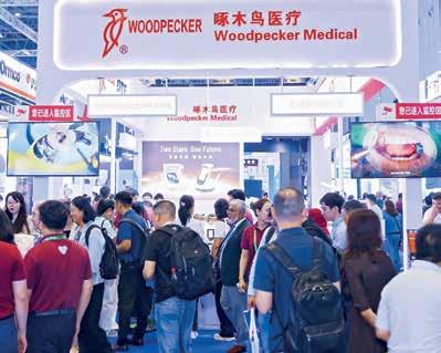

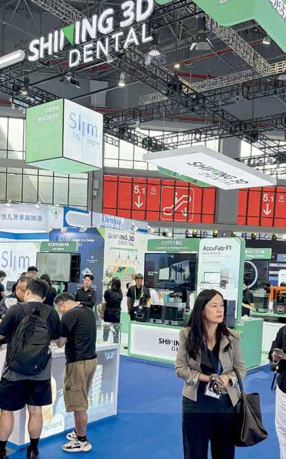

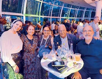


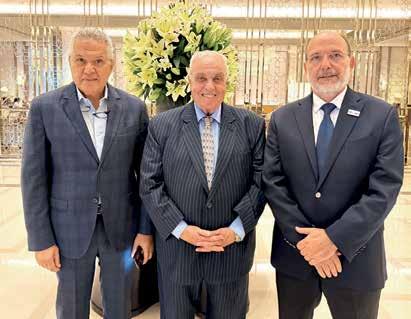
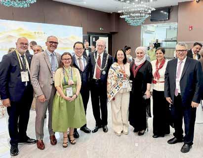
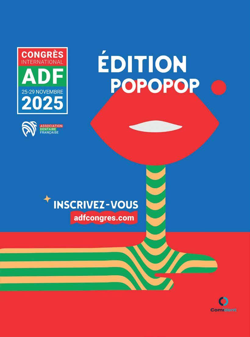


See you at our booth 7F01 19 – 21 January 2026
Dubai World Trade Center, UAE German Pavillion.
Founded in 1886, Dentaurum is the oldest dental company worldwide that has existed continuously.
We have been in the dental industry since the first day – and our company is still family-owned. This is both an obligation and an incentive for the management and for employees.
“Engineered and made in Germany”, at our company headquarters in Ispringen in the Black Forest – in the heart of the technology region of Baden-Wuerttemberg – more than 8,500 brand products are developed and produced in our highly complex, modern production facilities. As one of the few manufacturers

and full-range providers, Dentaurum, together with its 8 affiliates, sets standards in the fields of orthodontics, dental prosthetics, implantology and ceramics, in more than 130 countries worldwide. The company belongs therefore to the 5 % segment of the largest manufacturers of dental products in Germany.
We have unified environmental management, quality management as well as health and safety into one management system. It has always been a matter of course for us that our employees, more than 600 in number worldwide, have equal opportunities and are treated with respect.

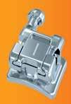


COMING SOON:
IT´S EASY #CLICK & GO! discovery® smart sl discovery® smart sl is the new, innovative addition to the internationally successful premium bracket line discovery®: it is passively self-ligating and smart to handle. The high material quality and reliable opening / closing function are convincing features of the bracket.


Skeletal anchorage with the tomas® -system for all indications


The tomas ® -pin head serves as an anchorage point for the various coupling elements to move teeth or to support tooth movement.


The connection of the future –Conical and platform united
Always the right implant, regardless of whether a conical or a platform connection is required: with every tioLogic® TWINFIT implant, the implantologist and the patient benefit from the freedom to choose at any time between conical and platform. See for yourself!

Elegance. Precision. Premium-Line Pliers. Whether for fixed or removable orthodontics, for use in the dental office or laboratory, the wide portfolio covers all fields of application. Discover the high quality of the Premium-Line pliers from Dentaurum and discover the benefits of the products for yourself.
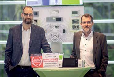
When asked which topics are particularly promising for future patents, Thomas Lang comments: “Digitisation is becoming increasingly important in dentistry, as is artificial intelligence. In the field of X-ray technology, for instance, we have recently filed two new patents covering both our software and hardware.”
Outstanding research from Austria: W&H has been named one of Austria’s 250 most innovative companies and honoured with the 2025 Innovation Award from the ÖGVS. The recognition is based on the company’s patent performance from May 2024 to June 2025. With 194 patents currently filed and already granted worldwide, W&H continues to drive innovation in medical technology.
“Our strength in development and engineering expertise is a key factor in W&H’s success. Around 100 R&D employees drive innovations that advance healthcare, always keeping people at the centre of our work. We support surgeons and physicians in providing the highest quality care to their patients,” says Thomas Lang, CTO of the W&H Group.
W&H’s patented innovations continue to set new standards internationally. A notable example shaping the dental industry is the world’s only sterilizable 5 × ring LED, which for the first time delivers shadow-free illumination directly at the treatment site. This allows dentists to work with precision – even in hard-to-reach areas – ensuring safe and efficient treatment.
Excellence in research and development has been at the heart of W&H’s corporate culture for over 130 years. The company –then operating as Weber & Hampel – filed its first patent back in 1893. Today, around 194 patents across roughly 70 patent families are protecting the company’s technology worldwide, primarily in Europe and the USA. “Our patented technologies are unique selling points that set us apart from our competitors while safeguarding our innovations and providing a competitive advantage. Patents therefore play a central role in our strategy,” explains Ralf Benda from W&H’s Intellectual Property team. Sarah Eder, BA Corporate Communication Manager Tel.: +43 664 780 25 480 E-Mail: sarah.eder@wh.com
Patented innovation: W&H’s 5 Xring LED provides shadow-free illumination directly at the treatment site.
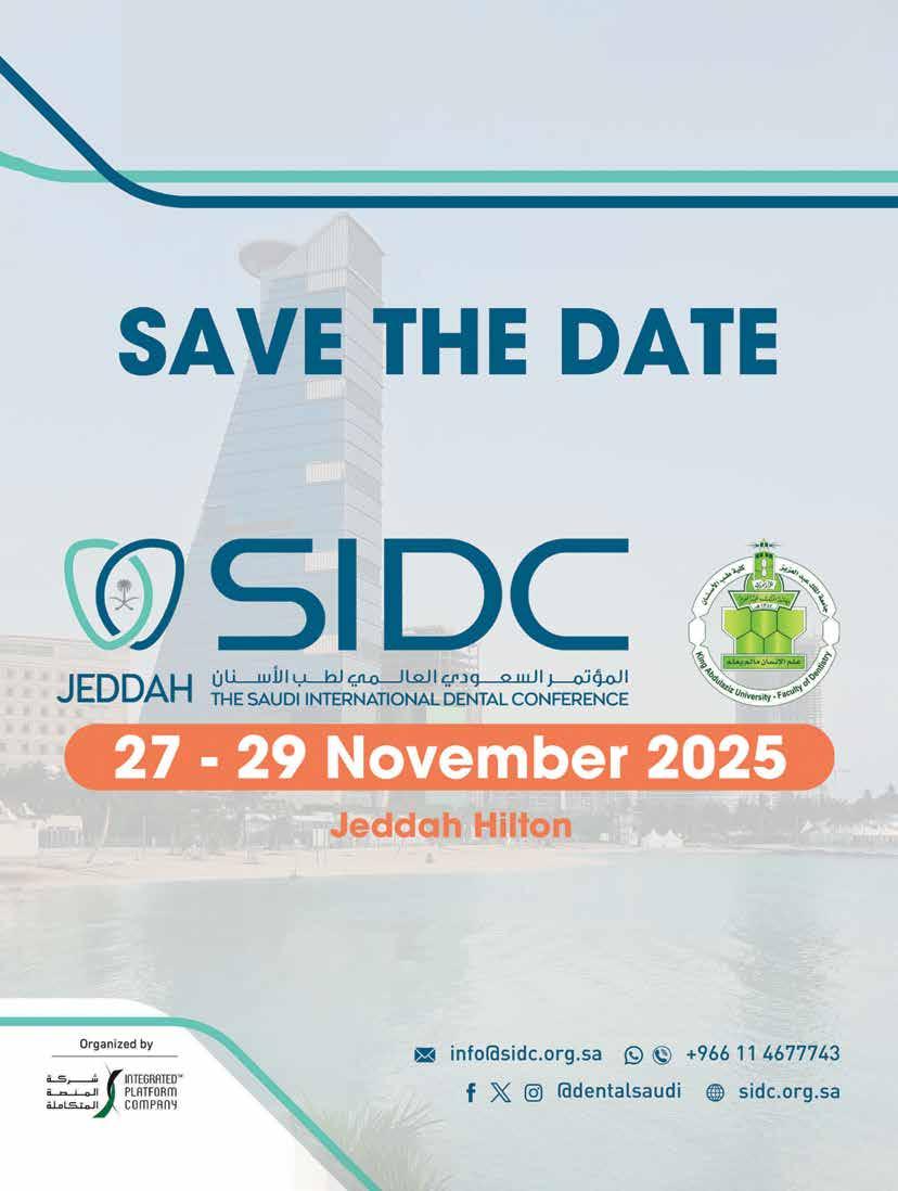

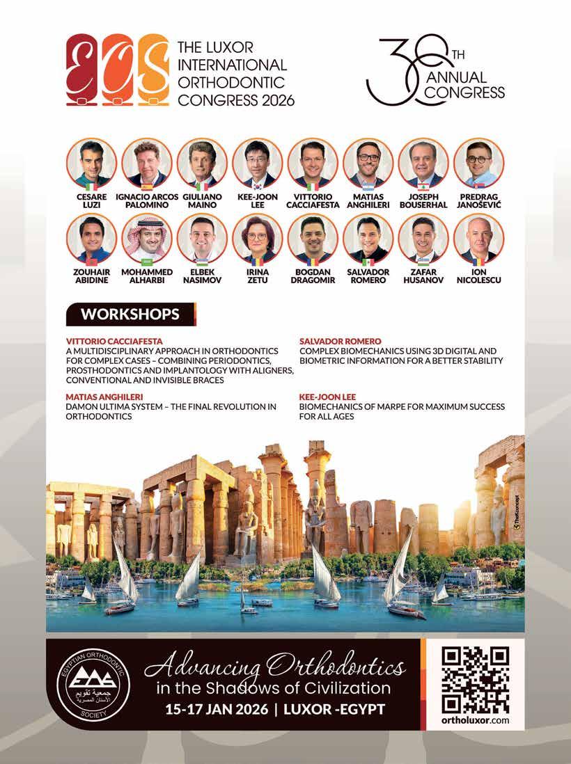


Dental News Magazine sat down with Dentsply Sirona’s Paolo Ambrosini, VP and General Manager, and Quynh Chu, Technical Director RTD and designer of RTD fiber posts, and creator of the proprietary ILLUSION X-RO technology to discuss innovations in fiber dental posts, their performance, and how clinical needs shape product development.
Dental News: Could you briefly introduce us to RTD ?
Paolo: Founded in 1968 under the name of “Recherches Techniques Dentaires”, today RTD is part of Dentsply Sirona. Located in southeastern France, RTD is a worldwide leader in the processing of Quartz fibers and in the production of FRC posts.
Dental News: What defines a high-quality fiber dental post in terms of material properties and clinical performance?
Quynh: In a fiber-reinforced composite (FRC) material, it’s the fibers that primarily dictate the mechanical properties. The greater the fiber content, the better the performance. In fact, a high-quality dental fiber post means a high ratio of fibers—typically around 80% by weight.
However, this high fiber content also presents manufacturing challenges. Impregnating the fibers and ensuring defectfree production requires deep expertise. Success depends not only on raw materials of proven clinical quality but also on highly
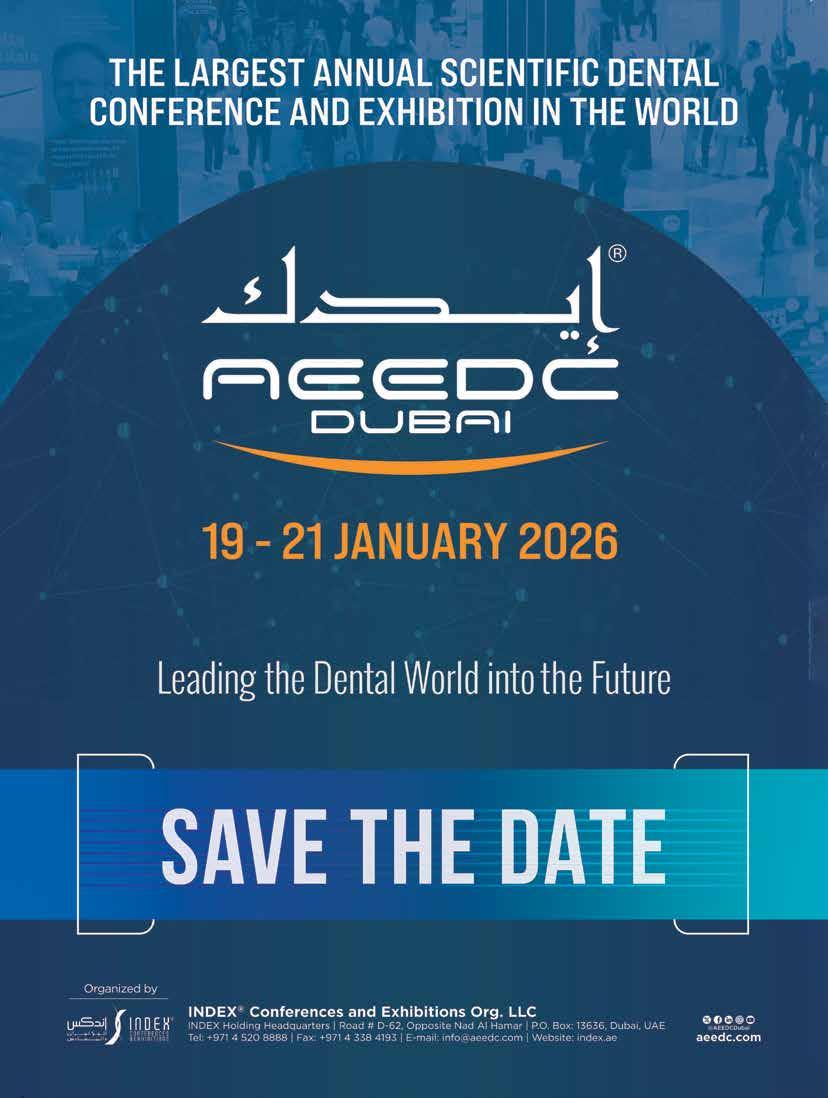
controlled manufacturing processes capable of delivering consistent quality across hundreds of thousands of units.
Dental News: Why is mechanical compatibility between the post material and dentin—such as matching the elastic modulus—so critical?
Quynh: That’s a fundamental principle in biomechanical engineering. Using materials with similar elastic moduli ensures uniform stress distribution. Dentin has an elastic modulus of around 18 GPa. If you use a metallic post with a modulus closer to 200 GPa—over ten times higher—you get stress concentrations at the post/cement interface, which increases the risk of loosening or root fracture1.
Dental News: Could you explain how fiber orientation—unidirectional versus multidirectional—affects performance under load?
Quynh: Absolutely. Fibers excel under tensile stress along their length, which makes orientation crucial. Dental posts mostly endure axial stress, so they must be reinforced with longitudinal fibers.
In the 1980s, we experimented with randomly oriented carbon fibers, but their strength was insufficient. That led to a breakthrough: in 1989, we patented a post using long, unidirectional fibers (French Patent FR 2654612), which provided superior strength and modulus closely matching dentin. Multidirectional fibers, by contrast, result in significantly lower modulus and performance.
Dental News: What role do surface treatments or coatings play in bonding with resin cements and core build up materials?
Quynh: Surface treatment is key to achieving strong adhesion between the fiber post and
the resin matrix. Our posts use a silane-based sizing formulation that enhances bonding. Given that the post surface is mostly fibers, proper treatment allows for mechanical micro-retentions and chemical bonding, both essential for reliable cementation and restoration durability.
Dental News: How do you balance strength, esthetics, and workability in product development?
Paolo: With over 35 years of experience, our development process is driven by ongoing feedback from clinicians and universities. Every product iteration incorporates clinical data to enhance physical and mechanical performance while also improving esthetics and ease of use. Our in-vitro tests simulate real-world conditions to ensure reliability before clinical deployment.
Dental News: How does RTD ensure new fiber post products meet the evolving needs of clinicians?
Paolo: Clinical users are at the heart of our development process. Dentists within the Company are actively involved from concept to launch. We gather external input early and validate through broad clinical evaluations.
Every project brings together a multidisciplinary team—Marketing, R&D, Regulatory, Clinical Affairs, and more—to make sure no need is overlooked, even those that aren’t explicitly stated. This cross-functional approach ensures that each product is robust, safe, and tailored to real clinical workflows.
1Wang X, Shu X, Zhang Y, Yang B, Jian Y, Zhao K. Evaluation of fiber posts vs metal posts for restoring severely damaged endodontically treated teeth: a systematic review and metaanalysis. Quintessence Int. 2019;50(1):8-20. doi: 10.3290/j.qi.a41499. PMID: 30600326.
From Cairo to the Middle East and beyond, Dr. Rami Gamil is helping to shape the future of digital dentistry. As CEO of 3DVision, a respected educator, and a skilled exocad expert, Dr. Gamil has become a driving force in advancing digital workflows across Egypt and neighboring countries. In this exclusive interview, he shares his professional journey, practical insights and why he considers exocad the “purple panther” of digital dentistry.
To begin, could you tell us a bit about your experience in the field of digital dentistry?
Dr. Gamil: I graduated from dental school in 2006 and have been actively engaged in digital dentistry since 2009. Based in Cairo, I serve now as the CEO and founder of 3DVision, a premium provider of digital dentistry solutions, which includes TriScan, our dedicated maxillofacial imaging centers, and 3DVision Education, a hub for handson digital dentistry training. My clinical and academic focus spans CBCT, 3D imaging, guided surgery, CAD/CAM, and smile design— areas where exocad software plays an integral role in my daily work.
What motivates you the most in your work?
Dr. Gamil: What drives me most is helping
clinicians transition into the digital world with confidence and clarity. I believe technology can humanize dentistry when used well—and that’s what I try to share through every case, lecture, and course I lead.
When did you first start working with exocad?
Dr. Gamil: A pivotal moment came in 2015 at IDS in Cologne, where I was first introduced to exocad. I immediately recognized its potential and began using it for digital wax-ups and bite splints.
How has it influenced your clinical practice? exocad software has become central to both our clinical and laboratory workflows. We work with DentalCAD and exoplan daily, using modules such as Smile Creator, Bite Splint, Virtual Articulator, Model Creator, and the Implant Module. These tools enable us to deliver highly precise, patient-specific restorations and surgical plans.
Can you describe a moment when exocad truly amazed you—something that made you think, “This is why I love what I do”?
Dr. Gamil: There have been several moments, but two really stand out. The first was in exoplan, when I started using exocad’s universal custom sleeves and anchor pin options. It was a “wow” moment—realizing that we could now support any surgical kit, whether keyed or keyless, and produce all types of guides, from fully sleeved to sleeveless designs.
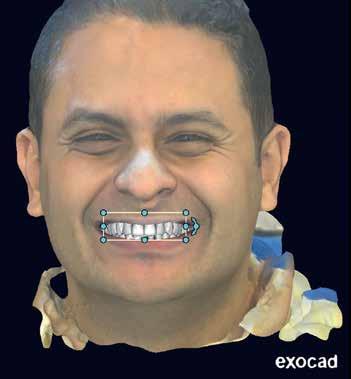
Gamil works with exocad’s DentalCAD and exoplan,
The second moment was with the Smile Creator Module in DentalCAD. In the past, we had to jump between multiple software platforms just to complete a digital smile design—3D face scans, wax-ups, and smile simulations all done separately. But seeing the Smile Creator Module bring all of that together into one seamless, streamlined workflow was transformative.
Education is key in digital dentistry. Could you tell us more about the current state of digital dental education in your region?
Dr. Gamil: There’s still a big gap in hands-on digital dentistry education across Egypt and the Middle East. Many clinicians are eager to learn but lack structured training. That’s why we launched 3DVision Education—a hands-on training platform that covers the entire digital workflow, from scanning to CAD design and production.
Today, digital skills aren’t just nice to have— they’re essential. For me, education is ultimately the bridge between technology and better patient care.
How do you address the issue of software piracy, and why is it important to use authentic exocad software?
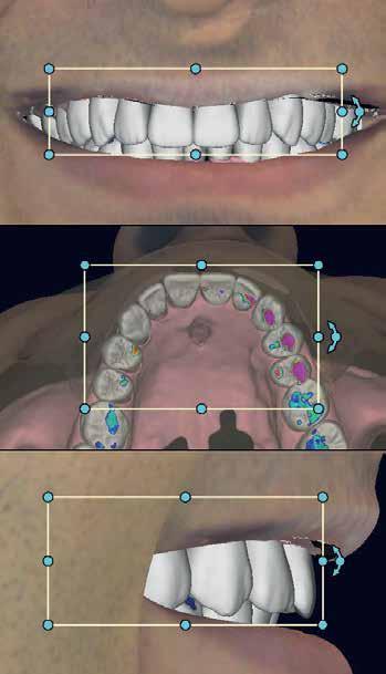
Dr. Gamil: Software piracy is a serious issue that undermines innovation and devalues the work of developers and clinicians. That’s why I’ve always avoided unofficial training programs and proudly support official exocad education.
With affordable student licenses and certified trainers, we’re building a trusted community committed to ethical, high-quality digital dentistry. It’s not just about software—it’s about protecting the future of our profession.
Dr. Gamil: One of the most exciting developments is the rise of AI integration in diagnostics and treatment planning. We’re seeing early adoption of AI-assisted planning
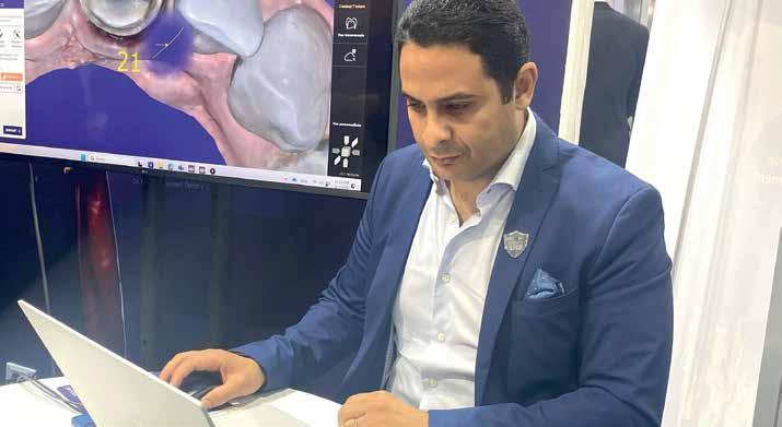
tools and annotation systems that enhance precision and speed. Smile design is also gaining strong momentum. Other key trends include all-on-x workflows, stackable guided surgery, and 3D printing for immediate loading—all of which are making dental treatments faster and more accessible.
You recently spoke at the AOI Congress in Delhi this September. What did you present, and how does digital dentistry in India compare to Egypt?
Dr. Gamil: At the pre-congress workshop, I presented guided surgery workflows using exoplan—from single implants to full-arch rehabilitations—covering the full digital process from scanning to surgical guide planning and clinical implementation.
India and Egypt share many similarities in digital dentistry: both countries have passionate clinicians and a growing interest in digital tools, especially in urban centers.
The main difference lies in scale—India’s market is larger and more complex.
But both countries are embracing cuttingedge technologies like exocad, and the real
opportunity lies in democratizing access to digital dentistry across all regions—ensuring that innovation reaches practitioners in both urban centers and rural communities.
What advice would you give to your younger self, just starting out in dentistry?
Dr. Gamil: I’d tell my younger self: Learn as much as you can—then keep learning. Be bold, take calculated risks, and don’t be afraid to challenge the status quo.
But at the same time, stay humble, always remembering that values come before titles, and people’s lives and well-being matter more than any material gain. In the end, it’s not about being the most successful—it’s about being the most meaningful.
And that’s a lesson I carry with me every day.
Last fun question: If exocad were an animal, what would it be—and why?
Dr. Gamil: If exocad were an animal, it would be a panther, “purple panther”—fast, slick, and always on target.
Thank you, Dr. Gamil, for sharing your insights and expertise with us.
Driven by quality, our goal is to make the professional feel comfortable in its daily endo workflow.
KENDO delivers a wide and reliable range of endodontic products made in Germany as: K-REAMERS, K-FILES, H-FILES, ROOT FILLERS.
A VDW brand - over 150 years of endo experience.
•


For nearly six decades, endodontists and patients have trusted RTD for the most high-stakes restorations. In fact, we were one of the firsts to develop and bring fiber posts to market, leveraging on deep knowledge and expertise. Today, we’re one of the largest producers all over the world, with a wide range of posts designed for optimal outcomes and unbeatable peace of mind.
Learn more about RTD at rtddental.com