Assoc. Prof. Lamyia Anweigi BDS MMedSc MFDS PhD (Qatar)
Dr Ana Cecilia Diniz Viana BDS MD PhD (Brazil)
Dr John Macken BDS PGCertMEd PhD MFDS RCSEng ACIEA FHEA (UK)
Prof. Leonardo Marchini DDS MSD PhD (USA)
Dr med dent Ramiar Karim BDS MSc Paed Dent (Germany)
Dr Elaine Smyth BA BDentSc DChDent (USA)
Prof. Murali Srinivasan BDS MDS MBA MAS PD (Switzerland)
Prof. Sayaka Tada DDS PhD (Singapore)
jida.scholasticahq.com
Irish Dental Association Unit 2 Leopardstown Office Park, Sandyford, Dublin 18.
T: +353 1 295 0072
F: +353 1 295 0092 www.dentist.ie
Follow us! irish-dental-association
Irish Dental Association irishdentalassociation irishdentalassociation
JIDA Science is an official publication of the Irish Dental Association. The opinions expressed in JIDA Science are, however, those of the authors and cannot be construed as reflecting the Association’s views. The editor reserves the right to edit all copy submitted to JIDA Science
Owning a high risk of dental caries is the first step for Irish elite sport
In this edition of JIDA Science, we are delighted to feature a guest editorial from Prof. Ian Needleman.
The new study by Hughes and colleagues (Diet and dental caries in elite athletes in Ireland, Journal of Human Nutrition and Dietetics , January 2026) 1 is an important contribution to the evidence regarding athlete health. Notably, the research was conducted by a multidisciplinary team contributing expertise in oral health, and both sport and food sciences. This is important to ensure that the methods are of the highest quality and therefore as reliable as possible in striving to achieve the research aims. The main finding is striking: 90% of athletes had caries requiring intervention. Unsurprisingly, nutrition was strongly associated with caries. So why is this important and what should be done? There is no health without oral health. 2 Furthermore, consistent evidence highlights the impact of oral health on elite sportspeople’s performance and well-being.3 High levels of dental caries in elite athletes is a consistent finding in many studies globally. Even more worrying is that it starts early in youth sport, and will create a life-long treatment burden and therefore potentially disadvantage those affected.4
The authors of this new study also suggest that the relationship between diet and dental caries may be more complex than previously suggested. Currently, the major dietary emphasis is on the intake of sugars, and these will unquestionably continue to be the most important. Rapidly digestible starches (e.g., white bread, pasta) have also recently been implicated, although the quality of evidence to support this is low.5 A more recent study in Finnish adults did not find an association between starch type and dental caries with 11-year follow-up.6
Why the uncertainty? These studies are difficult to perform and analyse. Collecting accurate data on dietary intake can be problematic, as the authors of this present study testify. Does this alter our recommendations for athletes and the general population? Probably not. Whole foods are still strongly advised over processed for oral and general health, as well as for athlete performance and recovery.7 Elite athletes will continue to need additional sugars to support training, performance and recovery. Therefore, as we have previously recommended, athletes and those supporting them should implement risk mitigation, which can be successfully introduced into highperformance sport.8
Prof. Ian Needleman is Professor of Restorative Dentistry and Evidence-Based Healthcare at the University College London Eastman Dental Institute and Honorary Consultant in
Risk mitigation is simple in theory and athletes are keen to do the right thing.9 Crucially, athletes should ‘own’ their oral health so that they can make decisions on self-management. However, their support teams (including coaches, dieticians, physiotherapists and sport medicine professionals) and federations also need to support the integration of oral health within overall athlete welfare and performance.
Manage elite athletes as at high risk of caries (until proven otherwise) and implement a recognised management strategy.10 Typically, this will include advice to limit sugars to those essential for training, competition and recovery, and to aim for whole foods rather than supplements as far as possible. Increasing fluoride availability is key, including high-concentration fluoride toothpaste (5,000ppm) twice daily and a fluoride mouthrinse (0.05% NaF; 230ppm fluoride) at a different time to toothbrushing with the recommendation to spit and not rinse after use. The toothpaste is not available in all countries and, in many, requires prescription or supply by medical or dental professionals.
In addition, dental professional intervention must not be forgotten. This includes regular dental recall/examination as well as application of fluoride varnish to teeth twice yearly (2.26% NaF). We have demonstrated that this can work in high-performance sport (Olympic teams and professional rugby) with two crucial provisos. Firstly, engage and preferably co-develop the tailored programme with the athletes and support staff, and secondly, employ simple validated behaviour change methods.8
While the focus of Hughes et al. was on caries, future studies should also report on broader measures of oral health, including periodontal health (which can affect performance) and erosive toothwear, which is a canary in the coalmine for both erosive dietary intakes and eating disorders. Clearly, standard oral health screening must be a routine part of athlete periodic health evaluations11 to provide support for athlete performance ambitions.12 It should also be considered duty of care for elite athletes.
References available on page 61.
Periodontology with UCLH. He leads a research programme in oral health and elite sport, which has included an investigation at the London 2012 Olympic Games and professional football in the UK.
Wisdom teeth: back to basics part 1
A shift away from the status quo has led to confusion among general dental practitioners about best practice for wisdom tooth referrals.
Learning outcomes:
n to review common pathologies that can present with mandibular third molars (Md3Ms);
n to equip the general dentist with the necessary tools to conduct a comprehensive wisdom tooth assessment; and, n to support decision-making around specialist referral for Md3M surgery.
Introduction
There exists an abundance of published literature highlighting the problems associated with the retention of asymptomatic, partially erupted third molars, in particular the mandibular mesioangular third molar. The issues surrounding rigid adherence to the 2000 UK National Institute for Health and Care Excellence (NICE) guidance are well documented, and it is no surprise that its place in modern oral surgery practice has been called into question. This shi away from the old status quo has led to confusion among general dental practitioners (GDPs) about the appropriate timing of wisdom tooth referrals, and indeed whether to refer patients in the first place.
Third molar impaction
An impacted tooth is defined as “a tooth that is all the way or partially below the gum line and is not able to erupt properly,”1 and is in essence one that fails to assume a functional position within the dental arch. Impaction is recognised by
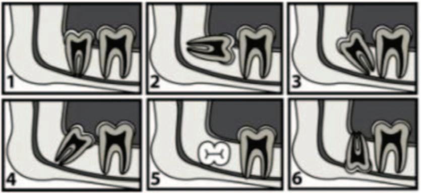
the World Health Organization (WHO) as a disease entity in its own right within the ICD-11 Classification of Diseases (DA07.8).1 Mandibular third molars (Md3Ms) are the last teeth in the series to erupt into the oral cavity, typically between 18 and 24 years of age,2 and are the most commonly impacted of all teeth. Winter’s classification of third molar impaction is commonly used by clinicians as a descriptive tool based on the radiographic inclination of the Md3M (Figure 1). The prevalence of third molar impaction in the 20-30 year age group is as high as 72.7%.2 A mesioangular orientation is observed most frequently in impacted Md3Ms,2 reflecting their developmental process. The diseases most commonly associated with impacted Md3Ms include:
n pericoronitis;
n caries;
n periodontitis; and,
n distal cervical caries (DCC) of the second molar.
Laura O’Sullivan
BDS FDS RCSEd DClinDent(OS) MFDS PDTLHE
Specialist in Oral Surgery
Cork University Dental School and Hospital
University College Cork
Katy Martin
BDS FDS OS RCSEd MOralSurg RCSEd MFDS RCSEd
MA Dent Ed
Specialist in Oral Surgery
Dublin Dental University Hospital Lincoln Place
Corresponding author: Laura O’Sullivan E: laura.osullivan@ucc.ie
FIGURE 1: Winter’s classification of mandibular third molar impaction. Third molars are classified according to their inclination relative to the long axis of the adjacent second molar: 1. Vertical; 2. Horizontal; 3. Distoangular; 4. Mesioangular; 5. Transverse; and, 6. Inverse.4
Pericoronitis
Pericoronitis is the single most common indication for lower third molar removal.2,4 There are currently no internationally agreed diagnostic criteria for pericoronitis, but it is typically diagnosed where there is clinical evidence of swelling of the so tissues around an impacted tooth, with or without a history of food packing and purulent discharge from the associated so tissues.5 Although most cases of Md3M-related pericoronitis will respond well to conservative management, there is a risk that more virulent infections could spread throughout the fascial spaces of the neck, with potentially lifethreatening consequences.
Caries
Caries is the second most common indication for lower third molar removal, and is seen more commonly in older patients, where the tooth has been in communication with the oral cavity for a period of time.2 Unfavourable tooth orientation and difficult tooth access are frequent barriers to restorative intervention in such instances.
Periodontitis
It is estimated that periodontitis accounts for 5% of all Md3M extractions.2 In one retrospective analysis, it was found that periodontal disease accounted for the removal of 13% of horizontally impacted Md3Ms and 9% of vertical, nonimpacted Md3Ms.2
Distal cervical caries of the second molar DCC in the mandibular second molar is believed to be uniquely related to the mesioangular Md3M, and has been widely reported in the literature in recent years.2,6-8 DCC arises at the cementoenamel junction of the second molar and is considered a variant of root surface caries (Figure 2).
In many cases, DCC is diagnosed at an advanced stage, by which point the second molar is oen deemed unrestorable. This contentious issue has been a topic of much research, and there is a strong argument in favour of prophylactic removal of mesioangular third molars to prevent those problems associated with their retention long term.7
Other
Cystic change affects around 2-3% of impacted third molars.9 Dentigerous cysts are typically associated with unerupted teeth, and account for 20% of all odontogenic cysts.10 These cysts arise from separation of the reduced enamel epithelium once amelogenesis is complete, and are believed to affect Md3Ms almost 10 times more frequently than their maxillary counterparts.11
Inflammatory resorption is another phenomenon that may affect the crowns of impacted third molars. This tends to be observed in older age groups (>50 years) and results from the activity of multinucleate giant cells.12 Destroyed tooth tissue is replaced by bone, resulting in ankylosis. Surgical removal of affected teeth can be extremely challenging.
Both cystic and resorptive change are oen incidental findings with no presenting signs or symptoms; it is therefore prudent to monitor unerupted teeth radiographically on a regular basis to ensure that any pathology is picked up at an early stage.
Guidelines
As there are no robust clinical guidelines for third molar surgery in Ireland,
clinicians have traditionally turned to our neighbours in the UK and USA, and guidance extrapolated from international recommendations where appropriate.
National Institute for Health
and Care Excellence (UK)
In March 2000, NICE published its ‘Guidance on the extraction of wisdom teeth’ document (TA1).13 NICE recommends removal of third molars only where a clear clinical indication exists. Since the introduction of these guidelines, there has been considerable debate in situations where a malpositioned, partially erupted Md3M poses a risk to the neighbouring tooth. Particular interest surrounds the enigmatic mesioangular impaction,7 which has been shown in numerous studies to lead to an increased incidence of DCC in the adjacent second molar tooth,2 with late detection posing serious restorative challenges.
Studies have shown a trend towards third molar surgery later in life, with an increase in the mean age of patients from 25 years at the time of surgery in 2000 to 32 years in 2010.2 Increased age at the time of surgery is associated with increased surgical morbidity.4
In light of the inherent issues posed by the 2000 guidance that are reported in the literature, NICE has acknowledged that the guidelines contained in the TA1 document are “controversial”.14 Clinicians are urged to exercise their judgement in the context of patients’ best interests. NICE guidance remains under review at the time of publication.
American Association of Oral and Maxillofacial Surgeons (USA)
The 2024 clinical paper produced by the American Association of Oral and Maxillofacial Surgeons (AAOMS) proposes that “third molars should be removed in the younger-age patient because there is less transitory or permanent morbidity”, and that “a final determination must be based on an agreement between the surgeon and the patient”.15 In this document, the AAOMS acknowledges the potential for future disease development related to diseasefree third molars. It highlights the importance of the surgeon’s role in determining the risk of future disease development in cases of asymptomatic impacted Md3Ms, and endorses the role of the surgeon in contributing to the decisionmaking process in the patient’s best interests.
FIGURE 2: Radiograph of distal cervical caries in the LL7 associated with mesioangular LL8.
Faculty of Dental Surgery Guidelines (UK)
A guideline development group in the UK collectively reviewed the pre-existing faculty guidelines on management of Md3Ms “because evidence suggests increasing patient harm due to retention of Md3Ms”.11 The consensus document ‘Parameters of care for patients undergoing mandibular third molar surgery’ recommends a shi “from a solely therapeutic approach to a mixed range of interventions for patients with mandibular third molars based on a holistic and informed approach agreed with the patient”.11 Particular emphasis is placed on patient education regarding the risks of retention, as well as surgical removal of malpositioned Md3Ms, with special recognition of the evidence supporting prophylactic removal of the mesioangular Md3M.
Wisdom tooth assessment
Routine evaluation of third molar development in the adolescent population should be undertaken by the GDP as part of the general dental health assessment. This will allow any active disease, and indeed the potential for disease, to be identified at an early stage and managed appropriately.
A comprehensive history forms the basis of the wisdom tooth assessment. Complaints including pain, so tissue swelling, food trapping, antibiotic history, and difficulty maintaining oral hygiene should be documented. Table 1 outlines a structured approach to the examination of wisdom teeth, and should be broadly adhered to where possible.
Clinical examination
n Degree of eruption:
● partially erupted;
● fully erupted;
● unerupted; or,
● overerupted.
n Operculum health
n Crown:
● inclination;
● restorations; and, ● caries.
n Periodontal status:
● oral hygiene;
● food trapping; and, ● pocketing.
n Adjacent tooth
n Opposing tooth/teeth
n Occlusion
Is the third molar?
Partially erupted
Mesioangular horizontal Vertical distoangular History of pericoronitis
Radiographic examination
n Winter’s classification
n Pell and Gregory classification
n Crown:
● size;
● shape; and, ● disease.
n Roots:
● number;
● length; and, ● morphology.
n Alveolar bone support
n Pericoronal status:
● bone resorption; ● follicular enlargement; and, ● cystic change.
Table 1: What to document during a wisdom tooth assessment.
FIGURE 3: Decision flowchart for assessing Md3Ms.
Once the examination is complete, how does the clinician decide whether a referral to oral surgery is indicated? The following considerations will help to inform this decision:
n is the tooth fully erupted and functional?;
n is the tooth disease free?;
n is the orientation of the tooth favourable with respect to oral hygiene?; and, n if partially erupted, is the tooth likely to erupt further and assume a functional position within the arch?
If the answer to any of the above questions is ‘no’, intervention will almost certainly be required. The type of intervention will depend on a number of factors, such as the restorability of the tooth, functional importance, type of impaction, age of the patient, underlying medical conditions, and patient preference. A decision flowchart is presented in Figure 3
It is important to consider the negative impact that retention of partially erupted mesioangular and horizontal Md3Ms will invariably have on the neighbouring teeth. In the case of the former, we know that DCC is a risk to the second molar, even in otherwise healthy, disease-free mouths. With the latter, loss of alveolar bone support along the distal root of the second molar is a common finding that results in severely compromised periodontal health (Figure 4).
Specialist referral
For patients requiring specialist referral, it is essential that all basic information is included in the referral letter to support clinical triage and subsequent assessment. Table 2 outlines the recommended information for specialist referral. It is important to include all relevant information, including any previous interventions and appropriate imaging. If a cone beam computed tomography (CBCT) scan is available, both the scan and accompanying report should be attached to the referral. When referring patients for conscious sedation, it is prudent to first consider the suitability of the patient for sedation before referral. Factors in the medical history such as sleep apnoea, obesity, pregnancy, drug abuse, and extremes of age can make conscious sedation an unsuitable treatment modality.
Conclusion
Routine third molar assessment should form part of a patient’s general dental health check in the relevant age group, and timely specialist referral should be arranged where indicated, following consultation with the patient. Ignoring asymptomatic partially erupted wisdom teeth is no longer acceptable practice. To leave the partially erupted mesioangular, horizontal upper or lower third molar
References
1. World Health Organization. International Classification of Diseases: 11th Revision (ICD-11). https://icd.who.int/en/. 2019.
2. McArdle LW, Andiappan M, Khan I, Jones J, McDonald F. Diseases associated with mandibular third molar teeth. Br Dent J. 2018;224(6):434-440.
3. Miclotte A, Grommen B, Cadenas de Llano-Pérula M, Verdonck A, Jacobs R, Willems G. The effect of first and second premolar extractions on third molars: a retrospective longitudinal study. J Dent. 2017;61:55-66.
4. Bruce RA, Frederickson GC, Small GS. Age of patients and morbidity associated with
FIGURE 4: Radiograph showing severe alveolar bone loss along the distal aspect of LL7 as a direct result of LL8 impaction.
Table 2: Recommended information to include in an oral surgery referral.
Referral information:
n patient details, including name, date of birth and contact number;
n details of referring clinician;
n details of GP;
n reason for referral (including history of presenting complaint);
n full medical history, including medication list and allergy status;
n full social history, including smoking and alcohol status;
n height, weight and BMI (particularly if requesting conscious sedation);
n Modified Dental Anxiety Scale or equivalent anxiety score (particularly if requesting conscious sedation);
n relevant radiographs; and,
n CBCT with associated radiographic report if applicable.
in situ is to leave a patient open to significant preventable disease in the adjacent second molar. In an era where prevention is better than cure, timely patient education and referral are the mainstay of best clinical practice. The second part of this series will discuss complications of wisdom tooth surgery and management of common third molar problems that may present to the GDP.
mandibular third molar surgery. J Am Dent Assoc. 1980;101(2):240-245.
5. Mackie L, Wong-McDermott R, Bell GW. An evaluation of referrals requesting third molar tooth removal: clinical diagnosis and treatment outcome. Br Dent J 2019;226(8):577-580.
6. McArdle LW, Renton T. The effects of NICE guidelines on the management of third molar teeth. Br Dent J. 2012;213(5):E8.
7. Allen RT, Witherow H, Collyer J, Roper-Hall R, Nazir MA, Mathew G. The mesioangular third molar – to extract or not to extract? Analysis of 776 consecutive
third molars. Br Dent J. 2009;206(11):E23-587.
8. Toedtling V, Devlin H, Tickle M, O’Malley L. Prevalence of distal surface caries in the second molar among referrals for assessment of third molars: a systematic review and meta-analysis. Br J Oral Maxillofac Surg. 2019;57(6):505-514.
9. Güven O, Keskin A, Akal UK. The incidence of cysts and tumors around impacted third molars. Int J Oral Maxillofac Surg. 2000;29(2):131-135.
10. Hill CM, Renton T. Oral surgery II: Part 3. Cysts of the mouth and jaws and their management. Br Dent J. 2017;223(8):573-584.
11. Royal College of Surgeons of England, Faculty of Dental Surgery. Parameters of care for patients undergoing mandibular third molar surgery. 2020.
12. Schulz SD, Donath K. Histologische Untersuchung resorbierter Zahnkronen
nichtdurchgebrochener Weisheitszähne [Histological study of the resorbed dental crowns of impacted wisdom teeth]. Dtsch Zahn Mund Kieferheilkd Zentralbl 1992;80(1):33-36.
13. National Institute for Health and Care Excellence. Guidance on the extraction of wisdom teeth. 2000.
14. National Institute for Health and Care Excellence. Guidance on the extraction of wisdom teeth. Technology appraisal guidance. 2014. https://www.nice.org.uk/guidance/ta1/documents/wisdom-teeth-removal-reviewproposal-october-14.
15. American Association of Oral and Maxillofacial Surgeons. The Management of Impacted Third Molar Teeth. 2024.
Quiz
Submitted by Prof. Martina Hayes, Consultant in Restorative Dentistry, Cork University Dental School and Hospital.
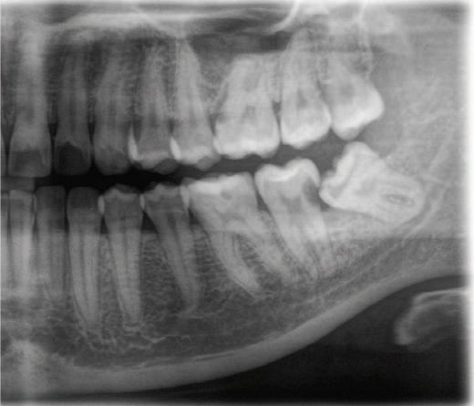
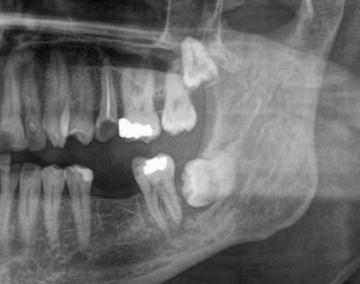
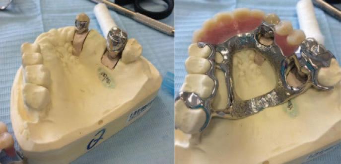
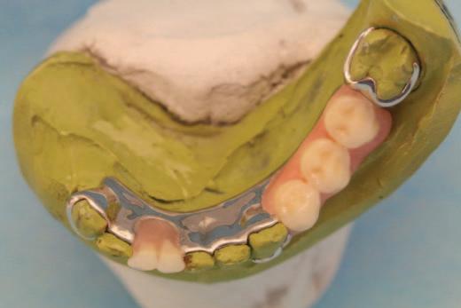
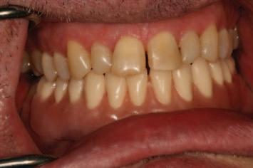
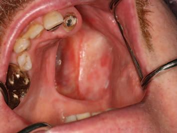
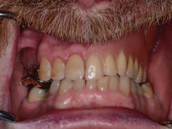
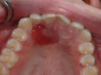
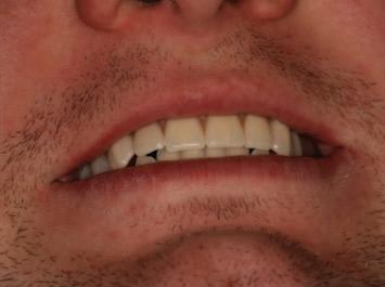
Figures 1 and 2 show surveyed crowns on abutment teeth that support the removable partial denture.
1. What is the primary purpose of the surveyed crowns visible on the prepared teeth as per the crowns on the cast displayed?
A. To improve aesthetics
B. To act as bridge abutments
C. To provide controlled contours, guiding planes, and rest seats for a removable partial denture (RPD)
D. To splint periodontally compromised teeth
2. Which of the following features would most likely be incorporated into these restorations?
A. Subgingival feather-edge margins
B. Occlusal rest seats and guiding planes
C. Veneered labial porcelain margins
D. Locator attachments
3. What is the main advantage of providing surveyed crowns rather than clasping natural tooth contours?
A. Reduces laboratory cost
B. Eliminates need for a major connector
C. Provides ideal path of insertion and controlled retention
D. Prevents the need for occlusal rests
Answers on page 59
FIGURE 1.
FIGURE 2.
Restorative and prosthetic challenges in oral cancer patients: a narrative review
Highlighting the restorative and prosthetic challenges that shape recovery, function and quality of life following treatment for mouth cancer.
Introduction
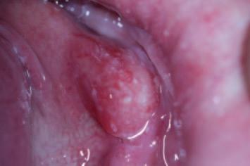
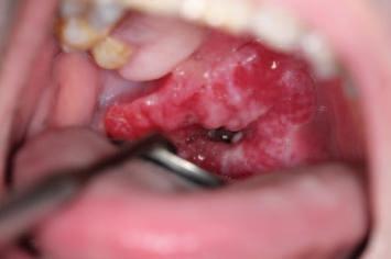
Oral cancer, as defined by the World Health Organization, includes cancers of the lip, oral cavity, and oropharynx, ranking as the 13th most common cancer globally.¹,² In 2020, there were 377,713 new cases and 177,757 deaths related to lip and oral cavity cancers.² The vast majority of these malignancies are squamous cell carcinomas (SCC), accounting for more than 90% of oral cancer cases. 3 Treatment options include surgery, radiotherapy, and chemotherapy, used individually or in combination depending on tumour location, stage, age, and comorbidities (Figures 1A-1C).4 Surgery remains the gold standard for many oral cancers. 5 However, both the disease and its treatments can lead to significant functional and aesthetic issues, including difficulties with mastication, deglutition, speech, and salivary hypofunction. 6 Consequently, oral rehabilitation plays a crucial role after cancer treatment, involving prosthetic reconstruction tailored to the restoration of function, appearance, and quality of life.7
Complications after oral cancer treatment
Trismus
Trismus, defined as an inter-incisal opening ≤35mm, occurs frequently aer radiotherapy due to fibrosis of the masticatory muscles.7 Trismus complicates eating and oral hygiene for the patient, and intra-oral access for vigilance, impression making, and jaw registration and prosthesis fit for clinicians.7 In severe cases (<15mm opening), prosthesis insertion becomes extremely difficult, and obturators may not achieve an adequate seal because of restricted access.8 Early implementation of jaw-stretching exercises improves long-term outcomes.7
Kumud Gogna Dental student1
Afnan Al Maini Dental student1
David McReynolds
BA BDentSC MFDS RCSEd DdChDent(Pros) FFD RCSI(Pros)
Hyposalivation
Radiation therapy oen reduces salivary flow by 50-60% within the first week, impairing buffering capacity and antimicrobial protection.9 Consequences include rampant caries, poor denture retention, and Candida infections.9 Thick, ropy saliva is common among post-radiotherapy patients, which increases denture friction, disrupts peripheral seal formation, and reduces impression accuracy.10 Management involves: saliva substitutes, such as BioXtra (BioXtra Ltd, Hatvan, Hungary) and Biotène (Haleon PLC, Weybridge, England), which act as oral lubricants and moisturisers; parasympathomimetic stimulants, such as pilocarpine and cevimeline, which act as muscarinic agonists; and, high-fluoride prophylactic toothpastes such as Colgate Duraphat 5,000 (Colgate-Pamolive, New York, USA), which act as anticaries agents.9 For many patients living with hyposalivation, oral moisturisers and stimulants remain unsatisfying and potentially expensive treatments. The regular sipping of plain water is typically a preferred management strategy for many.
Radiation caries
Teeth exposed to radiation undergo structural changes, including reduced microhardness of dentine and impaired enamel prism integrity, particularly around the dento-enamel junction (DEJ).11 Radiation-induced caries typically affects atypical tooth surfaces, progressing rapidly due to hyposalivation and enamel demineralisation.12 In many situations, teeth fracture at the gingival margin and are unrestorable. Preventive strategies include: fluoride varnishes, such as Colgate Duraphat High Fluoride Varnish (22,600ppm sodium fluoride); meticulous professional and at-home oral hygiene; and, regular recalls for supportive therapy at three-month intervals.12
1. Dublin Dental University Hospital Trinity College Dublin 2. Hermitage Dental Suite 10, Dublin
Corresponding author: David McReynolds E: david.mcreynolds@dental.tcd.ie
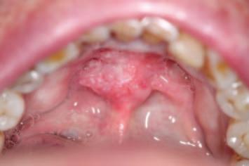
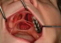
FIGURE 1: Examples of intra-oral squamous cell carcinoma (SCC) in three different patients. 1A and 1B are typical examples of early anterior floor of mouth invasive SCCs, both of which are amenable to surgical treatment. 1C illustrates a late presentation of a large oro-pharyngeal SCC with central necrosis and rolled borders, which is inoperable, but may undergo palliative treatment with radiotherapy.
A C B
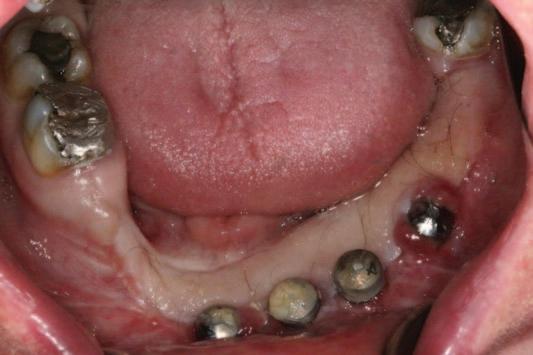
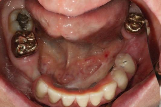
2: 2A shows an example of a hair-bearing fibula free flap reconstruction of the mandible following hemi-mandibulectomy, with poor oral hygiene, prior to maxillofacial prosthodontic reconstruction, including plaque-induced gingival hyperplasia of fibro-epithelial tissue adjacent to the transmucosal aspect of the LL4 dental implant. 2B illustrates the same patient post-prosthodontic reconstruction, with improved professional and at-home oral hygiene, including stabilisation of soft tissue inflammation, achieved through motivation and professional instruction.
Complication Cause
Prosthetic impact Management
Trismus Fibrosis from radiotherapy Limitations in prosthetic Jaw-stretching exercises insertion/removal
Hyposalivation Radiotherapy-induced salivary Poor prosthesis retention, oral Salivary substitutes and oral lubricants, gland damage discomfort, rampant caries high-fluoride varnishes and toothpastes, regular plain water sipping
Chemoradiation-induced mucositis manifests as erythema, ulceration, and pain, oen limiting denture tolerance and severely impacting quality of life.13 Mucositis reduces tolerance of any removable prosthesis, as minor trauma from denture flanges or borders can significantly worsen ulceration.13 Regular assessment and adjustment of dentures, including relining with tissue conditioners, are necessary to minimise friction and irritation. Any mucosal ulceration should be treated before fabricating new prostheses.13
Osteoradionecrosis
Osteoradionecrosis (ORN) is defined as exposed, non-healing irradiated bone persisting for >3 months.14 Radiation-induced hypovascularity reduces tissue resilience, and even minor denture trauma can precipitate ulceration and necrosis.14 Implant placement is high risk, with irradiated bone showing markedly increased implant failure and periimplant complications that may trigger ORN.15 Risk reduction includes: pre-radiation dental assessment and prophylactic dental extractions; avoiding post-radiation extractions where feasible; regular review to arrest and delay dental disease; and, relining ill-fitting dentures to prevent mucosal injury.14
Hair-bearing free flaps
Flaps harvested from the radial forearm or fibula regions may introduce intra-oral hair, leading to poor oral hygiene and halitosis. Dental implants placed in such flaps oen manifest with plaque-induced fibro-epithelial hyperplasia, which may engulf access to the implant head (Figures 2A and 2B).16 Management may include laser epilation or flap revision if at-home oral hygiene measures do not suffice (Table 1).16
Impact of oral cancer treatments on prosthetic rehabilitation
Altered oral anatomy
Surgical management of oral cancer can significantly alter intra-oral and peri-oral structures. Tooth loss, changes in ridge form, disrupted muscle attachments, loss of lip competence, bulky free flaps, sensory disturbances, trismus, and facial contour changes all affect prosthesis fit, retention, stability, and overall patient adaptation.8 These changes reduce available restorative space, alter functional boundaries, and oen require repeated adjustment as tissues continue to remodel postoperatively. Microstomia is a common consequence of lip reconstruction, particularly following techniques such as the Karapandzic flap.17,18 Reduced oral aperture restricts denture
Table 1: Complications and prosthetic implications.
FIGURE
Residual ridge of the mandible
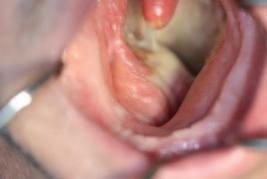
FIGURE 3: Residual surgical fenestration is illustrated, affecting the hard palate in 3A, creating an open communication between the oral cavity and nasal cavity. In a different patient, 3B illustrates the intra-oral view following total glossectomy. Both post-surgical defects significantly complicate impression making and increase the risk of aspiration during restorative procedures.
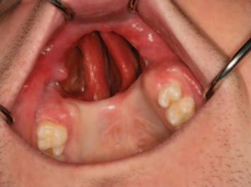
FIGURE 4: Surgical defect following hemi-mandibulectomy surgery, without flap reconstruction, demonstrating significant mandibular deformity, and deviation of the mandible to the side with the deformity (4A and 4B). A ‘J-shaped’ cobalt-chrome framed sectional removable partial denture is made to restore form, function and aesthetics insofar as is feasible given the nature of the defect (4C).
FIGURE 5: Scar tethering of the buccal and labial sulcus on the resected left side. Such defects will affect the development of a peripheral seal and stability of a removable reconstruction. The application of a tissue-borne removable modality in such a situation will require that denture teeth are placed in a more lingual position than is normative to accommodate a new centralised neutral zone.
insertion, limits border extension, and complicates impression procedures. Prosthodontic ingenuity may be required to overcome restricted access.17
Post-surgical fenestrations (Figure 3A) and glossectomies (Figure 3B) further increase aspiration risk during prosthodontic procedures and may complicate impression making.8 Appropriate blocking of undercuts, maintaining an upright patient position, and careful selection of impression techniques are essential to minimise these hazards, and ensure an accurate and safe prosthetic workflow.8
Mandibular prosthesis – challenges following oral cancer treatment
n Partial mandibulectomy compromises the stability, support, and retention of mandibular dentures, especially when resections extend to the midline (Figure 4).8
n Post-radiation lymphoedema in the floor of the mouth can alter posture, limiting lingual extension of the denture, and affecting retention and stability.8
n Loss of tongue bulk, sensation, and mobility complicates control of mandibular dentures. Effective mastication requires positioning the food bolus on mandibular occlusal surfaces, making tongue position relative to the occlusal plane critical. If resection extends to the tongue base, the tongue may become immobilised in a detruded position, losing effective interaction with teeth and palate.8
n Severe mandibular deviation and fibrosis-induced angular jaw motion pathways create lateral occlusal forces that dislodge dentures and increase mucosal irritation and ulceration.8
n Posterior lip retraction can prevent adequate denture flange extension, requiring lingual placement of teeth, which may create dislodging forces. If the lip and cheek are scarred on the resected side, denture extension attempts can worsen dislodgement, and complicate fit and function (Figure 5).8
n Increased so tissue bulk may encroach on tongue space, limit mobility, and obliterate the sulcus, compromising denture fit, retention, and stability.8
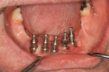
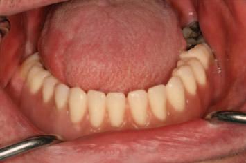
n Frequently, a fixed (Figure 2) or removable implant reconstruction (Figure 6) on a reconstructed neomandible represents a gold-standard approach in scenarios where tissue-borne removable modalities have a poor-hopeless prognosis.
FIGURE 6: Placement of dental implants in the mandible post resection, as illustrated in 6A, permits fabrication of a sophisticated and satisfying fixed or removable reconstruction. In this example, a complex implant overdenture (6B and 6C) provides a level of function that resembles a fixed dental reconstruction, while permitting prosthesis removal for at-home hygiene procedures, and personal and professional recurrence vigilance.
Maxillary prosthesis – challenges following oral cancer treatment
n Loss of anatomy from hard palate, alveolar ridge, or tuberosity ablation further reduces the prognosis of a removable prosthesis (Figure 3A, Figure 7A and Figure 8B).8
n Radiation-induced fibrosis of palatine salivary glands compromises the peripheral seal needed for denture retention.8
n Mandibular deviation post radiation may cause the maxillary tuberosity on the resected side to impinge on the coronoid process, hindering impression material extension and prosthesis fit.8
n Pedicle or free flaps for so tissue reconstruction complicate denture extension. Altered anatomy and scarring may reduce available space, challenging denture seating.8
n So tissue flaps (such as the radial forearm flap) reline the oral cavity and separate compartments, but may remove features like the mucocutaneous scar band and medial palatal shelf, worsening prosthesis stability.19 Such a lack of proper flap support is termed the “waterbed” or “trampoline” effect.20
n Tissue changes may continue for at least a year post healing (Figure 7). Obturator movement during function can worsen these changes, oen requiring prosthesis relining or rebasing (Figure 7B).8
n The presence of teeth adjacent to defects improves prognosis by assisting
retention, stability, and support (Figure 8). Sometimes, these teeth are splinted to enhance load-bearing capacity.8
Conclusion
Restorative and prosthetic rehabilitation of oral cancer patients is multifaceted and highly individualised. Treatment-induced complications including trismus, xerostomia, mucositis, ORN, altered anatomy, and microstomia create significant clinical challenges. Successful rehabilitation depends on early recognition of complications, appropriate timing of intervention, and meticulous prosthodontic planning tailored to each patient’s anatomical and functional limitations. Innovative approaches such as implant-supported prostheses, sectional dentures, flap reconstructions, and obturator design are increasingly central to restoring oral function and aesthetics. Ultimately, patient-centred, holistic care is essential to improve long-term quality of life.
CRediT author statement:
Kumud Gogna: original dra preparation and writing – review and editing.
Afnan Al Maini: original dra preparation and writing – review and editing.
David McReynolds: supervision, visualisation, project administration, writing –review and editing.
Denise MacCarthy: conceptualisation, supervision, writing – review and editing.
Edward Cotter: conceptualisation, supervision, writing – review and editing.
Conflict of interest: all authors declare that they have no conflicts of interest.
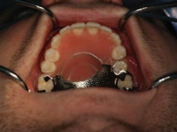
FIGURE 7: Intra-oral view of a patient following left hemimaxillectomy (7A). A survey crown and precision attachment cobalt-chrome framed obturator closes the maxillary defect (7B). Reline material is visible at the peripheral seal in response to post-surgical remodelling of the scar band. The obturator restores function, aesthetics, and separation of the oral and nasal cavities (7C).
A C B
A C B
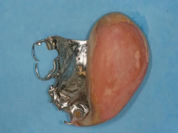
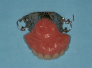
FIGURE 8: Surgical and maxillofacial prosthodontic management of a SCC associated with HPV infection in the anterior maxilla. 8A illustrates the pre-operative appearance of the SCC. 8B illustrates the surgical resection defect and resultant oro-nasal communication. The communication is obturated with a cobalt-chrome framed removable partial denture with an acrylic bulb, which restores aesthetics, phonetics and function, while permitting prosthesis removal for cleansability and recurrence vigilance.
References
1. World Health Organization. Oral health. https://www.who.int/news-room/factsheets/detail/oral-health. March 17, 2025.
2. Sung H, Ferlay J, Siegel RL, et al. Global Cancer Statistics 2020: GLOBOCAN estimates of incidence and mortality worldwide for 36 cancers in 185 countries. CA Cancer J Clin. 2021;71(3):209-249.
3. El-Naggar AK, Chan JKC, Grandis JR, Takata T, Slootweg PJ, eds. WHO Classification of Head and Neck Tumours. 4th ed. Lyon: International Agency for Research on Cancer; 2017.
4. Homer JJ, Winter SC. Head and neck cancer: United Kingdom national multidisciplinary guidelines, sixth edition – corrigendum. J Laryngol Otol. 2024;1.
5. Kröplin J, Reppenhagen JC. Best practices and future challenges in the treatment of oral cancer. Innov Surgl Sci. 2023;8(4):215-220.
6. Patton DW, Ali A, Davies R, Fardy MJ. Oral rehabilitation and quality of life following the treatment of oral cancer. Dent Update. 1994;21(6):231-234.
7. Warnakulasuriya S, Greenspan JS. Textbook of Oral Cancer: Prevention, Diagnosis and Management. Springer Nature; 2020.
8. Beumer J, Marunick MT, Esposito SJ. Maxillofacial Rehabilitation: Prosthodontic and Surgical Management of Cancer-related, Acquired, and Congenital Defects of the Head and Neck. Hanover Park, Il: Quintessence Pub; 2011.
9. Saleh J, Figueiredo MAZ, Cherubini K, Salum FG. Salivary hypofunction: an update on aetiology, diagnosis and therapeutics. Arch Oral Biol. 2015;60(2):242-255.
10. Mhatre S, Srichand R, Sethumadhavan J, et al. Dry mouth dilemma: a comprehensive review of xerostomia in complete denture wearers. Cureus. 2024;16(4):e58564.
11. Walker MP, Wichmann B, Cheng A-L, Coster J, Williams KB. Impact of radiotherapy
dose on dentition breakdown in head and neck cancer patients. Pract Radiat Oncol. 2011;1(3):142-148
12. Gupta N, Pal M, Rawat S, et al. Radiation-induced dental caries, prevention and treatment – a systematic review. Natl J Maxillofac Surg. 2015;6(2):160-166.
13. Madeswaran S, Saravanan D, Rethinam S, Muthu K. Oral mucositis: role of the dentist. J Oral Health Comm Dent. 2019;13(3).
14. Chronopoulos A, Zarra T, Ehrenfeld M, Otto S. Osteoradionecrosis of the jaws: definition, epidemiology, staging and clinical and radiological findings. A concise review. Int Dent J. 2017;68(1):22-30.
15. Toneatti DJ, Graf RR, Burkhard JP, Schaller B. Survival of dental implants and occurrence of osteoradionecrosis in irradiated head and neck cancer patients: a systematic review and meta-analysis. Clin Oral Investig. 2021;25(10):5579-5593.
16. Jeong WS, Oh TS. Oral and oropharyngeal reconstruction with a free flap. Arch Craniofac Surg. 2016;17(2):45-50.
17. Ahmad ZH, Gadbail AR, Shailesh Gondivkar, et al. Prosthetic rehabilitation of microstomia patients: a systematic review of published case reports and case series. J Contemp Dent Pract. 2019;20(4):508-515.
18. Miller LE, Greene JJ. The Karapandzic flap. Oper Tech Otolaryngol Head Neck Surg 2020;31(1):21-25.
19. El Fattah H, Zaghloul A, Pedemonte E, Escuin T. Pre-prosthetic surgical alterations in maxillectomy to enhance the prosthetic prognoses as part of rehabilitation of oral cancer patient. Med Oral Patol Oral Cir Bucal. 2012;17(2):e262-70.
20. Futran ND, Wadsworth JT, Villaret D, Farwell DG. Midface reconstruction with the fibula free flap. Arch Otolaryngol Head Neck Surg. 2002;128(2):161-166.
Jill Anne O’Driscoll MOrth RCSED MDSC (Res)
Post-CCST in Orthodontics
Dundee Dental Hospital and Research School
United Kingdom
Ghaly Adly Ghaly FDS FRCS(OMFS)
Consultant Oral and Maxillofacial Surgeon Ninewells Hospital
NHS Tayside Dundee
United Kingdom
Brian Stevenson PhD FDS(Rest Dent)
Consultant/Honorary Senior Lecturer in Restorative Dentistry
Dundee Dental Hospital and Research School
United Kingdom
Grant McIntyre PhD, FDS(Orth) FDS (Hon) Consultant and Honorary Professor in Orthodontics
Dundee Dental Hospital and Research School
United Kingdom
Corresponding author:
Jill Anne O’Driscoll E: jill.odriscoll3@nhs.scot
Ectodermal dysplasia: two-stage orthognathic surgery, digital planning and restorative rehabilitation
Précis: This case report highlights how a two-stage surgical approach and digital multidisciplinary team planning allowed for successful restorative rehabilitation of a patient with ectodermal dysplasia.
Abstract
Introduction: Ectodermal dysplasia (ED) is a hereditary disorder caused by dysplasia of one or more of the ectodermal tissues, with hypodontia or anodontia being common. Prosthetic rehabilitation is a routine process for people with ED from childhood to adulthood.
Objectives: This paper presents the multidisciplinary team (MDT) management of a 36-year-old female patient with anhidrotic ED, a hypoplastic maxilla and hypodontia, and with skeletal and dental concerns.
Methods: The MDT included consultants in oral and maxillofacial surgery, restorative dentistry and orthodontics. Following cone beam computed tomography (CBCT) imaging and digital MDT planning, treatment involved a two-step surgical approach with a bimaxillary osteotomy and bone grafting, followed by delayed insertion of transzygomatic and conventional dental implants. These were restored with a full arch maxillary bridge and a lower implant-retained complete denture. The piezoelectric system was used as part of the surgical procedure to ensure that the optimal patient outcome was achieved.
Results: Follow-up radiographic and clinical evaluation revealed appropriate healing, an enhanced facial profile, and a stable and functional occlusion. The patient is very satisfied, and her skeletal and dental concerns have been successfully addressed.
Conclusions: The integration of two-stage orthognathic surgery and digital MDT planning allowed for the restorative rehabilitation of a person with ED, a hypoplastic maxilla and hypodontia. This case demonstrates the favourable use of piezoelectric saws in the osteotomy of an underdeveloped maxilla, as well as outlining the need for MDT planning to ensure the optimal outcome for patients with ED.
Journal of the Irish Dental Association Science 2026;2(3): 54-59
Introduction
Ectodermal dysplasia (ED) is a hereditary disorder1 that is caused by dysplasia of one or more of the ectodermal tissues and their accessory structures.2 The disorders are congenital, non-progressive and diffuse, with a worldwide prevalence of approximately seven per 10,000 births.3 The primarily affected tissues include hair, skin, eccrine glands, teeth and nails. The two most common syndromes of ED may be broken down into hidrotic ED and anhidrotic ED (hypohidrotic ED).1
The more common phenotype is anhidrotic ED and is a rare X-linked condition. It is also known as Christ-SiemensTouraine syndrome and is characterised by several defects including onychodysplasia, hypohidrosis, hypotrichosis, and an atypical dentition.4
Males affected with ED may present with hypotrichosis, hypodontia or anodontia, and without eccrine sweat glands. In females, eccrine sweat glands may or may not be present.5
The most common oral characteristic in many cases of anhidrotic ED is hypodontia or anodontia,6 with Yenisey, Guler and Onal (2004)7 stating that prosthetic rehabilitation is a routine process for patients with ED. Literature comparing hypodontia between males and females found that almost all males had hypodontia, whereas hypodontia was present in 73% of females, and with great variability in terms of severity and presentation.8 However, Rosa et al. (2012)9 found that there were no statistically significant differences in hypodontia of one or more types of teeth between genders in patients with ED. Some people with ED may also present with prominent supraorbital ridges, sunken cheeks, frontal bossing, everted/thick lips, a saddle nose, hypoplastic alae nasi, large, low-set ears, and hyperpigmented skin.1 Other common symptoms include an absence of mucus glands in the oesophagus, duodenum, upper respiratory tract and bronchi, eye abnormalities including photophobia and decreased tearing, and a shortened stature.10
From a dental perspective, both the deciduous and permanent dentition are affected. Common dental concerns in patients with ED include conical or peg-shaped teeth (predominantly in the anterior dentition), hypodontia or anodontia, hypoplastic and atrophic maxillae, hypoplastic teeth, and a delayed eruption of the permanent dentition.11
Extensive dental intervention is needed from early childhood to manage the absence of deciduous and permanent dentition, with a multidisciplinary team (MDT) approach recommended to ensure the best outcome for patients affected by ED.12 Several authors have reported osseo-integrated implants as a prosthodontic treatment option for adults with anhidrotic ED.6
This case report outlines the positive application of the MDT approach in the successful management of an adult female patient with ED, a hypoplastic maxilla and hypodontia. It highlights the appreciation of sound diagnostic principles and treatment approaches in comprehensively addressing patient concerns. It hopes to highlight to dental professionals the clinical skills required, the benefit of piezoelectric systems in performing oral and maxillofacial surgery (OMFS) in patients with ED, and the crucial importance of interdisciplinary care when treating patients with ED.
Materials and methods
A 36-year-old female patient presented to the Restorative Department in Dundee Dental Hospital and Research School with concerns relating to her facial appearance and problems with an existing upper denture leading to social embarrassment. Medically the patient had anhidrotic ED, an associated lack of development of secondary dentition, and velopharyngeal incompetence. She had previously been considered for orthognathic surgery four years prior, but due to the significant risks to velopharyngeal function and a lack of available evidence associated with a LeFort 1 advancement at that time, it had been deemed inappropriate.
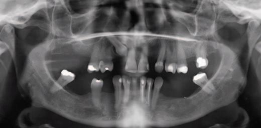
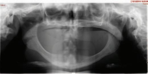
4: Pre-operative orthopantomogram of edentulous dentition revealing proximity of reduced maxillary ridge height to the maxillary sinuses.
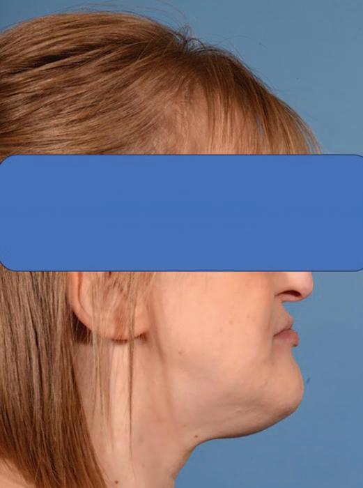
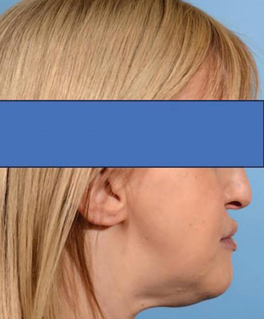
Upon clinical examination, she presented with a Class III skeletal pattern (Figure 1) with average vertical proportions, a hypoplastic maxilla, and she was edentate. She had undergone multiple previous attempts at denture construction; however, these were of limited success.
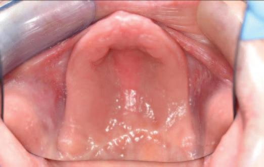
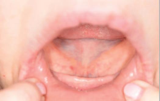
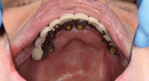
Intra-orally, the patient presented with an atrophic mandible and maxilla (Cawood and Howell13 Class VI and V, respectively) and was edentate (Figures 2 and 3). An orthopantomogram (Figure 4) confirmed the clinical findings and also demonstrated the proximity of the reduced maxillary ridge height to the maxillary sinuses.
Following clinical and radiographic findings, it was deemed that an MDT approach was required to comprehensively manage the patient’s skeletal and
FIGURE 1: Pre-operative lateral view of patient’s Class III skeletal pattern and hypoplastic maxilla.
dental concerns, and she was referred onwards to the OMFS Department, where she was jointly assessed by a consultant in OMFS, a consultant in orthodontics and a consultant in restorative dentistry. Using multiple 3D printed models in an OMFS laboratory, cone beam computed tomography (CBCT) imaging, 3D planning software (KLS Martin) and Straumann’s coDiagnostiX dental implant planning software, an OMFS consultant and a consultant in restorative dentistry planned the positioning of the implants on the 3D printed models. A consultant in orthodontics provided input to the planning of the skeletal movements in relation to the overall positioning and relationships of the dental bases for subsequent restorative treatment. They also provided input to the inclinations of the dental implants and subsequent denture teeth in association with the OMFS surgeon and restorative dentist throughout treatment. The three consultants decided upon the use of the piezoelectric system as part of the OMFS, to ensure that the optimal patient outcome was achieved.
Treatment options were discussed with the patient and it was agreed to pursue the following treatment plan:
1: Two-step surgical approach including:
a) bimaxillary osteotomy and bone graing, followed by, b) delayed insertion of transzygomatic and conventional dental implants (once adequate OMFS healing and successful bone graing were confirmed).
2. Restorative dental treatment to include a full arch maxillary bridge and a lower implant-retained and -supported denture (once adequate healing of implants was confirmed).
This staged sequence was carefully chosen by the MDT during pre-operative planning to ensure maximum implant stability and long-term success, and was based on sound clinical and biological principles to optimise bony healing and gra integration prior to implant insertion. The aims of treatment were:
1. To improve the patient’s skeletal and so tissue profiles.
2. To address the insufficient alveolar height and width, sulcus depth and incisal show of restorations.
First step of surgical approach: orthognathic surgery with bone graing
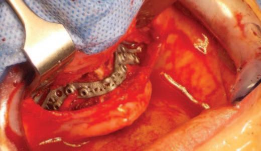
Following careful informed consent, the patient was treated in the OMFS Department in Ninewells Hospital. The first surgical stage was completed with a bimaxillary osteotomy with 3D guides and plates (KLS Martin) followed by bone gras from the le iliac crest to the maxilla and mandible (Figure 5). The maxilla was advanced 10mm and set down 7mm.
Second step of surgical approach: implant placement
Six months aer the first surgical procedure, a CBCT scan was taken in order to assess the position of the mandible to the maxilla and MDT discussions were repeated. Radiographic evaluation revealed appropriate healing with stable bone height. The patient then underwent the second stage of her surgical management, and transzygomatic implants (maxilla) and conventional dental implants (mandible) were inserted.
The piezoelectric system was very important in the management of this patient. It was used to split the maxillary and mandibular ridges (UR4, UR1, LL3 and LR2 regions) and also to remove the bone and open the alveolar ridge anteriorly and posteriorly on the right side of the maxilla.
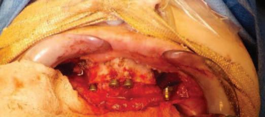
Bone level fixtures were placed in the intraforaminal region with submerged healing, and axially loaded. Aer alveolar ridge height reduction to facilitate implant placement in the apico-coronal position, one Southern zygomatic implant (Zygex with uniabutment and healing cap) was placed in the le quadrant and three 10mm bone-level implants (021.3310) were placed (UR4, UR1 and UL1 sites) (Figure 6). Two 12mm implants (021.5310) were then placed in the mandible (LL3, LR2 regions).
Restorative treatment
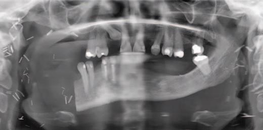
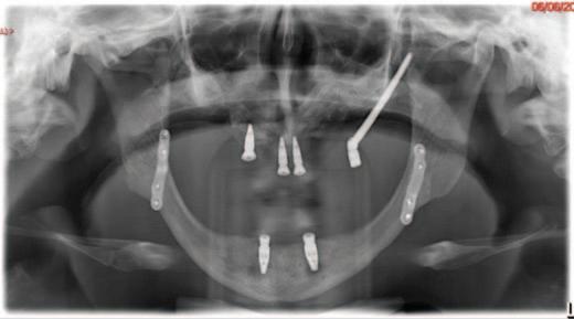
A follow-up orthopantomogram revealed that all implants were healing well (Figure 7). The shortened dental arch (SDA) concept was used to restore this patient’s occlusion, and a maxillary bridge and mandibular implant-retained
FIGURE 5: Intra-operative photograph of 3D plate.
FIGURE 6: Intra-operative photograph of implants in upper left quadrant and UR4, UR1 and UL1 sites.
FIGURE 7: Postoperative orthopantomogram.
denture were used to restore second premolar to second premolar in each arch (Figures 8 and 9). SDA was introduced in 198114 and is a problem-oriented method that aims to minimise complex restorative treatments.15 It outlines maintaining at least four posterior occlusal units or a minimum of 20 well-distributed teeth to ensure a functional, natural and healthy dentition.16
Follow-up
Follow-up clinical examination performed 24 months post treatment has revealed a clinically stable and functional occlusion, with minimal relapse post orthognathic surgery and an enhanced facial profile. The patient is very satisfied with the aesthetic and functional outcome, and her skeletal and dental concerns have been successfully addressed.
Discussion
Oral and maxillofacial surgery and the piezoelectric system
The piezoelectric system is a conservative and precise ultrasonic surgical system, which is used to perform orthognathic surgery and designed to be active on mineralised tissues only. It consists of a handpiece and various tips for cutting bone, bone substitutes and teeth. It has a narrow kerf for enhanced precision to perform bone surgery procedures and to reduce the risk to so tissues. This device maintains blood-free operation sites through a cavitation effect and allows for precise cutting of the bone by using micro-vibrations at frequencies of 24-29kHz with 60-200μm amplitude to cut through the bone.14 It has been suggested that in comparison to the traditional saw technique used in orthognathic surgery, the piezoelectric device has demonstrated more favourable outcomes in terms of neurosensory disturbance and operative blood loss, and is less traumatic and more efficient.17 The piezoelectric device is used in dentistry in maxillofacial
osteotomy, dental implantology, alveolar decortications, resection of cyst- and tumour-like lesions, corticotomy and temporomandibular joint (TMJ) ankylosis.18
The novelty lies not solely in the tool, but in the context-specific application, the piezoelectric instrumentation in the osteotomy of an underdeveloped maxilla, which remains under-reported in the literature. This approach allowed cuts to be within a controlled small width and avoided unnecessary bone loss. It also minimised the risk of so tissue injury. It was initially virtually planned that the patient would have one zygomatic implant placed on each side; however, unfortunately, due to limited zygomatic bone volume, it was felt intra-operatively that this was not appropriate. Instead, a ridge split was performed to allow placement of dental implants into the maxilla. Using the piezoelectric saw allowed for widening of the mandibular alveolar ridge without the risks associated with chisels and osteotomes. The precision and safety profile of a piezoelectric instrument made it the essential tool in this situation, as its use was necessitated by the patient’s exceptionally thin maxillary bone. Conventional drills or chisels posed a significant risk of iatrogenic fracture or collateral damage to adjacent so tissue structures and were therefore unsuitable in this case.
This case outlines the successful application of the piezoelectric system in managing a patient with ED and a hypoplastic maxilla with limited postoperative complications, and in achieving an aesthetically pleasing Class I skeletal profile (Figure 10).
Bone augmentation
Previously, patients with ED have had oral function restored with partial or complete removable prostheses supported by teeth or tissue. In recent years, dental implant-supported prostheses have provided an alternative method of restoring form and function to these patients.19
FIGURE 8: Postoperative intra-oral view with maxillary bridge.
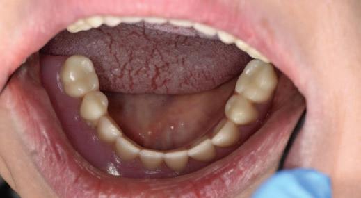
FIGURE 9: Postoperative intra-oral view with a lower implant-retained complete denture.
FIGURE 10: Postoperative lateral view demonstrating Class I skeletal profile.
As ED is a genetic disorder associated with premature tooth loss and severe oligodontia, the oral rehabilitation of these patients to improve quality of life and social confidence is of utmost importance.20 The demand for the prostheses to restore aesthetics and function while being comfortable for the patient is paramount. Fixed, implant-supported prostheses have been used to successfully restore some of the complex oral functions in patients with ED.21 However, the quantity and quality of alveolar bone greatly affect the success of implant restorations in these patients.20
As it is common for patients with ED to have hypodontia and thus a reduced quantity of bone, bone graing procedures are oen required prior to implant
placement. There are limited reports of successful reconstruction of both the mandible and maxilla with implants in patients with ED.22
In this case, both alveolar bone height and width were lacking as a result of hypodontia. The surgery was undertaken in two separate stages to allow for adequate bony healing post bone graing, and an accurate assessment of the relationship of dental bases to one another post orthognathic surgery. This was to ensure that a stable result would be achieved with restorative treatment and to reduce the risk of implant malpositioning. A CBCT scan allowed for the identification of alveolar ridge morphology and bone quality, and confirmed the success of dental implants prior to restorative treatment.
3D computer-aided planning system
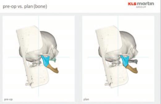
The use of a virtual 3D computer-aided planning system (KLS Martin) allowed the clinicians to accurately plan orthognathic surgical movements and acted as an excellent communication tool when communicating with the patient (Figure 11).
Restorative management
In 2013, the patient had been deemed unsuitable for standard Straumann implants due to insufficient bone. Polyetheretherketone (PEEK) implants were also unsuitable as there had not been a healing technology assessment of their efficacy in relation to this particular clinical situation, and further research and clinical trials are still required to explore the application of this material in orthognathic surgery.23
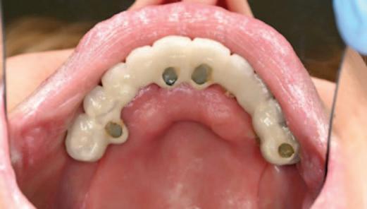
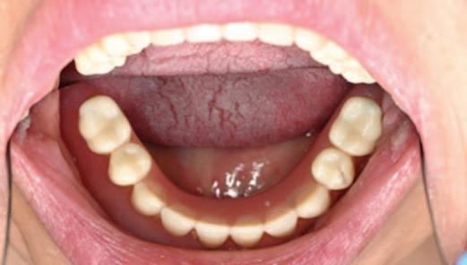
Careful digital restorative planning enabled a functional and aesthetically pleasing outcome to be achieved with a maxillary bridge and a mandibular implantretained and -supported denture (Figures 12 and 13). Enhancements in the speed of osseo-integration, the strength of dental implants and the range of prosthodontic materials contributed to the restorative success.
Multidisciplinary team planning
MDT planning was central to the success of the case, integrating input from OMFS, restorative dentistry and orthodontics, with careful surgical sequencing and patientspecific considerations. As noted in the aforementioned meta-analysis,17 piezoelectric techniques offer enhanced safety and control, particularly in delicate anatomical areas, further justifying their benefit in this case. While the use of the piezoelectric saw was technically significant, it was the MDT’s collaborative decision-making that led to this choice and ensured optimal patient safety and outcomes.
Conclusions
Treatment in this case has involved a lengthy course of OMFS and restorative treatment, including multiple MDT clinics. The two-stage surgical approach, with careful planning by OMFS, restorative dentistry and orthodontics, allowed for adequate bony healing and the successful placement of dental implants in a patient with ED, hypodontia and a hypoplastic maxilla. This interdisciplinary care was crucial in achieving a successful outcome for this patient. This case also demonstrates the favourable use of piezoelectric saws in the osteotomy of an underdeveloped maxilla. All patients with ED, and with concerns regarding their skeletal profile or dental function, should be referred onwards by their general dental practitioner for MDT planning. This will allow for all available treatment options to be discussed and for the patient to make the best-informed decision on their treatment.
Ethical statement
We can confirm that written informed consent for publication was obtained from the patient who was included in this case report.
FIGURE 11: 3D computer-aided planning system (KLS Martin) outlining pre- and postoperative orthognathic plan.
FIGURE 12 : Intra-oral view of maxillary bridge.
FIGURE 13: Mandibular implant-retained and -supported denture.
References
1. Bhakta P, Barthunia B, Nigam H, Pawar P. Ectodermal dysplasia – a rare case report. J Family Med Prim Care. 2019;8(9):3054-3056.
2. Itthagarun A, King NM. Ectodermal dysplasia: a review and case report. Quintessence Int. 1997;28(9):595-602.
3. Seraj B, Nahvi A. Hydrotic or hypohydrotic ectodermal dysplasia: diagnostic dilemmas. Int J Curr Microbiol App Sci. 2015;4:778-783.
4. Kumar A, Thomas P, Muthu T, Mathayoth M. Christ-Siemens-Touraine syndrome: a rare case report. J Pharm Bioallied Sci. 2019;11(1):102-104.
5. Nowak AJ, Christensen JR, Mabry TR, Townsend JA, Wells MH. Pediatric Dentistry-EBook: Infancy Through Adolescence. Elsevier Health Sciences; 2018.
6. Clarke A. Hypohidrotic ectodermal dysplasia. J Med Genet. 1987;24(11):659-663.
7. Yenisey M, Guler A, Unal U. Orthodontic and prosthodontic treatment of ectodermal dysplasia – a case report. Br Dent J. 2004;196(11):677-679.
8. Anbouba GM, Carmany EP, Natoli JL. The characterization of hypodontia, hypohidrosis, and hypotrichosis associated with X-linked hypohidrotic ectodermal dysplasia: a systematic review. Am J Med Genet A. 2020;182(4):831-841.
9. Rosa RR, Janeiro MM, Camargo SEA, de Oliveira Tocalino Walter Porto C, Kreich EM, Henriques JCG. Radiographic study of patients with ectodermal dysplasia and partial. Indian J Dent Res. 2012;23(6):801-805.
11. McDonald RE, Avery DR, Dean JA. Dentistry for the Child and Adolescent. Mosby Incorporated; 2004.
12. Farrington FH. The team approach to the management of ectodermal dysplasias. Birth Defects Orig Artic Ser. 1988;24(2):237-242.
13. Cawood JI, Howell RA. A classification of the edentulous jaws. Int J Oral Maxillofac Surg. 1988;17(4):232-236.
CPD questions
To claim CPD points, go to the MEMBERS’ SECTION of www.dentist.ie and answer the following questions:
1. The two most common syndromes of ectodermal dysplasia (ED) may be broken down into:
● A. Hidrotic ED and anhidrotic ED
● B. Regular ED and irregular ED
● C. Mild ED and moderate ED
14. Kayser AF. Shortened dental arch: a therapeutic concept in reduced dentitions and certain high-risk groups. Int J Periodontics Restorative Dent. 1989;9(6):426-449.
15. AlHmoudi H, Khamis AH, Elbishari H, Amir-Rad F. The shortened dental arch concept: awareness, knowledge, and practice of dentists in Dubai and the northern Emirates, United Arab Emirates. Int J Dent. 2022;6018650.
16. Eswaran B, Ranjani T, Ponsekar Abrahaam A, Praveen Kumar P, Vishva M. Shortened dental arch concept in prosthodontics: a review. Int J Res Publ Rev. 2024;5(12):13801384.
17. Alrefai M, Daboul A, Fleischhacker B, Landes C. Piezoelectric versus conventional techniques for orthognathic surgery: systematic review and meta-analysis. J Stomatol Oral Maxillofac Surg. 2022;123(5):e273-e278.
18. Akbar Z, Saleem H, Ahmed W. Critical analysis of piezoelectric surgery with oscillating saw in bimaxillary orthognathic surgery. J Coll Physicians Surg Pak. 2017;27(6):348-351.
19. Johnson EL, Roberts MW, Guckes AD, Bailey LJ, Phillips CL, Wright JT. Analysis of craniofacial development in children with hypohidrotic ectodermal dysplasia. Am J Med Genet. 2002;112(4):327-334.
20. Wang Y, He J, Decker AM, Hu JC, Zou D. Clinical outcomes of implant therapy in ectodermal dysplasia patients: a systematic review. Int J Oral Maxillofac Surg. 2016;45(8):1035-1043.
21. Ekstrand K, Thomsson M. Ectodermal dysplasia with partial anodontia: prosthetic treatment with implant fixed prosthesis. ASDC J Dent Child. 1988;55(4):282-284.
22. Bayat M, Khobyari MM, Dalband M, Momen-Heravi F. Full-mouth implant rehabilitation of a patient with ectodermal dysplasia aer orthognathic surgery, sinus and ridge augmentation: a clinical report. J Adv Prosthodont. 2011;3(2):96-100.
23. Hao Y, Shi C, Zhang Y, et al. The research status and future direction of polyetheretherketone in dental implant – a comprehensive review. Dent Mater J. 2024;43(5):609-620.
2. The most common oral characteristic in many cases of anhidrotic ED is:
● A. Supernumerary teeth
● B. Hypodontia or anodontia
● C. Ectopic teeth
Quiz answers
1. Correct answer: C
These are surveyed crowns designed to provide controlled contours, guiding planes, and rest seats for an RPD.
2. Correct answer: B
Surveyed crowns commonly include rest seats, guiding planes, and retentive undercuts for clasping.
3. From a dental perspective in people with ED, the following dentition/s are affected:
● A. Both the deciduous and permanent dentitions
● B. The deciduous dentition only
● C. The permanent dentition only
3. Correct answer: C
Surveyed crowns allow precise control of the path of insertion, retention, support, and stability of the RPD.
Questions on page 48
Elite teeth
Dr Annie Hughes works in private practice in Dublin, and supervises part-time in the Dublin Dental University Hospital (DDUH). She recently published research on the diet and oral health of elite athletes in Ireland.
Tell us about your background, what led you to dentistry, and to dental research. I was always interested in some sort of career in healthcare, but I felt dentistry offered an ideal blend of hands-on work and patient interaction. During my training, I particularly enjoyed prosthodontics – there’s a lot of problem solving involved in it and great satisfaction with completing technical treatment plans. Aer I graduated, I completed several house officer years, but still had a strong desire to go further in prosthodontics, so I completed the three-year clinical doctorate at the DDUH. I really enjoy the specialty because it combines the artistic and scientific sides of dentistry.
Can you share the primary focus of your recent research and what inspired you to pursue this topic?
Because the prosthodontic degree was a clinical doctorate, there was a large research component. One of my supervisors came to me with the idea, and I was interested because I was training for the Dublin Marathon and taking on energy gels during longer runs. We collaborated with Sport Ireland, and looked at the diet and oral health of elite athletes in Ireland. We found that 90% of participants had some evidence of caries, and they were all frequent users of sports nutrition products. The research highlighted the potential impact of sport-specific dietary practices on oral health.
What challenges did you encounter during your study? How did you overcome them?
In Ireland, we have a lack of up-to-date national oral health data for comparison, so we can’t compare to our general population, but we carefully interpreted our findings within the limitations of the available literature. Another challenge was gaining access to the elite athletes. They all have very demanding schedules and limited availability, so that required flexibility and good collaboration with Sport Ireland.
How do you see your research contributing to clinical practice or patient outcomes in dentistry?
I think it highlights a need to look at sport-specific dietary behaviours when assessing dental caries risk, and at a more tailored approach to prevention, particularly for athletes who have frequent carbohydrate exposure. We need to integrate oral health into wider athlete care, with collaboration between dental professionals, dietitians, sports scientists, and medics so that we can support their performance, but also their long-term health and oral health.
Are there any misconceptions in the field that your research helps to clarify?
A common misconception would be that athletes are generally very healthy, and maybe at lower risk of oral disease. But with their fuelling practices and dietary behaviours, the research has shown that they do seem to be at higher risk. Exerciseinduced physiological changes, such as reduced saliva flow and increased mouth
breathing, can lead to dehydration of the oral cavity, making it more prone to dental diseases. The risk is further increased by the frequent intake of carbohydrates and sports nutrition products.
Which emerging technologies or methods do you believe will most significantly shape the future of dental research?
Our research is cross-sectional, so it’s just a snapshot in time of this particular cohort. While this is valuable for identifying associations, there is considerable scope for future longitudinal research to better understand relationships and changes over time. Salivary diagnostics and oral microbiome profiling are likely to be key emerging tools, enabling a shi beyond clinical outcomes to better understand biological responses to diet and training stressors. I also think there’s significant scope for innovation within sports nutrition – opportunities to develop oral health-friendly fuelling strategies and product formulations that support performance but also protect dental health.
How important is interdisciplinary collaboration in your work, and can you give an example where it made a key difference?
It’s definitely essential in this field, and I think our research helped to bridge that gap between dentistry and sports nutrition. I believe the initial idea came from the sports nutritionist in Sport Ireland, who felt that these diets were likely impacting oral health. Since then, she’s put up infographics in the canteen for the athletes, and they’re provided with oral health products. I found it equally important to understand the sports nutrition perspective; it’s easy as dentists to advise patients to “reduce sugar”, but athletes need increased carbohydrate intake and frequency. The collaboration meant the advice we gave was practical, balanced, and realistic for athletes.
What advice would you give to young dental researchers just starting out?
I think it makes it a lot easier if it’s something you’re genuinely interested in, because it helps you stay motivated and enjoy the process. I was lucky that this was the case for me, and I really enjoyed the background reading on sports nutrition. Having good mentors always helps. I’d also say to stay open to interdisciplinary opportunities, because some very interesting research questions that haven’t been looked at lie between traditional fields rather than simply within your own field.
Outside of your research, how do you maintain balance and well-being in such a demanding field?
I’ve actually gotten a little bit more into sport, probably from learning a bit more about sports nutrition and getting inspiration from the athletes. I run a lot and I train regularly with a middle-distance group, so I do a little bit of competing on the track and some road races as well. Outside of that, I really like travelling, going on holidays, spending time with friends and family, and trying new restaurants.
References
1. Hughes A, O’Sullivan M, Winning L, et al. Diet and dental caries in elite athletes in Ireland. J Hum Nutr Dent. 2026;39(1):e70203.
2. World Health Organization. Bangkok Declaration – No Health Without Oral Health. 2024. cdn.who.int/media/docs/default-source/ncds/mnd/oral-health/bangkokdeclaration-oral-health.pdf.
3. Hollander K, Eshkol-Yogev I, Zech A, Buti J, Needleman I. The influence of oral health on sports performance: an interdisciplinary perspective. Br Dent J. 2026;240:277-283.
4. Konviser SN, Nurek M, Needleman I, Fine P. Disadvantage starts early: academy football has high levels of oral disease. BMJ Open Sport Exerc Med. 2025;11(2):e002245.
5. Halvorsrud K, Lewney J, Craig D, Moynihan PJ. Effects of starch on oral health: systematic review to inform WHO guideline. J Dent Res. 2019;98(1):46-53.
6. Jangda FH, Suominen AL, Lundqvist A, Mannisto S, Golkari A, Bernabé E. Starch intake and changes in dental caries among adults: a longitudinal study in Finland. J Public Health Dent. 2025;85(1):29-39.
7. Needleman I, Rankin A, Ashley P et al. Infographic. Nutrition and oral health in
sport: time for action. Br J Sports Med. 2019;53(22):1432-1433.
8. Gallagher J, Ashley P, Needleman I. Implementation of a behavioural change intervention to enhance oral health behaviours in elite athletes: a feasibility study. BMJ Open Sport Exerc Med. 2020;6(1):e000759.
9. Gallagher J, Ashley P, Petrie A, Needleman I. Oral health-related behaviours reported by elite and professional athletes. Br Dent J. 2019; 227(4):276-280.
10. Public Health England. Delivering better oral health: an evidence-based toolkit for prevention. 3rd Edition. 2021. https://www.gov.uk/government/publications/delivering-better-oral-health-anevidence-based-toolkit-for-prevention
11. Needleman I, Ashley P, Weiler R, McNally S. Oral health screening should be routine in professional football: a call to action for sports and exercise medicine (SEM) clinicians. Br J Sports Med. 2016;50(21):1295-1296.
12. Ljungqvist A, Jenoure P, Engebretsen L, et al. The International Olympic Committee (IOC) Consensus Statement on periodic health evaluation of elite athletes March 2009. 2009. www.olympic.org/Documents/Reports/EN/en_report_1448.pdf