Research Paper
Science
E-ISSN No : 2454-9916 | Volume : 5 | Issue : 8 | Aug 2019
STUDIES ON BIOLOGICAL ARCHITECTURE OF CEPHALIC MUSCLES IN THE FISH CHANNA ORIENTALIS Meena T. Nikam Shri Shivaji Science and Arts College Chikhli, Dist. Buldana, Pin- 443201.(M.S) India. ABSTRACT The present investigation shows that the muscles of the opercular region, the levator arecus palatine and dilator opercula are not well developed, due to air-breathing habits of fish. The frequent movements of operculum as seen in other fishes are reduced. Although the frequent opening and closing of the mouth is absent in this fish, the muscles of the adductor mandibulae are very well developed due to carnivorous habit which needs powerful muscles to crush the food. The eye muscles in Channa orientalis have similar pattern as found in majority of fishes. KEYWORDS: Study, Muscle, cephalic region, channa orientalis, Architecture. INTRODUCTION: The musculature in fishes attracted the attention of several workers in past. The pioneer workers were (Vetter 1878) and (Allis 1897) and who worked on the cranial musculature of Amia calva and Scomber. (Adams 1919) contributed an account on the jaw muscles of Acanthias, Polydons, Acipenser, Amia, Lepidosteus, Esox, Angulla etc. (Takahasi 1925) gave a comparative account of the phylogenetic significance of the cranial muscles in about 20 species of cypriniform fishes. (Edwards 1926) and (Eaton 1943) have worked on the muscles responsible for the protection of jaws in Catastomid and Cichilds respectively. (Kestevan 1943) has worked on the evolution of skull and cephalic muscles of several teleosts. ( Al-Hussaini1949) has studied the cranial muscles associated with feeding mechanism of Cyprinus carpio, Gobio gobio and Rutilus rutilus. ( Saxena 1960) investigated on the Hilisa ilisa Catla Catla, Notopterus chital, Mystus aor and Ophiocephalus. (Shann1921) in his notable studies on the comparative myology of the shoulder girdle and pectoral fins of fishes, has summarized the work done by earlier authors. (Dubale and Rao 1961) have described the pectoral fin musculature in Macrones cavasius, Callichrous macrothalamus and Heteropneusters fossils. (Dubale and Christian1963) have worked on the pelvic fin musculature and its function in Clarias magur. Heteropneustes fossils, (Lounder 1980) reported feeding mechanism and cephalic muscles of salmonid fishes. Nursall (1956, 1958 and 1963) has described the lateral and caudal musculature of fishes. Present investigation was launched for the study of cephalic musculatory system in the fish Channa orientalis. MATERIALS AND METHODS: INFORMATION OF THE FISH: The species which has been selected for the present study is of economic value and readily available throughout the year and it stands in capacity well. The fish is most sensitive. It represents the natural population in the river or water bodies of Amravati (Latitude 200-56N, Longitude 770-45E) and is resistant to handling and transportation. Locally the fish is known as “DOK” and is common edible fish in the region and fetches high price. COLLECTION, MAINTENANCE AND ACCLIMATIZATION METHODS OF FISH: The fishes were collected from localities around Amravati region. The fishes were first brought into the laboratory and transferred to the glass aquarium and were inspected for any possible injury or infection. Only the healthy fishes were selected and washed with dilute solution of potassium permanganate (KMnO4, 1.0mg/1) to remove dermal infection if any. Fishes were separated in different groups and were used for studying different parameters. A number of fresh live specimens of Channa orientalis were collected from localities around Amravati region, brought to the laboratory and dissected either in fresh or preserved condition for the study of muscles. The nomenclature for the girdle and fin muscles and cranial muscles was adopted after Shann (1921) and Munshi (1960) respectively.
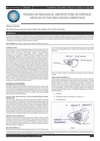
atine. It arises from the posterior part of the pterotic bone and extends beyond the process of the hyomandibula and is inserted on the antero dorsal edge of the operculum.
Fig. 1 This muscle helps in lifting the operculum and widening the gap of opercular opening. ADDUCTOR MANDIBULAE: These muscles are associated with the movements of the lower jaw in masticating the food and in opening and closing of the mouth during respiration. The opening and closing of the mouth is a frequent feature in aquatic respiration but as Channa orientalis can breath in atmospheric oxygen these movements are reduced. Only when the fish is chased or disturbed. It shows movements of mouth and operculum. Though strongly developed muscles are not required for the normal breathing, the carnivorous and predatory habits of the fish involve strong development of these muscles. The adductor mandibulae is divided into the following parts: a) Maxillaris b) Mandibularis which is subdivided into : Adductor 1-3 c) Intramandibularis
OBSERVATION AND RESULT: The present study deals with the myology and an attempt is made to explain architecture of the muscle in cephalic region. The levator arecus palatine is a broad sheet of muscles spread over the hyomandibulae. On the dorsal side it is very broad and tapers on the ventral side. It is triangular in shape. It is placed below the adductor mandibula (two) and arises from the ventrolateral surface of the frontal, sphenotic and pterotic. DILATOR OPERCULA: Dilator opercula is poorly developed and posed posterior to the levator arecus pal-
Fig. 2
Copyright© 2019, IERJ. This open-access article is published under the terms of the Creative Commons Attribution-NonCommercial 4.0 International License which permits Share (copy and redistribute the material in any medium or format) and Adapt (remix, transform, and build upon the material) under the Attribution-NonCommercial terms.
International Education & Research Journal [IERJ]
51
Research Paper
E-ISSN No : 2454-9916 | Volume : 5 | Issue : 8 | Aug 2019
MAXILLARIS: In Channa orientalis the maxillaries is a strongly developed muscle, which is partially covered by adductor 3 and adductor-1. On the upper side it is covered by adductor-3 and on the lower side by adductor-1. The muscle originate from the anterior face of the lower half of the operculum and runs straight to be inserted on one third part of maxilla from lower side by means of a narrow tendon. The upper Jaw is protractile in Channa orientalis. It is represented by the maxilla so the maxilla is not fixed at a place, it is protractile and retractile. The tendon of maxillaries makes the maxilla to protract and retract. MANDIBULARIS: ADDUCTOR 1: It is a very well developed muscle arising from the face of the lower portion of the pre-operculum. It covers the maxillaries completely and the adductor 3 partly. A slight line of demarcation is seen between the adductor 2 and adductor-3. The tendon of adductor 1 is very short, it is inserted partly on the articular and partly on the dentary. Fig. 5 INTERMANDIBULARIS POSTERIOR: It runs parallel to the body line of the fish and fuses posteriorly with the muscles of the hyoid region forming a continuous sheet of muscles leaving a very clear demarcation between intermandibularis posterior and the muscle of the hyoid region, the interhyoideus muscles. The intermandibularis posterior unites at the anterior region to the common tendon which is inserted at the symphysis. The continuous sheet which is formed by the union of hyoideus muscles and the intermandibularis posterior is known as protractor hyoideus.
Fig. 3 ADDUCTOR 2: This muscle is the largest muscle among the adductors 1, 2 and 3. It arises from the ventro lateral surface of the sphenotic, pterotic and surface of the hyomandibula. The muscles arising from these bones unite together forming a large muscle. It has a very strong tendon which unites with the tendon of the adductor 3. Adductor 2 covers levator arecus palatine on the upper surface. Adductor 2 and 3 are differentiated by the Vth mandibularis, which lies on the lower side of the adductor 2, but emerges between adductors 2 and 3 and runs over the common tendon of adductors 2 and 3.
DISCUSSION: The cranial musculature of Channa orientalis is similar to that of Ophiocephalus striatus (Munshi, 1960). The muscles of the opercular region, the levator arecus palatine and dilator opercula are not well developed, due to air-breathing habits of fish. The frequent movements of operculum as seen in other fishes are reduced. Although the frequent opening and closing of the mouth is absent in this fish, the muscles of the adductor mandibulae very well developed due to carnivorous habit which needs powerful muscles to crush the food (Munshi, 1960). The eye muscles in Channa orientalis have similar pattern as found in majority of fishes. The adductor are formed of three groups of muscles as found in Calcichrous macropthalamus, Macrones cavasius and Heteropneustus fossils (Dubale and Rao, 1960) and in majority of fishes (Shann, 1921). CONCLUSION: The muscles of the opercular region, the levator arecus palatine and dilator opercula are not well developed due to air breathing habit of the fish. Maxilla is not fixed at one place. It is protractile and retractile due to tendeons. In this fish the muscles of the adductor mandibulae are very well developed due to carnivorous habit of the fish. REFERENCES:
Fig. 4 ADDUCTOR 3: It arises from the symplectic metapterygoid and the quadrate and unites into a broad and strong tendon which then unites with the tendon of adductor 2 and enters into the dentary. INTRAMANDIBULARIS: It is well developed and lies inside the supra-Meekelian fossa. It is placed along the whole length of the Meckel's cartilage, and it partially united with the common tendon of adductor 2 and adductor 3. INTERMANDIBULARIS: In Channa orientalis the intermandibularis is formed by two muscles the intermandibularis anterior and the intermandibularis posterior. Both the muscles are well developed. These muscles are present on the ventral surface, between the two rami of the lower jaw.
1.
Vetter B. (1878): Untersuch Zaur Vergl anatder Kiemenund-kiefmus-culature der fische, Theil II. Jena Zeitschr. F. Naturw, 12(3), 431-550.
2.
Adams L. A. (1919), A memoir on the phylogeny of the jaw muscles in recent and fossil vertebrates Ann., N. Y. Acad. Sci., 51-56.
3.
Shann E. W. (1921): The comparative mycology of the fish shoulder girdle and pector al fin of fishes. Trans R. Soc. Ednib, 52, 531-570.
4.
Edwards L. F. (1926): “The protectile apparatus of the mouth of the catastomoid fishes, Anat., Rec., 33, 267.
5.
Kestavan H. L. (1942-43), The evolution of the skull and cephalic muscles a comparative study of their development and adult morphology, Mem Aust., Mus., 8(1-63), 63132.
6.
Al-Hussaini A. H. (1949): “On functional morphology of the alimentary tract of some fish in relation to differences in their feeding habits. Anatomy and histology”, Q JI microsc., Sci. 90(2), 109-140.
7.
Nursall J. R. (1956): The lateral musculature and the swimming of the fish Proc. Zool., Soc., Lond. 126, 127-143.
8.
Nursall J. (1958): The caudal fin as a hydrofoil, Evolution 12(1), 116-120.
9.
Saxena (1960): The cranial muscularature of a hilstream cyprinil fish Garra mullya (Sykes). Proc., Natn. Inst. Sci. India, 26(40), 176-188.
10. Munshi J. D. (1960): The structure of the gills of certain freshwater teleost, Ind. J. Zootomy 1(3), 135-174. 11. Nursall J. R. (1963): The caudal musculature of holopagrus guntheri gill (Perci forms : Lutjanidae) Can. J. Zool. 41, 865-880. 12. Dubale M. S. and E. Christian (1963-64): “Functional anatomy of the pelvic fins of certain Indian freshwater siluroids” J. Uni. Bombay, XXXII 54,55, 104-116.
INTERMANDIBULARIS ANTERIOR: This muscle is transversely placed between the two rami of the lower jaw at the symphysis. It runs over the tendon and the anteriomost part of the intermandibularis posterior.
52
International Education & Research Journal [IERJ]