Original Article
Medical Science
E-ISSN No : 2454-9916 | Volume : 4 | Issue : 9 | Sep 2018
MANAGEMENT OF GRADE I AND II HEMORRHOID BY SCLEROSANT INJECTION –A STUDY OF 100 CASES 1
Dr. Priya D Dhandore | Dr. N N Hombalkar 1 2
2
Assistant professor, Department of General Surgery, Government Medical College, Miraj. Associate professor, Department of General Surgery, Government Medical College, Miraj.
INTRODUCTION: For as long as man has been blessed with an anus, it is fair to assume that he has also been doubly blessed with haemorrhoids. Haemorrhoids are one of the most common ailments to afflict mankind. It is almost impossible to calculate its prevalence as for many patients with haemorrhoids never have symptoms so whether such patients should be considered as diseased is still a question.
correlated Liver disease, Portal hypertension and Haemorrhoids. He proposed stopping of excess flow with ligation, excision and cauterization with hot iron rod. Haemorrhoidal disease has been referred to in the Edwin Smith Papyrus (1700 BC), in code of Hammurabi in Babylon (2250 BC) and in the Papyrus of Eber (1500 BC).
The word “HAEMORRHOID” is derived from Greek word Haemorrhoides, “Haemo” means blood and “rhoos” meaning flowing. The word “PILES” comes from Latin word “pila” meaning a pill or ball. To be accurate, we should call the disease as piles when the patient complains of a swelling and haemorrhoids when patient complains of bleeding.
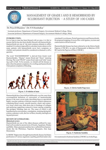
Fig no. 2: Edwin Smith Papyruses Fig no. 1: Evolution of man Haemorrhoids have been defined differently over the years from oversimplified definition of varicosities of haemorrhoidal plexus to the more recent study describing them as specialized highly vascular cushions of discrete masses of thick submucosa containing blood vessels, smooth muscles, elastic and connective tissue which may slide down due to breakage of collagen and anchoring supporting connective tissue causing symptoms like prolapse, bleeding, pain etc. The cause of which is still hypothesized as erect posture, constipation, straining during defecation, sedentary work, diet low in fibres, heredity and high resting anal pressure. REVIEW OF LITERATURE: Historical review: Haemorrhoids are one of the oldest diseases suffered by mankind well recorded in ancient texts of Greeks, Egyptians, Hindus and Bible. Many great personalities have suffered from haemorrhoids like the Philistines, Napoleon Bonaparte, Don Juan Demoranna. Fig no. 3: Sushruta Samhita In 400 BC Hippocrates thought haemorrhoids disease facilitates purification of various organisms expelling petrified matter also
It is mentioned in Sushruta Samhita (2500 BC) as Arsha Roga.
Copyright© 2018, IERJ. This open-access article is published under the terms of the Creative Commons Attribution-NonCommercial 4.0 International License which permits Share (copy and redistribute the material in any medium or format) and Adapt (remix, transform, and build upon the material) under the Attribution-NonCommercial terms.
International Education & Research Journal [IERJ]
1
Original Article
E-ISSN No : 2454-9916 | Volume : 4 | Issue : 9 | Sep 2018 orrhoids by Fraser and Gill.
The Italians following Galenic teachings referred to Profluvio de Sangue suggesting an overflow of blood. The ancient French referred to Flux d'or- flow of gold and ancient Germans called it the golden ader- golden veins implying that proctological symptoms may have been the prerogative of the rich. Polite society in 17th century referred to haemorrhoidal disease as le mal de St. Phiacre. In 1749 Morgagni attributed haemorrhoidal disease to erect posture of man. “Haemorrhoids are exceedingly rare in animals except for a few fat old dods”. In 1855 Verneuil suggested occlusion of superior haemorrhoidal veins by fecal bolus caused anal varicosities. Advances in treatment: Riverius 1657 advised topical application of nitric acid.
Ÿ Ÿ
In 1774, Jean Louis Petit became the father of sub epithelial haemorrhoide-ctomy.
Ÿ
In 1818, Boyer advised partial division of sphincter.
Ÿ
In 1829, Fecamier and 1864 Maisonneuve advised anal dilatation.
Ÿ
In 1869, Morgan described injection treatment using persulphate of iron.
Ÿ
IN 1879, Andrews of Chicago introduced the phenol secret of Dr. Mitchell to the medical profession using 30% phenol in arachis oil.
Ÿ
In 1882, Whitehead advocated total excision of pile bearing area with primary suture, in which an entire tube of mucosa and submucosal vascular tissue was removed. The subsequent wound breakdown and severe stenosis was called the post Whitehead deformity. This procedure had since been abandoned.
Ÿ
In 1969 and 1973, Lewis popularized it in the USA and in 1973 Lioyd Williams in United Kingdom.
Ÿ
In 1977, Nath developed the infrared coagulator, which was popularized by Neiger in 1979 in treatment of haemorrhoidal disease.
ANATOMY: The anal canal is the most terminal portion of the gastrointestinal tract. It begins at the anorectal junction and is about 4 cm long and terminates at anal verge. The anorectal junction is angulated in relation to the rectum due to pull of puborectalis muscle producing anorectal angle. It lies 2-3 cm in front and slightly below the tip of the coccyx. At this point the ampulla of rectum suddenly narrows and pierces the pelvic diaphragm. The anal verge is marked by a sharp turn where the squamous epithelium which lines the lower anal canal becomes continious with the skin of perineum. Embryologically the proximal two thirds of the anorectal canal develop from the distal part of the hindgut, whereas the distal third of anal canal is derived from ectodermal pit called the “anal pit” or “proctodeum”. The pit is created when the mesenchyme around anal membrane proliferates to form a raised border. The anal membrane thus separates the endodermal and ectodermal portions of anorectal canal; the former location of this membrane is marked in adults by irregular folding of mucosa called “pectinate line”. The anal canal consists of an inner lining epithelium, a vascular sub epithelium, the internal and external anal sphincters and fibromuscular supporting tissue. The anal canal is attached posterior to coccyx by the anococcygral ligament.This is a midline fibromuscular structure which possess some skeletal muscle elements. This fibromuscular structure runs between the posterior aspect of external sphincter and coccyx. Just above anococcygeal ligament is the raphe of the levator plate (the fusion of two halves of iliococcygeus muscle) which merges anteriorly with the puborectalis muscle. Between these two structures is the potential post anal space.
The 20th century is often described as era of the pectin band. Ÿ
In 1919, Miles described the wide V-shaped excision of preanal skin and division of the pectin band.
Ÿ
In 1928, Blanchard revived fixation techniques by sub mucus injection of 5% phenol in arachis oil.
Ÿ
In 1937, Milligan described the low ligation technique, which later became associated with the name of Naunton Morgan as the Milligan-Morgan technique nicknamed as the Smash and Grab haemorrhoidectomy or the 5-minute job.
Ÿ
In 1951, Eisenhammer popularized internal anal sphincterotomy.
Ÿ
In 1959, Ferguson popularized the closed haemorrhoidectomy technique.
Ÿ
In 1954, Blaisdell revealed his instrument for office ligation using a silk ligature, this was the forerunner of Barron's 1964 instrument for elastic band ligation.
Ÿ
In 1967, Cryosurgery was first used in management of haem2
Fig: 4 Anatomy of anal Canal Schwartz
RELATIONS OF ANAL CANAL: In both sexes the anal canal is related interiorly to the perineal body. Perineal body in males separates anal canal from membranous urethra and penile bulb. In females it separates anal canal from lower vagina. Posteriorly the anal canal is related to the tip of the coccyx and anococcygeal ligament and laterally to lose adipose tissue of ischiorectal fossa. International Education & Research Journal [IERJ]
Original Article
E-ISSN No : 2454-9916 | Volume : 4 | Issue : 9 | Sep 2018
INTERIOR OF THE ANAL CANAL: Anal Canal is divided into 3 parts: 1. Upper part 2. Middle part 3. Lower part Upper part (Mucous): This is in continuation of the rectum. The upper part is lined by columnar epithelium similar to the rectum. It contains secretory and absorptive cells with numerous tubular glands or crypts. The subepithelial tissues are mobile and relatively distensible and possess profuse submucosal arterial and venous plexus. Terminal branches of superior rectal vessels pass downwards towards the anal columns. The submucosal veins drain into submucosal rectal venous plexus and also through fibres of upper internal anal sphincter into an intermuscular venous plexus. There are 6-10 vertical folds in the mucosa called the anal columns. Each column contains a terminal radical of superior rectal artery and vein. The vessels are largest in left lateral, right posterior, right anterior quadrants of wall of anal canal, where sub epithelial tissues expand into three anal cushions. The lower end of columns form small cresentric folds called as anal valves. between these anal valves lie small recesses referred as anal sinuses. The anal valves and sinuses together form the dentate or (pectinate) line. About six anal glands open into small depressions in anal valves called crypts. Middle part (Pecten): The middle part is a transitional zone, also lined by bluish appearing mucosa because of a dense venous plexus that lies between the mucosa and muscle coat. It is non-keratinized stratified squamous epithelium lacking sweat / sebaceous glands and hair follicles but contains numerous somatic nerve endings extending down to the inter sphincteric groove.9 This is the lower limit of pectane line which often has a whitish appearance and is referred to as the “white line (of Hilton)”. Lower part (cutaneous): The lower part is lined by hair bearing, keratinsing squamous epithelium continuous with perianal skin.
Fig no. 5: Anal Canal Musculature sabiston Muscles of anal canal: The anal canal is encircled by internal and external anal sphincters separated by the longitudinal layer and has connection superiorly to puborectalis and transverse perinei muscles.9 Internal anal sphincter: The internal anal sphincter is a well-defined ring of obliquely oriented smooth muscle fibres and is continuation of the circular muscles of the rectum; the internal sphincter terminates at the junction of superficial and subcutaneous component of external sphincter. The thickness of internal sphincter varies between 1.5 to 3.5 mm. The lower portion of internal sphincter is crossed by fibres from conjoint longitudinal coat which passes into submuInternational Education & Research Journal [IERJ]
cosa of the lower canal. Vascular supply: The vascular supply of internal sphincter is from the terminal branches of superior rectal vessels and branches of inferior rectal vessel. Innervation: The internal sphincter has both sympathetic and parasympathetic innervation. The sympathetic innervation originate in lower two lumbar segments via inferior hypogastric plexus. The parasympathetic innervation originates in second to fourth sacral segments via inferior hypogastric plexus. External anal sphincter: The external anal sphincter is a elliptical cylinder of skeletal muscle surrounding the anal canal as 3 divisions (subsequently proved invalid by Goligher1). Goligher demonstrated that a sheet of muscle runs continuously upwards with the puborectalis and levator ani. The lowest portion occupies a position below and slightly lateral to internal sphincter a palpable groove at this level has been referred to as inter sphincteric groove.1 The lowest part is traversed by the conjoint longitudinal muscle. The intermediate portion is attached to coccyx by posterior extension of muscle fibres forming the anococcygeal ligament. The deep portion of external sphincter is devoid of posterior attachment and proximally becomes continuous with puborectalis muscle. Anteriorly the high fibres of external sphincter is attached to perineal body.1 Vascular supply: Terminal branches of inferior rectal vessels with contribution from median sacral artery. Innervation: Inferior rectal nerve, a branch of pudendal nerve originating, in anterior division of second to fourth sacral nerve roots. Conjoint longitudinal muscle: At the level of anorectal ring the longitudinal muscle coat of rectum is joined by fibres of levator ani and puborectalis, the conjoint muscle thus formed descends between internal and external anal sphincter. Many of these fibres traverse the lower portion of external sphincter to gain insertion into perianal skin referred as corrugator cutis ani1 some fibres of conjoint longitudinal muscle may form a longitudinal layer of muscle on the inner aspect of internal sphincter naming it muscularis sub mucosa ani. Some fibres that traverse the internal sphincter and become inserted just below anal valves returned as mucosal suspensory ligament. It has been suggested that the role of conjoint longitudinal muscle is to affix the anal canal and avert the anus during defecation. Anorectal ring: This is a muscular ring present at the anorectal junction, formed by fusion of puborectalis muscle, deep external sphincter and internal sphincter, less marked anteriorly where fibres of the puborectalis are absent.1 Surgical spaces related to anal canal: Ÿ Ischiorectal space on each side of anal canal. Ÿ
Perianal space surrounds the anal canal below the white line contains superficial part of external sphincter. External rectal venous plexus, terminal branches of inferior rectal vessels and nerves. 3
Original Article Ÿ
E-ISSN No : 2454-9916 | Volume : 4 | Issue : 9 | Sep 2018
Submucous space of the canal lies above white line between mucous membrane and internal sphincter contains internal rectal venous plexus and lymphatics.
Arterial supply of anal canal: The arterial supply of anal canal above pectinate line is by the superior, middle rectal arteries and the arterial supply below pectinate line is by the inferior rectal artery.
tal vein into internal pudendal vein, the middle part by middle rectal vein into internal iliac vein, upper part by superior rectal vein which continues as inferior mesentric vein a tributary of the portal vein. Anal veins: The anal veins are arranged radially around the anal margin. They communicate with internal rectal plexus and inferior rectal veins. Lymphatic drainage of anal canal: Lymphatics from the upper anal mucosa, internal anal sphincter and conjoint longitudinal coat drain upwards in to the submucosa and intramural lymphatics of the rectum. The lower anal canal epithelium and external anal sphincter lymphatics drain downwards via perianal plexus into vessels, which drain into external inguinal lymph nodes. The lymphatics of Puborectalis muscle drain into internal iliac lymph nodes.
Fig no. 6: Arterial supply of anal canal schwartz
Venous drainage of anal canal: The venous drainage of the upper anal canal mucosa, internal anal sphincter and conjoint longitudinal coat passes via terminal branches of the superior rectal veins into the inferior mesentric vein to portal system. The lower anal canal and external sphincter drain via inferior rectal branch of pudendal vein into internal iliac vein to the inferior vena caval system. Internal rectal venous plexus (haemorrhoidal plexus): The haemorrhoidal plexus lies in the submucosa of anal canal and drains mainly in superior rectal vein. It communicates freely with external plexus and thus with middle and inferior rectal veins, so internal plexus is an important site for porto-systemic anastomosis. Veins present in three anal columns at 3, 7, 11° clock positions are large and they constitute potential sites for primary internal piles.
SURGICAL PHYSIOLOGY: The function of the anorectal region is not only to act as reservoir for faces but also to facilitate effortless, unimpeded voiding during defecation. The physiology of anorectal region is very complex. Physiology is better understood by systematic and fundamental study utilizing anorectal manometry, defecography, continence test, electromyography of the anal sphincters and pelvic floor, nerve stimulation tests. Combining proctography with simultaneous pressure recordings and electromyography permits these investigations to present a more dynamic and physiological account of the state of anorectal region1 . ANAL INCONTINENCE: Maintaining anal continence is a complex mechanism because it is controlled by local reflex mechanisms. Normal continence depends on highly integrated series of complicated events. MECHANISM OF CONTINENCE: Stool volume and consistency: Stool weight and volume vary from individual to individual from time to time in the same individual, from one geographic region to other. The frequency of passing stools may play some role, as colonic transit time is rapid when large bowel content is liquid because left colon doesn't store fluid well. Ability to maintain normal control may depend on whether rectal contents are solid, liquid of gas. This fact is important as just changing the consistency of stool may be enough to regain control.1 Reservoir function of rectum: The distal part of large intestine has a reservoir function that is important for continence. Lateral angulations of sigmoid colon and the valves of Houston provide a mechanical barrier and retard progression of stools.1 The adaptive compliance of rectum along with rectal capacity and distensibility also contribute to differences in pressure patterns between distal and proximal levels of anal canal resulting in development of force vector in the direction of rectum. This contentious differential activity may be important in controlling the retention of small amounts of liquid matter and flatus in the rectum. Angulation between the rectum and anal canal due to to the continoius tonic activity of puborectalis muscle as well as high pressure zone in anal canal contribute to the reservoir function.
Fig no. 7: Haemorrhoidal plexus External rectal venous plexus: The external rectal venous plexus lies outside the muscular coat of rectum and anal canal, communicating freely with internal plexus, the lower part of external plexus is drained by inferior rec4
Sphincteric factors: Activity of the anal sphincters is believed to be the most important factor for continence. They are responsible for the highpressure zone (average 25-120 mmHg) in anal canal that appears to provide a barrier against high rectal pressure (average 5-20 mmHg). The high-pressure zone as demonstrated by pull International Education & Research Journal [IERJ]
Original Article
E-ISSN No : 2454-9916 | Volume : 4 | Issue : 9 | Sep 2018
through recordings has an average length of 3.5 cm and results from continuous tonic activity of both sphincters1. Internal sphincter: The internal sphincter contributes majorly to the high-pressure zone estimated to account for 55-85% of pressure recorded. Lestar et al concluded that when a 0.3 cm diameter probe was used 30% of maximum anal basal pressure is made up by striated sphincteric tone activity, 45% due to nerve induced internal sphincteric activity, 10% due to purely myogenic internal sphincter activity and 15% due to expansion of haemorrhoidal plexus.1 However when external sphincter is paralyzed the pressure is not significantly changed so resting pressure would seem to be largely due to internal sphincter. External sphincter: Continuous tonic activity at rest and even during sleep has been recorded in pelvic floor muscles and in external sphincter. External sphincter is unique in this regard because other striated muscles are electrically silent at rest. But basal tone of the external sphincter shows considerable variation.It is increased in upright posture and augmented by perianal stimulation (anal reflex). Basal tone is also increases with increase in intra abdominal pressure like coughing, sneezing and Valsalva maneuver and rectal distension with initial small volumes of feaces. Permanent activity of external sphincter is modulated by second sacral spinal segment. Sensory components: Extrinsic sensory receptors: The awareness of rectal distension as produced by feacal matter arrival is characterized by a distinct rectal sensation. Though stretch receptors are there in rectal wall itself there is evidence to suggest these receptors are located in puborectalis and pelvic floor muscles. Because these receptors for this proprioceptive reflex lie in pararectal tissues, this reflex remains intact even after resection of rectum.1 Intrinsic sensory receptors: A more precise perception of nature of rectal contents is achieved by receptors within the anal canal. Careful histologic studies have demonstrated free and organized nerve endings in epithelium of anal canal primarily in distal half of anal canal but may extend to 5-15 mm above dentate line. Neuropathway: Internal sphincter is supplied by a dual extrinsic innervation containing a motor supply from the sympathetic outflow via hypogastric nerve and an inhibitory supply from parasympathetic outflow. The sympathetic nerves have a direct effect on the internal sphincter muscle cells, which possess α and β adrenoreceptors. α receptors mediate contractions, β receptors mediate relaxation. It is suggested that there is a dominant population of α – adrenoreceptors explaining the overall excitatory effect.1 Reflexes: The reflex response of external sphincter is represented by transient increase in activity initiated by postural changes, perianal scratch and increased intraabdominal pressure. The reflex response of internal sphincter consists of transient relaxation stimulated by rectal distension or Valsalva maneuver. This does not involve peristalisis because sphincter relaxes before peristaltic wave reaches sphincter. The transient relaxation of internal sphincter allows rectal contents to be sampled by sensory epithelium of anal canal. During this sampling continence is maintained by synchronous contraction of external sphincter. International Education & Research Journal [IERJ]
The inhibition induced by rectal distension was thought to be under parasympathetic control. However recent evidence suggests reflex is predominantly of intramural origin.1 Mechanical factors: Angulation between rectum and anal canal: The angulation between rectum and anal canal is the most important component for gross fecal continence due to continuous tonic activity of puborectalis muscle. As measured by defecography the angle between axis of anal canal and rectum in resting state is about 90 degree and radiographic studies have elucidated changes during defecation.1 Flutter valve: It has been suggested that additional protection of continence might be afforded by intra abdominal pressure being transmitted laterally to the side of the anal canal just at the level of the anorectal junction. The anal canal is an anteroposterior slit like aperture and any increased intra abdominal pressure tends to compress it in a fashion similar to a flutter valve1.The flutter valve mechanism is controversial because the highest pressure is found in the middle part of the anal canal rather than in the upper part, and therefore intraabdominal forces should act at an infralevator level. Flap valve theory: The flap valve theory was advanced by Parks et al and the theory proposed that any increase in the intraabdominal pressure tends to accentuate the anorectal angle and forces the anterior rectal wall to lie firmly over upper end of anal canal producing a flap valve effect. For defecation to occur the flap valve must be broken, this breakage takes place by lengthening of the puborectalis muscle, lowering pelvic floor and obliterating the angle.1 Corpus cavernosum of anus: Stelzner postulated that the vascular architecture in submucosal and subcutaneous tissues of anal canal really represent what he called corpus cavernosum of rectum. These cushions consists of masses of blood vessels, smooth muscle fibres, and elastic connective tissue with constant configurative of left lateral, right anterolateral, right posterolateral segments. These cushions have a physiological ability to expand and contract taking up slack contributing to finest degree of continence. Supported by the fact some patients after haemorrhoidectomy have minor alteration in continence.1 HAEMORRHOIDS: Haemorrhoids are cushions of submucosal tissue containing venules, arterioles, and smooth muscle fibres that are located in the anal canal. Hemorrhoids are not varicose veins, and not everyone has haemorrhoids. But everybody has anal cushions. They are located in the upper anal canal, from the dentate line to the anorectal ring. Within the normal anal canal there exist specialized, highly vascular “cushions” forming discrete masses of thick submucosa containing blood vessels, smooth muscles, elastic and connective tissue? These cushions are located in the left lateral, right anterior and right posterior quadrants of the anal canal to aid in anal continence. Microscopically, hemorrhoids are sinusoids (vascular structures without muscular walls) with a blood supply arising from the middle and inferior rectal arteries. Bleeding from hemorrhoids is 5
Original Article
E-ISSN No : 2454-9916 | Volume : 4 | Issue : 9 | Sep 2018
arterial, arising from the pre sinusoidal arterial plexus, as evidenced by the bright red color, and having an arterial pH. Return of blood from the anal canal is via two systems: the portal and the systemic circulation. A connection between the two occurs in the region of the dentate line. The submucosal vessels situated above the dentate line constitute the internal hemorrhoidal plexus from which blood is drained through the superior rectal veins into the inferior mesenteric vein and subsequently into the portal system. Elevations in portal venous pressure may manifest as engorgement and gross dilatation of this internal hemorrhoidal plexus. Vessels situated below the dentate line constitute the external hemorrhoidal plexus from which blood is drained, in part through the middle rectal veins terminating in the internal iliac veins. But mainly the blood is drained through the inferior rectal veins into the pudendal veins, which are tributaries of the internal iliac veins.
they are abnormally mobile and trapped by tight anal sphincter, is also obsolete. The histologic studies by Thomson showed no vascular hyperplasia and they were similar to cadaveric specimens in which there was no evidence of haemorrhoids.1 Sliding downwards of anal cushions theory: Sliding downwards of anal cushions is the latest proposed theory. The association of haemorrhoids with straining and with irregular bowel habits is compatible with this theory. Repeated stretching of submucosal treitz muscle causes disruption and results in prolapse. The studies of Haas, fox, and Haas14 and Bernstein12 support this theory. These authors found that the anchoring and supporting connective tissue in haemorrhoids disintegrated and fragmented.
AETIOLOGY AND PATHOPHYSIOLOGY: Several theories have been postulated regarding the cause of haemorrhoids, however the precise etiology still remains elusive. Prevalence: The age distribution of haemorrhoids demonstrated a hyperbolic pattern with a peak between age 45 to 65 years and a subsequent decline after the age of 65 years.1 Epidemiologic studies report a prevalence ranging from 4.4% in adults in the United States to over 30% in general practice in London. A peak in prevalence is seen between 45 and 65 years of age and the development of hemorrhoids before the age of 20 is unusual. Haemorrhoidectomy are performed 1.3 times more commonly in males than in females.1 Most haemorrhoidectomies are performed in patients of age between 45-64 years of age.1 Taking both symptomatic and asymptomatic haemorrhoids into consideration the prevalence of haemorrhoids is marginally more in male. Socio-economic status and diet: Haemorrhoids are particularly rare in communities which have departed least from their traditional manner of life but more in economically developed communities.There is a close relationship with western type of diet, which is more refined and low in fibre increasing bowel transit time and forming hard stools.
Fig no. 8: Anatomy of Anal cushion1 Internal anal dysfunction theory: Dysrythmia within the internal sphincter15 and higher anal resting pressures have been demonstrated in patients with haemorrhoids. Hancock believed that an internal sphincter abnormality may be a causative factor. Roe et al found a reduction in anal pressure after haemorrhoidectomy and believed that, it might be possible that haemorrhoidal tissue itself is responsible for rise in pressure. Sun, Read, Shorthouse suggested that abnormally high pressures in anal canal in patients with haemorrhoids may be due to increased vascular pressures in the anal cushions.
Occupation: People whose occupation required prolonged sitting or standing are more prone for haemorrhoids. Washaw LJ and Turell noted a number of patients in whom occupation strain or stress played an important role in precipitating prolapsed of existing internal haemorrhoids.13 Varicose vein theory: The varicose vein theory stemmed from assumptions that dilatations of veins of internal rectal plexus result from pathological change, is shown as invalid by confirming the dilatations are in fact normal.1 The fact that haemorrhoids are no more common in patients with portal hypertension than in population at large is additional evidence against this theory. This theory also fails to account for the fact that haemorrhoids frequently occur singly and more common in right anterior position. Vascular hyperplasia theory: The vascular hyperplasia theory proposes that the principal cause of haemorrhoids seem to be congestion and hypertrophy of internal anal cushions as they fail to empty during defecation, 6
Fig no. 9: Pressure changes in anal cushions
Predisposing and associated factors: Many factors has been implicated in the causation of haemorrhoidal disease like chronic constipation,1 Heredity, erect posture, absence of valves in haemorrhoidal plexus and draining veins, obstruction of venous return from raised intraabdominal pressure. Portal hypertension may lead to venous engorgement in haemorrhoidal plexus and on rare occasions result in true International Education & Research Journal [IERJ]
Original Article
E-ISSN No : 2454-9916 | Volume : 4 | Issue : 9 | Sep 2018
1
varices . Pregnancy undoubtedly aggravates preexisting disease, usually becomes asymptomatic after delivery suggesting hormonal changes in addition to direct pressure effects. Paradoxically diarrhoeal states also predispose to development of haemorrhoids, patients with inflammatory bowel disease may in fact present with true haemorrhoidal symptoms. It has been suggested that chronic straining secondary to constipation or occasionally diarrhea may result in pathologic haemorrhoids. Eventually, with repeated straining, the haemorrhoids may loose their attachment ( Treitz's ligament) to the underlying rectal wall, leading to prolapse or the tissue into the anal canal. The engorged tissue becomes more friable which may contribute to bleeding. CLASSIFICATION OF HAEMORRHOIDS: Haemorrhoids are classified as external and internal on the basis of Dentate line. External hemorrhoids originate distal to the dentate line and are lined with modified squamous epithelium. Internal hemorrhoids originate proximal to the dentate line and are covered with mucosa. Interno-external haemorrhoids are those in which both external and internal components coexist. Internal haemorrhoids: Internal haemorrhoids are symptomatic, exaggerated, submucosal vascular tissue located above the dentate line. Internal haemorrhoids are classified by history and not by physical examination1. Internal haemorrhoids are further divided by “Goligher” into four grades on basis of bleeding and prolapse. 1) Grade I – bleeding without prolapse. 2) Grade II – Prolapse with spontaneous reduction. 3) Grade III - Prolapse with manual reduction.
granulomas, tuberculosis, malignant lymphomas, koilocytotic changes, dysplasia / carcinoma in situ, invasive squamous cell carcinoma or even malignant melanoma. CLINICAL MANIFESTATIONS: Bleeding: As the name haemorrhoids implies, bleeding is the principal and earliest symptom.2 At first it is slight and bright red in colour and occurs during defecation as splash in the pan. Bleeding may continue intermittently for months to years. Bleeding usually occurs at the end of defecation rarely may also be occult as guiac positive stools. Prolapse: Prolapse is a much later symptom.To start with protrusion is slight and occurs only at stool and reduces spontaneously.As time progresses it does not reduce spontaneously but have to be replaced digitally. Still later prolapsed occurs during day often during exertion and may go on to become permanently prolapsed.2 Discharge: A mucoid discharge is a frequent accompaniment of prolapsed haemorrhoids. It is composed of mucous from engorged mucous membrane sometimes augmented by leakage of ingested liquid paraffin.2 Pruritis almost certainly follows the discharge with excoriation of perianal skin and discomfort.1 Pain and anemia: Pain per se is not a symptom of uncomplicated haemorrhoids. It may indicate associated disease such as fissure, perianal abscess, or intersphincteric abscess. Anemia can be caused by bleeding haemorrhoids very rarely so this can be called as a complication rather than symptom. Clinical Examination: Clinical examination should be aimed at several aspects: General patient assessment to ascertain general health status and particularly to exclude associated disease.1 Notably bleeding disorders, liver disease with portal hypertension, Neoplastic growth in abdomen and pelvis, hepatosplenomegaly, presence of free fluid in the peritoneal cavity should be assessed. To rule out any other cardiovascular or respiratory disease that are responsible for causation of haemorrhoids.
4) Grade IV – incarcerated, irreducible prolapse. External haemorrhoids: External haemorrhoids comprise the dilated vascular plexus that is located below the dentate line and covered by squamous epithelium. Multiple skin tags are usually seen in such cases. Interno-Externo (mixed) haemorrhoids: Interno-externo haemorrhoods are those with elements of both internal and external haemorrhoids. PATHOLOGY: Histologically, haemorrhoids consist of dilated veins in the mucosa and submucosa. There may be evidence of hemosiderin deposition from a previous episode of bleeding16 depending on whether the haemorrhoids arise above or below the dentate line. They may be covered by columnar, transitional or nonkeratinzing squamous epithelium. The organization and recanalization of thrombi can lead to florid papillary endothelial hyperplasia. Microscopic examination of tissue submitted with clinical diagnosis of haemorrhoids, rarely may show non-specific International Education & Research Journal [IERJ]
Rectal examination: Position: Left lateral (Sim's position): This is the most popular position for anorectal examination with patient lying on left side buttocks projecting over the edge of the table with hips and knees are well flexed.18 Inspection: Anal tags and fourth degree haemorrhoids are usually visible. Third degree haemorrhoids become visible on making the patient strain as in passing stools. Thrombosed and strangulated external haemorrhoids can never be missed on local examination.18 Digital examination: This should be done gently explaining to the patient what is being done, with clear instructions to the patient to open his mouth and breathe in and out deeply. After wearing gloves and applying local anesthetic jelly the right Index finger is laid flat on anal verge, gentle pressure is exerted till sphincter yields and finger in slowly pushed in by rotatory movement.18 Uncomplicated piles are not usually felt unless thrombosed or fibrosed or sometime may be felt as a longitudinal fold when finger is swept around lower rectum. 7
Original Article
E-ISSN No : 2454-9916 | Volume : 4 | Issue : 9 | Sep 2018
Proctoscopy: Is crucial for diagnosis of haemorrhoids, the proctoscope is well lubricated and gently inserted in the rectum.18 It is first introduced in the direction of umbilicus till anal canal is passed and later directed posteriorly to enter the rectum. Now the obturator is withdrawn and interior examined with light. The haemorrhoids will protrude into the proctoscope as the instrument is being withdrawn. Note the position of haemorrhoids imagining a watch held against the anus when the patient is in lithotomy position will correspond to 3, 7 and 11° clock positions. Sigmoidoscopy, colonoscopy, and barium enema: These investigations may not be possible in all cases but in cases suspicious of sigmoid/rectal neoplasms, polyps may have to go through these investigations mandatorily.18 Complications of haemorrhoids: Haemorrhage: Profuse haemorrhage is not rare. Most oftenly it occurs in early stages of second degree haemorrhoids.Bleeding mainly occurs externally but it may continue internally after haemorrhoids has retracted. In such cases rectum is found to contain blood. Occasionally it can lead to severe anemia.2 Strangulation: One or more of the internal haemorrhoids prolapse and become gripped by the external sphincter. Further, congestion follows because the venous return is impended. Second and third degree haemorrhoids are most often complicated in this manner that is accompanied by considerable pain.Golingher Unless the internal haemorrhoids can be reduced within 1 or 2 hours of strangulation, haemorrhoids get thrombosis.2 Thrombosis: The affected haemorrhoid or haemorrhoids become dark purple and black and feel solid. Considerable oedema of anal margins is seen in thrombosis. Once thrombosis has occurred the pain of strangulation largely passes off but tenderness persists.Golingher Ulceration: Superficial ulceration of exposed mucous membrane often accompanies strangulation with thrombosis. Gangrene: Gangrene occurs when strangulation is sufficiently tight to constrict arterial supply to haemorrhoid. The resulting sloughing is usually superficial and localized. Occasionally a whole haemorrhoid sloughs off leaving a slow healing ulcer. Rarely massive gangrene extends to mucous membrane within anal canal and rectum and can cause spreading anaerobic infection and portal pyemia.2 Fibrosis: After thrombosis internal haemorrhoids sometime become converted into fibrous tissue, which is earlier sessile but repeated traction during defecation causes it to pedunculate to constitute a fibrous polyp.2 Fibrosis commonly occurs in subcutaneous part of primary haemorrhoid. Fibrosis of an external haemorrhoid favours prolapse of an associated internal haemorrhoid. Suppuration: Suppuration is uncommon, occurs due to infection of thrombosed haemorrhoid. Lal and Levitan have pointed out that haemomorrhoidectomy may be followed by transient bacteremia and a low-grade fever as a consequence of the relatively continuous release of bacteria into the blood stream from the maneuvering.STH Sitz baths, being routine in the postoperative management, may theoretically prevent most skin problems 8
(e.g., cellulitis and abscess).STH Pyelephlebitis (Syn. Portal pyemia): Portal pyemia is rare.Theoretically infected haemorrhoids should be a potent cause of portal pyemia.2 It can occur when strangulated haemorrhoids are taken for surgery and has even been reported following banding procedure. INVESTIGATIONS: Routine Blood examination: Haemoglobin percentage is done to rule out anemia. estimation of , if anemia is found is usually microcytic hypochromic. Total and differential counts to be done, ESR to be done. Fasting blood sugar, postprandial blood sugar, blood urea, serum creatinine to be done. Urine examination: Microscopy of urine to be done to rule out infection or diabetes. Stool examination: Stool examination is done to rule out occult blood in anemic cases. Chest x-ray: To rule out causes of chronic cough like chronic bronchitis, pulmonary tuberculosis which may secondarily cause haemorrhoids? MANAGEMENT OF HAEMORRHOIDS: Most patients with hemorrhoids present with painless bright red bleeding. However, the diagnosis should never be assumed and should be arrived at only after a thorough and detailed history and physical examination. ''Diagnosis must precede any treatment,'' is an important surgical dictum which is occasionally forgotten in the heat of events, often with tragic consequences.STH Medical treatment: Conservative management of haemorrhoids can be accomplished in majority of patients. These vary from advice with respect to Ÿ Ÿ Ÿ Ÿ
Defecation habits. Local hygiene. Dietary modifications. Topical applications.
Non operative treatment Ÿ Rubber band ligation. Ÿ Sclerotherapy. Ÿ Cryosurgery. Ÿ Bipolar and direct current therapy. Ÿ Infrared photocoagulation. Surgical treatment: Ÿ Anal stretch (Lords anal dilatation). Ÿ Lateral internal sphincterotomy. Ÿ Haemorrhoidectomy Closed haemorrhoidectomy Open haemorrhoidectomy Submucous haemorrhoidectomy Laser haemorrhoidectomy Ÿ Stapeled haemorrhoidopexy. Medical treatment: Defecation habits: Neglecting the first urge to defecate, spending a prolonged time at the toilet, straining at defecation are common defecation errors, which can be diagnosed by careful history taking and corInternational Education & Research Journal [IERJ]
Original Article
E-ISSN No : 2454-9916 | Volume : 4 | Issue : 9 | Sep 2018
rected by proper counseling. Local hygiene: Haemorrhoids particularly 3rd and 4th degree are associated with excessive mucous discharge and pruritus ani. These symptoms require advice about local hygiene to prevent perianal dermatitis and to decrease the symptoms. Sitz baths (soaking in a warm tub) are used to help soothe the uncomfortable perianal area and help reduce anal canal pressures. In some cases, baths also help in the manual reduction of prolapsed internal hemorrhoids by decreasing the swelling of the hemorrhoids and decreasing anal canal pressures. However, care must be taken to avoid prolonged soaking as this has the reverse effect and may cause perianal edema.STH Dietary modifications: The rationale of adding bulk to diet is to eliminate straining at defecation. Burkilt and Graham - Stewart1 observed that stools lacking in adequate fibre are small, hard and difficult to evacuate requiring prolonged straining. A diet which includes 20–30 g/day of fiber is recommended. This may be in the form of fruitsand vegetables, raw, unprocessed wheat, oat bran, or psyllium seed.STH Topical applications: A large variety of topical agents as creams, lotions, suppositories and local anesthetics have been employed with the purpose of improving haemorrhoidal symptoms. Anecdotal evidence suggests symptomatic relief has been achieved by topical medications. Topical nitric oxide has been reported as alternative for managing strangulated internal haemorrhoids by decreasing internal anal sphincter tone. Patients undergoing medical management should be reviewed frequently for upto 1 year to be sure that their symptoms are improving and bleeding has decreased. If complaints still persist or increase other treatment modalities should be considered. Non operative procedures: Rubber band ligation: Rubber band ligation has become one of the most frequently applied methods for treatment of internal haemorrhoids. The instrument for rubber band ligation was originally described by Blaisdell in 1954 and later modified by Barron in 1963. Principle: Rubber bands applied to the pedicle of mucosa of internal haemorrhoids causes ischemia, necrosis and scarring preventing further bleeding or prolapseschwartz of the respective haemorrhoids. This gradually cuts off through the tissues and within a period of seven to ten days these haemorrhoids slough off. Instruments required: Proctoscope: Ÿ Light source Ÿ Barron band applicator Ÿ Alligator forceps Ÿ Rubber bands (Barron bands) Proctoscope: Other than the usual proctoscope with handle and obturator, there are some self-illuminating proctoscopes, which can be used. The added advantage is there is no need for external light source, which usually gives poor illumination when compared to self-illuminating proctoscopes.
International Education & Research Journal [IERJ]
Light source: The light source for rubber band ligation is provided through a overhead lamp or illumination can be given from a light source attached to a headband of the operating surgeon or though selfilluminating proctoscopes. Barron band applicator: The barron band applicator consists of a pair of ligating drums at one end. It has a conical rubber band loader, which can be screwed into the inner ligating drum with the help of threading on the distal end. The diameter of the ligating drum is 11 mm.The ligating drum communicates with the handle through shafts which can be of variable length.The handles are interchangeable which fixes to the trigger. It has a spring action which brings it back to its original position. Once the bands on the drum are fired, the loading principle of Gravlee umbilical cord ligator1 has been utilized. Alligator forceps: Used to grasp the internal haemorrhoids to pull it through the hollow of the ligating drums the grasping surfaces are smooth with atraumatic tooth so that the haemorrhoids do not bleed on grasping them. Rubber bands (Barron bands): Black rubber bands with inside diameter of 1/16th of an inch are slid into the inner cylinder of the ligating drum. New version of rubber band applicator are available which makes use of suction pressure from a suction device to pull the internal haemorrhoid into the hollow ligation drum and so there is no need for alligator forceps, but has a disadvantage that it cannot regulate the amount of tissue sucked into the drum as sometimes when patient complains of pain while traction of haemorrhoids into the ligation drum the bands are fired a bit lower down on the pedicle.STH Procedure: Rubber band ligation is done as a minor Operation theatre procedure. Soap water or sodium biphosphate enema is given both on night before and in the morning of the procedure. Patient can be put in lithotomy or left lateral i.e. (SIMS position). Patient: Ideally for a case of II0 haemorrhoids and some selected cases of III0haemorrhoids. Technique: The proctoscope is well lubricated with local anesthetic jelly and gently introduced into the rectum, first in the direction of umbilicus till anal canal is passed and later directed posteriorly to enter rectum.18 Now the obturator is withdrawn to examine the interior of the proctoscope. As the proctoscope is withdrawn slowly the haemorrhoids protrude into the lumen of the proctoscope. The whole of the internal cushion is made to prolapse into the lumen so that the base of cushion is easy to recognize. The base of the cushion usually lies about 1.5 cm to 2 cm above the dentate line, here the haemorrhoids can be grasped with the alligator forces and pulled into the inner cylinder of the ligating drum without causing discomfort. Then with gentle downward traction with alligator forceps and upward pressure with ligating drum, the inner cylinder fills with the haemorrhoid and underlying vascular tissue forming the pedicle. Now the trigger is pressed so that the rubber bands on the inner cylinder of ligating drum strangulate about 1 cm diameter of tissue. It is preferable to treat all the hemorrhoids (2–4 piles) in one single session. Sequential single banding can be performed, but at least 21 days should elapse 9
Original Article between the sessions.
E-ISSN No : 2454-9916 | Volume : 4 | Issue : 9 | Sep 2018
STH
Post-procedure advice: Post procedure pain is relieved with oral analgesia. Warm sitz bath also helps greatly to relieve the pain. Patient is advised to take liquid and semisolids for one day following the procedure, so that attempts at defecation are minimal. Patients are advised oral intake of bulk forming agents for at least 6-8 weeks. Complications of rubber band ligation: Pain: Pain is the most common complication. It is usually in the form of mild discomfort subsiding with analgesics usually due to anal sphincter spasm.1 Immediate severe or progressive pain is an indication of misplaced ligation at or distal to dentate line and requires immediate removal of the rubber band.1 Urinary retention: Urinary retention occurs in approximately 1% patients and is more likely if ligation has inadvertently included a portion of internal sphincter.schwartz
Fig no. 9: Gabriel Syringe
Sclerosant: Numerous sclerosing agents are available for use; however, 5% phenol in vegetable or almond oil, 23.4% hypertonic saline, and 5% quinine and urea are among the most popular. Different types of sclerosants along with doses are as follows.STH
Bleeding: Bleeding may occur in approximately 7-10 days after rubber band ligation. Bleeding is usually self-limited, but persistent hemorrhage may require examination under anaesthesia and suture ligation of the pedicle.schwartz Acute perianal sepsis (Necrotizing infection): Acute perianal sepsis is an uncommon life threatening complication. Severe pain, fever and urinary retention are early signs of infection and should prompt immediate evaluation of patient under anesthesia. . Schwartz Treatment include debridement of necrotic tissue, drainage of associated abscess and administration of antibiotics.1 Because of this severe complication rubber band ligation should never be done in patients with immune deficiencies as in AIDS. SCLEROTHERAPY: John Morgan first attempted sclerotherapy to obliterate haemorrhoids as early as 1869 by using iron persulphate. Anderson (1924) and Bacon (1949) outlined injection treatment and later Albright used 5% phenol in almond oil in dose of 3.5 ml.1 Principle: Injection of Sclerosing agents into the submucosal tissue at the base of an internal haemorrhoid to create a focus of aseptic inflammation. This leads to fibrosis and contraction of submucous anal cushion.STH Indications: First degree haemorrhoids with bleed.2 Ÿ
Second-degree internal haemorrhoids.
Ÿ
Some III degree internal haemorrhoids. sabiston
Ÿ
5% Quinine and urea hydrochloride (3-5 ml).
Ÿ
1% Sodium tetradecyl sulfate (2-4 ml).
Ÿ
5% Phenol in almond or vegetable oil (3-5 ml).
Ÿ
23.4 % Hypertonic saline (3-5 ml).
Ÿ
Sodium Murrhuate (3-5 ml).
Ÿ
Aluminium potassium sulfate [OC-108] (9-13 ml).
Technique: No special preparation necessary other than a empty rectum. The patient is typically positioned in the left decubitus or modified prone position with the aid of a proctoscopy examination table. Adequate lighting for visualization of the entire anal canal is essential. Liberal use of topical lubricant is encouraged during placement of a standard side viewing proctoscope, with subsequent careful examination of the entire anal canal. The base of the hemorrhoid is identified and the needle is advanced into the submucosal tissue along the vertical plane approximately 1–2 cm. Prior to injection, brief aspiration will determine inadvertent cannulation of a haemorrhoidal vein, for which the needle should be removed and re-inserted. A total of 3–5 ml should be slowly injected into the submucosal tissue at the base of each hemorrhoid. If the injection is too superficial the mucosa will become tense and blanched. The injection should immediately be stopped upon suspicion of superficial placement to avoid mucosal necrosis. STHPatients may also feel discomfort during the injection of larger volumes of sclerosing solution, but in general it is a rare occurrence to require more than the standard 3–5 ml per column. Injection sclerotherapy may be performed in single or multiple sessions according to patient and practitioner preference.
Instruments required Proctoscope: Gabriel syringe with needle with lateral rings on the barrel and a ring at the end of the piston for fingers. Now a day's disposable syringes are used.
10
International Education & Research Journal [IERJ]
Original Article
E-ISSN No : 2454-9916 | Volume : 4 | Issue : 9 | Sep 2018 mum diameter of 6-7 mm after 2 minutes. Freezing should be continued for 3 minutes and once flow is stopped it takes 10-12 seconds for probe tip to get thawed and haemorrhoidal tissue detaches from the probe. Postoperative care and complications of cryosurgery: Patients can be sent home 20-30 minutes after the procedure. Profuse discharge associated with foul smell and irritation from necrosis was the rule.1 Cryotherapy involves pain and healing time was very long. Some cases even reported destruction of anal sphincter with anal stenosis and incontinence. Bipolar and direct current therapy: Bipolar therapy: Principle: Principle of bipolar therapy is the local application of heat produces tissue destruction, ulceration and fibrosis.
Fig no. 10: Sclerosant Injection STH ComplicationsSTH: The most frequent complication associated with injection sclerotherapy is minor patient discomfort that may be addressed with stool softeners and/or mild oral analgesics. Accidental injection into a haemorrhoidal vein has been linked to transient epigastric and precordial chest pain, along with an unpleasant taste. Ÿ Bleeding from injection site(s) is usually self-limited, but may be addressed with direct pressure, topical epinephrine, or banding if persistent and significant. Ÿ
Ÿ
Ulceration is seen due to mucosal necrosis.
Ÿ
Pelvic sepsis, necrotizing soft tissue infection, and anorectal necrosis following sclerotherapy are extremely rare but reported events.
Ÿ
A single case report of respiratory failure with the development of adult respiratory distress syndrome following sclerotherapy with 5% phenol oil has been documented.
Ÿ
Other reported urological complications include hematospermia and hematuria, dysuria, prostatitis, epididymo-orchitis, and erectile dysfunction.
CRYOSURGERY: Lewis introduced cryosurgery in treatment of haemorrhoids. Later followed by Frazer and Gill (1967), Lewis et al (1969) and Lloyd William et al (1973).
Technique: Disposable non-conductive anoscope is used. The side of the probe is applied directly over haemorrhoid above the dentate line producing coagulation of 3 mm deep. All haemorrhoids being treated in a single session. The advantage in this technique is that the depth of penetration is limited even after multiple application. Direct current therapy: Instrument: Monopolar low voltage instrument, which include generator, attachable handle, sterile probes, grounding pad, nonconductive anoscope. Technique: The probe is placed directly on the haemorrhoid electric current upto 16 milliamps is passed for 10 minutes. This does not produce heat but Sodium hydroxide at negative elect INFRARED PHOTOCOAGULATION: First described by Neiger in 1979. Principle: Infrared light penetrates the tissue and is converted to heat causing tissue destruction. Infrared light acts by coagulating tissue protein or evaporating water in the cells depending on the intensity and duration of application.1 Instrument: Infrared coagulator has a 15v tungsten halogen lamp as source. The light is reflected by a gold plated surface and carried through quartz light shaft to the side of mucosa through a proctoscope.
Principle: Cryotherapy is based on the concept that freezing at low temperature can destroy the haemorrhoidal tissues.1 Instrument: The freezing temperature is achieved by using a special probe through which nitrous oxide at –600 C to –800C or liquid nitrogen at –1960C is circulated.1 Technique: The technique of cryosurgery involves the application of the cryoprobe in long axis of haemorrhoid while nitrous oxide is circulated through it. Tip of the probe develops white frost and adheres to haemorrhoid. Probe becomes white reaching maxiInternational Education & Research Journal [IERJ]
Fig no. 11: Infrared Photocoagulation devices
11
Original Article
E-ISSN No : 2454-9916 | Volume : 4 | Issue : 9 | Sep 2018
Technique: Through proctoscopy probe tip is pressed directly onto the base of the vascular cushion. The use of 1.5 second pulse generates a tissue temperature of 100°C, which results in 3 mm depth of coagulated protein. The recommendation is to use for duration of 1.5 seconds three times on each haemorrhoid.1 it is a very fast procedure which can be complete in less than 30 seconds.
sole treatment for haemorrhoids and may be recommended if the patient has concomitant fissure. In a study by Schouten and Van Vroonhoven haemorrhoids associated with high anal pressure (>125 cm H2O) were treated with internal sphincterotomy. Evaluation of results 6 months after operation showed 78% patients with first and second degree and 65% of patients with third and fourth degree haemorrhoids had excellent results.1
Complications of infrared photocoagulation as sustained pain or superficial bleeding is unusual.
HAEMORRHOIDECTOMY Indications 2 Ÿ Third degree haemorrhoids.
SURGICAL PROCEDURES: Anal stretch (Lords anal dilatation): In 1968 Lord reported treating haemorrhoids by manual dilatation of the anus. Anal dilatation is based on the belief that haemorrhoids constitute a reversible condition caused by narrowing of lower anal canal by a fibrous deposit that Lord called "Pectin Band".1 the procedure is performed for third degree haemorrhoids under Intravenous sedation or general anesthesia. It is usually necessary to stretch the anal canal and lower rectum until four fingers of each hand are inserted. Anal dilator is provided for next 6 months to prevent recurrent anal stenosis. Lord claimed that pain and complications were low.1 ContraindicationsSTH Ÿ Asymptomatic haemorrhoids should not be treated by dilatation.
Types: Ÿ Closed haemorrhoidectomy
Ÿ
Ÿ
If the patient has large piles but the symptoms are not due to haemorrhoids but arise from another source (e.g., mucous leakage causing purities; mucosal prolapse), dilatation is contraindicated, not only because the symptoms will persist, but also because they may be worsened, after dilatation. Patients with a weak sphincter should not undergo dilatation, not least because their prolapsing tissue will consist of anorectal mucosa rather than haemorrhoids.
Post Dilatation CareSTH: Ÿ In the early days after dilatation, incontinence may occur, especially on exertion, sneezing or straining. Recovery from these initial effects can be speeded up by simple medications and sphincter exercises. Ÿ
Once the patient has established normal postdilatation defaecation, they are advised to add bran to their diet, along with plentiful fluids.
Ÿ
Inadvertent passing of flatus may persist for several months, but should respond within two or three months to sphinctertightening exercises that can be practiced within the home.
Lateral internal sphincterotomy: Lateral internal sphincterotomy is used widely for the management of patients with anal fissures, where the underlying problem is thought to be hyperfunction of internal sphincter. Some authorities claim similar dysfunction accounts for haemorrhoids too. Partial Internal sphincterotomy unlike anal dilatation has the advantage of division of sphincter under direct vision. It is done under local, regional or general anesthesia. Incontinence of varying degrees occurs in 25% of patients. Minor prolapse of redundant mucosa is common. This procedure has no effect on external haemorrhoids and skin tags. Postoperative care is simple and is aimed at providing patient comfort and ensuring early bowel movement. Although relatively simple this technique should not be used as a 12
Ÿ
Fourth degree haemorrhoids.sabiston
Ÿ
Second-degree haemorrhoids which have not been cured by non-operative treatment.2
Ÿ
Fibrosed haemorrhoids
Ÿ
Interno external haemorrhoids when external haemorrhoid is well defined.
Ÿ
Haemorrhoids complicated by ulceration, fissure, fistula, large hypertrophied and papilla and extensive skin tags.1
Ÿ
Open haemorrhoidectomy
Ÿ
Submucous haemorrhoidectomy
Ÿ
Laser haemorrhoidectomy
Closed haemorrhoidectomy: In 1931 Fansler1 described a technique where intra anal anatomic dissection was conducted which was later developed and modified by Ferguson and Heaton in 1959. Technique: Sodium biphosphate enema is given 1 to 2 hours before procedure. Under spinal anaesthesia the patient is put in Jacknife position with cheeks of buttocks taped apart. A suitable retractor as Hill Ferguson type is used. With scissors an elliptical excision is started at the perianal skin to include external and internal haemorrhoids and is ended at the anorectal ring. The mucosa and submucosa are dissected from the underlying internal sphincter with care taken not to injure the muscle. The pedicle is transfixed and ligated with 3/0 vicryl or Dexon2. The entire wound is closed with running 3/o chromic catgut. The strip of excision should not be more than 1 - 1.5 cm so that closure is without tension.1 If too much tissue is excised wound should be marsupialized and left open. The largest and most redundant haemorrhoid should be excised first. With this approach the original plans to excise three quadrants may be modified so that only two-quadrant haemorrhoidectomy is necessary. Open haemorrhoidectomy: Open haemorrhoidectomy is most commonly used in UK known as Milligan -Morgan operation named after surgeons who described it Technique: Under general or spinal anesthesia the patient is put in lithotomy position. Anal sphincter is gently stretched and the internal haemorrhoids are then prolapsed by traction on skin tags or skin of anal margin. Each haemorrhoid is picked up with dissecting forInternational Education & Research Journal [IERJ]
Original Article
E-ISSN No : 2454-9916 | Volume : 4 | Issue : 9 | Sep 2018
ceps and traction exerted on it. Traction displays a longitudinal fold (pedicle) above haemorrhoid, which is grasped with hemostat. The external haemorrhoid or skin tag connected to internal haemorrhoid is also held with hemostat. These pair of hemostats then held out by assistants forming a triangle. The operator takes the left lateral pair of hemostats in his palm and places the extended fore finger in anal canal to support internal haemorrhoid1. In this way traction is applied to anal margin. With scissors a 'V' shaped cut is made on either side of the skin holding hemostat the cut traversing the skin and corrugator cutis ani. Exerting further traction and little blunt dissection exposes lower border of internal sphincter. A transfixing ligature of Vicryl is applied to pedicle at this level. Each haemorrhoid dealt in this manner is excised 1.25 cm about 1 cm from knot. The stumps of ligated haemorrhoids are returned into rectum by tucking with a piece of gauze. The margins of skin wounds are trimmed so as not to leave overhanging edges. Bleeding subcutaneous arteries have been secured. At the corners, the three pieces of petroleum Jelly gauze are tucked into the anus so as to cover the area denuded of skin.2 Submucous Haemorrhoidectomy: The operation is carried out in lithotomy position with a special self-retaining retractor. The submucous plane is infiltrated with saline and adrenaline, which controls bleeding and helps dissection. A vertical incision is made through the mucosa from top of the anal canal to the anal margin where any skin tag or external haemorrhoidal component is removed. The mucosal flaps are lifted on both sides so that the haemorrhoid is completely exposed. It is then dissected away from the internal sphincter muscle. The pedicle is transfixed, ligated and excised. The mucosal flaps are allowed to pull back in place and are approximated with several interrupted catgut sutures. Prolapse of mucosa must be prevented by including part of the internal sphincter muscle in the suture.
incisionssth. Pain may demand analgesics, xylocaine jelly repeatedly.2 Retention of urinesth Urinary retention is the most common complication following hemorrhoidectomy. Bleday and coworkers reported a 20% incidence. Pain and fluid overload are the primary factors that contribute to urinary retention. If pain medication is inadequate, the patient cannot relax the sphincter mechanism sufficiently to urinate. Reactionary hemorrhage: Reactionary hemorrhage is more common than secondary hemorrhage. Hemorrhage may be mainly or entirely concealed. Suitable analgesics are given for pain relief and bleeding points secured with diathermy or under running with ligature on a needle. If no bleeding point is found suspected areas are underrun with sutures and rectum and the anal canal is packed. Secondary hemorrhage: Secondary hemorrhage is uncommon, occurs around 7th-8th day, occurs due to sepsis in pedicles, is usually controlled by morphine but if severe a catgut suture is used to occlude bleeding vessel.2 Late postoperative complications of haemorrhoidectomy: Anal stricture: This condition is a consequence of replacement of normal tissue with fibrous scar. This may develop following extensive removal of encircling hemorrhoids, thereby leading to contraction of the anorectal outlet. When healing is complete, a narrow, foreshortened, stenotic orifice may remain. sTH Anal stricture is a rare complication may need anal dilatation under general anesthesia with daily use of anal dilators.
On 5th postoperative day a digital per rectal examination is done to see the progress of healing. Patient can be discharged on 6th postoperative day but healing may prolong till 6 weeks.
Anal Incontinence: Fecal soilage or incontinence following hemorrhoidectomy, although infrequent, is not as rare as the physician might expect. A possible explanation is the loss of anal canal sensation resulting from removal of sensory-bearing tissue and its replacement by scar. STHn.Anal incontinence may occur rarely if there is injury to internal sphincter, which might have been inadvertently damaged. Incontinence is a serious problem and is very difficult to treat. Anal fissure: An anal fissure may develop in a patient who has a contracted anorectal outlet after hemorrhoidectomy. Usually, the fissure is situated posteriorly. Repeated trauma from defecation results in laceration of the eschar, which may become a chronic painful anal ulcer. Such postoperative fissures may respond to conservative management (e.g., laxatives, enemas, suppositories, topical creams such as cortisone) and dilation..sTH
Patients are advised on discharge to take high fiber diet, plenty of oral fluids, laxatives, sitz bath daily. Patients are advised to return for review after three weeks.2 on review digital per rectal examination is done to check for stenosis.2 Proctoscopy is not advised as it may injure the healing wounds and precipitate development of acute fissure.
Abscess or fistula: Abscess or fistula may rarely develop if the patient is left with narrow external wounds.Redundant skin edges may become adherent and lead to pocketing of purse resulting in abscess or fistula. They may require reshaping of the wound under general or local anaesthesia.
Haemorrhoidectomy complications: Early complications of haemorrhoidectomy: Pain: The etiology of pain after hemorrhoidectomy is multifactorial the trauma caused by the incision to the sensitive anoderm, internal sphincter spasm, and inflammation consequent to the
Laser HaemorrhoidectomySTH Laser has been used with some success in patients for treatment of polyps and rectal carcinoma. Lasers destroy tissue by concentrating the energy of electromagnetic radiation. The energy can be deployed diffusely over an area to ablate tissue or as a concentrated beam to cut tissue. Compared to diathermy there is less dif-
Though this technique was much to recommend due to less postoperative pain and less chances of postoperative anal stenosis, drawbacks like longer time to perform the surgery, considerable hemorrhage, and higher recurrence rate had withheld its wider adoption. Postoperative care: Dressings are changed after 24 hours. Patient is advised washing the area and new dressings are applied after each bowel evacuation. Laxative in the dose of 15 ml 8hourly is started the next day of surgery. If there is no bowel movement by 3rd postoperative day, sodium hydrogen phosphate enema will be given.
International Education & Research Journal [IERJ]
13
Original Article
E-ISSN No : 2454-9916 | Volume : 4 | Issue : 9 | Sep 2018
fusion of thermal energy, which in theory causes less destruction of adjacent tissue and should promote more rapid healing. Both the Nd: YAG and CO2 laser have been used to perform hemorrhoidectomies, and the techniques have varied greatly. The disadvantages of using a laser are many and include: The equipment, especially the Nd: YAG is expensive and is not used in many other procedures or specialties that can defer the cost.
Ÿ
Ÿ
Use of the laser does not speed up the procedure, even though with some techniques pedicle ligation is not necessary.
Stapled Haemorrhoidopexy: Introduction: The search for a less painful operation for treating hemorrhoids has always been a major concern for colorectal surgeons, and stapled hemorrhoidopexy has represented an important step in this direction. The possibility of using a circular stapler in the treatment of hemorrhoids was first proposed by Allegra in 1990 , but the originality of Longo's procedure lay in his proposal to treat hemorrhoids by resecting a circular ring of prolapsed mucosa above the dentate line rather than completely removing the haemorrhoids. Based on Thompson's theory, this procedure aims to lift the hemorrhoid tissue into the upper anal canal, preventing further prolapse during straining, by resecting and stapling a circumferential band of prolapsed rectal mucosa above the hemorrhoids. An additional tip is closure by stapler stitches of the terminal branches of the rectal arteries, causing a reduction of the blood flow.sth Most importantly, the suture line should be applied to the rectal mucosa, which lacks somatic innervations, thus causing only minor postoperative pain. The distal anal mucosa and the anal skin, with its valuable sensitivity, are spared. Indications: Nowadays, almost all colorectal surgeons agree that the best indication for stapled hemorrhoidopexy is the third-degree haemorrhoidal prolapse, since the second can be successfully treated by less invasive techniques like rubber band ligation. The fourth degree, despite some positive experiences, cannot be effectively corrected because of the absence of mobility of the prolapsed haemorrhoids.
dure, together with a gentle traction of the sutures around the anus, facilitates the subsequent introduction of the CAD. It is then tied to the anal verge using the sutures already in place. The site for placement of the purse string should be identified; this is the most crucial point in the entire procedure. sth the transparent CAD enables the operator to control the position of the dentate line, which appears paler compared to the rest of the mucosa and is usually in the middle of the CAD. After introduction of the purse string suture anoscope, the site for the purse string is identified about 2–3 cm proximally to the edge of the CAD (rectal mucosa), 4–5 cm from the dentate line. The purse string is fashioned clockwise by applying non absorbable sutures symmetrically around the entire circumference of anal canal. The purse string suture anoscope should be retracted and reinserted into the anus after each passage of the suture to prevent any rotation of the mucosal folds. The purse string should include only mucosal and submucosal tissue. sth at this point the purse string suture anoscope is retracted and the stapler introduced through the completely opened anoscope, placing the anvil well beyond the purse string. Attention should be paid not to close the purse string inadvertently before positioning the head of the stapler proximal to it. The suture is then gently pulled and tied around the stapler shaft. The loose ends of the suture are extracted through the stapler casing channel using the suture threader. By applying appropriate traction, the mucosa is pulled into the chamber of the casing while the stapler is closed. It is important to keep the stapler aligned along the axis of the anal canal and to look at the centimeter scale on the stapler casing, in order to control the position of the stapler in the anal canal. The closure of the stapler must be complete and maintained for a few seconds. The stapler is then fired after removal of the safety catch. The stapler cannot be retracted unless it is slightly opened by one or two rounds. The resected specimen should be inspected after its removal from the casing chamber. The suture line is then carefully inspected to detect any bleeding spots. This step of the procedure must be carried out very diligently, since postoperative bleeding occurs as a result of an incomplete hemostasis at the staple line. Post operative complication rates have been similar with that of conventional haemirrhoidectomy. The cost of instrument is high thus increasing the cost of surgery. But undoubtedly this new technique is an exciting development in the search of relatively painless procedure to treat haemorrhoidal disease.
Device: A modified 33mm circular stapler is used to perform the stapled haemorrhoidopexy. This included a modified 33 mm haemorrhoidal circular stapler (HCS33) a circular anal dilator (CAD33) a purse string suture anoscope (PSA33) and a suture threader (St100).sth In 2003 a further option was introduced, in the form of a new version of the PPH03stapler, which is more ergonomic (having a shorter shaft that is easy to handle), with faster opening and closure. But the most important modification is the reduced height of the closure of the metal stitches (0.75 mm), which ensures better hemostasis.sth Technique: The first step of the procedure involves positioning of four sutures (silk 0 for example) at the four cardinal points of the anal verge. Then the anus is gently dilated with a lubricated finger and the prolapsed mucosa and hemorrhoids are repositioned within the anal canal with the help of a small piece of gauze. This proce14
International Education & Research Journal [IERJ]
Original Article
E-ISSN No : 2454-9916 | Volume : 4 | Issue : 9 | Sep 2018 lateral region of anal margin is noted. If left untreated it may resolve, suppurate, fibrose,2 or burst to extrude the clot or continue bleeding. In majority resolution or fibrosis occurs. Thrombosed haemorrhoids if noticed within first 36 hours is treated as an emergency, under local anesthesia the haemorrhoid is bisected and excised with 1.25 cm of adjacent skin. The pear shaped wound is left to granulate, the relief of pain is immediate and a permanent cure is certain.2 Special situations: Haemorrhoids in pregnancy: Haemorrhoidal symptoms commonly occur and intensify during pregnancy and in most instances they resolve after delivery. Haemorrhoidectomy is indicated in pregnancy only if acute prolapse and thrombosis occur. Haemorrhoidectomy is done under local anesthesia in the second and third trimester with patient put in left antero-lateral position.1 Prolapse and thrombosis, which occur during delivery is an indication for operation in immediate postpartum period. Haemorrhoids in portal hypertension: The incidence of haemorrhoids in portal hypertension is not greater than normal population. Although massive bleeding is uncommon, it can be life threatening. Most commonly it occurs during treatment of encephalopathy with administration of nonabsorbable antibiotics and potassium supplements which cause severe diarrhea causing breakdown of anal canal lining. The bleeding site is spotted with a proctoscope under local anesthesia (0.25% bupivacaine with 1:200,000 epinephrines) and a stick tie figure1 of eight suture with 3-0 vicryl incorporating mucosa, sub mucosa and internal sphincter is placed. The associated coagulopathy should be corrected. Haemorrhoidectomy should be reserved for rare cases when stick tie method fails to control bleeding.
Fig no. 12: Stapled haemorrhoidopexy Treatment of complications of haemorrhoids: Strangulation, thrombosis and gangrene: In cases of strangulation and thrombosis it was earlier believed that surgery would promote Pyle phlebitis. If adequate antibiotic cover is given from start Pylephlebitis does not occur and immediate surgery is justified. Besides adequate pain relief, bed rest with frequent hot baths and warm saline compress usually cause haemorrhoidal masses to shrink considerably in 3-4 days. Then standard ligation and excision can be carried out. Some surgeons consider operation at this stage increases the risk of postoperative stenosis and delay surgery for a month or so. Lnspite of low risk of pylephlebitis caution should dictate a non-interventional 2 policy whenever practical. Severe haemorrhage The cause of severe hemorrhage usually lies in a bleeding diathesis or use of an anticoagulant. Such cases need local compress containing adrenaline with an injection of morphine. Blood transfusion is given if found necessary. Hemorrhage is to be controlled after which ligation and excision of piles may be required. Thrombosed external haemorrhoid: Commonly termed perianal haematoma is a small clot occurring in perianal subcutaneous connective tissue usually superficial to corrugator cutis ani muscle. This condition appears suddenly and is very painful. On examination a tense, tender swelling in International Education & Research Journal [IERJ]
Haemorrhoids in inflammatory bowel diseases: Haemorrhoids are uncommon in inflammatory bowel disease. Most anal problems result from perianal irritation and swelling caused by diarrhoea rather than haemorrhoids them. Surgical treatment may be indicated if necessary in ulcerative colitis but relatively contraindicated in Crohn's disease as the rate of severe complications are high.1 Haemorrhoids in patients with HIV and AIDS: Haemorrhoids in HIV and AIDS cases can be safely managed as in non-infected patients in early stages. Patients with AIDS however are at high risk of complication (infection, non-healing wounds) and probably should not undergo surgery except under well-controlled circumstances. Injection sclerotherapy may provide a safer alternative for the treatment of symptomatic Grades I and II hemorrhoids in the HIV/AIDS patients.sth AIMS AND OBJECTIVES: Ÿ To study the cases of haemorrhoids in relation to age and sex of the patient. Ÿ
To find out common presentation, associated signs and complications of haemorrhoids.
Ÿ
To analyze in brief certain common precipitating factors of haemorrhoids.
Ÿ
To highlight the role of injection sclerotherapy as primary 15
Original Article
E-ISSN No : 2454-9916 | Volume : 4 | Issue : 9 | Sep 2018
treatment modality in haemorrhoids with its complications. Ÿ
In the study, patients who found out to be symptom free are considered as cured.
METHODOLOGY: Source of Data: The duration of study was from July 2009 to July 2011. The total numbers of cases included in the study were 100. Materials and Methods: In the present study 100 cases of Grade I and Grade II internal haemorrhoids were selected for study with complaints of bleeding per rectum, pain during defecation, prolapse, discharge and irritation. Patients with Grade III haemorrhoids, thrombosed haemorrhoids and large skin tags were excluded from the study. A detailed history of each patient was taken with personal history, family history, diet history with systemic examination of respiratory, cardiovascular, per abdominal examination to know any associated disease and to rule out any cause predisposing to haemorrhoids and local examination including proctoscopy was done as per profarma made for study and the data entered in the proforma. The patients were explained in detail about their disease and the treatment modality of sclerotherapy with its advantages and disadvantages. Analysis was made on the basis of percentages, mean, binomial probability tests.
Ultimately this causes fixation of the cushion in its normal anatomic position, avoiding prolapsed and reducing the size of the cushion to limit future mucosal trauma. Instruments required: Ÿ Proctoscope Ÿ
Light source
Ÿ
Sclerosant (polidocanol)
Ÿ
Syringe (10 ml)
Ÿ
Long needle
Proctoscope: Proctoscope with obturator was used in our study. Light source: The needed illumination was provided by an over head lamp or a torch tilted to the needed angle. Sclerosant: Polidocanol injection was used as a sclerosant. Polidocanol has a sclerosant and at the same time has local anaesthetic effect. The active ingredient, polidocanol is a non-ionic detergent, consisting of two components, a polar hydrophilic (dodecyl alcohol) and an apolar hydrophobic (polyethylene oxide) chain. Polidocanol has the following structural formula:
Inclusion criteria: Ÿ Patients with complaints of bleeding per rectum, mass per rectum, pain on defecation, irritation, discharge per rectum. Ÿ
Patients with Grade I and Grade II haemorrhoids.
Ÿ
Both Male and Female aged between 20 years to 60 years.
Exclusion criteria: Ÿ Patients with Grade III haemorrhoids, thrombosed haemorrhoids and large skin tags. Ÿ
Patients with bleeding disorders.
Ÿ
Patients with deranged Liver function tests.
Ÿ
Pregnant patients.
Procedure: Sclerosant injection was done in minor operation theatre on outpatient basis for this study. Patient was asked to come for follow up. Preparation: Laxatives were given on the night before the morning of planned day of procedure. Position: Patient was put in left lateral (Sims) position while the procedure was performed. Principle: A Sclerosant (polidocanol) solution is injected into the submucosal tissue of the base of an internal haemorrhoid leads to fibrosis and contraction of submucosa and cushion, thus relieving the engorgement of the venous plexus.STH 16
C12H25(OCH2CH2)nOH Polyethylene glycol monododecyl ether. Polidocanol is a sclerosing agent that locally damages the endothelium of blood vessels. When injected intravenously, polidocanol induces endothelial damage. Platelets then aggregate at the site of damage and attach to the venous wall. Eventually, a dense network of platelets, cellular debris, and fibrin occludes the vessel. Finally, the occluded vein is replaced with connective fibrous tissue. With paravasal applications of Polidocanol, the local edema formation leads to compression of the varices and cicatriclal consolidation. In few cases severe allergic reactions have been reported following polidocanol use, including anaphylactic reactions, some of them fatal. Polidocanol should not be used in pregnancy. The effectiveness and safety of polidocanol in pediatric and geriatric age have not been fully established. Syringe: Disposable 10 ml syringe was used for injecting sclerosant. Long Needle: Long 25G needle was used for injection with mimimum length of 7.5 cm.Goligher Technique: The proctoscope was well lubricated with local anesthetic jelly and gently introduced into the rectum, first anal canal is International Education & Research Journal [IERJ]
Original Article
E-ISSN No : 2454-9916 | Volume : 4 | Issue : 9 | Sep 2018 18
passed and later directed posteriorly to enter rectum. Now the obturator was withdrawn to examine the interior of anal canal. As the proctoscope was withdrawn slowly the haemorrhoids protruded into the lumen of the proctoscope. The whole of the internal cushion was made to prolapse and visualized with help of light source. Sclerosant loaded in 10ml syringe with long 25G needle was introduced in the lumen of proctoscope. The base of haemorrhoids was identified and the needle was advanced into the submucosal tissue along the vertical plane approximately 1-2 cm deep.STH Prior to injection brief aspiration was done to confirm the position of needle. A total of 3-5 ml of polidocanol was slowly injected into the submucosa at the base of each haemorrhoid. Post procedure advice: After sclerotherapy analgesics and laxatives were given to the patients to relieve pain and constipation. Advise regarding avoiding straining while defecation and watch for bleeding was given to the patient. Patient was advised to return for evaluation in case of hematuria, retention of urine and erectile dysfunction.STH Follow up: Patient was advised to follow up after one week. At one week follow up, patients were asked about any complaints of bleeding, pain, prolapsed, pruritis and discharge from anal canal. Further follow up was advised at 1st and 3rd month and then at 6th month. RESULTS: Total 100 cases of first degree haemorrhoids underwent injection sclerotherapy during the period from July 2009 to June 2011.
Graph 2 2. Age and Sex Distribution: The age and sex distribution of cases that underwent sclerotherapy is as follows Table 2: Age And Sex Distribution Age (yrs)
No.
%
No.
%
No.
%
<20
-
-
1
6.6
1
1.0
21-30
6
7.0
2
13.3
8
8.0
31-40
24
28.3
3
20.0
27
27.0
41-50
23
27.0
4
26.6
27
27.0
51-60
31
36.4
5
33.3
36
36.0
61-70
1
1.1
-
-
1
1.0
Total
85
100
15
100
100
100.0
Mean
Male
46.30
Total
Female
43.66
45.91
1. Age distribution: The age distribution of the cases that underwent injection sclerotherapy is as follows: Table 1: Age distribution: Total
Age (Yrs.)
No.
%
<20
1
1.0
21-30
8
8.0
31-40 41-50 51-60 61-70 Total
27 27 36 1 100
27.0 27.0 36.0 1.0 100
Mean
45.91
In the present study the youngest patient was 20 years and the eldest patient was 63 years of age. In the present study the mean age for males was 46.30 years. Whereas in females the mean age was 43.66 years. The overall mean age was 45.91 years.
Graph 1
International Education & Research Journal [IERJ]
17
Original Article
E-ISSN No : 2454-9916 | Volume : 4 | Issue : 9 | Sep 2018
3. Bowel habits: The bowel habits were grouped under regular bowel habits and with history of constipation and straining at stools. Constipation is passing stools fewer than three times in a week. Faulty habits of defecation and postponement of urge of defecation causes hard stool formation and straining. Straining habit is seen in many individuals with otherwise normal bowel habits. Regular bowel habits means passing stools of normal consistency without straining even two to three times a day.
5. Occupation: Based on whether the occupation of patients needed them to do manual laborer cases were allotted in manual laborer group or sedentary worker group. And the observations were: Table 5: Occupation Occupation Manual Laborer Sedentary
Number (n=100) 77 23
% 77 .0 23 .0
Table 3: Bowel habits Bowel Habits Constipation & straining Regular
% 71 .0 29 .0
Number (n=100) 71 29
About 77% of patients who underwent sclerotherapy were manual laborer whereas 23% were sedentary workers.
It was observed that 71% of cases had history of constipation and straining at stools. Regular bowel habits were seen in 29% of cases.
Graph 5 6. Hemoglobin: The cases were grouped as hemoglobin < 9 gm% signifying anemia and hemoglobin > 9 gm % as normal. The observations were as follows: Table 6: Hemoglobin Graph 3 4. Diet: The dietary habits of the patients of the study is as follows:
Hemoglobin (Gram %)
Number (n=100)
%
< 9.0 gram %
49
49 %
> 9.0 gram %
51
51 %
Table 4: Dietary habits of study group
Diet Mixed Diet Vegetarian
Number (n=100) 82 18
% 82.0 18.0
Most patients i.e., about 82% of the subjects were on mixed diet consuming low fiber mostly non-vegetarian diet.
Graph 6 It was observed that 51% of patients were having hemoglobin > 9 gm% and 49 % of patients were having anemis i.e. hemoglobin < 9 gm %.
Graph 4
18
7. Presenting Symptoms: The presenting symptoms of the 100 cases that underwent sclerotherapy are as follows:
International Education & Research Journal [IERJ]
Original Article
E-ISSN No : 2454-9916 | Volume : 4 | Issue : 9 | Sep 2018
Table 7: Presenting Symptoms of the study group Presenting Symptoms Bleeding Pain Prolapse Irritation Discharge
Number 67 53 4 10 5
% 67 .0 53 .0 4 .0 10 .0 5 .0
Majority of cases i.e., 67% had bleeding as the predominant symptom, followed by pain, irritation in 10% cases, and discharge in 5% cases and prolapse in 4% cases.
discharge, anal stenosis and anal incontinence. Table 9: Effect of sclerotherapy on Symptom improvement Symptoms At presentation
At At At At 6 1week 1month 3month month
Bleeding
67 (67%)
6(6%)
-
4(4%)
3(3%)
Pain
53 (53%)
8(8%)
5 (5%)
3(3%)
-
Prolapse
4 (4%)
-
-
-
-
Irritation
10 (10%)
-
1(1%)
-
-
Discharge
5 (5%)
-
-
-
-
Anal stenosis
-
-
-
-
-
Anal Incontinence
-
-
-
-
-
Graph 7 8. Immediate post procedure complications: Following sclerotherapy of 100 cases the following post procedure complications were observed in the respective member: Graph 9 Table 8: Post procedure complications Symptoms Discomfort Pain Bleeding Urinary symptoms Sepsis
Number (n=100) 20 12 7 -
% 20.0 12.0 7.0 -
10. Present study with other associated diseases: Table 10 Associated Disease Portal hypertension Immunodeficiency state
Number (n=100) 4 2
% 4.0 2.0
Around 20% of cases had discomfort; pain was seen in 12% of cases and bleeding in 7% of cases. Complications such as urinary retention and sepsis were not seen in the study.
Graph 10 11. Association of dietary habits with recurrent bleeding: Graph 8 9. Symptoms at follow up: The effect of sclerotherapy on symptom improvement was assessed using parameters as bleeding, pain, prolapse, irritation, International Education & Research Journal [IERJ]
Table 11 Diet
Number of pts. with rebleeding (n=100)
%
Mixed diet
5
5%
Vegetarian diet
1
1%
19
Original Article
E-ISSN No : 2454-9916 | Volume : 4 | Issue : 9 | Sep 2018
In the present study total number of patients having recurrent bleeding after one week are 6 %. Out of this 5% of patients having recurrent bleeding are consuming mixed diet and only 1 % of patients are consuming Vegetarian diet.
Table 13 Study Present study Khoury Kanellos Santos G.
Male 85 31 154 106
Female 15 31 86 83
Etiology: As the etiology is not exactly defined some etiological factors were taken into consideration as occupation, diet and bowel habits. Taking the type of occupation into consideration cases were divided as manual laborer and sedentary worker. There was haemorrhoids preponderance in manual laborer with 77%. This observation is supported by William, LJ and Turell who noted that occupational strain and stress played important role in precipitating haemorrhoids.13
Graph 11 DISCUSSION: Injection Sclerotherapy proved to be a simple technique to perform and did not need any expertise. Rather no anesthesia, no pre-procedure elaborate work up, no expensive equipments and no need of bed rest. Sclerotherapy can be done on OPD basis. Age distribution: In the present study, numbers of patients in the study were of age from 20-70 years. The mean age of individual in the study is 45.91 years. Table 12 Study
Range of age (years)
Mean age (years)
Present study
20-70
45.91
Khoury
45.2
Takano
20 yrs and above
Santos G
20-85
51
Gartell
23-90
52
In the study of khoury et al, the mean age of individual was 45.2 years. In the study of Gartell et al, the mean age of individual was 52 years and range of age was 23-90 years. In the study of Santos G. et al, the mean age of individual was 51 years and range of age was 20-80 years. In the study of Takano et al, numbers of patients in the study were of age from 20years and above. Sex distribution: In the present study the sex distribution of cases of haemorrhoids were compared to other studies as follows: The sex distribution in present study with male: female ratio of 85:15 was on higher side as compared with other studies of Khoury who showed male: female ratio 31:31, kanellos showed male: female ratio 154:86 and Santos G. who showed male: female ratio 106:83.
20
The dietary habits of the patient, 82% cases were on mixed diet and low in fiber. This finding is similar to studies which showed close relationship of haemorrhoids with western type of diet which is more refined and low in fibre.1 The low fiber diet causes increase in bowel transit time and forms hard stools. This causes constipation and straining of stools. Constipation and straining was seen in 71% of cases. Presenting symptoms: The presenting symptoms of 100 cases that underwent sclerotherapy was as follows in comparison to other studies. The principle presenting symptom in most studies was bleeding pre rectum seen in 67% in present study, 80% in Kanellos I et al, and 100% in study of Santos G et al. The next common symptom in present study was pain which was seen in 53% of cases and complains of prolapse in 10% cases of present study and in study of Kanellos et al and Takano et al. Immediate post procedure complication: The cases were observed for immediate complications and compared with studies of Tokunaga et al, Khoury et al, Santos G et al, Kanellos et al and Gartell et al. In the present study, 20% of cases were complaining of discomfort and mild discomfort after sclerotherapy was also mentioned in studies of Khoury et al, Santos G et al, Kanellos et al and Gartell et al. Pain was noted in 12 % in present study, 1.8% in Tokunaya et al study and immediate bleeding was seen in 7% cases in present study which was stopped afterapplying pressure and packing in anal canal. Symptoms at follow up: The effect of sclerotherapy on symptom improvement was assessed using parameters as bleeding, pain, prolapse, purities, discharge, anal stenosis and incontinence. At one week post sclerotherapy, bleeding seen in 67% cases at presentation decreased to 6% which further decreased to 4% at 3 months and 3% at 6 months. Pain was seen in 53% cases at presentation decreased to 8% at one week and further decreased to 5% at 1 month and 3% at 3 months. Prolapse was seen in 4% of cases at presentation had disappeared completely. International Education & Research Journal [IERJ]
Original Article
E-ISSN No : 2454-9916 | Volume : 4 | Issue : 9 | Sep 2018
Table 14: Effect of sclerotherapy on symptom improvement At At At At At % of Symptoms 1 1 3 6 presentation improvement week month month month
From the above done study the following conclusions were derived. Ÿ
Sclerotherapy is a simple outpatient treatment, and easy to expertise.
Ÿ
Sclerotherapy does not necessitate the need for expensive equipment and can be done with minimal infrastructure without need for major operation theatre.
Ÿ
Sclerotherapy is beneficial procedure for initial symptom improvement in all grades of haemorrhoids who are unfit for surgery.
Irritation and discharge was seen in 10% and 55 of cases respectively and symptom improvement is 100% at 6 months follow up in both symptoms.
Ÿ
Youngest patient in the study was of 20 years and eldest was of 63 years, so wide range of patient acceptance for procedure was noted.
Comparison of success rate: Comparison of success rate of different studies was done with duration of each study.
Ÿ
No anesthesia is required for the procedure.
Ÿ
Careful patient selection was important with grade I haemorrhoids.
In the present study the numbers of cases were 100 and duration of study was for 6 months with success rate of 97%.
Ÿ
Duration of procedure was very small consuming about 5-10 minutes for the procedure.
Gartell et al did a similar study with 109 cases and duration of study was for 33 months with success rate of 67.8%.
Ÿ
Multiple haemorrhoidal sclerotherapy injection can be done at single session.
Ÿ
After sclerotherapy on severe complications were noted which required hospitalization.
Ÿ
No case of infection or sepsis or death occurred in the study.
Ÿ
Sclerotherapy was found effective based on significant symptom improvement with 97 % symptom free at 6 months follow up..
Ÿ
Post sclerotherapy complications like discomfort was seen in 20% cases. Pain in 12 % of cases and bleeding in 7% of cases. Discomfort and pain was relieved in 2-3 days without requiring analgesics and bleeding stopped spontaneously in one day.
Ÿ
Sclerotherapy was given to grade I haemorrhoids with few cases of grade II haemorrhoids, 4 cases of portal hypertension and 3 immunocompromised cases with haemorrhoids.
Ÿ
Almost all patients were able to resume their daily routine on next day of procedure.
Bleeding
67 (67%)
6(6%)
Pain
53 (53%)
8(8%) 5 (5%) 3(3%)
Prolapse
4 (4%)
-
-
-
3(3%)
97%
-
100%
-
-
100%
4(4%)
Irritation
10 (10%)
-
1(1%)
-
-
100%
Discharge
5 (5%)
-
-
-
-
100%
Anal stenosis
-
-
-
-
-
-
Anal Incontinence
-
-
-
-
-
-
Table 15: Comparison of success rate of different studiesSTH Study
No. of Duration Total no. Patients No response of study of Patients Cured (months)
Gartell et al
109
74
Cheng et al
30
Greca et al
33
%
35
33
67.8
24
6
12
80.0
23
10
12
69.6
Sim et al
24
18
6
12
75.0
Ambrose et al
42
31
11
12
73.8
Walker et al
35
31
4
48
88.5
Present study
100
97
3
6
97.0
Cheng et al had 80% success rate with sclerotherapy. The numbers of cases were 30 and duration of study was for 12 months. Similar studies with small number of cases were done by Greca et al, Sim et al and Ambrose et al for 12 months with success rate of 69.6%, 75% and 73.8% respectively. Walker et al did a study with 35 cases for 48 months and success rate was 88.5%. Serious complications: None of the cases in the present study had any serious complications as acute perianal sepsis, anal stenosis and fecal incontinence. But there have been studies showing some rare complications of sclerotherapy like recto urethral fistula, impotence, necrotizing fasciitis and adult respiratory distress syndrome.
Based on these conclusions, sclerotherapy can be recommended as a effective outpatient treatment for Grade I haemorrhoids.
Long term results: The present study followed up patients only for six months. There have been studies evaluating long term results of sclerotherapy by Walker who followed up the cases for 4 years and reported 88.5% cure rate. Another study by Kenellos who followed up cases for 3 years with success rate of 20.2%. CONCLUSION: This was the study of 100 cases who underwent injection sclerotherapy to evaluate the efficacy of sclerotherapy in Graed I haemorrhoids with reference to post procedure complications, symptom improvement on follow up and to know its limitations. International Education & Research Journal [IERJ]
21