Research Paper
Medical Science
E-ISSN No : 2454-9916 | Volume : 6 | Issue : 2 | Feb 2020
RELATION BETWEEN MEAN BLOOD PRESSURE (MAP) AND LINDEGAARD RATIO IN PATIENTS UNDERGOING SURGERY FOR A NON-RUPTURE BRAIN ANEURYSM. THE ROLE OF USING CATECHOLAMINES 1
Monov. D | Tsonev. Hr
2
1 2
University Hospital “St. Ivan Rilski” Sofia, Department of anaesthesiology and intensive care University Hospital “St. Ivan Rilski” Sofia, Department of neurosurgery
ABSTRACT Summary: The aim of the study is to analyze and establish the relation between mean arterial pressure (MAP) and cerebral blood supply, respectively, cerebral vasospasm, in patients operated for non-ruptured cerebral aneurysms in the early postoperative period by measuring the Lindegaard ratio. Materials and methods: The study included 48 patients operated on non-ruptured brain aneurysms for the period from May 2018. until June 2019. The patients were divided into two groups, depending on the MAP values and the use of catecholamines in the early postoperative period, with the Lindegaard ratio measured by transcranial Doppler ultrasound. Results: Depending on the values of MP, patients are divided into two groups: MBP 70-90 mm / Hg and 90 mm / Hg. Better brain perfusion was reported in the 90 mm / Hg group with 23.23%, respectively, a lower Lindegaard ratio, despite better results in comparing the two groups of patients, the improvement in cerebral blood flow was not statistically significant, p value < 0.5. Depending on the use of catecholamines in the early postoperative period, we divide patients into two groups: with and without catecholamine . In the catecholamine - treated group, brain perfusion was reported to be better by 32.14%, despite these results, comparing the two groups of patients, improvement in cerebral blood supply was not statistically significant with p value <0.5. Conclusion: It is theoretically permissible to assume that the use of catecholamines and maintaining a MBP above 90mmHg leads to better brain perfusion and prevention of cerebrovascular vasospasm in patients operated on nonruptured cerebral aneurysms. Forward is necessary to cover a larger cohort of patients in order to demonstrate statistical significance in the results obtained. KEYWORDS: MBP, Lindegaard ratio, Transcranial Doppler ultrasound, cerebrovascular vasospasm. INTRODUCTION: Intracranial aneurysms are acquired lesions that are most commonly located in the area of cerebral artery bifurcations in their cisternae segments, where there is also a maximum of hemodynamic stress. Most of the aneurysms are located in the anterior part of the Willis Circle (85-90%), where the most common localization is the anterior cerebral artery / anterior communicating artery - 30%, followed by the posterior communicating artery - 25%, and the meddial cerebral artery - 20 %. About 20-30% are multiple aneurysms1. The most common clinical manifestation in intracranial aneurysms is in a rupture that causes subarachnoid hemorrhage (SAH) or SAH, which is associated with intra-parenchymal, subdural, and / or intraventricular hemorrhages. Subarachnoid hemorrhage is an acute neurological condition leading to severe disability and mortality in a significant proportion of patients. About 12% of patients die before receiving medical care, and approximately 40% of hospitalizations graduate fatally by the end of the first month after hemorrhage. About 65% of patients die in the first episode of SAH2. 46% of patients, without neurological deficits after subarachnoid hemorrhage, experience a full recovery and only 44% return to their normal lifestyle3.
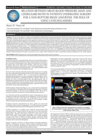
Radiographic vasospasm (angiographic vasospasm) is stenosis of the arteries, demonstrated by conventional angiography, often with delayed contrast fulfillment. The diagnosis is confirmed by previous or subsequent angiographs showing the same vessel of normal caliber. In some cases, DIND corresponds to a region of vasospasm. The incidence of angiographic vasospasm after SAH is about 50% (range: 20-100%)10. Conventional angiography is the most accurate and reliable method for detecting vasospasm, but it is invasive and carries risks in serial tests11. Transcranial Doppler (TCD) is a non-invasive method that uses pulsed-wave Doppler ultrasonography to measure blood flow velocity in the main branches of the anterior Willis Circle. (Fig. 1)
Sudden headache without hemorrhage is a very common clinical manifestation a.k.a warning leaks, with headache often alleviating after drug usage3. It has been described by patients as "the most severe headache in life"4. This is due to aneurysmal expansion, thrombosis or intramural bleeding without rupture5. Less often brain aneurisms are manifested by a space-occupying process, also in symptoms of compression of the optic nerves and pathways, as well as symptoms of compression of some of the other CNs, compression of the brain stem, hypothalamic-pituitary insufficiency, disorder in CSF drenaige, cerebral ischemia which are results from distal thromboembolism. Incidental aneurysms (asymptomatic) are those, which were discovered accidentally on the occasion of angiography, CT or MRI performed on another case. Incidentally detected aneurysms in the population are between 5-10% 6. Cerebral vasospasm is a condition most commonly observed after SAH associated with an aneurysmal rupture, but may also occur in other intracranial hemorrhages (eg, intraventricular hemorrhage from arteriovenous malformations7, and SAH with unknown etiology), trauma (with or without SAH8), brain surgery (endovascular embolization, the so-called iatrogenic vasospasm that occurs in difficult navigation through a tortuously altered vascular anatomy). The term vasospasm was originated in 1951 by Ecker9, and vasospasm can be clinical and radiographic. Clinical vasospasm is referred to as delayed ischemic neurological deficiency (DIND) or symptomatic vasospasm. Delayed ischemic neurological deficits after SAH are clinically characterized by decreased level of consciousness, sometimes with focal neurological deficits (spoken or motor).
Fig 1: TCD is non-invasive method that uses pulsed-wave Doppler ultrasonography to measure blood flow velocity in the main branches of the anterior Willis Circle. The introduction of TCD in 1982 by Aaslid12 provides a non-invasive method for the evaluation of cerebral vasospasm, especially valuable in the screening of the cerebral spasm of the middle cerebral artery and for monitoring the development and resolution of this condition13,14,15,16. Stenosis of the arterial lumen, as occuring in vasospasm, increases the rate of blood flow, which can be detected by TCD17,18,19. Detectable changes can precede clinical symptoms by up to 24-48 hours. Findings are often more useful when baseline studies performed prior to the onset of vasospasm are available. Several studies have shown that the degree of cerebral vasospasm and the clinical development of delayed ischemic neurological deficits cannot be estimated using only the absolute velocities of
Copyright© 2020, IERJ. This open-access article is published under the terms of the Creative Commons Attribution-NonCommercial 4.0 International License which permits Share (copy and redistribute the material in any medium or format) and Adapt (remix, transform, and build upon the material) under the Attribution-NonCommercial terms.
International Education & Research Journal [IERJ]
38
Research Paper
E-ISSN No : 2454-9916 | Volume : 6 | Issue : 2 | Feb 2020
flow through brain vessels measured by TCD20,21,22. Typical MCA values are shown in Table. 1. Likewise, daily increases of > 50 cm / sec signal vasospasm23. There is less correlation between velocity and vasospasm in the anterior cerebral arteries (ACA). Differentiation of vasospasm from hyperemia (in which blood flow velocity is increased in both MCA and ICA) is facilitated by using the ratio of these velocities, the so-called Lindegaard ratio. Tab. 1: Interpretation of TCD results. Average speed through MСА см/sec
MСА:ICА (Lindegaard) ratio
Interpretation of cerebral blood flow
<120 120-200
<3 3-6
Normal blood flow Moderate vasospasm
>200
>6
Severe vasospasm
MATERIALS AND METHODS: The study includes 48 patients operated on for non-ruptured brain aneurysms. Of these, 45 patients were operated on by endovascular procedure and 3 by craniotomy.
Preoperative evaluation of patients is in accordance with the classification of the American Association of Anesthesiologists (ASA), 42 of the patients are evaluated with grade II ASA and grade 6 - grade III ASA. The postoperative evaluation of patients was according to a modified Ranking scale (mRS), with 40 of the patients rated mRS = 0; 6 of the patients rated mRS = 1; 2 of the patients rated mRS = 2. Depending on the medication used, patients was divided into two groups. The first group included patients who were given catecholamine infusion. It contained 19 patients [39.6%], most commonly used catecholamines in our practice are dopamine 20-35 mcg / kg / min or noradrenalin 0.08 mg / ml-2ml / h. The second group consisted of 29 patients [60.4%] who were treated without catecholamine. In 2 patients in the first group who received catecholamine drug therapy, was reported a severe cerebrovascular spasm with a Lindegaard ratio higher than 3, interestingly, in both patients the MBP was higher than 90 mmHg. (See Chart 1.)
The average stay in the Intensive Care Unit (ICU) was 17.5 hours.
Fig. 2: TCD demonstrated the blood flow through the right internal carotid artery
Chart 1: Relation between mean blood preasure (MAP) and Lindegaard ration in patients who received catecholamine drug therapy In the second cohort of 29 patients [60.4%] without catecholamine infusion, 3 patients had evidence of severe cerebral vasospasm. (see Chart 2.)
All patients were followed by advanced hemodynamic monitoring, transcranial Doppler and Lindegaard ratio measurements at 6-th and 12-th postoperative hours. In (Fig. 2 and Fig. 3), is demonstrated direct measurement of blood flow through the right internal carotid artery (ICA) and, respectively, the right middle cerebral artery (MCA) in a patient intervening endovascularly for a non-ruptured right middle cerebral artery aneurysm.
Chart 2: Relation between mean blood preasure (MAP) and Lindegaard ration in patients without catecholamine drug therapy Fig. 3: TCD demonstrated the blood flow through the right middle cerebral artery RESULTS AND METHODS: According to localization, in 41 of the patients [85.4%] the aneurysm was located in the anterior part of the Willis circle, while in 7 [14.6%] they were located in its posterior part. In 29 patients [60.4%] were located in the area of the internal carotid artery (ICA) and 11 patients [22.9%] were in the area of the middle cerebral artery (MCA).
39
DISCUSSION: The very operative treatment of intracranial aneurysms (clipping, coiling or stenting) does not prevent the possibility of vasospasm. Surgical intervention and manipulation of intracranial vessels may increase the risk of so-called iatrogenic vasospasm. Prophylactic hyperdynamic therapy - triple H therapy, has not been indicated and may even lead to complications without result24. The most commonly used medicines for the treatment of cerebral vasospasm are calcium channel blockers (Nimodipine, Nicardipine), vasodilators (Papaverine),
International Education & Research Journal [IERJ]
Research Paper
E-ISSN No : 2454-9916 | Volume : 6 | Issue : 2 | Feb 2020 respectively, a lower Lindegaard ratio in patients with higher MBP. However, the analysis shows that this is not statistically significant, p <0.5.
magnesium drugs. After analyzing the results in our study (Application 1) in both groups of patients was found that those with catecholamine infusion had a MBP of 95.63mm / Hg and an average Lindeard ratio of 1.4, while those without catecholamine infusion have a MBP of 93.62mm / Hg and an average Lindegaard ratio of 1.85. From the statistical analysis is clear that the catecholamine patient group has a lower Lindegaard ratio and 32.14% better brain perfusion. This, given the small number of patients, is statistically insignificant, p <0.5. In the exploration of the other important criteria (MBP) and, accordingly, in dividing the patients into two groups - with MBP higher or lower than 90mm / Hg, the first group of 16 patients had a MBP 83.63mm / Hg, and 32 of the patients in second group have a MBP of 99.81mm/Hg. The mean Lindegaard ratio in patients with a MBP less than 90mm / Hg is 1.91 and in patients with a MBP above 90mm / Hg is 1.55. This shows 23.23% better brain perfusion,
The risk of cardiovascular and pulmonary complications in patients with higher MBP should be considered. Out of the 48 cases described, all were carried by the ICU in good general condition, without pathological abnormalities in cardiac and pulmonary status, and no aggravation in the preoperative neurological symptoms was observed in the study group of patients. CONCLUSION: It can be theoretically suggested that in patients, operated on non-ruptured brain aneurysms, the use of catecholamines and maintenance of MBP above 90 mm / Hg leads to improved brain perfusion and prevention of cerebrovascular vasospasm. In the future, it is necessary to cover a larger group of patients in order to prove the statistical significance of the obtained results, and to include catecholamine therapy as a standard treatment in such patients.
Application 1:
MBP groups <=90
Catheholamines >90
No
Yes
Count
Mean
Standard Deviation
Count
Mean
Standard Deviation
Count
Mean
Standard Deviation
Count
Mean
Standard Deviation
MBP
16
83,63
5,67
32
99,81
6,63
29
93,62
10,82
19
95,63
8,55
Lindegaard ratio
16
1,91
1,54
32
1,55
1,88
29
1,85
1,19
19
1,40
1,05
Comparisons of Column Meansa MBP
Catheholamines
<=90
>90
No
Yes
(A)
(B)
(A)
(B)
MBP
A( ,000)
Lindegaard ratio. The results are based on two-sided tests and assume equal deviations. For each significant pair, the key of the smaller category appears in the higher value category. Capitalization statistical level (A, B, C): 05a a. The tests are adjusted for all pairwise comparisons, with each row using the Bonferroni correction on the inner subtable. MBP
Catheholamines
<=90 Mean
>90 Mean
No Mean
Yes Mean
MBP
83,63
99,81
93,62
95,63
Lindegaard ratio
1,91
1,55
1,85
1,40
Comparisons of Column Meansa MBP groups
Catheholamines
<=90
>90
No
Yes
(A)
(B)
(A)
(B)
MBP
A( ,000)
Lindegaard ratio The results are based on two-sided tests and assume equal deviations. For each significant pair, the key of the smaller category appears in the higher value category. Capitalization statistical level (A, B, C): 05a a. The tests are adjusted for all pairs of comparisons in a row, using the Bonferroni correction for each row in the inner subtable. MBP groups
MBP
Catheholamines
<=90 (A)
>90 (B)
No (A)
Yes (B)
Mean
Mean
Mean
Mean
83,63
99,81
93,62
95,63
Lindegaard ratio 1,91 1,55 1,85 1,40 The results are based on two-sided tests assuming equal deviations. For each significant pair, the key of the smaller category appears in the higher value category. Capitalization statistical level (A, B, C): 05a 1.. a. The tests are adjusted for all pairs of comparisons in a row, using the Bonferroni correction for each row in the inner subtable.
International Education & Research Journal [IERJ]
40
Research Paper
E-ISSN No : 2454-9916 | Volume : 6 | Issue : 2 | Feb 2020 MBP groups
Catheholamines
<=90
>90
No
Yes
Mean
Mean
Mean
Mean
MBP
83,63a
99,81b
93,62a
95,63a
Lindegaard ratio
1,91a
1,55a
1,85a
1,40a
Note: Values in the same row and sub-table do not share the same index differ significantly at p <0.5 in the two-sided column equality test. Non-index cells are not included in the test. Tests assume equal variances.1 1. The tests are adjusted for all pairwise comparisons using the Bonferroni correction for each row in the inner subtable REFERENCES: 1.
Nehls DG, Flom RA, Carter LP, et al. Multiple Intracranial Aneurysms: Determining the Site of Rupture. J Neurosurg. 1985; 63:342–348
2.
Tew JM, Thompson RA, Green JR. In:Guidelines for management and surgical treatment of intracranial Aneurysms. Controversies in Neurology. New York: Raven Press; 1983:139-154
3.
Raps EC, Galetta SL, Solomon RA, et al. The Clinical Spectrum of Unruptured Intracranial Aneurysms. Arch Neurol. 1993; 50:265–268
4.
Day JW, Raskin NH. Thunderclap Headache: Symptom of Unruptured Cerebral Aneurysm. Lancet. 1986; 2:1247–1248
5.
Verweij RD, Wijdicks EFM, van Gijn J. Warning Headache in Aneurysmal Subarachnoid Hemorrhage: A Case-Control Study. Arch Neurol. 1988; 45:1019–1020
6.
Wirth FP. Surgical treatment of incidental intracranial aneurysms. Clin Neurosurg. 1986; 33:125-135
7.
Maeda K, Kurita H, Nakamura T, et al. Occurrence of Severe Vasospasm Following Intraventricular Hemorrhage from an Arteriovenous Malformation. J Neurosurg. 1997; 87:436–439
8.
Martin NA, Doberstein C, Zane C, et al. Posttraumatic Cerebral Arterial Spasm: Transcranial Doppler Ultrasound, Cerebral Blood Flow, and Angiographic Findings. J Neurosurg. 1992; 77:575–583
9.
Ecker A, Riemenschneider PA. Arteriographic Demonstration of Spasm of the Intracranial Arteries: With Special Reference to Saccular Aneurysms. JNeurosurg. 1951; 8:660–667
14. Aaslid R, Huber P, Nornes H: Evaluation of cerebrovascular spasm with transcranial Doppler ultrasound. J Neurosurg 60: 37–41, 1984 15. Grosset DG, Straiton J, McDonald I, Bullock R: Angiographic and Doppler diagnosis of cerebral artery vasospasm following subarachnoid haemorrhage. Br J Neurosurg 7:291–298, 1993 16. Seiler RW, Grolimund P, Aaslid R, Huber P, Nornes H: Cerebral vasospasm evaluated by transcranial ultrasound correlated with clinical grade and CT-visualized subarachnoid hemorrhage. J Neurosurg 64:594–600, 1986 17. Seiler RW, Grolimund P, Aaslid R, et al. Cerebral Vasospasm Evaluated by Transcranial Ultrasound Correlated with Clinical Grade and CT-Visualized Subarachnoid Hemorrhage. J Neurosurg. 1986; 64:594–600 18. Lindegaard KF, Nornes H, Bakke SJ, et al. Cerebral Vasospasm After Subarachnoid Hemorrhage Investigated by Means of Transcranial Doppler Ultrasound. Acta Neurochir. 1988; 42:81–84 19. Sekhar LN, Wechsler LR, Yonas H, et al. Value of Transcranial Doppler Examination in the Diagnosis of Cerebral Vasospasm After Subarachnoid Hemor- rhage. Neurosurgery. 1988; 22:813–821 20. Creissard P, Proust F, Langlois O: Vasospasm diagnosis: theoretical and real transcranial Doppler sensitivity. Acta Neurochir (Wien) 136:181–185, 1995 21. Ekelund A, Saveland H, Romner B, Brandt L: Is transcranial Doppler sonography useful in detecting late cerebral ischaemia after aneurysmal subarachnoid haemorrhage? Br J Neurosurg 10:19–25, 1996
10. Dorsch N. A clinical review of cerebral vasospasm and delayed ischaemia following aneurysm rupture. Acta Neurochir Suppl. 2011; 110:5–6
22. Mizuno M, Nakajima S, Sampei T, Nishimura H, Hadeishi H, Suzuki A, et al: Serial transcranial Doppler flow velocity and cere- bral blood flow measurements for evaluation of cerebral vasospasm after subarachnoid hemorrhage. Neurol Med Chir (Tokyo) 34:164–171, 1994
11. Leffers AM, Wagner A. Neurologic complications of cerebral angiography: A retrospective study of complication rate and patient risk factors. Acta Radiol 2000;41:204-210.
23. Malhotra K, Conners JJ, Lee VH, Prabhakaran S. Relative changes in transcranial Doppler velocities are inferior to absolute thresholds in prediction of symptomatic vasospasm after subarachnoid hemorrhage. J Stroke Cerebrovasc Dis. 2014;23:31–6.
12. Aaslid R, Markwalder TM, Nornes H: Noninvasive transcranial Doppler ultrasound recording of flow velocity in basal cerebral arteries. J Neurosurg 57:769–774, 1982
24. Egge A, Waterloo K, Sjoholm H, Solberg T, Inge- brigtsen T, Romner B. Prophylactic hyperdynamic postoperative fluid therapy after aneurysmal sub- arachnoid hemorrhage: a clinical, prospective, randomized, controlled study. Neurosurgery. 2001; 49:593–605; discussion 605-6
13. Aaslid R, Huber P, Nornes H: A transcranial Doppler method in the evaluation of cerebrovascular spasm. Neuroradiology 28: 11–16, 1986
41
International Education & Research Journal [IERJ]