Research Paper
Medical Science
E-ISSN No : 2454-9916 | Volume : 5 | Issue : 8 | Aug 2019
SOLITARY RECTAL ULCER SYNDROME MIMICKING RECTAL NEOPLASM ON COLONOSCOPY Samer Talib MD,Mohamed Alani, MD, NISAR AHMED M.D., F.A.C.G., Park Plaza Hospital, Houston, Texas. ABSTRACT We describe a highly unusual case of solitary rectal ulcer syndrome in a 90 years old African American female patient with a history of distal sigmoid colon resection for colon cancer with hysterectomy for uterine cancer. This patient presented with constipation, abdominal discomfort. Rectal showed positive fecal occult blood test. This finding raised suspicion for a possible recurrent malignancy. Colonoscopy shows the presence of an ulcerative rectal lesion. Histopathology study of the Biopsy showed granulomatous lesion with no evidence of neoplastic tissue. INTRODUCTION: Solitary rectal ulcer syndrome (SRUS) is a rare benign disorder characterized by a combination of symptoms, endoscopic findings, and histological abnormalities[1]. It was first described by Cruveihier[2,12] in 1829. When he reported four unusual cases of rectal ulcers. A disorder, with an estimated annual prevalence of one in 100000 persons. It is a disorder of young adults, occurring most commonly in the third decade in men and the fourth decade in women. Men and women are affected equally, with a small predominance in women[5,15]. However, The solitary rectal ulcer is a misnomer because ulcers are found in 40% of patients, while 20% of patients have a single ulcer, and the rest of the lesions differ in shape and size, including hyperemic mucosa to broad-based polypoid lesions[7,17]. There is even a suggestion that the disease process also may involve the sigmoid colon.[8,18] A 90-year-old African American female patient presented to the clinic with low abdominal pain and discomfort, and constipation. She complained of a feeling of incomplete evacuation. There was no associated nausea or vomiting. Past medical and surgical history was significant. She had colon resection for distal sigmoid colon cancer. She also had a history of hysterectomy for uterine cancer. Clinical examination showed typical vital signs, and her abdomen was soft and non-tender. On rectal examination, there was a finding of positive fecal occult blood. CBC and electrolytes were normal. Colonoscopy showed the indications of possible recurrent cancer, gastro-intestinal occult blood loss, and iron deficiency anemia.
Fig. 2: Shows presence of granulomatous and fibro-purulent debris.
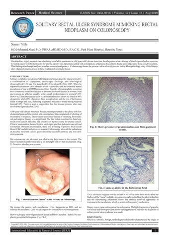
On colonoscopy: An ulcerated non obstructing large mass in the rectum. The mass was noncircumferential and 2 cm in length with 14 mm in diameter. (Fig. 1). No active bleeding was present.
Fig. 3: same as above in the high power field.
Fig. 1: shows ulcerated “mass” in the rectum, on colonoscopy. We treated the patient with mesalamine 1Gm. Suppositories BID, and we referred the patient to a Colo-rectal surgeon for consultation and management. However, biopsy showed granulation tissue and fibro- purulent - debris. No neoplastic growth in the biopsies. (Fig. 2 & 3 )
The Colo-rectal surgeon saw the patient in his office some three weeks after her finding of the “mass” and did a proctoscopy and reported that the ulcer is healing and the surrounding edematous tissue had entirely resolved apparently in response to the mesalamine which is an anti-inflammatory medication. Biopsy report came out negative for malignancy. Multiple fragments of granulation tissue and fibrinopurulent debris were appreciated, and thus the diagnosis of solitary rectal ulcer syndrome was made. DISCUSSION: SRUS is a chronic, benign, underdiagnosed disorder characterized by single or
Copyright© 2019, IERJ. This open-access article is published under the terms of the Creative Commons Attribution-NonCommercial 4.0 International License which permits Share (copy and redistribute the material in any medium or format) and Adapt (remix, transform, and build upon the material) under the Attribution-NonCommercial terms.
International Education & Research Journal [IERJ]
1
Research Paper
E-ISSN No : 2454-9916 | Volume : 5 | Issue : 8 | Aug 2019
multiple ulcerations of the rectal mucosa, with the passage of blood and mucus, associated with straining or abnormal defecation[1]. The average time from the onset of symptoms to diagnosis is five years, ranging from 3 mo to 30 years in adults, which is longer than in pediatric patients (1.2-5.5 years)[22]. Clinical features include rectal bleeding, copious mucous discharge, prolonged excessive straining, perineal and abdominal pain, feeling of incomplete defecation, constipation, and rarely, rectal prolapse[13,14]. The amount of blood varies from a little fresh blood to severe bleeding that requires blood transfusion[15]. Although the passage of blood during defecation is the hallmark, up to 26% of patients can be asymptomatic, discovered when investigating other diseases[7,17]. The underlying etiology and pathogenesis are not fully understood, but multiple factors may be involved. The most accepted theories are related to direct trauma or local ischemia as causes. It is believed that descent of the perineum and abnormal contraction of the puborectalis muscle during straining on defecation or defecation in the squatting position result in trauma and compression of the anterior rectal wall on the upper anal canal, and internal intussusceptions or prolapsed rectum[6,17]. Mucosal prolapse, overt or occult, is the most common underlying pathogenetic mechanism in SRUS This may lead to venous congestion, reduced blood flow, and edema in the mucosal lining of the rectum and ischemic changes with resultant ulceration. The cause of ischemia may also be related to fibroblasts replacing blood vessels, and pressure by the anal sphincter. Moreover, rectal mucosal blood flow is low in SRUS to a level similar to that seen in normal transit constipation, suggesting similar impaired autonomic cholinergic gut-nerve activity[18]. Self-digitation maneuver to reduce rectal prolapse or to evacuate an impacted stool may also cause direct trauma of the mucosa and ulceration[19]. Although this hypothesis seems plausible, it remains unproven because rectal mucosal intussusception is common even in healthy subjects, but rectal prolapse and SRUS are rare[20]. Besides, not all patients with rectal prolapse have SRUS and vice versa[21]. Furthermore, ulcers usually occur in the mid rectum, which cannot be reached by digital examinations[22].
14. Rutter KR, Riddell RH. The solitary ulcer syndrome of the rectum. Clin Gastroenterol. 1975;4:505–530. [PubMed] [Google Scholar] 15. Martin CJ, Parks TG, Biggert JD. Solitary rectal ulcer syndrome in Northern Ireland. 1971-1980. Br J Surg. 1981;68:744–747. [PubMed] [Google Scholar] 16. Tandon RK, Atmakuri SP, Mehra NK, Malaviya AN, Tandon HD, Chopra P. Is solitary rectal ulcer a manifestation of a systemic disease? Clin Gastroenterol. 1990;12:286–290. [PubMed] [Google Scholar] 17. Tjandra JJ, Fazio VW, Church JM, Lavery IC, Oakley JR, Milsom JW. The clinical conundrum of solitary rectal ulcer. Dis Colon Rectum. 1992;35:227–234. [PubMed] [Google Scholar] 18. Burke AP, Sobin LH. Eroded polypoid hyperplasia of the rectosigmoid. Am J Gastroenterol. 1990;85:975–980. [PubMed] [Google Scholar] 19. Ignjatovic A, Saunders BP, Harbin L, Clark S. Solitary ‘rectal’ ulcer syndrome in the sigmoid colon. Colorectal Dis. 2010;12:1163–1164. [PubMed] [Google Scholar] 20. Bonnard A, Mougenot JP, Ferkdadji L, Huot O, Aigrain Y, De Lagausie P. Laparoscopic rectopexy for solitary ulcer of rectum syndrome in a child. Surg Endosc. 2003;17:1156–1157. [PubMed] 21. Sharara AI, Azar C, Amr SS, Haddad M, Eloubeidi MA. Solitary rectal ulcer syndrome: endoscopic spectrum and review of the literature. Gastrointestinal Endoscopy. 2005;62:755–762. [PubMed] [Google Scholar] 22. Dehghani SM, Malekpour A, Haghighat M. Solitary rectal ulcer syndrome in children: a literature review. World J Gastroenterol. 2012;18:6541–6545. [PMC free article] [PubMed] [Google Scholar] 23. Suresh N, Ganesh R, Sathiyasekaran M. Solitary rectal ulcer syndrome: a case series. Indian Pediatr. 2010;47:1059–1061. [PubMed] [Google Scholar]
Histopathological examination is a gold standard for the diagnosis of SRUS. A fibroblast and smooth muscular obliteration of the lamina propria, crypts' distortion, and surface serration can establish the diagnosis in most cases [7]. The degenerative-regenerative process occurring in the mucosa may cause such changes[7]. Some pathological studies reveal cases where thrombosis, fibrin deposition, and atherosis could be appreciated [9]. Diamond shaped crypts seen by Warren[8]. Tendler and his colleagues [7]also identified mucosal capillary abnormalities, including dilatation, congestion, and thrombosis. It is important to note that these pathological findings are varied and diverse and can be affiliated with multiple disorders, e.g. inflammatory bowel disease, irritable bowel syndrome or adenoma. Most respond well to laxatives, enemas, and bowel retraining. Our patient above was treated with mesalamine suppositories 1gm, twice a day and did very well with complete resolution. RECOMMENDATION: we highly recommend the usage of Mesalamine suppository for the treatment of solitary rectal ulcer syndrome. REFERENCES: 1.
Tjandra JJ, Fazio VW, Church JM, Lavery IC, Oakley JR, Milsom JW. The clinical conundrum of solitary rectal ulcer. Dis Colon Rectum 1992;35:227-34.
2.
Preston DM, Lennard-Jones JE. Anismus in chronic constipation. Dig Dis Sci 1985;30:413-8.
3.
Lam TCF, Lubowski DZ, King DW. Solitary rectal ulcer syndrome. Clinic Gastroenterol 1992,6:129-43.
4.
Ming Sun w, et al. A common pathophysiology for full-thickness rectal prolapsed, anterior mucosal prolapsed and SRUS. Br J Surg 1989, 78:290-J
5.
Mackle EJ, Parks TG. The pathogenesis and pathophysiology of rectal prolapsed and SRUS. Clin Gastroenterol 1986;15(4):985-1000.
6.
Madigan MR, Morson BC. Solitary ulcer of the rectum. Gut1969;10:871-81
7.
Tendler DA, Aboudola S, Zacks JF, O'Brien MJ, Kelly CP. Prolapsing mucosal polyps: An underrecognized form of colonic polyp-A clinicopathological study of 15 cases. Am J Gastroenterol 2002;97:370-6
8.
Warren BF, Dankwa EK, Davies JD. "Diamond-shaped" crypts and mucosal elastin: Helpful diagnostic features in biopsies of rectal prolapse. Histopathology 1990;17:129-34.
9.
Lonsdale RN. Microvascular abnormalities in the mucosal prolapse syndrome. Gut 1993;34:106-9
10. Chiang JM, Changchien, CR, Chen JR. Solitary rectal ulcer syndrome an endoscopic and histological presentation and literature review. Int J Colorectal Dis 2006;21:34856. 11. Felt-Bersma RJ, Tiersma ES, Cuesta MA. Rectal prolapse, rectal intussusception, rectocele, solitary rectal ulcer syndrome, and enterocele. Gastroenterol Clin North Am. 2008;37:645–668, ix. [PubMed] [Google Scholar] 12. Cruveihier J Ulcer Chronique du rectum. In: Bailliere JB. Anatomie pathologique du crops human. Paris: 1829. [Google Scholar] 13. Madigan MR, Morson BC. Solitary ulcer of the rectum. Gut. 1969;10:871–881. [PMC free article] [PubMed] [Google Scholar]
2
International Education & Research Journal [IERJ]