Making waves
IN SONOGRAPHY RESEARCH
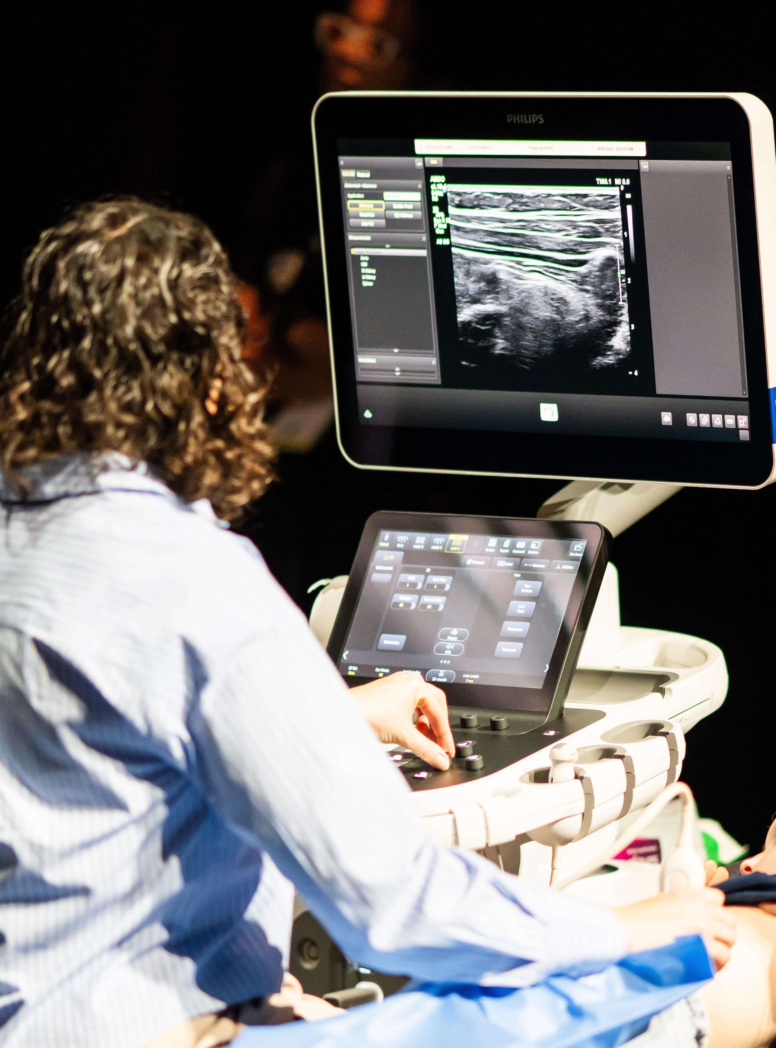
It is a pleasure to introduce this edition of Making Waves. What makes this publication so valuable is that it is truly created by sonographers, for sonographers. Drawing on the expertise of the ASA Special Interest Groups (SIGs), each summary reflects not only the latest research but also the real-world experience and clinical judgement that shape our daily practice. Together, these contributions offer a snapshot of a profession that is evolving, adapting, and continually striving to do better for our patients.
As you move through this edition, you will see the breadth of our discipline, from technical innovation and emerging technologies through to clinical education, wellbeing, and supervision. Just as importantly, you will see the willingness of our members to share knowledge and make research accessible to others. This spirit of collaboration is at the heart of Making Waves. I hope you find this edition both useful and thoughtprovoking, and that it encourages you to keep asking questions, sharing insights, and contributing to the future of sonography.
Paula Kinnane MMedUS, BAppSc, Grad Dip Medical Ultrasound, AMS, FASA General Manager – Professional Development, Clinical Standards and Research

Ellen Chen is a general sonographer based in Sydney, with approximately nine years of scanning experience as a qualified sonographer and a developing interest in paediatric ultrasound.

Joyce Chen has a particular interest in tertiary fetal diagnostics and complex pregnancies. She is actively involved in teaching and training obstetric ultrasound, promoting high-quality care. Joyce has travelled to rural regions in Cambodia to provide obstetric ultrasound training to local midwives, equipping them with the skills to identify pregnancies at risk of complications, enabling mothers to plan for safer deliveries and improve maternal and neonatal outcomes.

Deanne Chester is a senior lecturer and head of course in medical sonography at CQUniversity. She has extensive experience in clinical education, curriculum design, and assessment in ultrasound, and is particularly interested in feedback literacy, cognitive apprenticeship, and supporting clinical educators.

Susan Diep is a clinical sonographer and an ultrasound applications specialist for GE HealthCare based in Melbourne, Victoria. Her clinical work revolves around general ultrasound, with a particular clinical interest in musculoskeletal (MSK) imaging. She is passionate about teaching and is actively involved in education, conferences, and developing evidence-based ultrasound practice to support and improve patient outcomes. MSK ultrasound continues to fascinate Susan as it is patient-dependent, sonographer skills oriented and integrates clinical assessment with dynamic imaging.

Peter Esselbach is an MSK sonographer and MSK physio with over 30 years’ experience. He has enjoyed combining his two professions using POCUS to develop better management for his patients. He also enjoys conducting training courses in MSK ultrasound to medical professionals.

Charlotte Harman is a dedicated sonographer and clinical leader with a strong passion for women’s health, education, and innovation in ultrasound practice. She has completed advanced studies in women’s health and has held leadership roles within Monash Health, where she is committed to high quality, patient-centred care. Charlotte has a particular interest in education and mentorship, contributing to clinical training, governance, and service improvement initiatives that support both sonographers and the patients they care for.
Simone Haami is an early career sonographer with a passion for obstetrics, gynaecology and women’s health. She thrives in both clinical and educational spaces, mentoring upcoming sonographers and creating learning platforms. Simone advocates for evidence-based practice, compassionate care and collaboration within the profession.

Emma Jardine is a senior sonographer working in Victoria and a research and standards manager with the ASA. She has a passion for professional growth and believes that investing in ongoing education and training will benefit individual sonographers, contribute to better patient outcomes, and improve the overall healthcare experience.

Jane Keating is an experienced ultrasound clinical educator at Royal Melbourne Hospital. She has played an active role in the organisation of major professional conferences for the ASA, ASUM, as well as the WFUMB 2019 Congress. Jane has presented internationally and extensively across Australia at national conferences, travelling workshops, and educational forums.
In recognition of her excellence and contribution to the profession, she was awarded the ASA Pru Pratten Award for Ultrasound Excellence in 2006.
Her clinical and educational interests include chronic liver disease, liver lesions, renal transplantation imaging, and contrastenhanced ultrasound (CEUS).

Paula King is the co-supervising sonographer at the Royal Melbourne Hospital Radiology Department. Paula has been a passionate sonographer for over 30 years, with extensive experience in abdominal imaging. Her main interests include contrast-enhanced ultrasound, renal transplantation, and emergency department ultrasound. She has a long-term attachment to all things breast and neck and has recently expanded her acute clinical skills in bowel and appendix ultrasound. Paula loves to make new experiences in ultrasound understandable and accessible to all.

Lucy Law is an Australian-trained echocardiographer with a PhD in the field of cardio-rheumatology from Umeå University (Sweden) and is certified by the ESCVI (European Society of Cardiovascular Imaging) as a congenital echocardiographer. Lucy is currently working as a senior research officer for Menzies School of Health Research (NT), focusing on increasing access to echocardiography screening for rheumatic heart disease (RHD) in remote Australia and Timor-Leste. Additionally, Lucy is contributing to a Swedish national study reviewing the palliative pathways and outcomes of Fontan circulation for hypoplastic left heart syndrome (HLHS). Lucy works clinically in the NT.

James Maunder studied zoology, anthropology and biochemistry at the Australian National University (ANU), graduating in 2000, before beginning his vascular ultrasound training (DMU Vasc) in 2004.
James is currently the chief vascular sonographer for Dr Peter Bray, MBBS [UWA] FRACS [Vasc] in Subiaco, WA. He has worked as a dedicated vascular sonographer for over 20 years, and has presented at local, state, national and international workshops and conferences. He was awarded the ASA Western Australian Sonographer of the Year in 2019 and the ASA Associate Fellowship (AFASA) in 2025.

Lisa McGuire is a PhD candidate at the University of Sydney. Her thesis centres on equitable healthcare for those in remote and rural regions and focuses on enhanced ultrasound diagnosis of supraspinatus tendon tears with the addition of deep learning artificial intelligence.

Lino Piotto (FASA, FASMIRT, MMedRad, DMU, AMS) is the tutor sonographer at the Women’s and Children’s Hospital, Adelaide. He has been specialising in paediatric ultrasound for 36 years.

Sheri Anne Rae is the chief sonographer and clinical supervisor at I-MED Radiology Network. Holding a Master of Ultrasound, a Cert IV TAE, and with over two decades of scanning experience, Sheri is committed to pursuing endeavours that promote continued personal and professional growth. She takes pride in sharing her knowledge, developing tailored learning plans, and creating supportive and productive learning environments for students. Sheri embodies the spirit of stewardship by volunteering for several industry stakeholders that advocate for the sonography profession, champion the development of quality sonography education, and contribute to improved patient healthcare outcomes.

Tristan Reddan is the consultant radiographer/ sonographer at the Queensland Children’s Hospital in Brisbane, a role in which he manages medical imaging research and provides paediatric sonography advice, as well as some clinical scanning. He is also a clinical associate professor and sessional lecturer in paediatric ultrasound at QUT, where he completed his PhD and continues to mentor and collaborate on research projects. Tristan was awarded the Pru Pratten Memorial Lifetime Achievement Award 2026.

Jacqui Robinson is the chief vascular sonographer at Liverpool Hospital, Southwest Sydney. With over 30 years of experience in clinical sonography, research, and education, she regularly presents at national and international conferences, supporting patient care and professional growth in vascular ultrasound.

Louise Smith is a clinical nurse consultant and cardiac sonographer specialising in critical care echocardiography at Nepean Hospital’s intensive care unit and cardiac diagnostic department. With expertise in both echocardiography and intensive care nursing, she integrates these skills to provide high quality care for critically ill patients.

Caterina Watson is a senior lecturer at Edith Cowan University, Western Australia. She enjoys participating in outreach training programs in the South Pacific nations.
Caterina has extensive clinical experience in public, private and rural locations, with a passion to promote student sonographer learning with cultural safety and First Nations People. Caterina is a member of the ASA Research Special Interest Group and a PhD candidate with Edith Cowan University. Her studies focus on the application of AI technologies in fetal ultrasound images.
Craig Winnett is a sonographer with nearly two decades of clinical experience across private practice and tertiary hospitals in Brisbane, Queensland. He is passionate about sonographer education, mentoring, point-of-care ultrasound training, and advancing the profession with a strong interest in clinical research and expanding the scope of sonography practice in Australia.
Craig has a subspeciality interest in musculoskeletal ultrasound and is committed to promoting continuous professional development in this area. He values the versatility of ultrasound as a diagnostic tool and its role in supporting accurate diagnoses and improving patient outcomes.
CARDIAC Echocardiography in cardio-oncology: Optimising service delivery 7
CARDIAC Guidelines for the standardization of adult echocardiography reporting: Recommendations from the American Society of Echocardiography 8
CLINICAL SUPERVISORS
Describing ultrasound skills teaching by near-peer and faculty tutors using cognitive apprenticeship 10
CLINICAL SUPERVISORS
Work integrated learning in sonography: Barriers, enablers, and comparative perspectives 11
EMERGING TECHNOLOGIES
Application of artificial intelligence to ultrasound imaging for benign gynecological disorders: Systematic review 13
EMERGING TECHNOLOGIES
Hybrid deep learning models for automatic segmentation and classification of breast lesions in ultrasound images 14
GENERAL Association between left-sided varicocele and epididymo-orchitis with sonographic findings 16
GENERAL Correlation of ultrasound-based TIRADS and the Bethesda system for reporting of thyroid cytopathology: A study in a tertiary care centre 17
HEALTH AND WELLBEING
Factors influencing healthcare workers’ attitude toward delayed retirement: A crosssectional survey 18
HEALTH AND WELLBEING
Navigating sonographer work integrated learning: A thematic analysis of workforce pressures, training barriers and student wellbeing 19

MSK
Ultrasound-based knee osteoarthritis severity assessment and its association with KellgrenLawrence grading 20
MSK
High resolution ultrasound of the annular pulley system in the toes: Sonographic anatomy and pathological cases 21
PAEDIATRIC
Imaging of acute musculoskeletal infections in children and their differential diagnoses 23
PAEDIATRIC
Benign enlargement of the subarachnoid spaces and subdural collections – when to evaluate for abuse 24
RESEARCH
What the forks? A longitudinal quality improvement study tracking cutlery numbers in a public teaching and research hospital staff tearoom 25
RESEARCH
Medical imaging privacy: A systematic scoping review of key parameters in dataset construction and data protection 26
VASCULAR
Accuracy of Duplex ultrasound for detecting renal artery stenosis: A systematic review 28
VASCULAR
Ultrasound characteristics of carotid web 29

WOMEN’S HEALTH
How to get the most from axial views of the fetal brain: Applying the principles of neurosonography to screening examination of the supratentorial central nervous system 30
WOMEN’S HEALTH
Morphological changes of endometriomas during pregnancy and after delivery detected using ultrasound 31
Sonographers Association 2026. Disclaimer: The information in this publication is current when published and is general in nature; it does not constitute professional advice. Any views expressed are those of the author and may not reflect the views of ASA. ASA does not endorse any product or service identified in this publication. Use of this information is at the reader’s own risk. The ASA is not responsible for any errors or consequences arising from its use. Please visit www.sonographers.org for the full version of the Australasian Sonographers Association publication disclaimer.
This article discusses the development and implementation of specialised cardio-oncology services at a major UK tertiary health centre (University College London Hospital) and the role this has played in streamlining and optimising patient care. The article examines the benefits of the service, creation of the service and protocols, as well as role diversification for cardiac physiologists. The authors aimed to highlight the mutual benefits of creating and implementing such a service, especially in the context of increasing demand for cardio-oncology services and the use of cancer therapies that have known cardio-toxic effects.
Examination of how the department integrated the cardio-oncology services into everyday practice was discussed, looking at the steps of protocol development, required infrastructure and equipment, training and education, as well as quality control. A review of lymphoma patients accessing this service over 12 months was presented to demonstrate the effectiveness of the service. Additionally, the authors discussed the usefulness of the physiologist-led ‘late survivors’ clinics over the same period.
The study found that having a dedicated cardio-oncology service and physiologist-led clinics within this service was beneficial for both the patient and the clinician. For patients, the number of clinical visits decreased, and there was timely assessment and management of cardiovascular issues and non-cardiac concerns. Creating a ‘one-stop shop’ for the patient acted to minimise patient anxiety and optimise patient-health service contact time. For the clinicians, designing and appropriately positioning echocardiography services and
protocols allows for accommodation of the significant complexities that come with caring for oncology patients. Having specialist physiologists lead clinics with appropriate escalation pathways, as well as short-notice availability bookings, streamlined assessment and optimised clinical time management and resource use.
Echocardiography plays a significant role in monitoring cardiac function through all phases of the patient’s cancer journey (initial evaluation, during treatment and into remission). The effects of cancer treatments, including chemotherapy agents, which are known to be cardio-toxic, and radiation, greatly increase the risk of cardiac dysfunction (such as decreased left ventricular systolic and diastolic function, right heart dysfunction and valvular pathology). Having established clinical assessment and management pathways and protocols implemented within an oncology service, as well as dedicated education for cardiac physiologists, ensures timely expert care and management. For patients, this contributes to preventing adverse complications and comorbidities that may arise due to treatment and decreases the number of clinical visits. Creating physiologist-led clinics increases departmental capacity, reduces time to patient assessment and follow-up, and increases job satisfaction for the physiologist through specialist education and increased autonomy.
As new chemotherapy and immunotherapy agents are discovered and become available, and diagnostic and treatment methods improve and evolve, the importance of, and demand for, cardio-oncology services is only increasing. Developing and evaluating service integration pathways will help provide strong evidence, as well as refined protocols and service delivery models, which will benefit both the patient and health service providers and foster better outcomes all around.
Reviewer: Lucy Law
Authors & Journal: Lushington J, Kandala A, Bingcang J, Ferreira A, Byrne A, Sirker A, et al. Echo Res Pract. 2026;13(1):11.
Open Access: Yes
Read the full article here
“The physiologistled late effects clinic model highlights the expertise of cardiac physiologists, who report higher job satisfaction as a result of the expanded role.”
The previous American Society of Echocardiography (ASE) guideline addressing standardised reporting of adult echocardiography was published in 2002. Since then, echocardiographic practice has evolved substantially, with major advances in imaging techniques, digital reporting platforms, structured data, interoperability, and expectations for quality improvement. Increasing variability in report structure, terminology, and content across institutions was recognised as a barrier to clear communication, clinical usability, and consistency of care.
The 2025 ASE Guidelines for the Standardization of Adult Echocardiography Reporting were developed to address these issues. The guidelines aim to standardise report structure, terminology, and core content to improve clarity, support accurate clinical decision-making, enable meaningful comparison across serial studies and institutions, and facilitate quality assurance, research, and data integration. A further objective is alignment with contemporary digital health systems and emerging applications such as artificial intelligence.
The guidelines were produced by a multidisciplinary ASE expert writing group using a structured consensus-based process informed by review of existing standards and prior guidelines. The committee comprised 13 members with diverse expertise, including cardiac sonographers, a paediatric representative, members with Intersocietal Accreditation Commission (IAC) experience, experts in artificial intelligence and data registries, and representation from the National Institutes of Health. Several members also contributed international training perspectives. To ensure consistency with established frameworks, the committee reviewed published echocardiographic data standards,
IAC requirements, ASE guidelines, and guidance from other professional societies, including the European Association of Cardiovascular Imaging (EACVI). Consensus recommendations were developed to define standardised report structure, terminology, and content for adult transthoracic, transoesophageal, and stress echocardiography.
The guideline underwent formal peer review and a 21-day public comment period following online publication. Feedback from expert reviewers, including members and chairs of the ASE Guidelines and Standards Committee, as well as public submissions, was incorporated into the final document.
The guidelines concluded that standardised echocardiography reporting, using defined structure, terminology, and core measurements, improves clarity, consistency, and clinical utility. Standardisation supports accurate interpretation, facilitates longitudinal comparison, enhances data analysis and interoperability, and strengthens quality assurance across echocardiography laboratories.
The guidelines support clear, consistent, and clinically meaningful echocardiography reporting. They improve communication with referring clinicians, enable reliable serial comparisons, support informed clinical decision-making, and promote high quality patient care. Adoption of standardised reporting also facilitates laboratory accreditation, quality improvement initiatives, and data sharing.
Major highlights comparing the 2002 and 2025 guidelines:
• Defines a standardised report structure and mandatory core elements for adult transthoracic (TTE), transoesophageal (TEE), and stress echocardiography (SE) reports (the 2002 guidelines were limited to TTE only).
• Introduces a dedicated reporting framework for congenital heart disease (CHD).
Reviewer: Louise Smith
Authors & Journal: Taub C, Stainback R, Abraham T, Forsha D, Garcia-Sayan E, Hill J, et al. J Am Soc Echocardiogr. 2025;38(9):735–774.
Open Access: Yes
Read the full article here
“The guideline underscores the need for consistent, accurate reporting of clinically relevant findings and sets a new standard for clear and uniform communication.”
• Establishes uniform terminology, abbreviations, and definitions to improve clarity and reduce misinterpretation.
• Recommends inclusion of demographic information to enable comparison with normal reference ranges and highlight pathological findings.
• Clarifies required versus optional elements; while the 2002 guidelines encouraged flexibility, the 2025 guidelines emphasise required components with optional additions.
• Emphasises comparison with prior echocardiograms and other relevant imaging to support longitudinal patient care.
• Supports structured data use for analysis, interoperability, quality improvement, and future artificial intelligence applications.
• Incorporates guidance on artificial intelligence, machine learning, DICOM structured reporting, and standardised data storage.
• Recommends cross-modality comparison (e.g. TTE with CT/CMR) where available.
In summary, the 2025 ASE reporting guidelines represent a substantial change in the approach to cardiac ultrasound reporting, moving away from the 2002 guidelines, which were flexible, descriptive and TTE-focused, to a document that is highly structured, multimodality-focused and recognises changes in technology.
Ultrasound skills education commonly relies on both faculty (teacher/supervisor) and near-peer (fellow student) tutors, particularly in simulation-based learning environments. While near-peer teaching is widely used in several healthcare disciplines, there is limited understanding of how teaching behaviours differ between near-peer and faculty tutors, and how these behaviours support learners’ development.
Cognitive apprenticeship (CA) is an educational approach that supports skill development by making expert decisionmaking and clinical reasoning visible through modelling, guided practice, and gradual transfer of responsibility to learners. Using this framework, Hari et al. aimed to describe how both near-peer and faculty tutors enact elements of CA during ultrasound skills teaching. The study addressed a gap in the literature by examining how different tutor groups teach in practice and how these approaches may be leveraged to strengthen ultrasound education.
This observational study quantitatively analysed video-recorded ultrasound teaching sessions involving medical students, taught by either near-peer tutors or faculty tutors. Teaching interactions were examined using directed content analysis mapped to the six elements of CA: modelling, coaching, scaffolding, articulation, reflection, and exploration. Comparisons were made between the tutor groups to identify patterns and differences in instructional practice.
Both groups demonstrated similar overall patterns of CA, with minor differences in specific teaching behaviours. There were no significant differences in overall CA categories. A key finding was that teaching was dominated by coaching and articulation, with relatively little time spent on modelling or exploration.
Faculty tutors more frequently engaged in modelling expert performance, articulating
clinical reasoning, and encouraging reflection, thereby making tacit thinking explicit for learners. Near-peer tutors were more likely to focus on coaching, step-by-step guidance, and practical tips, often drawing on their recent experience as learners.
Near-peer tutors also appeared to create psychologically safe learning environments, offering reassurance and normalising difficulty. However, neither tutor group consistently enacted all components of CA. In particular, opportunities for articulation, reflection, and exploration were sometimes missed, suggesting scope for targeted educator development.
Although conducted with medical students, the findings are relevant to sonography education, which similarly relies on simulation and/ or supervised clinical practice. Importantly, near-peer learning is not routine in sonography clinical placement environments; however, this study suggests it may be a valuable model to consider, particularly for early skill development and confidence building.
Both near-peer and faculty teaching models are widely used in simulation-based scanning education, and this study highlights that neither group should be assumed to be inherently ‘better’. Instead, each brings complementary strengths that can be optimised through a shared understanding of CA principles.
For sonographers involved in education, the study reinforces the need for explicit tutor development regardless of seniority. Both teacher and fellow student educators can benefit from support in articulating clinical reasoning, deliberately scaffolding learning, and gradually transferring responsibility as learner capability increases. Embedding CA as a shared framework may improve consistency, learner engagement, and safe progression towards independent scanning.
While CA provides a valuable model for understanding teaching interactions, ultrasound skill acquisition also draws on psychomotor learning principles and deliberate practice, emphasising progressive skill development, repetition, and structured feedback, areas not specifically addressed in this study.
Reviewer:
Deanne Chester, AFASA
Authors & Journal: Hari R, Caprez R, Dolmans D, Huwendiek S, Robbiani S, Stalmeijer R. Teach Learn Med. 2024;36(1):33–42.
Open Access: Yes
Read the full article here
“Although they employ slightly different approaches, near-peer and faculty tutors provide complementary strengths that collectively enhance ultrasound education.”
This study addresses a gap in understanding the challenges associated with work integrated learning (WIL) in sonography and their impact on trainee emotional, social, and psychological wellbeing. The aim of the study was to inform the development of targeted strategies to enhance training quality, strengthen trainee support, and promote the sustainability of sonography clinical education.
How the study was performed
This study recruited Australian sonographer trainees (n = 165) and clinical supervisors (n = 266) registered with the Australian Sonographer Accreditation Registry (ASAR). Participants were asked to complete an online survey if they had either actively engaged in or participated in clinical sonography training within the past 12 months.
The analysis was conducted using validated tools, including the Kessler K6 Scale to assess psychological distress and the single-item social identification measure to evaluate professional identity.
This study found that many students struggle to maintain a healthy work-life-study balance, while also facing significant logistical and financial pressures. Sixty-two per cent (n = 102/165) of trainees reported incurring accommodation or travel expenses to facilitate engagement in clinical training, with financial stress identified as the most common pressure. Trainee sonographers identified multiple factors that contributed to stress, including mental clarity, family responsibilities, physical health and social connections, with balancing academic and clinical demands a consistent challenge. Kessler K6 scores identified 24% of trainee sonographers (n = 40/165) were at risk of probable serious mental illness, higher than the general population at 15%.
A key finding was the disconnect between trainees and clinical supervisors related to understanding trainee stress, with clinical supervisors consistently underestimating both the frequency and severity of stress experienced by trainees (Figure 1). While both groups supported strategies such as longer appointment times, trainees placed greater value on wellbeing-focused initiatives such as flexible working arrangements and leave entitlements.
CLINICAL SUPERVISORS
Reviewer: Sheri Anne Rae, AFASA
Authors & Journal: Boucaut P, Tunny R, McMahon K, Osborne B, Lamb K, Wardle J, et al. Radiography. 2025;31(6):103177.
Open Access: Yes
Read the full article here
“Twenty-four per cent of trainees had K6 scores indicating risk of serious psychological distress.”
Work integrated learning in sonography: Barriers, enablers, and comparative perspectives (continued)
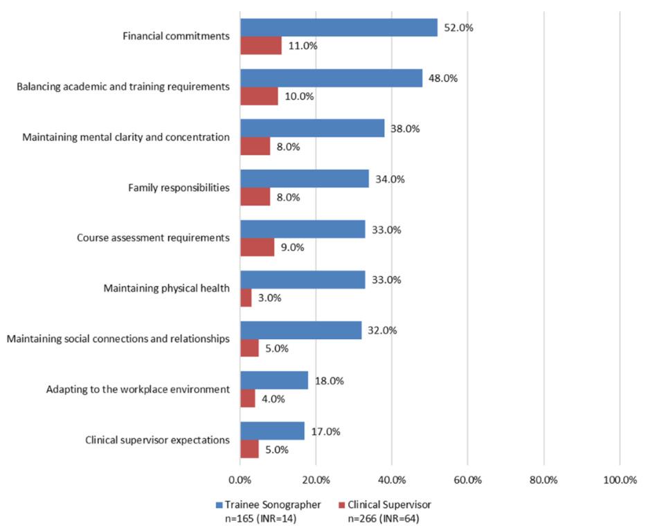
This study highlights the significant pressures faced by sonography trainees and the critical need for more supportive and sustainable clinical training environments. Without meaningful change, there is a risk of increased trainee burnout, potential exploitation, and compromised patient care, ultimately threatening the sustainability of the sonography workforce. The findings emphasise the need for targeted strategies, including improved financial support, accessible mental health services, structured mentorship, and more flexible training pathways, to better support trainees and strengthen clinical education outcomes.
Artificial intelligence (AI) is becoming increasingly integrated in medical imaging with growing interest and evidence supporting its potential role in assisting sonographers. Benign gynaecological conditions, including fibroids, adenomyosis, endometriosis, and ovarian lesions, can be challenging to diagnose due to operator dependency and variable sonographic appearance.
This systematic review aimed to evaluate how AI is currently being used in gynaecological ultrasound and whether these technologies may improve diagnostic accuracy and support clinical practice in the future.
The authors reviewed published studies investigating AI applications in the sonographic assessment of benign gynaecological disorders. Included studies assessed machine learning and deep learning systems designed to interpret ultrasound images and assist with diagnosis.
The review assessed AI model types, ultrasound techniques, and diagnostic performance measures, including sensitivity and specificity. Risk of bias and study methodology were also critically evaluated.
The review found that AI demonstrated promising results in identifying and classifying several benign gynaecological conditions, particularly in differentiating adnexal lesions and identifying features of adenomyosis
and endometriosis. The literature also demonstrated growing interest in AI applications for automated ovarian follicle segmentation and volume measurements, with potential benefits for workflow efficiency and measurement standardisation.
However, many studies documented limitations of small sample sizes, retrospective designs, and a lack of external validation. AI performance also depended heavily on image quality and expert-labelled datasets. The authors concluded that while AI shows promising potential as a support tool, there is currently insufficient evidence to support independent clinical use.
This review is highly relevant to sonographers as it demonstrates that AI in gynaecological ultrasound is currently best suited as a supportive technology rather than a replacement for human expertise. AI may eventually assist with workflow efficiency, lesion classification, and reporting support, but it still relies on high quality sonographic imaging and experienced interpretation.
Importantly, the study emphasises that AI systems are likely to enhance efficiency and confidence in sonographer practice while still requiring human oversight, advanced scanning skills and clinical reasoning. Rather than replacing sonographers, current evidence suggests AI is more likely to function as an adjunct tool that may enhance diagnostic consistency and support clinical decisionmaking. However, further large-scale prospective studies are required before routine implementation.
EMERGING TECHNOLOGIES
Reviewer: Charlotte Harman
Authors & Journal: Moro F, Giudice M, Ciancia M, Zace D, Baldassari G, Vagni M, et al. Ultrasound Obstet Gynecol 2025; 65(3):295–302.
Open Access: Yes
Read the full article here
“Incorporating AI systems into clinical practice can improve patient management and prognosis, reduce healthcare costs and reduce gynecologists’ workload by increasing their efficiency and accuracy.”
Breast cancer is the most common cancer in women and the second leading cause of cancer-related death in women [1]. Early detection improves treatment outcomes [2]. However, radiologist fatigue and elevated workload can compromise diagnostic accuracy. In this context, AI-based systems provide valuable support by reducing radiologists’ workload and streamlining diagnostic workflows [3]. Evidence shows that AI, particularly deep learning models, can enhance breast cancer screening and diagnosis, with performance in some studies comparable to or exceeding that of experienced radiologists. Consequently, computer-aided analysis has become increasingly utilised to assist radiologists in identifying and characterising suspicious findings [4].
This study aimed to develop hybrid deep learning models to improve the segmentation and classification of breast tumours, thereby enabling radiologists to diagnose breast cancer with greater efficiency and accuracy.
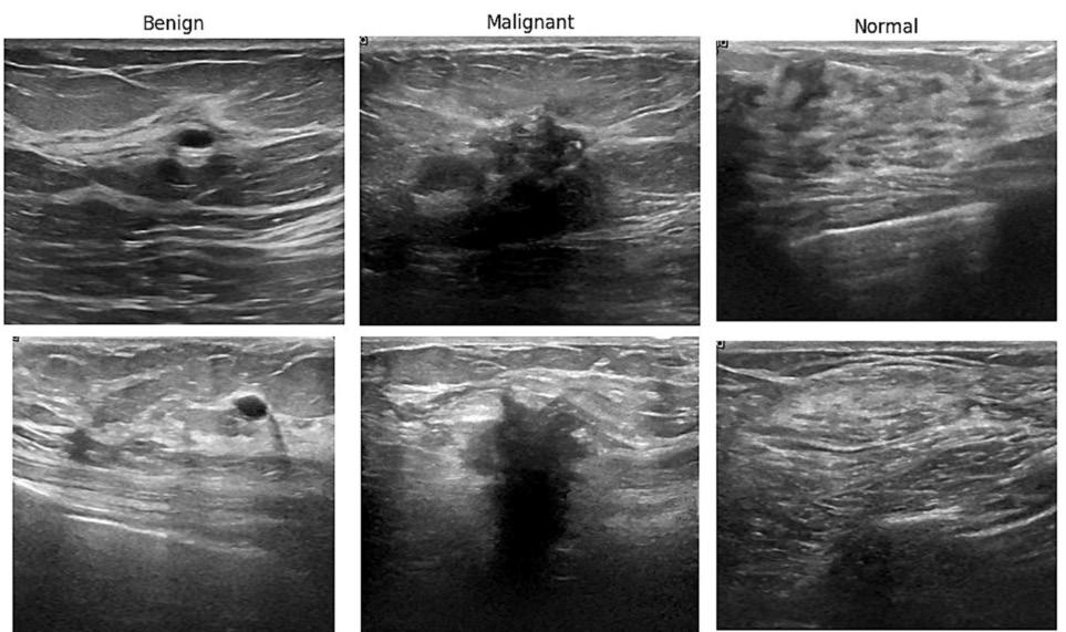
This study acquired 780 images from 600 patients from a public database and classified ultrasound images into 437 benign, 210 malignant, and 133 normal. Four pretrained deep learning models – VGG-16, DenseNet-121, DenseNet-169, and ResNet-50 – were fine-tuned for classification tasks. These models were evaluated based on accuracy, precision, recall, F1-score, and area under the curve (AUC). The U-Net model was used for generating segmentation masks to highlight regions of interest, with performance assessed through intersection over union (IoU) and Dice coefficient. The study compares classification performance using original ultrasound images versus segmented images. Segmentation enables precise, objective quantification and visualisation by generating masks that outline target areas. It classifies each pixel and, in doing so, identifies areas of interest (ROI) such as tumours.
EMERGING TECHNOLOGIES
Reviewer: Lisa McGuire, AFASA
Authors & Journal:
Rahimnezhad A, Rezaeijo S, Bayet M, Heydarheydari S. Egypt J Radiol Nuc Med. 2025;56(243).
Open Access: Yes
Read the full article here
“The study incorporated segmentation masks as a preprocessing step to enhance classification by concentrating the model’s attention on pertinent image features.”
The final hyperparameters were selected based on the highest intersection over union (IoU) score on the validation set. In addition, model stability and training time were considered to ensure robust and efficient performance across runs. This approach aims to improve the model’s overall classification.
In this study, a comparative analysis focused on evaluating the performance of two approaches for classifying breast ultrasound images was performed using pre-trained classification models directly on the original images versus applying these models to images processed with U-Net segmentation masks.
(IoU) = accuracy of an object segmentation algorithm by computing the ratio of the area of overlap between the predicted bounding box or segmentation and the ground truth, relative to the area encompassed by their union.
Applying U-Net segmentation before classification improved model performance on the breast ultrasound images dataset, particularly for DenseNet-121 and DenseNet-169, by helping the classifiers focus
on lesion-specific regions. These results indicate that by applying segmentation before classification and focusing the classifier on lesion-specific regions, prediction quality can be enhanced.
This framework should be regarded as a supportive tool, not an independent diagnostic system. It has the potential to help sonographers and radiologists identify subtle findings and reduce errors through structured lesion-focused analysis. However, a major limitation of these findings was the singlecentre dataset used. Further external validation in multicentre prospective studies is needed before clinical integration can be considered.
1. Siegel RL, Miller KD, Jemal A. Cancer statistics. CA Cancer J Clin. 2018;68(1): 7–30.
2. Han L, Huang Y, Dou H, Wang S, Ahamad S, Luo H, et al. Semi-supervised segmentation of lesion from breast ultrasound images with attentional generative adversarial network. Comput Methods Programs Biomed. 2020;189:105275.
Epididymo-orchitis and varicoceles are both common conditions encountered in scrotal ultrasound and are important contributors to male infertility. Epididymo-orchitis is typically regarded as an infectious or inflammatory process, whereas varicocele is traditionally associated with venous reflux and impaired spermatogenesis rather than infection. Emerging evidence, however, suggests that chronic venous stasis associated with varicoceles may predispose patients to epididymal inflammation. The study was therefore undertaken to investigate whether a left-sided varicocele is associated with an increased risk of epididymitis or epididymoorchitis, and to determine whether varicoceles represent merely a coincidental finding or a potential contributing risk factor.
This retrospective study included 72 patients with left-sided epididymitis or epididymoorchitis (Group 1) and 72 control patients without scrotal pathology (Group 2). Patients with right-sided disease were excluded to maintain cohort consistency.
All participants underwent scrotal ultrasound using a high frequency (15 MHz) linear transducer, incorporating B-mode, colour and spectral Doppler imaging. Varicoceles were assessed in both supine and standing positions using established diagnostic criteria, including pampiniform plexus vein diameter greater than 3 mm and reflux duration exceeding two seconds during the Valsalva manoeuvre. Varicoceles were graded on a 5-point scale.
Epididymitis and epididymo-orchitis were diagnosed based on sonographic features, including enlargement, increased vascularity, heterogeneous echotexture, reduced echogenicity, and scrotal wall thickening. Statistical analysis compared varicocele prevalence, vein diameter, and varicocele grade between groups using Mann-Whitney U tests, Chi-square tests, and Cochran-Armitage trend analysis.
The prevalence of left-sided varicocele was significantly higher in patients with epididymitis or epididymo-orchitis compared to controls (66.7% vs 22.3%). Additionally, affected patients demonstrated larger pampiniform plexus vein diameters and higher varicocele grades.
A significant positive trend was observed between increasing varicocele grade and the likelihood of epididymo-orchitis, with highergrade varicoceles conferring greater risk of inflammatory disease.
The authors propose that chronic venous stasis may impair local microcirculation, promote oxidative stress, and reduce immune defence mechanisms, thereby increasing susceptibility to infection and inflammation within the epididymis and testis.
This study is highly relevant to sonographers performing scrotal ultrasound, as it highlights a potentially under-recognised association between varicocele and epididymo-orchitis. It reinforces the importance of actively assessing for varicocele in patients presenting with scrotal pain or suspected infection, rather than dismissing it as an incidental finding.
From a clinical perspective, identifying coexisting varicocele may influence patient management, particularly in cases of recurrent or chronic epididymo-orchitis where venous congestion may contribute to persistent symptoms or infertility risk.
Limitations of this study include the relatively small sample size, single-centre design, and absence of longitudinal follow-up to establish causation or recurrence risk. Nevertheless, the findings suggest a clinically meaningful association that warrants further investigation.
Overall, this study encourages a more integrated approach to scrotal ultrasound assessment, considering vascular, inflammatory, and fertility-related factors collectively rather than in isolation.
Reviewer: Jane Keating
Authors & Journal: Atay M, Toprak H, Akcay A, Mehdi E, Yilmaz M. J Clin Ultrasound. 2024;52(6):700–704.
Open Access: Yes
Read the full article here
“Left-sided varicocele was significantly more common in patients with epididymoorchitis and is an important cause for its development due to chronic venous stasis.”
This study aimed to address the need for improved understanding of the relationship between ultrasound-based risk stratification (TIRADS) and cytological classification (Bethesda system) in the assessment of thyroid nodules. While both systems are widely used in clinical practice, there is a need to better define how well they correlate and support one another in predicting malignancy and guiding management decisions.
This was a retrospective study involving 1,100 patients with thyroid nodules. The authors analysed ultrasound findings classified using Thyroid Imaging Reporting and Data System (TIRADS), fine needle aspiration (FNA) cytology results reported via the Bethesda system (TBSRTC), and histopathological outcomes where available. Correlation between imaging, cytology, and histology was assessed to evaluate concordance across the systems.
The study demonstrated a strong correlation between TIRADS categories and Bethesda cytological grades.
• High concordance was observed between TIRADS 5 and Bethesda 5, both indicating high suspicion of malignancy.
• TIRADS 2 nodules correlated well with Bethesda 2, confirming benign findings.
• Histopathological data supported these correlations, strengthening the validity of the results.
An important finding was a reported malignancy rate of approximately 5% in TIRADS 2–4 nodules that proceeded to histopathology. While this may reflect selection bias, as only certain nodules progressed to surgery, it reinforces the rationale for recommending FNA in selected cases despite apparently lower-risk ultrasound appearances.
This study reinforces the importance of accurate TIRADS assessment by sonographers and highlights how imaging findings directly influence downstream clinical decisions. Understanding the correlation with Bethesda categories helps sonographers appreciate the diagnostic and management implications of their reporting.
The authors also highlight the importance of solitary thyroid nodules as a potential indicator for FNA assessment. In current practice, considerable emphasis is placed on TIRADS scoring features, and the significance of a discrete solitary nodule may sometimes be under-recognised. This serves as a useful reminder that clinical decision-making should incorporate the broader context of thyroid pathology alongside formal risk stratification systems.
The article is particularly valuable for sonographers involved in thyroid imaging and those considering advanced practice roles in thyroid FNA procedures.
This is a well-constructed study supported by a large patient cohort and a comprehensive reference list. The inclusion of ultrasound, cytological, and histopathological images enhances understanding of the relationships between imaging appearances and pathological outcomes.
The article successfully demonstrates that TIRADS and Bethesda classifications are complementary systems that work effectively together in the investigation and management of thyroid nodules.
I would recommend this article to all sonographers performing thyroid ultrasound examinations, as well as those interested in thyroid pathology and advanced practice in undertaking FNA. It offers valuable insight into the relationship between imaging and cytological diagnosis and reinforces the importance of accurate thyroid nodule characterisation.
Reviewer: Paula King
Authors & Journal: Dhar L, Singh M, Jain S, Rana D, Kumar J, Meher R, et al. Cytopathology. 2023;34(6):590–596.
Open Access: Yes
Read the full article here
“The article successfully demonstrates that TIRADS and Bethesda classifications are complementary systems that work effectively together in the investigation and management of thyroid nodules.”
Given the current shortages of sonographers and other health workers in Australia, there is a need to encourage the delayed retirement of sonographers. This article aimed to identify factors contributing to the decision facing healthcare workers contemplating retirement.
How the study was performed
Healthcare workers in China were asked to complete a cross-sectional survey guided by the role theory and the push-pull model. The questionnaire covered data on demographics, work conditions, job satisfaction, occupational fatigue, self-related health, and chronic illness. Results were analysed to identify predictors that may support a decision to delay retirement.
The survey of the technical staff showed 58.5% were in good health, 30% in fair health and 11.5% in poor health. Technicians reporting chronic illness comprised 43% of their cohort, which was the highest amongst all the healthcare workers.
Healthcare workers with the highest job satisfaction and lowest occupational fatigue tended to support delayed retirement. Supporters also, in general, reported their health as excellent or good (72% vs 57%) and had fewer incidences of poor health (5% vs 13%). Those opposed to delayed retirement
reported a higher incidence of chronic illness (46.7%) compared to supporters (33%).
Supporters of delaying retirement also tended to be male, have greater work experience, work longer weekly hours and have more frequent night shifts. Supporters generally had high job satisfaction, while occupational fatigue showed an inverse association.
This article shows that occupational fatigue, poor health, chronic illness, and work strain may influence sonographers’ decisions to prolong their professional careers. Conversely, good job satisfaction and a high level of organisational support suggest a positive influence on sonographers to delay retirement.
Given the shortage of sonographers in Australia, an increasing workload and an ageing population of experienced sonographers, healthcare administrators need to factor these findings and implement them to encourage sonographers to consider delaying their retirement. Failing to address low occupational wellbeing, job satisfaction and burnout risks will lead to high staff turnover and compromised quality of patient care.
Clinical relevance of this study is limited by the study population confined to Chinese health workers and not specific to the sonographer cohort.
Conceptual Framework: Role Theory & Push-Pull Model
Push Factors
Occupational fatigue, Poor health, Chronic illness, Work strain
Pull Factors
Job satisfaction, Professional identity, Financial need, Organisational support
Demographics Age, Gender, Education
Attitudes towards delayed retirement (Outcome)
HEALTH AND WELLBEING
Reviewer: Peter Esselbach
Authors & Journal: Sun X, Wu Z, He L, Wang S. BMC Public Health. 2025;25(1):3070.
Open Access: Yes
Read the full article here
“Failing to address low occupational wellbeing, job satisfaction and burnout risks will lead to high staff turnover and compromised patient care.”
Work integrated learning (WIL) is a core component of sonography training in Australia and New Zealand. Yet the profession continues to face persistent workplace pressures, including staff shortages, limited opportunities to provide supervision, and growing clinical demand. Despite these challenges, there has been limited research capturing the lived experiences of trainee sonographers and their clinical supervisors. This study addresses that gap by examining how structural, systemic, financial, and interpersonal factors shape the training environment, with a particular focus on trainee wellbeing. By identifying key pressure points and potential avenues for improvement, the authors aimed to highlight opportunities for reform across education providers, clinical sites, and the broader health system.
The study used a qualitative research design, with an online survey distributed between July and November 2024. A total of 431 participants across Australia responded, including 266 clinical supervisors and 165 trainee sonographers. All participants were registered with the Australian Sonographer Accreditation Registry (ASAR) and had been actively involved in WIL within the previous 12 months. The survey contained five open-ended questions exploring barriers to WIL, learning challenges, support needs, wellbeing, and perceptions of training quality.
Data was analysed using semantic thematic analysis, with codes generated inductively, refined collaboratively, and organised into major themes. The analysis focused on identifying recurring challenges, differences in perspectives between trainees and supervisors, and proposed strategies for improving WIL experiences.
The study revealed a complex web of workforce, institutional, and emotional pressures shaping the current sonography training landscape. The availability of training positions emerged as the most significant barrier, with many clinical sites being unable to accommodate trainees. Even when trainees
were able to secure a training position, they were not always guaranteed a supportive or structured learning environment. Many described their training as ad hoc, fragmented, and heavily dependent on individual supervisors’ availability.
Financial pressures also featured prominently. Trainees reported the burden of travel costs, unpaid placements, and reduced income, all of which contributed to considerable strain. Mental health and wellbeing concerns were common, with some trainees reporting anxiety and depression associated with the intensity of ultrasound training. Poor work-life balance was also highlighted, as trainees struggled to manage academic requirements alongside demanding clinical workloads.
The study identified several strategies to strengthen the WIL experience. Participants emphasised the need for stronger collaboration between universities and clinical sites to support trainee learning and enhance supervisor engagement. Improved vetting of clinical training sites was recommended to ensure they provide consistent, high quality learning experiences. Additionally, participants called for stronger safeguards to protect trainees from burnout and exploitation, including improved institutional oversight and clearer mechanisms for reporting concerns.
This study highlighted the increasing strain on the clinical workforce, where service demands are restricting the ability to train the next generation of sonographers effectively. The authors emphasise the need for stronger partnerships between universities and clinical training sites to ensure that education and service delivery are better aligned. They also highlighted that trainee wellbeing is closely linked to learning outcomes, patient safety, and workforce retention, making it an important element of creating a sustainable sonography workforce.
The study offered a roadmap for change, calling on stakeholders to reimagine WIL as a shared responsibility. Rather than viewing learning, wellbeing, and service delivery as competing priorities, the authors argue for an integrated approach that supports trainees, strengthens the workforce, and enhances the quality of patient care.
HEALTH AND WELLBEING
Reviewer: Emma Jardine, FASA
Authors & Journal: Boucaut P, Tunny R, McMahon K, Osborne B, Lamb K, Wardle J, et al. Radiography. 2026;32.
Open Access: Yes
Read the full article here
“Trainee wellbeing is closely linked to learning outcomes, patient safety, and workforce retention.”
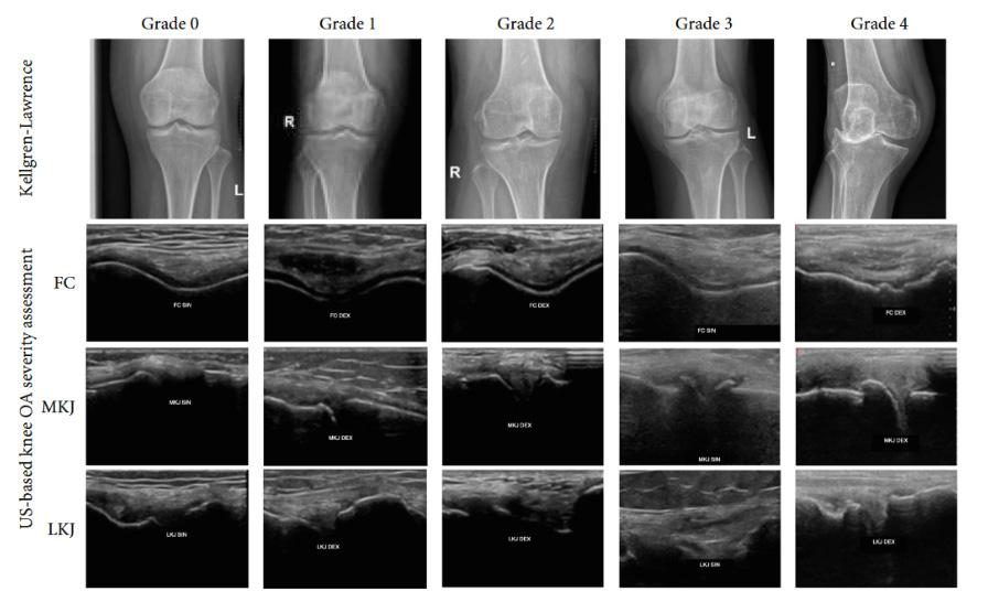
Osteoarthritis (OA) of the knee is typically diagnosed on imaging using conventional radiography; however, it cannot predict OA severity due to its inability to assess meniscal extrusion, femoral cartilage loss and other soft tissue abnormalities. Magnetic resonance imaging (MRI) can also be used, but it is often not available in low-resource settings. Ultrasound imaging can be used to identify femoral condyle cartilage changes, knee osteophytes and meniscal protrusion, all of which can be used as indicators for monitoring of knee OA. This study compared the use of ultrasound imaging and conventional radiography to determine the severity of knee OA.
A radiography-based validated KellgrenLawrence (KL) grading system used to determine the severity of knee OA was used for radiograph assessment. This system grades knee OA based on knee joint space narrowing, osteophyte formation, bone sclerosis and bone end deformity. An ultrasound-based scoring system was developed from the KL system to determine knee OA severity using femoral condyle cartilage thickness and the presence of osteophyte formation and meniscal protrusion at the lateral and medial knee joints and was used to assess knee ultrasound imaging.
Participants (112) with clinically diagnosed knee OA using the American College of Rheumatology (ACR) criteria were included in the study. Participants under 50 years of age, with a history of knee injury, prior knee surgery, a diagnosis of autoimmune disease or those who had intra-articular injections in the last six months before imaging were excluded from the study.
Three key sonographic criteria were used to assess knee OA, which included:
1. femoral cartilage contour, regularity and degree of femoral cartilage loss
2. meniscus protrusion
3. presence of osteophytes.
Ultrasound imaging of the femoral cartilage was obtained with the knee in full flexion, whereas the medial and lateral knee joints were imaged with the knee flexed 30 degrees,
as this is most sensitive for detecting meniscus protrusion, joint narrowing or osteophyte formation. Imaging was obtained by two observers and interobserver reliability was assessed using the scoring system using kappa analysis.
Excellent interobserver reliability was demonstrated using the modified KL protocol for knee ultrasound assessment. Additionally, a significant association was demonstrated between the radiographic and ultrasoundbased knee OA severity scaling. This study demonstrated that ultrasound was superior to conventional radiography in detecting knee OA changes, as it could directly identify femoral cartilage thickness loss (Figure 1).
Cartilage thickness loss is frequent in older adults, with or without OA, but can also be a biomarker in OA development. Femoral cartilage degradation is a hallmark of knee OA and in the disease process progression, which is not truly appreciated in conventional X-rays and is better assessed using ultrasound. Patients with knee OA will have thinner femoral cartilage and morphological changes compared to the healthy population. Inclusion of femoral cartilage assessment in a standard knee ultrasound examination can improve the detection of early knee OA changes.
Reviewer: Susan Diep
Authors & Journal: Pane R, Handayani H, Aisyah A, Hisamud-Din N, Al Hajiri A, Rahman A. Sci World J. 2026;(1):1–7.
Open Access: Yes
Read the full article here
“Cartilage thickness loss frequently occurs in older adults, with or without knee OA, and serves as an important biomarker for knee OA development.” MSK
Although ultrasound is commonly used to evaluate the finger flexor pulley system, there has been a paucity of studies investigating the use of ultrasound imaging to reliably visualise the toe flexor pulley system. Previous research has been limited to a small number of studies involving cadaveric toe specimens, cross-examined with magnetic resonance imaging. This study investigated whether high frequency ultrasound could reliably image the flexor pulley system of the toes. The paper also describes the normal sonographic appearance of the toe pulley structures and evaluates whether ultrasound imaging can identify pathological changes to the pulleys, and how the sonographic findings resemble those seen with pathological pulleys of the fingers.
The study involved a two-phase observational design. Firstly, cadaveric specimens (eight feet from four cadavers) were examined using ultrasound, and imaging findings were validated via dissection following ultrasoundguided marking. Secondly, 15 healthy adult participants underwent bilateral toe ultrasound imaging using high frequency linear transducers (18–5 MHz and 24–8 MHz) by two experienced sonographers. They assessed pulley visibility, anatomical location, and thickness, with interobserver agreement of pulley thickness measurements analysed using intraclass correlation coefficients (ICC).
The cadaveric component of this research defined four annular pulleys and two cruciform pulleys in the second to fifth toes, with the most
distal pulley (defined as A4 pulley) situated at the distal interphalangeal joint. In the great toe, three annular pulleys and two cruciform pulleys were identified.
Ultrasound imaging was demonstrated to be able to reliably identify annular pulleys in toes two–four, with slightly reduced reliability in the fifth toe due to the smaller-sized anatomy of this toe. Sonographic toe pulley thickness measurements showed minimal interobserver variability, although statistical agreement was moderate (ICC up to 0.529). The lower frequency 18–5 MHz transducer demonstrated better reliability than the higher 24–8 MHz transducer due to improved penetration through the plantar soft tissue of the toe.
The paper also presented clinical cases to demonstrate how toe pulley partial tears, pulley inflammatory changes, and a trigger hallux can be demonstrated sonographically (Figures 1 and 2 on following page).
Relevance to clinical practice
The anatomy of the toe flexor pulley system is underappreciated. Despite this study having a small sample size and a limited number of pathological cases, it demonstrates how sonographic imaging can be used to assess the toe flexor pulley system and identify injury and pathology to these small structures. This small study demonstrates that highfrequency ultrasound is a feasible modality for dynamically evaluating the toe pulley system, highlighting pathologies that may otherwise be overlooked or underdiagnosed if the anatomy and sonographic appearance are not well understood.
Injuries and pathologies such as partial tears, inflammatory change and trigger toe can all be demonstrated sonographically.
Reviewer: Craig Winnett
Authors & Journal:
Pistoia F, Macciò M, Susi M, Picasso R, Zaottini F, Marcenaro G, et al. Skeletal Radiol. 2025;54:2045–2054.
Open Access: No
Read the full article here
“High-resolution ultrasound has been proven to be a valuable diagnostic tool for identifying and assessing pathologies of the toe annular pulley, potentially leading to more accurate diagnoses and better treatment planning.”
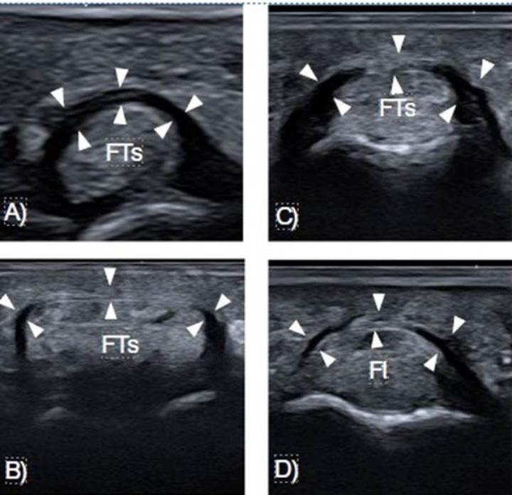
Figure 1. Images from the Pistoia et al. demonstrating flexor pulleys of the 2nd toe in a healthy participant. A. A1 pulley. B. A2 pulley. C. A3 pulley. D. A4 pulley. Reproduced images from Skeletal Radiol. 54:pg 2049.
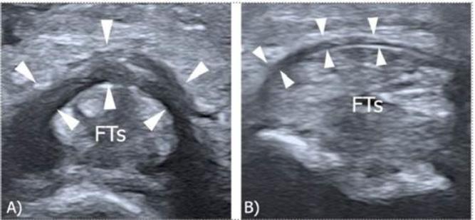
Figure 2. Images from Pistoia et al. demonstrated sonographic demonstration of the A3 toe pulley. A. Post-traumatic partial tear of the pulley in short axis which demonstrates hypoechoic thickening. B. Comparison with uninjured contralateral pulley, showing normal thickness. Reproduced images from Skeletal Radiol. 2025;54:pg 2053.
The review was performed to focus on paediatric infection, which can cause substantial morbidity, and the role of different imaging modalities, including ultrasound, currently used in the United States to aid diagnosis or assessment of differential diagnosis.
The authors assessed currently documented knowledge in the literature to describe the aetiology, incidence/prevalence and imaging characteristics of acute musculoskeletal infections in children. They examined imaging features that may help differentiate between self-limiting conditions (e.g. transient synovitis) and those requiring urgent treatment to prevent long-term complications (e.g. malignancy).
Skin and soft tissue infections (SSTIs): primarily a clinical diagnosis.
Cellulitis is the non-necrotising type of infection of superficial soft tissues. Radiographically, it demonstrates non-specific soft tissue swelling with effacement of fat planes. US will also show skin thickening, increased echogenicity in the subcutaneous fat, and subcutaneous oedema with ‘cobblestoning’, and hyperaemia (Figure 1). Complicated cellulitis includes investigation for a foreign body (e.g. wood, plastic or metal) or for alternative causes of soft tissue swelling (e.g. fracture or DVT).
Abscesses: can be challenging because clinical appearance shows significant overlap with that of uncomplicated cellulitis. Imaging better identifies abscesses, helps to determine the size and location(s) of the collection and helps plan drainage.
US, often at the point of care, is highly accurate in the diagnosis of an abscess. This shows as a fluid collection on US with possible hyperaemic and hyperechoic rims. The use of dynamic compression, looking for swirling of internal contents, may help differentiate an abscess from a phlegmon.
MRI still has the highest sensitivity and specificity.
PAEDIATRIC
Reviewer: Ellen Chen
Authors & Journal: McLuckey M, Kemp J, and Meyers A. Pediatr Radiol. 2026;56:1018–1032.
Open Access: Yes
Read the full article here
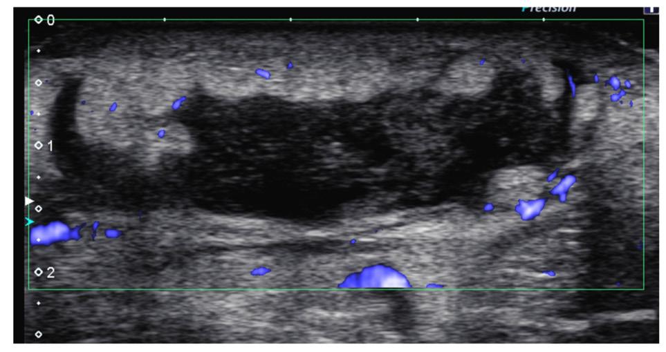
Figure 1 shows a 14-year-old girl who presented to the emergency department with proximal right forearm swelling in the area of bug bites. Power Doppler transverse image of the volar soft tissues of the area shows a large 5 cm hypoechoic collection with peripheral hyperaemia. Note the surrounding soft tissues show increased echogenicity.
Necrotising soft tissue infections (NSTIs): primarily a clinical diagnosis, first establishing the type of infection involved. In the absence of traumatic or iatrogenic causes, gas extending along fascial planes is diagnostic on MRI.
Pyomyositis: contrast MRI is the main modality. US may show liquefied contents as an intramuscular collection with surrounding hyperaemia. Differential: haematoma.
Infectious tenosynovitis: The closed nature of the paratendinous synovial compartment predisposes to infection. Hands/wrists and feet/ankles are the most commonly involved sites due to direct inoculation. US and MRI are the best imaging modalities. US will demonstrate hyperaemia with normal or slightly thickened tendons. US may show fluid or synovial thickening around tendons, with the potential for infection to spread between deep compartments.
This study describes how the choice of and interpretation of imaging findings may contribute to determining causes of acute MSK infections in children, and their differentials.
“Musculoskeletal
infections are a common reason children present to emergency departments and can be difficult to diagnose. This is especially the case in younger children, who often show vague symptoms such as fever, limping, or reduced use of a limb.”
The study aimed to investigate the occurrence of subdural collections in infants with benign enlargement of the subarachnoid spaces (BESS), a common cause of macrocephaly. Given the potential overlap in imaging findings between BESS and abusive head trauma, the researchers sought to determine when further evaluation for abuse is warranted.
The authors conducted a comprehensive review of existing literature and clinical data to assess the prevalence and characteristics of subdural collections in infants diagnosed with BESS. They examined imaging features, clinical presentations, and outcomes to differentiate between benign and potentially abusive conditions.
The study found that while BESS is typically a benign condition, the presence of subdural collections in these infants can complicate the clinical picture. These collections may mimic signs of abusive head trauma, leading to challenges in diagnosis. The exact prevalence of occult injuries and abuse in these infants remains unknown. Therefore, thorough evaluations, including skeletal surveys and clinical assessments, are recommended even when initial findings appear unremarkable.
This study underscores the importance of careful interpretation of imaging findings in infants with macrocephaly. Healthcare providers should maintain a high index of suspicion for potential abuse, especially when subdural collections are present. Implementing comprehensive diagnostic protocols can ensure appropriate management and safeguarding of vulnerable infants.
PAEDIATRIC
Reviewer: Lino Piotto, FASA
Authors & Journal: Raissaki M, Adamsbaum C, Argyropoulou M, Choudhary A, Jeanes A, Mankad K, et al. Pediatr Radiol. 2023;53,752–767.
Open Access: Yes
Read the full article here
“While BESS is typically a benign condition, the presence of subdural collections can complicate the clinical picture and mimic signs of abusive head trauma.”
Many of you will be familiar with the frustration of reaching for a teaspoon at break time and finding none. Turns out it is a measurable and statistically significant phenomenon. This paper investigated cutlery attrition in a hospital tearoom [1]. You may be familiar with the legendary 2005 study by Lim et al., who applied rigorous research methods to the cutlery problem in a Melbourne research institute [2]. This Brisbane team’s aim was to extend that work to a busy hospital environment, and to see how forks compared to spoons.
This study is in the tradition of the special Christmas issues of the British Medical Journal, which have been published for decades. Papers that apply genuine scientific rigour to questions that are a little quirky are featured, such as: 27 is not a dangerous age for famous musicians [3], and that men are idiots [4]. The point is not just to be humorous, but a reminder that the scientific method is a tool, not a subject, and that good methodology can illuminate any question.
This was a prospective longitudinal quality improvement study. The researchers purchased 18 stainless steel forks and 18 teaspoons, marked each with a discreet red spot using nail polish, and introduced them into the staff tearoom alongside the existing 81 items of cutlery. Tearoom users were blinded to the purpose of the study and were unaware that the cutlery was under surveillance. Counts were conducted twice weekly for seven weeks, with a baseline and end-of-study census of all items on day 45. Outcome measures were the number of marked items remaining and the rate of utensil loss per 100 days. Statistical comparison between fork and teaspoon attrition used a standard test for differences in proportions.
Results demonstrated that by day 45, six of the 18 marked teaspoons had vanished, compared to just one of the 18 forks, a statistically significant difference (p = 0.038). The overall attrition rate was 2.2 items per 100 days
for spoons and –2.2 per 100 days for forks, meaning forks were somehow accumulating.
The authors noted, dryly, that teaspoon disappearance ‘may reflect different kleptomaniacal or individual appropriation tendencies’ and that the symbolism of fork ‘rebirth or resurrection’ makes it an ideal Christmas gift.
While a pivot from the usual focus of Making Waves, there’s value in this paper for sonographers through the celebration of methodology. Research is not reserved for academics studying complex issues; it is a framework for answering questions. The same longitudinal cohort principles that underpin studies of clinical management or diagnosis can apply to cutlery. There is also an Australasian connection with this study and the one that inspired it. This group of clinicians and researchers at a busy hospital took the time to pursue this question, with ethics oversight and peer review. It’s a great reminder that research does not always have to begin with a grant application and a sophisticated hypothesis. Sometimes it begins on-call on a Saturday night with a lukewarm cup of hospital-issued instant coffee and no spoon to stir in the milk and sugar.
1. Mattiussi M, Livermore A, Levido A, Starr T, Lassig-Smith M, Stuart J, et al. What the forks? A longitudinal quality improvement study tracking cutlery numbers in a public teaching and research hospital staff tearoom. Med J Aust. 2020;213(11):521–3.
2. Lim MS, Hellard ME, Aitken CK. The case of the disappearing teaspoons: longitudinal cohort study of the displacement of teaspoons in an Australian research institute. BMJ. 2005;331:1498–500.
3. Wolkewitz M, Allignol A, Graves N, Barnett AG. Is 27 really a dangerous age for famous musicians? Retrospective Cohort Study. BMJ. 2011;343.
4. Lendrem BA, Lendrem DW, Gray A, Isaacs JD. The Darwin Awards: sex differences in idiotic behaviour. BMJ. 2014:349.
Reviewer: Tristan Reddan, FASA
Authors & Journal: Mattiussi M, Livermore A, Levido A, Starr T, Lassig‐Smith M, Stuart, et al. Med J Aust. 2020;213(11):521–3.
Open Access: Yes
Read the full article here
“Teaspoons tend to go missing more often than forks in a shared multidisciplinary staff tearoom, possibly reflecting different kleptomaniacal or personal appropriation tendencies.”
There are no globally adopted protocols for the de-identification of patient image-based data. Global heterogeneity for preprocessing patient data increases the risk of patient data breaches. This paper reviews current methods used to protect patient privacy when patient images are used for research and technology development. The health professional can refer to this framework to inform the responsible handling of patient image data for the use of research development.
Many regional protocols and guidelines have been developed to comply with local legislation. Australian legislation for patient data preprocessing relates to Australia’s Privacy Act 1988 [1]. Professional guidelines for the principles of handling patient data are outlined in standard ten of the Royal Australian and New Zealand College of Radiologists (RANZCR) Standards of Practice for Clinical Radiology [2]. The addendum provides a summary of AI standards requirements and evidence.
The systematic review was conducted according to the Preferred Reporting Items for Systematic Reviews and Meta-Analyses for Scoping Reviews (PRISMA-ScR) guidelines [3]. The review was conducted across 5 databases (PubMed/Medline, Scopus, Web of Science, Embase, Google Scholar). The following keywords were used in modified search strings: ‘Anonymization’, ‘De-identification methods’, ‘patient identity protection’, ‘data masking’, ‘privacy’, ‘Medical Images’, ‘Clinical Images’, ‘radiography’, ‘Patient images’, ‘disease image’, ‘lesion image’, ‘data sharing’ and ‘dataset creation’. Studies were included that identified the creation of datasets from de-identified medical images as outlined in Table 1. Exclusion criteria included reviews, systematic reviews, editorials, correspondence, letters to editors, proceedings, white papers and conference abstracts.
Parameter
Inclusion criteria
Population (P) Datasets with medical images
Exposure (E) Pseudonymisation strategies
Comparator (C)
Anonymisation techniques and the parameters used for medical images/ datasets
Outcome (O) Parameters that must be de-identified in medical images and the method
Study design (S)
Original research studies, cross-sectional studies, dataset creation, and randomised control trials
Table 1: PECOS criteria for inclusion eligibility. Adapted from Sood et al. J Med Imaging Radiat Sci. 2025;56(5): pg 3.
The final study set included 13 papers, a relatively small number in comparison to the many papers published using anonymised medical image datasets. Most papers used open-source datasets (92%), which are preanonymised before archiving in the named repository.
The most common imaging technique was magnetic resonance (n = 8), of which seven studies focus on adult brain images. The commonly used formats for MR imaging data were BIDS (Brain Imaging Data Structure) (n = 3) and Digital Imaging and Communication in Medicine (DICOM) (n = 3). Methods of deidentification were listed as burn-in annotation (permanently redacted sensitive information embedded in the medical image pixels), defacing processes, removal of DICOM tags, and facial de-identification.
Findings from this review shaped a medical image checklist which closely aligns with the British Medical Journal published standards and anonymisation checklist [4]: 1. After anonymisation, perform a manual check to confirm the removal of patientidentifiable information.
Reviewer: Caterina Watson, AFASA
Authors & Journal: Sood A, Pattnaik T, Malhotra R, Nayyar V, Narayan B, Mishra D, et al. J Med Imaging Radiat Sci. 2025;56(5):1–9.
Open Access: No
Read the full article here (Full access has been granted by the journal for six months from date of publication.)
“There is currently no universally accepted standard for de-identifying medical imaging datasets, creating significant variability in how privacy is protected across studies.”
2. Use encryption methods to store and transmit images. Store copies of data in case of system failure.
3. Ensure only authorised personnel can access data by using access control mechanisms. A data security officer should review the framework for compliance with internal policies.
4. Keep a record of any actions performed on the data, such as anonymisation dates, personnel involved, and any modifications made.
5. Ensure patient consent for research purposes, even if the data is anonymised.
Patient image de-identification is the responsibility of the healthcare worker before data sharing. There are recommended standard actions for effective image deidentification. The review also highlights the need for discernment in selecting critical patient privacy information and balancing sufficient quality and richness of information for future research analysis.
1. Australia: Act No. 119 of 1988, Privacy Act 1988 (Cth). Available from: https://www. refworld.org/legal/legislation/natlegbod/1988/ en/104174. [accessed 25 November 2024].
2. Standards of Practice for Clinical Radiology v12.0 © Royal Australian and New Zealand College of Radiologists. Standards of practice for clinical radiology. Version 12.0. Sydney: RANZCR; 2025 Nov 7 [cited 2026 May 3]. Available from: https://www.ranzcr. com/wp-content/uploads/2005/08/20251124Standards-of-Practice-for-Clinical-Radiologyv12-FINAL.pdf
3. Tricco AC, Lillie E, Zarin W, et al. PRISMA extension for scoping reviews (PRISMAScR): Checklist and explanation. Ann Intern Med. 2018;169(7):467–473.
4. BMJ Standards on Anonymisation (Checklist) – 2020, https://s16086.pcdn.co/ wp-content/uploads/2020/05/External-BMJStandards-of-Anonymisation-checklist.pdf.
Duplex ultrasound (DUS) is a well-tolerated non-invasive imaging technique; however, a systematic review of the effectiveness of DUS has been reviewed only up to 2005 (Williams et al., AJR Am J Roentgenol), with that review looking at studies conducted up to 40 years ago, reporting that there was inconclusive, mixed evidence for the validity of DUS to detect renal artery stenosis (RAS). However, there has been significant improvement in the capability of DUS in the decades since. The purpose of this systematic review was to determine whether DUS accurately detects RAS in comparison with the gold standard of digital subtraction angiography (DSA).
Search strategies were developed using ‘ultrasound’ and ‘stenosis’ as primary search terms by researchers with prior experience conducting review studies. No search restrictions were placed on the type, language of the study, year of study after 2005, or population.
Studies not published in a peer-reviewed journal, or published as an editorial, opinion, review, or conference abstract, were excluded. Grey literature studies were excluded. Post2005 and available in English studies were included.
To be included in the review, studies must compare the detection of RAS by DUS with digital subtraction angiography (DSA, gold standard criterion) or any of the other reference criterion standards, such as MRA and CTA. There were 1,114 papers screened for applicability, with 34 meeting their inclusion criteria.
In comparison with the gold standard assessment of fluoroscopy DSA, most studies exhibited moderate to high agreement with DUS based on the literature, with a low risk of bias and low applicability concerns. Compared with angiographic methods of detecting RAS, DUS generally exhibited moderate to high agreement.
Some of the limitations of DSA may also be addressed with DUS and might be a suitable alternative imaging technique for evaluating RAS that practitioners should consider.
Duplex ultrasound (DUS) of the renal arteries is a non-invasive, cost-effective and highly relevant first-line screening tool for detecting and monitoring significant renal artery stenosis (RAS), offering high sensitivity and specificity in specialised vascular laboratories. It is a radiation-free and non-contrast test, particularly beneficial for patients with renal impairment.
This systematic review of published papers post-2005 demonstrates that Duplex ultrasound exhibited moderately high agreement with DSA and other well-established angiographic criteria, with the existing literature having low risk of bias.
Limitations include technically challenging patients, operator dependence and the need for experienced vascular sonographers or dedicated vascular laboratories.
Reviewer: James Maunder, AFASA
Authors & Journal: MacLeod J, Kivell M, Shivgulam M, Liu H, O’Brien M. J Vasc Ultrasound. 2024;48(1):15–27.
Open Access: Yes
Read the full article here
“DUS demonstrates moderate to high concordance with gold standard and other established angiographic criteria, with the existing literature showing low risk of bias and good patient applicability.”
Carotid web is now recognised as an important, often underdiagnosed cause of ischaemic stroke, particularly in younger patients and in cases initially labelled cryptogenic. On routine carotid duplex, however, webs can be subtle and are easily mistaken for minor plaque or artefact, and there are no universally adopted ultrasound criteria. This is problematic because carotid ultrasound remains a first-line imaging test in many stroke pathways, yet sonographers and clinicians may not have a clear mental model for how a web should look or behave haemodynamically.
The article ‘Ultrasound characteristics of carotid web’ set out to close this gap by carefully describing how carotid webs appear on high-resolution ultrasound and how advanced slow-flow modes contribute. The authors sought to identify consistent B-mode and Doppler features, and to test whether microflow imaging (MFI) provides meaningful detail beyond standard colour Doppler. In practical terms, the question was: Can duplex ultrasound, used thoughtfully, become a more reliable tool for detecting carotid web in patients being investigated after stroke?
This was a retrospective observational imaging study of patients in a cerebrovascular cohort who had a carotid web confirmed on crosssectional imaging, such as CT angiography. The series included 24 carotid webs in patients being assessed for ischaemic events or suggestive symptoms, representative of the population sonographers see in stroke work-up. Each patient underwent carotid ultrasound with high-resolution B-mode, colour Doppler, spectral Doppler and microflow imaging focused on the carotid bulb. Two experienced reviewers assessed stored clips for lesion location, morphology, echogenicity and associated flow patterns, with particular interest in flow separation and zones of sluggish flow distal to the lesion. The analysis was descriptive, comparing how confidently webs could be recognised using conventional views versus when microflow imaging was incorporated.
The study showed that the carotid web has a recognisable ultrasound signature when actively sought. Typical appearance was a thin, shelf-like, usually echogenic projection from the posterior wall of the carotid bulb, best appreciated on longitudinal B-mode with a smooth interface and minimal background plaque. Doppler commonly demonstrated disturbed flow around the web with a small area of low-velocity or stagnant flow just distal to the projection, consistent with the proposed mechanism of local thrombus formation and embolic risk.
Adding microflow imaging improved visualisation of both the web and the adjacent slow-flow zone, and in this series, increased the detection rate compared with B-mode alone. MFI helped distinguish webs from small low-profile plaques by better outlining the intraluminal shelf and the pocket of recirculating flow.
For sonographers, the implications are very practical. The paper supports deliberate interrogation of the posterior carotid bulb in young or cryptogenic stroke patients, and the routine use of sensitive slow-flow or microvascular modes when appearances are equivocal. It also highlights the importance of naming a suspected web in the report rather than using vague terms, so that stroke and vascular teams can pursue confirmatory imaging and consider targeted intervention.
The study is limited by its small, singlecentre, retrospective design and the fact that all webs were already known from reference imaging, so it does not provide full sensitivity or specificity for ultrasound screening. Microflow capability is also vendor-dependent. Nevertheless, the work is highly relevant to contemporary practice because it equips vascular sonographers with clearer visual and haemodynamic cues to recognise a lesion that can materially change secondary stroke prevention.
Reviewer: Jacqui Robinson, FASA
Authors & Journal: Fontaine L, Guidolin B, Viguier A, Gollion C, Barbieux M, Larrue V. J Neuroimaging. 2022;32:894–901.
Open Access: Yes
Read the full article here
“Carotid web (CaW) is a cause of recurrent ischaemic stroke that remains underdiagnosed using Duplex ultrasound (DUS). Improved methods and description of its sonographic features could allow better detection of CaW.”
How to get the most from axial views of the fetal brain: Applying the principles of neurosonography to screening examination of the supratentorial central nervous system
In clinical practice, routine screening examinations of the fetal brain are primarily performed using axial imaging planes. In contrast, dedicated neurosonography provides a more detailed assessment of fetal brain anatomy but is generally performed in cases in which an abnormality is suspected, when there is a family history of inherited central nervous system (CNS) anomalies, or a previous pregnancy has been affected by a CNS abnormality.
The authors demonstrate how a systematic, stepwise evaluation of structures visible on routine axial views may improve the prenatal detection of supratentorial CNS abnormalities.
This article was an expert opinion paper discussing how the axial planes used in routine fetal brain assessment can be evaluated in a more systematic and detailed manner. A series of descriptions, annotated diagrams, and ultrasound images were included to demonstrate normal supratentorial brain anatomy and illustrate how different abnormalities or clinical scenarios may appear on the axial views.
The authors proposed a diagnostic algorithm to encourage a consistent, pattern-recognition
approach that can help raise suspicion of supratentorial brain anomalies. The series of key questions suggested to be applied during all routine assessments of the fetal brain include:
• Is the calvarium visible, and does it have a normal shape?
• Is the interhemispheric fissure visible and undistorted, and does it separate the two cerebral hemispheres symmetrically?
• Is the CSP visible, rectangular or trapezoidal in shape and anechoic?
• Are the anterior horns of the lateral ventricles visible, symmetrical and anechoic, and do their medial walls approach the cavity of the CSP?
• Is the third ventricle visible, anechoic and of normal width?
• Are the posterior horns of the lateral ventricles of normal width and anechoic, and do they contain a homogeneous choroid plexus?
• Is the periventricular area hypoechoic?
• Are the cerebral fissures and sulci visible and symmetrical?
• Is the brain parenchyma normal?
Relevance to clinical practice
This article provides a practical and systematic approach to routine evaluation of the fetal brain, which may lead to increased detection of CNS abnormalities, earlier diagnosis and triaging for specialist referral.
WOMEN’S HEALTH
Reviewer: Joyce Chen
Authors & Journal: Della Valle L, Pilu G, Khalil A, Rizzo G, Pooh R, Galindo A, et al. Ultrasound Obstet Gynecol. 2026;67(4):417–434.
Open Access: No
Read the full article here
“Some of the most common supratentorial anomalies are associated with a unique rearrangement of the intracranial structures, which can be suspected even from the axial views of the fetal brain.”
Endometriomas – those familiar ‘ground-glass’ ovarian cysts we recognise so easily have a secret life during pregnancy. Hormonal changes can alter their appearance in fascinating but sometimes misleading ways. What were once benign, stable lesions can suddenly display solid, vascularised nodules that look suspiciously neoplastic.
This morphing of endometriomas during pregnancy, known as decidualisation, is completely physiological, yet it can cause clinical confusion, anxiety, and unnecessary intervention if misinterpreted.
A recent prospective study conducted at Skane University Hospital, Sweden, aimed to clarify exactly how these changes occur through pregnancy, when those changes appear, and whether they truly resolve after pregnancy.
Fifty-seven pregnant women with sonographically confirmed endometriomas were followed from early pregnancy through to 12 months postpartum.
Each participant underwent serial ultrasound scans documenting cyst size, internal structure, and vascularity to capture their morphological evolution in real time.
The researchers focused on:
• timing of decidualised change (appearance of vascularised solid nodules)
• size variations of both cysts and nodular components
• postpartum regression or persistence of features.
By combining these data points, they created a timeline of endometriomas transformation through the hormonal arc of pregnancy.
The findings were both reassuring and illuminating:
• Seventy-four per cent of endometriomas changed morphology during pregnancy.
• Fifty-eight per cent showed clear decidualisation with solid vascularised nodules typically first appearing around 17 weeks (range 6–29 weeks).
• While the overall cyst size decreased in most cases, the decidualised nodules sometimes grew or became more vascular later in gestation.
• Postpartum, all decidualised features regressed, with most cysts returning to their pre-pregnancy appearance within 3–12 months.
These results highlight the dynamic nature of endometriomas: living, responsive tissue that reflects the hormonal symphony of pregnancy rather than malignancy.
For sonographers, this study reinforces the power of context and continuity in ultrasound interpretation. When we see a pregnant patient with a vascularised ovarian cyst, it’s tempting to sound the alarm, but knowing that decidualisation is common, transient, and benign can transform how we report and reassure.
Key practice points:
• Document cyst morphology early in pregnancy for future comparison.
• Use wording such as ‘Findings are consistent with a decidualised endometrioma’ and recommend postpartum follow-up to guide safe, conservative management.
• Encourage postpartum imaging (3–12 months) to confirm regression and close the diagnostic loop.
Recognising the natural transformation of endometriomas during pregnancy helps us reduce unnecessary surgical referrals, protect maternal wellbeing and strengthen trust in the power of ultrasound interpretation.
WOMEN’S HEALTH
Reviewer: Simone Haami
Authors & Journal: Orlov S, Sladkevicius P, Rivano Eckerdal I, Jokubkiene L. Fertil Steril. 2025;123(2):211–220.
Open Access: Yes
Read the full article here
“About threequarters of endometriomas show changes in appearance during pregnancy, and features that may seem concerning during gestation often resolve after delivery.”