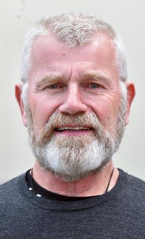
Terry Taylor, BSc DipMLS MNZIMLS, Southern Community Laboratories, Dunedin
Dr Donna Rudd, BappSci, PhD, MAIMS, CCS, FFSc (RCPA), James Cook University, Australia
Formatting
Sharon Tozer, AT DipBusStud, Executive Office NZIMLS, Rangiora
About the Journal
The New Zealand Journal of Medical Laboratory Science (the Journal) is the official publication of the New Zealand Institute of Medical Laboratory Science (NZIMLS). The Journal is peer reviewed and publishes original and review articles, case studies, technical communications, and letters to the Editor on all subjects pertaining to the practice of medical laboratory science. The Journal is open access (www.nzimls.org.nz/nzimlsjournal) and is published three times per year in March, July, and November. Hard copies are circulated to all NZIMLS members and universities and research units in New Zealand and overseas. Current circulation is about 2,800 copies per issue.
Printed by Spectrum Print, Christchurch, using environmentally responsible paper from elemental chlorine free, third-party certified pulp sourced from well‑managed and legally harvested forests. Manufactured under Spectrum’s Toitū Diamond Environmental Management Certification.
The Journal is indexed by CINAHL, EMBASE, SCOPUS, Informit, Thomson Gale, EBSCO and Biosis Citation Index, and the Journal Editors are members of the World Association of Medical Editors (www.wame.org).
Brief instructions to authors
The Journal accepts original submissions from anyone and anywhere. Comprehensive instructions can be found on the NZIMLS website (www.nzimls.org.nz/instructions-to-authors. html). All submissions will undergo single-blind peer review and possibly plagiarism checking with iThenticate™ software. If accepted for publication, copyright is vested in the author(s) under terms of the Creative Commons Attribution License (www. creativecommons.org/licenses/by/2.5/legalcode). The authors are responsible for the scientific content and views. Opinions expressed in the Journal are not necessarily those of the Editors, Editorial Board, or Council of the NZIMLS.
Advertisement bookings and enquiries should be addressed to the NZIMLS Executive Officer, Sharon Tozer: sharon@nzimls.org.nz. Phone +64 3 313 4761.
Medical Laboratory Science
Volume 80 Number 2
July 2026
ISSN 1171-0195
80th Anniversary Guest Editorial
The real life from inside the governance bunker.
Terry Taylor 72
Reviews
The role of artificial intelligence in shaping New Zealand’s medical laboratories: current trend and future direction.
Yanfang Hu 73-76
Artificial intelligence in genetic research and diagnostics: a scoping review.
Mona Ellaithi and Aya Ziyada ............................................ 77-82
Original articles
Role of triple immunohistochemical stains CK20, CD44 and p53 in differentiation between urothelial carcinoma in situ and reactive urothelial atypia.
Zahraa Sh Elalfy, Elia A Ishak, Wafaa E Abdelaal, Samira M Abd-Allah and Nora N Kamel 84-89
Determination of the frequency of malarial infection and HCV antibody prevalence in a cohort of antenatal patients in Calabar Municipality, Nigeria.
Glory Philemon Bebia, Ugwu Joy Chinweokwu, Edim Sunday Nyambi, Emmanuel Onyekachi Ibeneme, Paul Columbus Inyang-Etoh, Eldad Akong Akpang and Goodluck Samuel Ekanem 90-94
Case Studies
Assessment of haemoglobin HbA2 stability in refrigerated EDTA samples using capillary electrophoresis.
Danyi Zhang, Yii Sen Wee, Samantha Enger and Alan Neal 95-96
Conference proceedings
A solution to beating plastic waste in a pathology laboratory. Lynn Brott 97
Phlebotomy now and back then: a lived experience.
Gaye Duffill 98
White paper report
The impact of living with a rare disorder in Aotearoa New Zealand.
Angela Nielson 101
Book review
The blood says otherwise by Ruben Miller.
Reviewed by Michael Legge 97
Welcome to our second issue celebrating 80 years of the NZIMLS with some parting words from Terry Taylor, past NZIMLS president who reflects on his involvement in the profession, NZIMLS, governance and political arenas in our guest editorial.
Artificial Intelligence (AI) is reshaping almost every sector of modern society, with healthcare at the forefront of this transformation. In the first of two reviews on AI in this issue, Yanfang Hu, from AUT in Auckland, considers the role of AI in New Zealand medical laboratories and the increased need for technological augmentation due to expanding demographic pressures, workforce shortage, patient safety, equity and regulatory compliance. AI specific applications were explored in three disciplines, haematology, microbiology and histopathology. Current technological barriers and strategic solutions for the New Zealand health environment were also considered.
In the second AI review, researchers Ellaithi and Ziyada from AlNeelain University in Sudan and CCPMG in Qatar, respectively, performed a comprehensive literature review to analyse the state-of-the-art based genomics techniques and assesses their potential uses in precision medicine, drug development and clinical diagnostics. Using PRISMA Extension for Scoping guidelines, 71 peer-reviewed articles met the inclusion criteria. This review also sought to highlight significant developments in computing, ethical and legal issues and identify new technologies that are influencing how AI will be incorporated into genetic research and clinical practice in the future.
Both reviews stressed the need for control and care in building infrastructure, regulatory frameworks, ethical oversight, privacy and training throughout the research and medical industries.
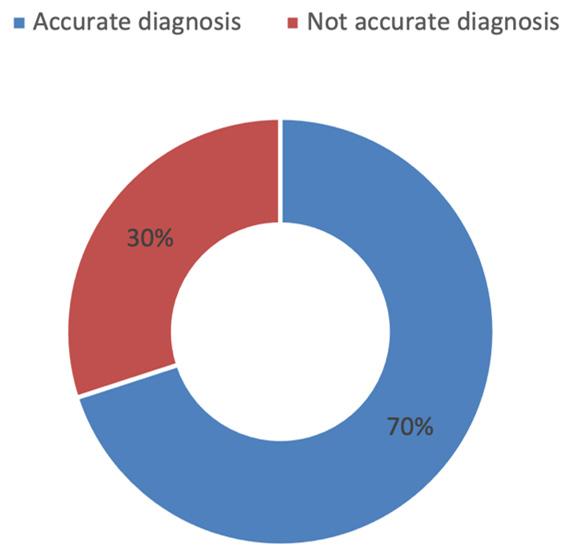
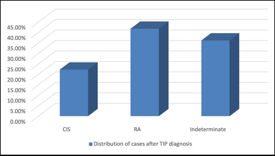
Bladder cancer is the fourth most common cancer in men and the twelfth in women in the USA, in Egypt, carcinoma of the bladder accounts for approximately 31% of all cancer cases. Urothelial carcinoma in situ (UCIS) is a malignant flat intraepithelial lesion of the urinary tract and considered high risk in non-muscle invasive bladder cancer. Classic diagnosis relies on identifying nuclear features and morphologic patterns. However, the diagnosis of UCIS can be challenged by atypia seen in association with other benign conditions. This significant morphological overlap results in an atypical or indeterminate diagnosis and uncertainty in management. Elalfy and colleagues from the National Research Centre and Cairo University in Egypt, conducted a study of seventy-two patients and assessed the usage of immunological markers CK20, CD44 and p53 in a triple immunostaining panel (TIP) and compared them with the accuracy of H&E staining. Thirty-two showed accurate diagnoses, with identical H&E and TIP based decisions (70% accuracy). In 14 of 46 cases (30%) the diagnoses were changed, 3 reactive atypia (RA) were changed to UCIS, 11 cases of UCIS appeared to be RA. Twelve were related to clinical data associated with malignancy of UCIS (p<0.05) indicating a significant diagnostic bias and over diagnosis. Triple staining provided an important supporting tool for distinguishing UCIS from RA, however TIP requires complex translation of the staining pattern and are costly to perform.
Malaria is an endemic disease of the tropics caused by the bite of the female Anopheles’ mosquitoes. Hepatitis C virus (HCV) causes an inflammation to the liver and in pregnant women it is more serious as it can cause jaundice in babies. A study conducted by researcher Bebia and colleagues at the University of Calabar and University of Cross River in Nigeria, assessed the seroprevalence and associated risk of Malaria and Hepatitis C virus among pregnant women in Calabar, Nigeria. Results were 0% seropositive for HCV across all 340 of the anti-natal cohort and may have limited by the ability of the Accu-tell HCV Rapid Test Strip to detect early infection. The cohort had an overall prevalence of 11.8% malarial infection, with younger women having significantly higher prevalence of malaria compared to that of older females and primigravida females were more susceptible to malaria infect than multigravida. This study highlighted the importance of malaria prevention and control strategies among pregnant women including improved access to antimalarial prophylaxis and treatment, expanded distribution
environmental sanitation programmes. Furthermore, routine malaria screening during antenatal visits, especially among younger and primigravid women, may facilitate early detection and timely management.
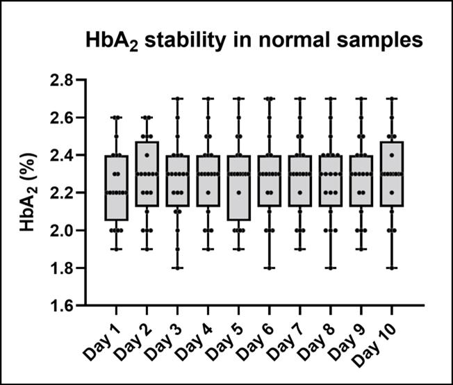
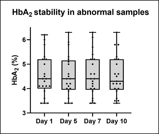
Yii Sen Wee and colleagues at Haematology, PathLab in Waikato present a case study assessing the stability of haemoglobin HbA2 in refrigerated EDTA samples using capillary electrophoresis. Previous studies reported significant drop in HbA2 levels after 3-7 days when samples were kept at room temperature, leading to misdiagnoses of heterozygous beta-thalassemia. This case study aimed to build on this published information to determine stability of samples stored at 4°C. Under routine refrigerated storage, HbA2 level in EDTA blood samples remained stable over a 10-day period with minimal degradation and supports flexibility in routine practice to allow for batching and minor delays without compromising accuracy.
Recently retired Lynn Brott, presented at the NZIMLS Annual Science Meeting, Christchurch, August 2024 offering solutions and alternatives in reducing waste and sustainability of biohazard bags in the medical laboratory and summarises her talk in the first of two conference proceedings in the issue. In our second proceeding, Gaye Duffill, a senior phlebotomist from Waikato summarises her unique experiences in phlebotomy in New Zealand, from her presentation given at the Pre-analytical Special Interest group held in Auckland, in November 2025.
We announce the retirement of Karen Glover, from LabPlus The NZIMLS and profession thank her for her service and wish her all the best for a well-earned retirement.
Savannah Young, NZIMLS recipient of the Barrie Edwards & Rod Kennedy Scholarship, reports on her attendance at the Blood and Regional Congress of the ISBT in Perth, held in October 2025.
Rare Disorder NZ Communication Manager, Angela Nielsen informs us on a recently commissioned white paper launched in New Zealand Parliament in 2026. This white paper; “The impact of living with a rare disorder in Aotearoa, New Zealand” addresses the systematic barriers that persist in diagnosis, care pathways, access to medicines, social support and access to appropriate expertise for people living with a rare disorder. This white paper informs the implementation of the Rare Disorders Strategy, that sets the direction for government health entities to become more responsive to people living with rare disorders, their families and the service providers.
The Journal is excited to have been given permission from the New England Journal of Medicine, to reprint Eve Rittenburg’s article on Inheritance, published in N Engl J Med 2026; 394: 425427. We also include a reprint of an article on Denise Zuze Carter, Otago University’s first student of the Postgraduate Diploma of Medical Laboratory Science, published on the University of Otago Newsroom website on the 6th May 2026.
Dr Michael Legge offers a review of the recently published book; “The blood says otherwise”, by Ruben Miller
Regular features include the Pacific Pathology Training Centre (PPTC) update in the Pacific Way, CPD Questionnaire and some pointers from our CPD coordinator, meeting reports from the recent South Island Seminar and Biochemistry Special Interest Group, the Science Digest and Recent Reviews.
Lisa
Cambridge, Editor
Advertisers in this issue:
Abacus dx Helena Laboratories Outside back cover Siemens Healthineers ................................... Inside front cover New Zealand
The real life from inside the governance bunker
Terry Taylor
It is both an honour and also with sadness that I write about what the NZIMLS has meant for me personally and the medical laboratory science sector over many years. As you all read this, I am no longer practising in New Zealand but never-the -less am continuing to do what I enjoy and spreading the gospel so to speak. I never say ‘never’ because you never know where I may pop up in the future.
I am probably a medical science professional enigma because I have always enjoyed being in the public eye and speaking to large groups about all sorts of medical and sporting topics. I excelled in the multidisciplinary forums we used to be part in at Dunedin Public Hospital when we were part of the old District Health Board (DHB). Unfortunately, many things changed since we were the first region in New Zealand to fall under the dreaded experiment of ‘contracting out’ of pathology services back in 2007.
My involvement with the NZIMLS council began in 2009 with prior involvement as a seminar and educational forum convenor. The NZIMLS has been the cornerstone and the one constant across Pathology over the 80 years of its existence.The absolute honour I felt when I became President in 2017 was clear to everyone who was close to me. This wasn’t just a title, but an opportunity to repay the profession I loved so much by working to get us into the health and political spaces we needed to be in. I remember my first job was drafting a letter to the then Minister of Health, Dr Jonathan Coleman. I did not get to meet him in person as there was an election coming up, with the expectation that this would be sorted after the election. This was the start of my long and interesting involvement at the top level of health and political leadership which continues to this day. I have worked across seven Health Ministers, numerous health spokespeople and have personal ‘thank you’ letters from many of them as well as forming lifelong friendships from my many political forays to Wellington.
I was fortunate in 2018 to have both the government Health Minister and shadow Health Minister living in Dunedin whom I met with regularly and struck up important wider leadership relationships. I was given what I can only describe as exceptional insights from sitting and past Cabinet Ministers on the workings of Parliament and how to get Ministers attention. I was a sponge to this and although not apparent to anyone at the time, this was the work that the whole profession benefitted from with what was coming.
Late in 2018, I was invited by the then Minister of Health, Dr David Clark, to submit a submission on behalf of the medical laboratory science sector to then national Health and Disability Review (HDR) committee. The results of this review, often referred to as the ‘Simpson Report’, led to the formation of the national entity; Health NZ (HNZ). This whole process involved many hundreds of pages of advisory documents, meetings with officials at the Department of the Prime Minister and Cabinet (DPMC) and Health Select Committee appearances. This was as far away from my frontline scientist job as one could get, but our voice simply had to be heard. Many of our wider professional and workforce documentation was incorporated in the regulatory and operational mandates. The big pity has always been that little has been proactively acted on across our sector.
We all know what hit in 2020 and it is fair to say this was the single biggest impact that the NZIMLS ever had to deal with. My relatively calm world was completely upended and the impacts from this time have changed our profession and the expectations on medical laboratories forever. To put this into context, the NZIMLS had had less than half a dozen media excursions from 2009 to 2019. Suddenly as a professional leader, I was thrust firmly into the spotlight and did literally hundreds of appearances over the pandemic. Plus, I was instantly in the political rooms with partyspokespeopleallplayingoffagainsteachothertryingtogain the moral high ground and boss the pandemic response. It was a crazy time, I was now effectively a fulltime advisor, advocate and spokesperson, as well as still trying to do my fulltime scientist
role. Great for the NZIMLS publicity wise, but the work behind the scenes was extraordinary and unprecedented and we barely had time to come to grips with what was happening all around us.
When I stood down as President of the NZIMLS in 2023, Her Excellency, The Right Honourable Dame Cindy Kiro was a guest at the Annual Science Meeting and I consider this a very telling and proud moment not just for me personally but for the entire profession.
We all learnt a lot, the country learnt a lot, and then the inevitable reviews came with recommendations to nationalising our governance and having leadership within the big health rooms. None of which has happened and despite warning after warning and presenting a doomsday paper to Parliament in 2022, regarding the state of the workforce, its training and educational opportunities. The inertia continued regards transforming how our Pathology services are structured and delivered and as of 2026, we still have a mix of private and public providers within our public hospital laboratories and no let-up in workforce issues and challenges. It is hoped that a National Pathology Clinical Governance Network and Pathology sector review may provide a positive direction and pathway to a brighter future for those currently in the profession but also importantly for those entering the profession in years to come.
I do however leave everyone with hope from many years of advocating and lobbying across health and social interfaces. We have many allies across the new age political and health leadership environment and patient advocate leaders are now all firmly on the side of needed changes for diagnostic services. They are all acutely aware just what a desperate state many of our hospital laboratories are in. The lack of investment in senior experience and deskilling staff and service by stealth has not gone unnoticed and we have a gifted chance to put things right. Afterall No Pathology equals No Healthcare.
The NZIMLS has been the profession’s stability throughout its history, the amazing and dedicated professionals, and equally engaged councils and executives. This is what our medical laboratory sciences sector is built on. I firmly believe, if our operational leadership are given the keys, that after many difficult years, the overdue transformational change to strong national governance and firm and decisive leadership to drive regional and local service delivery, is upon us.
He aha mea nui o te ao, he tangata, he tangata, he tangata ‘What is the most important thing in the world? It is people, it is people, it is people’
AUTHOR INFORMATION
Terry Taylor, BSc, DipMLS, MNZNIMLS, Past President New Zealand Institute of Medical Laboratory Science (Inc.) and Life Member.
Email: terry.taylor707@gmail.com
Note added in proof: Terry has now left New Zealand to take up the role of Clinical Flow Cytometry Specialist based at the Princess Alexandra Hospital, Queensland, Australia. His commitment to the profession will have long-term outcomes for all NZIMLS members.
The role of artificial intelligence in shaping New Zealand’s medical laboratories: current trends and future directions
Yanfang Hu
ABSTRACT
New Zealand’s medical laboratories are experiencing a transformational shift characterised by the transition from modular automation to integrated Artificial Intelligence (AI). This article considers the current state of AI adoption into three phases: haematology, which exhibits the most mature application in digital morphology; microbiology, which is upgrading from laboratory automation to intelligent plate reading; and histopathology, currently undergoing a digital revolution through a national rollout of digital pathology. However, the introduction ofArtificial Intelligence faces various challenges.At the regulatory and ethical levels, laboratories must address data privacy risks, Māori Data Sovereignty and Treaty of Waitangi obligations, algorithmic bias, and the risk of hallucinations in generative AI. At the technical level, major barriers include the scarcity of high-quality annotated data, poor model generalisation, the black box nature of deep learning models, and substantial interoperability issues, particularly the difficulty of integrating legacy Laboratory Information Systems (LIS) with modern AI tools. Artificial Intelligence application in New Zealand clinical laboratories has become a reality, with future success relying on ethical integration, local validation, and the creation of models specific to the unique characteristics of Aotearoa.
Keywords: Artificial Intelligence (AI), digital pathology, Haematology, Microbiology, Māori Data Sovereignty, New Zealand.
NZ J Med Lab Sci 2026; 80(2): 73:76
INTRODUCTION
The global integration of Artificial Intelligence (AI) is reshaping almost every sector of modern society, with healthcare at the forefront of this transformation. Advances in machine learning, deep learning, and data-driven automation have fundamentally altered how clinical information is generated, interpreted, and applied to patient care. Within this broader healthcare ecosystem, clinical laboratories constitute a key component, serving as the primary source of diagnostic data that supports most evidencebased clinical decisions. Medical decision making relies, at least in part, on laboratory test results, highlighting the strategic importance of laboratory services in ensuring patient safety, diagnostic accuracy, and system wide efficiency.
In Aotearoa New Zealand, the adoption of AI within clinical laboratories is migrating from an experimental or speculative innovation into a strategic operational requirement. Unlike larger healthcare systems that may prioritise rapid technological deployment, New Zealand’s approach has historically been cautious and values-driven, emphasising patient safety, equity, and regulatory compliance (1). However, expanding demographic pressures, workforce shortages, and increasing test volumes have created conditions in which traditional laboratory workflows are no longer sustainable without technological augmentation. Artificial intelligence, when deployed responsibly, offers a mechanism to enhance diagnostic capacity without compromising quality or ethical standards.
The most compelling strategic argument for AI in New Zealand healthcare is the growing workforce crisis. Projections from Te Whatu Ora (Health New Zealand) indicate widening gaps across multiple laboratory professions. By 2033, the system is expected to experience a 36.2% shortfall in medical laboratory technicians, a 13.4% deficit in medical laboratory scientists, and a 14.7% increase in demand for pathologists based on existing training pipelines and work models (2). These shortages are further intensified by an ageing workforce, limited domestic training capacity, and international competition for skilled professionals. Without intervention, these trends pose a direct threat to service continuity, turnaround times, and diagnostic quality across both urban and regional laboratories.
Artificial intelligence offers a proactive response to these challenges through workforce augmentation rather than workforce replacement. By automating repetitive, highvolume, and low-complexity tasks, AI enables laboratories to reallocate highly trained professionals toward complex diagnostic interpretation, quality assurance, and multidisciplinary collaboration. This “human-in-the-loop” model is consistent with New Zealand’s clinical governance culture, ensuring that AI functions as a decision-support tool rather than an autonomous
decision-maker. Consequently, AI adoption in New Zealand laboratories prioritises controlled integration over full automation, with accountability remaining with qualified health professionals.
DISCIPLINE-SPECIFIC AI APPLICATIONS AND INSTRUMENTATION
Haematology: the maturity of digital morphology
Among all laboratory disciplines, haematology represents the most mature and widely adopted application of artificial intelligence within the medical laboratories of New Zealand. Traditionally, blood film examination has relied on manual microscopy, a process that is time-consuming, subjective, and highly dependent on individual expertise. The transition from manual microscopy to Digital Cell Morphology (DCM) is predominantly facilitated by the CellaVision DM96/DC-1 systems in New Zealand (3). The incorporation of Artificial Neural Networks (ANN) within these platforms enables the automated localisation and consistent pre classification of leucocytes, while also offering high resolution imaging for the morphological assessment of erythrocytes and platelets, supporting the identification of anaemias, haemoglobinopathies, and other haematological abnormalities (4)
The widespread adoption of CellaVision systems across Te Whatu Ora laboratories reflects both their clinical effectiveness and their suitability for the geographically dispersed healthcare infrastructure of New Zealand (5). Digital morphology enables remote review and validation, allowing expert haematologists and scientists to support smaller or rural laboratories without the need for physical slide transport (4). This capability is particularly valuable in regional settings, where access to specialised expertise may be limited. Furthermore, digital archiving supports quality assurance, training, and audit activities, contributing to improved standardisation across the national laboratory network (6)
Microbiology: transitioning to intelligent plate reading
The field of microbiology within Aotearoa New Zealand is currently advancing beyond conventional Total Laboratory Automation (TLA) toward more advanced forms of AI-enabled analysis. This transition is primarily driven by platforms such as BD Kiestra™ and Copan WASPLab, which apply AI-based Digital Plate Reading (DPR) technologies (7). These systems analyse high resolution images of culture plates to differentiate between “growth” and “no growth” outcomes, thereby reducing the need for manual plate inspection. In routine specimens such as urine cultures, where a high proportion of samples are negative, this
capability delivers substantial efficiency gains. A significant local case study is provided by Pathlab in the Bay of Plenty, which implemented the BD Kiestra™ automation line in early 2016 (8). This transition laid the foundation for AI-enhanced urine culture screening, which enables automatic validation of negative results and streamlines laboratory throughput. As a result, specialised microbiology staff were able to focus their expertise on complex positive cultures, antimicrobial susceptibility testing, and clinically significant isolates. This reallocation of human resources not only optimised operational efficiency but also enhanced diagnostic quality across the regional laboratory network.
Histopathology: the digital revolution
Histopathology is currently experiencing a transformative phase of expansion, driven by the national rollout of Digital Pathology (DP). Historically, histopathological diagnosis has relied on glass slides and optical microscopy, a workflow that limits scalability, remote consultation, and computational analysis. Awanui Labs has taken a leading role in this domain by implementing Whole Slide Imaging (WSI) at its histology laboratory in Dunedin (9) Using Leica digital scanners, the laboratory is establishing a unified national digital slide repository, providing remote access, consultation, and long-term data storage. This infrastructure enhances diagnostic safety by reducing the risk of slide loss or damage and supports cross-site workload balancing. Importantly, digital pathology provides the foundation for future AI applications in histopathology, including tumour detection, grading assistance, and quantitative biomarker analysis. While most AI tools in this field remain in validation or research phases, the establishment of a national digital slide architecture enables New Zealand to integrate these technologies safely and aligned with evolving statutory requirements
REGULATORY AND ETHICAL FRAMEWORKS IN AOTEAROA
According to the NAIAEAG (National AI and Algorithm Expert Advisory Group) assessment (10), New Zealand laboratories must manage several core regulatory, ethical, and operational risks when introducing artificial intelligence into clinical workflows.
Data privacy and retention risks
The use of public or externally hosted generative AI systems (e.g., ChatGPT or similar large language models) for processing laboratory data raises serious privacy concerns. Inputting identifiable patient health information (PHI) into such systems is strictly prohibited under the New Zealand Privacy Act 2020, as third-party AI providers may archive telemetry and user prompts for model optimisation (11). This creates a systemic vulnerability of unauthorised data storage and cross-border data exposure. Clinical laboratories must therefore ensure that any AI tools used for operational or analytical purposes are either locally hosted or governed by contractual guarantees that fully comply with New Zealand privacy legislation.
Māori data sovereignty and Treaty obligations
Māori data sovereignty represents a distinct and non negotiable consideration unique to the New Zealand context (12). Most public and international AI models lack both the governance structures and cultural authority to process Indigenous health data appropriately. The use of such models risks the misuse, misinterpretation, or extraction of Māori health data without consent or oversight. Consequently, any AI system deployed within New Zealand laboratories must demonstrate explicit compliance with Te Tiriti o Waitangi principles, including partnership, protection, and participation (13). This includes governance mechanisms that guarantee Māori participation and stewardship, ensuring that the handling of Māori and Pasifika data is both culturally safe and methodologically transparent (12)
Algorithmic
bias and health inequity
Many AI models are trained predominantly on international datasets that over-represent global mainstream populations (14) This creates a risk of algorithmic bias, whereby disease patterns, biomarker distributions, or diagnostic thresholds relevant to Māori and Pacific peoples are under represented or characterised (12). Within the laboratory environment, such bias could lead to overlooked abnormalities, misclassification, or diminished diagnostic sensitivity for minority populations, thereby intensifying current health inequities. Laboratories therefore have a responsibility to critically evaluate training datasets and performance metrics to ensure equitable diagnostic outcomes for New Zealand’s diverse populations.
Hallucination and reliability risks
Generative AI systems are susceptible to phenomena known as ‘hallucinations’, where they generate fabricated clinical data, including erroneous medical facts and non-existent citations (15). Given the critical role of laboratory outputs in clinical decision making, the potential for fabricated insights conflicts with the high-quality standards expected in patient care. The use of AI-generated outputs should be restricted to decision-support purposes and must operate within clearly defined, transparent, and auditable frameworks, while ultimate accountability remains with suitably qualified laboratory professionals.
Local validation and contextual performance
AImodelstrainedoninternationaldatasetscannotbeassumedto perform equivalently in New Zealand laboratories. Differences in local reagents, instrumentation, analytical protocols, population demographics, and pathogen prevalence should be considered during local validation prior to clinical deployment. Performance must be assessed under real world conditions specific to New Zealand to guarantee analytical accuracy, validity, and clinical relevance (12)
TECHNOLOGICAL BARRIERSAND STRATEGIC SOLUTIONS
Despite the promising technological potential of artificial intelligence, its implementation within the Aotearoa New Zealand clinical sector is constrained by considerable technical complexities. These barriers extend beyond mere software availability, touching upon the fundamental data structure, laboratory instrumentation, and informatics framework.
Data scarcity and the annotation bottleneck
A primary barrier is the scarcity of high-quality, expert-annotated data, which serves as the ground truth for training supervised AI models. Although large international datasets are available, laboratories in New Zealand often face data silos, where highvalue diagnostic images and results are dispersed across regional servers and institutions.
Moreover, there is a severe shortage of locally relevant datasets. AI systems trained predominantly on European or North American cohorts may fail to capture diagnostic patterns specific to New Zealand, including regional pathogen variants or distinctive biomarker distributions within Māori and Pasifika populations. To overcome this challenge, transfer learning (16) and style transfer (17) have become pivotal approaches. These techniques allow models pre-trained on extensive global datasets to be fine tuned with smaller local data, adapting AI systems to New Zealand’s unique biological context without millions of local samples.
Model generalisation and the imperative for local validation
Another critical challenge is model generalisation, defined as the ability of an AI system to maintain consistent performance across different environments. Research shows that AI models trained on data from a single laboratory often experience domain shift when deployed in new settings (18) In pathology and diagnostics, even minor variations in lighting conditions, slide
preparation procedures, reagent suppliers, or digital scanning hardware can lead to substantial reductions in model accuracy (19)
In the Aotearoa New Zealand landscape, this reality requires a systematic process of comprehensive local validation. Since diagnostic services range from tertiary urban hospitals to remote rural laboratories, each with distinct operational characteristics, AI implementations must be locally calibrated to maintain reliability under specific regional conditions. Without site specific validation, models risk producing unreliable and non generalisable outputs, undermining diagnostic integrity and clinical confidence which is essential for AI adoption.
Model opacity and regulatory requirements for transparency
The opacity of deep learning and generative AI systems, commonly described as the black box problem, creates a substantial obstacle to clinical application (20). These models rely on complex internal parameter interactions to generate outputs yet frequently lack the mechanisms to explain the reasoning behind a given prediction or recommendation.
In New Zealand, the inherent opacity of certain AI models conflicts with the statutory requirement for explainable clinical outcomes (10). Because diagnostic decisions must be transparently communicated to patients, the deployment of non-interpretable models is categorically limited in high-stakes settings to uphold professional accountability (10). In this context, Explainable AI (XAI) serves as a foundational bridge, translating opaque outputs into transparent, auditable, and clinically justifiable insights that adhere to the high standards of the Aotearoa New Zealand health system (21)
Interoperability challenges and legacy infrastructure
Interoperability remains a major technical barrier, particularly in integrating AI-generated outputs with existing Laboratory Information Systems (LIS). Much of New Zealand’s LIS infrastructure was implemented decades ago, prior to the emergence of real-time AI-driven analysis. Therefore, these systems often lack modern application programming interfaces (APIs) and flexible data models required for smooth interaction with advanced AI platforms.
The extent of this challenge is exemplified by Te Whatu Ora (2024), which reports that the New Zealand health sector relies on more than 6,000 fragmented and disconnected digital systems (22). This legacy infrastructure represents a central bottleneck, as it lacks the semantic or structural requirements necessary for AI integration within live clinical workflows. In addition, without consistent national data standards and open APIs, fully automated AI workflows (23) connecting laboratory systems and clinicians remain severely constrained.
Strategic approaches to systemic integration
Overcoming these technical barriers requires a coordinated approach that combines technological solutions with organisational reform. From a technical perspective, achieving interoperability between AI applications and existing laboratory information systems depends on adopting standardised data exchange frameworks, such as Health Level Seven Fast Healthcare Interoperability Resources (HL7 FHIR) (24). As the leading standard for modern digital health ecosystems, FHIR delivers structured, machine-readable data, commonly in JSON or XML formats, required for real-time AI inference (25). However, because legacy LIS platforms commonly employ proprietary formats, the deployment of middleware is therefore necessary. This middleware acts as a key translator or abstraction layer, converting outdated data into FHIR-compliant resources, hence bridging the technological gap without extensive system modification (26).
Equally important is the development of a coherent organisational strategy to ensure that AI technologies are introduced in a manner consistent with clinical governance and regulatory requirements. This includes the establishment
of national guidelines for health data exchange and sustained investment in the digital infrastructure modernisation. Through managing interoperability at both technical and systemic levels, Aotearoa New Zealand can progress toward a unified digital health ecosystem in which AI-enabled diagnostic support can be safely and effectively scaled to optimise clinical efficacy and patient outcomes at a population level.
CONCLUSION
Artificial intelligence in New Zealand medical laboratories is no longer a theoretical concept but an emerging clinical reality. As organisations such as Awanui Labs and Te Whatu Ora continue to expand digital pathology, pathology informatics, and microbiology automation, the focus must remain on ethical integration, patient safety, and the protection of Indigenous data rights. In parallel, the rapid development of pathology informatics emphasises the growing need for a skilled workforce capable of implementing, interpreting, and governing these technologies in routine practice. Future research and policy development should prioritise not only the creation of locally validated, culturally responsive AI models that reflect the distinctive biological and socio-cultural landscape of Aotearoa New Zealand, but also education and training pathways to support this evolving field.
ACKNOWLEDGEMENTS
The author would like to thank the Auckland University of Technology for providing the library resources and database access necessary for this review. This research received no specific grant from any funding agency in the public, commercial, or not for profit sectors.
AUTHOR INFORMATION
Yanfang Hu, MMLSc Student, Auckland University of Technology, Auckland, New Zealand Email: hyf0418@gmail.com
Article Preprint
Hu Y. The role of artificial intelligence in shaping New Zealand’s medical laboratories: current trends and future directions. 4 Jan 2026. Available at SSRN. 6100628. 2026. (https://papers.ssrn. com/sol3/papers.cfm?abstract_id=6100628)
REFERENCES
1. New Zealand. Health and Disability Commissioner Act 1994. Available from: https://www.legislation.govt.nz/act/ public/1994/0088/latest/whole.html
2. Health New Zealand | Te Whatu Ora. Health Workforce Plan 2024: Profession specific analysis – pathology. Wellington: Health New Zealand; 2024. Available from: https://info.health.nz/about us/what we do/planning and performance/health-workforce-planning/health-workforceplan-2024-detailed-analysis-and-data/workforce-planprofession-specific-analysis/health-workforce-planpathology-analysis
3. Briggs C, Longair I, Slavik M, et al. Can automated blood film analysis replace the manual differential? an evaluation of the CellaVision DM96 automated image analysis system. Int J Lab Hematol 2009; 31(1): 48 60. doi: 10.1111/j.1751 553X.2007.01002.x.
4. Al Qudah R, Suen CY. Improving blood cells classification in peripheral blood smears using enhanced incremental training. Comput Biol Med 2021; 131: 104265. doi: 10.1016/j.compbiomed.2021.104265.
5. Peters J. New hematology automation solution helps Middlemore Hospital hematology laboratory improve productivity and accuracy. Sysmex J Int 2010; 19(2): 1 6.
6. Kratz A, Lee SH, Zini G, et al. Digital morphology analyzers in hematology: ICSH review and recommendations. Int J Lab Hematol 2019; 41(4): 437 447. doi: 10.1111/ijlh.13042.
8. Pathlab. Microbiology and molecular diagnostics are moving. Tauranga: Pathlab; 2016 Jan 23. Available from: www.pathlab.co.nz/ static/01784e09f75b37b3537e943cda741b7c/ 1601%20 Pathlab%20Micro%20move.pdf.
9. Awanui Labs takes significant step with Digital Pathology. Awanui Group; 30 May 2025. Available from: https:// awanuigroup.co.nz/news/awanui labs takes significant step-with-digital-pathology
10. Artificial Intelligence and Algorithm Expert Advisory Group. Health New Zealand | Te Whatu Ora. Available from: https://info.health.nz/about us/who we are/expert groups and networks/expert groups/artificial intelligence and-algorithm-expert-advisory-group
11. New Zealand Privacy Act 2020. Available from: https:// www.legislation.govt.nz/act/public/2020/0041/latest/ whole.html.
12. Whittaker R, Dobson R, Jin CK, et al. AI Governance Group. An example of governance for AI in health services from Aotearoa New Zealand. NPJ Digit Med 2023;6(1): 164. doi:10.1038/s41746 023 00882 z.
13. New Zealand Ministry of Health. Te Tiriti o Waitangi Framework. Wellington: Ministry of Health; 2020.Available from: https://www.health.govt.nz/our work/populations/ maori-health/te-tiriti-o-waitangi-framework
14. Panch T, Mattie H, Atun R. Artificial intelligence and algorithmic bias: implications for health systems. J Glob Health. 2019;9(2):010318. doi:10.7189/jogh.09.020318.
15. Ji Z, Lee N, Frieske R, et al. Survey of hallucination in natural language generation. ACM Comput Surv 2023; 55(12): 1–38. doi:10.1145/3571730
16. Gallardo García R, Jarquín Rodríguez S, Beltrán Martínez B, et al. Efficient deep learning architectures for fast identification of bacterial strains in resource constrained devices. Multimed Tools Appl 2022; 81(16): 22723 22748. doi: 10.1007/s11042 022 13022 8.
17. Pawłowski J, Majchrowska S, Golan T. Generation of microbial colonies dataset with deep learning style transfer Sci Rep. 2022; 12: 5212. doi: 10.1038/s41598 22-09264-z.
18. Roland T, Böck C, Tschoellitsch T, et al. Domain shifts in machine learning based COVID-19 diagnosis from blood tests . J Med Syst 2022; 46(4): 23. doi: 10.1007/s10916 022-01807-1.
19. Alouani DJ, Ransom EM, Jani M, et al. Deep convolutional neural networks implementation for the analysis of urine culture. Clin Chem 2022; 68(4): 574 583. doi: 10.1093/ clinchem/hvab270.
20. Pantanowitz L, Hanna M, Pantanowitz J, et al. Regulatory aspects of artificial intelligence and machine learning. Mod Pathol 2024; 37(12): 100609. doi: 10.1016/j. modpat.2024.100609.
21. Mienye ID, Obaido G, Jere N, et al. A survey of explainable artificial intelligence in healthcare: concepts, applications, and challenges. Inform Med Unlocked 2024; 51: 101587. doi: 10.1016/j.imu.2024.101587.
22. Health New Zealand | Te Whatu Ora. Health Digital Investment Plan 2024-2034. Beehive.govt.nz. 2024 Nov 25.Available from: https://www.beehive.govt.nz/release/ digitising healthcare put patients first
23. AI Forum New Zealand. Artificial Intelligence for Health in New Zealand. 2019. Available from: https://aiforum.org. nz/wp-content/uploads/2019/10/AI-For-Health-in-NewZealand.pdf
24. Duda SN, Kennedy N, Conway D, et al. HL7 FHIR-based tools and initiatives to support clinical research: a scoping review. J Am Med Inform Assoc 2022; 29(9): 1642 1653. doi: 10.1093/jamia/ocac105
25. Gazzarata R, Almeida J, Lindsköld L, et al. HL7 Fast Healthcare interoperability resources (HL7 FHIR) in digital healthcare ecosystems for chronic disease management: scoping review. Int J Med Inform 2024; 189: 105507. doi: 10.1016/j.ijmedinf.2024.105507.
26. Yang Y, Sun K, Gao Y, et al. Preparing data for artificial intelligence in pathology with clinical-grade performance. Diagnostics (Basel) 2023; 13(19): 3115. doi: 10.3390/ diagnostics13193115
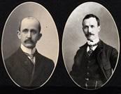
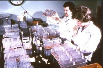
Extracted from: Vol 4, No. 3, July 1949 Extracted from: Vol 2, No. 2, July 1947
Artificial Intelligence in genetic research and diagnostics: a scoping review
Mona Ellaithi and Aya Ziyada
ABSTRACT
Objectives: Artificial intelligence (AI) is rapidly reshaping genomic research and clinical genetics, yet its clinical utility, limitations, and future impact remain incompletely characterised. This scoping review evaluates current AI-based methods in genomics and their applications in clinical diagnostics, drug discovery, and precision medicine, while addressing key computational, ethical, and regulatory challenges.
Methods: A systematic literature search of peer reviewed English language literature was conducted using PubMed, Scopus, Web of Science, and Google Scholar. Studies related to Artificial intelligence, machine learning, deep learning, genomics, genetic diagnostics, precision medicine, and drug discovery were screened for relevance and methodological quality. Findings were synthesized qualitatively without meta-analysis.
Results: Artificial Intelligence driven approaches ranging from traditional machine learning to advanced deep learning models such as convolutional, recurrent, transformer, and graph-based networks, have substantially improved variant calling, biomarker discovery, multi omics integration, and disease risk prediction. In drug discovery and pharmacogenomics, Artificial intelligence has accelerated target identification, molecular design, ‘Absorption, Distribution, Metabolism, Excretion, and Toxicity’ (ADMET) prediction and personalized therapy development. Persistent challenges include data bias, limited interpretability, regulatory uncertainty, and ethical concerns surrounding privacy, equity, and accountability.
Conclusion: Artificial Intelligence represents a transformative force in genomics and precision medicine, with significant potential to enhance research and clinical care. Realising this potential will require advances in explainable Artificial intelligence, more diverse genomic datasets, and robust ethical and regulatory frameworks to support safe, equitable, and clinically meaningful implementation.
Keywords: Artificial Intelligence (AI), machine learning, genetics analysis, deep learning, precision medicine
NZ J Med Lab Sci 2026 80(2): 77:82
INTRODUCTION
From primarily diagnostic applications in radiology and pathology (1,2) to more therapeutic and interventional uses in cardiology and surgery, artificial intelligence has been used in many facets of medicine (3,4). The first artificial intelligence based software solution was approved by the U.S. Food and Drug Administration in April 2018. The application employs fundus image analysis to help diagnose diabetic retinopathy.
Due to numerous regulatory, legal, financial, logistical, ethical, computational, and other issues, as well as the fact that many different stakeholders disagree about the best methods and results, it is challenging to predict exactly how medicine, especially clinical genetics, will change as a result of AI. Even though AI will significantly alter medicine, it is still unclear exactly what the future of medicine contains and how these changes will impact patients, physicians, researchers, and society.
This scoping overview's aim is to present a thorough analysis of the state-of-the-art AI-based genomics techniques and assess their potential uses in precision medicine, drug development, and clinical diagnostics. The review also seeks to highlight significant developments in computing, ethical and legal issues, and new technologies that are influencing how AI will be incorporated into genetic research and clinical practice in the future.
MATERIALS AND METHODS
This scoping review was conducted through a comprehensive and structured literature search to identify recent advances in the application of artificial intelligence (AI) in genomics,
clinical diagnostics, drug discovery, and precision medicine. Peer-reviewed articles were retrieved from major scientific databases, including PubMed, Scopus, Web of Science, and Google Scholar. Studies published between January 2015 and December 2025 was included to capture the most recent decade of AI developments in genetic research and diagnostics. The search strategy combined relevant keywords and Boolean operators such as artificial intelligence, machine learning, deep learning, genomics, genetic diagnostics, precision medicine, and drug discovery. Studies published in English were considered, with an emphasis on original research articles, systematic reviews, and landmark studies demonstrating methodological or clinical significance. Retrieved articles were screened based on relevance, scientific rigor, and contribution to the field. Information was synthesized qualitatively, focusing on AI methodologies, clinical applications, computational frameworks, challenges, and ethical considerations. As this work is a scoping review, no statistical meta-analysis was performed.
RESULTS
Following PRISMA Extension for Scoping reviews (PRISMASCR) guidelines 150 records were screened, and 71 peerreviewed articles published between 2015 and 2025 met the inclusion criteria, comprising 60% original research and 40% reviews or policy analyses. The majority (~70%) were published between 2023 and 2025, reflecting the rapid recent expansion of AI in genomics (Table 1).
Table 1: Distribution of included studies by research domain and proportion of total references (2015–2025)
Current AI methods in genomics
Artificial intelligence methods in genetics cover the range from established machine learning algorithms to complex neural architectures capable of representing detailed biological processes (5). Classical methods such as support vector machines and random forests remain relevant for highdimensional genomic data analysis and the development of interpretable genetic risk models (6). More recently, deep learning techniques, specifically convolutional neural networks (CNNs), recurrent neural networks (RNNs), and transformer models, have enabled advances in sequence-level analysis, variant calling, and long range regulatory sequence modelling (7–9).
Table 2: AI methods in genomics and their applications
AI Method Type
SVM, Random Forest Classical ML
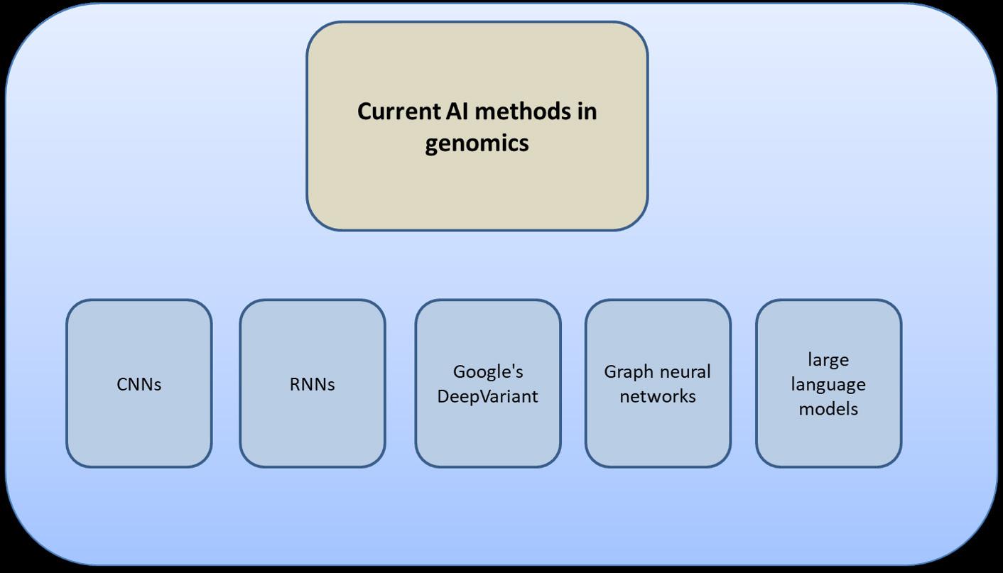
Tools such as Google's DeepVariant illustrate the superiority of AI over heuristic methods in calling single-nucleotide variants and small insertions/deletions (10). Building generative AI models even extend this role in imitating biological sequences and aiding synthetic biology and protein engineering (11). Graph neural networks allow efficient frameworks for protein–protein interaction modelling and genetic networks, and large language models perform automated annotation and functional genomics, reducing the need for manual effort (12–14). These methods collectively comprise the foundation of growing sophistication in AI-driven genetic science (Table 2) (Figure 1).
CNN Deep Learning Variant calling, sequence modeling
RNN Deep Learning
Transformer DL
Generative AI Generative Models
Long-range sequence dependencies
Regulatory sequence modeling
Synthesizing sequences, protein design
GNNs Graph Models PPI networks, pathway modeling
LLMs Large Language Models
Automated annotation, functional genomics
DeepVariant
RNN-based variant predictors
Enformer-like models
AlphaMissense, generative protein design
Graph-based genomics tools
Genome LLMs
1.
Clinical Applications and Diagnostics
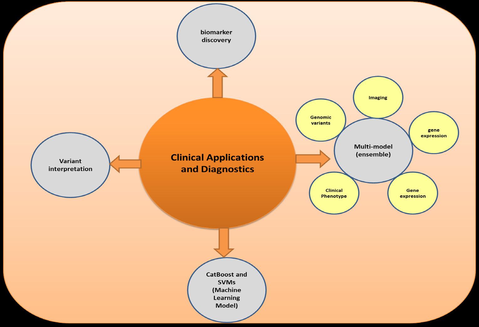
Clinical use of AI in genetics has resulted in an immense improvement in diagnostic accuracy, the diagnosis of a disease at an initial stage, and treatment programs customized for the patient (15–18). Variant interpretation remains one of the best uses, with AI-supported pipelines proving to be faster and reproducible compared to the traditional pipelines (19,10). AI has also enabled the tremendous advancements in biomarker discovery, and studies in Alzheimer's disease have also been able to state Area under the Curve (AUC) of 0.89 to 0.92, and
Clinical Area
Variant Interpretation
Early Disease Detection
Biomarker Discovery
Prognostic Modeling
Personalized Treatment
sensitivity and specificity values above 85% (20). (Table 2) Machine learning models like CatBoost and SVMs have also shown excellent utility in classifying early genetic disorders and their subtypes and have enabled efficient triage and screening even with incomplete data (21–23). Moreover, the potential of AI to integrate multi-modal (e.g. ensemble model) information like genomic variants, gene expression, clinical phenotypes, and imaging has improved prognostic modelling and facilitated personalized diagnostic trajectories (24,25) (Table 3), (Figure 2).
AI Contribution
Faster, reproducible pipelines
Classifies disorders, triage
Alzheimer’s biomarkers
Integrates genomic + clinical data
Predicts drug response
Figure
Current AI methods in genomics
Table 3: Clinical applications of AI in genetics
Drug Discovery and Precision Medicine
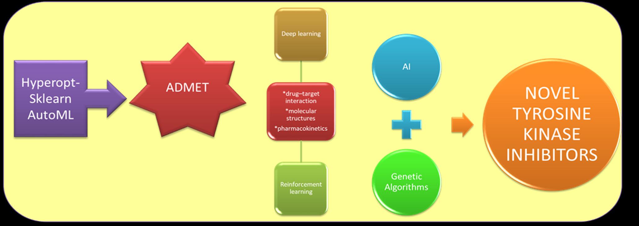
AI has transformed drug research, particularly in integrating genetic information with drug discovery pipelines and precision medicine platforms. In target selection, AI algorithms employ large-scale omics data and protein interactomes to prioritize new therapeutic targets (26). Deep learning and reinforcement learning approaches have been employed to make predictions of drug–target interaction, enhance molecular structures, and simulate pharmacokinetics (26–28). Generative AI models paired with genetic algorithms have resulted in novel tyrosine kinase
inhibitors of high bioactivity, and evolutionary algorithms are used to mimic molecular evolution for the design of therapeutics (29,30). Beyond pharmaceutical discovery, AI methods like Hyperopt-sklearn AutoML facilitate ADMET prediction, reducing costs and minimizing reliance on animals (31,32). In pharmacogenomics, AI algorithms in precision medicine predict drug side effects, tailor dosing, and risk stratify individuals for clinical trials (17,33). This application of genetic and clinical data represents a giant leap toward genuinely individualized medicine (Figure 3).
Computational Genomics and Data Analysis
The complexity and scale of genomic data today demand computationally efficient, AI enabled analytic pipelines (34) through rapid, accurate, efficient processing, and managing big volume of data. AI algorithms can integrate multi-omics data, including genomics, transcriptomics, proteomics, epigenomics, and metabolomics, into consistent structures that capture biological complexity (35). Advanced integration methods such as autoencoders, multi-modal deep learning, and graph frameworks allow for the identification of latent cross omic interactions (36,37). Of similar importance is the development of explainable AI (XAI), providing transparency to clinicians and researchers (38,39). Feature attribution methods, motif discovery software, and visualization frameworks are bridging the "black box" gap by offering transparency in AI driven variant interpretation and diagnostic recommendations (40).
Challenges and Limitations
There are, however, several critical challenges limiting the adoption of AI into genetic research. The first is the most important: data quality and representation. Most genomic datasets have serious biases toward European populations, resulting in variable model performance and reducing generalizability of results (41). Other technical factors that weaken the reliability of data are sequencing bias, batch effects, and inconsistent clinical annotations (42,43). Deep learning models raise concerns regarding transparency, trust, and accountability when used clinically because they are hardly interpretable (44,45). Clinician acceptance of AI, training gaps, and unresolved medicolegal responsibilities impede translation into routine practice (46,47). While vast genomic repositories certainly strengthen AI's capacity to model complex biology, they equally serve to escalate ethical challenges including privacy risks, algorithmic
Figure 2. Clinical applications and diagnostics
Figure 3. Drug discovery and precision medicine
bias, cognitive offloading, and the deep moral implications of altering human genetic information (43,48). Regulatory bodies such as the FDA and EU are setting up frameworks of validation
Table 4: Major challenges of AI in genetics
Challenge
Data Bias
Batch Effects
Future Developments and New Technologies
AI in genetics is poised to develop further by uniting with other technologies. Genomic sequence-training foundation models picture universal embeddings, which can be used in a range of tasks (50,51). Genomic pattern discovery and molecular simulation can be speeded up by quantum computing (52–54). Clinically, AI will most likely provide point-of-care real-time genetic diagnostics, for example, rapid pathogen classification and genetic profiling of antimicrobial resistance (55–57). Population wide, AI-driven genomic databases will transform public health surveillance and disease prevention interventions (15,58,59). The development of digital twins (patient specific computational models) will enable predictive monitoring and personalized treatment planning (60,61). Integration with Internet of Things (IoT) and blockchain will enhance security, accessibility, and data-sharing potential, enabling a robust ecosystem for AI-based precision medicine (62–64).
Ethical Concern and Regulatory Challenges
The integration of AI into genetics raises substantial ethical, legal, and societal challenges. Privacy remains a central concern, as genomic data are inherently identifiable and highly sensitive. Approaches such as enhanced encryption for data storage and computation, as well as federated learning, offer potential safeguards (65). Equitable access to AI-enabled genetic services is another critical issue, particularly given the overrepresentation of individuals of European ancestry in many genomic databases, which may limit model generalizability and exacerbate health disparities across ethnically diverse populations (66-68). Improving dataset diversification and mitigating algorithmic bias are therefore essential steps toward fairness and clinical validity (69). Additional concerns have emerged regarding the increasing use of synthetic or AI-generated data to augment limited datasets, which may amplify existing biases or introduce unrecognized artifacts if not rigorously validated (70). Clinically, AI systems should support rather than replace physician judgment, requiring appropriate training, transparency, and ethical oversight to balance human expertise with algorithmic recommendations (71,72). From a regulatory perspective, existing frameworks are evolving to address AI validation, accountability, and transparency; however, adaptive regulatory mechanisms will be necessary to manage continuously learning systems and to ensure that AI-driven analyses of large, complex genomic datasets translate into demonstrable improvements in diagnosis, treatment, and patient outcomes in real-world clinical settings (73).
CONCLUSIONS
Artificial intelligence has significantly advanced in genetics, from improved diagnostic potential to enabling personalized therapeutics and accelerating drug discovery. Its technical maturity and wide applications speak to its potential for revolution. Challenges, however, remain, particularly interpretability, data bias, equity, and regulation, and these need to be addressed to allow for responsible uptake. Scientists must develop interpretable models and expand training datasets with diverse populations. Healthcare systems must invest in infrastructure, training personnel, and governance frameworks. Policymakers need adaptive regulatory frameworks that provide privacy and transparency. Industry should strive for clinician-centred design,
and oversight to guarantee that AI-based genetic tools will remain safe, transparent, and aligned with ethical considerations (49)(Table 4).
Description
Mostly European genomes
Sequencing variability
explainability, and open collaboration.
In the future, the integration of AI with foundation models, digital twins, and real-time analytics promises precision medicine that is accurate, equitable, and transformative.
CONFLICTS OF INTEREST AND FUNDING
The authors declare no conflicts of interest related to this study and authors did not receive any financial support for this research.
ETHICAL APPROVAL STATEMENT
This work is a scoping review article and does not involve human or animal subjects; therefore, ethical approval was not required.
DATA AVAILABILITY STATEMENT
The data that support the findings of this study are available from the corresponding author upon reasonable request.
Use of AI
Artificial intelligence (AI) tools were utilized exclusively for minor grammar corrections and language polishing during the manuscript’s preparation. Specifically, OpenAI’s ChatGPT was applied to enhance the readability and linguistic precision of the English text. All scientific ideas, data analyses, and conclusions were independently conceived and developed by the authors. The authors carefully reviewed and revised the AI-assisted text to ensure accuracy and accept full responsibility for the final content.
AUTHOR INFORMATION
Mona Ellaithi, PhD, Associate Professor in Human Genetics1 Ayah Ziyada, MSc, Precision Medicine Program Manager2
1 Faculty of Medical Laboratory Sciences, Al-Neelain University, Khartoum, Sudan.
2 Center for Clinical Precision Medicine and Genomics (CCPMG), Hamad Medical Cooperation Doha, Qatar.
Corresponding Author: Mona Ellaithi, Faculty of Medical Laboratory Sciences, Al-Neelain University, Khartoum, Sudan. Email: mona.ellaithi@gmail.com
REFERENCES
1. Thrall JH, Li X, Li Q, et al. Artificial intelligence and machine learning in radiology: opportunities, challenges, pitfalls, and criteria for success. J Amn Coll Radiol 2018; 15(3): 504–508.
2. Salto-Tellex, Maxwell P, Hamilton P Artificial intelligence -the third revolution in pathology. Histopathology 2019; 74(3): 372–376.
3. Deo RC. Machine Learning in medicine. Circulation 2015; 132(20): 1920 1930.
4. Hashimoto DA, Rosman G, Rus D, Meireles OR. Artificial intelligence in surgery: promises and perils. Ann Surg 2018; 268(1): 70–76.
5. Anteghini M, Gualdi F, Oliva B. How did we get there? AI applications to biological networks and sequences. Comput Biol Med 2025; 190: 110064.
6. Ogbonna UE, Ofoezie EF, Babalola OO, et al. Advances
in machine learning for optimizing pharmaceutical drug discovery. Current Proteomics 2025; 22(2): 100015.
7. Tahir M, Norouzi M, Khan SS, et al.Artificial intelligence and deep learning algorithms for epigenetic sequence analysis: a review for epigeneticists and AI experts. Comput Biol Med 2024; 183:109302.
8. Kumar S, Goel P Deep Genomics: Deep learning based analysis of genome sequenced data for identification of gene alterations. In: Mandal S, [Editor]. Artificial Intelligence (AI), Cell and Genetic Engineering New York, NY: Springer US. 2025: 335–367.
9. Testagrose C, Boucher C. Tokenization and deep learning architectures in genomics: a comprehensive review. Comput Struct Biotech J 2025; 27: 3547–3555.
10. Abdelwahab O, Torkamaneh D. Artificial intelligence in variant calling: a review. Front Bioinform 2025; 5: 1574359.
11. Naderiyan Z, Shoari A. Protein engineering paving the way for next-generation therapies in cancer. IJTM 2025; 5(3): 28.
12. Zhang XM, Liang L, Liu L, Tang MJ. Graph neural networks and their current applications in bioinformatics. Front Genet 2021; 12: 690049.
13. Jin Q, Yang Y, Chen Q, Lu Z. GeneGPT: augmenting large language models with domain tools for improved access to biomedical information. Wren J, [Editor]. Bioinformatics 2024; 40(2): btae075.
14. Pancino N, Gallegati C, Romagnoli F, et al. Protein–protein interfaces: a graph neural network approach. IJMS 2024; 25(11): 5870.
15. Alowais SA, Alghamdi SS, Alsuhebany N, et al. Revolutionizing healthcare: the role of artificial intelligence in clinical practice. BMC Med Educ 2023; 23(1): 689.
16. Abdallah S, Sharifa M, I Kh Almadhoun MK, et al. The impact of artificial intelligence on optimizing diagnosis and treatment plans for rare genetic disorders. Cureus 2023; 15(10): e46860
17. Taherdoost H, Ghofrani A. AI’s role in revolutionizing personalized medicine by reshaping pharmacogenomics and drug therapy. Intelligent Pharmacy 2024; 2(5): 643–650.
19. De La Vega FM, Chowdhury S, Moore B, et al. Artificial intelligence enables comprehensive genome interpretation and nomination of candidate diagnoses for rare genetic diseases. Genome Med 2021; 13(1): 153.
20. Jabbar GMD, Aamir M, Hadiqa Gul, et al. AI in the Identification of genetic biomarkers for Alzheimer’s disease: a meta-analysis of computational approaches. JHWCR 2025; e109.
21. Gadde N, MohapatraA, Kanaparti R, et al. Machine learning for early detection of rare genetic disorders using multiomics data. IJWMN 2024; 16 (4/5): 29–36.
22. Kumbhar S, Gulhane M, Gavhale K, et al. AI-driven diagnostics for identifying genetic disorders from genome sequences. Proceedings of the 6th International conference on information management & machine intelligence Jaipur, India: ACM 2024: 1–9. doi: 10.1145/3745812.3745867.
23. Hamid M, Hajjej F, Alluhaidan AS, Bin Mannie NW. Finetuned CatBoost machine learning approach for early detection of cardiovascular disease through predictive modeling. Sci Rep 2025; 15(1): 31199.
24. Johnson KB, Wei W, Weeraratne D, et al. Precision medicine, AI and the future of personalized health care. Clin Transl Sci 2021; 14(1): 86–93.
25. Simon BD, Ozyoruk KB, Gelikman DG, et al. The future of multimodal artificial intelligence models for integrating imaging and clinical metadata: a narrative review. Diagn Interv Radiol 2024; 31(4): 303 312.
26. Ocana A, Pandiella A, Privat C, et al. Integrating artificial intelligence in drug discovery and early drug development: a transformative approach. Biomark Res 2025; 13(1): 45.
27. Bhat AR, Ahmed S. Artificial intelligence (AI) in drug design
and discovery: a comprehensive review. In Silico Research in Biomedicine 2025; 1: 100049. https://doi.org/10.1016/j. insi.2025.100049.
28. PathanI,RazaA,SahuA,etal.Revolutionizingpharmacology: AI-powered approaches in molecular modeling and ADMET prediction. Medicine in Drug Discovery 2025; 28: 100223.
29. Gangwal A, Lavecchia A. Unleashing the power of generative AI in drug discovery. Drug Discov Today 2024; 29(6):103992.
30. Romero R. Integration of genetic algorithms and deep learning for the generation and bioactivity prediction of novel tyrosine kinase inhibitors. Biomolecules ARIV 2024. Available from: https://arxiv.org/abs/2408.07155
31. Han H, Shaker B, Lee JH, et al. employing automated machine learning (autoML) methods to facilitate the in silico ADMET properties prediction. J Chem Inf Model 2025; 65(7): 3215–25.
32. Gangwal A, Lavecchia A. Artificial intelligence in preclinical research: enhancing digital twins and organ on chip to reduce animal testing. Drug Discov Today 2025; 30(5): 104360.
33. Ahmad F Optimizing treatment: the role of pharmacology, genomics, and AI in improving patient outcomes. Drug Dev Res 2025; 86(3): e70093.
34. Wang L, Wang H. Big data in genomics: overcoming challenges through high-performance computing. Comput Mol Biol 2024;1 4(4): 155 162.
35. Luo Y, Zhao C, Chen F Multiomics research: principles and challenges in integrated analysis. BioDes Res 2024; 6: 0059.
36. Ren F, Wei J, Chen Q, et al. Artificial intelligence driven multi omics approaches in Alzheimer’s disease: progress, challenges, and future directions. Acta Pharm Sin B 2025; 15(9): 4327 4385.
37. Wu Y, Xie L. AI-driven multi-omics integration for multi-scale predictive modeling of genotype-environment-phenotype relationships. Comput Struct Biotechnol J 2025; 27: 265–277.
38. Abgrall G, Holder AL, Chelly Dagdia Z, et al. Should AI models be explainable to clinicians? Crit Care 2024; 28(1): 301.
39. Alkhanbouli R, Matar Abdulla Almadhaani H, Alhosani F, Simsekler MCE. The role of explainable artificial intelligence in disease prediction: a systematic literature review and future research directions. BMC Med Inform Decis Mak 2025; 25(1): 110.
40. Hassija V, Chamola V, MahapatraA, et al. Interpreting blackbox models: a review on explainable artificial intelligence. Cogn Comput 2024; 16: 45 74. https:// doi.org/10.1007/ s12559-023-10179-8.
41. McKibbin KJ, Popejoy AB, Shabani M. Reconciling diversity in health and genomic data collection with the regulation of AI in clinical genomics. Genet Med 2024; 26(7): 101127.
42. Choon YW, Choon YF, Nasarudin NA, et al. Artificial intelligence and database for NGS-based diagnosis in rare disease. Front Genet 2024; 14:1258083.
43. Athanasopoulou K, Michalopoulou VI, Scorilas A, Adamopoulos PG. Integrating artificial intelligence in next generation sequencing: advances, challenges, and future directions. Curr Issues Mol Bio 2025; 47(6): 470.
44. Nouis SC, Uren V, Jariwala S. Evaluating accountability, transparency, and bias in AI-assisted healthcare decisionmaking: a qualitative study of healthcare professionals’ perspectives in the UK. BMC Med Ethics 2025; 26(1): 89.
45. Kiseleva A, Kotzinos D, De Hert P Transparency of AI in healthcare as a multilayered system of accountabilities: between legal requirements and technical limitations. Front Artif Intell 2022; 5: 879603. doi: 10.3389/frai.2022.879603.
46. Maliha G, Gerke S, Cohen IG, Parikh RB. Artificial intelligence and liability in medicine: balancing safety and innovation. Milbank Q 2021; 99(3): 629–647.
47. Foote HP, Hong C, Anwar M, et al. Embracing generative artificial intelligence in clinical research and beyond. JACC Adv 2025; 4(3): 101593.
48. Farhud DD, Zokaei S. Ethical issues of artificial intelligence in medicine and healthcare. Iran J Public Health 2021; 50(11): i v. doi: 10.18502/ijph.v50i11.7600.
49. Ménard T. Good quality practices for artificial intelligence in genetics. Eur J Hum Genet 2022; 30(9): 993–995.
50. Dalla-Torre H, Gonzalez L, Mendoza-Revilla J, et al. Nucleotide transformer: building and evaluating robust foundation models for human genomics. Nat Methods 2025; 22(2): 287–297.
51. Timilsina M, Buosi S, Razzaq MA, et al. Harmonizing foundation models in healthcare: a comprehensive survey of their roles, relationships, and impact in artificial intelligence’s advancing terrain. Comput Biol Med 2025; 189: 109925.
52. Hassan Ali. Quantum computing and AI in healthcare: accelerating complex biological simulations, genomic data processing, and drug discovery innovations. World J Adv Res Rev 2023; 20(2): 1466–1484.
53. Alrashed S, Min-Allah N. Quantum computing research in medical sciences. Informatics in Medicine Unlocked 2025; 52: 101606.
54. Braga DM, Rawal B. Harnessing AI and quantum computing for revolutionizing drug discovery and approval processes: case example for collagen toxicity. JMIR Bioinform Biotech 2025; 6: e69800–e69800.
55. Lee S, Park JS, Hong JH, et al. Artificial intelligence in bacterial diagnostics and antimicrobial susceptibility testing: current advances and future prospects. Biosens Bioelectron 2025; 280: 117399.
56. Miglietta L, Rawson TM, Galiwango R, et al. Artificial intelligence and infectious disease diagnostics: state of the art and future perspectives. Lancet Infect Dis 2025; S1473309925003548.
57. Waldock WJ, Thould H, Chindelevitch L, et al. Mitigating antimicrobial resistance by innovative solutions in AI (MARISA): a modified James Lind Alliance analysis. NPJ Antimicrob Resist 2025; 3(1): 75.
58. Abubaker Bagabir S, Ibrahim NK, Abubaker Bagabir H, Hashem Ateeq R. Covid 19 and artificial intelligence: genome sequencing, drug development and vaccine discovery. J Infect Public Health 2022; 15(2): 289–296.
59. Olawade DB, Wada OJ, David-Olawade AC, Kunonga E, Abaire O, Ling J. Using artificial intelligence to improve public health: a narrative review. Front Public Health 2023; 11: 1196397.
60. Vallée A. Envisioning the future of personalized medicine: role and realities of digital twins. J Med Internet Res 2024; 26: e50204.
61. Saratkar SY, Langote M, Kumar P, et al. Digital twin for personalized medicine development. Front Digit Health 2025; 7: 1583466.
62. Ghadi YY, Mazhar T, Shahzad T, et al. The role of blockchain to secure internet of medical things. Sci Rep 2024; 14(1): 18422.
63. Mane S, Singh Y. Advancements in AI, blockchain, and IoT for healthcare and automation: a comprehensive review. IJARCCE 2025; 14(2): 115 113. https://doi.org/10.171/ IJARCCE.2025.14216.
64. Othman SB, Getahun M. Leveraging blockchain and IoMT for secure and interoperable electronic health records. Sci Rep 2025; 15(1): 12358.
65. Bonomi L, Huang Y, Ohno-Machado L. Privacy challenges and research opportunities for genomic data sharing. Nat Genet 2020; 52(7): 646–654.
66. Garcia Saiso S, Marti M, Pesce K, et al. Artificial intelligence as a potential catalyst to a more equitable cancer care. JMIR Cancer 2024; 10: e57276.
67. Goktas P, Grzybowski A. Shaping the future of healthcare: ethical clinical challenges and pathways to trustworthy AI. J Clin Med 2025; 14(5): 1605.
68. Fatumo S, Chikowore T, Choudhury A, et al. A roadmap to increase diversity in genomic studies. Nat Med 2022; 28(2): 243–250.
69. Ueda D, Kakinuma T, Fujita S, et al. Fairness of artificial intelligence in healthcare: review and recommendations. Jpn J Radiol 2024; 42(1): 3–15.
70. Kapania S, Ballard S, Kessler A, Vaughan JW. Examining the expanding role of synthetic data throughout the AI development pipeline. In: Proceedings of the 2025 ACM Conference on Fairness, Accountability, and Transparency Athens Greece. ACM 2025; p45–60. doi: 10.1145/3715275.3732005.
71. Harishbhai Tilala M, Kumar Chenchala P, Choppadandi A, et al. Ethical considerations in the use of artificial intelligence and machine learning in health care: a comprehensive review. Cureus 2024; 16(6): e62443.
72. Hanna MG, Pantanowitz L, Jackson B, et al. Ethical and bias considerations in artificial intelligence/machine learning. Mod Pathol 2025; 38(3): 100686.
73. Pantanowitz L, Hanna M, Pantanowitz J, et al. Regulatory aspects of artificial intelligence and machine learning. Mod Pathol 2024; 37(12): 100609.
in differentiation between urothelial carcinoma in-situ and reactive urothelial atypia
Zahraa Sh Elalfy, Elia A Ishak, Wafaa E Abdelaal, Samira M Abd-Allah and Nora N Kamel
ABSTRACT
Objective: Urothelial carcinoma in situ (UCIS) is a high grade flat lesion with high potentiality of multifocality, spread and invasion. Our study aims to assess the role that the triple immunostaining panel (TIP) cytokeratin 20 (CK20), tumour protein 53 (p53) and CD44 has in detecting Urothelial carcinoma in-situ.
Methods: Our study included different urinary tract biopsies and cystectomies. Cases were revised then categorised into two groups as Urothelial carcinoma in-situ group and reactive atypia (RA) group. We included cases according to the availability of paraffin blocks and glass slides corresponding to the diagnosis, the presence of well oriented specimens and the preservation of area of interest (AOI) after re-section. Cases with concurrent urothelial carcinoma were included.
Results: Seventy two collected cases were constituted of 66 males and 6 females. The mean age was 61 years. 48.6% of our cases gave no history of previous or concomitant malignancy most of them (28 cases) having no previous history. While 51.4% showed a malignancy-related history. Most of reactive atypia (RA) cases (78.1%) did not have a history of malignancy, while 75% of cases diagnosed as Urothelial carcinoma in-situ were having a clear history and/or concurrent malignancy with highly significant statistical values. By the usage of the three immunohistochemical markers and application of the positivity criteria of triple immunostaining panel, new study group had been formed (indeterminate group). The accuracy of Haematoxylin and Eosin (H&E) staining diagnosis was assessed. A final decision was taken towards the diagnosis of 46 cases (64.89%) by triple immunostaining panels. Thirty-two of them showed accurate diagnoses, with identical Haematoxylin and Eosin staining and triple immunostaining panel-based decisions (70% accuracy).
Conclusion: The triple immunostaining panel is an important supportive tool for distinguishing high grade urothelial carcinoma from its mimics.
Bladder cancer is the fourth most common cancer in men and the twelfth in women in the USA. It is estimated that 67,160 Americans were diagnosed with bladder cancer and 13,750 died of the disease in 2007 (1). In Egypt, carcinoma of the bladder accounts for as many as 31% of all cancer cases (2). Currently, it ranks first in males representing 16.2% of male cancer (3). The estimated incidence in males in rural areas in Egypt is 32 per 100,000 (4).
More than 90% of bladder cancers are urothelial carcinomas and almost 80% of these are superficial, non muscle invasive tumours (5). Despite their favourable prognosis, a significant proportion (37-54%) of these tumours recur and up to 15% progress to a higher stage necessitating lifelong follow-up and surveillance (6).
Urothelial carcinoma in situ (UCIS) is a malignant flat intraepithelial lesion of the urinary tract and considered a high-risk feature in non-muscle-invasive bladder cancer. The diagnosis of UCIS classically hinges on nuclear features, typically requiring nuclear enlargement, nuclear hyperchromasia, nuclear membrane irregularity, and nuclear pleomorphism. Less specific features include increased nuclear to cytoplasmic (N/C) ratio, cellular decohesion, and increased mitotic activity. A variety of morphologic patterns have been described: large cell pleomorphic, large cell nonpleomorphic, small cell, clinging, pagetoid, undermining, plasmacytoid, and with glandular differentiation. The diagnosis of UCIS is frequently challenged by atypia seen in association with benign conditions such as inflammation, lithiasis, viral cytopathic changes and therapy induced changes. The significant morphologic overlap among these categories frequently results in an atypical or indeterminate diagnosis causing uncertainty regarding subsequent management. Atypical diagnoses constitute a significant proportion of all specimens with reported rates ranging from 2% to 23% (7-12).
Recently, immunohistochemical stains have been examined by several studies singly and in various combinations for their utility in identifying malignant urothelial lesions (13–15). Although
studies using single stains have reported improved detection rates, the addition of multiple stains has been shown to mitigate the high false positive results from staining of benign entities. Previous studies included E-cadherin, P16, CD138, cytokeratin 20, Ki67, Her2 neu, AMACR, p53, CK5/6 and CD44 as a potential markers for early detection of intraepithelial malignant changes with potential early detection of intraepithelial carcinogenesis, however, emphasizing the importance of combination usage for increase of diagnostic accuracy (13-29).
Based on these findings, this study aims to establish clear algorithm for combined interpretation of markers. The study was designed to determine the utility of a triple immunostaining panel (TIP) of CK20, p53 and CD44 on tissue samples as a potential test for proper diagnosis of urothelial carcinoma in-situ, as the positivity of CK20, diffuse pattern of p53 and negativity of CD44 help in differentiating UCIS from benign mimics favouring malignant changes. Our hypothesis is to reach a fixed panel interpretation that gives the highest accuracy of diagnosis. Specimens with suspicious foci frequently have limited material for testing which makes a TIP appealing. The combination of CK20, CD44 and p53 on these specimens has not been abundantly studied. As an ancillary test, it may have the potential for decisive diagnosis.
MATERIALS AND METHODS
Patients and data collection
A diagnostic retrospective study included seventy-two cases of different urinary tract biopsies and cystectomies collected from Kasr El-Ainy Hospital archives at Cairo University over three years duration between January 2020 and December 2023. Cases were revised for their data and diagnoses, then categorised into two groups as UCIS group and reactive atypia (RA) group. Selection criteria are determined by the research group. We included cases according to the availability of paraffin blocks and glass slides in the archives, the presence of intact well oriented urothelium as a prerequisite for subsequent
New Zealand Journal of Medical Laboratory Science
analysis and the preservation of area of interest (AOI) after resection. In addition, cases with concurrent urothelial carcinoma were included. AOI is defined as the area of maximum nuclear enlargement, nuclear membrane irregularities, hyperchromasia and distorted urothelial maturation from basal cell layer up to umbrella cell layer. All these features were standardised by comparing AOI to nearby normal urothelium of the same case. The Pathologists had full knowledge of patient age, sex, biopsy site and clinical history of previous malignancy or therapy during re-evaluation. In both groups, the entire resected tissue was subjected to microscopic re-evaluation. Only histologically confirmed AOI were examined for IHC reactivity.
Methods
Each selected paraffin block was sectioned at 4um thickness by microtome from the formalin fixed, paraffin embedded tumour blocks for Haematoxylin and Eosin (H&E) staining for light microscopy reassessment. The area showing the most histological cellular or nuclear atypia was labelled as AOI. Four unstained paraffin sections at 2um thickness were cut by microtome for each case. Sections were mounted on the positively charged glass slide and staining was performed in an autostainer (Benchmark Dako® autostainer, California, USA). The sections were deparaffinized in xylene and rehydrated through graded ethanol (100%, 95% and 70%) each for 5 minutes. Slides
were washed in distilled water then in phosphate buffered saline (PBS), for 5 minutes each. Endogenous peroxidase activity was blocked using 3% solution of hydrogen peroxide in methanol for 30 minutes then washed in PBS before immersion in citrate puffer (pH6.0). For antigen retrieval, sections were exposed to microwaves for 15 minutes under water emersion.
All antibodies were received ready-to-use and manufacturer appropriate dilution (Zymed® laboratories Inc., San Francisco, CA, dilution 1/100). One drop of the primary antibodies was applied according to kit instructions. The universal detection kit contains: A polymer-based detection system (Dako EnVision™ FLEX, K8000) biotinylated secondary IgG anti-immunoglobulin and Streptavidin biotinylated horse radish peroxidase complex. The chromogen 3-3' diaminobenzidine tetrahydro-chloride (DAB) was used as chromogen and Mayer's haematoxylin as counterstain (DAKO, Denmark). Slides were rinsed in PBS in different stages of staining. Finally slides were dehydrated in 70%, 95% then 100% ascending grades of alcohol. Positive control slides were included within each run of immunohistochemical staining. Tissue sections of normal ileum, colon cancer, and normal spleen were used to control the staining quality for CK20, P53, and CD44 respectively (Micrograph 1). Some of the urothelial lesion tissue sections were processed in the abovementioned sequences with the omission of the primary antibody and replaced by PBS, as negative controls.
Micrograph 1. Sections from positive control tissue for assurance of quality of IHC. A) Small intestine positive for cytoplasmic CK20. B)Colonic carcinoma positive for nuclear P53. C) Splenic white pulp positive for cell membrane CD44. (IHC, 200x).
Triple immunohistostaining panel
The criteria of positivity of the triple immunostaining panel (TIP) applied by Aron et al (23) were used as a reference in the recategorisationofourstudygroups.Thepanelusedwasasfollows: Cytoplasmic staining with CK20 antibodies was considered as positive result. Staining confined to umbrella cell only (UCO) was considered a negative result. Nuclear staining for P53 was considered as positive results. Weak patchy staining and staining confined to basal cell layer only (BCO) was considered as negative results. The CD44 stain is believed to be positive if any diffuseepithelial cell membrane staining detected. Restricted staining to the BCO was also considered negative. The study groups re categorized according to TIP results as; confirmed RA (pattern 1), confirmed CIS (pattern 2) and indeterminate group (pattern 3) these did not follow the panel.
RESULTS
Seventy-two collected cases constituted of 66 males and 6 female patients with approximate percentage 92% and 8% respectively. Mean age was 61 years (Table 1).
Clinical history
Thirty-seven cases (51.4%) showed a malignancy-related history as follows; 27 (37.5%) were known cases of malignancy with either previous history of BCG installation, previous TUR of tumours on follow up, or previous TUR of tumours with current cystectomy Ten cases (13.9%) were diagnosed having concurrent transitional cell carcinoma (TCC) for the first time (Table 2).
Histological diagnosis of study cases
Revision of the archived pathology reports and re-examination of H&E slides are shown in Figure 1. This study showed that the initial histological diagnoses of cases have been related to the clinical history of the patients. Seventy eight percent (78.1%) of RA cases did not have a history of malignancy, while 30 (75%) of cases diagnosed as UCIS had a clear history and/or concurrent malignancy with highly significant statistical values (Table 3).
Using TIP new study groups had been formed. Re-categorisation of the cases were done accordingly, 30 cases (41.7%) confirmed as RA and named pattern “1”, 16 cases (22.2%) confirmed as UCIS and named pattern “2”, while 26 cases (36.1%) were categorized as indeterminate group and named pattern “3”. The confirmed cases were 46 (30 RA and 16 UCIS) (Table 4, Figure 2, Micrographs 2 and 3). The accuracy of H&E diagnosis was assessed. By TIP a final decision was taken towards the diagnosis of 46 confirmed cases (64.89%). Thirty two showed accurate diagnoses, with identical H&E and TIP based decisions (70% accuracy). In 14 out 46 (30%) cases the diagnoses were changed, indicating inaccurate diagnoses in 30% of cases (Figure 3.)
Three cases of RA were changed into UCIS, while 11 cases of UCIS appeared to be RA. Twelve cases out of these 14 were related to clinical data of associated malignancy, therefore an over-diagnosis of CIS was seen. In other words, 85.7% of the inaccurate diagnoses were associated with the clinical data based on malignancy, with highly significant p value (p<0.05) indicating diagnostic biasing.
New Zealand Journal of Medical Laboratory Science
Micrograph 2. Sections from case of reactive atypia (Confirmed by immunohistochemical panel). A) Morphology revealed loss of orientation with scattered mitotic figures in H&E staining, B) Negative IHC staining for CK20, C) Negative IHC staining for P53, D) Diffuse positive cell membrane staining for CD44. (200x) (Resolution=220 dpi).
Micrograph 3. Sections from case of UCIS (Confirmed by immunohistochemical panel). A) Morphology revealed diffuse loss of orientation, marked nuclear enlargement and pleomorphism with H&E staining, B) Diffuse positive IHC staining for CK20, C) Positive IHC staining for P53, D) Cell membrane staining for CD44 is limited to basal layer. (200x) (Resolution=220 dpi).
Table 3. Relation between histological diagnosis and clinical data
*Significant p value (p <0.05)
Table 4. Distribution of cases after TIP with percentage of confirmed and changed diagnoses
Table 5. Relation between accuracy of diagnosis and clinical data
p value (p <0.05)
DISCUSSION
In this current study and according to the clinical history; the initial conventional histopathologic evaluation of our 72 cases, resulted in 2 groups, (1) RA group (32 cases, 44.4%) and, (2) UCIS group (40 cases, 55.6%). The association of positive clinical history of urothelial carcinoma with the histologic diagnosis of UCIS was found in 75% of cases. This showed disagreement with Aron et al (23), Steinestel et al (24) and Moch et al (25) studies which showed positive history in only 44.5% 25.9% and 45% respectively.
The false positive and false negative diagnoses which are based on H&E-stained sections with dependence only on the morphology of the cellular and architectural features; then shown to be out-of-diagnosis after studying the immunohistochemical characteristics of the lesions. The inaccurate diagnoses in 30% of cases, i.e. three cases in each ten were misdiagnosed (30%
Figure 1. Histological diagnosis of study cases.
Figure 2. Distribution of cases after TIP diagnosis
Figure 3. Accuracy of histological diagnosis after TIP in confirmed cases
inaccuracy with H&E), opens the door to explore the potential reasons for false positive and false negative results, and to emerge to light the power of data influence on the diagnostic decision. This discrepancy may be due to the non-blind initial diagnosis of our study and the biasing caused by knowing the clinical history influencing the pathological evaluation in questionable cases with over diagnosing of UCIS. Pathologists may feel more comfortable diagnosing UCIS in a patient with known history of malignancy than to give this diagnosis de novo, especially in the absence of strict morphological criteria e.g. nuclear size differences or mitotic count cut off point, increasing the chance of misdiagnosis.
Application of TIP to our cases results in 3 groups, namely, (1) RA group 41.7%, (2) UCIS 22.2%, and (3) A third group of indeterminate immunohistochemical results, which represents 36.1%. The TIP led to changing the diagnosis of some cases. Among 32 cases previously diagnosed as reactive atypia, 3 cases have proved to be UCIS by TIP, while among 40 cases previously diagnosed as UCIS, 11 cases have proved to be RA. The total confirmed UCIS cases were 16 cases (41.88%). These results agreed with Olivakih et al (26) and Jung et al (27) who showed confirmation of UCIS diagnosis by immunohistochemistry in 50% and 45% of cases respectively. These results disagreed with Kunju et al (18) and Yildiz et al (29) who showed confirmation of 68% and 62% of UCIS cases respectively.
After the TIP, new study groups were formed in the current piece of work. The cases were reclassified appropriately. Thirty cases (41.7%) were confirmed as RA and designated pattern "1", sixteen cases (22.2%) as UCIS and designated pattern "2", and twenty-six cases (36.1%) as indeterminate group (ICs) and designated pattern "3". Stated differently, there were 46 confirmed cases (30 RA and 16 UCIS). Aron et al showed 2 groups with 67 cases of UCIS and 68 cases of RA. Following the application of their "IUN-3 stain" TIP, they discovered that 20 cases (14.8%) had an indeterminate IUN-3 stain pattern, 53 cases (39.3%) had a malignant IUN-3 stain pattern, and 62 cases (45.9%) had a reactive IUN-3 stain pattern (23). Our study used individual markers on separate slides and resulted in a more accurate assessment rather than using a cocktail marker on a single slide. Sticking to the criteria of positivity and considering partial or faint stain as negative led to decreased figures in our study.
After applying CK20 and P53 to urothelial lesions diagnosed as "equivocal for atypia" Arias Stella et al (2017) reclassified their cases into four groups, 16% as RA, 13% as UCIS, 26% discordant group (one positive marker and one negative marker), and 45% as indeterminate group (equivocal positivity of the markers short of UCIS pattern). (28). The percentage of confirmed UCIS was nearly in agreement when our numbers and their findings were compared. We did, however, exhibit discrepancies in ICs and RA.
Our study was unique in that CD44 was added within the panel and showed that the loss of immunohistochemical expression of this protein gives an early indicator for intraepithelial malignant changes. The confirmatory role of CD44 negativity in our study may account for the decline in the percentage of our ICs. Additionally, their reduced rate of reactive atypia made it unclear if these were cases of RA because their study showed that seven of them had invasive lesions and UCIS at subsequent followup. Spiess et al developed the well-known dual-track theory of urothelial lesion development, which states that the majority of lesions start as occult expansions of preneoplastic non-invasive urothelium with little phenotypic deviation from normal urothelial morphology (30).
CONCLUSION
It is important to develop new reliable markers considering the previously shown variability and discrepancy in the results of various immunostaining panels. Its benefits will become apparent in terms of low cost, simple interpretation, and high sensitivity and specificity.
The triple immunostaining panel (TIP) is an important
supportive tool for distinguishing urothelial carcinoma in-situ from its mimics. Nevertheless, the test is costly and requires a complex pattern to be translated. More immunohistochemical markers and molecular testing should be included in further studies, for example, high Ki67 index, Her2 neu overexpression, upregulated MTOR, TP53 and RB1 gene.
ACKNOWLEDGEMENTS
Words cannot express my gratitude to Professor Dr Manal Badawy, Dr Abdelrazik Farrag and laboratories of the National Research Centre for their generous help and support.
AUTHOR INFORMATION
Zahraa Sh Elalfy, MBBch, MSC MD, Assistant Researcher 1
Elia A Ishak, MD, USCAP Member, Professor of Pathology 2
Wafaa E Abdelaal, MD, Medical Ethics Committee Chairman, Professor of Pathology 1
Samira M Abd-Allah, MD, Professor of Pathology 2
Nora N Kamel, MD, USCAP Member, Assistant Professor 1
1National Research Centre, Medical Research Institute, Department of Pathology, Dokki, Egypt
2Cairo University, Faculty of Medicine, Department of Pathology, Giza, Egypt.
Corresponding author: Dr Zahraa Sh Elalfy, National Research Centre, Medical Research Institute, Department of Pathology, Dokki, Egypt.
Email: drzahraa1983@gmail.com
REFERENCES
1. American Cancer Society. Cancer facts & figures 2014. American Cancer Society; [Available from] https://www. cancer.org/research/cancer-facts-statistics/all-cancerfacts figures/cancer facts figures 2014.html [Accessed April 2026]
2. World Health Organization. Public health impact of schistosomiasis: disease and mortality. Bull World Health Organ 1993; 71(6): 657–662.
3. Khaled HM. Systemic management of bladder cancer in Egypt: revisited. J Egypt Natl Canc Inst 2005; 17(3): 127 131
4. Amal SI, El-Sebai I. Epidemiology of bladder cancer and ligand binding. Cancer Bladder, Volume 1. Inc Florida Press, USA. 1983; 28–32.
5. Heney NM. Natural history of superficial bladder cancer. Prognostic features and long-term disease course. Urol Clin North Am. 1992; 19(3): 429 433.
6. Lutzeyer W, Rübben H, Dahm H. Prognostic parameters in superficial bladder cancer: an analysis of 315 cases. J Urol 1982; 127(2): 250 252. doi:10.1016/s0022 5347(17)53725 8.
7. Bastacky S, Ibrahim S, Wilczynski SP, Murphy WM. The accuracy of urinary cytology in daily practice. Cancer 1999; 87(3): 118 128.
8. Raab SS, Grzybicki DM, Vrbin CM, Geisinger KR. Urine cytology discrepancies: frequency, causes, and outcomes. Am J Clin Pathol 2007; 127(6): 946 953
9. Brimo F, Vollmer RT, Case B, et al. Accuracy of urine cytology and the significance of an atypical category. Am J Clin Pathol 2009; 132(5): 785 793.
10. McKenney JK, Desai S, Cohen C, Amin MB. Discriminatory immunohistochemical staining of urothelial carcinoma in situ and non neoplastic urothelium: an analysis of cytokeratin 20, p53, and CD44 antigens. Am J Surg Pathol 2001; 25(8): 1074 1078.
11. Bhatia A, Dey P, Kakkar N, et al. Malignant atypical cell in urine cytology: a diagnostic dilemma. Cytojournal 2006; 3: 28.
12. Kapur U, Venkataraman G, Wojcik EM. Diagnostic
significance of 'atypia' in instrumented versus voided urine specimens. Cancer 2008; 114(4): 270 274
13. Li HX, Li M, Li CL, et al. ImmunoCyt and cytokeratin 20 immunocytochemistry as adjunct markers for urine cytologic detection of bladder cancer: a prospective study. Anal Quant Cytol Histol 2010; 32(1): 45 52
14. Lin S, Hirschowitz SL, Williams C, et al. Cytokeratin 20 as an immunocytochemical marker for detection of urothelial carcinoma in atypical cytology: preliminary retrospective study on archived urine slides. Cancer Detect Prev 2001; 25(2): 202 209
15. Retz M, Lehmann J, Amann E, et al. Mucin 7 and cytokeratin 20 as new diagnostic urinary markers for bladder tumor. J Urol 2003; 169(1): 86 89
16. Mallofré C, Castillo M, Morente V, Solé M. Immunohistochemical expression of CK20, p53, and Ki-67 as objective markers of urothelial dysplasia. Mod Pathol 2003; 16(3): 187 191
17. Klein A, Zemer R, Buchumensky V, et al. Expression of cytokeratin 20 in urinary cytology of patients with bladder carcinoma. Cancer 1998; 82(2): 349 354.
18. Yildiz IZ, Recavarren R, Armah HB, et al. Utility of a dual immunostain cocktail comprising of p53 and CK20 to aid in the diagnosis of non-neoplastic and neoplastic bladder biopsies. Diagn Pathol 2009; 4: 35
19. Bhatia A, Dey P, Kumar Y, et al. Expression of cytokeratin 20 in urine cytology smears: a potential marker for the detection of urothelial carcinoma. Cytopathology 2007; 18(2): 84 86
20. Soyuer I, Sofikerim M, Tokat F, et al. Which urine marker test provides more diagnostic value in conjunction with standard cytology- ImmunoCyt/uCyt+ or Cytokeratin 20 expression. Diagn Pathol 2009; 4: 20. doi:10.1186/1746 1596-4-20
21. Golijanin D, Shapiro A, Pode D. Immunostaining of cytokeratin 20 in cells from voided urine for detection of bladder cancer. J Urol 2000; 164(6): 1922 1925
22. Sullivan PS, Chan JB, Levin MR, Rao J. Urine cytology and adjunct markers for detection and surveillance of bladder cancer. Am J Transl Res 2010; 2(4): 412 440.
23. Aron M, Luthringer DJ, McKenney JK, et al. Utility of a triple antibody cocktail intraurothelial neoplasm-3 (IUN-3-CK20/ CD44s/p53) and α methylacyl CoA racemase (AMACR) in the distinction of urothelial carcinoma in situ (CIS) and reactive urothelial atypia. Am J Surg Pathol 2013; 37(12): 1815-1823.
24. Steinestel J, Cronauer MV, Müller J, et al. Overexpression of p16(INK4a) in urothelial carcinoma in situ is a marker for MAPK-mediated epithelial-mesenchymal transition but is not related to human papillomavirus infection. PLoS One 2013; 8(5): e65189
25. Moch H, Cubilla AL, Humphrey PA, et al. The 2016 WHO classification of tumours of the urinary system and male genital organs part A: renal, penile, and testicular tumours. Eur Urol 2016; 70(1): 93 105.
26. Jung S, Wu C, Eslami Z, et al. The role of immunohistochemistry in the diagnosis of flat urothelial lesions: a study using CK20, CK5/6, P53, Cd138, and Her2/Neu. Ann Diagn Pathol. 2014; 18(1): 27 32
27. Oliva E, Pinheiro NF, Heney NM, et al. Immunohistochemistry as an adjunct in the differential diagnosis of radiation induced atypia versus urothelial carcinoma in situ of the bladder: a study of 45 cases. Hum Pathol 2013; 44(5): 860 866
Determination of the frequency of malarial infection and HCV antibody prevalence in a cohort of ante-natal patients in Calabar Municipality, Nigeria
Glory Philemon Bebia, Ugwu Joy Chinweokwu, Edim Sunday Nyambi, Emmanuel Onyekachi Ibeneme, Paul Columbus Inyang-Etoh, Eldad Akong Akpang and Goodluck Samuel Ekanem
ABSTRACT
Background: Malaria is an endemic disease of the tropics which is caused by the bite of the female Anopheles’ mosquitoes. Hepatitis C virus (HCV) causes an inflammation to the liver and in pregnant women more serious as it can cause jaundice in babies. This work was therefore aimed at assessing the seroprevalence and associated risk of Malaria and Hepatitis C virus among pregnant women in Calabar, Nigeria.
Materials and Methods: A total number of 340 pregnant women were selected by random sampling. Capillary bloods were collected from the pregnant women, and thick and thin blood film were stained with 3% Giemsa stain and viewed microscopically. Hepatitis C virus antibody testing was by Accu-Tell HCV Rapid Test.
Results: The results showed that none of the study subjects were infected with Hepatitis C virus and the overall prevalence of malaria was 11.8%, n=40 in the cohort. Primigravida females had a significantly higher (p=0.001) prevalence rate of 21.4% (n=30) compared with secundigravida females (10%, n=10). Although pregnant females in their first trimester had the highest prevalence of malaria infection (15.5%, n=56), compared with those in their second trimester (8%, n=8), the difference was not statistically significant (p =0.209). Younger women (18 25 years) had a significantly higher (p=0.007) prevalence of malaria (20%, n=24), compared to older females (26 30 years) in the cohort (12.3%, n=16). An interesting finding was that females >31 years of age that made up approximately 25% of the cohort (n=90) did not have any evidence of malaria. In the occupational category, although traders had the highest malarial prevalence rate of 17.1%, n=24 as compared to civil servants (11.6%, n=14) the difference was not statistically significant (p>0.05).
Conclusion: Malaria remains prevalent among pregnant women in Calabar, particularly among younger and primigravid women, underscoring the importance of routine malaria screening during antenatal care for early detection and management. Although no Hepatitis C virus infection was detected, the reliance on a single antibody based rapid test without molecular confirmation may have underestimated the true prevalence. Future studies incorporating confirmatory Hepatitis C virus RNA testing and larger sample sizes are recommended to better define the burden of malaria and potential co infection in antenatal populations.
Keywords: Malaria, Hepatitis C virus (HCV), Pregnant women.
NZ J Med Lab Sci 2026; 80(2): 90:94
INTRODUCTION
Malarial infection during pregnancy is characterized by the marked accumulation of parasites in the intervillous spaces of the placenta, where infected red blood cells adhere to the endothelial lining. This adhesion or sequestration is mediated through a specific parasite encoded variant surface antigen (VSA) that is found on the membrane of the infected red blood cell. The VSA binds to chondroitin sulphate A on the syncytiotrophoblast lining the intervillous space. The sequestration causes the release of inflammatory leukocytes, which causes necrosis of the placental tissue. Malaria in pregnancy leaves the woman and fetus extremely vulnerable. The parasite interferes with transmission of vital substances through the placenta, often resulting in stillbirth, spontaneous abortion, still birth or low birth weight (1,2). Persons with severe falciparum malaria can develop a range of clinical symptoms including bleeding, shock, kidney and liver failure, central nervous system dysfunction, and coma (3,4). Complications of malaria in pregnancy include maternal anaemia, low birth weight, prematurity and increased perinatal mortality. Multigravida women in endemic areas are somewhat protected from placental malaria, and this may be the result of maternal antibodies preventing cyto-adhesion of the parasite to the placenta. This protection is lost when women move away from endemic regions, their risk of infection increasing should they return after a period (≥ 5 years) of absence (5).
Hepatitis C Virus (HCV) is a small-enveloped RNA virus which has been allocated to a unique genus within the family Flaviviridae. The HCV genome is a single stranded RNA molecule of positive polarity that contains a single open reading frame with the potential to encode a protein with 3000 amino acids in length (6). Hepatitis C infection is a liver disease caused by HCV: the virus can cause both acute and chronic hepatitis, ranging in severity from a mild illness lasting a few weeks to a serious, lifelong illness. HCV is a blood-borne virus, where most infections are from parenteral transmission. From a transfusion perspective, after the identification of HCV, most
post-transfusion hepatitis cases, previously categorised as non-A non-B hepatitis (NANBH) were proven to be due to HCV (7). HCV replicates mainly in the hepatocytes of the liver (8) and extra-hepatic sites of peripheral blood mononuclear cells, giving rise to immunological disorders and systemic manifestation that are encountered in chronic infected individuals (9). Most HCV infected children acquire their infection due to either vertical transmission or during the birth process (ingestion of contaminated maternal blood) at a risk rate of approximately 5% (10-12). A high HCV viral load in the mother has a higher risk of infection to her newborn infant (13). Recent therapeutic advances due the introduction of direct acting anti-viral drugs (combination of Glecaprevir and Pibrentasavir) has resulted in a 98% clearance rate of HCV from chronically infected individuals after a two-month treatment regime (14).
Due to the tropical region where both HCV and malaria are endemic, this work is aimed at assessing the seroprevalence and associated risk of Hepatitis C virus and malaria co-infection among pregnant women in Calabar Municipality, Nigeria.
MATERIALS AND METHODS
Study area
This research was carried out in Calabar Metropolis. Calabar is the capital of Cross River State, and it is made up of two Local Government Areas: Calabar Municipality and Calabar South Local Government Areas. Cross River State occupies 20,156 square kilometres and shares an extensive boundary with Cameroon Republic to the East, with Benue State to the North, Enugu, and Abia Sate to the West and to the south by AkwaIbom State. It is in the rainforest belt where rainfall and humidity are high. Calabar is located on latitude 8’20” E and 4’58” N. The city is adjacent to the Calabar and Great Kwa rivers and creeks of the Cross River. It has an area of 406 square kilometres and a population of 371,022 as at 2006 census (15-17).
Study design
A comparative cross-sectional study was carried out.
Sample size calculation
Sample size formulae (18).
Sample size (SS) = Z2 x P (1-P) C 2 = (1.96) 2 x 0.40 (1-0.40) 0.052 = 3.8416 x 0.247 x 0.753 0.0025 = 368 patients
Where, Z = confidence level at 95% (standard value of 1.96) n, P = estimated prevalence rate of malaria (40%) (18), C = Confidence interval of 5% (standard value of 0.05). From calculated formula of 368 patients, 340 (92%) were enrolled.
Subject
selection
Subjects were selected randomly among pregnant woman attending ante-natal clinics in the University of Calabar Teaching Hospital, General Hospital and some selected secondary health institutions in Calabar Metropolis. Inclusion criteria included pregnant mothers who attended maternity clinics in the University of Calabar Teaching Hospital, General Hospital, and other clinics in Calabar and gave their consent will be included in this study. Exclusion criteria were pregnant mothers who attended maternity clinics in the University of Calabar Teaching Hospital, General Hospital and other clinics who refused to give their consent.
Ethical clearance was sought and obtained from ethical committee of Cross River State Ministry of Health. A questionnaire was administered to all participating pregnant mothers to obtain demographic and other vital information of the subjects. Verbal informed consent was obtained from all participating subjects prior to specimen collection.
Collection of blood samples
Five millilitres (5mL) of blood were collected from each subject, and few drops was used to make thick and thin films immediately; the prepared smears were allowed to air-dry by placing them in a horizontal position on the work bench. 3.5mL whole blood was allowed to clot and the serum for HCV antibody testing was separated and stored under refrigeration (2-8°C).
Processing of blood films for malaria
After fixing the thin film in absolute methanol for a few seconds and allowing to dry, the blood film was stained using 3% Giemsa for 45 minutes. The slides were arranged vertically on a staining rack, and enough volume of freshly prepared Giemsa stain was poured to cover the blood smear. The slides were allowed at room temperature free from direct sunlight for 30 minutes and rinsed with buffered-water (pH7.2). After the stained slides were completely dried on a drying rack, they were viewed under the
x100 (oil immersion) objective lens of the microscope to detect and identify the malaria parasite present.
Processing of blood samples for HCV antibodies
The refrigerated sera were allowed to attend the room temperature before testing. Anti-HCV antibodies were assessed using the Accu-Tell HCV Rapid Test Cassette/Strip (Whole Blood/ Serum/Plasma) (AccuBioTech, Beijing, China), a qualitative lateral flow immunoassay for the detection of antibodies to hepatitis C virus in sera. Testing was performed according to the manufacturer’s instructions (19, 20). The manufacturer reported a sensitivity of 100% and a specificity of 99.8% when compared with a commercial reference assay. A drop of patient’s serum were transferred to test strips and then 2 drops of buffer was added and observed for the emergence of line bands on the strips. Positive and Negative control sera were running alongside test. Interpretation of results were equally done according to manufacturer’s instructions. A positive result is consistent with the presence of HCV antibodies.
Statistical analysis
Quantitative variables were summarized using mean and standard deviation. Data generated from this study area were analysed using statistical package SPSS software (version 21.0), Chi square statistical tool was used to analyse the data obtained and the statistically significant level set at p<0.05.
RESULTS
None of the 340 ante-natal cohort had detectable HCV antibodies, however, the overall prevalence of malarial infection was 11.8%. Younger women (18-25 years) had a significantly higher (p=0.007) prevalence of malaria (20%, n=24), compared to older females (26-30 years) in the cohort (12.3%, n=16). An interesting finding was that females >31 years of age that made up approximately 25% of the cohort (n=90) did not have any evidence of malaria (Table 1).
Trimester stage did not statistically influence malarial prevalence (Table 2). However, female gravida history did significantly influence malarial prevalence with those in primigravida having twice the prevalence compared those in secundigravida. It is worth noting that in an effectively equivalent size class those women with a multiple live birth history had no evidence of malarial infection (Table 3). There was a statistically significant inverse correlation between decreasing educational level of participants and increasing malarial prevalence (Table 4).
Comparing the two highest occupational size classes of traders and civil servants, occupation type did not impact on the prevalence of malaria. Collection centre locality also had no influence of malarial prevalence (Tables 5-6).
Table 1. Prevalence of malaria parasite infection and HCV antibody categorized by study participant age (years)
Table 2. Prevalence of malaria parasite infection and HCV antibody categorized by study participant gestational age
Table 3. Prevalence of malaria parasite infection and HCV antibody categorized by study participant gravida history
Table 4. Prevalence of malaria parasite infection and HCV antibody categorized by study participants educational level attained
Table 5. Prevalence of malaria parasite infection and HCV antibody categorized by study participants occupation
Table 6. Prevalence of malaria parasite infection and HCV antibody across multiple collection sites in Calabar Municipality
DISCUSSION
The overall prevalence of malarial infection of this study is not dissimilar to other published works (range 5.6% - 18.7%) (2123). Minor variations in published malarial prevalence studies may be influenced by improved education of cohort participants, control strategies including long lasting insecticide treated nets, use of anti-malarial medications (sulfadoxine or pyrimethamine) and season of study (P. falciparum frequency is higher in the rainy season) (24-25).
Our findings confirmed those of a previous study which found primigravida females were more susceptible than those who had a history of multiple live births (26). Studies have identified the factors responsible for the susceptibility of primigravidae to malaria as inhibition of type 1 cytokine responses (interferoninterleukins 2 and 12 and TNE) (27) and additionally, cell mediated immune responses are markedly suppressed in the first as opposed to subsequent pregnancies (28). The multigravida females are presumably less affected due to
immunological memory activation (28).
While absence of malarial infection in older pregnant females as seen in this study can be explained by continual exposure to malaria and the consequent strength of their immune status, younger pregnant females were more prone to malaria (29, 30). However, other studies have reported different trends, with malaria and HCV prevalence rates of 4.2% and 1.9% in individuals over 26 years, and the highest malaria prevalence occurring in the 36-39 age group (31) This is because this age group may be more susceptible to severe malaria due to decreased immune function or underlying health conditions, delayed diagnosis and treatment due to atypical symptoms or comorbidities (32, 33).
The relationship between the education levels of the respondents showed that 25% of the study subjects had no formal education. This lack of formal education may contribute to difficulties in reading and understanding English efficiently, leading to poor awareness of malaria transmission, prevention, and control measures. Studies have shown that individuals with higher education tend to have lower malaria prevalence compared to those with lower educational qualification because pregnant women with lower educational qualification are more exposed to malaria parasite due to bad environmental condition and their lifestyles (34). Occupation is a recognized predisposing factor for malaria infection (35). In this study, pregnant women who worked as traders showed a particularly high malaria prevalence of 17.5%. This elevated prevalence may be linked to several factors, including reduced attention to their pregnancy due to demanding work routines, the possibility that their trading environments serve as breeding sites for malaria vectors, and limited awareness of the risks associated with their occupational conditions.
Although no participant tested positive for hepatitis C virus, interpretation of this finding should be made with caution. This study relied on a single rapid antibody-based assay for HCV screening. Despite the high manufacturer-reported sensitivity and specificity of the Accu Tell HCV Rapid Test Cassette/ Strip, antibody-based assays may fail to detect early infection or infection in individuals with delayed or impaired antibody responses. Consequently, false-negative results cannot be completely excluded, and the true prevalence of HCV infection may have been underestimated. Furthermore, the absence of confirmatory HCV RNA testing limited the ability to definitively exclude active infection. Additional limitations included incomplete participation, as 368 pregnant women were enrolled but 28 did not complete the study due to disruptions caused by an ongoing health sector strike during the study period. This reduction in sample size may have affected the precision and external validity of the findings.
Taken together, these findings underscore the importance of strengthening malaria prevention and control strategies among pregnant women. Improved access to antimalarial prophylaxis and treatment, expanded distribution of insecticide-treated nets, particularly during the rainy season, and enhanced communitybased environmental sanitation programmes may contribute to reducing malaria burden in antenatal populations. Furthermore, routine malaria screening during antenatal visits, especially among younger and primigravid women, may facilitate early detection and timely management. Future studies incorporating larger sample sizes and molecular diagnostic methods for HCV confirmation are warranted to provide a more comprehensive assessment of malaria and potential co-infection in pregnant women.
Paul Columbus Inyang-Etoh, PhD, FMLSCN, Professor4
Eldad Akong Akpang, MLSc, Student4
Goodluck Samuel Ekanem, Student5
1Department of Medical Bacteriology, Virology and Mycology, Faculty of Medical Laboratory Science, University of Calabar, Nigeria.
2Department of Microbiology, Faculty of Biological Science, University of Calabar, Nigeria.
3Department of Microbiology, Faculty of Biological Science, University of Cross River, Nigeria.
4Department of Medical Parasitology and Entomology, Faculty of Medical Laboratory Science, University of Calabar, Nigeria.
5Department of Science Laboratory Technology, Faculty of Biological Science, University of Calabar, Nigeria.
Corresponding Author: Glory Philemon Bebia, Department of Medical Bacteriology, Virology and Mycology, Faculty of Medical Laboratory Science, University of Calabar, Nigeria.
1. Felician EK, Ngoda OA, Jahanpour OF, et al. Placental parasitic infections and pregnancy outcomes among women delivering at a tertiary hospital in northern Tanzania. East Afr Health Res J 2022; 6(2): 141 146.
2. Lara Z, Tariq S. Plasmodium falciparum Malaria. [Internet] 2023 [Cited 2023 December 12]. Available from https:// www.ncbi.nlm.nih.gov/books/NBK555962/
3. Centers for Disease Control and Prevention. Malaria. [Internet] 2023 [Cited 2024 March 15]. Available from CDC -Malaria - Symptoms of Malaria.
4. Word Health Organization. Hepatitis C. [Internet] 2023. [Cited 2023 December 12]. Available from https://www. who.int/news-room/fact-sheets/detail/hepatitis-c.
5. Soma-Pillay P, Macdonald AP. Malaria in pregnancy. Obstet Med 2012; 5(1): 2 5. doi:10.1258/om.2011.110063.
6. Morozov VA, Lagaye S. Hepatitis C virus: morphogenesis, infection and therapy. World J Hepatol 2018; 10(2): 186 212.doi:10.4254/wjh.v10.i2.186.
7. Holland PV. Post transfusion hepatitis: current risks and causes. Vox Sang 1998; 74(Suppl2): 135 141. doi:10.1111/j.1423 0410.1998.tb05411.x.
8. Tallan A, Feng Z. Virus spread in the liver: mechanisms, commonalities, and unanswered questions. Future Virol 2020; 15(10): 707 715. doi:10.2217/fvl 2020 0158.
9. Baré P. Hepatitis C virus and peripheral blood mononuclear cell reservoirs. World J Hepatol 2009; 1(1): 67 71. doi:10.4254/wjh.v1.i1.67.
10. Dieye NL, Varol M, Zorich SC, et al. Retrospective analysis of vertical hepatitis C exposure and infection in children in Western New York. BMC Gastroenterol 2023; 23(1): 242. doi:10.1186/s12876 023 02871 8.
11. Kwabena OA, Hrishikesh S. Pregnancy and Viral Hepatitis. StatsPearl Publishing. [Internet] 2023 [Cited 2024 March 15]. Available from Pregnancy and Viral Hepatitis StatPearls - NCBI Bookshelf (nih.gov).
12. Narkewicz M. Hepatitis C in Children. [Internet] 2020 [Cited 2023 December 12]. Available from https://liverfoundation. org/resource-center/blog/hepatitis-c-in-children.
13. Levine H. Hepatitis and Pregnancy: What to Know. [Internet] 2023. [Cited 2023 October 25]. Available from https://www. wedmd.com.
14. Dobrowolska K, Brzdęk M, Rzymski P, et al. Revolutionizing hepatitis C treatment: next gen direct acting antivirals. Expert Opin Pharmacother 2024; 25(7): 833 852. doi:10.10 80/14656566.2024.2358139.
15. Ugwu JC, Bebia GP, Uwem E, et al. Phenotypic and molecular characterization of potential pathogens from raw fish, meat and milk samples sold and consumed in Calabar Metropolis, Cross River State, Nigeria. Babcock Univ Med J 2024; 7(2): 132 143. Available from: https://bumj.babcock.
edu.ng/index.php/bumj/article/view/528.
16. Joseph A, Edet U, Asanga E, et al. Spice-induced metal contamination and microbiological risk assessment of instant noodles prepared for human consumption. Biol Trace Elem Res 2024; 202(10): 4787 4801.
17. Bebia G, Eldad A, Ugwu J, Inyang-Etoh P. Prevalence of malaria and intestinal parasitic co-infection among diabetic patients in Calabar. Glob J Pure Appl Sci 2022; 28:193 200. doi:10.4314/gjpas.v28i2.9.
18. Inyang-Etoh P, Obi O, Udonkang M. Prevalence of hepatitis B, C and D among patients on highly active antiretroviral drug therapy (HAART) in Calabar metropolis, Nigeria. J Med Allied Sci 2018; 8:1. doi:10.5455/jmas.279731.
19. Blumberg BS, Millman I, Sutnick AI, London WT. The nature of Australia antigen and its relation to antigen-antibody complex formation. J Exp Med 1971; 134(3): 320 329.
20. Wilber J. Development and use of laboratory tests for hepatitis C infection: A review. J Clin Immunoassay 1993; 16: 204 207.
21. Oyerogba OP, Adedapo A, Awokson T, et al. Prevalence of malaria parasitaemia among pregnant women at booking in Nigeria. Health Sci Rep 2023; 6(6): e1337. doi: 10.1002/ hsr2.1337.
22. Inah A, Ejemot-Nwadiaro R, Inah J, et al. Prevalence of malaria among pregnant women and children under five years in Abi Local Government Area, Cross River State, Nigeria. Asian J Med Health 2017; 7:1 –7.
23. Oladosu OO, Adeniyi AV. A cross-sectional study of risk factors associated with malaria diseases in pregnant women attending a state hospital Iwo Osun State, Southwest Nigeria. Scientific African 2023: 20(2023); 1 10.
24. Villena OC, Arab A, Lippi CA, et al. Influence of environmental, geographic, socio-demographic, and epidemiological factors on presence of malaria at the community level in two continents. Sci Rep 2024; 14(1): 16734.
25. Minakawa N, Sonye G, Mogi M, et al. The effects of climatic factors on the distribution and abundance of malaria vectors in Kenya. J Med Entomol 2002; 39(6): 833–841. doi:10.1603/0022 2585 39.6.833.
26. Quanquin NM, Barres LG, Aliyari SR, et al. Graviditydependent associations between interferon response and birth weight in placental malaria. Malar J 2020; 19(1): 280. doi:10.1186/s12936 020 03351 0.
27. Pam VA, Landan S, Pam DD, et al. The prevalence of malaria and typhoid co-infection in pregnant women attending antenatal in Wuse general hospital Abuja, Nigeria. J Vet Adv 2015; 4: 39 50. Available from: https:// api.semanticscholar.org/CorpusID:78850283
28. Brabin BJ. An analysis of malaria in pregnancy in Africa. Bull World Health Organ 1983; 61(6): 1005 1016.
29. Inyang-Etoh P, Aganyi R, Agan T, Opara-Osuoha U.Occurrence of hepatitis B and C viral infections among pregnant women in Calabar, Cross River State, Nigeria. J Adv Microbiol 2016; 1(1): 1 9. doi: 10.9734/ JAMB/2016/30868.
30. Gebretsadik D, Assefa M, Fenta GM, et al. High seroprevalence of hepatitis B and C virus infections among pregnant women attending antenatal clinic in Borumeda General Hospital, Northeast Ethiopia. Biomed Res Int 2022; 2022:1395238. doi: 10.1155/2022/1395238
31. Adefioye OA, Adeyeba OA, Hassan WO, Oyeniran OA. Prevalence of malaria parasite infection among pregnant women in Osogbo Southwest, Nigeria. Am-Euras J Sci Res 2007; 2: 43 45.
32. Desai M, Hill J, Fernandes S, et al. Prevention of malaria in pregnancy. Lancet Infect Dis 2018; 18(4): e119–e132.
33. Minwuyelet A, Yewhalaw D, Siferih M, et al. Current update on malaria in pregnancy: a systematic review. Trop Dis Travel Med Vaccines 2025; 11(1): 14.
34. Monjol BE, Useh MF. Malaria endemicity among pregnant women attending antenatal clinic in the University of Calabar Teaching Hospital, Calabar, Nigeria. J Med Lab Sci 2017; 27(1): 1 7. Available from: http://www.jomls.org/en/ publications/acceptedpapers/vol27no1/Monjol_and_Useh. pdf
35. Haile M, Lemma H, Weldu Y. Population movement as a risk factor for malaria infection in high-altitude villages of Tahtay Maychew District, Tigray, Northern Ethiopia: a case control study. Am J Trop Med Hyg 2017; 97(3): 726 732. doi: 10.4269/ajtmh.17 0129.
Assessment of haemoglobin HbA2 stability in refrigerated EDTA samples using capillary electrophoresis
Danyi
Zhang, Yii Sen Wee, Samantha Enger and Alan Neal
In 2021, Jonas M Hildrum et al (1) published a study in International Journal of Laboratory Hematology (ISLH) regarding; “Assessment of Hemoglobin A2 stability at room temperature during 24 or 25 days as measured by high pressure liquid chromatography and capillary electrophoresis.” Their findings showed that HbA2 levels drop significantly after 3‑7 days when samples were kept at room temperature, which can lead to missed diagnoses of heterozygous beta-thalassemia, our recent work builds on this by examining whether HbA2 decreases or stabilises when samples are stored at 4°C.
InNewZealand,thalassaemiascreeningisprimarilyperformedatcentralisedlaboratoriesduetocostconstraintsandtherequirement for specialised expertise.As a result, blood samples may take several days to reach the main testing centre. To date, there have been no published New Zealand studies assessing whether samples transported in insulated containers (chilly bins or cool boxes) remain suitable for analysis, or whether such conditions may affect the measurement of HbA2 levels.
Pathlab Waikato serves as the main reference laboratory for haemoglobinopathy evaluations, receiving samples from other Pathlab sites including Tauranga, Whakatāne, Rotorua, and Taupō. We have undertaken an evaluation of samples sent from these locations to simulate transport conditions in insulated containers, with the aim of determining whether HbA2 remains stable for up to ten days under refrigerated conditions.
The HbA1c capillary electrophoresis method was utilised in this study.As HbA1c measurement also quantifies HbA2, Pathlab Waikato employs the HbA1c profile as a secondary method alongside the dedicated thalassaemia screening profile. The observed difference between HbA2 results from the two methods is typically around 0.2%, with the thalassaemia profile yielding slightly higher values.
NZ J Med Lab Sci 2026: 80(2): 95:96
METHOD
This study was conducted at Pathlab Waikato (Hamilton, New Zealand) between July and September 2025 as a collaboration between the Biochemistry and Haematology departments. Surplus venous whole blood samples collected in K2EDTA tubes from routine patient testing were identified and selected based solely on their haemoglobin A2 (HbA2) levels. Each sample was anonymised and fully de identified before use. No patient demographic information was recorded.
The variables we identified are the time and temperature conditions from when the blood was taken until it reached the referral laboratory, and from the referral laboratory to the reference laboratory. All the samples were collected by Pathlab staff; therefore, we assume that the samples were kept at the recommended room temperature before being placed in a insulated container and transported to the Pathlab Waikato Haematology Laboratory with data loggers that show the insulated container temperature is within the expected temperature.
The samples from different sites were used and were divided into two cohorts based on their HbA2 results: one cohort consisted of 20 samples with normal HbA2 levels (reference range 1.5–3.2%), and the other cohort consisted of 21 samples with elevated HbA2 levels (above 3.4%). At the testing laboratory, HbA2 values between 3.2% and 3.4% were considered equivocal (borderline). Borderline results were interpreted alongside red blood cell indices from a complete blood count (Beckman Coulter DxH 900 analyser) to distinguish true mild HbA2 elevations from analytical or biological variability. No additional selection criteria were applied. As a result, the two cohorts represented typical clinical specimens spanning the normal and elevated HbA2 ranges.
All HbA2 measurements were performed on a single Sebia CAPILLARYS 3 TERA using capillary electrophoresis (CE) method. In the normal HbA2 cohort, each sample was analysed once daily for 10 consecutive days (Day 1 through Day 10 postcollection). In the high HbA2 cohort, each sample was analysed on Day 1 (baseline), Day 5, Day 7, and Day 10. Between analyses, samples were stored at 4°C. Each sample was brought to room temperature before its run and returned to 4°C storage immediately afterward. The instrument was maintained according to routine laboratory protocols. Commercial quality control materials were run daily throughout the study period to verify assay precision and accuracy. All HbA2 results fell within the assay’s reportable range, and the daily quality control results remained within acceptable limits on each day of testing.
Data were compiled and analysed using GraphPad Prism (version 10). A repeated-measures analysis of variance (ANOVA) was used to assess the effect of storage time on HbA2 percentage in each group. Each specimen served as its own control over time, and this design accounted for within-sample correlations across days. Statistical significance was defined as p < 0.05 for detecting any time-dependent changes.
DISCUSSION
Under routine refrigerated storage, HbA2 levels in EDTA blood samples remained largely stable over a 10-day period. In the elevated HbA2 group, no significant change in HbA2 percentage was observed between Day 1, 5, 7, and 10 (ANOVA F = 1.126, p = 0.3396, R² = 0.0533). The normal HbA2 group also showed no visible trends or shifts across 10 days of testing. All values remained within the expected range of 1.5–3.2%. However, statistical analysis revealed a significant p value (F = 3.316, p = 0.0051), despite the low effect size (R² = 0.1486).
This statistical result likely reflects the assay's precision. The Sebia CAPILLARYS 3 TERA instrument consistently produces tightly clustered data. When within-sample variability is low, even minor fluctuations due to analytical noise can trigger significance in repeated-measures ANOVA. These minor shifts do not indicate a biological trend or meaningful degradation. Therefore, this finding shows statistical sensitivity, not clinical impact.
These findings extend the results reported by Hildrum et al (1), who stored samples at room temperature. In their study, HbA2 levels declined to about 90% of baseline by Day 7, and to 80% in some high-HbA2 samples by Day 24. Hildrum et al. used both HPLC and CE methods while they reported more substantial losses with HPLC. In contrast, our study used only CE under refrigeration and found no such decline. Our results show that cooling blood samples limits degradation.
Our results are also supported by previous work. Mercadanti et al (2) reported minimal HbA2 change after one week at 4°C, with a mean bias of −0.06%. Our study confirms similar stability and extends the timeframe to 10 days. This suggests that samples can be safely stored and tested within this period. In routine practice, this flexibility allows for batching and minor delays without compromising accuracy.
It is important to note that our aim was not to dispute Hildrum et al.'s findings. Instead, our focus was on different conditions relevant to our local setting. Their study simulated transport delays under ambient conditions. Our study reflects standard
New Zealand laboratory practice, where samples are kept refrigerated and analysed within days. Differences in storage and method explain most differences in outcomes.
Figure 1. Stability of HbA2 levels in stored whole blood samples measured over time. A) Normal HbA2 samples (n = 20) were measured daily over a 10-day period. B) High HbA2 samples (n = 21) were measured on Days 1, 5, 7, and 10. For both groups, box-and-whisker plots show median, interquartile range, and individual sample values.
ACKNOWLEDGEMENTS
We would like to thank Emma Tuhakarina and Trent Jones for their assistance in running the samples over the weekend to complete this evaluation.
AUTHOR INFORMATION
Danyi (Sandy) Zhang, BMLCs, Medical Laboratory Scientist1
Yii Sen Wee, BMLSc, Service Manager2
Samantha Enger, BMLSc, Medical Laboratory Scientist1
Alan Neal, FIBMS MSc CSci, Service Lead1
1Haematology, Pathlab Waikato, Hamilton, New Zealand
2Core Services, Canterbury Health Laboratories, Christchurch, New Zealand
Corresponding author: Yii Sen Wee, Canterbury Health Laboratories, New Zealand
Email: YiiSen.Wee@cdhb.health.nz
REFERENCES
1. Hildrum JM, Fjeld B, Rishagen SM et al. Assessment of hemoglobin A2 stability at room temperature during 24 or 25 days as measured by high pressure liquid chromatography and capillary electrophoresis. Int J Lab Hematol 2021; 43: e266–e270.
2. Mercadanti M, Romero A, Lippi G. The measurement of hemoglobin A2 and F in stored whole blood samples. Clin Lab 2011; 57(9 10): 777–780.
Notice is hereby given of the 80th Annual General Meeting of the New Zealand Institute of Medical Laboratory Science (Inc.) Thursday 20 August, 2026 7:30 am (Breakfast meeting)
Tākina Conference and Event Centre, Wellington Register to attend at www.nzimls-asm.org
(Please note: if you register for the breakfast and do not attend, you will be charged accordingly.)
CONFERENCE
Phlebotomy now and back then: a lived experience
Gaye Duffill
PRESENTATION
Phlebotomy now and back then, presented by Gaye Duffill, a senior phlebotomist from Waikato Hospital at the Pre-Analytical Special Interest Group, November 2025, Waipuna Events Centre, Auckland, New Zealand.
Bloodletting is an ancient medical practice. It goes way back to Hippocrates around 460-370BC. But we won’t go back that far. Phlebotomy in the 21st century is a very slick high technology operation, and Becton Dickinson (BD) supplies us with most of our equipment.
Founded in 1897 in America by Maxwell Becton and Fairleigh Dickinson (1). The story goes that they met on a train while going on a sales trip. Becton made syringes and Dickinson made needles. A match made in heaven and 125 years later, BD is one of the largest global medical technology companies supplying needles, butterflies, lancets and tubes. For example, the BD Vacutainer push button ultra touch blood collection set, fondly called the butterfly, has a 5-bevel designed needle, and thinner profile needles, for a smoother experience and hopefully a happier patient. The ultra-thin design extends the inner diameter, without changing the gauge size, resulting in a faster flow. Tubes are colour coded and vacuum calculated, specific for each department. The colour coded lancets for heal pricks and finger pricks are also 21st Century. The Safety Blood Collection Needles have the safety shield which can be flicked closed to help prevent needle stick injury. The butterfly also has the push button needle retraction. So, our equipment is now digitally manufactured, electronically sharpened, with high technology packaging.
Gone are the days of having to sharpen the reusable steel needles once a week. Monday was the best day for a blood test! Back then the tubes were glass with either screw lids or corks. So, blood would be taken using a glass syringe, then some squirted into each tube, and either screw on the lid or put in the cork. Very messy, and no one wore gloves.
At Waikato hospital we do a daily ward round in the Newborn Intensive Care unit (NICU). I have just completed a training video for the nurses there, to help them improve their technique, and avoid clotted CBCs and haemolysed chemistry samples. The lancets we use today, pink or green, are calibrated to deliver the measured appropriate incision for the tiny babies with an emphasis on accuracy and safety. An under 1Kg baby requires the small pink lancet, incision depth 0.85mm and width 1.75mm. For babies over 2kg the depth is 1mm and width 2.5mm. Remember the old metal lancets which we used for heel pricks? They were crude and cruel, and delivered the same depth of jab whether the baby was full term or only 25 weeks’ gestation. I think bone damage was a concern back then.
I started my laboratory training, a Certificate of Proficiency, in 1966 at Christchurch Hospital. Barry Edwards, Barry Rae, Gilbert Rose, Peter Skidmore and the ancient Adam, were pillars of the laboratory back then. I signed up for the five-year laboratory training which included haematology, bacteriology, biochemistry and histology with a bit of phlebotomy thrown in when required. In the lab we wore white lab uniforms. No gloves! Smoking and eating were quite normal. Ashtrays were on the bench, and people would flick their ash anywhere. A haze of smoke filled the tearoom! In haematology, diffs were done by hand and the films stained manually with Romanowsky and Leishman stains. We used to mouth pipet the ESRs, and if you got a bit of blood in your mouth you would just spit it out and go onto the next one. Back then there was no hepatitis that we knew about, no HIV and blood was considered quite harmless. Chemicals not so much! We became aware of Hepatitis in NZ in the late 1970’s.
The microtome in Histology was used to cut the wax blocks. We would take home the left-over wax shavings and melt them down to make candles. Sorry, there should be a health warning here!! Health and Safety? What was that? It was bad behaviour, but the candles were great at parties.
So, this was the swinging 60’s. The Beatles toured New Zealand in 1964 and had a concert in Christchurch at the old Majestic Theatre. Screaming teenagers. We were there.
I was a young scientist in Christchurch when Barry Edwards was still working on the bench. Today the Barry Edwards and Rod Kennedy scholarship is one of the most significant awards offered by the NZIMLS. So back then things were very different. There were no designated phlebotomists. No Med Lab, Path Lab, Lab Plus. General Practitioners and district nurses would take blood and made home visits.
I did my first venipuncture 60 years ago. At Christchurch Hospital, haematology workers would go onto the wards, collect the blood samples then come back to the lab and do the testing. I guess we were the first phlebotomists.
Blood Bank: A day out on the blood donor bus was always good fun. Our donors enjoyed the cup of tea and biscuits. The copper sulphate haemoglobin method, to establish whether a donor was fit to give blood when we were on the road was a quaint way of measuring the haemoglobin. We used a copper sulphate solution which had a certain specific gravity. One drop of blood from a finger prick or ear lobe using the metal lancet was dropped into the solution. If it sinks in 15 seconds, the patient is all good to give the pint of blood. If not the possibly anaemic person was given an extra biscuit with their cup of tea and sent home. So, this was one of our first Point of Care (POC) tests.
On Wednesdays the whole lab smell of cooked beef mince. The hospital butcher delivered the raw mince, and the lab kitchen ladies cooked it up to make the agar plates and various broths. And if you were lucky, you might get some leftovers for your lunch! In fact, today Fort Richard in the US still makes some agar plates, as the Americans say, from scratch. And fresh eggs were used in the Media Room to make up the TB culture media. Glass petri dishes were still being used and like most equipment back in the day it was recycled. Glass had to be washed, scrubbed, and autoclaved. No disposable plastic. The throw away society came later, and now we have gone full circle.
In Biochemistry, before automation, testing was done by hand, and we would only do around 100 tests a day. We used a very primitive auto analyser and all reagents had to made up and controls run manually on each test. Imagine that! There were storerooms full of chemicals, highly unstable and irritating to the skin. We would regularly use mercury and strong acids. Laddered stockings and acid burns were common.
My friend Jenny was in my 1966 intake at Christchurch Hospital. She recalls, in her own words; “It was terrible hygiene in the lab. People smoked and left their cigarettes between puffs on the bench beside the samples. There were ceiling fans which kept everything airborne. Girls’ hair was everywhere, not needing to be tied back. We cleaned the benches with meths or acid alcohol. We never had a problem with cross contamination, or ourselves getting sick. Girls wore button up white uniforms and men wore lab coats doctor style and in Haematology we mouth pipetted the ESRs. All blood screen tests were done manually, white cell counts, platelets, retics. Results were collated and checked from the bench books onto the back of the forms for the office to type out and send. Senior technologists checked the abnormals and signed out. We would go to the pub on Wednesdays and sometimes on Fridays after work. Noone worried about underage
drinking. The drinking age was 20 in those days, and the pubs had six o’clock closing until 1967. The best of times!”
The typing pool was a busy place. No computers, no bar codes and lots of clerical errors. Central Registration has evolved over the last 40 years. Early registration of specimens was done by separate departments, by hand. At Waikato Hospital in the 1980’s we were using Big Bench Day Sheets for Chemistry and Haematology. Microbiology and Immunology were using early computers which were very basic but also used for printing out labels. Test results were written on a results slip at the bottom of each form, then photocopied and hand delivered to the wards. The original request form was then filed in alphabetical order for that day and kept in boxes for years. In the early 1990’s to 2012 we used the Galen Computer programme, but registration was still done by each department separately. Then, 2012 to 2025 we went onto ISL and Central Registration as we know it today was in action, registering all departments, and delivering a 21st Century service. Information is entered into the computers, cross checked and alerts will come up if there is a problem. Test results are easily accessible and available immediately. Some labs now have e-ordering. With the modern Auto Analysers, it is amazing to stand back and watch the highly automated process 1, 2 or 3 thousand tests being done every day on the multiple tracks. Point of Care (POC) testing has come a long way since the “sink or swim” blood drop in copper sulphate or the early urine tests for diabetes. As far back as 1674 an English doctor, Thomas Willis described urine as “wonderfully sweet as if imbued with honey or sugar”. That was the taste test back in the day! How much better is the dip stick? We now have multiple handheld devises for dozens of tests, Haemocue HBA1c, Human chorionic gonadotropin (HCG), Troponins, Coagulation tests, including International Normalised Ratio (INR), thromboelastography (TEG), activated blood-clotting time (ACT). And of course, the glucose meter. One small drop of blood and in a few seconds a result. (2)
An article in a recent New Zealand Journal of Medical Laboratory Science makes for interesting reading. It is the Editorial by Michael Legge and Lisa Cambridge, entitled “Point of care testing: The elephant in the room”. (3) It starts by saying “Point of care, or near-patients testing, is one of the most rapidly growing areas in healthcare globally” The main growth is seen in Clinical Biochemistry, Microbiology and Haematology testing. The Editorial talks about the increasing sophistication of POC tests and points out that the public are now able to purchase a variety of “easy to use” devices for their own use. But the lack of formal standardisation and regulation of tests is a real concern. However, to bring us back to the hospital setting. POC testing has grown and developed massively in the last 20 years. In 2009 Geoff Herd, Northland District Health Board, set up the NZ POC Testing Advisory Group advocating for clinical governance and changes to legislation for the regulation of in vitro diagnosis devices.
In our Newborn Intensive Care Unit (NICU) at Waikato Hospital the blood gas machine has made a huge difference, with 25 tests done in 2 or 3 minutes on just a few drops of blood. And results are instantly accessible. We get a printout immediately, a comprehensive list of results with abnormallevels highlighted. Quick to do and using minimal blood. These premature babies may have less than 100mls of circulating blood, so every drop is precious. Quality Control (QC), and standardisation are monitored remotely through software by the lab. This machine is set to calibrate itself automatically every few hours and run QC regularly. The laboratory has fourteen POC machines around the hospital which they monitor remotely and visit when required.
Just Another Blood Sample: the NICU is overflowing with babies, some happy, and some not so much! In 2011 I did a study on the impact of blood sampling. My baby weighed only 680gms and was 25 weeks’ gestation. Over the first 28 days we took approx. 2/3 of the baby’s blood for testing. Lots of CBCs and U/Es. And that baby was transfused five times… 10mL each time. It is not surprising that blood sampling in premature
babies. Another study now, 15 years on, would see how things have improved.
So… Looking back, and looking forward, I think we should be proud of our modern-day laboratory AND our teamwork. I am glad that I was reborn a phlebotomist 30 years ago. I was inspired by the work and dedication of Ailsa Bunker (Middlemore Hospital) and was present when they established the PreAnalytical Special Interest Group of the NZIMLS in 2001. The group developed the first NZIMLS syllabus for Phlebotomy and Specimen Services. It was great to have the exam system set up… around 20 years ago. I enjoyed working with the team from the top the North Island, writing and moderating the QMLT exams. As much as we all hate sitting exams, I am sure it has raised our standards sky high!
I do enjoy my job, especially with the babies and kids, AND the easter eggs. Here at Waikato Hospital, we have a great team of phlebotomists and Central Reg technicians. We are proud to be on the front line… for the laboratory, the first link in the chain.
Figure 3. Haematology lab technologists with cigarette and ash tray (historic photo)
Figure 1. Maxwell Becton and Fairleigh Dickinson the founders of Becton Dickinson (1)
Figure 2. Lab workers, 1970’s (historic photo)
Figure 6. Phlebotomy Team, Waikato DHB, (photographed with permission, from left); Smitha Jayan, Maddie Fair, Stuart Rogers, Gaye Duffill, Jesse Rose, Shallu Lata, Marijon Geurts, Dante Rosillo
AUTHOR INFORMATION
Gaye Duffill, Certificate of Proficiency (COP) Senior Phlebotomist, Waikato District Health Board, Waikato Hospital, New Zealand Email: Gaye.Duffill@waikatodhb.health.nz
REFERENCES
1. Becton Dickinson, Overview and products. Available from: www.bd.com/en-anz
2.
3. Roche. The history and future of blood monitoring Available from www.roche.com/stories/diabetes history [Accessed March 2026].
Legge M and Cambridge L. Point of care testing: the elephant in the room. NZ J Med Lab Sci 2025; 79(2): 48.
RETIREMENT ANNOUNCEMENT
KAREN GLOVER
Medical Laboratory Scientist
Karen Glover commenced her training in 1987 at Princess Mary Children’s Hospital as a student Medical Laboratory Scientist. During this time, she completed a Diploma in Medical Laboratory Technology through Auckland University of Technology (AUT), along with an O Level qualification in Haematology
Following an opportunity to transition into Biochemistry, Karen undertook further extramural study through Massey University, completing a three-year Diploma in Medical Laboratory Science while continuing to work within the laboratory environment.
Through the amalgamation of laboratory services over the years, Karen’s career spanned Princess Mary Children’s Hospital, Auckland Hospital Biochemistry, and ultimately LabPlus, Biochemistry. She progressed through a number of senior roles, including Technical Specialist, Section Leader, and Acting Scientist Unit Manager for a period of three months.
Reflecting on her early training days, Karen recalls how far laboratory practice has evolved. She remembers, with a mix of amusement and disbelief, mouth pipetting sodium hydroxide during her training at AUT, on one occasion receiving an unintended mouthful, and witnessing a senior scientist use a microbiological wire spreader as a makeshift toothpick, practices that would be firmly discouraged today.
After a rewarding 39-year career in Medical Laboratory Science, Karen now looks forward to early retirement and a new chapter on her five-hectare block in Hokitika. She is eager to embrace a quieter rural lifestyle, surrounded by sheep, cows, pigs, chickens, ducks, cats, and dogs, and to enjoy the best of life in rural New Zealand.
LabPlus and the profession appreciates her commitment, knowledge and steady presence that have left a lasting impact on many. Her experience and guidance have been invaluable and she will be deeply missed.
The New Zealand Institute of Medical Laboratory Science (Inc.) wish to thank the following sponsors for their support of the 80th NZIMLS Annual Scientific Meeting:
Figure 5. Christchurch Hospital and laboratory, 1960’s.
Figure 4. Blood bus out on the road, 1960’s (historic photo)
The impact of living with a rare disorder in Aotearoa New Zealand
Rare Disorders NZ is the national peak body organisation for rare disorders in New Zealand. It provides a strong common voice to advocate for an equitable healthcare system that works for the 300,000 New Zealanders living with one or more of over 7,000 rare conditions. The organisation offers a central starting point for patients and families affected by rare disorders, and helps families, patients and healthcare providers find essential information and support groups. Rare Disorders NZ collaborates with its collective of over 150 support groups, the Government, clinicians, researchers, and industry experts to promote diagnosis, treatment, services and research.
The rare disorder information gap in New Zealand
For too long people living with rare disorders in New Zealand have felt invisible and underserved. It is estimated that one in every 17 New Zealanders lives with a rare disorder, but this is based on international data, as data on rare disorders in New Zealand is scarce. The classification system for diseases used in this country does not include most rare disorders and there is no systematic way rare disorder diagnoses are being captured. This has meant rare disorders have remained under the radar, overlooked and under resourced. Until 2024, New Zealand had never had a strategy for rare disorders, or even an official definition, resulting in an inconsistent and rather ad hoc approach to managing the health needs of people affected by rare disorders.
Capturing the voice of lived experience
“It has been a profoundly traumatic experience of fighting every inch of the way to get help”” (1).
Rare Disorders NZ was founded in 2000, and over the years it became clear from our enquiries line and through regular engagement with our support group collective just how many barriers people with rare disorders were facing every day to get the care they needed. Rare Disorders NZ recognised we had to capture this information to better identify gaps in health system responsiveness of rare disorders. Surveys were determined to be the best way to go about this. Collecting this information from lived experience would ensure we were prioritising and tailoring our advocacy to where it was needed most.
Every two years, therefore, Rare Disorders NZ undertakes a survey of the rare disorder community to get an understanding of the impact on individuals and families of living with a rare disorder in Aotearoa New Zealand. The first survey conducted in 2019 had 288 respondents (2), and the most recent in 2025 had 1,051(3), reflecting good progress in reach and engagement over the years. It is the largest survey of consumer reported outcomes for rare disorders in this country.
The surveys are designed to be self-completed online and cover the following areas: Health Profile, Healthcare services, Treatments (Medicine), Coordination of Care, Cost of the Disorder, Disability Supports, Employment, Care Services, Whānau/Family and social life, Stress and Wellbeing. Eligible participants are people with a rare disorder in New Zealand or a carer and over 18 years of age (3). The results from each survey are independently analysed and presented in a white paper.
Impact of living with a rare disorder in Aotearoa New Zealand in 2025
In March this year the latest white paper, Impact of Living with a Rare Disorder in Aotearoa New Zealand in 2025, (2) was launched at the New Zealand parliament and attended by a wide range of stakeholders, including top health officials and the Minister of Health himself.
The results presented in the white paper paint a picture similar to previous years, showing that systemic barriers persist in diagnosis, care pathways, access to medicines, social supports, and access to appropriate expertise for people living with a rare disorder. For example, over half of respondents waited more than a year for a diagnosis, and 23% waited more than 5 years.
Over half were misdiagnosed at least once. One third of people are self-funding their rare disorder medicines, the majority of whom are doing so out-of-pocket with a small proportion utilising private insurance (2%) or crowd funding (0.6%). Only four in ten people were confident their doctor had access to guidelines and pathways providing information on how to manage their rare disorder.
The results also found challenges for carers, with only 15% of carers getting some respite care in the last 12 months, and 18-20% reporting often feeling unhappy and depressed and that they could not overcome their problems. The survey results reflect a health system ill equipped to support people living with rare disorders and their families/whānau, consequently leading to inequitable health outcomes.
Addressing the gaps through the implementation of the Rare Disorders Strategy
In 2024, after years of advocacy from Rare Disorders NZ and other patient groups, the Ministry of Health released New Zealand’s first Rare Disorders Strategy. This Strategy sets the direction for government health entities to become more responsive to people living with rare disorders, their families and service providers over the next ten years.
In November 2025 the Minister of Health formally required Manatū Hauora | Ministry of Health, Te Whatu Ora | Health New Zealand, Pharmac and the Health Quality and Safety Commission to work with Rare Disorders NZ to develop specific plans to progress the implementation of the Strategy. The first implementation meeting of all five organisations took place in early May 2026.
As Rare Disorders NZ works with these entities on their implementation plans for the Rare Disorders Strategy, the gaps in service delivery highlighted through the survey results will be more important than ever to identify priority areas to address. As rare disorders encompass such a broad range of conditions and affect people of all ages and backgrounds, the lived experience data from the survey is central to ensuring an equity-focused plan for implementation.
White paper
The Impact of Living with a Rare Disorder in Aotearoa New Zealand in 2025 white paper was commissioned by Medicines New Zealand with all analyses and report written by Andrew Cameron, Principal Consultant at HealthNZ. The data was collected by Rare Disorders NZ through our biennial Voice of Rare Disorders Survey, conducted in late 2025. The white paper was launched on 11 March 2026 in New Zealand Parliament and is available on our website (https://raredisorders.org.nz/about rare disorders/white paper 2026/) and Medicines New Zealand Website: https://www.medicinesnz.co.nz/resources/publications
1. Qualitative anonymised response from a respondent living with a rare disorder to the 2025 Voice of Rare Disorders Survey, conducted by Rare Disorders NZ.
2. HealthiNZ (2020) Impact of Living with a Rare Disorder in NZ (https://raredisorders.org.nz/media/pages/file/a0/voice of-rare-disorders-whitepaperv5.pdf)
3. HealthiNZ (2026) Impact of Living with a Rare Disorder in Aotearoa New Zealand in 2025 (https://raredisorders.org.nz/ media/pages/file/25/impact of living with a rare disorder feb-2026-web.pdf)
The future of phlebotomy?
SCIENCE DIGEST
Contributed by Michael Legge
It has been over 75 years since the evacuated blood collection tubes were first introduced and it has been relatively unchanged since then except for updated tubes and systems. A recent publication with research from the Netherlands has the potential to change how phlebotomy might be undertaken in the future (1).
The researchers conducted a muti-centre trail of an autonomous robotic phlebotomy device (Alertta Autonomous Robotic Phlebotomy Device, Vitestro, Netherlands). The machine can perform venesections and dispense bloods into selected tubes. In summary, it uses near infra-red imaging to identify a starting position for an ultrasound probe, then the area is sterilised and the ultrasound probe scans the antecubital area and uses doppler to distinguish veins from arteries. A torniquet is applied and a needle is inserted and blood drawn and put into the respective tubes. Following collection, the venesection area is compressed with an adhesive bandage. Samples can then be retrieved form the machine. The Dutch group conducted two trials, the first comparing machine and manually collections (150 patients) and the second machine only (1,633) patients. The analytes from the first cohort showed no statistically significant difference between machine and manual collected bloods. In 95% of patient blood collection a suitable vein was identified with an adverse event rate of 0.6% and 90% patients reported of far less or similar pain from the remote blood collection. The haemolysis rate was 0.3%. A total of 82% of participants indicated they would strongly prefer or prefer the use of the machine for future blood collections. The authors considers that by automating phlebotomy procedure it would improve performance and enhance patient experience in outpatient settings.
Mouthguards and bacterial contamination
Mouthguards were initially introduced in the 1800’s as protection for boxers to prevent loss of teeth and were crude protection. Modern mouthguards are now a common feature in contact sports, and the majority are made of ethylene vinyl acetate (EVA) which can absorb shock. In a recent publication from Australia the researchers investigated bacterial attachment to sports mouthguards in young (children under 12 years) football players (2). The authors identified culturable isolates using 16S tRNA sequencing and biofilm formation. In addition, they also investigated surface toughness using 3D profilometry and scanning electron microscopy. Overall, 30 culturable bacterial isolates were obtained representing 13 genera with Pseudomonas pitida being the most common isolate. Other common bacterial isolates were Staphylococcus spp. Streptococcus spp, Mycobacterium spp, Enterobacteriaceae spp plus other bacterial contamination. Mouthguards that had been used exhibited significantly rougher surfaces compared with new ones which correlated with bacterial adhesion. The authors also investigated removal of bacterial contamination and found that brushing with toothpaste removed 98% of bacteria from both new and used mouthguards. Other cleaning methods were less effective: rinsing in water (60% to70%) and chemical mouth washes (86% to 88%). Biofilm formation was described as ‘robust’ with Klebsiella oxytoca having the highest activity. The authors conclude that children’s mouthguards are significant source of infection and require better attention for maintenance to reduce health risks to young athletes.
Climate change and antimicrobial resistance in the Western Pacific Research has shown that climate change is being linked to increasing antimicrobial resistance, to accelerate bacterial growth and to enhance the frequency of horizontal gene transfer. In an international collaboration with China, Australia and Singapore the researchers conducted a three-stage mixed methods systematic analysis of regional studies, mapping the landscape and empirical quantitative analysis using a longitudinal panel data set (3). The authors identified that with increasing climate temperature there was a strong association with increased clinical resistance rates and enhanced dissemination
of antibiotic resistance genes. They identified that a one-degree Celsius increase in mean ambient temperature was associated to a higher antimicrobial resistance for carbapenem-resistance Actinobacter humannii and Peudomonas aeruginosa. They interpret their data that climatic changes and socioeconomic vulnerabilities together shape the antimicrobial resistance risk in the Western Pacific Region and that extreme weather events with antibiotic misuse will increase antibiotic microbial resistance and that an antimicrobial resistance surveillance network is essential.
Coffee and gut microbiome
Coffee is a complex mix of phytochemicals including caffeine and additional compounds which result from the roasting procedures. Moderate coffee consumption is associated with many health benefits including the reduction cardiovascular and liver disease, type 2 diabetes and cancer. It has also been associated with a 27% reduction of Alzheimer’s disease. Habitual coffee drinkers have also been shown to changes in the sensory and emotional processing in the brain. Coffee also has multiple effects in the gut, stimulating hormones to facilitate digestion, prevention of constipation and there is now a growing body of evidence the gut microbiome has a direct interaction with coffee with coffee metabolites promoting gut bacterial growth, and a range beneficial compounds to both bacterial growth and brain function. In a recent publication from an international collaboration from Ireland and Italy the authors investigated the effects of coffee withdrawal and the reintroduction of coffee on mood behaviour and cognition (4). They also profiled the gut microbiome of healthy adult coffee drinkers compared to those of non-coffee drinkers using shotgun metagenomics. Significant differences of the microbiome form coffee drinkers and noncoffee drinkers was identified with coffee drinkers showing increased abundance of Crytobacterium and Eggerthelia spp alongside reduction of metabolite such as indole-3-propionic acid, indole-3-carboxyaldehyde and γ-aminobutyric acid, and an increase in theophylline and caffeine Coffee drinkers exhibited greater impulsivity and emotional reactivity whereas non-coffee drinkers demonstrated better memory performance. The authors concludes that coffee consumption has a direct effect on the gut microbiome which increases the microbial diversity and enhance resilience and cognitive performance.
Physical activity and psychological wellness
Mental health and associated disorders are a leading cause of world-wide health issues. Estimates place one in eight people worldwide are affected by a mental health problem and that approximately one in two will experience a mental health disorder in their lifetime with depression a leading cause of a mental health burden to which COVID-19 added to that burden. While many forms of treatment and therapy are provided, in Australia management of lifestyle is the first recommended approach but often pharmacotherapy may be the first line a solution. In a multiauthor publication from Australia the authors conducted a metaanalysis of 97 trials where physical activity interventions were used with adults diagnosed with mental health disorders but otherwise healthy (5). A cohort was including people with chronic disease. The overall outcome was that benefits were identified in people with depression, HIV and kidney disease. Women who were pregnant and post-partum also reported significant benefits from physical exercise. However, the effectiveness of physical exercise diminished over time. The authors concluded that physical activity is significantly beneficial for people identified as having mental health problems and with chronic disease and proposed that it should be a mainstay of management in treating mental health issues.
Australia bans the use of genetic testing by life insurers
On April 1st 2026 Australia passed a law which banned life insurance companies from restricting or denying life insurance based on the results of genetic tests (6). In 2019 a moratorium was placed on the use of genetic testing, but it was found to be ineffective in preventing discrimination based on genetic tests by
life insurance companies. The new legislation “Genetic Testing Protections in Life Insurance and Other Measures Act” will ban both the use by Life Insurance companies of individual genetic testing information but also their genetic relatives. Ignoring the ban insurers will face both civil and criminal penalties. This will only apply for policies applied for post the legislation. This is world leading legislation but unfortunately, New Zealand does not have the equivalent legislation.
REFERENCES
1. Performance safety and patient experience of an autonomous robotic phlebotomy device: a multicenter trial. Clin Chem 2026. https://doi.org/10.1093/clinchem/hvag029. [Open Access]
2. Badan RN, Zaferanloo B, Butardo V, Sumer H. Surface deterioration and poor handling of sports mouthguards for young football players promote bacterial attachment and colonisation requiring mechanical cleaning. BMC Oral Health 2025; 25(1):1743. https://doi.org/10.1186/s12903 025 07016 9. [Open Access]
3. Yang L, Ma Z, Meng F et al. Climate change and antimicrobial resistance in the Western Pacific: a mixed methods systematic analysis. The Lancet Reg Health West Pac 2026; 16: 67: 101772 [Open Access]
4. Boscaini S, Bastlaanssen TFS, Moloney GM et al Habitual coffees intake shapes the gut microbiome and modifies host physiology and cognition. Nat Commun 2026; 17(1): 3439. https://doi.org/10.1038/s41467 026 71264 8. [Open Access].
5. Singh B, Olds T, Curtis R et al. Effectiveness of physical activity interventions for improving depression anxiety and distress: an overview of systematic reviews. Br J Sports Med 2023: 57(18): 1203 1209. [Open Access]
6. Nogrady B. Australia bans use of genetic testing information by life Insurers to deny or restrict coverage. Medscape News Australia, April 08, 2026. [Accessed 21 May 2026].
RECENT REVIEWS
The reviews below can be accessed for their Abstracts, and “Open Access” is indicated where applicable. Unfortunately, the NZIMLS cannot provide full access due to copyright restrictions. However, full access may be available through various institution agreements with publishers. Any feedback on the Reviews can be sent to: editor@nzimls.org.nz.
1. Lorsch ZS, Liddle RA. Mechanisms and clinical implications of gut-brain interactions. J Clin Invest 2026; 136(1): e196346. [Open Access]
2. D’Antonio F, Flacco ME, Valle L et al. Prenatal paracetamol exposure and child neurodevelopment: a systematic review and meta-analysis. Lancet Obstet Gynaecol Womens Health 2026; 2(3): e190 198. [Open Access]
3. Yang L, Ma Z, Meng F et al. Climate change and antimicrobial resistance in the Western Pacific: a mixed methods systematic analysis. Lancet Reg Health West Pac 2026; 67: 101772. https:/doi.org/10.1016/j.lawpc.2025.101772. [Open Access]
4. Schunkert H, Natarajan P, Samani NJ. The inherited basis of coronary heart disease. New Eng J Med 2026; 394(6):576 587.
5. Zwaenpoel K, Sucet Y, Ramadhan A et al. Are H&E- based computational models transforming molecular pathology diagnostics in cancer. Academia Oncology 2025; 2(4): https://doi.org/10.20935/AcadOnco8050. [Open Access]
6. Wagner M, Koyasu S. Cancer in disguise: a parasite within. EMBO J 2026; http://doi.org/10.1038/s44318 025 00691 y. [Open Access]
7. Yan Y, Lewey J, Arany Z. Cardiovascular complications of pregnancy. J Clin Invest 2026; 136(1): e198808. [Open Access]
8. Haanen JB, Schmacher TNM, Kagen JC et al. Clinical development of cancer vaccines. Nature Med 2026; 32(3): 828-840.
9. De Giovanni M, Inverso D, Lanncone M. Understanding local immunity to enable regionalized medicine. EMBO J 2024; 43(23): 5788 5792. [Open Access]
10. Nolan B, Cheung AS. Laboratory monitoring in transgender and gender-diverse individuals. Clin Chem 2025; 71(3): 358 177. [Open Access]
11. Aba N, Ducos C, Morel E et al. Influence of genetic biomarkers on cardiac diseases in childhood cancer survivors: a systematic review. Pharmocogenomics J 2025; 25(3): 15. [Open Access]
12. Licas F, Cherian S, Linden MA, Tashakori M. Flow cytometry in acute myeloid leukemia (AML): A critical tool for accurate diagnosis, classification and monitoring. Clin Chem 2026: 72(2): 238 253.
13. Li X, Yang Y, Zhang B et al Lactate metabolism in human health and disease. Signal Transduct Targeted Ther 2022;7(1): 305. [Open Access]
14. Zho S, Ohukainen P, Kettunen J et al. Understanding lipidomics associations and the lipoprotein-related caveats in population biology. Am J Epidemiol 2025; 194(10): 2800 2812. [Open Access].
15. Zhang R, Martin L, Mandal A et al. a review of advancements and challenges in nanoplastics detection. Cell Reports Physical Science 2025; 7(1): 103042. https:// doi.org/10.1016/j.xcrp.2025.103042. [Open Access]
16. Vashist V, Vashist A, Mondal AK et al. Genomics for emerging pathogen identification and monitoring prospects and obstacles. BioMedInformatics 2023;3(4): 1145 1177. https://doi.org/10.3390/biomedinformatics3040069. [Open Access].
Prepared by: Michael Legge
Inheritance
Eve Rittenberg, M.D.
I come from a line of Ashkenazi Jews who have intermarried for as long as a genetic ancestry kit can detect. The genetic isolation dates back far longer than the three generations that I can trace my family tree, centuries before the ancestors who escaped Eastern Europe and its waves of slaughter. My connection to this family sometimes seems to me to be in vented: I speak no Yiddish, I am unsure of what towns my family comes from, I have only tenuous, dreamlike memories of my grandparents.
Seven years ago, in an ongoing population study, I was tested for the likely cause of my grandmother’s ovarian cancer: the three founder BRCA1 and BRCA2 genetic variants that speckle the Ashkenazi genes. I knew that this set of variants in tumorsuppressor genes reflected the imprint of some distant common ancestor and explained clusters of cancer and early deaths among Jewish people. And I received my negative result with relief; I thought I had emerged pristine out of my ancestral genetic ocean.
This year dawned brightly. I left the United States on a much awaited months-long sabbatical to India with my husband. I traveled: in Delhi and Chennai, I tasted freshly ground spices with new acquaintances; in Jaisalmer, I rode on the back of a motorcycle and behind the hump of a camel; in Mahabalipuram, I touched stone monuments carved more than a millennium ago. But just 3 days after I returned home, an anonymous hospital number appeared on my cell phone. I listened to a young voice rattle off words that seemed impossible, that must have been meant for someone else. In my blood sample, donated several years earlier to a research data-base called the MGB Biobank, investigators had found a genetic mutation that increases the risk of breast and ovarian cancer. The caller politely declined to say more, except to recommend that I obtain clinical confirmation.
A few weeks later, the confirmed diagnosis appeared in my patient portal: an ironically non founder BRCA2 mutation one of the hundreds of variants that were not included in the screening test. I had not escaped my grandmother’s legacy after all.
I learned that BRCA2 is one of more than 50 genes that have “actionable” mutations - they increase the risk of disease, but there are effective interventions to reduce that risk. Statistics that I had memorized in medical school and then relearned to counsel patients now pounded in my head. According to the National Comprehensive Cancer Network, over a lifetime, the BRCA2 mutation confers a roughly 5 to 10% risk of pancreatic cancer, a 20% risk of ovarian cancer, and a 65% risk of breast cancer. These health consequences can be mitigated with intensified screening involving MRI, mammography, and endoscopic ultrasonography and with risk-reducing surgeries - salpingooophorectomy and bilateral mastectomy. Once I learned of my mutation, my calendar filled with medical appointments, exams, and surgery. The repercussions rippled outward, onto my loved ones, whom I informed of their own risk. These days, when sleep eludes me, I feel encircled by the tendrils of my past. I ruminate on a single base mutation that passed through the generations before me, that has taken root within my body. I can narrate my story as wreck or miracle; eventually, I settle on miracle.
A series of lucky events enabled my mutation to be detected in time for me to take steps to improve my chance of finding a cancer early, and to reduce my risk of cancer overall. First was my offhand decision to donate blood for unknown research purposes. Then, years later, the biobank analyzed my blood sample for all known disease associated and actionable mutations. And finally, crucially, the MGB Biobank has a policy of notifying participants if they are found to have an actionable mutation.
As both a patient and a primary care physician, I am aware of how unlikely it was for my story to unfold in this way. Currently, only a tiny fraction of people with a high-risk family history obtain the
recommended genetic testing; one study of women with a family history of BRCA-related cancer found that a mere 2.7% underwent testing.1
As I try to make sense of my history, I revisit my earlier mistaken decision to be tested specifically for the founder BRCA mutations rather than a broad genetic panel. A colorful pile of leaflets had been dropped at my office, advertising a study of the founder mutations in Ashkenazi Jewish women. All I had to do was fill out an online questionnaire and give a blood sample at a nearby laboratory. I am sure that my decision was influenced by the comparative hassles of clinical genetic testing: obtaining a referral, scheduling an appointment, and meeting with a genetic counselor. I suspect, too, that I got stuck on an anchoring heuristic tied to my ethnic heritage. And somewhere, deep inside, I also know that I preferred a happy ending.
These and other barriers, both systemic and personal, limit the uptake of genetic screening for anyone with risk factors: scheduling logistics, financial costs, risks to privacy and insurance eligibility, a dearth of genetic counselors, lack of awareness of the benefit of testing, distress at facing diseases that have affected family members. But screening is the first step needed so that medical and surgical oncologists, genetic counselors, and well established peer support groups such as FORCE (Facing Hereditary Cancer Empowered) can help people with pathogenic mutations decide whether, and when, to undergo surgery or take other preventive measures. I believe that to reduce the burden of inherited cancer syndromes, screening people with high-risk family histories should be clearly explained, affordable, accessible, and covered by robust privacy protections; currently, the Genetic Information Nondiscrimination Act (GINA) protects against health insurance and employment discrimination based on genetic information but does not cover life, disability, or long-term care insurance. Potentially even more impactful than improving rates of targeted screening would be population screening, which several trials are exploring -pointing to a possible future in which genetic screening could become a routine part of clinical medicine for everyone.2
Even without broadened screening, existing research databases contain results that participants deserve the opportunity to know. Disclosing results requires effort and has costs for recontacting people, confirming research results, and connecting participants to clinical care.3 Despite these challenges, there is a growing consensus that biobanks have an ethical obligation to return clinically important research results to participants.4 Several biobanks, including the one in which I was fortunate enough to have participated, have implemented protocols for communicating results; the large All of Us biobank from the National Institutes of Health recently partnered with a clinical lab to offer complimentary clinical testing to participants. However, despite expert consensus, published guidelines, and successful examples, many research biobanks do not routinely return abnormal results to participants.5
I lived 57 years before learning that I have a pathogenic BRCA2 mutation. I had imagined my future as one of decades in which I would pick back up my old loves - painting, calligraphy, poetry, classical piano. This capacious future now telescopes inward, as I imagine percentages and prognoses, estimate years of life, weigh benefits and harms. Now that I have completed the first round of responses - disclosures to family, a gamut of screening tests, and risk-reducing surgery - I can try to reenvision that future.
As I do so, I think about genetic science’s enormous potential for alleviating suffering. Achieving this potential requires clinical, institutional, and policy efforts to facilitate screening in time for effective intervention. I am filled with gratitude for having learned of my own mutation: the biobank disclosure feels like an oar in my hand, allowing me to steer against the current of my inheritance.
Disclosure forms provided by the author are available at NEJM.org. 1Brigham and Women’s Hospital, Boston; 2Harvard Medical School, Boston. This article was published on January 24, 2026, at NEJM.org. Rittenberg E. Inheritance. N Engl J Med 2026; 394: 425 427
REFERENCES
1.
2. Hull LE, Haas JS, Simon SR. Provider discussions of genetic tests with U.S. women at risk for a BRCA mutation. Am J Prev Med 2018; 54: 221 8. David SP, Dunnenberger HM, Ali R, et al. Implementing primary care mediated population genetic screening within an integrated health system. J Am Board Fam Med 2021; 34: 861 5.
3. Blout Zawatsky CL, Shah N, Machini K, et al. Returning actionable genomic results in a research biobank: analytic
validity, clinical implementation, and resource utilization. Am J Hum Genet 2021; 108: 2224 37.
4. De Clercq E, Kaye J, Wolf SM, Koenig BA, Elger BS. Returning results in biobank research: global trends and solutions. Genet Test Mol Biomarkers 2017; 21: 128 31.
5. Bledsoe MJ, Grizzle WE, Clark BJ, Zeps N. Practical implementation issues and challenges for biobanks in the return of individual research results. Genet Med 2012;14: 478-83.
Reprinted with permission from The New England Journal of Medicine. https://www.nejm.org.nz
Working mum first graduate of scientist diploma
The first student of the Postgraduate Diploma of Medical Laboratory Science's bridging pathway will graduate this Saturday.
A passionate scientist and dedicated mother, Denise Zuze Carter says she is elated to be graduating from this programme as she never thought she would have the opportunity to return to University.
This diploma is designed to fill the knowledge gaps of existing medical laboratory technicians, so they might qualify for work as medical laboratory scientists. The University of Otago’s diploma is a unique and competitive offering in that it allows distance and flexible learning, which was hugely appealing to Denise and her busy life as the mum of two young girls.
“Like many other technicians, I didn’t have a degree specifically in medical laboratory science, instead having got a Bachelor of Science majoring in Biological Science many years ago,” Denise says.
“This enabled me to get my first job working the graveyard shift in a histology lab in Sydney, and I would go on to work in three different laboratories over 10 years while starting my family.
“Working full-time and caring for young children meant returning to University to complete a Medical Laboratory Science degree would be impossible; a challenge other technicians like myself are often faced with, but this diploma gave me the opportunity to turn things around.”
Denise was able to complete her studies by adjusting Zoom classes, lectures and tutorials to her busy schedule, all while living and working in Tauranga.
She credits much of her success to the Head of the Department of Medical Laboratory Science, Associate Professor Tania Slatter, as well as all the other lecturers who were incredibly helpful and responded promptly to queries.
“Before I started the programme, I was really overwhelmed as I hadn't studied in years and didn't know how it was going to work, but Tania and her team helped me immensely.
“Never in a million years did I picture myself going back to University or studying again, but I’ve learnt so much in a short amount of time that I'm very grateful for.”
Looking forward, Denise says she hasn’t lost her love for working in histology labs and sees herself bringing her new expertise into her current workplace.
“I love working in histology and, maybe it's the formaldehyde talking, but I see myself hanging around for a few more decades while actively promoting this programme that I benefited so much from.”
Kōrero by the Division of Health Sciences Communications Adviser, Kelsey Swart, The University of Otago Reproduced with permission of Kelsey Swart
Figure 1. As a busy mum of two young girls, Denise completed the diploma via distance from Tauranga by adjusting her Zoom classes, lectures and tutorials around her full-time job.
2026 SOUTH ISLAND SEMINAR REPORT
Dunedin, April 2026
Convenors: Kay Jones, Awanui Labs Dunedin and Tessy George, Pacific Edge Ltd Dunedin
Report by: Tessy George
The NZIMLS South Island Seminar was held on Saturday 11 April 2026 at the Scenic Hotel Southern Cross in Dunedin. After several years away, the seminar returned to Dunedin and generated strong interest, with registrations reaching full capacity weeks in advance. A total of 135 attendees and presenters came from hospital laboratories, community laboratories, industry, and academia, offering a wide range of practical and clinical perspectives.
The programme covered a broad mix of topics relevant to laboratory medicine. Sessions included an introduction to the NZIMLS Council and coagulation, point-of-care testing and competency requirements, a haemolytic uraemic syndrome (HUS) case study, and an overview of lipid testing. Presentations also highlighted the investigative and analytical nature of laboratory science, challenges in achieving national coverage for ovarian cancer care, and the importance of reducing unnecessary blood testing to promote quality over quantity while minimising patient discomfort.
Further sessions included a Nocardiosis case study that emphasised the value of multidisciplinary input in diagnosis, a reminder of the importance of cultural awareness through Ipu whenua and Ipu wairua raranga and their connection to Te Tiriti o Waitangi, advances in flow cytometry technology, and a discussion on iron studies and treatment considerations.
Lisa Cambridge (NZIMLS Journal Editor) and Mike Legge (NZIMLS Professional Advisor and Journal Deputy Editor) awarded Rezina Vu the Best Presentation prize for When the
Gut Strikes Back, with Olivia Hurnard named Runner-Up for A Bug’s Life: Featuring Disseminated Nocardiosis. These awards recognised the high quality of presentations and encouraged early-career scientists for their contributions to the seminar and the laboratory medicine.
Olivia Hurnard (Runner-up) and Rezina Vue (Best presentation) winners.
We would like to thank all presenters and attendees for making the seminar such a success. We also acknowledge Sharon Tozer (CEO) and Fran Rae (Finance Administrator) from NZIMLS for their support, Rosie Shanks and her team at the Scenic Hotel Southern Cross for accommodating requests for additional attendees, extra seating, and dietary requirements and to our sponsors Abacus dx, Stago and Roche for their support. We look forward to hosting another seminar in Dunedin in the near future.
Ngā mihi nui, Kay and Tessy
2026 BIOCHEMISTRY SPECIAL INTEREST GROUP MEETING
This year, the Biochemistry SIG Seminar was successfully held at the Beachside Conference & Events Centre in Nelson on 6 June. We were honoured to have the event sponsored by Abacus Dx, Abbott and Roche. Their support helped provide medical laboratory professionals with an opportunity to share knowledge, receive industry and Institute updates, and most importantly, network with peers from around the country.
Approximately 50 people attended the seminar despite rising travel and fuel costs. This strong turnout demonstrated that the Biochemistry SIG Seminar has continued to build a reputation for providing quality education across the biochemistry community. Although a technical issue involving the projector connection caused a 30-minute delay, this was resolved by the supportive staff at the conference centre.
The seminar covered a broad range of topics relevant to contemporary clinical biochemistry practice. The programme included case-based discussion of discrepant and aberrant results, practical laboratory risk and troubleshooting, macro troponin, HbA1c, coeliac screening, glycosylation effects on protein measurement, and metrology and standardisation in clinical mass spectrometry. These presentations highlighted the importance of analytical quality, clinical interpretation,
standardisation and innovation in laboratory medicine. The CPD and NZIMLS update also provided attendees with important professional and Institute-related information.
Attendees were highly engaged throughout the day, with many thoughtful questions asked following the presentations. The variety of topics encouraged discussion across both routine and specialist areas of biochemistry and provided valuable opportunities for professional learning and networking.
Following the presentations, Wendy Shaddick and Alisa Chhiba from Middlemore Hospital received the Best Presentation award for their presentation, “Updating our Roche Lines”, while William Gouws from Awanui Labs, Canterbury received the Best Presentation Runner up award for his presentation: HbA1c. Overall, this year’s Biochemistry SIG Seminar was a successful event that achieved its objectives of providing high-quality professional development and fostering engagement within the biochemistry community We hope to continue building on this success in the future, and to see the NZIMLS Biochemistry SIG Seminar maintain its position as a flagship educational event within the profession.
Leo Luk, Biochemistry SIG Convenor
Left to right: Leo Luk (Convenor), Alisa Chhiba and Wendy Shaddick (Best Presentation winners) and Mike Legge (NZIMLS).
Mike Legge (NZIMLS) presenting the Runner Up prize to William Gouws.
Read the articles carefully as most questions require more than one answer. Answers are to be submitted through the NZIMLS website. Make sure you supply your correct email address and membership number, it is recommended that you write your answers in a word document and then cut and paste your answers on the website. You are reminded that to claim valid CPD points for successfully completing the journal questionnaire you must submit an individual entry. It must not be part of a consultative or group process. In addition, members who have successfully completed the journal questionnaire cannot then claim additional CPD points for reading the articles from which the questions were derived.
The site will remain open until Friday 16 October 2026. You must get a minimum of eight questions correct per questionnaire to obtain 5 CPD points.
The Editor sets the questions but the CPD Co-Ordinator, Jillian Broadbent, marks the answers. Direct any queries to her at cpd@nzimls.org.nz.
JULY 2026 QUESTIONNAIRE
1. Artificial intelligence (AI) is transforming every sector of society, what is cited by Yanfang Hu’s article as the most compelling argument for AI in New Zealand healthcare laboratories? What is the ‘human-in-the-loop’ model?
2. List the three laboratory disciplines currently using specific AI applications and instrumentation in New Zealand. As organisations continue to expand their lab AI automation, what is recommended to remain focused on?
3. When was the first artificial intelligence based software approved by Food and Drug Administration (FDA) in USA? What does approved application employ and for which health condition?
4. What challenges remain for AI in genetics? What can health systems do to address these challenges and allow for responsible uptake?
5. Urothelial carcinoma in situ (UCIS) is a malignant flat intraepithelial lesion of the urinary tract and a high-risk feature in non-muscle-invasive bladder cancer. What nuclear features are classically used to diagnose UCIS? What associated atypia seen with benign conditions make its diagnosis challenging?
6. In the study conducted by Elalfy and colleagues what percentage of inaccurate UCIS diagnoses in their cases were reported with H&E staining? Triple immunostaining panels (TIP) were shown to be important tools for distinguishing UCIS from its mimics, however what are the potential problems of this method?
7. Malaria in pregnancy can leave the woman and the fetus extremely vulnerable, so how does the malaria parasite interfere? What clinical symptoms can develop with severe malaria in pregnant women?
8. Based on reported studies, what are the factors responsible for increased susceptibility to malaria infection in primigravida (first pregnancy) females than compared to multigravida (multiple pregnancies) females? How is an absence of malaria seen in multigravida pregnant females explained? However, what reasons are given by opposing studies reporting that the 36-39 age group may be more susceptible to severe malaria?
9. What did levels of HbA2 in EDTA blood samples show when they were stored under routine refrigeration? And what does this finding confirm? What would be the advantages?
10. How long do plastic sample bags used in the laboratory reportedly take to decompose in landfill? compared to what other plastics?
ANSWERS MARCH 2026 QUESTIONNAIRE
1. Diabetic nephropathy (DN) remains a major cause of end-stage renal disease in diabetic patients, what is DN characterized by? What are the four causal factors involved in the pathogenesis of diabetic nephropathy? Progressive albuminuria and a decline in glomerular filtration rate (GFR). Hyperglycaemia, haemodynamic alterations, oxidative stress (OS), and inflammatory processes.
2. What is EC-SOD? What does it do? What important role does it play in the prevention of the progression of Diabetic nephropathy?
Extracellular superoxide dismutase (EC-SOD) is a key antioxidant enzyme that scavenges superoxide radicals from the extracellular matrix. (or it catalyses the dismutation of superoxide anion into hydrogen peroxide and oxygen –either is ok) Plays a role in protecting against oxidative stress and preventing the progression of DN by maintaining vascular and renal homeostasis.
3. How is case based learning (CBL) defined? What are core laboratories? What do they process? And why are core laboratory staff highly sought after? CBL is defined as “an inquiry structured learning experience utilizing live or simulated patient cases to solve or examine a clinical problem with the guidance of a teacher and stated learning objectives” Core laboratories are generalist and multidisciplinary laboratories that can process haematology, biochemistry, transfusion science, and some microbiology and molecular medicine in one laboratory. Core laboratory staff are highly sought after for their ability to simultaneously process and analyse specimens in multiple disciplines.
4. What does Christoff Tzazaroff et al report from reviewed studies that implemented case-based learning methods? Reviewers found that the implementation of CBL in these studies resulted in consistently higher exam scores compared to LBL. Students of CBL also reported generally positive perceptions towards the teaching method
5. What important role does Glutathione play in metabolism? What roles have increased levels of Gamma-glutamyl transferase (GGT) and cellular Glutathione (GSH) (γ glutamyl cysteinyl glycine) homeostasis been associated with in carcinogenesis? List the types of cancers are that found to be associated with elevated GGT levels. Glutathione plays major important role as the cell antioxidant, neutralizing reactive oxygen compounds and other free radicals which are produced during normal metabolism. Tumour progression, invasion and anticancerdrug resistance Prostate cancer, hepatocellular carcinoma, gynaecological cancer (ovarian, cervical and endometrial) and breast cancer.
6. How did serum GGT levels in patients with breast tumours compare to those with benign disease in the Olaogun et al study? What correlation was observed? How could serum GGT levels be used in a clinical setting? GGT elevation is more prevalent in breast cancer compared to benign breast lumps There was correlation between GGT levels and age, the presence of elevated GGT level among
younger age groups presenting with breast lump is more predictive of cancer compared to elevated GGT among older patients presenting with breast lump. Serum GGT level may be a novel biomarker for diagnosing, predicting the efficacy of treatment, prognosticating and monitoring of breast cancer patients.
7. How many genetic mutations have been described for the cystic fibrosis transmembrane conductance regulator (CFTR) gene? What is the most common CFTR mutation found in individuals of European ancestry?
Nearly 2,000 mutations in the CFTR gene have been described CFTR c.1521_1523delCTT (p.Phe508del) mutation is the most common CFTR mutation found in individuals of European ancestry
8. What diagnostic value does Multiplex Ligation Probe Amplification (MLPA) add to CFTR studies compared to what other conventional methods?
MLPA provides added diagnostic value in CFTR studies by
reliably detecting exon-level deletions and duplications that conventional methods including, Sanger sequencing, PCR panels, NGS pipelines without copy number analysis, and the Vienna Lab CF PROB kit cannot identify.
9. Complete blood count analyses are essential in assessing anaemia, infection and other haematological abnormalities. How is the stability of CBC parameters affected? What increases the stability of most CBC parameters including mean corpuscular volume (MCV) and reticulocyte count? The stability of CBC parameters is affected by storage duration and temperature. Storing samples at 4-8°C has been found to be effective in increasing the stability of most CBC parameters especially MCV and reticulocyte count
10. What were the study limitations and suggestions made by Algershi and Akhafaji to increase the validation and reliability of their study results? Increasing sample size, examining a wider range of temperatures and their comparison with room temperature storage, and the effect of different anticoagulants.
Some Useful Journal Questionnaire Information
The Journal Questionnaire was originally introduced as an alternative to requiring members to write a 150-word synopsis after reading articles in the NZ Journal of Medical Laboratory Science. Its purpose was to provide evidence that members had read and understood the article, while offering a concise and efficient way to earn CPD points.
On average, there are about five articles in each edition of the Journal, meaning there are usually two questions set on each article. If members want more than five points for reading the Journal, they can read each of those articles and write 150word summaries on each article to give a total of 10 points, however points cannot then be claimed for completing the Journal Questionnaire as well. Also note, reading Journal articles is capped at 10 articles per year (20 points or two editions of the Journal). If members claim points for reading each article individually it could then shut them out from claiming CPD points for reading articles from other journals.
Why do some questions have several parts?
The questions are designed so that, when answered correctly, they provide a summary of the paper’s key points. They also guide members through the main concepts and indicate the
specific information required in each response. For questions with multiple parts, only partial marks are deducted if some required information is omitted. 10/10 is usually only achieved by about 20% (or less) of members; there would be a much higher fail rate if there were not parts to each question.
Most questions require no more than four to five lines of text, yet there are some members who submit answers with over 20 lines of text. This indicates that they are just copying and pasting from the on-line version hoping that the correct answer is embedded in there somewhere. This shows that either (a) they are lazy or (b)b)they didn’t understand the article or the questi Members who submit like this are only cheating themselves and the system and should perhaps be looking elsewhere for CPD activities relevant to their Scope of Practice.
The Medical Sciences Council describes CPD as a “hightrust model.” Accordingly, the Journal Questionnaire must be completed as an individual activity and should not involve group consultation or discussion. This approach ensures that the learning achieved is personal, reflective, and represents new learning for each participant.
Barrie Edwards & Rod Kennedy Scholarship
The Barrie Edwards and Rod Kennedy Scholarship is one of the most significant awards offered by the NZIMLS. The scholarship pro-vides the successful applicant with support to attend an international or national scientific meeting up to a maximum value of $7,500. Applications for this prestigious scholarship are invited from Fellows, Members and Associate Members of the NZIMLS. Applicants must be a current financial member of the Barrie Edwards NZIMLS and have been a financial member for at least two concurrent years prior to application. To be eligible applicants must make an oral presentation or present a poster as 1st author at the nominated scientific meeting.
There is one scholarship awarded in each calendar year. Closing date is December 20th in any given year..
Successful applicants will be required to provide a full written report within three months on return from the conference, which will be published in the New Zealand Journal of Medical Laboratory Science. If not intending to publish elsewhere, successful applicants will be required to submit their study results for consideration by the New Zealand Journal of Medical Laboratory Science within 12 months following the conference.
More information and application forms are available a thttps://www.nzimls.org.nz/scholarships.
2026 Haematology Course 1: 23 February – 02 April 2026, Pacific Pathology Training Centre, Wellington. Course Lecturer: Phil Wakem (NZCSc, Dip MLSc, MMLSc, MNZIMLS, RNZMLS.
The Pacific Pathology Training Centre (PPTC) once again delivered a cornerstone programme in its regional training portfolio with the successful completion of the 2026 Haematology Course. Held over six weeks from 23 February to 02 April 2026, this course brought together medical laboratory personnel from hospital laboratories across the Pacific region, continuing the PPTC’s long-standing commitment to strengthening diagnostic services in low-resource settings.
Hosted at the Wellington Hospital campus in New Zealand, the programme offered a comprehensive blend of theoretical instruction and hands-on laboratory training. Participants engaged intensely with the diagnostic science of Haematology, with a particular focus on blood film morphology, an essential skill for accurate diagnosis in many Pacific laboratories where automated analysers are often limited or unavailable.
The curriculum covered a wide spectrum of Haematological disorders, laboratory methodologies, quality practices, and interpretive skill. Practical workshops formed the backbone of the course, enabling students to consolidate their learning through direct examination of clinical specimens, guided case studies, and structured microscopy sessions. This approach ensured that participants not only understood the underlying theory but could confidently apply it in real‑world diagnostic environments. For many attendees, the course represented a rare opportunity for intensive professional development, peer learning, and exposure to updated laboratory practices. The PPTC’s training model is designed with the Pacific context in mind - recognising the challenges faced by laboratories in remote or resource‑constrained settings and equipping staff with the skills needed to deliver reliable, high-quality results.
The overarching aim of the programme is clear: To empower laboratory professionals to return home with enhanced competence, improved diagnostic accuracy, and renewed confidence in their ability to support clinicians and contribute to better patient outcomes. By strengthening the capability of individual practitioners, the course contributes directly to the resilience and quality of laboratory services across the region.
Karmi Soar Federated States of Micronesia, Pohnpei State Laboratory
Matthew Thingag Federated States of Micronesia, Yap State Laboratory
Vaimaila Teitala Tuvalu Princess Margaret Hospital Laboratory
A special moment during the course was an arranged farewell held for Dr. Ron Mackenzie (PPTC- Co founder), who is relocating to the South Island. Students and PPTC staff gathered to acknowledge his long-standing contribution, leadership, and support. Although Dr. Mackenzie is moving geographically, he remains a valued and active member of the PPTC Board, continuing his commitment to the Centre’s mission and the development of laboratory services in the region.
2 and 3: Students, staff and guests at Dr Ron Mackenzie’s farewell
Laboratory Quality Management Course, 27 April – 22 May 2026, Pacific Pathology Training Centre, Wellington Course Lecturer: Russell Cole (NZCSc, Dip MLSc, MNZIMLS, RNZMLS)
ThePPTCiscurrentlydeliveringitsfour-weekLaboratoryQuality Management Course from 27April to 22 May 2026. Participants from across the Pacific are engaged in an intensive programme focused on strengthening laboratory quality systems aligned with ISO 15189 The course combines theory with practical application, covering quality system essentials, document control, internal auditing, non-conformance management, and continuous improvement. Hands-on auditing exercises and case-based learning equips students with the skills needed to support quality initiatives and accreditation readiness in their home laboratories. By enhancing the capability of laboratory professionals, the programme contributes directly to improved diagnostic reliability and better patient outcomes across the Pacific region. This 2026 cohort demonstrates strong commitment to advancing quality practices and strengthening laboratory services within their respective countries. Further updates will be provided in the next issue, when the course concludes. The courses we deliver constantly stand as an example of the PPTC’s enduring impact on workforce development and health system strengthening in the Pacific. Through targeted training, practical skill-building, and sustained regional partnerships, the PPTC continues to play a vital role in improving diagnostic services and supporting the health of Pacific communities.
UPCOMING COURSES
All PPTC Wellington-based courses are NZQA-accredited and designed to provide high-quality, reputable training that strengthens diagnostic pathology services across the Pacific region.
For further information please contact the PPTC Education Programme Manager: Emmanuel Marshall, Pacific Pathology Training Centre.
Tonga: The PPTC’s first country visit of 2026 took place in Tonga from 18–27 February, supporting the national laboratory network across Vaiola, Ha'apai, and Eua. The visit focused on strengthening quality systems, reviewing progress toward ISO15189 alignment, and providing practical guidance on workflow design, documentation, and laboratory management. Telesia Apikotoa met with laboratory staff, hospital leadership teams at the Tonga Ministry of Health, and the New Zealand High Commissions Office, reinforcing strong national collaboration support for ongoing quality improvement. Overall, the mission highlighted continued collaboration, visible progress, and a shared commitment to advancing laboratory standards throughout Tonga.
Papua New Guinea: Russell Cole visited the Port Moresby General Hospital Laboratory in March to provide Laboratory Quality Management Systems (LQMS) support, strengthening laboratory practices and quality standards. During his visit, he also attended the graduation of 15 laboratory professionals who successfully completed the PRIDA - PPTC LQMS course. This six-month programme, which began in October 2025, marked an important milestone in building local capacity and supporting ongoing improvements in laboratory quality and accreditation readiness.
Samoa: Filipo Faiga conducted a visit to Samoa on 7 15 April to support and progress Samoa towards ISO 15189 accreditation at the TTMH (Apia) and MTIIH (Savaii) laboratories. TTMH and MTIIH laboratories continue to perform strongly, with effective leadership, structured quality systems, and steady improvement.
Cook Islands: From 7–11 April 2026, Phil and Emmanuel travelled to the Cook Islands to deliver essential in-country technical support to the Rarotonga Hospital Laboratory. This visit formed part of the PPTC’s ongoing commitment to strengthening laboratory services across the Pacific region. During the week, targeted support was provided in Haematology, Quality Management, and Laboratory Information Systems, addressing key areas identified by the laboratory as priorities for improvement. The visit also offered valuable opportunities for hands‑on mentoring, workflow review, and discussion with laboratory staff and management on progressing quality initiatives and enhancing diagnostic reliability.
Upcoming visits: PPTC’s ongoing programme of visits to Pacific laboratories reflects a deep and enduring commitment to strengthening health systems across the region. We extend
our heartfelt appreciation and utmost respect to the Ministries of Health in Pacific countries for their resilience, leadership, and unwavering dedication to improving laboratory services and patient care. The work carried out by PPTC is both meaningful and inspiring, made possible through the passion and commitment of its staff, who continue to deliver essential support, training, and capacity building with professionalism and care. Upcoming visits to Vanuatu, Kiribati, Solomon Islands, and Fiji will occur later in the year, reinforcing partnerships and supporting continued progress toward high-quality, sustainable laboratory systems across the Pacific.
Staff Professional Development: WHO Global Forum Meeting
The PPTC was happy to be part of the historic first Global Forum of Collaborating Centres - one of the world’s largest and most diverse public health networks - bringing together representatives from over 800 institutions designated as WHO Collaborating Centres (CCs) across more than 80 countries. The main objectives of the meeting were to:
• Harness and optimize WHO Collaborating Centres contributions.
• Expand and strengthen collaborative networks to address emerging global health.
The meeting took place in Lyon, France from April 7th – 9th 2026. Our PPTC consultant Telesia Apikotoa attended the meeting on behalf of the PPTC. The PPTC have been a WHO Collaborating Centre for External Quality Assessment in Health Laboratory Services since 1990 and we are happy to continue this collaboration, and to continue improving the quality of laboratory testing in Pacific countries. “WHO's network of Collaborating Centres is an immensely valuable but underutilized resource for global health," said Dr Tedros Adhanom Ghebreyesus, WHO Director-General. "It brings together the world’s leading institutions to translate evidence into action to support countries, strengthen health systems, and protect populations. Collaborating centres are a powerful demonstration of international cooperation, and what it means to stand with science."
Associate Professor Henry Crawford (Sandy) Ford Jr 6 April 1932 – 29 May 2024
The Pacific Pathology Training Centre acknowledges with gratitude the life and significant contributions of Sandy Ford to the Centre and to clinical laboratory training in the Pacific region.
Sandy was born in New York City and raised in Swarthmore, Pennsylvania, USA. A talented sportsman in his youth, he earned soccer All-American recognition—an annual honour awarded to outstanding amateur athletes in the United States. Academically gifted, Sandy graduated with a Doctor of Medicine from the University of Pennsylvania. He went on to complete a PhD in Biochemistry at the University of British Columbia, followed by postgraduate studies at Harvard University. While working as a lecturer in Biological Chemistry at Harvard, he was recruited to New Zealand.
In 1977, Sandy, his first wife Sue, and their five children moved to Wellington, where he took up the first Senior Lectureship in Chemical Pathology at the newly established Wellington School of Medicine. He was also appointed as a Chemical Pathologist at Wellington Hospital. During this time, Sandy became involved in training initiatives for Pacific Island laboratory technicians run by the Wellington Hospital Department of Laboratory Services. At the time, these efforts were informal, supported by the Red Cross and the goodwill of the department. Recognising that this arrangement was not sustainable, and with Ron Mackenzie, Principal Technologist, a proposal was developed for submission to the External Aid Division of the Ministry of Foreign Affairs and the Department of Health. This led to the establishment of what is now the Pacific Pathology Training Centre.
Sandy played a key role in this foundational work, and his guidance during the Centre’s formative years was invaluable. The inaugural steering committee meeting of the Centre was held in March 1980, with Sandy Ford and Ron Mackenzie serving as co-chairs. Sandy remained actively involved as co-chair and financial manager until his retirement in 1995. He was known for his forward-thinking approach, including being instrumental in introducing Apple computers to the Centre. His academic and clinical interests focused on endocrinology, particularly thyroid disorders, spanning both laboratory science and patient care in collaboration with the Endocrine Department. Sandy also served as Chairman, Department of Laboratory Services 1986-1990
Sandy was widely respected for his generosity, integrity, and kindness. He treated colleagues and staff with respect, both within the Pacific Pathology Training Centre and at Wellington Hospital. In later years, Sandy moved to Auckland with his wife, Mee Ling Yeong, where he lived until his passing in May 2024. Mee Ling recalls that Sandy remained immensely proud of the Centre and his role in its development, maintaining close contact
and celebrating its milestones.
Sandy will be remembered as an intelligent, thoughtful, and compassionate man whose legacy continues through the work of the Centre.
Written by: Clare Murphy, with thanks to PPTC and to Mee Ling Yeong, Michael Crooke, Christine Story, Lynne Pomare and Mike Lynch.
CAN YOU HELP?
If any New Zealand medical laboratories have items of diagnostic instrumentation that have been recently upgraded or continue to be stored in the laboratory but are surplus to requirements, the PPTC would be most grateful if such items could be donated through its Centre to Pacific Island laboratories where there is an exceptional need. Pacific laboratories have very restricted budgets and often cannot afford to replace troublesome instrumentation that continues to breakdown and which is often discontinued because it is so outdated.