Explore the new cutting-edge scientific technologies in our shared research facilities at Rutgers New Jersey Medical School, where innovations drive groundbreaking scientific discoveries and medical progress.

At Rutgers New Jersey Medical School (NJMS), we are dedicated to advancing medical research and training the next generation of scientists. Our core research facilities serve as an integral part of this mission, providing the tools and resources needed to drive medical breakthroughs that can have a global impact.

The core research facilities at NJMS are at the forefront of scientific discovery, equipped with cutting-edge technology, and home to internationally renowned researchers This resource plays a vital role in advancing medical knowledge and fostering innovation in key research areas such as neuroscience, cancer research, and infectious diseases The facilities are led by world-class researchers and external partners, driving breakthroughs that improve patient care and public health
TheOfficeofResearchprovides:
A collaborative research environment that encourages cross-disciplinary innovation
High-quality equipment and cuttingedge technology to support your research endeavors.
Opportunities to work alongside worldrenowned researchers who are making groundbreaking discoveries.

Learn more about the NJMS Office of Research.
njms.rutgers.edu/research
FlowCytometry&Immunology CoreLaboratory
Cryo-ElectronMicroscopyCoreFacility
CellularImaging&HistologyCore
GenomicsResearchCore
RegionalBiocontainmentLaboratory
AdvancedPreclinicalImagingLab
CenterforAdvancedMetabolomicsand ProteomicsandMetabolomicsResearch (CAMPR)
GnotobioticCore,jointwithRutgersAnimal CareServices
MolecularandGenomicInformaticsCore (MaGIC)
Scientific Director: Patricia Fitzgerald-Bocarsly, PhD; bocarsly@njms.rutgers.edu
Technical Director: Sukhwinder Singh, PhD; singhs1@njms.rutgers.edu

Scan the QR code to view the lab’s website
njms.rutgers.edu/fcic
Housed in the Regional Biocontainment Laboratory (see below)
Intellifex-RUO xMAP
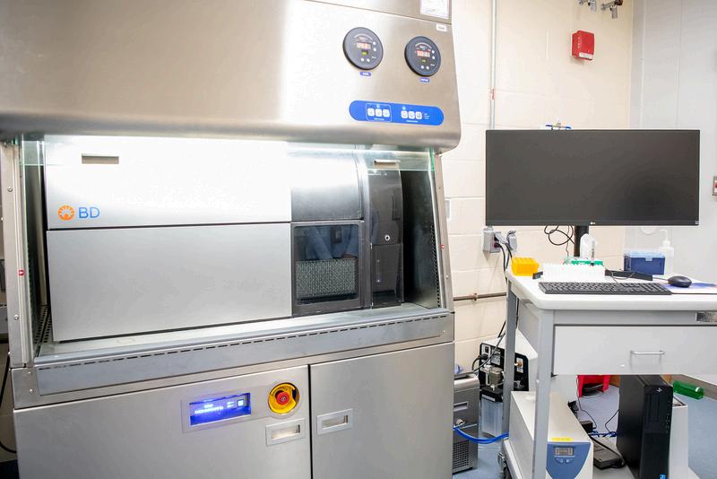
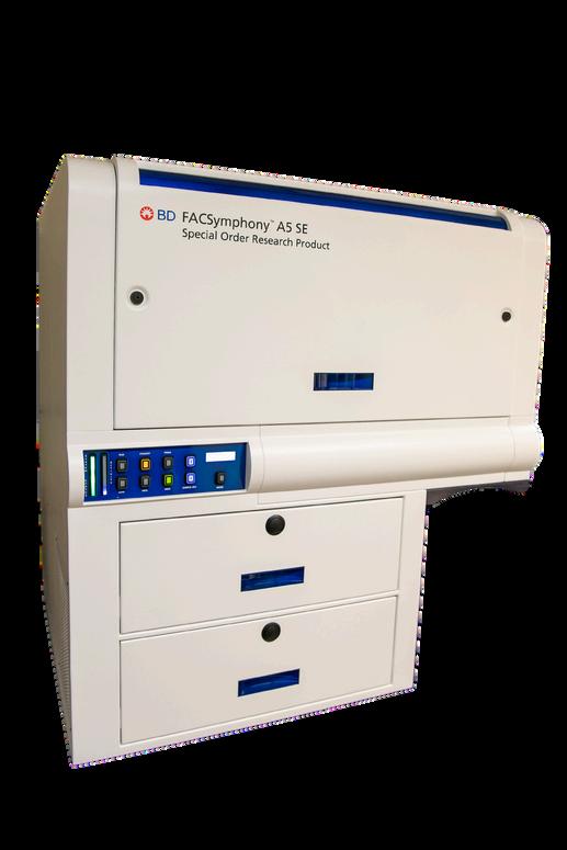
The BD FACS Symphony A5 SE is a high-performance flow cytometer analyzer for single-cell multiparameter analysis, equipped with a full-spectrum detection of light emissions by fluorescent probes. This technology allows researchers to detect up to 48 different biomarkers parameters.
The Intellifex-RUO xMAP is a high-throughput multiplex platform designed for simultaneous analysis of up to 48 biomarkers using advanced Luminex® technology. This system excels at measuring key immune factors such as cytokines, chemokines, and growth factors, and supports immune assays, including ELISA and ELISpot, and suitable for analyzing serum, plasma, and cell culture samples.
ScientificDirector:VasileiosPetrou,PhD;vasileiospetrou@rutgersedu
TechnicalDirector:AlexWei,PhD;h.wei@rutgers.edu
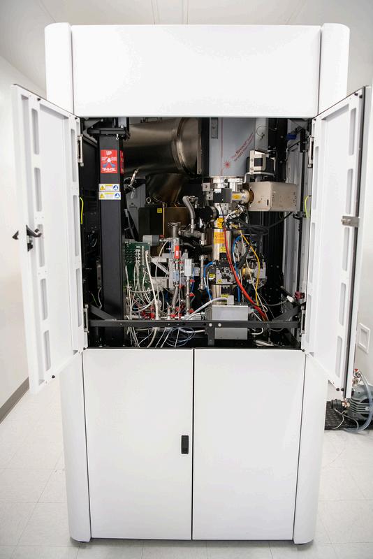
The Tundra Next-Generation Cryo-TEM is a cutting-edge tool for microscopic imaging of proteins and macromolecules. Featuring a 100kV Transmission Electron Microscope with autogrid capability, it offers high-resolution 2D and 3D imaging at an entry-level. Support instruments include the Thermo Fisher Vitrobot Plunge-Freezer for rapid sample vitrification ensuring high-quality cryo-fixation results and a high sample preparation throughput prior to cryo-TEM observation and the Agar Scientific Easiglow Glow Discharge System to support sample grid preparation.
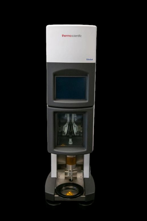
Thermo Fisher Vitrobot Plunge-Freezer
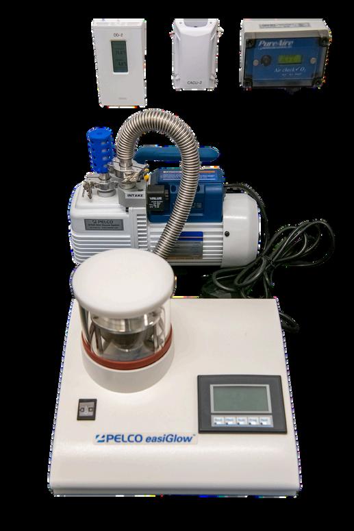
Agar Scientific Easiglow Glow Discharge System
ScientificDirector:DongfangLiu,PhD;dongfangliu@rutgersedu
TechnicalDirector:LukeFritzky;fritzklf@njms.rutgers.edu

njms.rutgers.edu/cihc
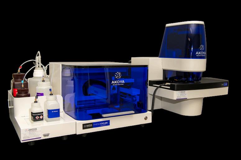
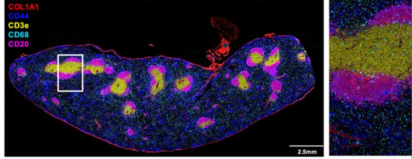
The Akoya Phenocycler-Fusion 2.0 offers spatial microscopic imaging with precise multiparameter detection of proteins within tissue samples. This system can detect more than 60 different protein biomarkers in a single tissue section. Funded by an NIH S10 Grant, PI Jason Weinstein, PhD.
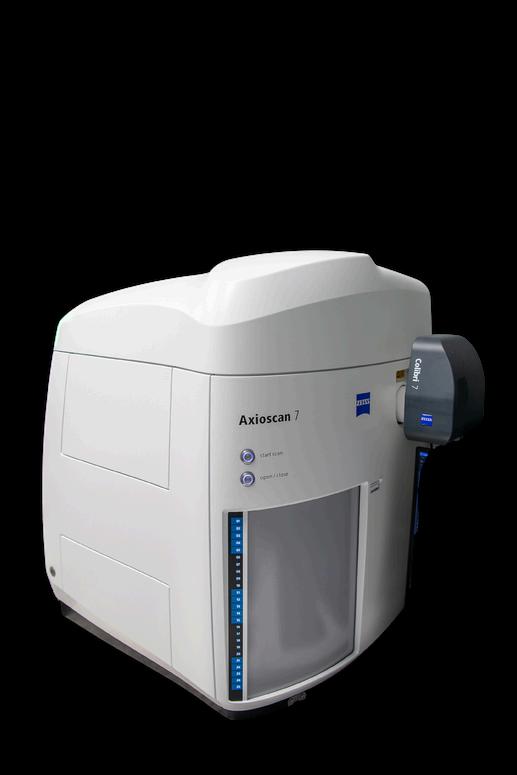
The Zeiss Axioscan 7 is a rapid, high-resolution slide scanner designed for the microscopic whole slide imaging of standard brightfield illuminated histology slides as well as slides labeled with multiple fluorescent dyes. The digitized slides can be viewed and analyzed using standard computer systems.
ScientificDirector:DongfangLiu,PhD;dongfangliu@rutgersedu
TechnicalDirector:LukeFritzky;fritzklf@njms.rutgers.edu
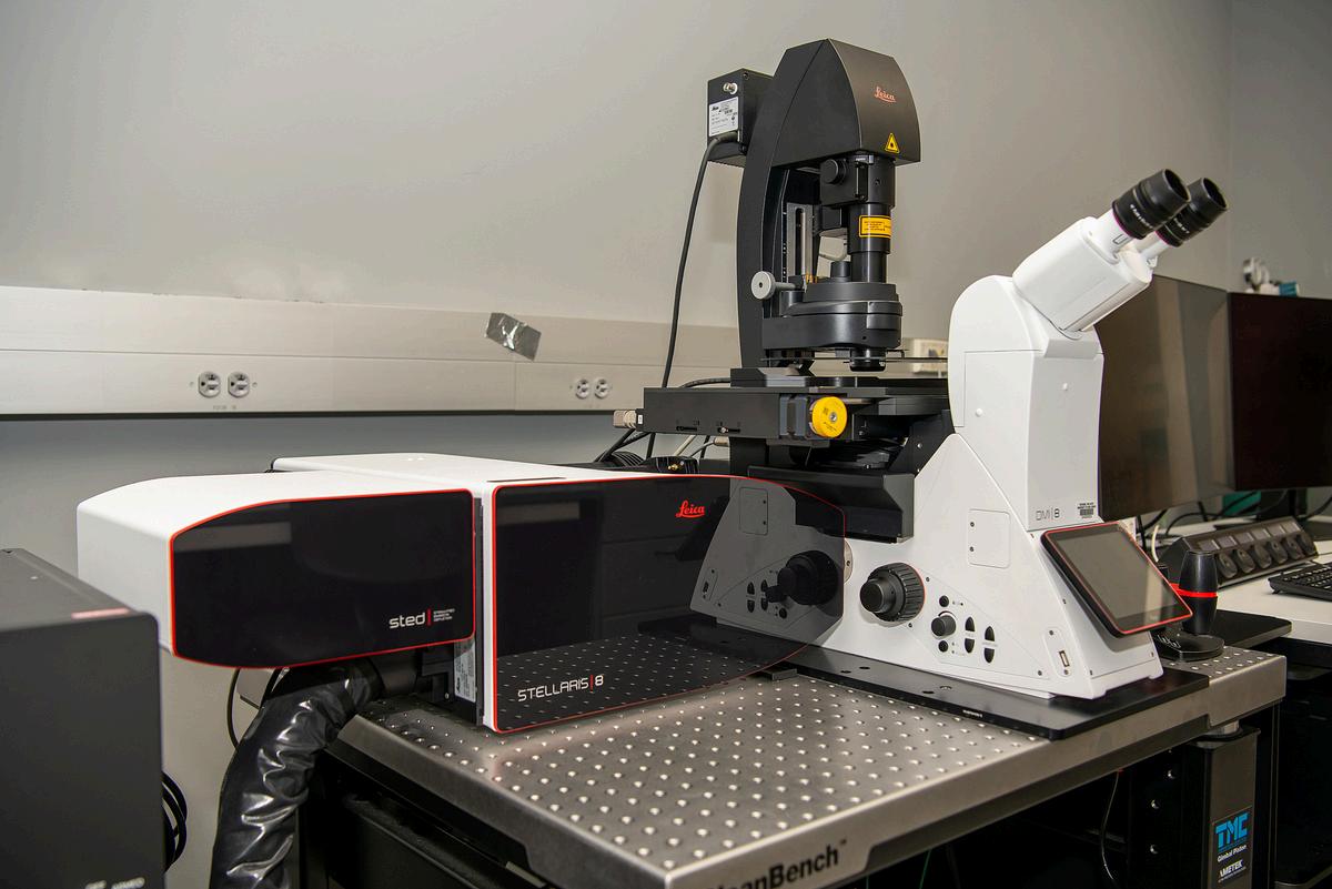
The Leica Stellaris 8 tau-STED is a fully tunable spectral laser scanning confocal microscope. The instrument features Leica’s White Light Laser enabling excitation of fluorochromes from 440 nm ton 790 nm and includes a tunable spectrophotometer-based detector. The system is also equipped with two STED depletion lasers enabling Super-Resolution imaging with axial resolutions below 50 nm.
Imaris is a sate-of-art 3D/4D visualization and analysis software platform for advanced multidimensional microscopy images. Imairs for Core Facilities includes packages for automated object detection, filament tracing, 4D tracking, AI powered object classification, GPU accelerated deconvolution and large format tile stitching. The Cellular Imaging and Histology Core maintains a high-end Xenon workstation dedicated to running Imaris.
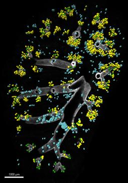
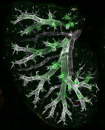
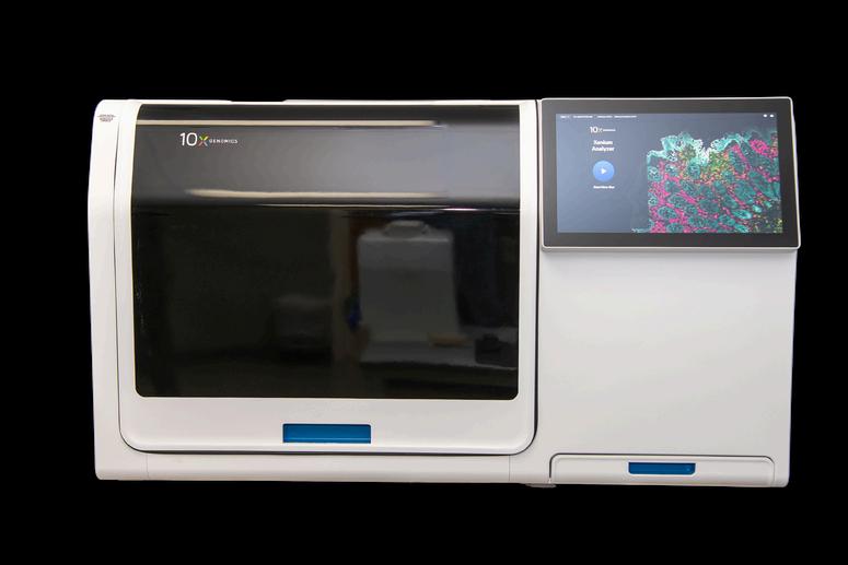
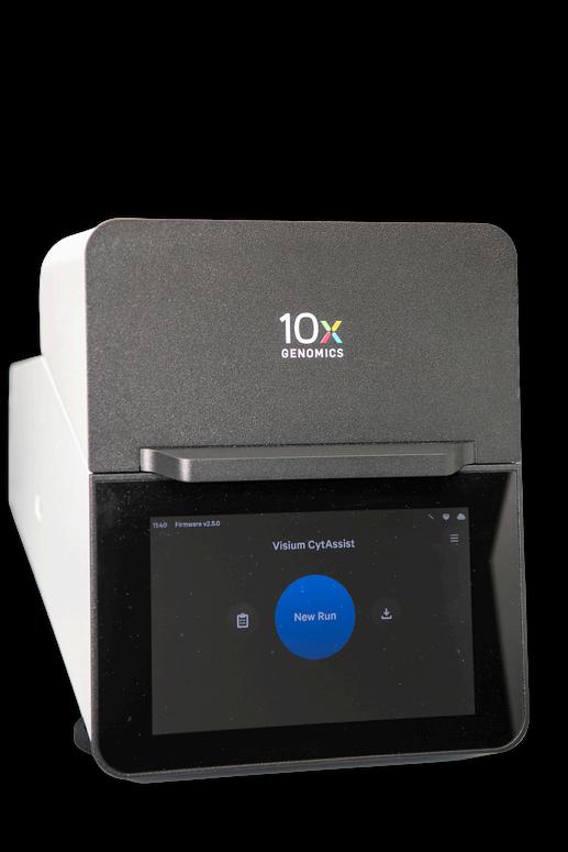
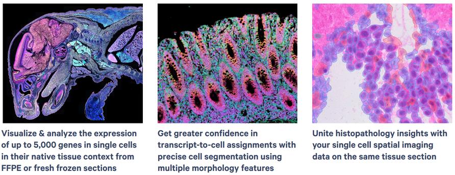
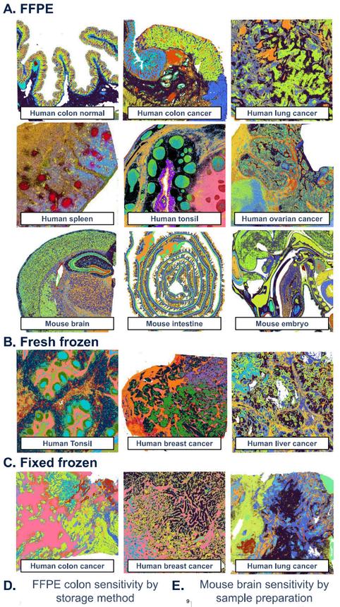
The 10x Genomics Xenium instrument is an imaging based in situ spatial biology platform providing subcellular expression profiling of hundreds to thousands of genes in a single tissue section. 10x Genomics offers both predesigned off-the-shelf panels as well as custom designed panels for use in the Xenium Analyzer. The 10x Genomics VisiumHD is a sequencing based spatial gene expression platform enabling the unbiased analysis of the whole transcriptome in a single tissue section. The 10x Genomics CytAssist ensures the accurate transfer of probes from a tissue section to the capture area on a VisiumHD Slide. The resulting spatially encoded cDNA library is then sequenced on an Illumina Next Gen Sequencer and gene expression data is then mapped back to a scan of the original tissue section preserving spatial context.
IncollaborationwithwiththeMolecularandGenomicInformaticsCore(MaGIC)
Image courtesy of 10x Genomics, Inc.
Visium HD
Image courtesy of 10x Genomics, Inc. ®
Human and mouse gene expressio
Director:MilindMahajan,PhD;mm3711@njms.rutgers.edu
Scan the QR code to view the lab’s website
njms.rutgers.edu/genomics
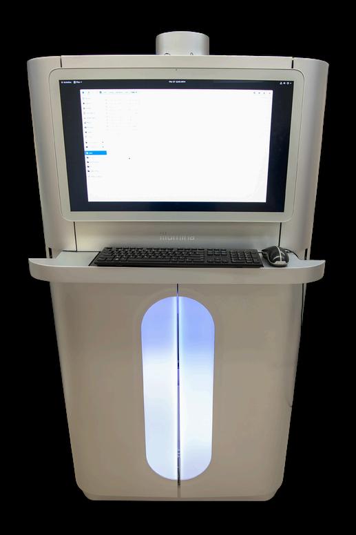
The Illumina NovaSeq X Plus is an upgraded highthroughput deep genome sequencing platform designed for faster and more cost-effective sequencing. The new XLEAP-SBS chemistry delivers increased accuracy and instrument performance. With a higher throughput volume, it offers single-cell sequencing at half the cost. This system is capable of up to 52 billion single reads and 104 billion paired-end reads with onboard cluster generation, and accelerated data analysis using an onboard DRAGEN platform
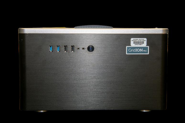
The Nanopore GridION is a long-read next-generation sequencing system that supports multiple platforms running together, offering microbiome strain identification and clinical biomarker-filling analysis, filling in sequencing gaps, and providing improved statistical power.
Multi-Mode
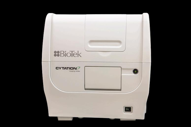
The Agilen BioTek Cytation 7 is a versatile, highthroughput multimodal microplate reader with advanced imaging capabilities It supports automated digital upright and inverted widefield microscopy for fluorescence and brightfield detection with a widerand more sensitive range of fluorophore detection.
**New Spatial Transcriptomics Technology
Joint with Cellular Imaging & Histology Core**
Illumina 10x Genomics Visium hd/CytAssist
Xenium technologies
RBLDirector:DavidAlland,MD;allandda@njms.rutgers.edu
Animalmodelsandrelatedservices(AMRS)core CoreDirector:PadminiSalgame,PhD;salgampa@njms.rutgers.edu
Co-Directors:RicardoRajsbaum,PhD;ricardo.rajsbaum@rutgers.edu
JohnChan,MD;jc2864@njms.rutgers.edu
TechnicalDirector:PreetiBharaj,PhD;pb607@njms.rutgers.edu

njms.rutgers.edu/rbl
Funded by an NIH UC7 Grant, PI Dr. David Alland, MD.
Sireq flexiVent® Complete System Solution (FV-FXCS)
An advanced system for monitoring and measuring in vivo respiratory functions. It goes beyond traditional resistance and compliance mechanics of pulmonary ventilation, and captures crucial details about the mechanical properties of conducting airways, terminal airways and parenchyma
RBLDirector:DavidAlland,MD;allandda@njms.rutgers.edu
Animalmodelsandrelatedservices(AMRS)core CoreDirector:PadminiSalgame,PhD;salgampa@njms.rutgers.edu
Co-Directors:RicardoRajsbaum,PhD;ricardo.rajsbaum@rutgers.edu
JohnChan,MD;jc2864@njms.rutgers.edu
TechnicalDirector:PreetiBharaj,PhD;pb607@njms.rutgers.edu
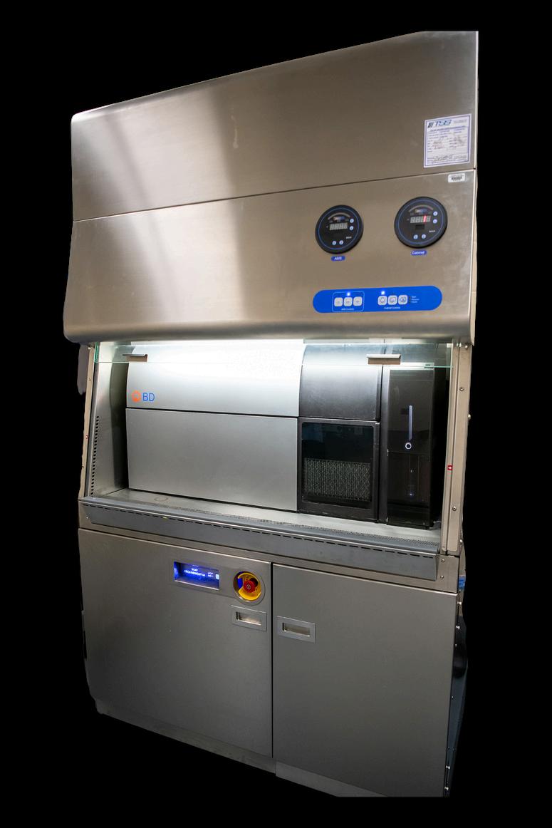
The BD FACSSymphony S6 offers single-cell, multi-color flow cytometry analysis and cell sorting, with the ability to simultaneously collect up to 6 distinct cell subpopulations. This system features full-spectrum detection of light emissions from fluorescent probes, identifying up to 50 biomarker parameters.
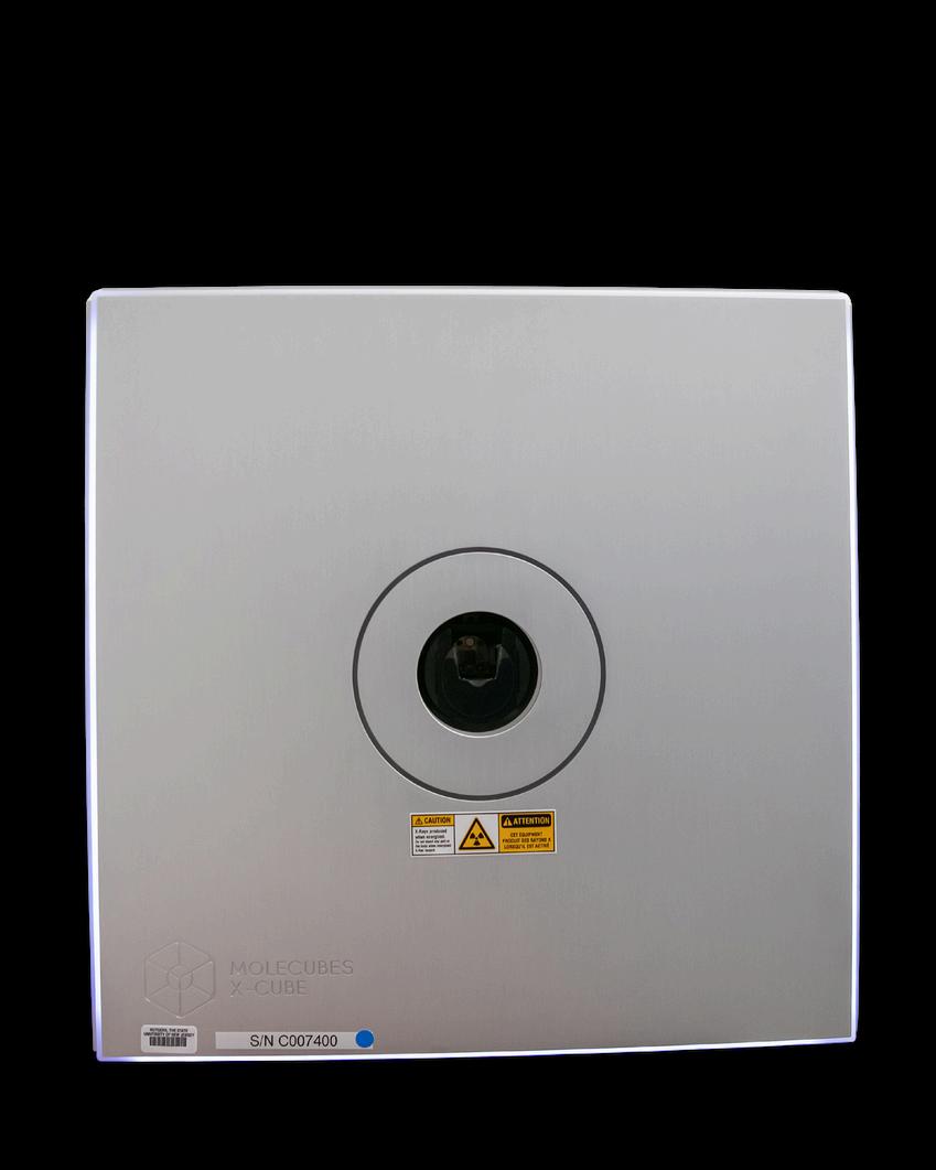
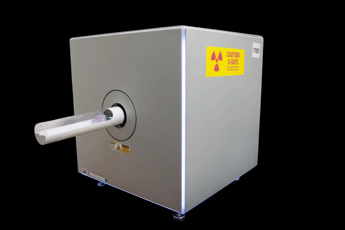
A high-throughput benchtop micro-CT scanner designed for preclinical imaging analysis. This scanner will be part of an respiratory infectious disease animal modeling lab in the RBL, supporting studies of bacterial and viral transmission and chronic infection.
TechnicalDirector:SushilTripathi,PhD;st1123@research.rutgers.edu
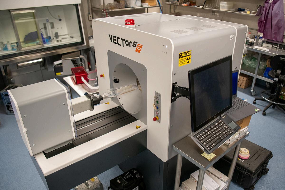
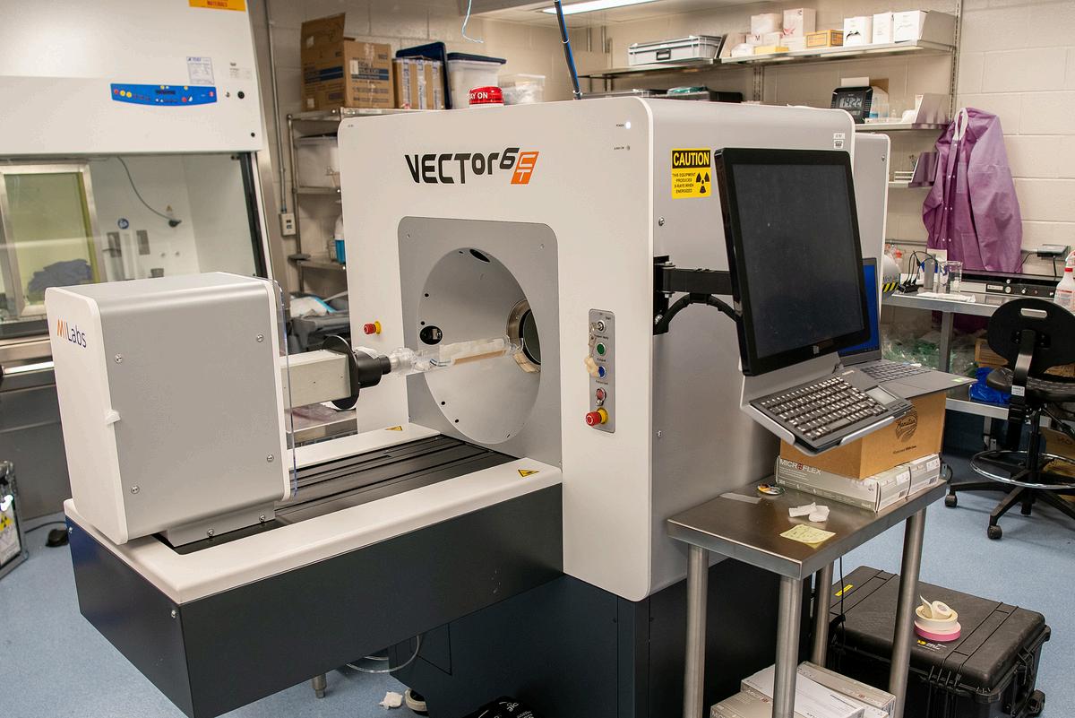
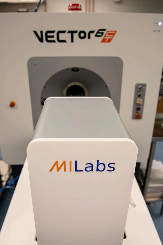
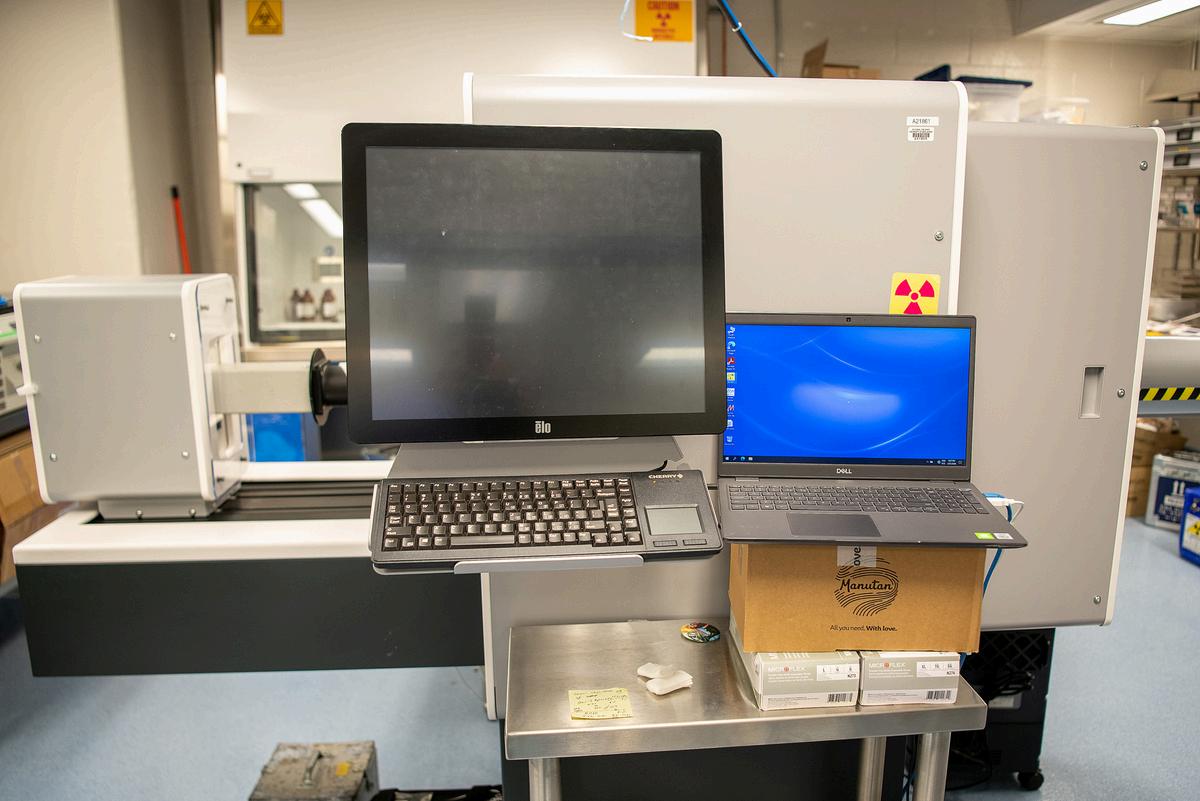
MiLab VECTor6 PET/SPECT/CT
The MiLab VECTor6 is a multi-modal live imaging system for small animal studies. This system includes PET, microCT, and SPECT imaging capabilities. Applications include regional glucose uptake in tumors/brains; lung injury; bone mineral density and studies monitoring biomaterials and devices. Funded by an NIH S10 Grant, PI Roger Howell, PhD. Scientific Director: Roger Howell, PhD.
TechnicalDirector:SushilTripathi,PhD;st1123@researchrutgersedu
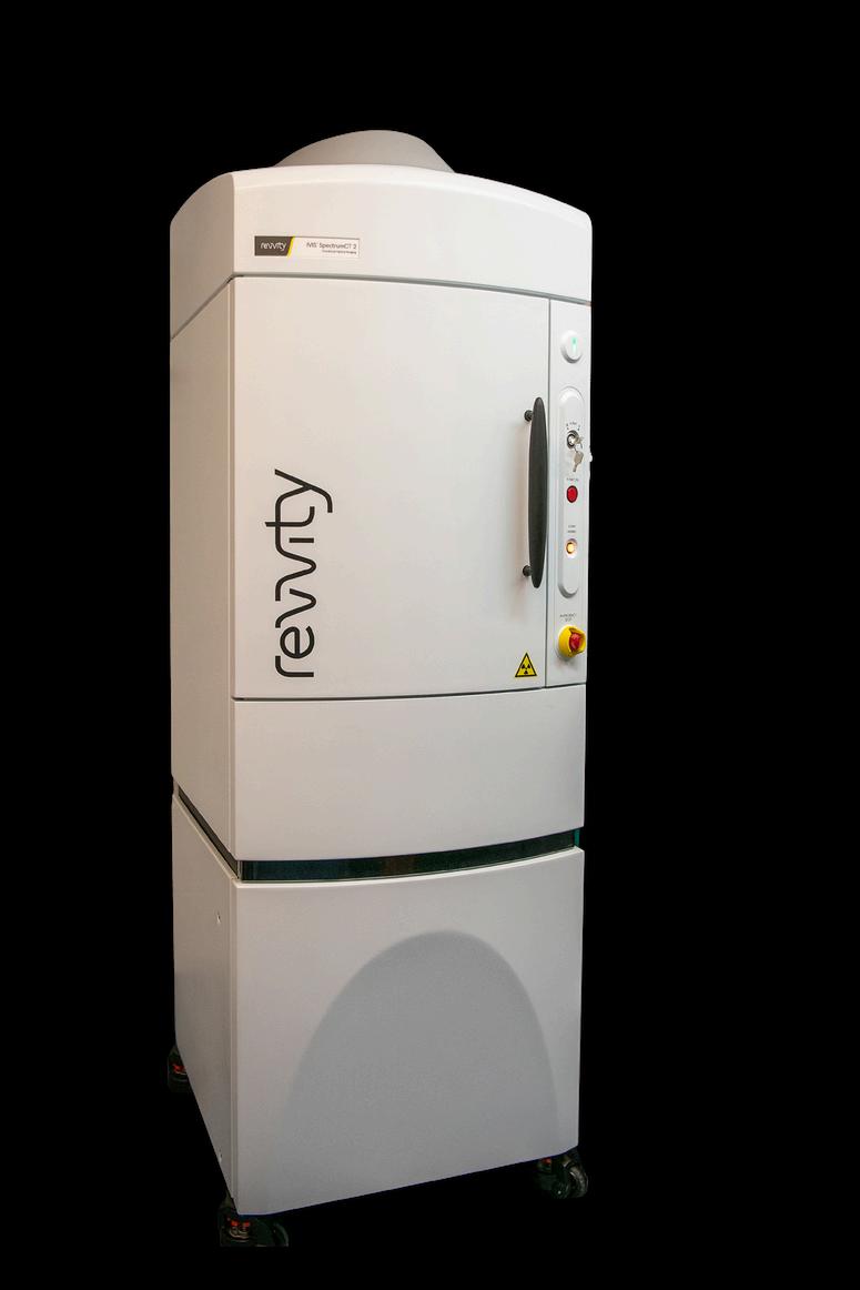
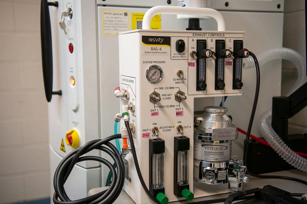
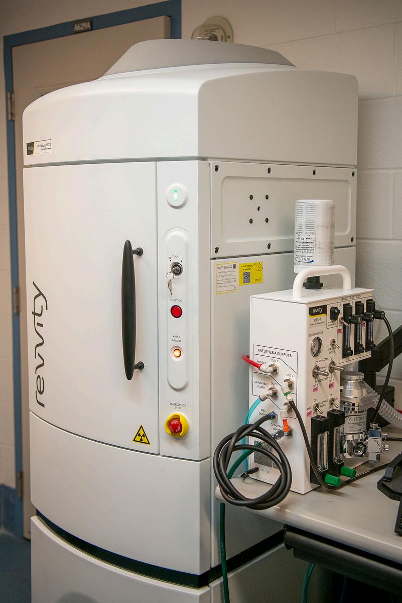
An in vivo animal imaging platform for non-invasive monitoring of disease progression through bioluminescence and fluorescence detection. It offers high sensitivity in deep tissue and can simultaneously image up to 5 mice. The rotating camera enables 3D capabilities, while the software allows co-registration with MiLabs PET, SPECT, and microCT. Funded by an NIH S10 Grant, PI: Punima Bhanot, PhD.
Director:HongLi,PhD;liho2@njmsrutgersedu
njms.rutgers.edu/capr
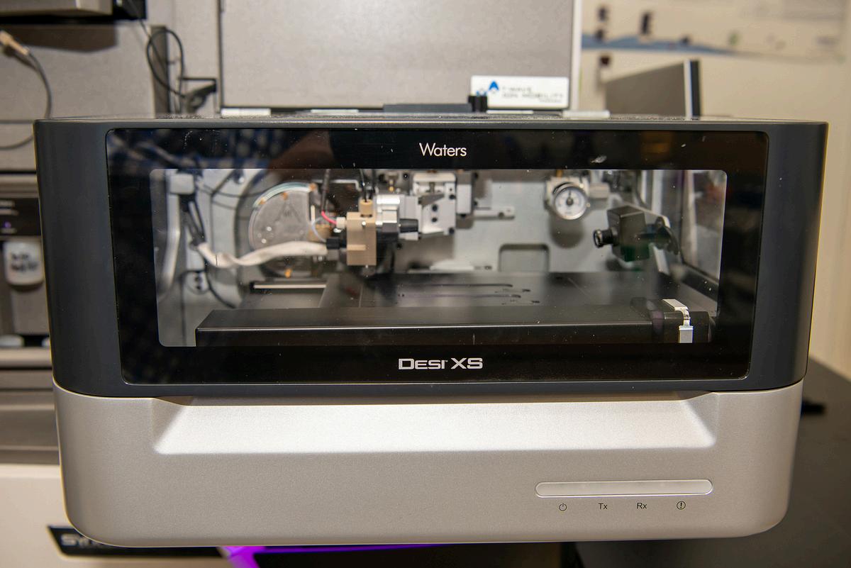
Spatial Metabolomics: Waters DESI-Synapt
This instrument combines mass spectrometry with microscopic detection of metabolites allowing for spatial mapping of molecular events of metabolism within tissue architecture. Scientific Director: Tania Wong, PhD.
**New Mass Spectrophotometer Now Operational** NIH S10 Awarded Bruker timsTOF Ultra 2 LC-MS System for high performance proteomics, PI Hong Li, PhD
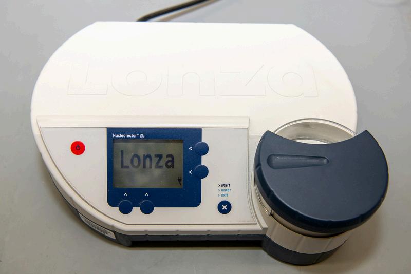
A cutting-edge platform for efficient transfection of even non-dividing primary cells, such as resting T cells or neurons, with rapid gene expression and modification. Key applications include the CrisprCas9 technology for site directed mutations, as well as gene expression and deletions.
For in vivo metabolic and behavioral phenotyping. This system provides highresolution, physiological metabolic and behavioral phenotyping in a 22-cage housing setup. It is equipped with a triple gas analyzer (O₂, CO₂, H₂O), 3D infrared beam detection, body weight monitoring, a running wheel module, and an environmental sensor array for recording ambient temperature, light, RH, sound, BP, noise, and motion data.
Scientific Director: Tania Wong, PhD.
This instrument is a high throughput, accurate, precise, and contact-free acoustic transfer system to dispense sample volumes as low as 2.5 nL. Transfers from 96-well to 1536-well microplates are supported, enabling assay miniaturization in a broad range of applications. Funded by an NIH S10 grant, PI David Alland, MD, Public Health Research Institute Center.