IMC21 Preview: ‘Olympics of Microscopy’ coming to Liverpool, UK
RMS Section Award-winners announced!
Connecting Wales Through Microscopy: The Launch of the Welsh Microscopical Society
Wood Under the Microscope: A Week at Kew Gardens
IMC21 Preview: ‘Olympics of Microscopy’ coming to Liverpool, UK
RMS Section Award-winners announced!
Connecting Wales Through Microscopy: The Launch of the Welsh Microscopical Society
Wood Under the Microscope: A Week at Kew Gardens



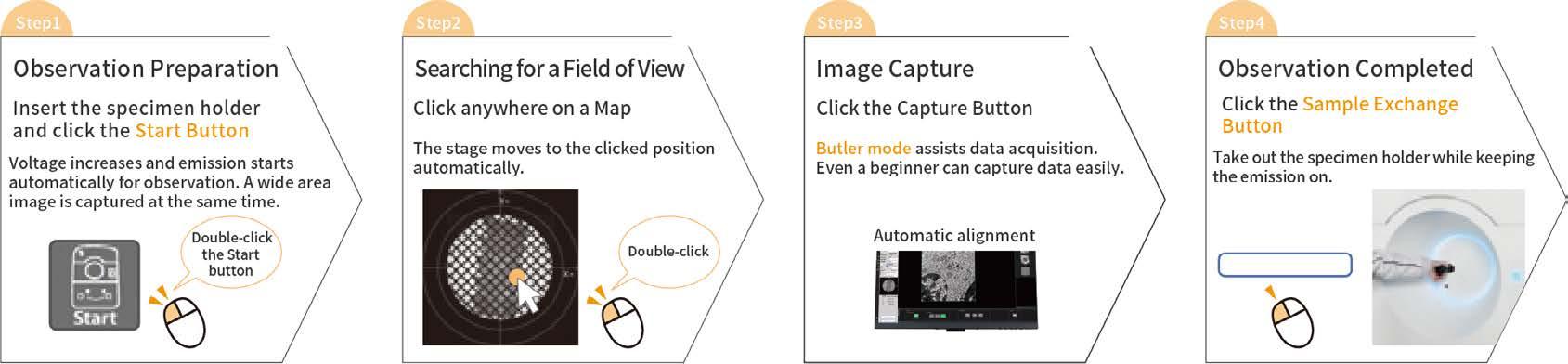
Specimen observation in 4 steps
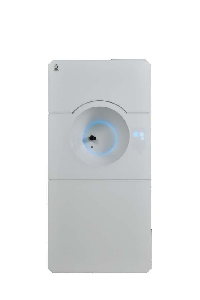
We developed the JEM-120i with the concept of "Compact", "Easy To Use", and "Expandable". With the new external appearance, this instrument has evolved into a useful tool that anyone can use easily, from operation to maintenance.
It takes only 4 steps from loading a specimen to completing observation. The JEM-120i is equipped with an enhanced TEM control system and fully automated apertures, eliminating the need for switching magnification modes and selecting an aperture. Observation operations can be performed more smoothly than with previous models.

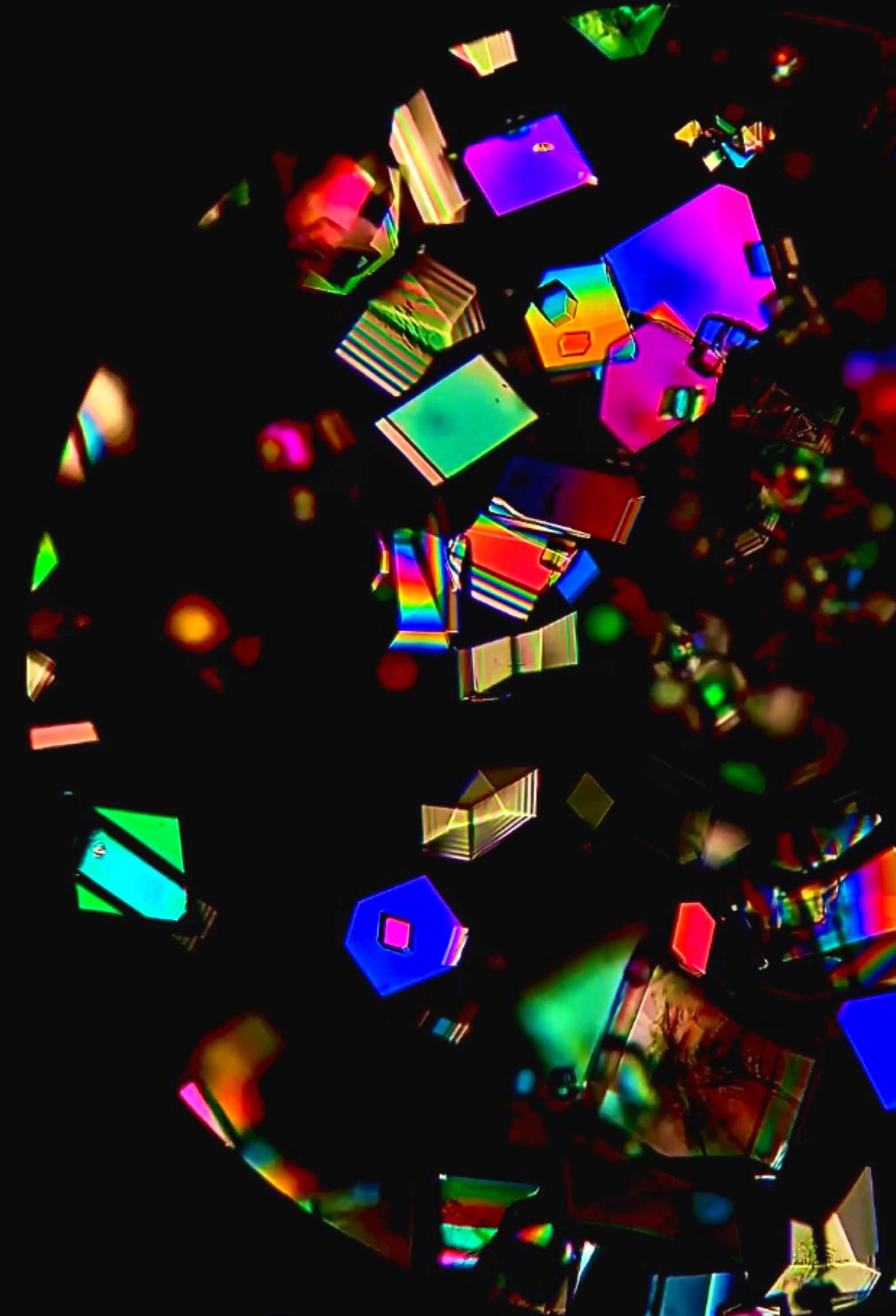
infocus is the Magazine of the Royal Microscopical Society (RMS) –the only truly international microscopical society. The RMS is dedicated to advancing science, developing careers and supporting wider understanding of science and microscopy.
infocus Magazine
37/38 St Clements
Oxford, OX4 1AJ, UK
Tel. +44 (0)1865 254760
Email: infocus@rms.org.uk Website: www.infocus.org.uk
Scientific Editor
Leandro Lemgruber, University of Glasgow, UK
Editor
Owen Morton
Tel. +44 (0)1865 254763, Email: editor@infocus.org.uk
Editorial Board
Myfanwy Adams, John Innes Centre, Norwich, UK
Maadhav Kothari, Zeiss Microscopy, UK
Hilary Sandig, Cancer Research, UK
Trevor Almeida, University of Glasgow, UK
Tim Young, University of Cambridge
Ferran Valderrama, City St George’s, University of London
Thomas Slater, Cardiff University
Advertising
Email: advertising@infocus.org.uk
ISSN: 1750-4740
© 2026 Royal Microscopical Society
infocus is published four times per year by the RMS. Designed and produced by Creative Design. Reproduction in whole or in part without permission from the RMS is forbidden. Views expressed in the Magazine are those of the individual contributors and do not necessarily reflect those of the RMS.
Dear Readers,
With summer almost upon us here in the northern hemisphere, I am delighted to introduce our June 2026 issue of infocus!
Among our offerings in this edition is a preview of this year’s premier event for the global microscopy community – The 21st International Microscopy Congress (IMC21), which takes place in the wonderful city of Liverpool at the end of August into early September.
It really is a special event for the global microscopy community, coming around only once every four years – just like the Olympics! The event is returning to Europe for the first time in over 10 years, and to the UK for the first time since the 1950s, would you believe! Our editorial team will be there to chat about infocus and hear your ideas for new content. We’ll also be asking attendees their views on the future of Artificial Intelligence (AI) in microscopy – for a special feature in our next issue.
Elsewhere in this issue we have an article by one of our editorial board members, Myfanwy Adams, about the launch of the Welsh Microscopical Society – a fantastic new initiative aimed at unlocking the full potential of the microscopy community in Wales and boosting research, industry and education.
On a botanical note – and with the approach of summer once again in mind - Lee Bilson provides an interesting account of a wood-themed microscopy course at the world-famous Kew Gardens.We also have a creepy-crawly-inspired article by Muneebah Fatimah, a student in Pakistan, about her role in last year’s Lahore Science Mela – an annual event that attracts thousands of visitors.
Finally, I would like to highlight the achievements of all our recent RMS award-winners, who are featured in this issue. It is always exciting to learn about the ground-breaking work of some of the leading figures across the microscopy community, and I hope you enjoy reading about their work as much as I have. Slàinte!
Leandro Lemgruber
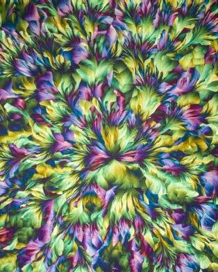
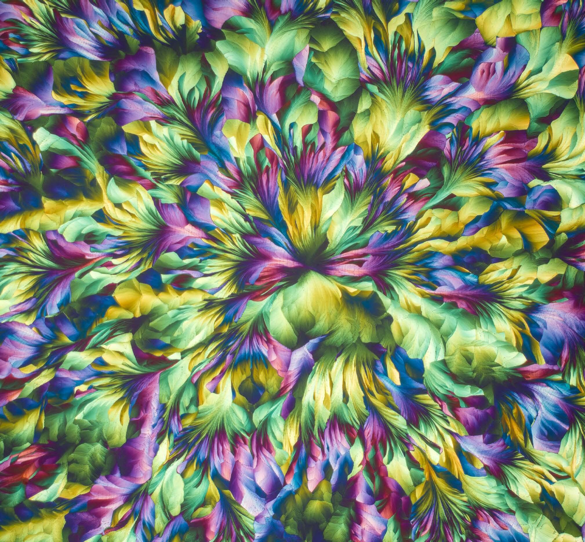
By Karl Gaff, Art of Science Photography. This abstract floral landscape is a magnified view (150x) of a birefringent thin crystalline film, captured using compensated polarisation microscopy. The image centres on a nucleation point, from which plant-like structures radiate outward. The entire formation shown is confined within a single cell of the film. The film itself consists of many such cells, fitting together like pieces of a jigsaw puzzle. Each cell contains numerous crystal grains, whose individual lattice orientations and thicknesses determine the colours observed.

The 21st International Microscopy Congress (IMC21) 2026
31 August - 4 September 2026, Liverpool, UK
Pre-Congress Events 30 August 2026

“The Next Generation – Embracing the
The abstracts are in, the speakers are confirmed, and the stage is set for the 21st International Microscopy Congress (IMC21)!
With less than three months to go before this year’s premier event for the global microscopy community kicks off in Liverpool, UK, we’re taking a closer look at what’s in store for attendees – from the blockbuster scientific programme to a world-class exhibition, workshops, networking and more!
The RMS is honoured to be delivering this year’s premier event for the global microscopy community alongside the International Federation of Societies for Microscopy (IFSM), Liverpool University and all our other partners.
We look forward to seeing you there!
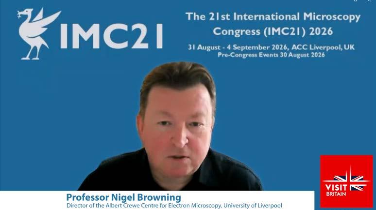
“The IMC is known as the Olympics of Microscopy, and is quite simply the biggest and best event in the global microscopy calendar this year.”
Professor Nigel Browning, IMC21 Organising Committee Chair
IMC21 features a blockbuster scientific programme, organised into three separate themes and comprising no fewer than 32 different symposia.
With almost 1,700 talk and poster abstracts submitted – plus a surge of late-breaking posters - a huge range of scientific content awaits; so now’s the time to familiarise yourself with what’s on the menu, and start to plan your IMC21 experience!
Overview by Co-Chairs Gail McConnell and David Bhella
As the world of microscopy focuses on Liverpool for the 21st International Microscopy Congress, we look forward to an exciting programme of lifescience research. IMC21 will showcase innovation across imaging modalities and the application of
cutting-edge microscopy methods to address key questions in biology that span length scales from molecules to organisms.
Cryo-EM - Innovations in macromolecular structure determination will showcase how this disruptive technology continues to transform structural biology, highlighting technological advances that improve data quality and throughput. Integration of new AI tools better enables interpretation of these data, yielding insights into protein dynamics and accelerating model building. Moving the focus of structural biology into the cellular and tissue context, innovations highlighted in Cryo-ET –Macromolecular Imaging in Cells, Organoids and Tissues include advances in specimen preparation, highspeed automated tomography, and computational tools to uncover mechanism through interrogation of noisy, challenging cryo-ET datasets. The promise of molecular imaging at tissue scale has led to an explosion of interest in Volume Imaging in the electron microscope and this session will be a real highlight of IMC21, showcasing how techniques such as serial block-face imaging and array tomography can map the complexity of biological systems.
IMC21’s life sciences programme places light microscopy firmly at the centre of biological research, with a series of dedicated symposia reflecting both technological progress and expanding application areas. In Live Imaging Cells, Tissues, Organs and
“The congress creates a platform for groundbreaking scientific research for inspiring the next generation of scientists and for establishing collaborations across all scientific disciplines.”
Nigel Browning
Organisms, the focus will be on capturing dynamic biological processes in real time, from intracellular events to development at the organismal level. This is complemented by Resolution Revolution in Light Microscopy, where ongoing advances in superresolution and optical engineering continue to push beyond traditional diffraction limits, opening new windows on molecular organisation.
The importance of imaging in three dimensions is further highlighted in Volumetric Imaging, with techniques enabling increasingly detailed reconstruction of complex biological structures across scales. At the same time, Democratisation and Widening Access to Microscopy signals a growing emphasis on accessibility, reproducibility, and global collaboration, ensuring that cutting-edge methods are widely available across the international research community.
Light microscopy is also deeply embedded within
the broader life sciences themes. Strong links to enabling technologies via Machine Learning in Biological Imaging point to the increasing role of artificial intelligence in image acquisition and analysis, while multimodal sessions such as Neurosciences, Pathogen Biology, Plant Sciences, and Correlated Imaging Across Length Scales highlight how a broad range of cutting edge imaging technologies are being brought to bear to provide comprehensive biological insight.
Together, these symposia highlight a field that is not only rapidly evolving but increasingly unified, where technologies converge to reveal biology in unprecedented detail. IMC21 will capture this momentum, showcasing how imaging innovation is reshaping our understanding of life across spatial and temporal scales.
Physical Sciences – ‘An exciting, forward-looking programme’
The International Microscopy Congress (IMC) conference is a highlight for any microscopist, and has often been described at the “Olympics” of microscopy as it only occurs once every four years. For IMC21 this summer we are looking forward to welcoming you to the vibrant city of Liverpool. Bringing together leading researchers, early career scientists, innovators, practitioners, trade suppliers and scientific societies from across disciplines, the congress offers a chance to meet and interact with colleagues from across the globe.
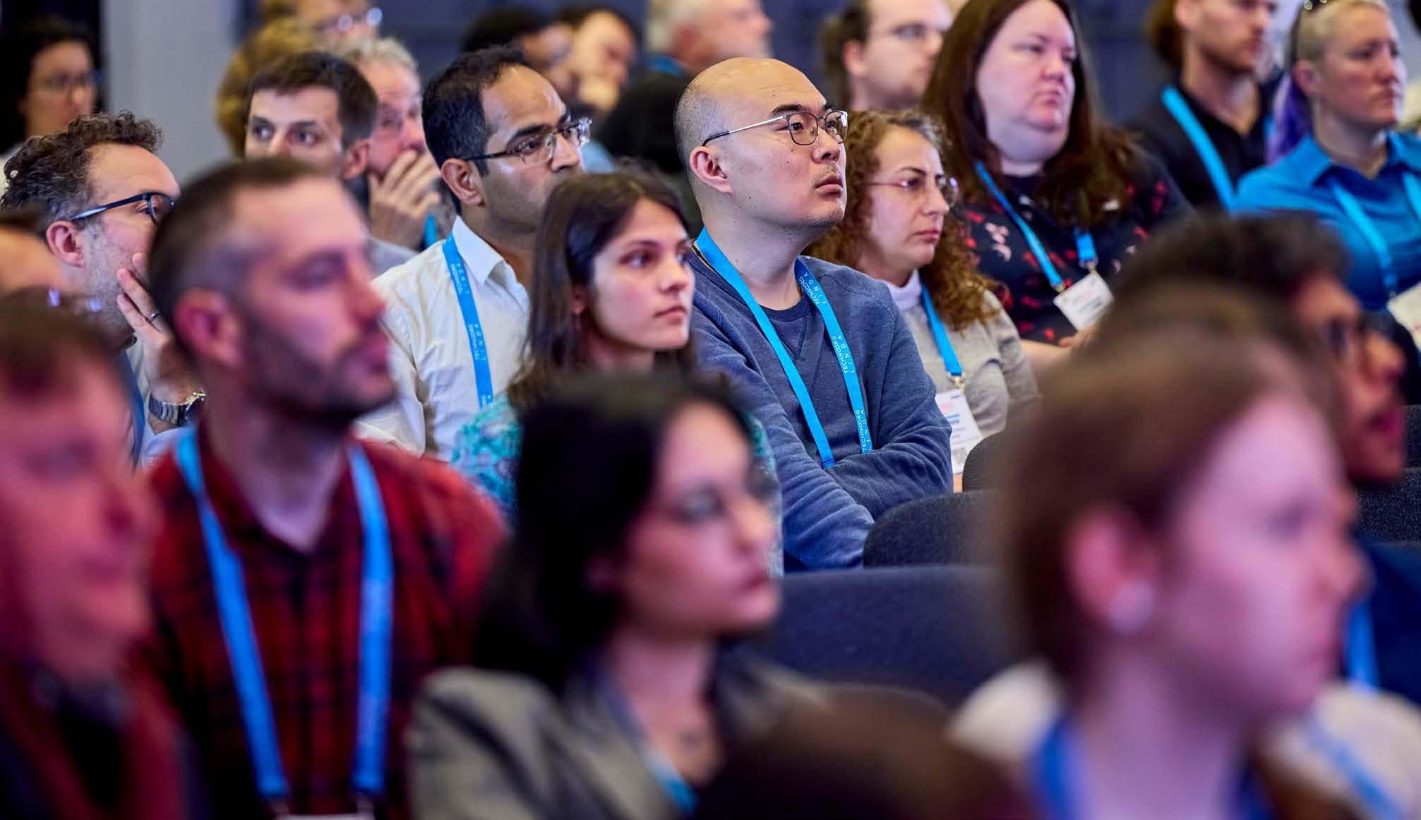
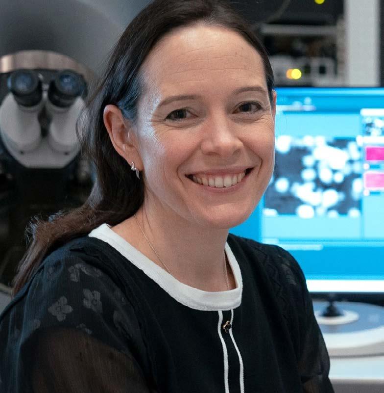
It has been our pleasure to co-chair the Physical Sciences theme of the congress. With this theme there are 11 individual symposia with a wide range of topics covering the applications of advanced microscopy methods within physical sciences. The breadth of materials science covered is impressive. Dedicated sessions explore soft materials, polymers, and beam-sensitive materials, alongside twodimensional materials, nanomaterials and catalysts, functional ceramics and oxides, and structural materials such as metals and alloys.
Further sessions delve into highly topical areas including semiconductors, quantum materials, and ICT, magnetic materials, ferroelectrics, energy storage and conversion, and radiation damage and nuclear energy under extreme conditions. Along with earth and planetary materials, these themes underline the crucial role microscopy plays in addressing global challenges, from sustainable energy to next-generation computing technologies. The inclusion of phase transformations and high-speed dynamics sessions also emphasises the importance of understanding materials in motion—capturing
processes as they happen rather than after the fact.
This diversity ensures that the Physical Sciences theme is relevant to researchers working across both fundamental and applied domains. What makes this theme truly exceptional is how it integrates all these topics into a coherent and forward-looking programme which will allow us to hear from leaders in the field and early career scientists with new ideas. It reflects not only where microscopy is today, but where it is heading. We are delighted with the number of abstracts submitted to this theme at IMC21 – more than 700 in total!
– and don’t forget, ‘late-breaking’ poster abstracts will continue to be accepted until 26 June. We are genuinely excited about the science we are going to

hear about and the discussions we will have, and we look forward to seeing you there.
‘Instrumentation Matters: Come to see the latest driving force for advances in microscopy in action’
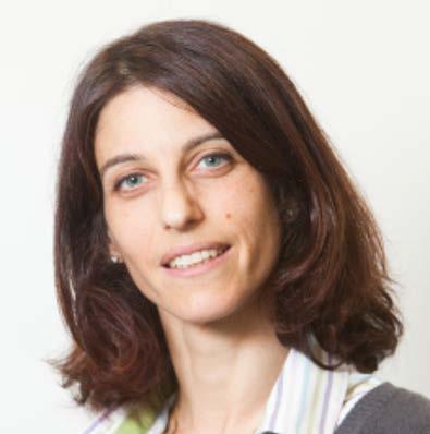
Overview by Co-chairs Jun Yuan and Cate
Breakthroughs in microscopy are often celebrated for the biological or materials discoveries they enable. The Instrumentation theme of IMC21 focuses on the advances in hardware, software and methodology that make those breakthroughs
possible. Progress in nanoscale imaging depends on sustained development in optics, detectors, contrast mechanisms, sample environments, automation and data handling. This is where many of our future discoveries will begin.
From aberration correction and ptychography to ultrafast imaging, environmental microscopy and AI-assisted analysis, the Instrumentation theme provides a forum for those who develop the methods that determine what can be seen, measured and interpreted. At a time when microscopy is becoming increasingly specialised, this kind of crossdisciplinary discussion is particularly important.
Across eight symposia, the theme brings together recent advances in instrument design and measurement approaches: hardware, automation and AI; diffraction and 4D-STEM; dynamic and environmental techniques; phase imaging; quantitative 3D tomography; surface-sensitive methods, including SEM, FIB, helium ion microscopy and LEEM/PEEM; scanning probe and advanced light microscopy; and spectroscopy, including emerging laser–electron approaches. The common thread is quantitative, multimodal and, where possible, operando characterisation. Examples include mapping magnetic fields using ptychography, following catalyst evolution in a gas cell, and combining FIB-SEM with atom probe tomography for 3D analysis. In each case, instrumentation and computation are closely linked: better experiments depend not only on better signals, but also on better ways of extracting reliable information from them.
We are also pleased that the theme brings together physical and life sciences. Instrumentation ideas often move productively between fields, sometimes in unexpected directions. We are grateful to the symposium organisers for assembling a strong programme of invited speakers and contributed presentations, and for helping to create a theme that reflects the breadth of current instrumentation development.
We are particularly looking forward to discussions on real-time automation and AI-assisted microscopy. The prospect of a microscope that can acquire data, recognise features or phases, adjust acquisition conditions and guide the next measurement is now within reach. We are also keen to hear about ultrafast and phase-sensitive approaches, including structured beams, PINEM and other laser-coupled or quantum-coherent methods that can probe dynamics on intrinsic length and time scales. Finally, the growing connections between scanning probe microscopy, super-resolution light microscopy and electron microscopy should make the correlative sessions especially interesting.
We hope the Instrumentation theme will provide a lively and technically focused forum for discussing not only what microscopy can currently do, but what the next generation of instruments should be able to do.
Topping the IMC21 bill are our brilliant plenary speakers.
Joanne is an Australian Research Council Laureate Fellow in the School of Physics and Astronomy at Monash University. She conducts research in the theory and development of new electron scattering methods for determining the atomic and electronic structure of condensed matter. She is a Fellow of the Australian Academy of Science.
Peter obtained a PhD in biophysics from Harvard University, Cambridge, Massachusetts. He then moved to the Medical Research Council (MRC) Laboratory of Molecular Biology in Cambridge, UK, before joining the Division of Physical Biochemistry at the MRC National Institute for Medical Research in 2005 (now part of the Francis Crick Institute).
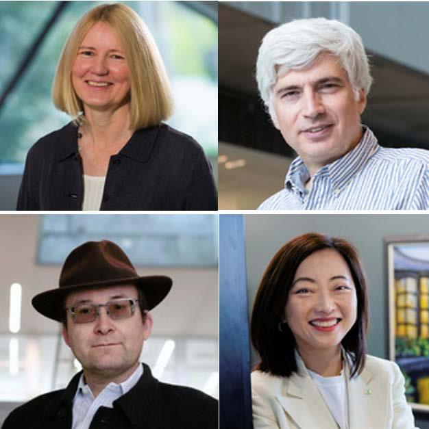
Shirley is the Liew Family Professor at the Pritzker School of Molecular Engineering at the University of Chicago. She is the director of Energy Storage Research Alliance (ESRA), an innovation hub funded in 2024 by US Department of Energy, Office of Science.
Sergei is a Weston Fulton chair professor at the University of Tennessee, Knoxville. From 2022 to 2023, he was a principal scientist at the Amazon Grand Challenge (moon shot factory). Before then, he spent 20 years at Oak Ridge National
The congress kicks off with more than 20 different pre-congress workshops taking place on 30 August at the University of Liverpool. All workshops will be running in parallel – which means it is only possible to attend one – so choose carefully!
Covering a host of exciting topics and techniques across the sciences, this is a great way to begin your IMC21 experience in Liverpool - so be sure to book your place alongside your congress ticket. If you
have already registered, you can add a pre-congress workshop to your existing booking by logging back into your online account.
The Life Sciences pre-congress workshops at IMC21 are, for me, one of the most exciting parts of the entire programme. It is also a real privilege to see so many excellent colleagues from across the community leading these sessions, bringing both depth of expertise and a genuine commitment to sharing knowledge. They set the tone for the meeting in a very real way: They are bringing people
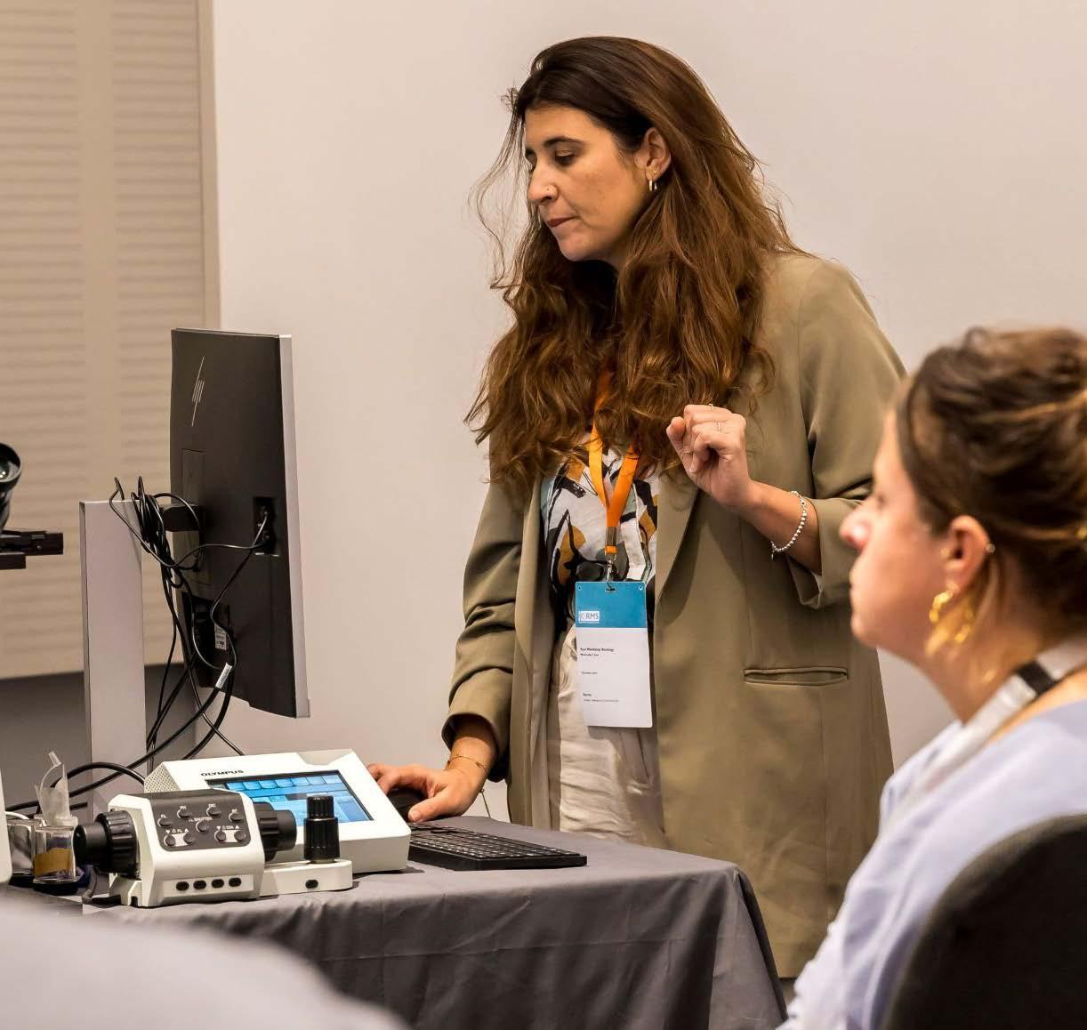
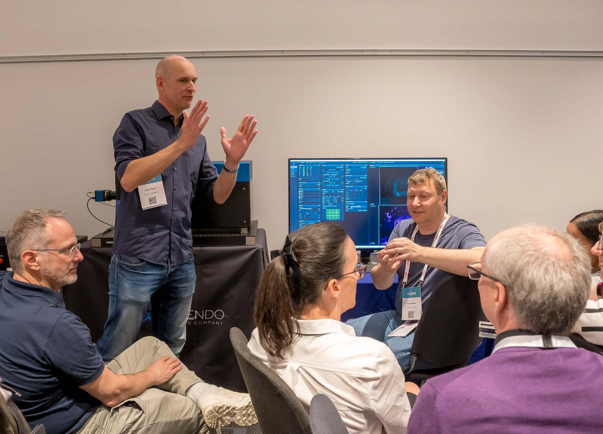
together early, creating space for discussion, and grounding the congress in the practical realities of how we design, acquire, and interpret imaging data. What stands out is the breadth of topics, but also the deliberate focus on connecting technology to biological insight. These are not passive sessions; they are designed to encourage people to engage critically with both methods and data from the outset.
A number of workshops really highlight where the field is heading. The session on artefacts in electron microscopy for the life sciences is particularly important, not just from a technical perspective, but in terms of scientific rigour and reproducibility. Understanding what we are looking at, and just as importantly what we are not looking at, is fundamental. Alongside this, the workshops focused on volume electron microscopy and X-ray imaging reflect the continued move towards multiscale, multimodal approaches that are reshaping how we interrogate biological systems. These are
complemented by sessions on digital pathology and multiplexed analysis, which speak to the growing importance of data-rich imaging and the need for robust, scalable analytical frameworks. Equally, the pedagogy workshop is a real highlight: recognising that how we train, teach, and support the next generation of microscopists is just as critical as the technologies themselves.
More broadly, these workshops are about building confidence and capability across the community. They provide an invaluable opportunity for early career researchers, in particular, to develop practical skills, ask questions in a supportive environment, and begin to establish networks that will carry through the rest of the congress and beyond. At the same time, they create space for more experienced researchers and facility scientists to reflect on best practice, share expertise, and engage in the kinds of conversations that ultimately drive the field forward.
For me, the Life Sciences pre-congress workshops really capture the spirit of IMC21, collaborative,
forward-looking, and grounded in both technical excellence and biological relevance. They are an opportunity not just to learn new approaches, but to challenge assumptions, refine how we work, and set the agenda for the days that follow.
With no fewer than 12 different Physical Science Workshops to choose from, there’s sure to be something to interest all our attendees.
All the latest developments in the field are represented here, with a diverse range of microscopy techniques covered - from time-resolved electron microscopy, cryo-electron microscopy and EBSD, to Electron Holography, atom probe tomography and artificial intelligence analysis methods.
The full list of workshops is as follows:
• From nanobeam to ptychography: 4D-STEM data acquisition, processing, and interpretation
• Time-resolved Electron Microscopy
• Electron Energy Loss Spectroscopy: Learning Past, Present and Future Applications
• The Fundamentals of Quantitative In-Situ liquid and In-situ Gas EM
• Advances in Cryo-Electron Microscopy: Quantum Materials and Energy Storage
• How EBSD can help to solve real-world problems and accelerate developments by Oxford Instruments
• Electron Holography
• Artificial Intelligence Methods for Microscopy Analysis and Knowledge Extraction
• Atom Probe Tomography
• In-Situ Electrical Characterization in the TEM: Techniques and Applications
• EMAG Workshop - Introduction to Python for electron microscopists
• In-situ liquid electrochemical TEM
Book your workshop now!
Overview by Demie Kepaptsoglou & Jordi Arbiol, Young Scientist Assembly Chairs
Walking into a major international congress like the International Microscopy Congress can be an intimidating experience for an early career researcher. It’s easy to feel lost among thousands of delegates and the legendary names from textbooks
or papers, and it is often daunting to approach a senior scientist you don’t know. The IFSM Young Scientist Assembly (YSA) exists to break this barrier and help the next generation find their path in the global science community.
Established in 2018 for the IMC19 meeting in Sydney, Australia, the YSA is organised as a pre-meeting of IMC under the auspices of the International Federation of Societies for Microscopy (IFSM). The IFSM’s mission is to contribute to the advancement of microscopy in all its aspects worldwide, and this assembly is the primary platform for supporting the next generation.
The response to the 2026 IMC21 YSA has been incredible, with more than 100 applications for just 50 available places. The selection was really
challenging, as the quality of the applications was excellent, but this overwhelming response highlights how much the early career researcher community values this dedicated space. The final 50 young scientists (PhD students and researchers within five years of their doctorate) were selected based on their scientific accomplishments and their IMC21 abstracts, while ensuring geographical and gender balance.We are proud to host a cohort representing 23 different countries, with delegates travelling from as far afield as Australia, Brazil, China, Saudi Arabia and the USA, alongside a strong European presence. This diversity ensures that the assembly is a melting pot of ideas from every corner of the globe and every branch of microscopy.
To spark this new chapter in their careers, the programme features a series of inspiring lectures from leaders who have shaped the field.This year, we are particularly thrilled to include a session with Dr Jessica Wade, from Imperial College London, who will discuss her path towards becoming a science advocate and excelling in science outreach. In today’s academic landscape, the ability to communicate our work and advocate for science is an essential skill. By listening to the personal experiences of established researchers from all around the world, our delegates can see that success in science is
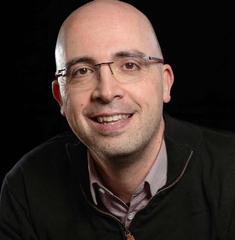
Jordi Arbiol.
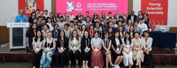
rarely a straight line, helping to demystify the path forward.
Beyond the lectures, the YSA features a roundtable format designed to facilitate open, honest discussions. By sitting down with senior scientists in a relaxed setting, the meeting aims to break the fear of the unknown and decrease the perceived distance between student and expert. Just as importantly, the YSA allows delegates to meet peers at the same career level from all around the world, representing
different walks of life and various career paths. These are the friends who will become future collaborators and, in turn, will eventually inspire the generations of scientists that follow them.
We also believe in celebrating the beauty of our work. Our Imaging Contest is a light-hearted science-meets-art snippet, showcasing the beauty of the microscopic world and reminding us of that curiosity that first led us to see far beyond the reach of the naked eye.
The YSA is as much about community as it is about science. Our social programme gives attendees an overview of this historic city, featuring a live tour bus through Liverpool’s fascinating history and a celebratory dinner at the iconic Victoria Gallery & Museum. By the time the main Congress opens, we hope that these 50 delegates aren’t just attendees; they are friends and future colleagues, establishing a connection for the future.
Our hope is that the YSA does more than just help you navigate a week in Liverpool; we hope it will be a catalyst for your future career. Whether you
continue in academia, move into industry, or forge a path in outreach, the connections made here are the foundation of a lifelong network. We want you to leave with the realisation that you aren’t just a member of the microscopy community; you are its future.
This meeting was made possible thanks to the support of the IFSM executive board, the Royal Microscopical Society (RMS) and especially the support of Kate Wooding. We are also deeply grateful to all our invited guests and senior scientists who have generously used their own time to come and share their experiences with the YSA attendees. Your mentorship is what makes this assembly possible.
“The Next Generation – Embracing the Responsible AI Revolution” – speak to infocus at IMC21!
In terms of hot topics in microscopy, it’s fair to say Artificial Intelligence (AI) is right up there, and it will be a recurring theme at IMC21.
Questions surrounding AI, its influence over the future of microscopy, and how it can best be incorporated into research, are many and various
across the sciences – and infocus would like to hear your answers!
Members of our Editorial Board will be asking for your contributions throughout IMC21, with the aim of including a special feature in our September issue.
Be sure to have your say on AI during the Congress!

Photo by Igor Omilaev on Unsplash

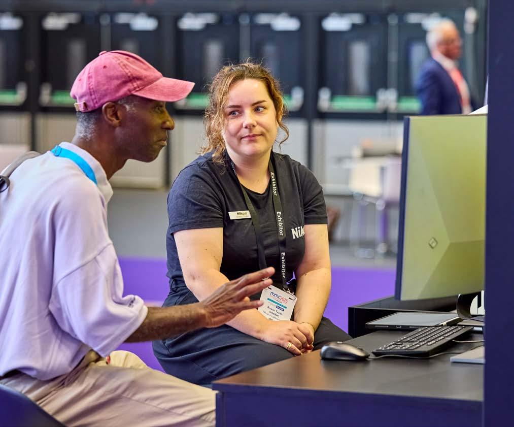
“The exhibition is very much going to be at the heart of this year’s Congress, and our visitors are sure to have plenty to explore.”
RMS Chief Executive Sali Davis
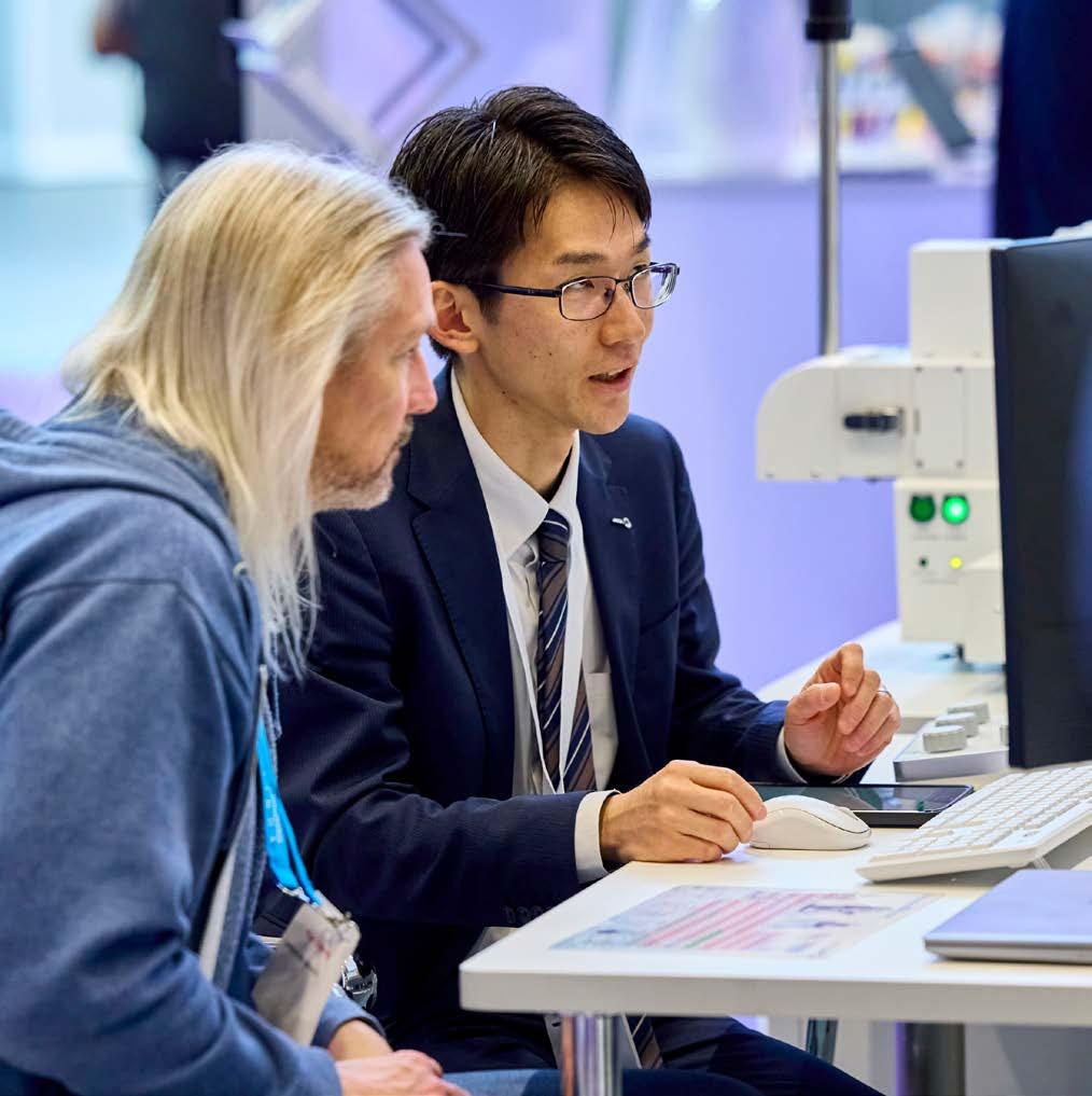
The IMC21 exhibition will provide a fantastic opportunity to network with local and international companies, catch up with old contacts, view demonstrations of the very latest equipment and technology, and much more!
In what promises to be this year’s biggest gathering of global microscopy manufacturers, a host of leading international companies will be showcasing their latest products and technologies.
More than 100 exhibitors have already signed up to be part of the showpiece exhibition at IMC21,
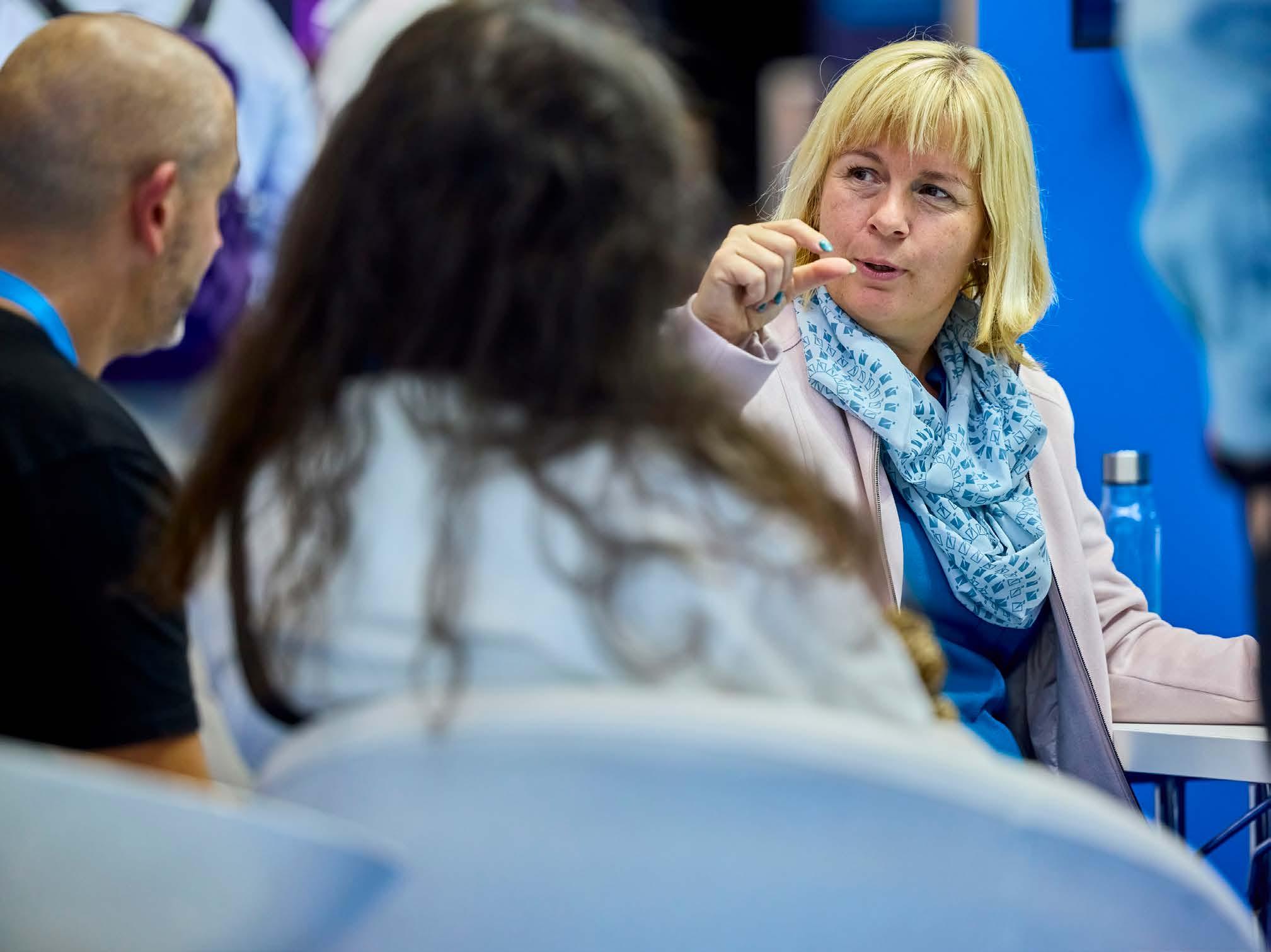
and at the time of publication, a limited number of exhibition stands were still available – but going fast! (book online).
RMS Chief Executive Sali Davis said: “We would like to thank all our exhibitors and sponsors for supporting IMC21. The exhibition is very much going to be at the heart of this year’s Congress, and thanks to the response from so many companies, our visitors are sure to have plenty to explore.”
A full list of companies and other exhibitors taking part in the IMC21 Exhibition can be found here
IMC21 is being held at Liverpool Experience Campus (the new name for ACC Liverpool), a purpose-built arena and convention centre in the heart of Liverpool on the King’s Dock.
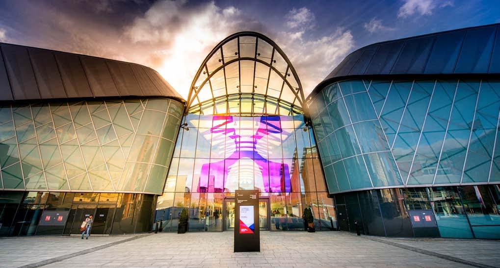
Liverpool Experience Campus (LEX) has great transport links as well as being a short distance from a large number of hotels, restaurants, shops and bars. The full address is King’s Dock, Port of Liverpool, Kings Dock Street, Liverpool L3 4FP, UK.
Voted best UK Convention Centre seven times, the venue boasts a modern, integrated design which ensures that the conference and exhibition space, plus all associated facilities, are within very close proximity for delegates to have a great experience at IMC21.
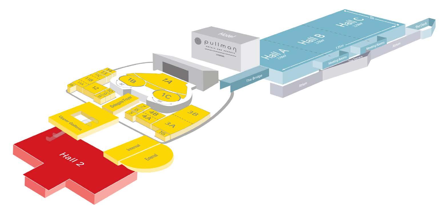
The congress will be taking place in Hall 2 and the meeting rooms (coloured yellow in the building plan on the opposite page); the exhibition will fill Halls A, B and C (shown in blue).
The LEX’s central location in Liverpool means everything is within walking distance, however, the venue is also well served by great public transport links and motorway networks. The city’s main railway station (Lime Street) is just a 20-minute walk or a five- minute taxi ride away. The venue boasts three on-site hotels and an array of amenities close by.
There will certainly be no shortage of networking opportunities at IMC21, and a number of official
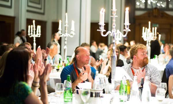
social events have also been organised to enable delegates to connect, relax and unwind in an informal atmosphere.
Here’s what to look forward to:
The IFSM Young Scientist Assembly (YSA/ ECR) brings together 50 early-career scientists recognised for their achievements and the quality of their IMC21 abstract submissions. The YSA/ECR programme includes an excursion and dinner on Saturday 29 August 2026, followed by a full-day meeting on Sunday 30 August 2026.
Network with colleagues from around the world and introduce yourself to the wider world of microscopy at the welcome reception.
The gala dinner will be taking place inside the conference centre and will include a drinks reception and entertainment. This is always a memorable highlight of IMC, providing the perfect opportunity to catch up with friends and colleagues in an informal setting.
You can add a gala dinner ticket to your booking when registering online - or you can update an existing booking by logging back into your online account. Spaces at the gala dinner are limited, so early booking is advised to avoid disappointment.

“The city of Liverpool is rich in culture and history. It’s a very affordable and safe location for international travellers and everything is within walking distance of the conference centre.”- Nigel Browning
Since being named the European Capital of Culture in 2008, Liverpool has regenerated into a worldclass destination with a dynamic reputation.
The city is an iconic and exciting events destination rich in heritage, culture, music and sport. A walkable, compact city, Liverpool offers delegates a unique opportunity to immerse themselves in a welcoming, vibrant and diverse atmosphere.
The city is centrally located in the UK, and with a population of 1.38 million, it is the fifth-largest city in the country. Liverpool was voted the UK’s friendliest city by readers of Condé Nast and placed in Rough Guide’s top three cities in the world to visit. Liverpudlians are known for their warm welcome and their great sense of humour!
Liverpool City Region has more than enough to appeal to delegates looking for culture, sports, culinary experiences, the outdoors, music and the arts. Congress delegates will be able to book tours through an award-winning travel and tour operator. From day trips within the city to exploring further afield, a tour is a brilliant way to discover more about Liverpool’s unique culture and heritage.
Many notable attractions are also within a 15-minute walk of the convention centre. These include: the Grade 1-listed Royal Albert Dock, home to Tate
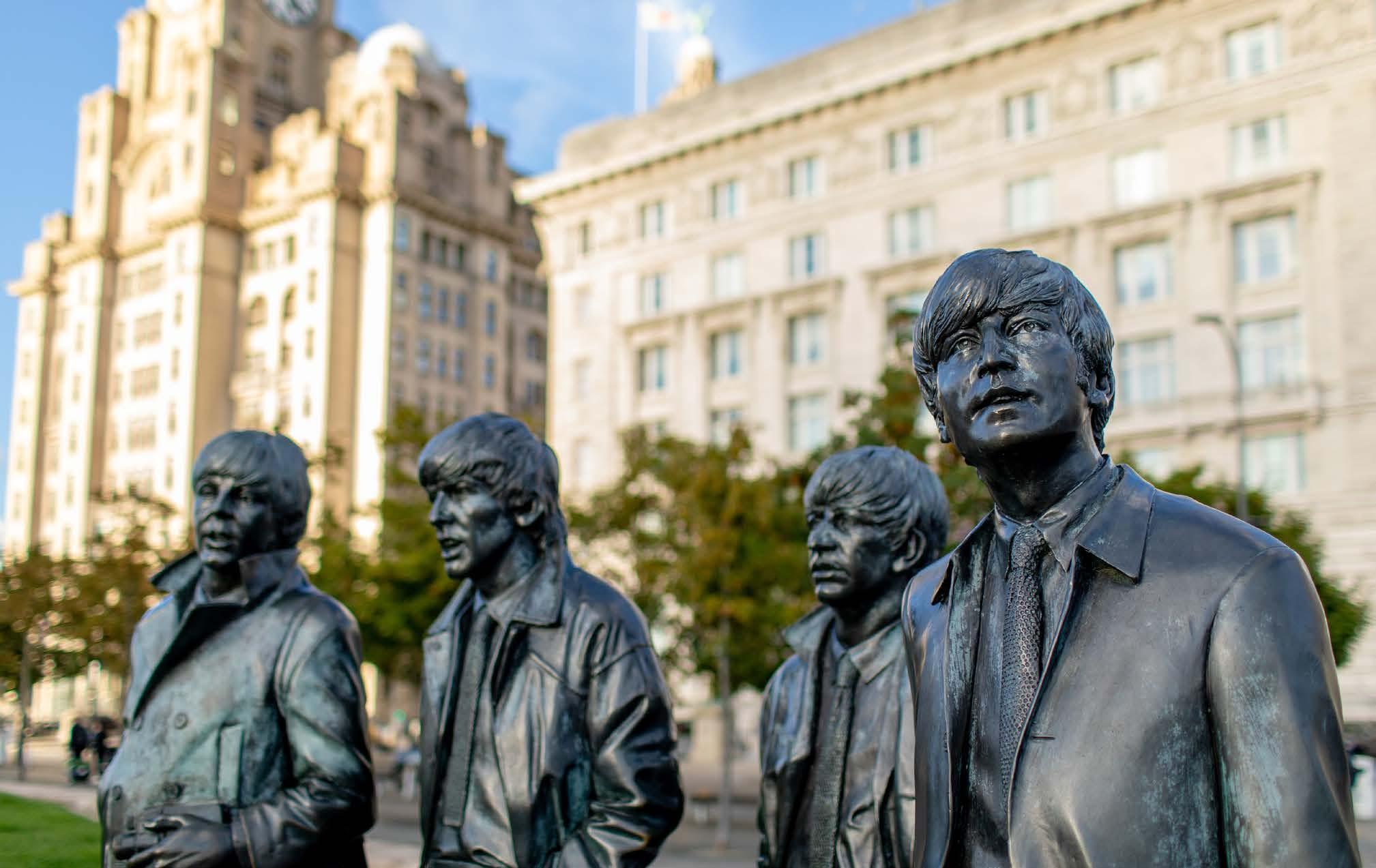

Liverpool, The Beatles Story, Liverpool ONE shopping centre and the famous Mersey ferry terminal. A diverse range of cafes and restaurants are also close by.
With two international airports within a 45-minute drive and high-speed rail service from London in just over two hours, it has also never been easier to get to Liverpool.
Liverpool John Lennon Airport features low-cost and charter flights to and from domestic, European and other international destinations. Alternatively, Manchester Airport, just 45 minutes away, brings even more international destinations within reach.
The city is very well served by the rail and bus networks, and a taxi from Liverpool John Lennon Airport to the conference centre takes around 20 minutes. A taxi from Liverpool Lime Street, the city’s main rail station, takes just five minutes.
Visit our International Delegates page for
further information on transport and travel, visa requirements and registration.
A limited number of bursaries are still available for those attending IMC21, including:
Congress Bursaries – Available to those who have submitted an IMC21 Congress abstract. As a limited bursary fund, awards will be made on a firstcome-first-served basis. IMC21 bursaries are open to both RMS members and non-members.
Care and Support Grants - We are pleased to announce that we are piloting a Care and Support Grant scheme for IMC21.These grants are designed to help with the additional caring or childcare costs that are incurred when trying to attend a conference.
Exhibition Day Visitor Bursaries - Although it is free to attend the IMC21 exhibition, the Royal Microscopical Society understand that funding your travel to events can be difficult. For this reason, the RMS are offering a number of day visitor travel
bursaries of up to £50 to help those planning to attend the IMC21 exhibition.
Find out more and apply on the bursaries webpage
You’ve booked your ticket, so now it’s time to get your accommodation sorted!
A range of great hotels are located within a short walk of the convention centre, and you can take advantage of the exclusive rates offered to congress delegates and exhibitors by booking online through Liverpool Convention Bureau
Liverpool Convention Bureau is our official IMC21 accommodation booking provider and we strongly advise you to book your accommodation through this service.
IMC21’s official headquarters hotel is Pullman Liverpool.
Keep updated in the lead-up to IMC21 by visiting our official website: www.imc.org.uk





We are very pleased to continue offering a range of ‘in-person’ and virtual events, in order to maximise accessibility and provide opportunities to those who might not otherwise be able to attend.
The following information was correct at the time infocus went to print but could potentially be subject to change in the coming weeks. Please visit our event calendar at www.rms.org.uk for the latest updates.
If you have any questions about a booking you have already made for an event, or need any help or advice, please contact us at info@rms.org.uk
7 – 12 Signaling by Adhesion Receptors GRC 2026, (RMS sponsored event), New Hampshire, USA
18 Thermo Fisher Scientific - Accessing cryo-EM services with Instruct-ERIC , Online (RMS-hosted event)
23 – 25 AFM & SPM 2026, Leeds, UK
30 June – 1 July Light Microscopy Summer School 2026, York, UK POSTPONED
2 – 3
Getting the most from your Confocal Course 2026, York, UK POSTPONED
6 – 10 Flow Cytometry Course 2026, York, UK
7 – 8 Premier European meeting of the Society of Biomolecular Imaging and Informatics (SBI2), (RMS sponsored event), cambridge, UK
8 – 9 The Advanced Materials Show, (RMS Affiliate event) Birmingham, UK
12 – 16 Synergy of Science, Institute of Physics (RMS sponsored event), Glasgow, UK
2 – 6
Microscopy & Microanalysis, (RMS attending), Milwaukee, Wisconsin, USA
17 – 21 Workshop on Stereology, (RMS affiliate event), Bern, Switzerland
31 August – 4 September IMC21, Liverpool, UK
30 Microscopy: Advances, Innovation, Impact 2026 - incorporating the RMS AGM & Section AGMs, London, UK (and online)
12 BioImagingUK 2026 Meeting, (RMS attending), London, UK
November
12 flowcytometryUK 2026, Cambridge, UK
24 – 27 32nd Annual New Zealand Microscopy Conference, (RMS sponsored event), Wellington, New Zealand
3 – 4
Virtual European Flow Core Meeting 2026, Online
For further information on all these events, please visit our Event Calendar at www.rms.org.uk
Thermo Fisher Scientific - Accessing cryo-EM services with Instruct-ERIC 18 June, Online (RMS-hosted event)
Single particle analysis (SPA) has become one of the most powerful approaches in structural biology, helping researchers resolve macromolecular structures at near-atomic resolution and accelerate discovery across drug development, virology, neuroscience, cell biology, and beyond.
But while structural biology can accelerate your research and provide unique molecular insights, accessing the technologies required can be a very significant barrier to access for new or occasional users. Instruct-ERIC, the panEuropean research infrastructure, resolves this issue by offering funded access to cutting-edge
6 - 10 July, York UK
This Flow Cytometry Course is aimed at both clinical applications and applications in cell biology, with the common fundamentals covered on days one and two. The course then splits into clinical applications and applications in cell biology streams, from practical demonstrations to lectures highlighting not just the applications, but best practise as well.
The course is constructed as a set of three modules. You can elect to attend the course from between two to five days, depending on the
structural biology tools.
In this webinar, we will detail how Instruct-ERIC can help you add structural biology techniques to your research toolkit.
Learn how to access cryo-EM and sample preparation services across Europe with Instruct-ERIC
Discover from Lefteris Zarkadas, facility manager at IBS, Grenoble how an Instruct-ERIC cryo-EM experiment runs
Hear the user’s perspective from José María de Pereda, CSIC, Salamanca about their experiences with Instruct-ERIC
Gain the knowledge and confidence to start planning your next - or first - adventure in structural biology.
modules selected.
The modules consist of lectures interspersed with sessions in the laboratory. It is anticipated that instruments from two manufacturers will be available for practical work.
This course is open to all and is suitable for those who are relatively new to flow cytometry and who wish to expand their experience with applications and specific analysis.
A trade exhibition will be held at this event on Wednesday 8 July, if you are interested in exhibiting please contact Jess Cole.
Microscopy & Microanalysis 2026
2 - 6 August, Milwaukee, Wisconsin, USA (RMS attending)
The M&M 2026 conference is the largest scientific meeting and gathering of microscopy and microanalysis professionals, academics, technicians, students and exhibitors in the world. Microscopy & Microanalysis will provide a forum for the presentation and discussion of a wide range of microscopy and microanalysis techniques and their application to the biological and physical sciences.
Microscopy: Advances, Innovation, Impact 2026 - incorporating the RMS AGM & Section AGMs
30 September, London, UK (and online)
The RMS Annual General Meetings (AGMs) will take place on Wednesday 30 September
The Royal Microscopical Society (RMS) will be attending this event to promote our wide ranging activities in support of microscopy, imaging and flow cytometry. We are grateful for the opportunity to support this community event and look forward to discussing the benefits of RMS membership with attendees.
Please visit our stand to find out more about the RMS, and discover what we have to offer both new and existing members – and the scientific community as a whole!
2026. This will be a hybrid event.
This meeting will be the 8th one-day meeting in the Microscopy: Advances, Innovation, Impact series, having previously taken place in 2016, 2018, 2020, 2021, 2022 and 2024
29th March – 1st April 2026
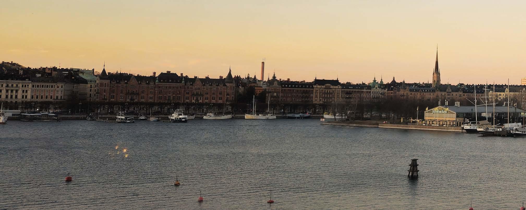
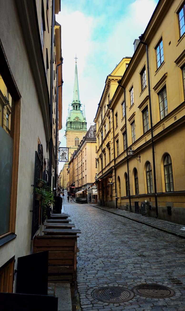
Ascientific conference in Stockholm is special. No, not because of ABBA, but because the city is the home of the Nobel Prize. I spent an afternoon perusing the objects donated to the Nobel Prize Museum by laureates, where optics and microscopy are very well represented. A highlight was Osamu Shimomura’s fishing net he used to catch the jellyfish from which green fluorescent protein (GFP) was isolated. I also came across Stefan Hell’s copy of the book ‘The Quantum Theory of Light’, a slightly surreal moment having attended his keynote presentation just the day before.
Stefan Hell delivered a particularly disruptive talk, unveiling a new “inverse” approach to super-resolution microscopy in which imaging is performed using a focused light field with a central diffraction minimum rather than a maximum. He boldly claimed that the method was not limited by diffraction and would outperform any existing detection strategy. FRET, he said, was “dead”. The remarks might have been designed to ruffle feathers, but the creativity was unmistakable, and history tells us that many transformative ideas were met with scepticism at first airing. Time will tell.
Stefan was not the only highlight and Focus on Microscopy continues to be a forum not only for optical developments but also for advances in image analysis. In 2026, this inevitably included the growing incorporation of artificial intelligence. I particularly enjoyed the keynote lecture from Carolina Wählby on image processing and AI to map tissue organisation, as well as several dedicated parallel sessions covering tools enabling image segmentation, object tracking, and feature classification. It was also motivating to hear about the major investment the Wallenberg Foundation is making in Sweden’s
bioimaging research community.
This year, I was joined at Focus on Microscopy by my colleagues from the bioimaging and biophotonics community at Guy’s Campus, King’s College London – all of whom presented in different capacities. This provided an opportunity to showcase the breadth of a long-standing collaboration between the labs of Simon Ameer-Beg, Simon Poland, and Siân Culley. The work presented spanned fast fluorescence lifetime imaging (FLIM), tomographic FLIM, and dual view light sheet microscopy.
I presented a poster on a new AI methodology that enables transfer learning to be applied to the typically limited datasets found in biological and medical research. We hope this will lower the barrier to using deep learning for tasks such as identifying drug-resistant cancer cells. Although this was my first time presenting on a computational topic, I received great feedback and had plenty of stimulating discussions during the busy poster session and afterwards.
Our work fitted well with emerging themes from the programme this year, which included a push toward
extracting more information beyond intensity, alongside continued efforts to image faster and at larger scales. For example, Gail McConnell’s group at Strathclyde are combining FRET with mesoscale microscopy to enable functional imaging at the millimetre scale. There was also a trend towards embedding AI into imaging pipelines and addressing the practical limitations of its deployment.
The conference concluded with a gala dinner at the museum of modern art, offering panoramic views across the island-city of Stockholm. I had the pleasure of sharing a table with Carolina Wählby, Hans Gerritsen (scientific programme head of Focus on Microscopy), and a mix of post-doctoral
researchers and company representatives – none of us from the same country. For me, this perfectly captured the diverse and welcoming nature of the conference, bringing together people of different backgrounds and career stages.
This year’s organisers, Ilaria Testa and Francesca Pennacchietti from KTH Royal Institute of Technology, did a fantastic job in coordinating this edition of Focus on Microscopy. And, as ever, appreciation goes to Mariska Timmers who tirelessly orchestrates the conference every year. Without Mariska, and the yearly committees, this conference would not be possible.
Focus on Microscopy alternates between Europe and Asia, helping to maintain diversity in the delegation and foster a truly global community of imaging scientists. Next year’s meeting will take place in another special location: Osaka, Japan. As a global centre of optical manufacturing – home to companies such as Nikon, Olympus, and Hamamatsu – Japan has long been a pillar of the microscopy community, and I’m sure this rich history will be reflected in the exhibition. For anyone needing a reason to attend the conference for the first time, a trip to Osaka may well be it.
My thanks go to the Royal Microscopical Society for their financial support in attending this conference. The continued support of societies such as the RMS makes it possible for students and early career researchers to engage with a wider scientific community and gain experience presenting on an international platform.
Tommy Pallett
Postdoctoral Research Fellow
Comprehensive Cancer Centre, King’s College London, UK
We are delighted to announce the latest winners of the RMS Section Awards, celebrating outstanding scientific achievements in the field of microscopy.
These prestigious awards are bestowed every two years by each of the Society’s Scientific Committees, covering all areas of microscopy, imaging and flow cytometry.
RMS President Peter O’Toole said: “As always, it is a privilege to announce our latest award-winners, who have all made outstanding contributions to their various scientific fields.
“Each winner has been chosen by one of our Scientific Committees - or ‘Sections’ - which represent all branches of microscopy, imaging and flow cytometry. As such, these awards are about receiving recognition from others working in the same field - which means an enormous amount for the recipients.
“The nominations were of exceptional quality and our committees certainly had a difficult job making their final decisions. My warmest congratulations go to all our winners.”
The latest winners are as follows:
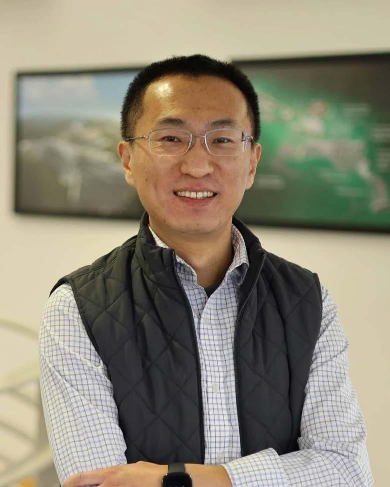
Dr Yongtao Liu - Center for Nanophase Materials Science at Oak Ridge National Laboratory, TN, USA
Yongtao, who is currently based at the Center for Nanophase Materials Science at Oak Ridge National Laboratory, has made major advances in AI/MLenabled automated and autonomous atomic force microscopy, fundamentally reshaping how AFM experiments are conceived and executed.
His work has transformed AFM from a predominantly manual characterisation technique into an AI/MLpowered, hypothesis-driven, theory-in-the-loop scientific instrument capable of autonomously uncovering new nanoscale physical phenomena.
Yongtao has introduced new experimental paradigms in AFM, enabling AFM experiments to adapt in real time, dynamically selecting measurement locations, modes, and parameters in response to emerging physical insight rather than relying on predefined experimental scripts. This represents a significant methodological advance in AFM.
His work has culminated in the development of
AEcroscopy, a software–hardware framework that enables automated and autonomous AFM experimentation with Agentic AI–enabled control. AEcroscopy does not merely increase experimental throughput; rather, it redefines the role of AFM as an active participant in scientific discovery.
Beyond individual scientific contributions,Yongtao is also an internationally recognised leader within the AFM community. His publications are widely cited and have become reference points for researchers developing next-generation AFM methodologies, helping to establish best practice in automated and machine-learning-enabled microscopy.
He has given extensive invited presentations, including keynote talks, at major international conferences and institutions, and has contributed significantly to organizing symposia and workshops in advanced and autonomous microscopy.
Through the mentorship of postdoctoral researchers, visiting scholars, and students, he has also made a significant contribution to the training of the next generation of scanning probe microscopists.
Dr Ignacio Arganda-Carreras - University of the Basque Country (UPV/EHU), Biofisika Institute, DIPC and Ikerbasque
For many years, Ignacio has been at the forefront of developments in Artificial Intelligence (AI) applications to bioimaging. As one of the founders of Fiji, a globally renowned open-source image processing package, he has made huge contributions to the bio-image analysis community. His research lab specialises in image processing and machine learning, with a primary focus on developing opensource computer vision methods tailored for biomedical images.
Ignacio has trained numerous researchers in
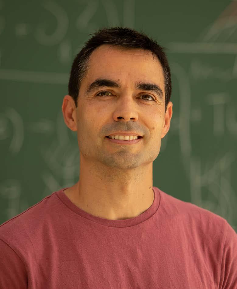
the application of deep and machine learning to bioimage analysis, and has created several Fiji plugins that have been extensively used by the community, and still serve as a solid build for other software developers.
He has more than 87,200 citations for his research work, classifying him as first worldwide for the “bioimage analysis” tag.
Ignacio is an Ikerbasque Research Associate Professor affiliated with the Department of Computer Science and Artificial Intelligence at UPV/ EHU, located in Donostia-San Sebastian, Spain. He is also associated with the Donostia International Physics Center (DIPC) and the Biofisika Institute.
Dr Laura Niermann - Technische Universität Berlin
As an independent early-career researcher at the Technische Universität Berlin, Laura is a central figure in the emerging field of computational microscopy. She has made
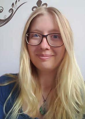
significant contributions to the research areas of dynamical diffraction and 4D-STEM, specifically regarding the reconstruction of 3D strain fields.
Laura’s research addresses a key challenge in electron microscopy: extracting depth-resolved strain information. So far, information along the beam direction has been inaccessible due to the tensorial nature of the strain field. She has established that this information is encoded in dynamical diffraction data, and that 3D strain fields can therefore be investigated from 4D-STEM datasets acquired from a single orientation of the specimen. In her latest work,Laura achieved a major breakthrough by demonstrating the reconstruction of a strain field along the beam direction. She accomplished this by the numerical inversion of strain-induced dynamical diffraction effects from experimental data. To further advance her research, the German Research Foundation (DFG) recently awarded her funding to establish her own Emmy Noether Junior Research Group.
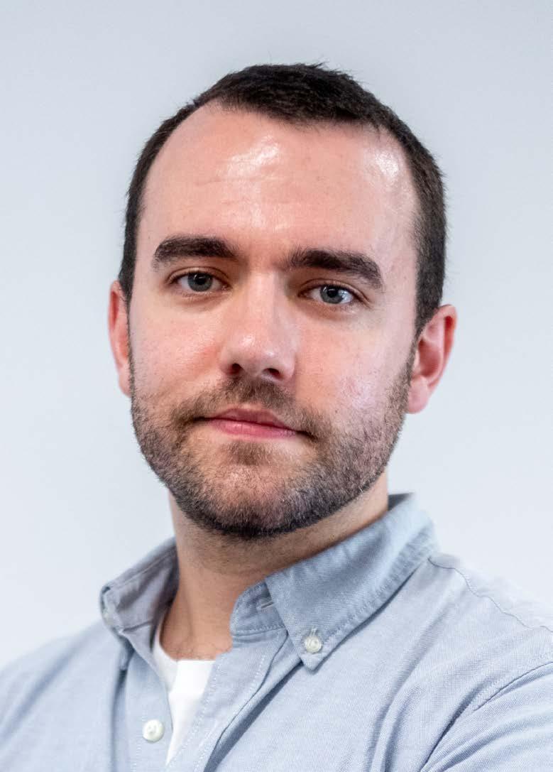
Dr Adam Tyson – University College London
Adam is Head Research Engineer and Head of the Neuroinformatics Unit at the Sainsbury Wellcome Centre for Neural Circuits and Behaviour and Gatsby Computational Neuroscience Unit, University College London, UK
He is the co-founder and lead of the BrainGlobe Initiative, and has made significant contributions to the field of neuroscience.
The BrainGlobe Initiative is an ecosystem of tools that help neurobiologists, microscopists, and imaging scientists to understand their data in the accurate neuroanatomical context. In recent years, digital high-resolution 3D brain atlases have been produced for several model organisms providing an invaluable resource for the research community.
Effective use of these atlases depends on the availability of an application programming interface (API) that enables researchers to develop software to access and query atlas data. However, while
some atlases come with an API, these are generally specific for individual atlases, and this hinders the development and adoption of open-source neuroanatomy software. The BrainGlobe Atlas API overcomes this problem by providing a common interface for programmers to download and process data across a variety of model organisms.
By adopting the Atlas API, software can then be developed agnostic to the atlas, increasing adoption and interoperability of packages in neuroscience and enabling direct integration of different experimental modalities and even comparisons across model organisms.
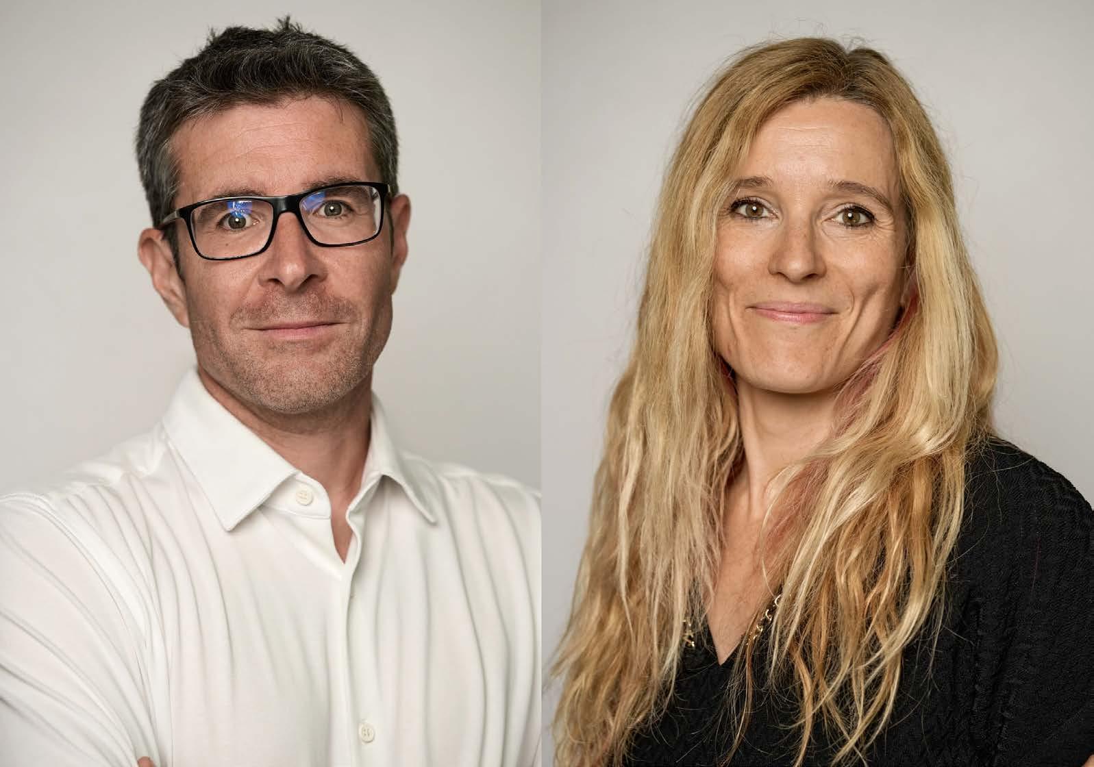
Prof. Paul Guichard and Dr. Virginie Hamel – University of Geneva
Together, Paul and Virginie have made significant contributions to the development, validation,
application, and broad dissemination of Ultrastructure Expansion Microscopy (U-ExM) and its derivatives, including cryo-ExM and iU-ExM.
As co-leaders of the CentrioleLab at the University of Geneva, their work has focused on extending the capabilities of light microscopy to resolve cellular architecture such as centrioles at the nanoscale.
A major objective of their research has been to bridge the longstanding resolution gap between fluorescence light microscopy and electron microscopy in light of understanding organelle architecture. They were among the first to recognise that expansion microscopy could be adapted to faithfully preserve cellular ultrastructure, enabling nanoscale imaging using conventional light microscopes.
They have applied and refined U-ExM across a broad range of biological systems, including human cells1,4–6, parasites such as Toxoplasma, Trypanosoma, and Plasmodium7–9, yeast10, plankton11, and diverse tissues including retina12 and glioblastoma samples in collaboration with
clinicians13.These studies illustrated how expansion microscopy can uncover ultrastructural principles that were previously accessible only by electron microscopy,
Building on this foundation, they developed cryoExM, combining cryo-fixation with expansion microscopy via established freeze-substitution workflows used in electron microscopy14. This approach significantly improves structural preservation and minimizes fixation-induced artefacts, demonstrating that high-resolution fluorescence imaging can be achieved alongside near-native ultrastructural preservation.
Dr Demie Kepaptsoglou – University of York
We are very pleased to announce Dr Demie Kepaptsoglou as winner of the RMS Award for Innovation in Applied Microscopy for Engineering, Physical and Material Sciences (EPMS).
Demie is a Senior Lecturer in the School of Physics, Engineering and Technology at the University of York, and also the Deputy Director of SuperSTEM, the Engineering and Physical Sciences Research Council (EPSRC) National Research Facility for Advanced Electron Microscopy. She has made outstanding contributions to physics and materials science using electron microscopy.
In the last few years Demie has created an entirely new field of research in the form of magnon spectroscopy in the scanning transmission electron microscope. She launched this new direction in the midst of the COVID-19 pandemic through an ambitious EPSRC New Horizons project, with the aim of developing electron energy loss spectroscopy (EELS) at ultrahigh energy resolution (<10 meV) to detect and spatially resolve these quasiparticles
describing the collective excitations of the spin structure of magnetic solids.
Demie has spearheaded collaborations in theoretical and computational work, successfully making use of the Royal Society’s International Exchanges scheme to establish work with Prof. Ján Rusz’s group at Uppsala University in Sweden. This approach led to now-seminal theoretical papers.
She has worked exhaustively to develop acquisition and data processing procedures to produce convincing spectral peaks as well as dispersion curves matching calculated response characteristics and even demonstrated the potential for spatially resolved mapping.
Demie’s collaborative work spans a vast range of other applications across chemistry, physics, engineering materials science, and earth science. Remarkably, she has accomplished these feats while also teaching at York, supervising PhDs, lecturing at international training schools, leading the SuperSTEM Daresbury Lab open day outreach activities (5,000 site visitors), and organising symposia and conferences.
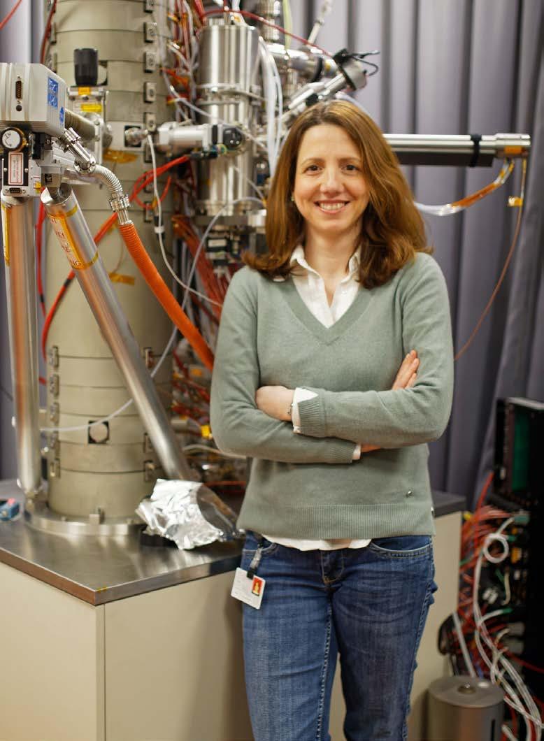
We are very pleased to announce Dr Oliver Burton as winner of the RMS Flow Cytometry Award.
Oliver has been a senior scientist at the University of Cambridge since 2023, assisting with research in the Liston-Dooley lab on topics such as regulatory T cell biology, neuroinflammation and T cell trafficking to tissues. In recent years, he has focused on technical development for the lab, sharing technical knowhow and tips for cytometry via an informal blog.
Flow cytometry has been central to Oliver’s research programme. He has consistently applied high-parameter cytometry approaches to dissect complex immune populations, enabling detailed characterisation of T cell subsets, regulatory networks, and tissue-resident immune cells. His studies have combined rigorous experimental design with advanced multiparameter analysis, contributing to high-quality, reproducible datasets that have informed both basic immunology and translational research.
Oliver has contributed to the wider adoption of robust cytometry practices within his research environment. His work exemplifies how flow cytometry can be effectively integrated with complementary approaches, including in vivo models and molecular immunology, to address biologically meaningful questions. Through his publications and collaborations, he has helped demonstrate the value of cytometry as a core tool in modern immunological research.
Oliver is recognised as a collaborative scientist who actively engages with multidisciplinary teams. His work has contributed to several high-impact immunology studies, and he continues to support the development of early-career researchers
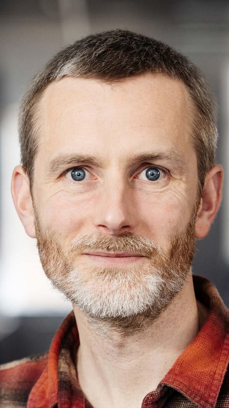
and cytometrists through supervision, talks and tutorials with the flow cytometry community.
While his primary identity is as an immunologist rather than a technology developer, Oliver represents an important category of cytometry users whose work drives the field forward through application, innovation in experimental design, and biological discovery.
The RMS is very pleased to announce Dr Melina Schuh as the latest recipient of the Pearse Prize – a prestigious international honour in the field of histochemistry and life sciences.
Melina is a ground-breaking biochemist known for her studies on how chromosome segregation errors occur during the meiotic divisions of mammalian eggs, and what causes the age-related decline in female fertility.
She is currently Director of the Max Planck Institute for Multidisciplinary Sciences in Göttingen, Germany, where she heads the Department of Meiosis. Her laboratory has developed a new method for acute degradation of endogenous proteins called Trim-Away. She has also revealed the cause of spindle instability in human oocytes, and discovered how mRNAs and proteins are stored in oocytes for the early embryo.
Melina is co-founder of Ovo Labs, a biotech startup developing therapies to combat egg-quality decline
errors in human oocytes. She is also an honorary professor at the University of Göttingen.
Melina obtained her PhD in 2008 at the University of Heidelberg and the European Molecular Biology Laboratory (EMBL), where she established methods for high-resolution microscopy of live mouse oocytes in the group of Jan Ellenberg. In 2009 she became a group leader at the MRC Laboratory of Molecular Biology (LMB), where her group carried out the first studies of meiosis in live human oocytes and developed strategies for high-content screens for meiotic genes in mammals.
Melina is a member of the Leopoldina and the EMBO and has received multiple awards and accolades, including the Leibniz Prize, the EMBO Gold Medal, and the European Young Investigator Award.

The Pearse Prize was established by the RMS Histochemistry and Cytochemistry Section (now Life Sciences) in 1982 to honour the contribution made to histochemistry by Professor AGE Pearse. The prize is awarded to a scientist who has made a significant contribution to histochemistry and life sciences and is still active in their field.
It is typically awarded every four or five years (and not more frequently than every two years) and only when it is felt there is a suitable candidate. It is generally regarded as one of the international honours in histochemistry and the life sciences, with an emphasis on microscopy.
Melina Schuh.
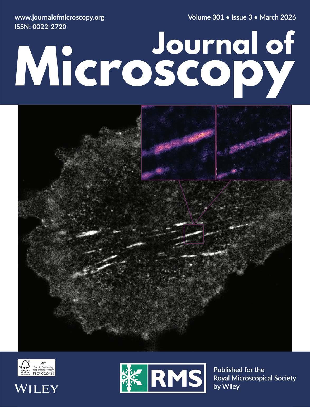
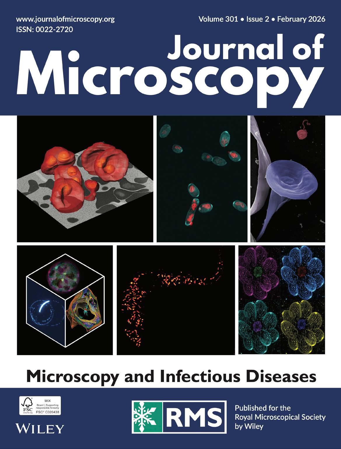
The Journal of Microscopy publishes top quality research articles, review articles and Hot Topic papers covering all aspects of microscopy and analysis. This includes cutting-edge technology and innovative applications in physics, chemistry, material and biological sciences.
You can read the latest Early View papers online at www.journalofmicroscopy.org
They include:
ORIGINAL ARTICLE
Global survey of Flow Cytometry core facilities 2021
Jessica B Back, Alexander Scott, Zhenxi Guo, Masilamani Elangovan, Kirk J. Czymmek, Peter O’Toole, Derek Davies
Two surveys were conducted of international core facilities that focused on Microscopy and Flow Cytometry for an in-depth snapshot of the landscape. Here we focus on the Flow Cytometry survey which had 148 respondents across ‘The Americas’, ‘Asia Pacific’ and ‘Europe and Africa’. A variety of subjects were covered, including instrument types and quantities, staffing, fees and funding, and specific questions related to COVID response. Broadly, the global trends were similar but there were some geographical differences notably in staff to machine ratio and funding models. As this serves as a baseline for longitudinal studies, we will be able to use these data to follow trends across all metrics in the future as facilities and technologies evolve.
Closer to the native state: Sandwich freezing and freeze-substitution of microorganisms, cultured cells, and animal and plant tissues for electron microscopy
Masashi Yamaguchi, Katsuyuki Uematsu, Toshikatsu Kuramoto, Shinichi Ohno, Hiroji Chibana, Azusa TakahashiNakaguchi
Since the invention of the electron microscope, morphologists have developed numerous techniques to visualise the native ultrastructure of biological cells and tissues. Although conventional chemical fixation followed by room temperature dehydration remains widely used, it often fails to preserve native structures, leading to distortion and leakage of cellular components. In contrast, cryofixation combined with freeze substitution offers a superior alternative, producing micrographs that more accurately reflect native ultrastructure. We have developed a ‘sandwich freezing method’ for rapid cryofixation, in which small biological
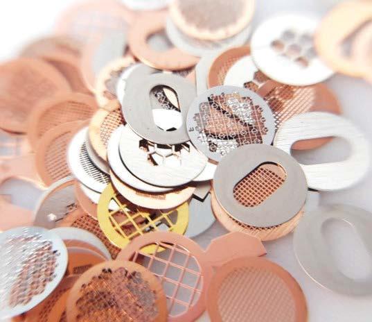
specimens are placed between two copper disks, rapidly frozen by plunging into melting propane cooled with liquid nitrogen, then freeze-substituted in acetone containing osmium tetroxide at low temperature, and finally embedded in epoxy resin. This technique yields exquisitely preserved, nearnative ultrastructure in living microorganisms and plant tissues. For cultured cells and animal tissues, prefixation with glutaraldehyde prior to sandwich freezing results in even more nativelike preservation compared to specimens frozen without fixation. We propose that glutaraldehyde prefixation prevents the immediate distortion and ultrastructural changes that occur upon removal of cells and tissues from their culture medium or host organism. Importantly, glutaraldehyde rapidly immobilises cellular components without altering cellular architecture.
We recommend the routine application of the sandwich freezing–freeze substitution technique to achieve ultrastructural preservation that is as close to the native state as possible in electron microscopy.
ORIGINAL ARTICLEOpen access
Making the invisible visible: A global examination of careers and recognition for Imaging Scientists in core facilities
Kerry Thompson, Johanna
Bischof, Yara Reis, Constadina Arvanitis, Olubunmi A. Balogun, Claire M. Brown, Mariana De Niz, Marcela
Díaz, Lize Engelbrecht, Eunice FabianMorales, Elnaz Fazeli, Georgina Fletcher, Adan Guerrero, Michelle S. Itano, Eleanor Kable, Gregory T.
Kitten, Shinya Komoto, Xiaoxiao Ma, Peter John O’Toole, Andreia Pinto, Cora Noemi Pollak, Clara Prats, Joanna W.
Pylvänäinen, James Douglas Riches, Jean Salamero, Virginia Silio, Stefan Terjung , Sophie Winter, Graham Daniel Wright, Lisa Yen, Antje Keppler
Imaging facilities underpin a growing share of modern life-science research, yet the career conditions and scholarly recognition of the Imaging Scientists in core facilities who design, deliver, and
interpret imaging research remain uneven. This mismatch has previously been shown to risk loss of expertise, reduced service quality, and weaken longterm sustainability of shared research capabilities.
To provide an evidence base for practical change, we synthesise findings from two complementary international community surveys run by Global BioImaging: a ‘Top 5’ survey on career development and job conditions (>290 responses spanning 43 countries) and an ‘authorship and acknowledgement’ survey examining how publication credit is assigned for imaging-related contributions of core facility staff (>330 responses spanning 43 countries). Across both surveys, respondents consistently link limited recognition to constrained career prospects. The career survey highlights recurring bottlenecks around progression routes, job stability and attractiveness, access to professional development, and the day-to-day consequences of being perceived primarily as service providers rather than scientific partners. The authorship and acknowledgement survey documents substantial variability in credit practices across facilities, disciplines, and regions, with many respondents reporting uncertainty about expectations, difficulty initiating credit discussions, and concerns about bias and power dynamics.Taken together, the results suggest a reinforcing cycle: inconsistent credit reduces visibility and leverage for Imaging Scientists in core facilities, which in turn entrenches fragile career pathways and inhibits retention.
We translate these findings into targeted, stakeholder-specific actions. Recommendations focus on defining career paths that match facility roles, aligning evaluation with the full range of facility outputs (including methods, data, software,
and training), and ensuring that institutions, funders, publishers, and research teams share responsibility for fair and consistent credit.
Jackson Rodrigues, Manikanth Karnati, Gagan Raju, Edwin Pious, Ankur Gogoi, Guan-Yu Zhuo, Nirmal Mazumder, Fu-Jen Kao
Collagen remodelling and dysregulation are the hallmarks of diverse pathological conditions. In this context, second-harmonic generation (SHG) imaging has emerged as a powerful label-free modality for assessing collagen. This offers submicron resolution, intrinsic optical sectioning, and deeper imaging capabilities without the need for exogenous agents. Additionally, polarisation-resolved SHG (P-SHG) further enhances orientation-sensitive information on collagen architecture. This enables the elucidation of subtle structural alterations that are often invisible to conventional imaging techniques. This nonlinear technique has demonstrated utility in diagnosing diseases, particularly in assessing tumour progression, where collagen remodelling is correlated with disease severity and prognosis. Furthermore, quantitative image analysis, using metrics such as fibre density, orientation, anisotropy, and alignment, provides objective measures of collagen remodelling. P-SHG complements these analyses by enabling the retrieval of molecular susceptibility ratios (e.g. χ33/χ31), degrees of
polarisation (DOLP), and orientation distribution functions, yielding more profound insights into the fibrillar ultrastructure and molecular organisation. These quantitative descriptors are being increasingly integrated into clinically relevant feature extraction methods. The incorporation of these features in artificial intelligence (AI) methods has enhanced SHG-based pathological assessment. AI-driven models can automatically classify tissues, detect pathological patterns, and correlate the collagen microstructure with clinical outcomes, addressing the bottleneck of manual interpretation and enhancing reproducibility. Future perspectives highlight the integration of SHG and P-SHG with deep-tissue multiphoton imaging,
accompanied by explainable AI/ML-driven quantification, which will improve diagnostic precision, reproducibility, and clinical adoption. Ultimately, the convergence of optical innovation, deep learning, and translational engineering will lead to the establishment of collagen pathology assessment through SHG and P-SHG as reliable, accessible tools for diverse clinical applications.
Jiayi Chen, Yue Sun, Liujia Tian, Yiling Huang , Xuemei Song , Fan Peng , Wei Zheng , Yi Zeng
This study investigates the f-ratio quantitative method applicable to Cold Field Emission Scanning Electron Microscope/Energy Dispersive X-Ray Spectroscopy (CFE-SEM/EDS) systems. To address the issue of cumbersome calculations arising from the reliance on real-time simulation in traditional f-ratio methods, this study proposes an efficient strategy of ‘preset curve + ZAF correction’. This method employs pre-constructed ‘f-ratio vs. concentration’ models for individual elements derived from standard samples to substitute the
traditional real-time simulation of the unknown sample, and introduces ZAF factors to effectively compensate for matrix effects, thereby achieving quantitative analysis of unknown samples with matrix differences. An iterative approach was implemented to resolve the challenge of determining the absorption parameter (x), which arises due to the unknown concentration of the unknown sample. F-ratio models were established based on the P–In, As–Ga, S–Zn, Se–Cd, and S–Fe systems. Quantitative f-ratio analysis was subsequently performed on the binary system As–In and Se–Zn, as well as the ternary system S–As–Fe. The results demonstrate that the ZAF correction f-ratio method achieves satisfactory precision.
Open Access
Ann Wheeler, Dale A Moulding
Light microscopy core facilities are widely established in research institutions, providing access to advanced instrumentation with expert guidance. Centralising technology and expertise within these facilities improves research output, fosters collaboration between research groups and reduces costs through resource consolidation. As core facilities proliferate, national and international networks have emerged that facilitate global knowledge exchange. Here we examine the benefits of establishing local networks of light microscopy facility staff. These small networks complement their larger, formal counterparts by creating a sense of community and offering practical day-to-day
benefits, such as rapid technical support, resource sharing, and informal mentorship.
METHODS AND PROTOCOLSOpen Access
Sourena Azidhak, Douglas J. Taatjes, Ian R. Campbell, Eleftheria Roumeli, Linda S.
Schadler
Plastic pollution is impacting global ecosystem health. Bioplastics made from unprocessed biomass, including whole or fragmented tissues, are one potential solution. They are biodegradable and exhibit competitive mechanical performance. Seaweed and microalgae have gained popularity over the last decade as bioplastic feedstocks due to their abundance and ease of cultivation. To grasp the complexity of transforming biomatter into bioplastics and to optimise their performance, it is essential to develop processing–structure–property relationships. Transmission electron microscopy can be used to observe the morphology of bioplastics; however, current infiltration techniques are designed for biomaterials in their natural state, and harsh reagents can cause artefacts in bioplastic structure. This paper examines four different fixation methods for Spirulina bioplastics and investigates the impact of fixative, contrasting, dehydration, and resin embedding on Spirulina ultrastructure before and after bioplastic transformation. Our study suggests that the key to preserving bioplastic morphology is to prepare the specimen for electron microscopy by exposing it to gas-phase infiltration.
ORIGINAL ARTICLE - Open Access
Ilkka Miinalainen, Eija Jokitalo
As electron microscopes became more costly, technically complex, and integral to a wide range of scientific fields, centralised electron microscopy (EM) core facilities have become essential for maintaining accessibility, performance, and quality. Drawing on the experiences of running two EM core facilities in Finland, we outline key considerations for establishing and operating a biological and biomedical EM facility. We cover facility infrastructure and technical requirements, user training, sample preparation, safety and occupational considerations, personnel and cost management, and finish by outlining three areas, sustainability, research data management, and artificial intelligence, that we consider to be increasingly defining the strategic direction of EM facilities. By distinguishing the specific requirements of EM from those of light microscopy, this article provides practical guidance to support efficient, safe, and high-quality facility operations within institutional and national imaging networks.


1. No submissions fees
2. No page or colour charges
3. No page limit
4. Simple online submission
5. Helpful, friendly editorial team
6. Average time from submission to first decision is less than 50 days
7. High readership figures
8. Online tracking system – authors can easily check the status of an article in production and receive emails at key stages
9. Rapid publication with Early View papers published online in advance of print, significantly shortening time from acceptance to publication
10. Free electronic offprints

Search for Journal of Microscopy on the App Store or Google play and access your personal or institutional subscription wherever you are, whenever you want.
View the Guidelines for Authors and full submission details online at:














How a new national network aims to strengthen collaboration, visibility and opportunity across Welsh science.
By Dr Myfanwy Adams
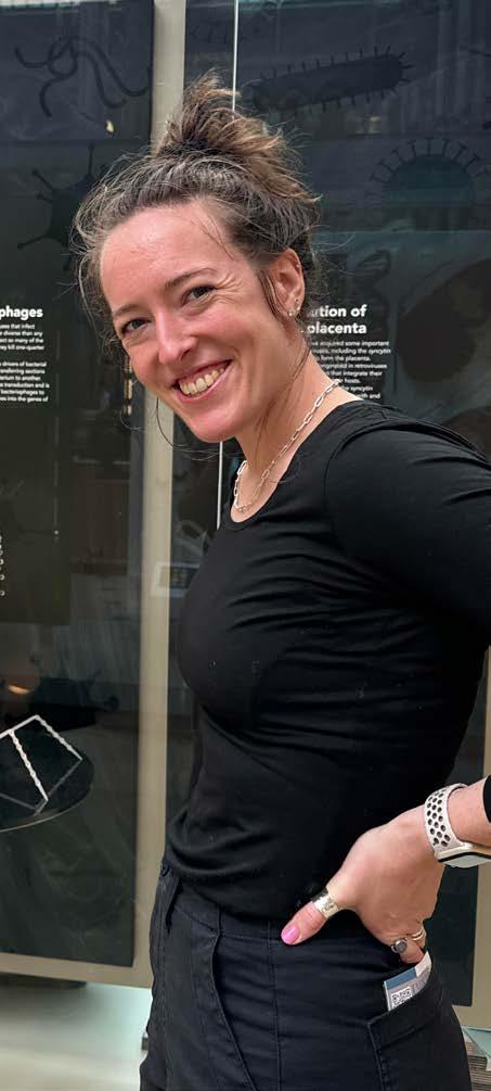
Dr Myfanwy Adams.

I do microscopy because I love being the first to see the unseen. Cryo-EM reveals atomic level details of proteins and helps us understand how these tiny molecular machines power every biological function in every single cell. I am a Welsh-American structural biologist and in 2022 I moved to the UK to start my postdoctoral research. Much like my proteins, finding the Welsh scientific community proved elusive but that is not due to a lack of STEM excellence. Wales is home to world-class research institutions, advanced imaging facilities, industrial innovation, expertise and ambition. It has people doing cutting-edge, technically demanding work however, these strengths have not always been easy to see, connect, coordinate or advocate for at a national level.
On May 19th 2026, this changed with the launch of the Welsh Microscopical Society (WMS) and I hope you enjoy reading the origin story in the following pages.

Microscopy is massively prevalent throughout every sector; enabling discovery across medicine, biotechnology, materials science, agriculture, environmental science, aerospace, engineering, heritage science and many other fields.The challenge is that these techniques are also infrastructureheavy, expertise-rich and often highly specialised, requiring highly skilled users and rapid adaptation to developing methodologies. Collaboration often depends not only on the availability of a microscope, but on knowing who has it, who can run it, and what it can do. In Wales, these questions are often difficult to answer due to a lack of visibility, coordination and connection.
Across Wales, researchers and technical specialists are already using microscopy to support highquality science. Cardiff, Swansea, Aberystwyth and Bangor, alongside science parks, incubator sites and industry-linked environments, all form part of a wider research and innovation ecosystem. But without a clear national network for microscopy, these activities can remain institutionally siloed, making it harder to advocate for infrastructure,
close skills gaps, sustain public engagement, share resources equitably and build opportunity across a geographically diverse nation.
Other parts of the microscopy community have shown the value of coordinated national and regional networks. The Royal Microscopical Society (RMS) provides a long-established professional
home for microscopists across the UK and internationally. The Microscopy Society of Ireland (MSI) and Scottish Microscopy Society (SMS) offer examples of regional and national communities that support connection, identity and shared activity. Until now, Wales has lacked an equivalent national network dedicated to microscopy and imaging.
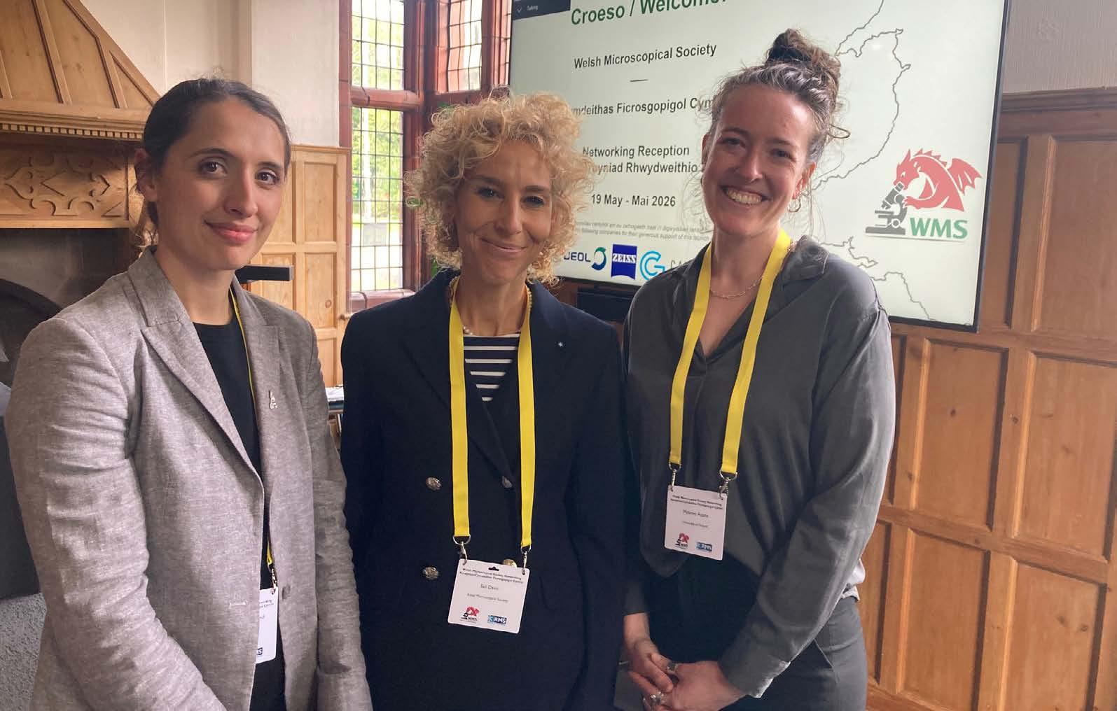
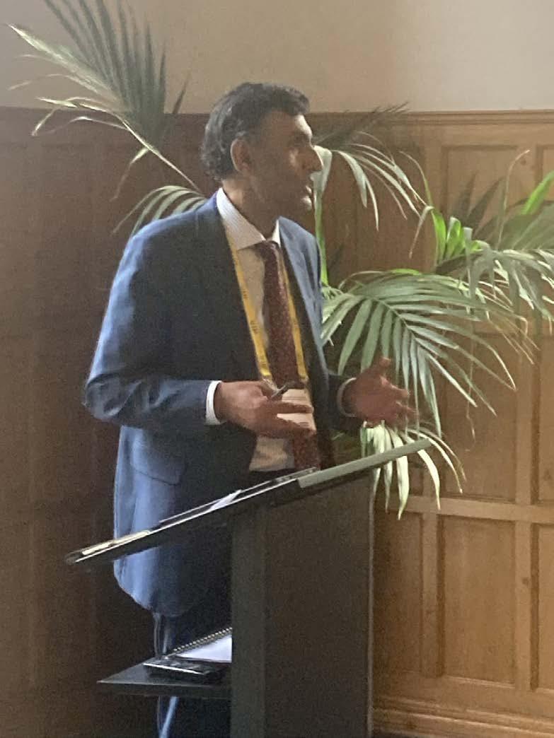
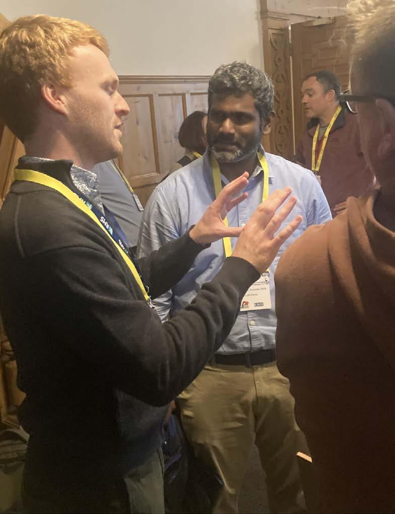
On 19 May 2026, in a packed room with drinks aplenty, all of this changed. Microscope users and enthusiasts, researchers, technicians, industry representatives, policy and public engagement professionals, educators, and supporters of Welsh science gathered at Cardiff University to launch the Welsh Microscopical Society (WMS).
Its aim is to connect people across Wales with an interest in microscopy and its applications, and to build a stronger, more coordinated community across research, industry, education and public engagement. The scope is deliberately broad, not intended only for one technique, discipline, or career stage; this initiative is intended to unite everyone across both sectors and geography. By doing so, the Society hopes to support collaboration, strengthen
advocacy, and help develop the infrastructure, visibility and long-term resilience needed to support research and development across the life sciences, physical sciences and wider STEM landscape in Wales.
This transition, from the unseen corners of Wales into a visible network, was mirrored in the society naming. Prior to this initiative, there was no exact translation in Welsh for ‘microscopical’. With enormous gratitude to Bangor University and Termau Cymru, a Welsh term, ‘Figrosgopical’, has now been created and added to the Welsh National Terminology Portal. In this way, the Society’s launch has not only brought people together around microscopy; it has also helped create language for a new national community.
Early conversations around the Society kept returning to practical questions. Where is the equipment? Who has the expertise? How can people find new collaborations? How can Wales make the case for investment in microscopy if it cannot first describe what it already has?
To move from ambition to action, WMS is built around four connected ideas: Knowledge, Access, Culture and Innovation.
Wales needs a clearer picture of its microscopy capabilities: which techniques are available, where facilities are based, what equipment can do, who the key contacts are, and which facilities are already working with external users or industry partners. A national facilities and expertise database would help researchers, students, companies, educators and collaborators understand the breadth of microscopy available across Wales. It would also provide an evidence base for future advocacy, funding bids and strategic planning. Knowledge makes capacity visible.
Visibility alone is not enough. WMS also aims to
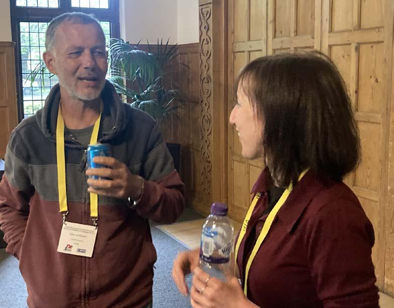
improve routes into microscopy expertise, training and infrastructure. This includes understanding funding needs, developing training initiatives, supporting more equitable access to equipment, and identifying shared priorities across the Welsh microscopy community. Researchers and technical specialists should not always have to look outside Wales, or travel to established research clusters elsewhere in the UK, to access equipment, training or expertise that could be supported closer to home.
Improving access also means recognising the realities of geography. A national community must
work for people across Wales, not only those located near major urban centres. Over time, WMS hopes to support visible national moments for Welsh microscopy, including meetings, roadshows, showcase events and conferences that help bring opportunities closer to different communities. Access opens routes into capability.
Science moves through relationships as much as equipment, and WMS aims to build the social infrastructure needed for microscopy in Wales to thrive. That means creating spaces where people can meet across disciplines, sectors, institutions and career stages; where technical specialists and researchers are shaping broad-reaching decisions; where industry, academia, education and public engagement can speak to one another; and where people who may not yet see themselves as part of a microscopy community are actively welcomed in.
The Society also offers a way to connect Welsh culture and scientific identity. By bringing people together through the shared medium of microscopy, WMS can help build a community that is both technically ambitious and deeply rooted in place.
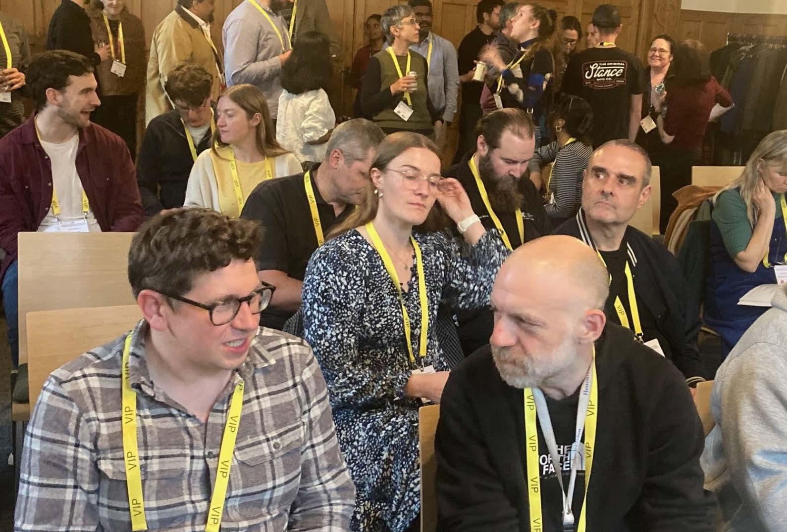
Culture builds trust, belonging and shared purpose.
Microscopy contributes directly to innovation in healthcare, biotechnology, materials, agriculture, engineering, manufacturing, environmental science and beyond. Wales has a significant opportunity to use microscopy not only as a research tool, but as a platform for economic development, industrial collaboration, skills growth and public value.
WMS aims to become a shared voice for this community: gathering evidence, identifying needs, advocating for investment, and helping explain why microscopy matters to the future of Wales. Longer term, this may include contributing to policy discussions, engaging with Senedd crossparty groups, and supporting conversations about how to attract research and development activity, companies and talent to Wales. Innovation explains why capability matters.
Together, the four themes form a practical plan. Knowledge makes capability visible. Access makes it usable. Culture makes it inclusive. Innovation makes it nationally significant.
The people: who made this possible?
The Welsh Microscopical Society has emerged from
a group of people with professional expertise in microscopy and strong personal or institutional connections to Wales.
Dr Myfanwy Adams has been one of the driving voices behind the initiative. A postdoctoral researcher at the University of Oxford specialising in structural biology and cryo-electron microscopy, she brings both technical expertise and over a decade of experience in outreach, teaching and engagement across multiple scientific disciplines. She has been a member of the Royal Microscopical Society since 2020 and currently serves on both the RMS Early Career Committee and the infocus magazine editorial board, giving her direct insight into the research culture challenges and opportunities facing early career researchers. For Myfanwy, WMS is about uniting the entirety of Wales across the ‘medium’ of microscopy to create a sustained platform for innovation, support and advocacy.
Sali Davis, Chief Executive of the RMS, has also played a central role. Born and raised in Cardiff and a Welsh speaker,she brings leadership experience from professional membership organisations and a clear understanding of how learned societies can support people across disciplines, sectors and career stages. Prior to joining the RMS, she was Chief Executive of Optometry Wales, a national membership body representing primary care optometrists. She has also worked at Deloitte and Touche LLP, where she led a practice review programme for Tax Partners, and previously held roles with the Royal College of General Practitioners. In addition, she serves as a lay panel member for Social Care Wales and as a mentor supporting new mothers returning to the workplace. Sali continues to split her time working between Wales and the RMS offices in Oxford. Her strong personal and professional connection to Wales underpins her commitment to supporting Welsh research communities. Within this project, Sali will play a strategic role in shaping the network’s direction, supporting the planning and delivery of activities, and ensuring alignment with wider
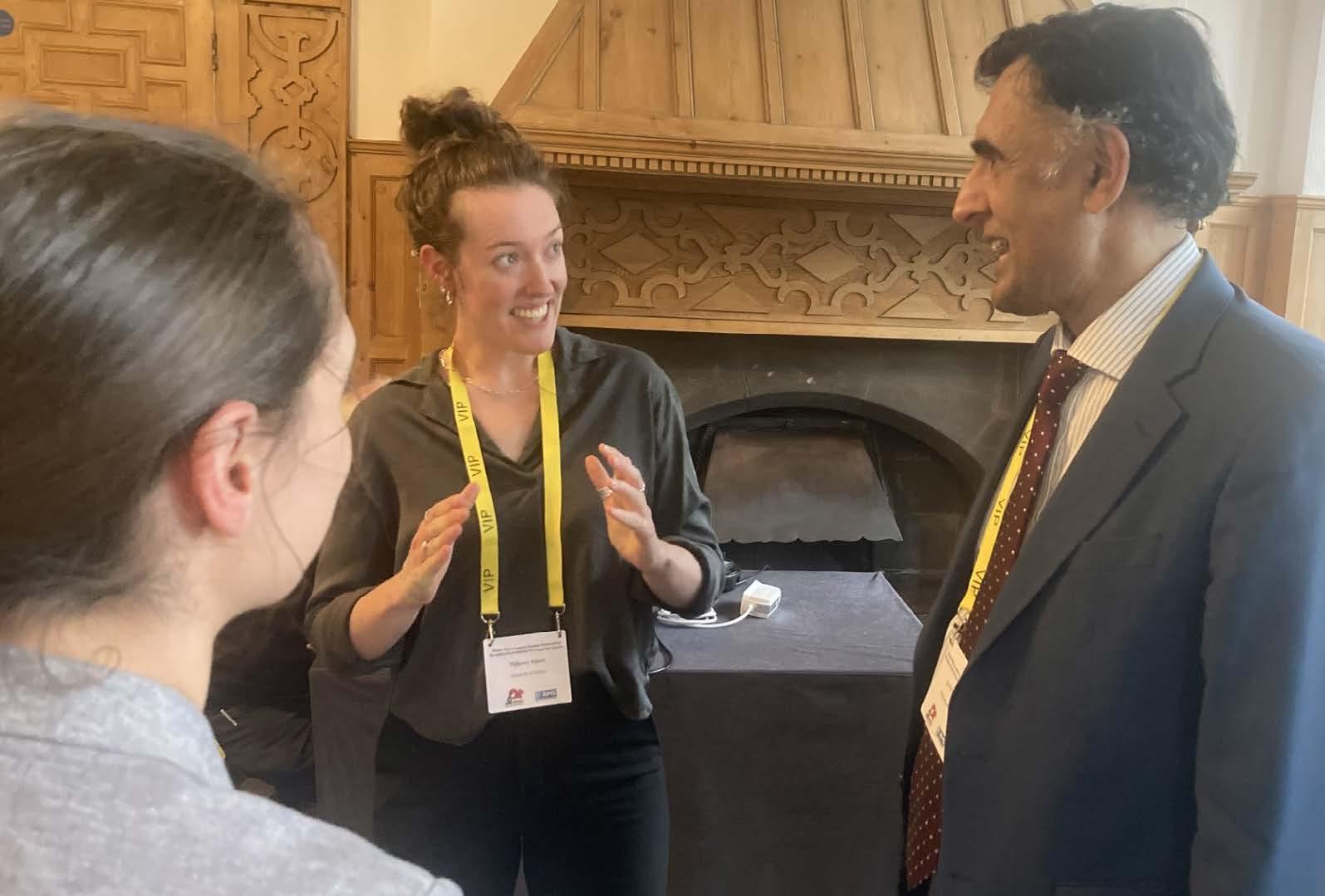
disciplinary and learned society initiatives, helping to embed the network within sustainable national and international structures.
Dr Ainhoa Dafis-Sagarmendi is a founding member of the WMS and a Key Account Manager at Nikon Healthcare UK for South Wales and the South West. Originally from Aberystwyth, she has over a decade’s experience in microscopy including cryo-electron tomography and electron crystallography as part of her PhD at the University of Sheffield, and a range of light microscopy techniques in her role at Nikon. During her PhD, she also held a position at the Unit of Electron Microscopy Solutions at Thermo Fisher Scientific (Eindhoven, the Netherlands).
Ainhoa works to strengthen the links between researchers and industry to foster collaboration and innovation in Wales. As part of her role as founding member of the WMS, she is dedicated to showcasing Welsh research and promoting its global impact.
Gwilym Hughes is an electron microscopist and applied scientist with over 20 years’ vocational experience of innovating, de-risking and
commercialising novel technologies across a broad range of industries (including MEMS semiconductors, agritech and food technology, fuel cells, cell-andgene therapy, drug delivery, and medical diagnostics).
This includes working as an applied science and technology consultant for almost 10 years, during which he procured, managed and operated an inhouse SEM capability.
Gwilym’s primary microscopical expertise covers SEM and TEM imaging, alongside EDS spectroscopy and FIB-SEM cross-sectioning / in-situ lift-out. He has also made extensive use of optical and metrological microscopy, including the use of fluorescence imaging to invent and patent a novel bioburden monitoring cassette for use within medical and healthcare settings. Gwilym is passionate about bringing his applied microscopy and imaging experience to bear in promoting Welsh microscopy, identifying new opportunities for industry collaboration and research funding, and making microscopy more accessible for all within Wales.
This launch event builds off the back of numerous attempts, countless discussions and shared longing for such a network. Having backing from the four
major institutions has been pivotal to determine what equipment is housed at each of the respective institutions, what is needed and ideas about how the society can tangibly help within the scientific community here in Wales. The wider founding cohort includes contributors from key Welsh research environments, including Chris Von Ruhland, facility lead for electron and light microscopy in Cardiff University, Lydia Powell, lecturer in microbiology and infectious disease at Swansea University, Susan Girdwood, senior teaching and research technician at Aberystwyth University and David Williams, senior lab technician at the Nuclear Futures Institute at Bangor University.
WMS is still in its infancy, and early support from sponsors and industry partners has been vital in helping launch the society with strong momentum. One of the first approaches came from Roy Edward, also a founding member of the society, recently retired after two decades with BioStatus and more than 43 years in the life sciences industry. BioStatus has strong links to De Montfort and Cardiff Universities, with key work on DRAQ5 carried out in the laboratories of the Tenovus Institute in Cardiff by the team of Professor Paul Smith and Professor Rachel Errington. BioStatus products have since been cited more than 14,000 times in peerreviewed journals and are distributed globally.
The Society has also received support from companies with a significant instrumentation footprint in Wales and across the UK, including Zeiss, JEOL, Gatan and Hitachi. Representatives from these organisations, many with Welsh connections themselves, have offered support, enthusiasm and long-term encouragement for the initiative.
This early backing highlights three important points. First, WMS must be cross-sector, wide-reaching and inclusive. Second, the Welsh scientific community extends beyond geography; people with ties to Wales continue to give their time, energy and expertise to support Welsh science. Third, the equipment, skills and expertise already present in
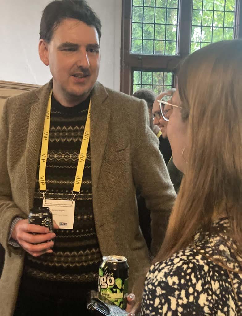
Wales are significant and deserve to be made visible.
The launch event was held alongside the RMS Introduction to Image Analysis workshop at Cardiff University, which meant there was already a concentration of microscopy expertise in the city. The evening reception widened that gathering to include the broader Welsh community.
Myfanwy opened the event by setting the scene, welcoming attendees and acknowledging the numerous people who had helped bring the Society to life. She spoke about the long-standing desire for a community of this kind and the years of conversations, attempts and shared hopes that had led to this moment. Sali then spoke about the founding story – how she and Myfanwy met one year ago at the Microscience Microscopy Congress (mmc) in Manchester and both wanted to turn ambition of STEM advocacy into structured delivery to make Wales a truly world-leading force in science and innovation. Ainhoa followed with an industry perspective, encouraging academics and technical specialists to look beyond their own institutions and consider the value of wider partnerships.
A highlight of the evening was an address from Professor Jas Pal Badyal FRS, Chief Scientific Adviser for Wales. Professor Badyal’s research has used microscopy creatively and he is internationally recognised for his pioneering research of solid surfaces and deposition of functional nanolayers. His remarks placed the Society’s ambitions in a broader Welsh science and policy context, underlining that microscopy has a role to play not only in research, but in national conversations about skills, infrastructure, innovation and economic development.
Endorsing the WMS, Professor Jas Pal Badyal said: “One of our strengths in Wales is that, because we are a relatively small nation, there is a real opportunity for close collaboration and increasing the visibility of the excellent work that is taking place here.
“Microscopy is at the absolute heart of science and contributes so much to society - for example through medicines, healthcare, computer chips and other products. Initiatives like the WMS have the potential to benefit the people of Wales, not just through research, but also by boosting industry and economic growth.”
The evening continued with lively discussion, new introductions and clear enthusiasm for the establishment of a national society. The launch showed that the appetite for connection already exists. WMS now offers a structure through which that energy can grow.
The Welsh Microscopical Society has launched with administrative support from the RMS, but the ambition is to become an independent, memberbased society. Inclusivity sits at the heart of this mission. Wales’ strength lies not only in the quality of its science, but in the relationships, identities and shared ambitions that connect people across its communities.WMS aims to make those connections visible, practical and sustainable.
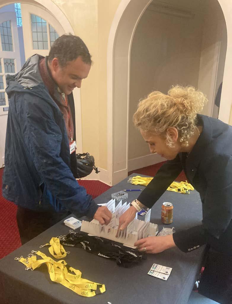
The immediate task is to convert enthusiasm into structure. Attendees at the launch, whether in person or online, will become part of a growing database of interested members. The next meeting will take place virtually on June 25th (email wms@ rms.org.uk if interested in attending), providing an opportunity to reflect on the launch, define the Society’s structure and values, and begin shaping its next steps. Future plans include debuting WMS at IMC21 in Liverpool at the end of August, developing a Welsh microscopy database by the end of the year, and exploring showcase activities including a proposed Welsh microscopy focus in the Journal of Microscopy and a Celtic session at mmc in the Summer of 2027.
Microscopy is, at its heart, about looking closely enough to see what was always there. The Welsh Microscopical Society begins from the same idea. Wales already has the people, expertise, infrastructure, ambition and community spirit needed to make microscopy a national strength. WMS offers a way to bring that strength into focus: visible, connected, inclusive and ready to contribute to the future of science, innovation and society in Wales.
The Royal Microscopical Society would like to welcome our new members who have joined us in the last three months. We hope they enjoy a long and rewarding membership with the RMS.
Dr Urška Jančič
Dr Kendra Leigh
Dr Ainsley Beaton
Dr Lourde Raj Irudayanathan
Ms Abbie Scholefield
Miss Felecia Sutton
If you know of anyone who might be interested in becoming a member of the Royal Microscopical Society and if you would like us to contact them, please send their details to our Membership Administrator, Debbie Hunt – debbie@rms.org.uk
Application forms are available to download at www.rms.org.uk/membership
Don't forget you can now log into the RMS website and check your membership status, renew and download receipts. If you have never logged into the RMS website, please enter the email address that is linked to your membership and then click 'forgotten password'.
If you have any queries or questions about your membership please contact Debbie Hunt debbie@rms.org.uk
Name A Nandakishore
Tell Us About You?
I am currently pursuing an MSc in Biotechnology at Christ University, with a strong academic focus on molecular biology, biochemistry, and advanced analytical techniques. I have developed a solid foundation in core areas including gene expression systems, CRISPR-Cas9 technology, and protein expression in microbial systems. Additionally, I am actively preparing for competitive examinations such as CSIR-NET, strengthening my theoretical and analytical skills in life sciences.
Why did you become a member of the RMS?
Through my membership of the Royal Microscopical Society, I aim to deepen my understanding of microscopy techniques and their applications biological research, particularly in pathogen detection and cellular analysis. I am keen to engage with the scientific community, stay updated with emerging imaging technologies, and contribute to interdisciplinary research.

Name Aszad Hossain Akib
Tell Us About You?
I am a Master’s student currently working on my thesis titled ‘Fabrication of bio incorporated nanostructured thin film for food spoilage detection’
Why did you become a member of the RMS?
My thesis contains a section on

Scanning Electron Microscopy, Transmission Electron Microscopy, and Field Emission Electron Microscopy. Currently, in my country, there is no available opportunity to study these technologies. I became a member of the RMS for having better understanding of these technologies.
How do you feel being an RMS member benefits you?
I am grateful for the opportunity and hope to connect with scientific professionals worldwide

Name Mahima Kerai
Tell Us About You?
I am a second-year Biochemistry undergraduate student at King's College London. I am an aspiring researcher and want to build a career exploring genetics, epigenetics, and molecular changes within cells. I am eager to keep up with everything the RMS is doing and read about what’s going on in and around the world of microscopy!
Name Preksha Hooda
Tell Us About You?
I am a PhD student in the School of Chemical Materials and Biological Engineering at University of Sheffield. My work focuses on looking at mRNA structure for rational therapeutic design, using AFM.
Why did you become a member of the RMS?
I became a member of the RMS because my PhD research relies on AFM to investigate mRNA structure for rational therapeutic design. The RMS provides a great community of microscopists, and membership gives me access to valuable resources, training opportunities, and
Name Sahaja Raj Kandiraju
Tell Us About You?
I am an MRes student in Biological and Biomedical Sciences with a specialisation in the Cancer Biology Pathway at the University of Liverpool, and I completed my undergraduate degree in B-Tech Biotechnology. My current research focuses on Wilms Tumour, using immunofluorescence and microscopy-based techniques to study tumour biology and the tumour microenvironment and I enjoy making sense of complex biology one image at a time.
Why did you become a member of the RMS?
I want to learn more and keep updated with all the wonderful stories and research being done to develop my knowledge and exercise my curiosity.
How do you feel being an RMS member benefits you?
I want to build a research career, and so keeping up with the latest developments through the RMS will help me build my understanding and grow my scientific knowledge.
networking with others working in microscopy-based research.
How do you feel being an RMS member benefits you?
Being an RMS member benefits me by keeping me connected to the wider microscopy community, which is invaluable as a PhD student developing my skills in AFM. Access to training resources, workshops, and conferences helps me advance my technical expertise, while networking with other microscopists exposes me to new techniques and ideas that can inform my own research into mRNA structure and therapeutic design.

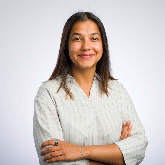

Why did you become a member of the RMS?
I became a member of the Royal Microscopical Society to deepen my understanding of microscopy beyond the lab bench, learn from experienced researchers, and stay connected with advances in imaging that shape how we study cancer biology.
How do you feel being an RMS member benefits you?
eing an RMS member benefits me by giving me access to expert-led training, up-to-date resources, and a network of researchers who share a strong interest in microscopy, supporting both my technical growth and research confidence.
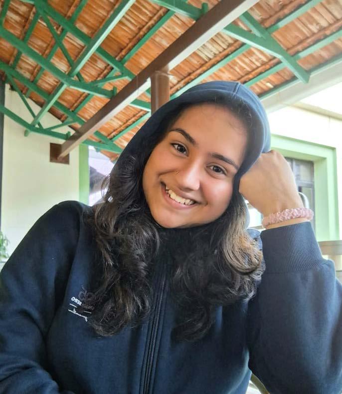
Tell Us About You?
I am a junior undergraduate at the Indian Institute of Science Education and Research, India, where the mechanistic thinking I have developed so far has crystallised my interest in cell biology. Beyond academics, I enjoy exploring new ideas and perspectives; in my free time, I like dancing, long walks, travelling, and cooking.
Why did you become a member of the RMS?
“It’s all very well to look at a dividing cell and going, ‘Gosh, this is marvellous’! but it doesn’t help you understand what’s going on behind the scenes.”
-Paul Nurse (Interview, December 2001). In my early twenties, there was a surge in public attention to cancer through campaigns and widespread media coverage, and once this struck close to home, it ignited a burning question: why does it happen, and what forces lead to cell loss over division? My upbringing and love for movies taught me to appreciate what we see, as it is often formed by layers of unseen organisation and control, which connects me to Paul Nurse's Nobel talk.
This curiosity compelled me to explore the unseen pathways of cell biology, where processes orchestrated at microscopic scales become visible through advanced imaging, revealing a precise cellular ballet that continues to shape my scientific interests. I highly value opportunities to engage with diverse scientific communities, as learning from different perspectives strengthens both my research approach and collaborative mindset.
Name Mezida Saeed
Tell Us About You?
I am an Advanced Imaging Specialist at the COMPARE Advanced Imaging Facility at the University of Birmingham. With a background in biology, my work focuses on advanced light microscopy, quantitative imaging, and imaging infrastructure development. I support superresolution, single-molecule and large-volume light-sheet imaging, with particular interests in quality control, training frameworks, and data stewardship as microscopy becomes increasingly data intensive.
Why did you become a member of the RMS?
I became a member of the RMS because it brings together a uniquely broad microscopy community, spanning biological, physical, and technical perspectives. As someone working in a core facility role, I value the RMS
as a space where methodological development, best practice, and training are taken seriously alongside biological discovery.
How do you feel being an RMS member benefits you?
Being an RMS member connects me to a community that is actively shaping the future of microscopy, particularly around quality, reproducibility, and training. It provides opportunities to engage with experts across disciplines, stay informed about new technologies, and contribute to discussions around standards and good practice.
The RMS is especially valuable to me as I work on developing imaging infrastructure within a facility context. The Society supports the exchange of ideas around calibration, data management, and education, which are increasingly critical as microscopy enters an era of large, complex datasets and integrated analysis pipelines. Membership helps me situate my work within a wider professional and scientific framework.
Name Muhammad Zeeshan Arshad
Tell Us About You?
I am a final-year Medical Laboratory Science student at The University of Faisalabad, Pakistan, with a passion for clinical diagnostics and research. My background includes clinical internships in Hematology, Microbiology, and Pathology at Allied Hospital and Alkhidmat Foundation. Beyond the lab, I am a published researcher in antimicrobial nanotechnology and have been recognized for my work in designing BSL-4 laboratory models. I am also an active member of the ASM and ASLM.
Why did you become a member of the RMS?
I joined the RMS to bridge the gap between academic theory and the rapidly evolving world of microscopy technology.As a student specializing in

Tell Us About You?
I am a first year PhD student within the Thermal Methods Research Unit at the University of Huddersfield, researching novel hyphenation and applications of thermomicroscopy, in collaboration with Linkam Scientific Instruments Ltd. I recently graduated from Huddersfield with my MSci in Forensic and Analytical Science, with academic interests spanning polarised light
molecular diagnostics and Immunohistochemistry (IHC), I believe that staying connected with a world-class society like the RMS is essential for mastering the high-resolution imaging techniques required for modern cancer biomarker testing and pathogen identification.
How do you feel being an RMS member benefits you?
Being a member provides me with a gateway to international standards in microscopy, specifically through the infocus magazine and technical user groups like Expansion Microscopy. It allows me to align my practical clinical experience with global best practices in quality management (ISO 15189).The networking opportunities and access to discounted professional events, such as the IMC21 Congress, are invaluable for my transition from a stuent to an international diagnostic professional.
thermomicroscopy, thermoanalytical chemistry, and forensic chemistry. Outside my academic life, I enjoy martial arts and reading.
Why did you become a member of the RMS?
I joined RMS to connect with an international community of microscopy enthusiasts and grow my network as a PhD student, as well as highlight the importance and utility of microscopical techniques in chemistry and thermal analysis. Additionally, with my forensic-based background and the fact that microscopy is a cornerstone technique in this field, an RMS membership is highly relevant, especially for the expansion of my knowledge and awareness of cutting-edge microscopical innovations.
How do you feel being an RMS member benefits you?
Tell Us About You?
I’m a student of BS MLT. I study in Faisalabad Medical university, Pakistan
Why did you become a member of the RMS?
I aim to utilise this platform to track funding opportunities, manage my investigator profile, and collaborate on institutional research proposals.
Access to the RMS will allow me to maintain a centralised research profile, ensuring my publication and funding history are accurately represented for future applications. It will allow me to contribute to the research community as a peer reviewer in my field

Name Aidan Gallagher

I am a Biomedical Science student in my third year, I also have a degree in History and Politics (1st Class) and I have a vested interested in pharmacology and drug interactions.
Why did you become a member of the RMS?
I became a member of the RMS as I work as a placement student with EMResolutions, who work on precise manufacturing of EM grids so I felt it only natural to become a member! I also wanted to participate or listen to lectures on the latest technologies.
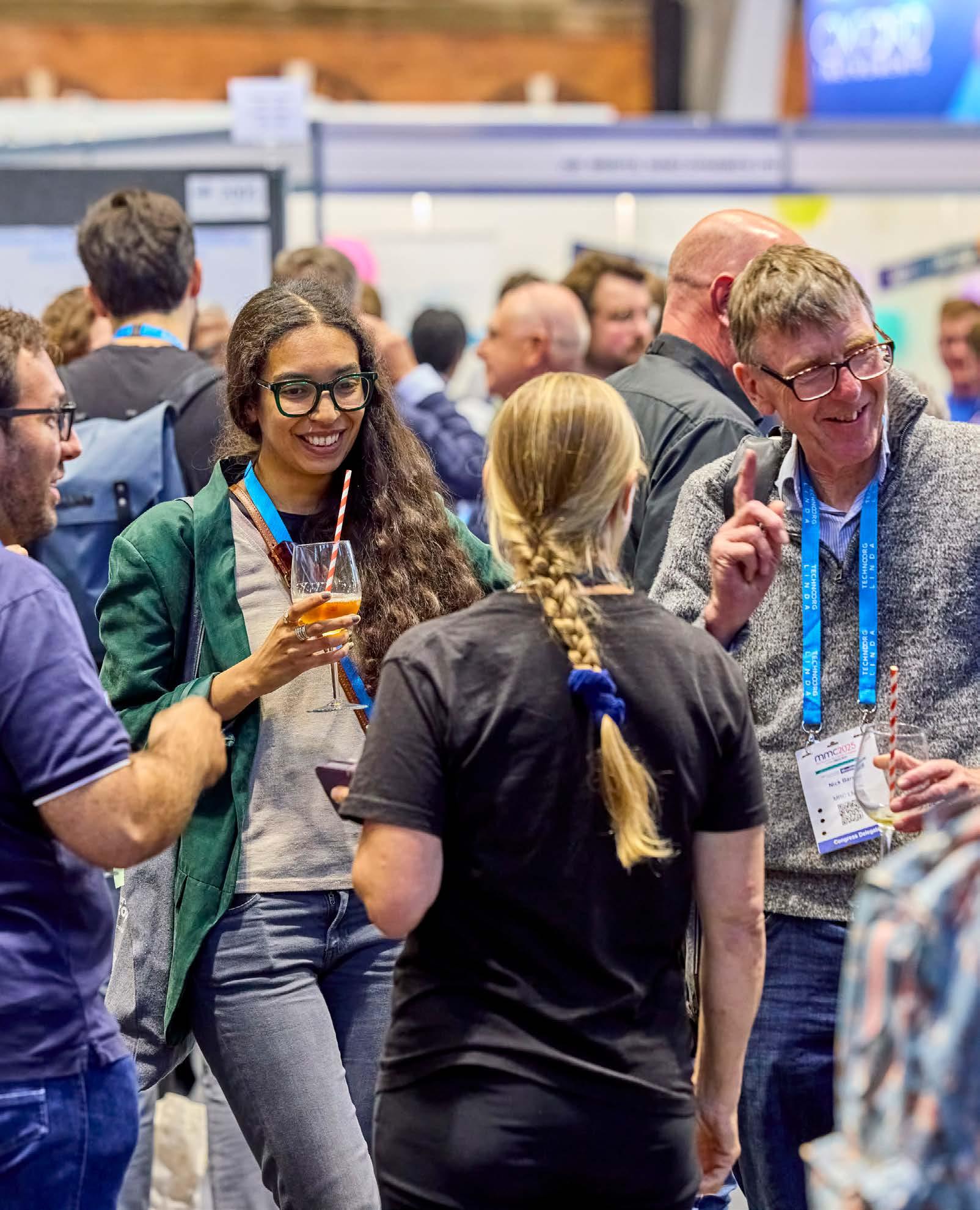

Lee R Bilson FLS FIIC
Head of Conservation at Recclesia and Co-Founder at Darwin Biological Microscopy and Wood Identification at the Royal Botanic Gardens, Kew
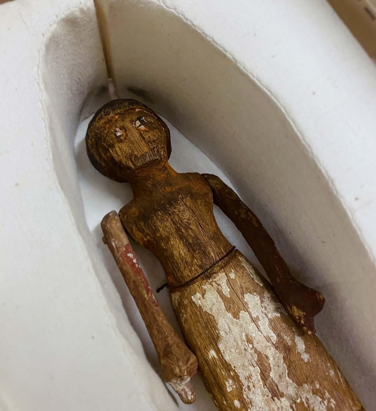
For those who work with microscopy, the study of wood reveals an intricate and often overlooked world of cellular complexity. In February, I had the privilege of attending the Wood Identification course at the Royal Botanic Gardens, Kew, an intensive, week-long programme dedicated to the microscopic analysis of wood anatomy. Led by Dr Peter Gasson and supported by a team of experienced specialists, the course provided a rare opportunity to develop advanced microscopy skills while working with one of the most extensive wood collections in the world.
The Jodrell Laboratory, where much of the course was based, has been a hub for botanical research at Kew since 1877. Originally founded to study plant physiology, it has since expanded into wood anatomy, forensic botany, and chemical wood identification, making it an ideal setting for this highly specialised course.
For RMS members, the true value of this course would lie in the hands-on experience with light
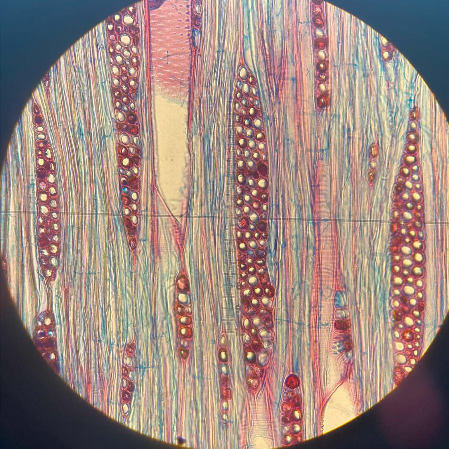
microscopy and the opportunity to analyse anatomical structures of wood at a microscopic level. Much of the week was spent refining the skills needed to distinguish hardwoods from softwoods, identify key diagnostic features such as vessel arrangements, ray structures, and axial parenchyma patterns, and understand the cellular variations that define different species.
We worked extensively with Kew’s vast microscope slide collection, which includes reference samples from around the world, allowing for detailed comparisons between species. The Banks Building wood collection, another invaluable resource, provided understanding of real-world specimens, including an ancient Egyptian figure of a boatman, carved from Ficus sycomorus (sycamore fig), demonstrating the role of wood anatomy in heritage conservation, archaeology, and material science.
A key addition to this year’s course was an introduction to Direct Analysis in Real Time (DART) mass spectrometry, a non-destructive chemical identification method that is transforming forensic wood analysis. While microscopy remains the gold standard for wood identification, the DART approach offers a complementary tool for verifying species by analysing their chemical fingerprint – where one might struggle the other might complement. This was a perfect example of cross disciplinary potentials: seeing how Kew employs both traditional and modern techniques reinforced the value of integrating microscopy, chemical analysis, and digital databases — an approach highly relevant to scientists and researchers in a range of sectors.
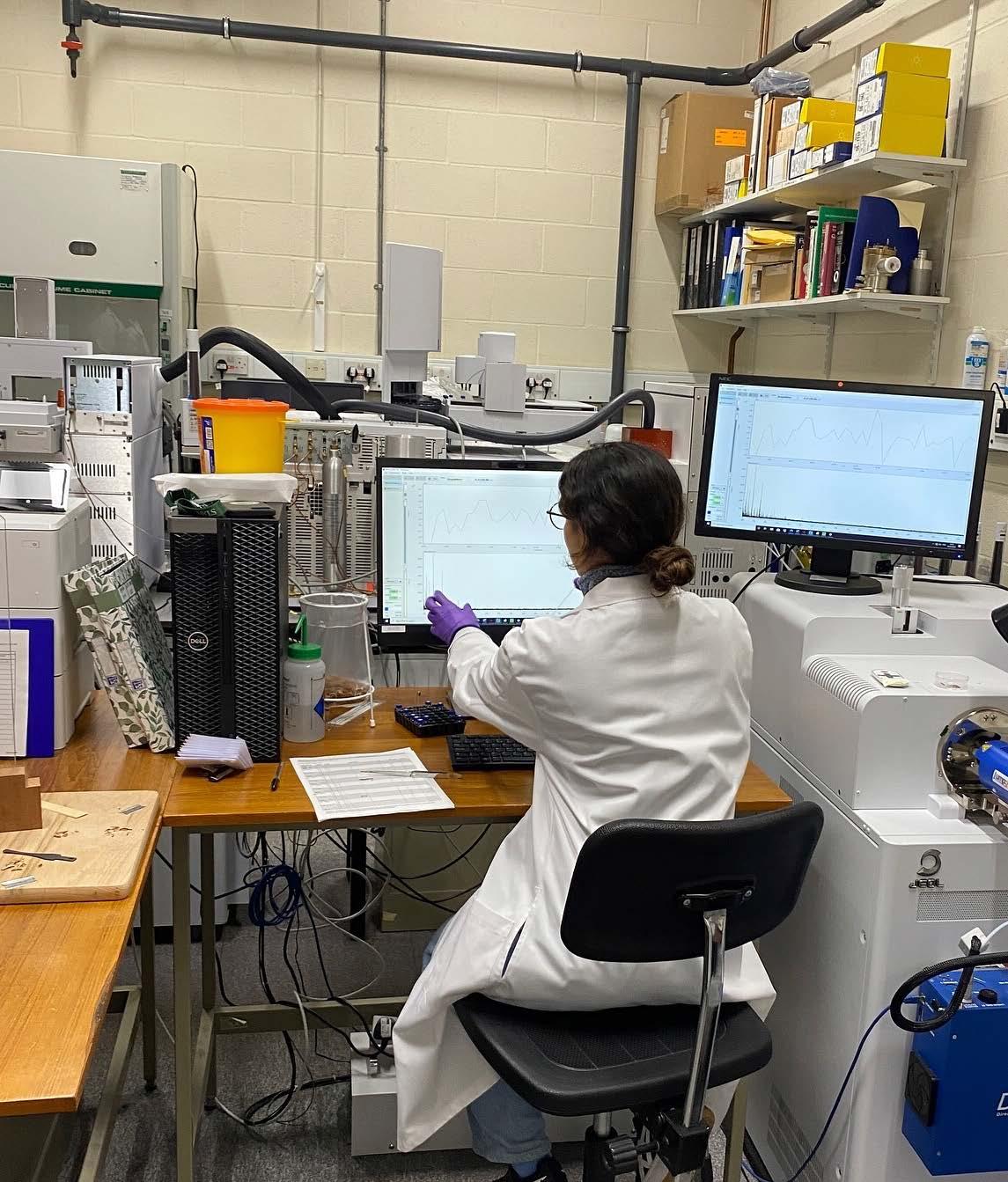
For those in the microscopy community, wood anatomy presents a rich and technically demanding area of study. The skills developed on this course could be directly applicable to many fields, from Forensic Science & Border Control, to Heritage Conservation & Archaeology, Environmental Science & Forestry, or even Material Science & Engineering. As a CPD week of study this was a very insightful and engaging course indeed.
Studying wood in the historic setting of Kew Gardens, surrounded by a globally significant collection of specimens, state of the art microscopy facilities, and fantastic botanic gardens, made this course a truly unique experience. The combination of expert tuition, extensive practical work, and exposure to cutting-edge analytical techniques
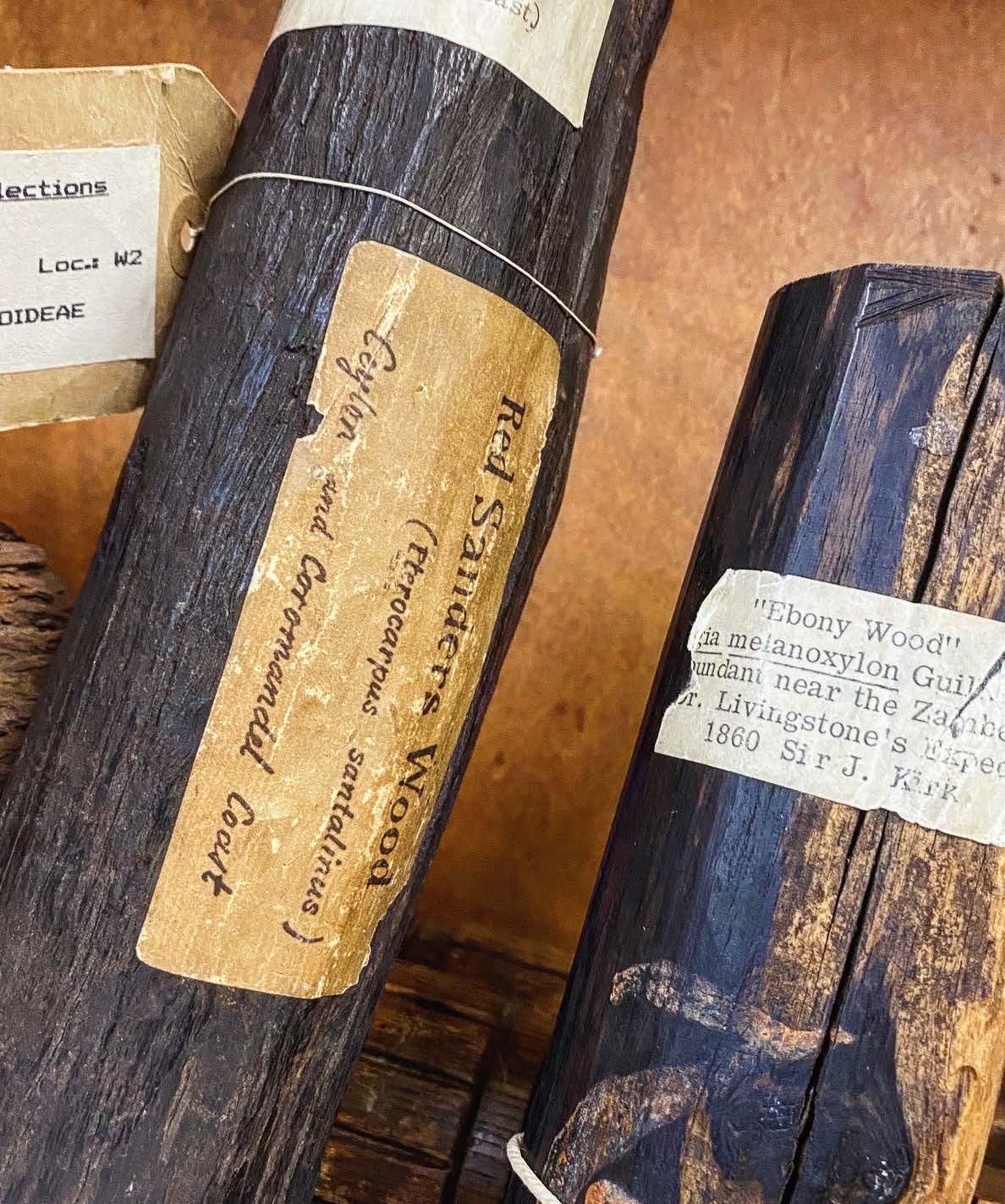
makes this an invaluable training opportunity for any RMS member interested in the intersection of microscopy, material science, and conservation.
For those looking to expand their microscopy expertise in wood anatomy, I would highly recommend the Wood Identification course at Kew — an experience that brings botanical research, forensic analysis, and heritage science together
under one microscope.
Useful links:
https://www.kew.org/science/training-andeducation/continuing-professional-development/ wood-identification
https://www.kew.org/science/training-andeducation/continuing-professional-development
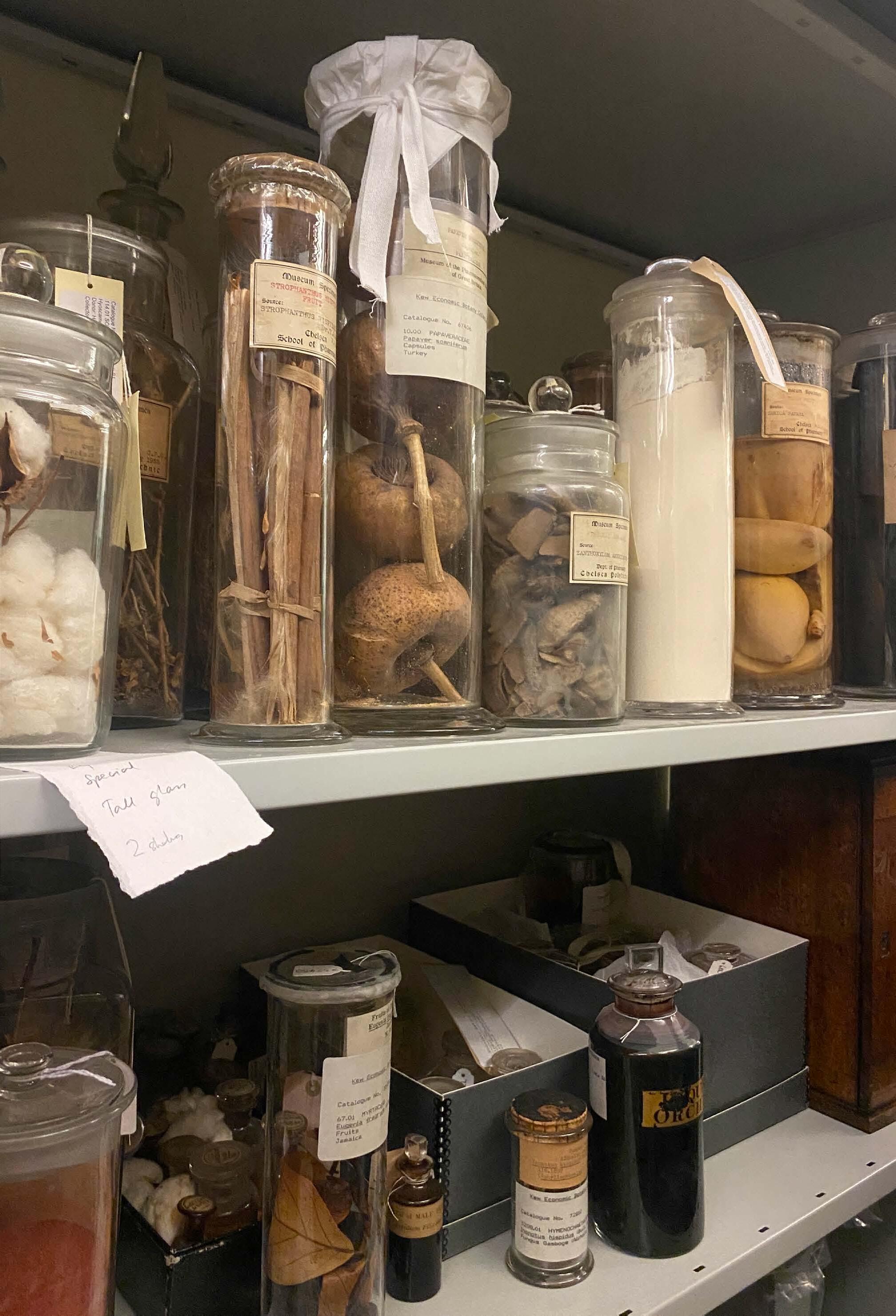
Dear readers,
It seems like very little time has passed since our March issue of infocus – and yet so much has been going on at the RMS!
As I sat down to write this piece, a very simple but powerful thought came into my head; it occurred to me what a wonderful thing it is to be part of the microscopy community.
These days it seems there is so much to worry about in the world – from international conflicts and economic shocks to climate fears and other crises –that we all need a good dose of positivity now and again. I personally find that in abundance both within the RMS and across the wider microscopy, imaging and cytometry communities.
It is perhaps something that is easy to take for granted, but it really is a privilege to be part of this thriving and friendly community, to see and hear about the achievements of others, and to celebrate all that is good about the role of microscopy in the

Over the Spring (or Autumn, for our Southern Hemisphere colleagues) we saw fantastic attendance at several RMS training courses and other events. These included ‘Virtual Flow Cytometry Data Analysis Spring Course’, ‘All Things Cryo’, and ‘Electron Microscopy Spring School’. The fact that these events continue to be enthusiastically supported is really heartening, especially against a backdrop of difficult financial circumstances for
We were also pleased to assist ThermoFisher Scientific in hosting a series of highly successful webinars in late April and early May, under the title of ‘Cryo-
It is perhaps something that is easy to take for granted, but it really is a privilege to be part of this thriving and friendly community, to see and hear about the achievements of others, and to celebrate all that is good about the role of microscopy in the world.
EM SPA introductory mini-course’. The three-date course attracted an incredible 868 registrations in total, with a high of 360 attendees logged-in at one point. Taken together, these Spring events demonstrate the value of continuing to offer a mix of in-person and online events to meet the needs of attendees, and to improve accessibility for an increasingly global audience.
Also last month, the RMS was proud to assist in launching the Welsh Microscopical Society in Cardiff. This is a really important development for microscopy in Wales, which has significant expertise and facilities distributed across major institutes, but until now has lacked a regional national network to unlock its full potential.
The new Society aims to unite the microscopy community across Wales, showcase national research infrastructure and foster collaboration across
research, industry, education, and public engagement. I look forward to seeing this exciting initiative gather momentum in the coming months.
Looking ahead, there is a rather significant event coming into focus on the horizon, in the shape of IMC21!
This summer will mark the first time IMC has been held in the UK since the 1950s, and the event’s first return to Europe in over 10 years. As such, the RMS is very proud to have been chosen to assist in delivering this year’s Congress, alongside the International Federation of Societies for Microscopy (IFSM), the University of Liverpool and our other partners. It is hard to overstate just how big IMC is for the global microscopy community – with thousands of attendees from across the globe, an unparalleled scientific programme, and the largest gathering of microscopy manufacturers you will find anywhere. If you haven’t yet booked your ticket (and also if you have!) then take a look at what’s on the menu in our special preview on page 4. We look forward to seeing you there!
Finally, I would like to return to the theme of positivity, and celebrating all that is good within our community. One important way we do this at the RMS is by formally recognising the achievements of some of the leading figures in microscopy, imaging and flow cytometry. The winners of our Section Awards – chosen by each of our RMS committees covering all branches of microscopy across the sciences – are all featured in this issue: Yongtao Liu (AFM & SPM); Ignacio Arganda-Carreras (Data Analysis in Imaging - DAIM); Laura Nierman (Electron Microscopy); Adam Tyson (Life Sciences); Demie Kepaptsoglou (Engineering, Physical and Materials Sciences - EPMS); Paul Guichard and Virginie Hamel (Light Microscopy); Oliver Burton (Flow Cytometry). We also recently announced Melina Schuh as winner of the RMS Pearse Prize - a prestigious international honour in the field of histochemistry and life sciences.
My warmest congratulations to all our winners, whose achievements really do represent the very best of our community.
Best wishes to all, Professor Peter O'Toole
A massive 1,694 talk and poster abstracts have been submitted for the 21st International Microscopy Congress (IMC21), coming to Liverpool this summer.
With submissions now closed, the figure represents a fantastic response from the global microscopy community and ensures a blockbuster scientific programme lies in wait for attendees.
IMC21 will consist of three congress themes, comprising 32 individual symposia covering all forms of microscopy and imaging across the sciences. It will also feature pre-congress workshops, the IFSM Young Scientist Assembly (YSA/ECR), and a worldclass exhibition showcasing the latest microscopy products and technologies
Late breaking poster abstracts will continue to be accepted until Friday, 26 June 2026.
Late breaking poster submissions are invited to report the very latest results in microscopy. This is an opportunity for researchers to present the most exciting recent developments in the field, rather than an extension of the general submission deadline. Authors may submit more than one abstract.
IMC21 takes place in Liverpool, UK, from 31 August - 4 September 2026, with pre-congress events on 30 August.
RMS Chief Executive Sali Davis said: “We would like to thank everyone who has submitted abstracts for IMC21, and for supporting this crucial event for the global microscopy community. We are absolutely thrilled with the response, which means we can look forward to a fantastic scientific programme and a truly action-packed Congress.”
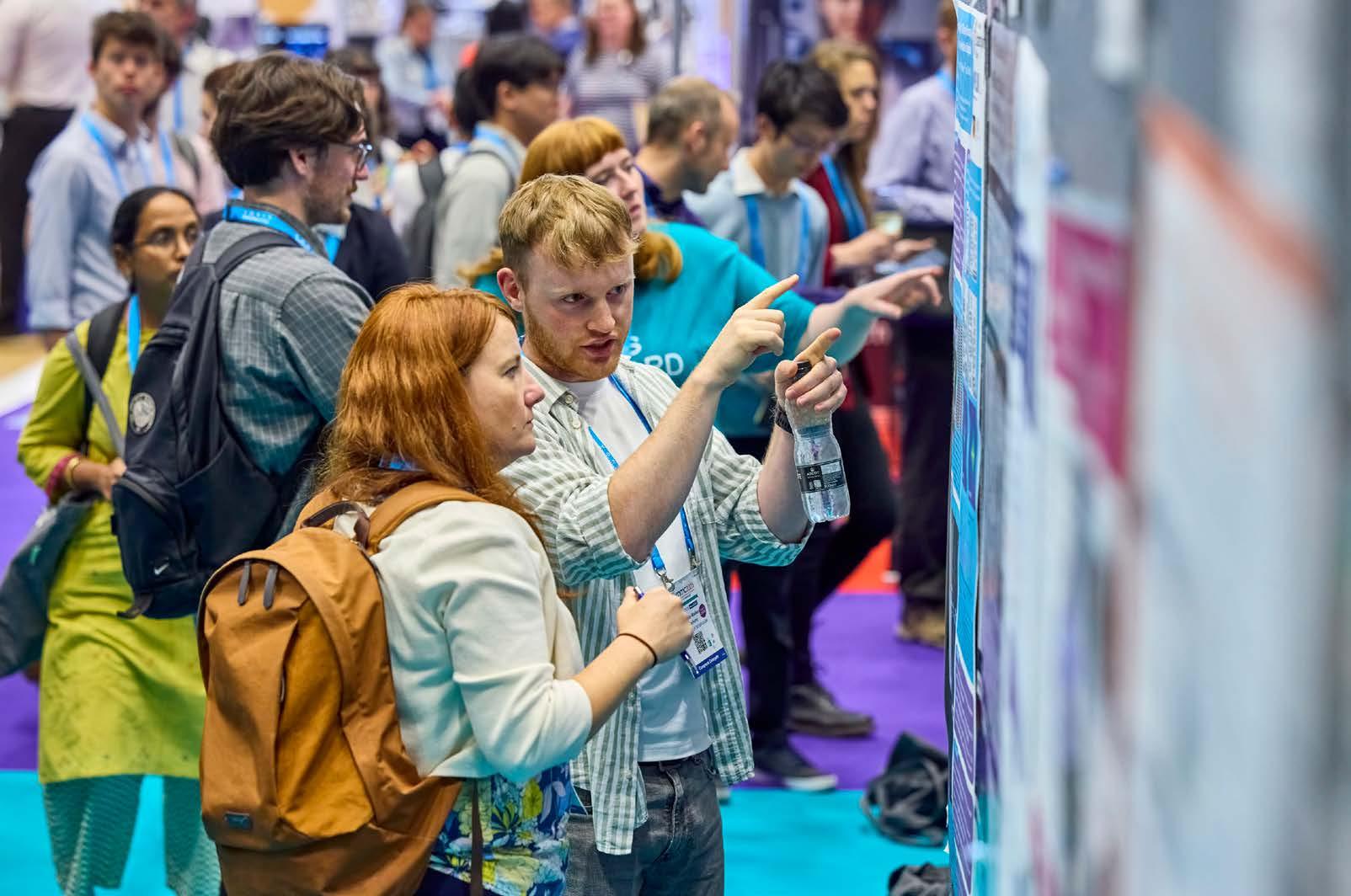
The Journal of Microscopy’s newest Editorial Board Member recently demonstrated his athletic prowess at the 2026 Manchester Marathon.
Dr V V G Krishna Inavalli, who joined the Journal team in April, swapped his microscope for a pair of trainers and completed the city’s annual 26.2-mile race in just three hours, 49 minutes and 58 seconds.
Krishna is currently the Head of Microscopy Group at the University of Southampton’s Centre for Cancer Immunology, where he develops cuttingedge technology to investigate antibody-based cancer therapeutics. He is also a member of the RMS Light Microscopy Committee.
However, it was his recently-discovered passion for running which was to the fore in Manchester on 19 April, as he crossed the finish line for a personal best time.
Krishna said: “What began in 2022, when I started
running with the simple goal of completing a 5km park run, gradually grew into a passion for taking on bigger challenges.
“Marathon running pushes you both physically and mentally, but that’s exactly what makes it so rewarding. After learning some tough lessons at the Manchester Marathon last year, I was determined to return better prepared. Returning this time, with more consistent training and a smarter approach, I was able to achieve a personal best, which made all the hard work worthwhile. I’m proud not just of the result, but of the journey it took to get there.”
Krishna brings a wealth of experience to the Journal editorial team, including nearly two decades working in optics and more than 10 years working in an interdisciplinary environment. He has developed various light microscopy techniques ranging from diffraction-limited to super-resolution microscopy techniques to probe cellular biology.
His developments have aided in the understanding of morpho-molecular interactions of Neuronal Synapses, as well as the exploration of the Brain’s extracellular space. He also secured a £1M BBSRC ALERT grant for an open-access STED.
If you’ve yet to secure your ticket for the 21st International Microscopy Congress (IMC21)and even if you have - don’t forget to add a precongress workshop and the Congress gala dinner to your booking.
Delegates can kick-start their IMC21 experience by taking part in one of the fantastic pre-congress workshops taking place on 30 August, before the main event gets underway. More than 20 different workshops are available, covering a huge range of microscopy techniques, technology and hot topics across the Physical and Life Sciences.
The IMC21 Gala Dinner will be taking place inside the conference centre on Wednesday 2 September, and will include a drinks reception
and entertainment. This is always a memorable highlight of IMC, providing the perfect opportunity to catch up with friends and colleagues in an informal setting.
You can add your chosen workshop and the gala dinner to your booking as additional options when registering online - or you can update an existing booking by logging back into your online account. Places on the workshops are allocated on a first-come-first-served basis, with the most popular workshops expected to fill up quickly in the coming weeks. Spaces at the gala dinner are also limited, so early booking is advised to avoid disappointment.
IMC21: Register now
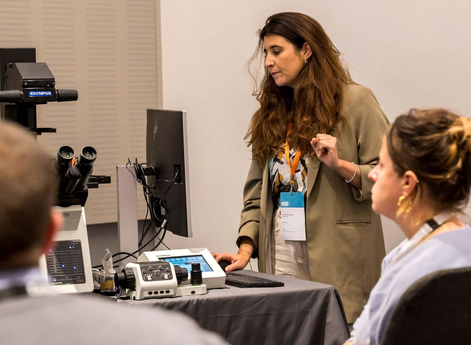
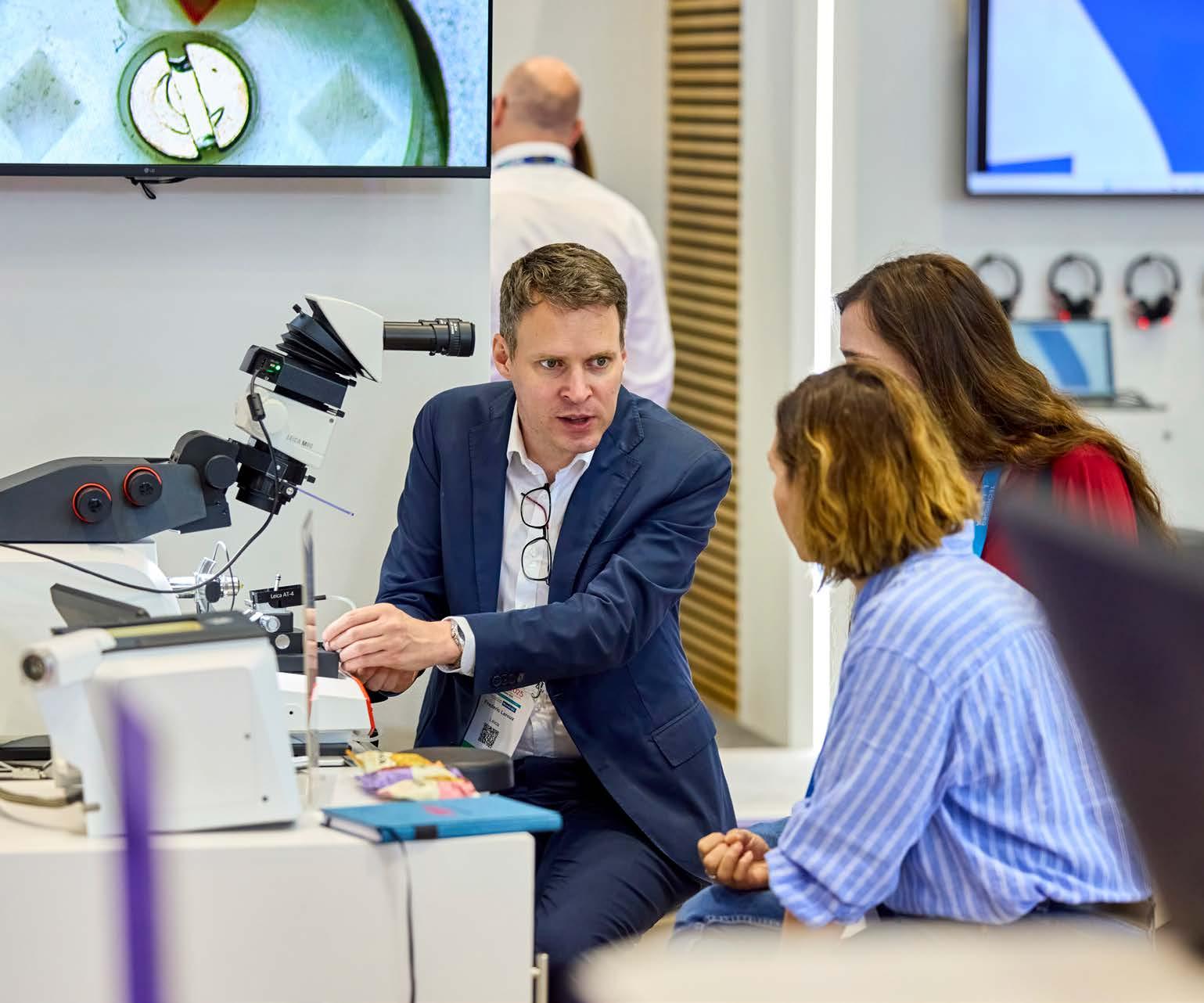
More than 100 exhibitors have now signed up to be part of the showpiece exhibition at the 21st International Microscopy Congress (IMC21).
In what promises to be this year’s biggest gathering of global microscopy manufacturers, a host of leading international companies will be bringing their latest products and technologies to the ACC Liverpool conference centre for the event, which takes place from 31 August - 4 September (precongress events 30 August).
The response from companies has been fantastic since exhibition space went on sale in September - and there are still a limited number of exhibition stands available (book online).
RMS Chief Executive Sali Davis said: “We would like to thank all our exhibitors and sponsors for supporting IMC21 - which is really shaping up to be a superb event for the international microscopy community. The exhibition is very much going to be at the heart of this year’s Congress, and thanks to the response from companies so far, our visitors are sure to have plenty to explore.”
“We still have a limited number of stands and sponsorship opportunities available, so I would urge any companies who might be interested in joining us in Liverpool to check out the exhibition information on our dedicated IMC21 website.”
A list of companies and other exhibitors taking part in the IMC21 Exhibition can be found here
The Editors of the Journal of Microscopy are pleased to announce the winners of the 2025 ‘Best Paper’ awards for Early Career Researchers.
Each prize was £200 and was judged by General Editor Professor Michelle Peckham and our team of Scientific Editors.
For life sciences, Dr Elnaz Fazeli was awarded the prize for ‘From cells to pixels: A decision tree for designing bioimage analysis pipelines’.
General Editor, Michelle Peckham, said: “This paper was a great, well thought out introduction to bioimaging analysis pipelines that is likely to be really useful to new starters in the field”.
Elnaz said: “From cells to pixels: A decision tree for designing bioimage analysis pipelines’ was a collaborative effort that actually began on Twitter as a light and fun exchange of ideas between bioimage analysts from Finland, Germany, France, Hong Kong and Japan. Over time it grew into a passion project, strongly inspired by challenges we encounter daily while working in a microscopy core facility.
“A recurring issue is communication: biologists often have rich imaging data but are not always sure what kinds of quantitative information can be extracted from their images, while image analysts may struggle to understand exactly what biological questions need to be answered. The goal of the paper was to help bridge this gap.We developed a guide for researchers by categorizing the types of shapes and patterns present in biological images and connecting them to suitable bioimage analysis approaches.
For physical sciences, Dr Alexandra Sheader was awarded the prize for ‘Subcellular localisation and identification of single atoms using quantitative scanning transmission electron microscopy’.
Scientific Editor, Vinayak Dravid, said: “Alexandra Sheader et al. deliver a notable methodological advance by demonstrating the localisation, identification, and quantitative weighing of individual heavy atoms within biological tissue using calibrated high-angle ADF STEM. Through meticulous detector normalisation, cross-section quantification, and simulation-validated analysis, the authors achieve atomic-scale compositional mapping in cryosectioned dorsal root ganglia—revealing nanoscale platinum aggregations following oxaliplatin treatment and establishing a pathway for subcellular elemental detection with unprecedented sensitivity.
“The work stands out for its conceptual clarity, technical sophistication, and broader implications for biomedical microscopy, representing a substantive contribution to the field.”
Alexandra said: “I’m really delighted to have been awarded the Journal of Microscopy ECR ‘Best Paper’ prize.This research focused on identifying the species of single atoms within sections of peripheral nerve tissue using HAADFSTEM imaging, in order to trace platinum that had accumulated inside cells following treatment with a Ptbased drug.
“My hope is that this framework helps biologists think more clearly about what they can measure from their images and facilitates more effective communication with bioimage analysts/ core facility staff when designing analysis pipelines.”
“Quantitative STEM imaging is always challenging - especially when you’re trying to detect individual atoms in a cell that is hundreds of thousands of times larger! This work took a lot of iteration and many hours at the microscope, but I think it really highlights the power and versatility of electron microscopy in tackling complex problems - as well as the value of attempting experiments that you might initially assume won’t work!”
www.journalofmicroscopy.org.uk

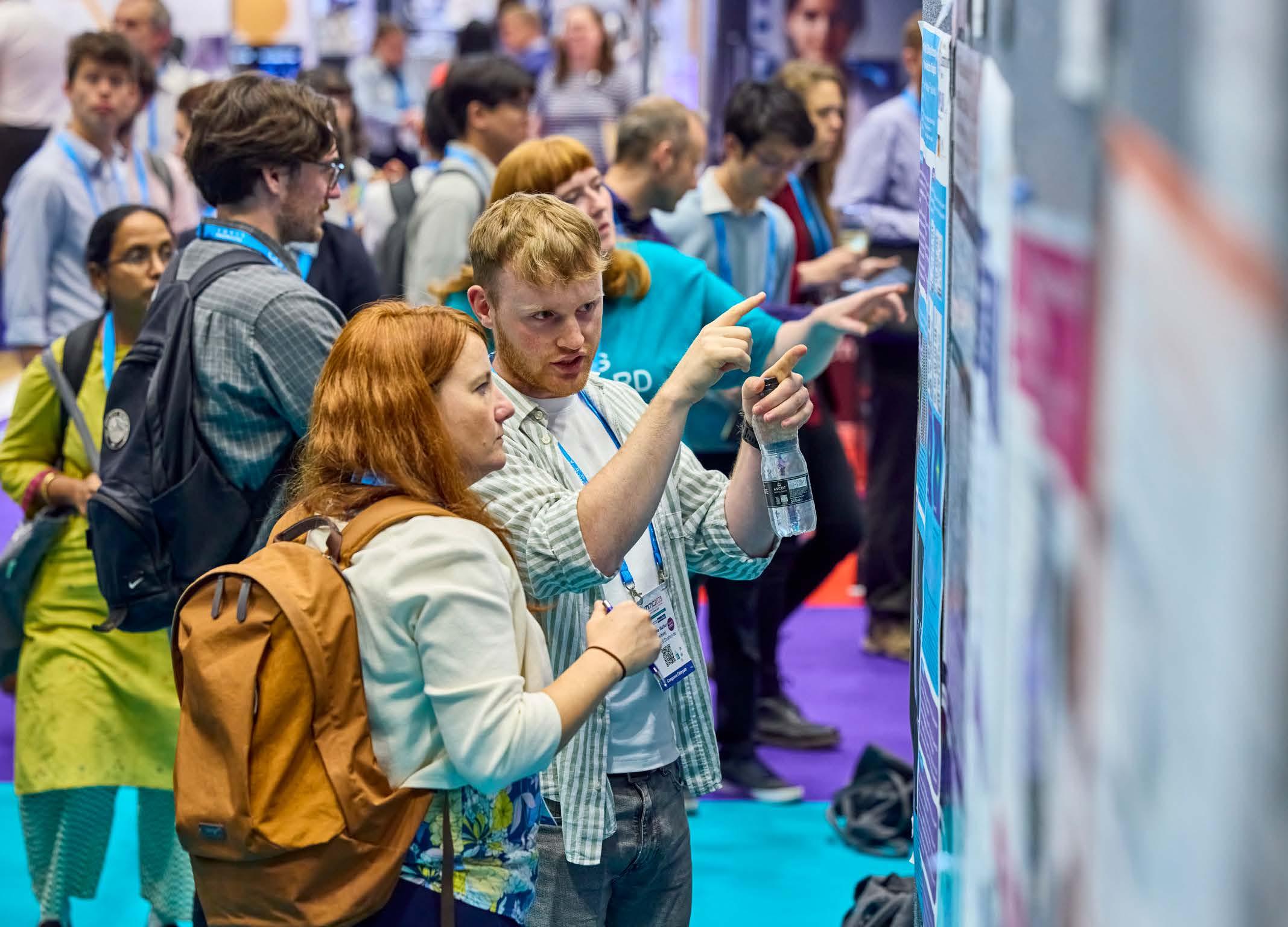
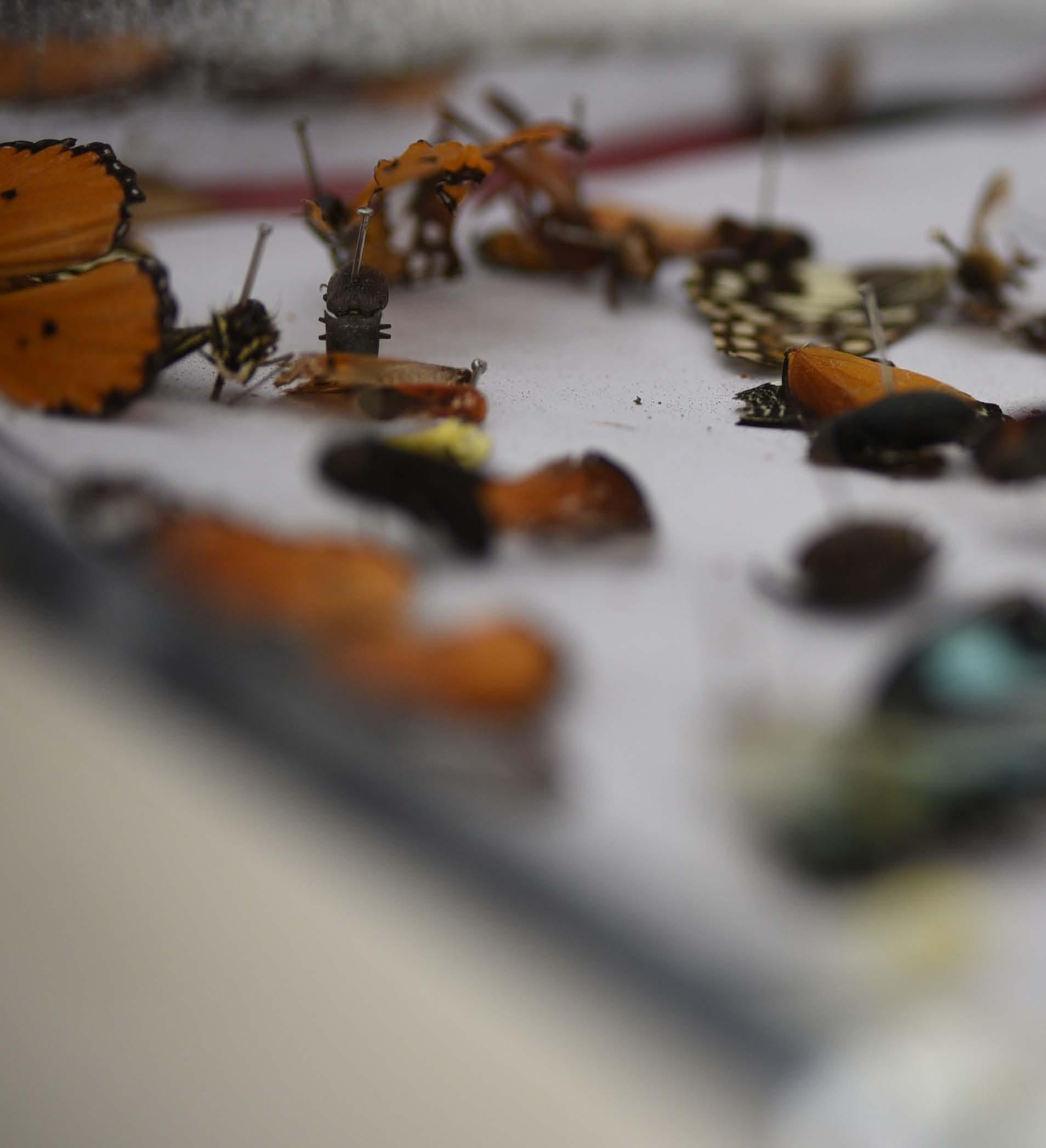
An account of the Lahore Science Mela 2025, by
Muneebah Fatimah
“Nature is full of infinite causes that have never occurred in experience.”
- Leonardo da Vinci
Nature’s mastery is evident even in the seemingly insignificant. Some insects are recluses, refusing to be divulged in the open, existing solely in the margins beneath bark and stones and who, alone, value the coolness of night. They thrive in subtlety, shielding themselves with camouflage.The butterfly can be considered an exception. With wings brushed in amber, turquoise, and sapphire, she perches where a flashing petal resides, as if aware that beauty deserves an audience. When one gets too close, she flies teasingly allowing admiration only, probably laughing at our foolishness.
In the Lahore Science Mela 2025 (LSM25), Pakistan’s most active grassroots science education and awareness association, the Khwarizmi Science Society (KSS) allowed minimalists like me to indulge the public in the grandeur of the minute through the art of microscopy.The society allowed the display of both the ascetics and the inviting untouchables. The setup consisted of three different microscopes, ranging from the portable HD LCD digital microscope to stereo microscopes. One of the microscopes was in fact donated to KSS by insect lovers at The Caterpillar Lab in New Hampshire, USA. A smaller screen was attached to the digital
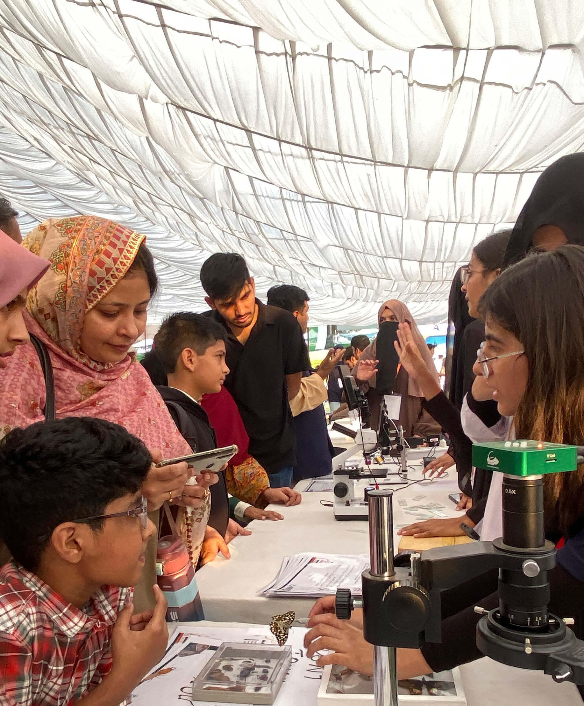
and light microscope while a TV-sized LED for better vista. Once assembled, the digital microscope displays the specimen at 1-1200x magnification. In contrast to the digital microscope, the light microscope provides higher magnification, enabling
detailed observation with improved resolution. This microscope was equipped with three objective lenses (4x, 10x, 40x) and two eyepiece lenses (10x, 16x).
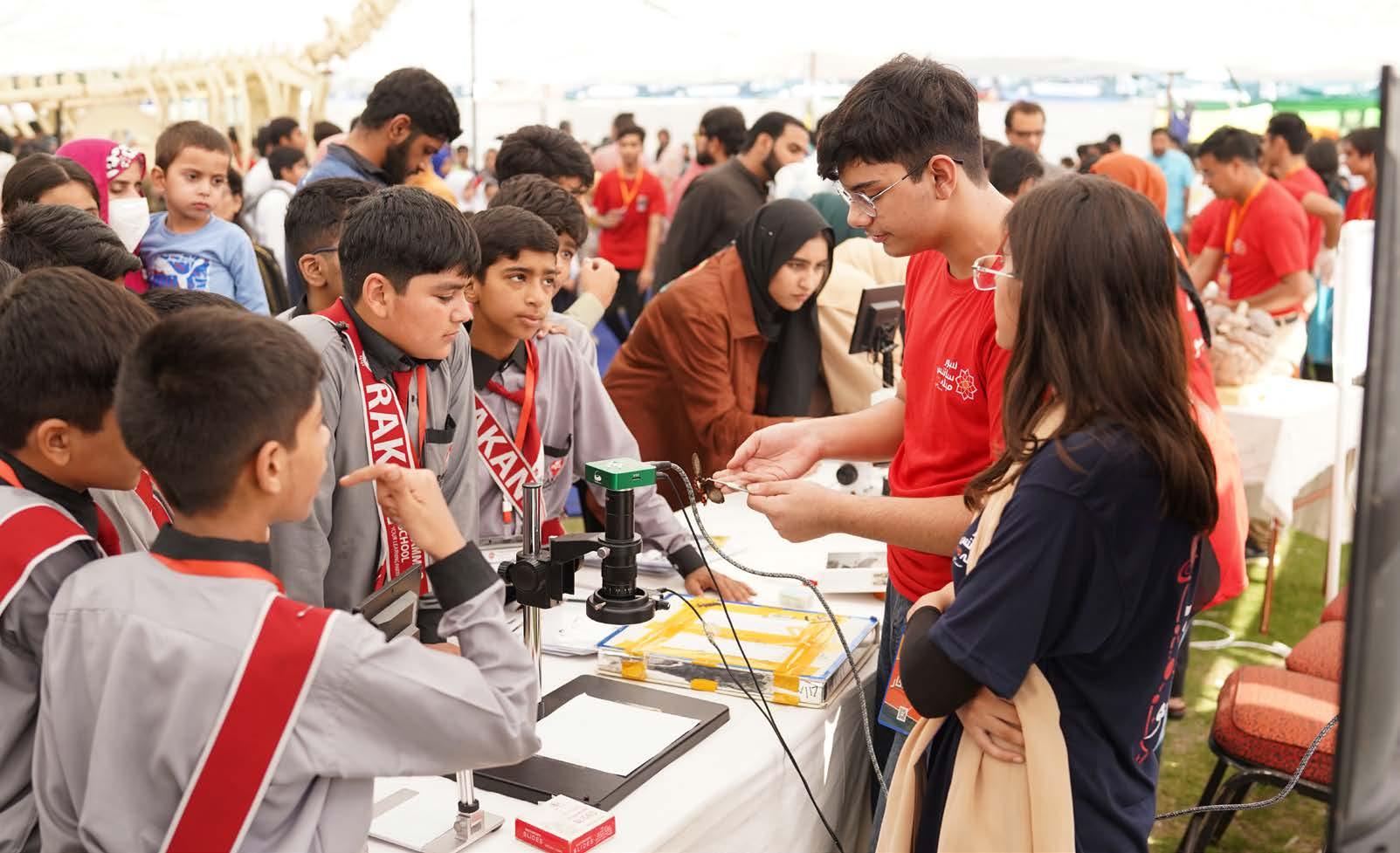
The practice and preparation began months before the big day. Our team of six diverse microscopists met every week, befriending the marvellous machine, assembling slides, making flashcards, and mounting insect specimens. We familiarized ourselves with the microscopes in advance and finetuning magnification levels.
The first time I handled a microscope, I had no clue what I was stepping into. I pictured myself taking it home, turning the magnification to its limit, and uncovering baffling realms. While I witnessed remarkable sights, the process was far more measured. It involved minor adjustments, and postprocessing, and even then, a perfectly clear view of the specimen wasn’t guaranteed. However, the symphony of technicalities and dramatized story telling kindled curiosity for the unseen.
The Lahore Science Mela 2025, held on 25th and 26th October at Crescent Model Higher Secondary School, brought together more than 145 academic exhibitors and more. The event showcased live demonstrations spanning multiple disciplines, including KSS’s own 80 in-house creations featured at the Surpriseum ( ). Over the course of two days, approximately 150,000 visitors from diverse backgrounds attended.
the heart of the
the
Science Society’s Surpriseum. Hundreds of people pooled their intellect and skill to pillar for what felt like Pakistan’s most electrifying experimental science extravaganza. From the thudding of dominoes to the electric murmur of coils and the sigh of chemical reactions, with togetherness, the environment turned science into a megaplex. And in the busy atmosphere, was the station entitled ‘Micro-World’ where our mighty microscopes charmed the bypassers.
On these eventful days, children admired the venation of the Akk grasshopper and the compound eyes of a dragonfly. Others marveled at the mandibles of black digger wasps, mosaics on butterfly wings, and muted tones of carpenter bees. I felt that there is a quiet wizardry in the art of science communication, especially when displaying microscopes. Behind every slide lies a macrocosm, and the role of a communicator is to bring life to it without accidentally poking anyone with the slides.
It appeared to me that the visitors were like pilgrims, travelling through the arena, yearning for scientific enlightenment. And never were there the audience of the same age. Hence, the diversity of the questions they put forward was both puzzling
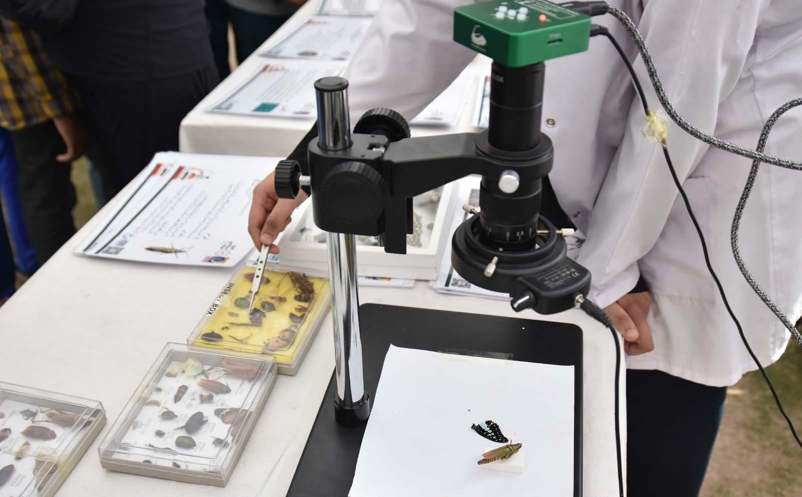
and august. The cornerstone of our demonstrations was the question, ‘what if the world we see is only a fraction of what truly exists?’
The bedazzled spectators viewed one of our ardent demonstrators catching butterflies on the spot, some were hooked, while some remained sentimental once they were quelled. Meanwhile, specimens under varying magnifications inspired questions about both the technicalities and the subjects themselves. Undoubtedly, a microscope is a benison for the meticulous, and a cruel joke for anyone with shaky hands.
Science, with its discipline, grants us the power to handle nature with the fidelity it deserves. It encourages us to observe with a curious mind and a humility that acknowledges the complexity of what is perceived as divine revelation. Art speaks where analysis falls silent, and it was evident when youngsters’ jaws dropped at both the architecture of the microscope and the wonders it revealed. The Micro-World booth at the Lahore Science Mela combined all these traits.
Observing under the lens reveals the spatial relationships between structures invisible to the naked eye, from the iridescent scales of a butterfly
to the intricate ommatidia of its eyes and fine setae on its exoskeleton, inspiring awe in connoisseurs. And then there is the crowning marvel: the textures themselves, a delicate interplay of velvet, glass and steel.
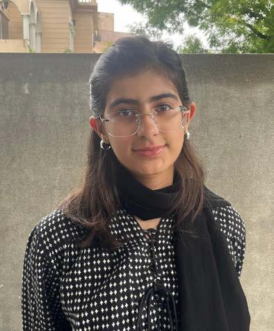
My name is Muneebah Fatimah and I am a school student in Lahore, Pakistan. I am a life member of the Khwarizmi Science Society which aims to enlighten people with knowledge that covers all disciplines. I am an avid science communicator and a biology enthusiast. Furthermore, I am very fond of microscopes and insects. KSS organises an annual festival, called the Lahore Science Mela KSS LSM where tens of thousands of people come in the pursuit of scientific awareness. At the Lahore Science Mela of 2025, I participated as a science demonstrator in the microscopy station.
Back-illuminated sCMOS sensor
Ultra-low read noise

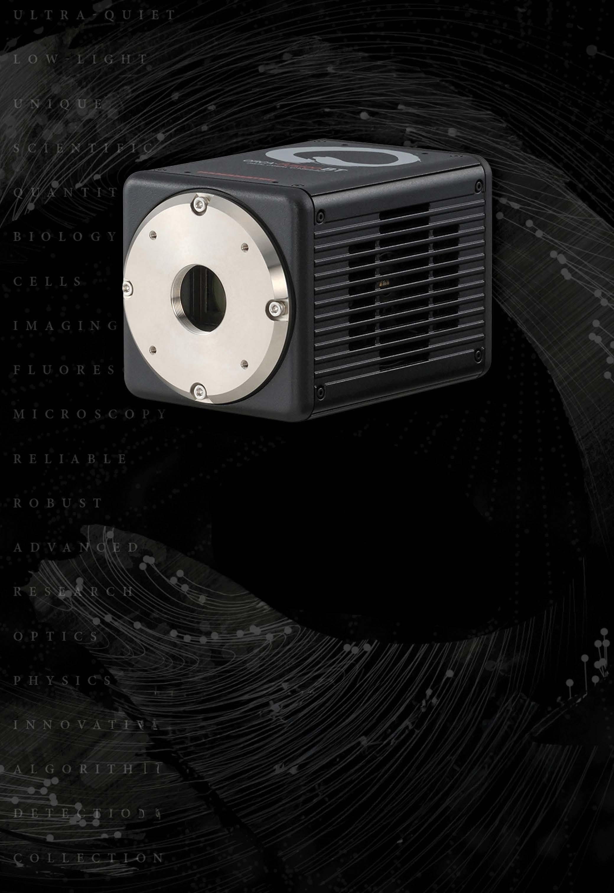
In this issue, we speak to the new Chair of the RMS Qualifications Committee, Marie Kokolski, of the University of Nottingham.
A subgroup of the RMS Outreach and Education Section, our Qualifications Committee oversees the RMS Diploma programme – a flexible, portfolio-based qualification suitable for those using microscopy or cytometry as part of their career. Diploma candidates design their own project with the assistance of their line-manager, and with input from existing Fellows of the Society. This approach ensures that the study is both challenging and rewarding whilst fitting with, and complementing, the candidate's existing employment.
The Qualifications Committee assesses applications from candidates, assigns mentors to support them through their chosen projects, and assesses candidates’ progress through to the completion of their assignments.
Let’s find out more…
Can you tell us a little bit about your current working role, and your background in microscopy?
Currently I’m an Associate Professor at the University of Nottingham. My research is in Science Education, so I’m not involved in research involving microscopy techniques, but rather teaching students about the wonders of microscopy and how it can be used, at university-level, and also through outreach work.
My teaching is mainly around light microscopy and electron microscopy, in terms of using it to show students ultra structure and understanding human health and disease. I previously worked as a technician in a specialist imaging unit, so I had
experience with a whole range of microscopy techniques, and that was how I was first introduced to the RMS –more than 15 years ago.
How did you become interested in supporting the RMS Diploma programme?
Initially I was involved in the RMS Outreach and Education Committee, and then joined the Qualifications Committee a few years later. It was really just my interest in teaching and outreach, and seeing how people were presenting what they were learning and how they were developing their projects. I also think it’s important for people to have the opportunity to develop their skills and interests within the workplace – rather than just traditional educational routes.
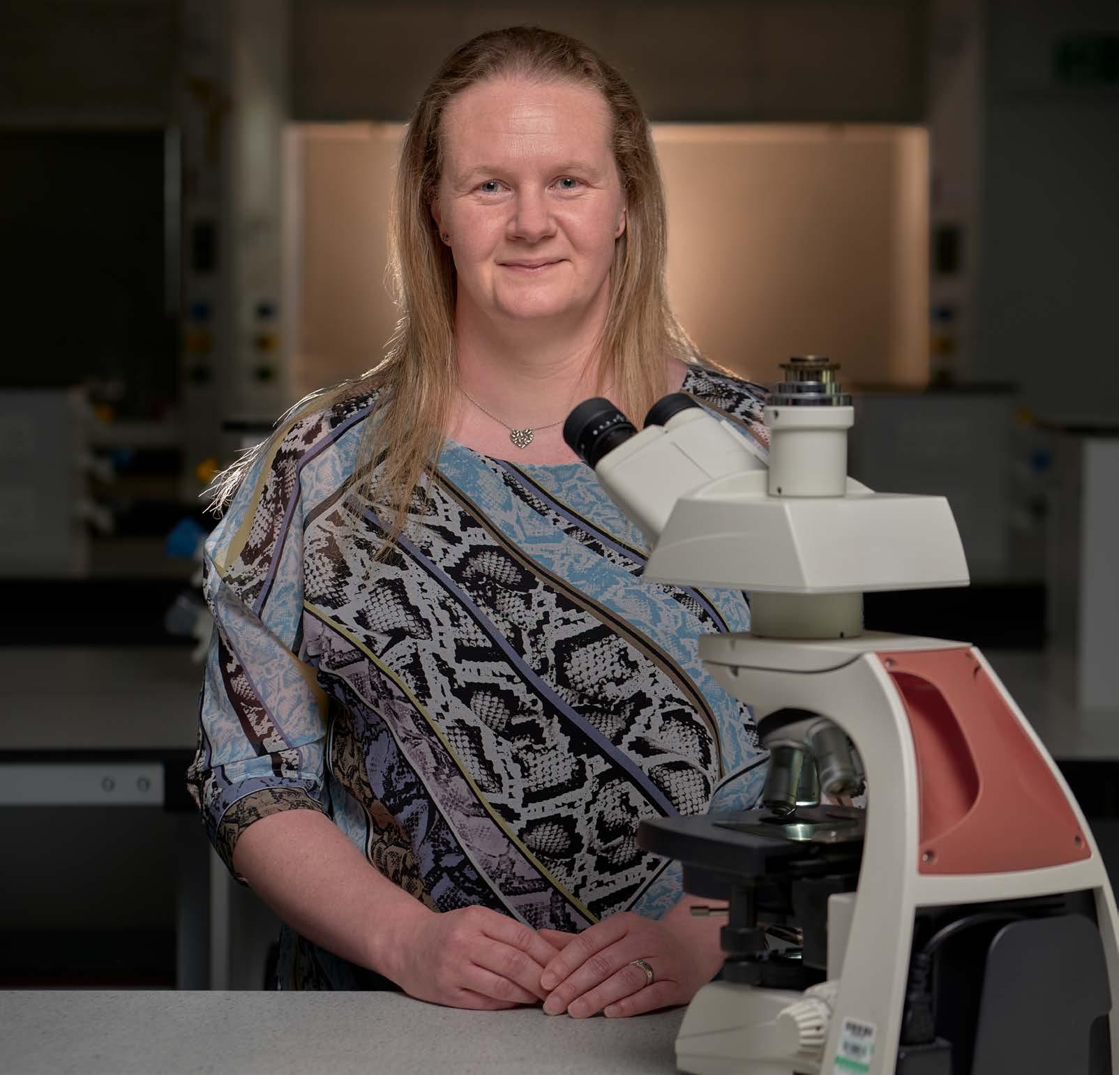
What is unique about the RMS Diploma in terms of what it offers candidates?
I think it can sometimes be difficult for people who are working in a technical role to find opportunities for development. Often they are involved in particular projects at work, and they may have limited input into those, but the Diploma enables you to be the principle investigator. It’s an opportunity to develop your own projects, interests and expertise – and to be able to disseminate that expertise as well – which you might not otherwise have the opportunity to do without going away and doing a PhD.
The candidates also get the opportunity to present
their work at a meeting or conference, or to write a report, or get involved with outreach activities. This also really helps them to develop their skills and gain new experiences.
How do Diploma candidates decide on a project and what input does the Qualifications Committee have?
Our Diploma candidates carry out their projects within their own workplace, so it will be something related to what they are already doing, but which allows them more ownership.
We review the applications when they are submitted and make sure that it is something that looks
feasible, and something that we could support. It will be a short study proposal, a summary of what it is they will be researching, and we take it from there.
What role do the mentors have in the Diploma programme?
Each candidate has a supervisor from their workplace, with whom they have day-to-day contact, but then we will also assign a mentor. This will be someone within the RMS who is working in the same field as the candidate, and they have a really important role. The key thing is that they are external to the workplace, so it’s an alternative person that candidates can speak to and seek advice about the technical side of things.
They will have regular contact, and the mentor is also there if the candidate needs to reach out to them at any time. Having someone involved who is outside of work, who can provide a different perspective, and who they are not speaking to every day is really valuable, and the feedback we have had from candidates confirms this.
What ongoing involvement does the committee have during the course of a Diploma candidate’s project?
In general, it is a case of maintaining the relationship with the candidates as they move through their studies. We meet with them formally every year to discuss their progress, and address any challengesincluding any support they may need.We have found this to be really beneficial both for ourselves and for the candidates; it enables them to ask questions and refocus, and it enables the committee to be more than just an approving body.
take?
As a guide, we anticipate people could complete the Diploma within two years, but we are realistic about the challenges of completing a qualification alongside
full-time employment – so if people need to extend for whatever reason, then we are supportive of that because, ultimately, we want to provide that support and encouragement to help people to complete their projects.There are many challenges to navigate within the projects themselves – the development of research or challenges around kit, for instance. We have even had people change jobs in the middle of their project and move elsewhere, but still find a way to continue.
What do you find most rewarding about your role?
I think it is seeing the development of the candidates and how they grow in confidence. It’s great to be able to meet with them at RMS events and see the confidence they have gained in that time and the new things they have attempted – for example, attending conferences, taking part in outreach activities, writing an article, or presenting a talk or poster at a meeting. They are trying new things that are unlocking new skills, and it’s really rewarding when you see them get to the end point and they have completed the diploma.
At present we have candidates carrying out such a wide range of projects, so being able to see all of those projects progress, and the fascinating things that people are doing, is fantastic. This is also an international Diploma, so it is great to see what is happening with our candidates in different countries and to have those connections through the RMS. I’m very happy to be involved in it and to help develop it in the best way possible for the people who take part.
What would you say to anyone considering applying for the RMS Diploma?
I think the message to anyone working in microscopy is that the diploma exists for you – and whether you’re ready for it now, or later in your career, it’s a great opportunity to develop your skills and confidence in microscopy, while gaining a fantastic qualification.
The 8th Great Wall Symposium was held at the Grand Hotel Baia Verde, Catania, Sicily from the 15th – 17th September. Accompanied by serene views of the Ionian Sea and stunning backdrops of Mount Etna, this biennial conference was an opportunity for researchers working on everything related to the robust bacterial cell wall to come together, share recent findings, exchange ideas and form new collaborations. As a second year PhD student, it was an exciting prospect to be surrounded by other researchers who were just as interested in peptidoglycan synthesis as I was.
The conference programme spanned six sessions, split over three days. The most interesting session for me was how peptidoglycan synthesis is coordinated with the cell cycle, comprised of six different talks. I gained a huge appreciation for the variety of imaging modalities that research groups around the world are utilising to answer some of the most important unsolved questions in bacterial cell biology- seeing is believing! I took great interest in two talks: Nils Meiresonne (ITQB, Portugal) presented compelling bacterial FLIM-FRET data on cell division machinery with exciting implications for advancing in vivo microscopy studies of proteinprotein interactions, and Paula Navarro (University of Lausanne, Switzerland) presented a tour-de-force study combining in situ cryo-ET, AFM and singleparticle tracking methods to better characterise the role of a class A penicillin-binding protein called PBP1b, with divergent insights from previous literature that extend our understanding of division site reinforcement.
I presented my very first poster which was a gratifying experience. My poster, entitled “Singlemolecule studies of peptidoglycan synthases in Pseudomonas aeruginosa” was an introduction to my PhD project and summarised the first instance of establishing single-molecule FRET studies of PG synthase conformational dynamics in the context of P. aeruginosa cell division. I interacted with many group leaders and early career researchers at other
institutions whose names I recognised from my favourite research papers, and it felt like a great triumph to receive lots of interest and thoughtful questions from them regarding my ongoing work.
On the social front, there was ample opportunity to interact with other researchers. Every day included a substantial lunch break and a poster session with refreshments, where I formed various connections with scientists closer to home in the UK and much further afield, which I’m sure will turn into fruitful collaborations in the future! It was so refreshing to be immersed in science in this manner; the enthusiasm at the conference was infectious. We enjoyed an extensive Italian gala dinner on the evening of 16th September which facilitated in-depth discussions about various talks and posters earlier in the day, and there were two excursions planned for the final day by the conference organisers, including a trip to Catania Old Town, and a visit to the alluring Cyclopean Isles.
I would like to extend my heartfelt thanks to the Royal Microscopical Society and the BBSRC-MIBTP for providing funding which allowed me to attend this conference; this trip has instilled an invigorated sense of excitement about my research!
Saba Alhagagi University of Warwick
Get ready for one of the key highlights of this year’s light microscopy calendar! If you’re attending ELMI in Coimbra, you’ll have the opportunity to see CoolLED’s latest LED Illumination Systems in action at Booth #17 and across a range of workshops throughout the meeting.
At CoolLED, we design and manufacture LED illumination solutions that give scientists precise control, stability and flexibility across every stage of fluorescence imaging, including:
• High-speed live cell applications –elevate your research with the 8-channel pE800, offering eight individually controllable LEDs and ultra-fast TTL triggering speeds of <7 µs.
• Spatial biology – the new 10-channel
pE-10 when paired with the Kromnigon SpectraSplit® 10 filter sets enables fast, robust multiplex imaging for advanced spatial biology applications.
• Routine to advanced fluorescence –the pE-400 Series provides four LEDs covering DAPI and YFP through to Cy5, delivering reliable performance with intuitive control for everyday imaging needs.
Visit the CoolLED team at Booth 17 to learn more about our LED Illumination Systems and where they’ll be on show across the workshop programme, giving you the opportunity to see first-hand how they perform under real experimental conditions.
www.coolled.com

Before you section for 2D multiplex imaging, see what is happening within your sample in 3D. With 3D investigation, you can find the ideal planes to section within your sample. So every slice is a treasure trove of data.
Spatial biology has revolutionised our understanding of tissue architecture by enabling the contextual analysis of cellular and molecular constituents within their native microenvironments. However, conventional methodologies are confined to twodimensional thin sections, limiting the capacity to capture large-scale tissue organisation and potentially missing critical cellular relationships. To overcome these limitations, Miltenyi Biotec presents an integrated workflow that combines volumetric
3D imaging with high-plex spatial analysis, enabling multiscale investigation of complex biological systems within intact specimens.
Harold Cremer, PhD, (Université Aix-Marseille) says of the workflow: “The quality is super – I’ve never seen such a thing before. I think it’s the first time this really worked, and I’m glad that we were chosen as beta testers for this new approach.” See the whole interview with Harold Cremer PhD here.
Contact Jak at jakg@miltenyi.com for more information.
https://www.miltenyibiotec.com/GBen/lp/3d-2d-spatial-biology-workflowfor-precision-tissue-profiling.html
If you would like your Company News to appear on these pages, please contact infocus Magazine at advertising@infocus.org.uk
The announcements in this Section are compiled by the manufacturers. They in no way represent a recommendation by the Royal Microscopical Society for any particular instrument or equipment. The Royal Microscopical Society does not endorse, support, recommend or verify the information provided on these pages.
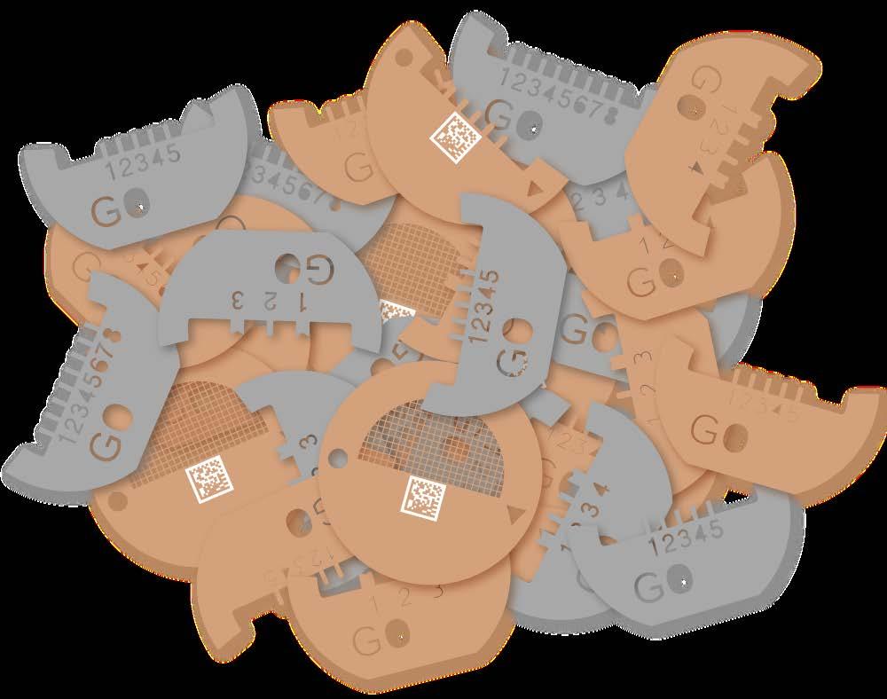
In recent times the need for different types of EM grids has expanded, and the commercial introduction around 15 years ago of Nanomanipulators was perhaps the start of this.Their requirement for halfmoon type grids with pins or slots to hold lamella samples led to the introduction of FIB Lift-Out Grids.
Having made custom grids for many OEM companies since the inception of these products, Graticules has recently introduced their own brand of products with multiple pin configurations and in copper or molybdenum.
Over the years the design and quality requirements of the grids has changed, driven by the increased demands of the analysis and automated workflow systems. This is something that Graticules has addressed with improvements in processing and investment in new plant. The latest innovation has been the addition of unique DMC marking on each grid for true traceability. This has meant significant development and major investment in bespoke systems that are capable of achieving the 100% readable micro coding demanded.
www.graticulesoptics.com
Nikon Corporation (Nikon) has launched new ECLIPSE microscope models, the LV150NA LED, LV150N LED, and MA200 LED, to strengthen the company’s comprehensive range of industrial microscopy solutions. Following the LV100N series announced in March 2025, the new models use a high colour-rendering LED light source and improve maintainability.
The ECLIPSE LV150NA LED and LV150N LED are upright microscopes designed specifically for reflected illumination, with accessories selectable to suit your observation method and purpose. The MA200 LED is an inverted microscope with reflected illumination that is ideal for inspecting industrial parts and utilising for research and development.
• Improved maintenance: The newly developed
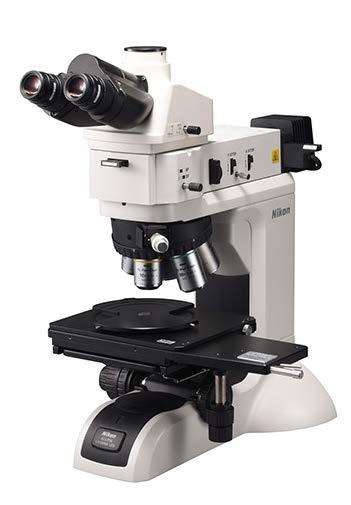
high colour-rendering LED light source upgrades brightness, colour, and colour rendering properties compared to the existing halogen source, extending lamp life from 1,100 to 50,000 hours. For the MA200 LED, space efficiency is improved by incorporating the LED light source into the microscope stand, reducing footprints by up to approximately 100 mm.
• Enhanced usability: The microscopes offer high scalability through the compatibility of accessories between old and new products. Existing accessories can be used with the new microscopes providing extensive expandability with a wide range of accessories.
More information about the microscopes can be found here.
LV150NA LED / LV150N LED / MA200 LED
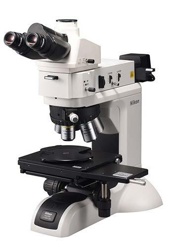
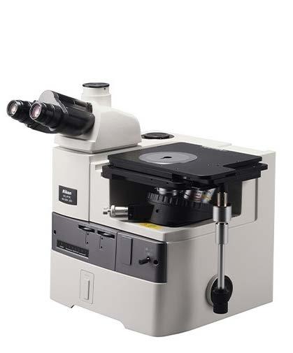
If you would like your new product information to appear on these pages, contact infocus Magazine at advertising@infocus.org.uk
The announcements in this Section are compiled by the manufacturers. They in no way represent a recommendation by the Royal Microscopical Society for any particular instrument or equipment. The Royal Microscopical Society does not endorse, support, recommend or verify the information provided on these pages.
infocus is the Royal Microscopical Society’s (RMS) vibrant and striking quarterly magazine for members. It provides a common forum for scientists & technologists who use any form of microscope, including all branches of microscopy. Published four times a year, infocus is free to members of the RMS. infocus features articles on microscopy related topics, techniques and developments, an events calendar, news, event reports, book reviews, new product information, and much more. infocus welcomes submissions of:
Articles - Full articles or reviews of general interest to microscopists, of approximately 30004000 words (excluding references), with images/ figures (as many as appropriate, 4-8 as a guide). Longer articles can also be considered.
Short Articles - Short topical articles, review articles or articles providing hands-on help for microscopy methods.
Primer Articles - Short general articles that are focussed on specific techniques.
Debuts - Student articles publishing emerging results from a project. Results may still be incomplete, but areas of progress/problems should be highlighted, with the aim of provoking feedback.
Book Reviews – if you are a member of the RMS and are interested in writing book reviews for infocus, please contact Owen Morton owen@rms.org.uk
Please see recent issues of infocus for examples of articles and reviews.
If you are interested in submitting to infocus, contact: editor@infocus.org.uk
Article Text
• Text should be in a standard font (e.g. Times New Roman or Arial) at a size of 12 pt.
• Articles should begin with a brief summary, which accurately summarises the content and is intelligible without reference to the text.
• Footnotes and appendices should not be used unless absolutely necessary.
• The hierarchy of headings within the text should be clear.
• Spelling should conform with The Concise Oxford Dictionary and SI units must be used.
• Abbreviations should be used sparingly and only if a lengthy name/expression is repeated throughout the article. When used, the abbreviated name or expression should be cited in full at first usage, followed by the abbreviation in parentheses.
• Authors should provide a photograph, brief biography as well as contact information that will be published.
References in the text should be in the form Joy (2000) or Joy & Williams (2000). For three or more authors, use the form Echlin et al. (2000). The reference list should:
• be listed in alphabetical order of first authors’ surnames.
• (where a journal is cited) - include authors’ surnames and initials, date of publication, title of paper, name of journal, volume number, and first and last page numbers.
• (where a book is cited) - include authors’ surnames and initials, title of book, year of publication, edition, followed by publisher and town, county/state (and country if necessary) of publication.
• (where a URL is cited) – include authors’ surnames and initials, year of publication, title of page, URL and date accessed.
• Figures can be one column/half page width, 65.5 mm or two column/full page width, 135 mm.
• Larger images may fill the page/spread. A full page of the magazine is 170 x 250 mm, a double page is 340 x 250 mm.
• Text in figures (labels, axis labels, legends, etc) should be Helvetica or Arial, 8pt size.
• Figure and table captions should be listed numerically at the end of the article text
• Line weights and line strokes should have a maximum value of 1 and a minimum of 0.25.
• For graphs and plots, whenever possible, please submit vectorized images.
• As much as possible, please avoid white spaces.
• All images must be high resolution – 300dpi or more.
Double page of magazine, 340 x 250mm (Trim size)
• Submission files should be in CMYK format and can be supplied as tiff, jpeg or eps files.
• Images MUST include scale bars or field widths where relevant.
• Total number of images/figures/tables should not exceed 15 including tables.
Prior to publication, authors will be sent a PDF of the article by email for approval.
Authors should ensure articles are thoroughly checked before submission – proof amendments should be limited to minor corrections only.
Authors are requested to assign copyright to the RMS. However, authors may make copies of their own articles without seeking permission from the RMS, provided that such copies are for free distribution only (they must not be sold) and provided that infocus is properly acknowledged (issue number, month and page number should be given). Permission to reproduce material from infocus in other publications will not be given to third parties except with the consent of the authors concerned.
Authors are responsible for obtaining permission to reproduce copyright material from other sources. Approval for reproduction/modification of any material (including figures and tables) published elsewhere should be obtained by the authors before submission of the manuscript and the source of the material should be properly acknowledged. Authors are responsible for any copyright fee involved.
One column/half page width, 65.5mm
Authors are requested to complete and submit a signed copy of our copyright sign-off form. This is available on the RMS website (www.infocus.org.uk).
Two column/full page width, 135mm