ISSUE 81 MARCH 2026
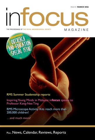
RMS Summer Studentship reports Inspiring Young Minds in Malaysia: infocus speaks to Professor Kang-Nee Ting RMS Microscope Activity Kits reach more than 200,000 children! …and much more!
Plus...News, Calendar, Reviews, Reports 1
ISSUE 80 DECEMBER 2025
JEM-120i
NEW!
Compact, Easy to Use, Expandable TEM
Specimen observation in 4 steps
We developed the JEM-120i with the concept of "Compact", "Easy To Use", and "Expandable". With the new external appearance, this instrument has evolved into a useful tool that anyone can use easily, from operation to maintenance. It takes only 4 steps from loading a specimen to completing observation. The JEM-120i is equipped with an enhanced TEM control system and fully automated apertures, eliminating the need for switching magnification modes and selecting an aperture. Observation operations can be performed more smoothly than with previous models.
www.jeol.com • salesinfo@jeol.com 978-535-5900
Learn more
contents features 6
RMS Summer Studentship reports Ben Davies, Charlotte Howman, Darcy Gabriel, Elsie Sheldrake, Shang Wang,Yeli Xia
32
Inspiring Young Minds in Malaysia: infocus speaks to Professor Kang-Nee Ting
48
RMS Microscope Activity Kits reach more than 200,000 children!
60
Dipping in and out – Balancing a Diploma with work (a follow-up article) Tim Young
70
Picturing Science in Schools: A Science Ambassador Programme with the Crick’s Electron Microscopy Team Jenny Hounsome, Helen Spiers, Martin Jones and Lucy Collinson
76
Opening Doors: How Summer Studentships are transforming research careers Georgina Fletcher
86
Bringing research to the public: Outreach at CRUK Cambridge Tim Young
regulars 29 40 54 64 80 84
Calendar Journal of Microscopy New Member Welcome Office News Company News New Products
reports and other features 17 47 60 90
Darcy Gabriel wins RMS Beginners Competition at SEMT one-day meeting IMC21: Connect Globally. Discover Science. Experience Liverpool! RMS Diploma News In Memoriam: Michael Ivan (Spike) Walker 1
MAGAZ I N E
infocus is the Magazine of the Royal Microscopical Society (RMS) – the only truly international microscopical society. The RMS is dedicated to advancing science, developing careers and supporting wider understanding of science and microscopy. infocus Magazine 37/38 St Clements Oxford, OX4 1AJ, UK Tel. +44 (0)1865 254760 Email: infocus@rms.org.uk Website: www.infocus.org.uk Scientific Editor Leandro Lemgruber, University of Glasgow, UK Editor Owen Morton Tel. +44 (0)1865 254763, Email: editor@infocus.org.uk Editorial Board Myfanwy Adams, John Innes Centre, Norwich, UK Maadhav Kothari, Zeiss Microscopy, UK Hilary Sandig, Cancer Research, UK Trevor Almeida, University of Glasgow, UK Tim Young, University of Cambridge Ferran Valderrama, City St George’s, University of London Thomas Slater, Cardiff University Advertising Email: advertising@infocus.org.uk ISSN: 1750-4740 © 2026 Royal Microscopical Society infocus is published four times per year by the RMS. Designed and produced by Creative Design. Reproduction in whole or in part without permission from the RMS is forbidden. Views expressed in the Magazine are those of the individual contributors and do not necessarily reflect those of the RMS.
RoyalMicroscopicalSociety @RoyalMicroSoc @RoyalMicroSoc
2
ISSUE 81 MARCH 2026
FROM THE SCIENTIFIC EDITOR Dear Readers, I hope you have had a great start to the year, wherever you are in the world. For our first edition of 2026, we have something a little bit different – a Special Issue highlighting some of the fantastic Outreach and Education activities taking place both within the RMS, and the wider microscopy community. At the heart of this issue, we are very proud to publish reports from each of six undergraduate students who successfully applied for RMS Summer Studentship projects in 2025 - gaining invaluable microscopy experience in the process.We also include an overview of the BioImagingUK and UK Physics of Life summer studentship scheme, which provided research placements for students from low-income backgrounds across the UK. One of the Society’s long-standing outreach and education initiatives – the Microscope Activity Kits (MAKs) for primary schools – recently passed a significant milestone, with more than 200,000 children having benefitted from the scheme since it was established in 2011! We’re delighted to include a feature celebrating that success and sharing some of the feedback submitted to us by participating schools. We also speak to last year’s winner of the Chris Hawes Award for Outreach and Education, Kang-Nee Ting, about her work bringing microscopy to children in deprived communities in Malaysia with the help of the MAKs. On the subject of educational opportunities and career development, we have the latest news from the RMS Diploma, including a follow-up article from diploma candidate Tim Young, in which he updates us on his progress completing the Society’s portfolio-based microscopy qualification alongside his day job. Finally, we are grateful to the Francis Crick Institute and Cancer Research UK - Cambridge Institute, for providing articles about some of their recent outreach initiatives. I really hope you enjoy reading all the content in this Special Issue of infocus.
Slàinte!
Leandro Lemgruber
COVER IMAGE: Tied up in Notts By Nicola Weston, University of Nottingham. Image of a knot in a spider silk thread taken using a Quanta 650-ESEM at the Nanoscale & Microscale Research Centre (nmRC), University of Nottingham. Image was acquired in esem mode with Peltier stage cooling at 2C providing high humidity (95%). Image has been coloured using Affinity 2 software. Magnification: x1500.
3
Foreword from Dr Kerry Thompson, Chair of the RMS Outreach & Education Committee and Honorary Secretary for Education The Royal Microscopical Society (RMS) Outreach and Education Committee sits at the heart of the Society’s commitment to sharing the wonder and impact of microscopy with the widest possible audience. Bringing together volunteers from across academia, core facilities, industry, and education, the Committee works to champion imaging as both a powerful research tool and an accessible entry point into science. Our activities span the creation of high-quality teaching resources, support for schools and universities, and the delivery of hands-on workshops, public engagement events, and national initiatives. We collaborate closely with partner organisations to extend reach and ensure that microscopy is visible not only within specialist communities but also in classrooms, community spaces, and public forums. A particular focus is placed on inclusivity, developing opportunities that welcome new audiences, support educators, and highlight the diverse career pathways connected to imaging science. Through competitions, training events, and outreach programmes, we aim to spark curiosity while building confidence and practical skills. STEM outreach grounded in microscopy is uniquely powerful because it makes the invisible visible. By revealing the structures that underpin life, materials, and the environment, microscopy offers an immediate sense of discovery that can inspire learners at any stage. Investing in outreach therefore strengthens scientific literacy, nurtures future talent, and helps ensure that the benefits of imaging science are understood and valued across society. By bringing microscopy beyond the laboratory and into classrooms and communities, we help build a more scientifically literate, inspired, and inclusive future. I hope you enjoy this snapshot of the activities the committee contribute to as part of this special edition. Dr Kerry Thompson
4
ISSUE 81 MARCH 2026
Dr Kerry Thompson.
IMC21 The 21st International Microscopy Congress
31 August - 4 September 2026, Liverpool, UK Pre-Congress Events 30 August 2026
Connect globally. Discover science. Experience Liverpool
Find out more
© Visit Liverpool
2 0 2 5 S U M M E R S T U D E N T S H I P R E P O RT
Correlative Microscopy of a 2D Graphene Flake via Scanning Probe Microscopy Student: Ben Davies Supervisor: Oleg Kolosov Project location: Lancaster University Lay Summary:
Atomic force microscopy (AFM) is a powerful technique often used to study surfaces at the nanoscale, helping researchers to better understand the structure and properties of materials. This project focused on harnessing the adaptability of the simple AFM to perform correlative microscopy on a sample of graphene, allowing the properties of the heavily researched material to be presented through visual images. I began by looking at the standard topography of the graphene flake to determine the thickness relative to the surrounding substrate. I followed this on by looking at the effect of friction when the probe was in contact with the sample. To go beyond surface imaging, I experimented with Kelvin probe force microscopy (KPFM) to map electrical potential under different bias voltages, before then utilising ultrasonic force microscopy (UFM) to reveal variations in surface elasticity. I implemented a thermal probe to analyse the thermal conductivity through scanning thermal microscopy (SThM), and tested the novel technique of scanning thermal gate microscopy (STGM) to investigate the Seebeck effect. By combining all these approaches, I built a detailed portfolio of complementary measurements, showing how AFM can provide a comprehensive picture of graphene’s physical, electrical, and thermal properties.
Main Report
Contact mode also enabled measurements of
Project Aim
surface friction and probe deflection. I then used
The aim of this project was to perform correlative
Kelvin force probe microscopy (KPFM) to explore
microscopy on a conductive graphene flake using
scanning
probe
microscopy
(SPM)
to
comprehensively investigate its structural, electrical,
6
the difference in surface potential between the sample surface and the probe tip under varying bias voltages. To examine mechanical properties,
mechanical, and thermal characteristics.
I employed ultrasonic force microscopy (UFM),
How did you address the aim?
generate images that reflect variations in elasticity.
exploiting the extreme stiffness of the cantilever to
To address this aim, I applied a series of scanning
Next, to measure the thermal conductivity of the
probe microscopy techniques, starting with basic
material, I utilised a thermal probe to perform
imaging and then extending to more advanced modes.
scanning thermal microscopy (SThM). Varying
The most basic of these tests involved using the
AC and DC voltages enabled the heating of the
atomic force microscopy (AFM) to perform simple
cantilever tip which was then placed onto the sample
topography scans through both contact and tapping
in contact mode, allowing heat to be conducted
mode, allowing mapping of the graphene flake and
onto the surface. Finally, I implemented the novel
determining its thickness relative to the substrate.
technique of scanning thermal gate microscopy
ISSUE 81 MARCH 2026
Figure 1. Image of the selected sample for testing.The sample consists of exfoliated graphene with two contacts and a backgate.The left image shows an overhead view of the sample mounted on a piezoelectric material and circuit board, while the right image is an optical microscope view at 5× magnification, highlighting the overlap between the wires and the graphene flakes, which appear as dark blue shards.
(STGM), which combines SThM with scanning gate
with the graphene flake being substantially brighter,
microscopy to allow the visualisation of the gradient
indicating a lower work function than the substrate.
of the Seebeck coefficient through thermal-electric
Attempts to measure the intrinsic potential
interaction. Both SThM and STGM were performed
difference without applied bias were completed, but
under ambient and vacuum conditions to determine
the electrostatic coupling from the large cantilever
how restricting thermal conduction to the sample
dominated over the probe tip. UFM was utilised to
influenced image quality.
investigate the elastic properties, with both a doped
What did you find out?
silicon probe and a thermal probe.The doped silicon
For consistency, all tests were completed at the same section of the flake using 25µm wide scans at a 2:1 scan ratio and a scan rate of 0.2 Hz. The first analysis was of the surface topography of the sample. Due to surface debris (visible as white dots in figure 2a), the precise height difference between the flake and substrate was uncertain. However, in several areas the separation was less than 1 nm, consistent
probe produced higher quality images, possibly due to increased cantilever stiffness. The resulting image can be seen in figure 2d showing that the graphene layer had a lower lock-in output and therefore was more compliant than the substrate. Thermal properties were probed using SThM. Figure 2e shows the resulting image conducted inside a vacuum with a 4 V DC input and 1 V peak-to-peak modulation.
with the claim of a two-dimensional surface. In
The substrate appeared substantially darker than
contact mode it was also possible to view the effect
the graphene, indicating higher thermal conductivity.
of friction on the probe as it was dragged over
While placing the sample into the vacuum chamber
the surface. While there was only a small change
the wire connections broke, not allowing images of
in voltage (-100 mV to 50 mV), there was a clear
the same region of the flake. Instead, figure 2f shows
contrast between the substrate and flake, with the
the visible change in the gradient of the Seebeck
substrate appearing slightly darker as seen in figure
coefficient in air with an input of 7 V DC. The bright
2b.Tests of the surface potential were completed via
and dark spots correspond to the polarity of the
KPFM at DC bias voltages ranging from -2 V to +2
contacts which is relative to the thermoelectric
V, with the most distinct images created at ±1 V The
response, simply swapping the polarity flips the sign
resulting image at +1 V DC can be seen in figure 2c,
of the Seebeck-induced voltage.
7
2 0 2 5 S U M M E R S T U D E N T S H I P R E P O RT a)
b)
c)
d)
e)
f)
Figure 2. Correlative microscopy of the same section of graphene: (a) Topography, (b) Friction, (c) KPFM at +1 V DC bias, (d) UFM, (e) SThM in vacuum, (f) STGM in air.
What did you learn from participating in this project? Before taking part in this project I had absolutely no experience in the field of microscopy! This summer project unveiled a whole new area of research and fascinating physics that I never even considered
Lancaster University. After completing this, I plan on pursing a PhD in materials science, potentially continuing in utilising AFM or branching out into electron microscopy. I can confidently say that this project has reinforced my commitment to continuing through academia and into research.
exploring. Learning about the characteristics of graphene and gaining experience in using an AFM was incredibly rewarding. My favourite part of this project was performing ultrasonic force microscopy. I enjoyed understanding the use of piezoelectric materials and how using them to vibrate samples at such incredibly high frequencies can uncovering detailed information about their elasticity.
How has this project affected your long-term goals? This project resonated with me and has inspired me to complete my masters degree in microscopy, focusing on the creation and characterisation of energy-efficient atomic scale organic materials at
8
ISSUE 81 MARCH 2026
Ben Davies.
Investigating the localisation of PEX10 in-line with patient mutations Student: Charlotte Howman Supervisor: Dr Triana Amen Project location: University of Southampton Lay Summary:
Peroxisomes are essential organelles found in all eukaryotic cells. They are membrane bound compartments that play a critical role in lipid metabolism and detoxifying toxic products, such as hydrogen peroxide. Peroxisome Biogenesis Disorders (PBDs) are inherited metabolic diseases that affect children and are caused by a disruption in function of peroxisomes. There is a current lack of understanding surrounding the contribution of individual mutations and how this results in clinical symptoms for patients, so this project investigated a compound mutation in the PEX10 gene of a patient with PBD. PEX10 is a peroxin that resides on the peroxisomal membrane and is part of the ubiquitin ligase complex (PEX2, PEX10, PEX12), which is involved in protein import into peroxisomes. The mutation we studied is in-line with the patients missense mutation at residue 290, where histidine replaces aspartic acid. We explored the cellular localisation of this mutated PEX10 protein to determine whether it remains in the peroxisomal membrane or mislocalises to the cytosol, and to begin to explain the loss of peroxisomal function observed in the patient.
Methods
at regions of interest (peroxisomes). For each cell
To quantify the localisation of PEX10, we used a
line fluorescence data for 30 cells per experiment
conventional GFP reporter as our negative control,
were collected across at least three biological
and PXMP2-mCherry was used as a spatial and
repeats. Statistical significance was assessed using
quantitative reference for peroxisome identification.
Kruskal-Wallis tests for non-parametric data, which
Plasmids were amplified in DH5α E.coli, plated on
enabled us to determine differences in localisation
LB agar plates, colonies transferred to secondary
between constructs.
ampicillin
Results
LB
plates, plasmids
isolated
and
measured using Thermo Scientific™ NanoDrop™ spectrophotometer. HEK293T cell lines were co-transfected with PXMP2-mCherry and either, GFP, GFP-PEX10 or GFP-PEX10-H290D (1µg of plasmids). These were fixed (4% PFA), visualised using the Leica SP8 confocal microscope and images were processed
Based on previous work by members of our lab, we hypothesised that the H290D variant of PEX10 would not localise to peroxisomes. Our aim was to determine whether PEX10-H290D co-localises with peroxisomes to gain insight into the mechanism underlying this loss of import.
allowed for visualisation and comparison of
Visualisation of PEX10-H290D mutation by confocal microscopy
fluorescent intensity for GFP and mCherry channels
To test this hypothesis, we performed co-expression
using Fiji (Image J). The co-expression of plasmids,
9
2 0 2 5 S U M M E R S T U D E N T S H I P R E P O RT A
B
Figure 1. A. Confocal microscopy of fixed wild-type Human Embryonic Kidney (HEK293T) cells co-expressing; PXMP2-mCherry (yellow, 1µg/ml) and either GFP, GFP-PEX10, or GFP-PEX10-H290D (cyan, 1µg/ml). Nuclei were stained with Hoechst (10 µg/ml). Scale bar, 10µm. B. Quantification shows average fluorescent intensity of the same region of interest (peroxisomes) between the red channel (mCherry) and the green channel (GFP), mean ± SEM, N= 30 cells, Kruskal-Wallis (**** = <0.0001).
experiments in HEK293T cells co-expressing
(Figure 1B, p<0. 0001), this is unexpected as it
PXMP2-mCherry, as a peroxisomal marker, with
suggests PEX10 fusion localisation may not be
either GFP, GFP-PEX10 or GFP-PEX10-H290D.
strictly peroxisomal, and means there may be a
This allowed for comparison of the fluorescent
methodological or construct related error. This
intensity between channels and visualisation of the
was also supported by the lack of significant
distribution PEX10 variants between the cytosol
difference between GFP-PEX10 and GFP-PEX10-
and peroxisomes (Figure 1A).
H290D (Figure 1B). Future experiments will include
Quantification of co-expression experiment shows a significant reduction in fluorescence intensity from PEX10-H290D to control
establishing the baseline localisation of GFP-PEX10,
Statistical analysis revealed a significant difference in fluorescent intensity between PXMP2-mCherry and GFP signals, consistent with GFP being cytosolic (Figure 1B, p<0. 0001). Similarly, a significant difference between PXMP2-mCherry and GFPPEX10-H290D, as well as between GFP and GFPPEX10-H290D (p<0. 0001) indicated that the mutated PEX10 variant is predominantly cytosolic (Figure 1B).
10
colocalisation analysis and performing fusion assays to independently confirm subcellular localisation of PEX10-H290D.
What did I learn from participating in this project and how has it affected my long-term goals? Participating in this project significantly strengthened my technical and analytical abilities. I gained handson experience using the Leica SP8 confocal microscope, which deepened my understanding of high-resolution microscopy. Through experimental planning and data interpretation, this experience
However, the data does show that GFP-PEX10
helped to refine my critical thinking and problem-
also differed significantly from PXMP2-mCherry
solving skills.
ISSUE 81 MARCH 2026
This project has reinforced my passion for
Mark Whillet for providing access to the facilities,
cell biology and microscopy, as well enhancing
training and technical support.
my ambition to pursue a PhD in this field. My experience using the Leica SP8 confocal system motivated me to explore other imaging techniques to advance our understanding of this field, and I am particularly inspired by the application of advanced imaging techniques to explore model systems in relation to disease.
Acknowledgments I would like to thank the Royal Microscopical Society for funding this studentship, as well as my supervisor, Dr Triana Amen for her expert guidance, insightful supervision and continuous support throughout this project. I would also like to thank Emma Spencer for her previous work on PEX10 and ongoing assistance during this study. Finally, I wish to acknowledge the Imaging and Microscopy Centre (IMC) at the University of Southampton and
Charlotte Howman.
Submit to infocus infocus welcomes submissions of articles of general interest to microscopists. You provide the text and images and we take care of the rest. It’s the ideal way to share your work with the microscopical community. Full submission information and guidelines are available at www.infocus.org.uk. To submit an idea or if you have any questions about the process please email the Editor (editor@infocus.org.uk)
11
2 0 2 5 S U M M E R S T U D E N T S H I P R E P O RT
Age-dependent changes of alpha-Synuclein in the motor cortex Student: Darcy Gabriel Supervisor: Carola Radulescu Project location: Department of Brain Sciences, Faculty of Medicine, Imperial College London Lay Summary:
Alpha-synuclein (aSyn) is a protein found within the nerve cells of the brain. This protein controls the release of nerve signals and interacts with other nerve proteins to regulate neural function (Emamzadeh, 2016). Abnormal accumulations of aSyn lead to the brain disorder Parkinson’s Disease (PD) (Van Den Berge et al., 2021). PD, generally associated with older age, causes difficulties with movement, as well as behavioural and functional symptoms (Bloem, Okun and Klein, 2021). Despite its significance, the role of aSyn within the body and ageing process remains unclear. Here we used young (3-months) and older (12-months) mice to investigate how the expression of aSyn and 2 other nerve proteins Synapsin-1 (Syn1) and Homer1 (H1) change with age. We looked at the motor cortex area of the brain, as this has previously shown age-dependent change. We used staining, involving proteins tagged with a light-sensitive marker; and imaging, using a microscope, to visualise these proteins within our samples. We then used two programmes, ImageJ and MATLAB, to quantify protein expression and run statistical tests. We observed an increase in aSyn and a reduction in H1 expression in older samples. Subsequently we suggest that aging leads to a change in protein expression within the brain, which may be involved in brain disorders observed in older age.
What was the aim of your project?: Our study focused on three aims: 1. 2.
3.
Optimisation of experimental protocols, imaging and analysis parameters. Identify age-related changes in the size and density peaks in the molecular expression of aSyn, Syn1 and H1. Determine pre- and postsynaptic colocalisation changes with ageing.
How did you address the aim? Aim 1 involved antibody optimisation. I tested primary/secondary antibody combinations, concentrations, and incubation times to improve signal detection and reduce nonspecific binding. Aim 2 focused on identifying age-related changes
12
ISSUE 81 MARCH 2026
in puncta. I used a custom-written MATLAB code and assessed the optimal peak detection parameters through cross-referencing peak detection in specific regions of interest (ROIs) to intensity data from FIJI ImageJ. Aim 3 focused on determining age-related colocalisation changes in puncta. I verified the overlapping peak detection in the protein fluorescence. Briefly, the methods involved in this project: adult (P120-390) PC57Bl/6 mice (n=8) were divided into two, sex matched, groups based on age. Brains were sagittally sliced into 40μm-thick sections using a frozen microtome and selected if they contained the motor cortex before primary and secondary antibodies (Figure 1a.) were applied for staining.
a)
b)
Merged overview
c)
Merged
aSyn
NeuN
H1
Syn1
Figure 1. Experimental workflow, representative images and summary of findings. (a) Schematic illustration of experimental workflow.YA = young mice, 3 months old. LA = older mice, 12 months old. Primary antibodies: anti-alpha-Syn mouse, anti-Neun guineapig, anti-Homer 1 rabbit, antiSynapsin1 chicken Secondary antibodies: Goat anti-mouse ALEXA 405 fluorophore (AF 405), goat anti-guineapig ALEXA 488 fluorophore (AF 488), goat anti-rabbit ALEXA 568 fluorophore (AF 568), goat anti-chicken ALEXA 647 fluorophore (AF 647). Created in https://BioRender.com (b) Merged overview of a sagittal section showing the motor cortex. (c) Example of high-resolution images from the motor cortex, layer 2/3 expressing aSyn (red), H1 (blue), Syn1 (magenta) or NeuN (green). Merged image of all 4 channels. Scale bar: 15μm.
Imaging used the SP8 Lightning confocal microscope and DMI8 platform. Overview images required the 20x/0.4 dry objective lens, 1.00x digital zoom and high-magnification images required 40x/1.30 oilimmersion objective lens, 4.00x digital zoom. FIJI ImageJ and MATLAB were used to analyse molecular expressions. Using FIJI ImageJ, 12 ROIs were selected to measure the EGFP fluorescent signal intensity. A MATLAB code then analysed peaks in synaptic protein expression and colocalisation.
What did you find out? aSyn is a presynaptic protein involved in regulating neurotransmission and vesicular trafficking (Calabresi et al., 2023). By interacting with other pre- and postsynaptic proteins it facilitates neural function and plasticity (Emamzadeh, 2016). The
abnormal accumulation of aSyn within neurones, referred to as Lewy bodies, is thought to underly the pathophysiology of PD and contribute to disruption of dopaminergic neurones function (Van Den Berge et al., 2021). PD, which is generally associated with older age, causes a syndrome of motor dysfunction and systemic symptoms including gut, sleep and psychiatric disturbance (Bloem, Okun and Klein, 2021). Motor symptoms which include rigidity, bradykinesia and impaired motor learning/adaptation are closely associated with disease pathology within the basal ganglia and motor cortex, making these notable areas for PD and general age-related research (Lindenbach and Bishop, 2013). At present, the physiological role of aSyn remains poorly understood. Evidence from aSyn knock
13
2 0 2 5 S U M M E R S T U D E N T S H I P R E P O RT out (KO) studies demonstrates a reduction in dopaminergic neurones within the substantia nigra, supporting the assumption that aSyn plays a role in neuronal development. Furthermore, triple KO studies, where all synuclein proteins (alpha, beta, gamma) are removed, demonstrate neurodegenerative impairment (Greten-Harrison et al., 2010). Interestingly, models of aSyn overexpression demonstrated similar deficits within the dopaminergic system. These findings suggest that a coordinated balance of aSyn within the central nervous system is required for physiological function, and that even slight deviations from baseline precipitate significant neurological deficit.
H1, regulate receptor expression and synaptic signalling, and may provide a route through which aSyn may influence synaptic plasticity (Luo et al., 2012, Radulescu et al., 2023). With ageing, the expression and colocalisation of these synaptic proteins may shift, influencing synaptic architecture and plasticity. Overall, changes in protein expression and colocalisation may present a risk factor for neurodegenerative disease, including synucleinopathies such as PD. This work raises the question as to how the molecular expression of pre- and postsynaptic proteins is altered through the ageing process, especially within the higher cortical regions such as the motor cortex.
aSyn interacts with multiple presynaptic binding partners, such as the phosphoprotein Syn1, to regulate the sequestration of synaptic vesicles within presynaptic boutons (Hoffmann et al., 2023). Imbalances in Syn1/aSyn expression potentially precipitate impaired vesicular trafficking hindering neurotransmitter release and overall neural communication (Hoffmann et al., 2021). Although presynaptic mechanisms are central to the function of aSyn, neurotransmission ultimately relies on coordinated pre-/postsynaptic processes (Emamzadeh, 2016). Postsynaptic scaffolds, including
Here we find that the mean density of aSyn increased from YA to LA motor cortex, mirroring previous work demonstrating an accumulation of aSyn within the aged brain (Saito et al., 2003, Van Den Berge et al., 2021) (Figure 2a). Additionally, we demonstrate a significant reduction in the expression of H1 (Figure 2b), corroborating work showing agedependent changes within the dendritic spines and reduced capability for synaptic plasticity (Luo et al., 2012). Despite elevation in aSyn expression, we find no significant change in the expression of Syn1, suggesting this protein may be less vulnerable to age
a)
b)
c)
d)
e)
14
ISSUE 81 MARCH 2026
Figure 2. Age-dependent changes in alpha-Synuclein, Homer1, and Synapsin1. Analysis of the motor cortex in YA and LA cohorts. Molecular expression is determined as a ratio between ROI and background signal intensity. Data are presented as mean expression/ colocalized puncta ± s.e.m, box plot or cumulative fraction distribution (a) Mean density (per ROI) of aSyn, Homer-1, and Syn1. aSyn (YA vs LA, P=0.0028), Homer-1 (YA vs LA, p=0.1920), Syn1 (YA vs LA, P=0.5316).Two variable t-test (t-test) was used to compare mean density of aSyn and Homer-1. Mann-Whitney U test was used to evaluate molecular expression of Syn1. (b) Box plot showing molecular expression of aSyn (YA vs LA, P=0.1629), Homer-1 (YA vs LA, p=0.0016), Syn1 (YA vs LA, P=0.1182. MannWhitney U test was used to evaluate molecular expression of aSyn and Homer-1. t-test was used to evaluate molecular expression of Syn1. (c) Mean density (per animal) of aSyn, Homer-1, and Syn1. aSyn (YA vs LA, P=0.2443), Homer-1 (YA vs LA, p=0.7625), Syn1 (YA vs LA, P=0.7456). t-test was used to compare mean density of all proteins. (d) Mean colocalised puncta. Colocalisation of aSyn, Homer-1 and Syn1 (YA vs LA, P=0.8854). t-test was used to compare the extent of colocalisation between cohorts. (e) Peak size plotted as a cumulative fraction distribution. aSyn (YA vs LA, P=5.7921e-04). Homer-1 (YA vs LA, P<0.0153). Syn1 (YA vs LA, P<1.5751e-23). *P < 0.05, **P<0.01, ***P < 0.001, NS not significant.
related change (Figure 2a, Figure 2c). The co-expression of pre- and postsynaptic proteins was shown to be associated with the regulation of neurotransmission and neural plasticity (Radulescu et al., 2023). When evaluating the mean colocalised puncta in YA vs LA samples, we find no change in the number of colocalised puncta (Figure 2d). Our findings suggest that interactions between the preand postsynaptic remain stable through ageing to maintain their connection and neurotransmission. With previous work in mind, we conclude that multiprotein expression changes occur in the ageing brain and may be involved in the pathophysiology of age related proteinopathies such as PD. Future work in the lab will use expansion microscopy and cyclic immunofluorescence to investigate the colocalisation of a greater number of proteins that may contribute to age-dependent mechanisms of PD. Specifically, we will use an animal model of synucleinopathies to understand age-related changes in aSyn aggregation and presynaptic protein expression. Through this work we will investigate synaptic dysfunction that may contribute to overall neurological decline.
What did you learn from participating in this project? Participating in this project provided the opportunity to gain hands-on experience across various aspects of laboratory work such as brain sectioning, immunofluorescent staining, confocal imaging, data analysis and writing-up of reports. I particularly enjoyed the challenge of learning to use the confocal microscope. In a previous project with Barnes Lab, I worked on analysing pre-acquired images. However, this was my first opportunity to acquire the images myself.This experience felt like a natural progression from my earlier work, building upon my previous understanding. Additionally, I was able to deepen my understanding of data analyses, using FIJI ImageJ in combination with statistical software such as MATLAB to interpret our experimental data. Other skills that I learnt through this project include free-floating immunofluorescent staining, cryostat sectioning, and basic coding in MATLAB.
Overall, this experience has furthered my understanding of how microscopy can be used to understand cell and synapse physiology and the pathological mechanisms underlying neurodegenerative disease. It has affirmed my interest in pre-clinical research and taught me a range of valuable skills that I will take forward into my academic medical career.
How has this project affected your long-term goals? This project has provided a valuable opportunity to explore microscopy and its applications in preclinical research. The experience has deepened my interest in research, particularly in the area of neurodegenerative disease. My time on this project has highlighted how much I enjoy all aspects of the research process, from reading and understanding the existing literature to producing and analysing original data. As a result of both my BSc in Neuroscience and Mental Health, and my recent experience on this project, I am now eager to pursue a PhD in neuroscience soon after completing my medical studies. Not only will this prove invaluable for my medical career, but it will allow me to engage in research alongside clinical practice. In the short term, I hope to present the findings from this project at the RMS Beginners’ Competition. This will provide an opportunity to share our work and further develop my presentation skills, an essential skill for the career I hope to build.
Reference list Bloem, B.R., Okun, M.S. and Klein, C. (2021). Parkinson’s Disease. The Lancet, [online] 397(10291), pp.2284–2303.
doi:https://doi.org/10.1016/S0140-
6736(21)00218-X. Calabresi, P., Mechelli,A., Natale, G.,Volpicelli-Daley, L., Di Lazzaro, G. and Ghiglieri,V. (2023). Alpha-synuclein in Parkinson’s disease and other synucleinopathies: from overt neurodegeneration back to early synaptic dysfunction. Cell Death & Disease, [online] 14(3), pp.1– 16.
doi:https://doi.org/10.1038/s41419-023-05672-
9. Emamzadeh, F. (2016). Alpha-synuclein structure,
15
2 0 2 5 S U M M E R S T U D E N T S H I P R E P O RT
The Barnes Laboratory team.
Darcy Gabriel.
functions, and interactions. Journal of Research in
[online] 37(10 0 2). doi:https://doi.org/10.1016/j.
Medical Sciences, [online] 21(1), p.29. doi:https://doi.
neubiorev.2013.09.008.
org/10.4103/1735-1995.181989.
Luo, P., Li, X., Fei, Z. and Poon, W. (2012). Scaffold
Greten-Harrison, B., Polydoro, M., Morimoto-Tomita,
protein Homer 1: Implications for neurological
M., Diao, L., Williams, A.M., Nie, E.H., Makani, S., Tian,
diseases. Neurochemistry International, 61(5), pp.731–
N., Castillo, P.E., Buchman, V.L. and Chandra, S.S.
738. doi:https://doi.org/10.1016/j.neuint.2012.06.014.
(2010). αβγ-Synuclein triple knockout mice reveal
Radulescu, C.I., Nazanin Doostdar, Nawal Zabouri,
age-dependent neuronal dysfunction. Proceedings of the National Academy of Sciences, 107(45), pp.19573– 19578. doi:https://doi.org/10.1073/pnas.1005005107.
Pavlina Pavlidi, Airey, J., Kopanitsa, M.V., Clopath, C. and Barnes, S.J. (2023). Age-related dysregulation
Hoffmann, C., Rentsch, J., Tsunoyama, T.A., Chhabra,
of homeostatic control in neuronal microcircuits.
A., Aguilar Perez, G., Chowdhury, R., Trnka, F.,
Nature
Korobeinikov, A.A., Shaib, A.H., Ganzella, M.,
s41593-023-01451-z.
Giannone, G., Rizzoli, S.O., Kusumi, A., Ewers, H.
Saito, Y., Kawashima, A., Ruberu, N.N., Fujiwara, H.,
and Milovanovic, D. (2023). Synapsin condensation
Koyama, S., Sawabe, M.,Arai,T., Nagura, H.,Yamanouchi,
controls synaptic vesicle sequestering and dynamics.
H., Hasegawa, M., Iwatsubo, T. and Murayama, S.
Nature
(2003). Accumulation of Phosphorylated α-Synuclein
Communications, [online]
14(1), p.6730.
Neuroscience.
doi:https://doi.org/10.1038/
doi:https://doi.org/10.1038/s41467-023-42372-6.
in Aging Human Brain. Journal of Neuropathology &
Hoffmann, C., Sansevrino, R., Morabito, G., Logan,
Experimental Neurology, 62(6), pp.644–654. doi:https://
C., Vabulas, R.M., Ulusoy, A., Ganzella, M. and
doi.org/10.1093/jnen/62.6.644.
Milovanovic, D. (2021). Synapsin Condensates
Van Den Berge, N., Ferreira, N., Mikkelsen, T.W., Alstrup,
Recruit alpha-Synuclein. Journal of Molecular Biology,
A.K.O.,Tamgüney, G., Karlsson, P.,Terkelsen, A.J., Nyengaard,
433(12),
J.R., Jensen, P.H. and Borghammer, P. (2021). Ageing
p.166961.
doi:https://doi.org/10.1016/j.
jmb.2021.166961.
promotes pathological alpha-synuclein propagation
Lindenbach, D. and Bishop, C. (2013). Critical Involvement
of
the
Motor
Cortex
in
the
Pathophysiology and Treatment of Parkinson’s Disease. Neuroscience and biobehavioral reviews,
16
Leire Melgosa-Ecenarro, Wang, X., Sadra Sadeh,
ISSUE 81 MARCH 2026
and autonomic dysfunction in wild-type rats. Brain, 144(6), pp.1853–1868. doi:https://doi.org/10.1093/ brain/awab061.
Darcy Gabriel wins RMS Beginners Competition at SEMT one-day meeting Congratulations to Darcy Gabriel, who won the
changes of synaptic alpha-synuclein and molecular
RMS Beginners Competition at the Society of
binding partners, was also the subject of her RMS
Electron Microscope Technology (SEMT) one-day
Summer Studentship Project (pages 12-16).
meeting in December.
She said: “Taking part in the beginners’ competition
Darcy, an undergraduate at Imperial College
was a great experience. As my first time presenting
London, was among a number of shortlisted
my work at a conference, it was a complete surprise
entrants who were invited to deliver a talk at the event, which took place at London’s Natural History Museum on 8 December.
and an absolute privilege to receive the beginners’ award for my presentation.
The Beginners Competition encourages those
“I learned a lot while preparing for and delivering
relatively new to microscopy to present their
my presentation, including that simply taking a deep
work before an audience at a scientific meeting.
breath and smiling can go a long way in boosting
The presentation slot is always a highlight of the
confidence in the moment. Overall, taking part in
meeting, giving entrants the chance to deliver a
this competition has continued to motivate me
relatively short and well-thought-out talk in an
towards a career in medical research.”
economical style.
The judges also awarded a runner-up prize, funded
Participants are allocated a 10-minute slot, to include a recommended seven-minute talk and three minutes for questions. Darcy’s
winning
presentation,
by the SEMT, to Charlie Butterworth, of the University of Strathclyde, for her talk on Refractive index manipulation of 3D printed lenses using
Age-dependent
ZrO2 nanoparticle doping.
Darcy Gabriel (centre) pictured with Alex Ball and Patricia Goggin at the SEMT one-day meeting 2025.
17
2 0 2 5 S U M M E R S T U D E N T S H I P R E P O RT
Hybrid Machine Learning to Classify STM Tip States Student: Elsie Sheldrake Supervisors: K. R. Rusimova, P. Keenan Project location: University of Bath Scanning tunneling microscopy (STM) is the premier technique for atomic-scale imaging. By raster scanning a surface with an atomically sharp tip, an image topography can be deduced via the amount of tunneling current induced by quantum tunneling electrons. A serious drawback is the maintenance of the tip itself. The state that it is in can be inferred only by the images taken, a process that is not only time consuming, but also unreliable. A solution to the problem of classifying these STM tip states is to train a neural network on years of already-collected data. Once trained, the programme aims to identify the tip state of the STM based on the image and spectroscopy data. Given the constraints of what would be an unreliable and time-consuming labeling process, the script uses hybrid learning, incorporating large sets of unlabeled training data and smaller sets of labeled data. This allows the network to learn invariance to physically irrelevant information, while also learning what the expected classes of data are to look like.
Aims and Methods
then refined by the addition of a ResNet-50 neural
The aim of this project was to construct the best
network trained on ImageNet that places like STM
learning algorithm to classify a broad selection of STM
images close together in feature space, and unlike
data of Si(111)-7x7 (image crops, adatom images and
ones apart. This was heavily influenced by irrelevant
spectroscopy data) into groupings based on the tip state.
image features, prompting the use of a contrastive
This amounts to training the learner to ignore details that
loss. This loss trains the learner to recognise an
do not depend on the tip state.
image and a transformed one as belonging to the
To address this requires the learner to have access to a large set of varied image data, else features that are not relevant for tip state identification may be used to falsely classify the image. On the other hand, the process of labeling such a large set of images is not only impractical, but there is often disagreement even amongst experts as to the particular tip state
18
same class, aiding in learning to ignore unimportant image features. A supervised contrastive layer was then added to finetune the learner and give it deliberately curated images from which to build its classes from. The learner was then made multimodal, incorporating both adatom and spectroscopy data in its feature vectors.
used for a given image.
Results
The first means by which the data were clustered was
The results demonstrated that the process of
a simple t-stochastic neighbour embedding (t-SNE).This
classifying STM tip states is a more involved
crude technique demonstrated the extent to which the
procedure than many other image classification
layers to be added must be specifically tailored
problems
to this specific form of image. The approach was
qualitative good/bad tip classification has been
ISSUE 81 MARCH 2026
in
machine
learning. While
more
achieved by convolutional neural networks, the
been indispensable when considering my future
process of identifying the specific configuration of
prospects. Not only have I gained strong proficiency
the tip has eluded the standard methods of image
in machine learning and the experimental methods
classification by machine learning.
of STM, I feel as though no other avenue could give
The final model of the learner consisted of
me so much experience as part of a research team.
two layers: an unsupervised blind image quality
The nature of scientific collaboration, and how my
assessment (BIQA) contrastive learning network,
role as an individual integrates into it, has been at
trained on ImageNet, that takes in a large dataset
the forefront of my time during this project. This
of unlabelled STM image and spectroscopy data to
has been invaluable to me, as someone looking to
build the backbone; and a supervised contrastive
undergo a PhD in the future, as I feel much more
(SupCon) classifier script for fine-tuning of the
confident within the research environment. My skill in
network’s weights and header.The high-dimensional
communicating, presenting and explaining concepts
feature space of the data is then visualised using
and new ideas has increased significantly. I am now
t-SNE and examined to review the performance of
very comfortable discussing papers, presenting my
the network.
work, and proposing new approaches. The on-the-
The multi-modal approach of including both two
go learning necessary to undertake serious research
types of image data (square image crops and adatom
has also been extremely engaging, not just because
images) and spectroscopy data provided the learner
the subject matter concerns the latest research,
with as much tip
but also that it was inherently tied to my own
state-dependent information as possible, while the
immediate research objectives, with new ideas put
hybrid aspect allows for a much smaller labeled
into practice at a stimulating pace. Prior to research
dataset to be used.
for this project, I was unaware of the current
Experience The opportunity to partake in this research has
limitations of STM; being a part of improving such vital technology has been a fascinating experience, and I eagerly await engagement in future research.
Elsie Sheldrake.
19
2 0 2 5 S U M M E R S T U D E N T S H I P R E P O RT
Tracking Neutrophils in the Early Metastatic Niches Using Ex Vivo Live Imaging Student: Shang Wang Supervisors: Leo Carlin & Lindsey Arnott Project location: Cancer Research UK Scotland Institute Lay Summary:
Metastasis is the leading cause of cancer mortality. Neutrophils, a type of immune cell, play important roles in cancer with both potent anti- and pro-tumour activities. They are produced in both the bone marrow and the spleen, and recent research has established that their phenotype was much more heterogenous than we previously thought. Our lab has also shown that the spleen may serve as an important extramedullary production site in cancer, capable of producing neutrophils distinct from those that originated in the bone marrow. My summer project explored this by studying how neutrophils from the bone marrow and spleen behave and interact with tumour cells in the lung at early stages of metastasis. Using confocal microscopy ex vivo live imaging, I tracked neutrophil movements in lung tissue from a mouse model of metastasizing pancreatic cancer. I found that neutrophils from both sources clustered near tumour cells, but spleenderived neutrophils may have higher arrest times and moved slower when near tumour cells, suggesting a potential role in early metastasis.
Aim of project
over 40 minutes (Figure 1a, b). Imaging data were
To investigate whether bone marrow- and spleen-
analysed in Imaris, and neutrophil motility metrics—
derived neutrophils showed distinct motility
including speed, track straightness, arrest coefficient,
behaviours in the lung early-metastatic niche,
tumour approach speed, and clustering—were
and how these differences influence tumour cell
quantified in R.
interactions.
In
How the aim was addressed We delivered a fluorescently labelled murine pancreatic
cancer
cell
line
(KPC-ZsGreen),
intravenously into mice, alongside healthy controls for comparison. Lungs were harvested two days post-injection, and neutrophils isolated from the bone marrow and spleen were labelled with CellTrace Yellow before being added to lung slices. Pre-existing tissue endogenous neutrophils were labelled with anti-Ly6G AF647. After a 20-hour incubation, live imaging with Zeiss LSM 880 confocal microscope captured 3D Z-stack time-lapse videos
20
ISSUE 81 MARCH 2026
addition,
mathematical
I
constructed
model
to
an
agent-based
stimulate
neutrophil
behaviours. The positions of the neutrophils are governed by random walk dynamics, with a bias towards the tumour, a tendency for clustering and a probability of arrest (see equation below). X_(t+1,i)=X_(t,i)+m_(t,i) v_(t,i) s_mult v _ ̂ (t,x,i) ∆t Y_(t+1,i)=Y_(t,i)+m_(t,i) v_(t,i) s_mult v _ ̂ (t,y,i) ∆t Where X_i and Y_i are the x and y coordinates of the neutrophils i=1,2,…200 respectively at time t. Simulation performed on a 500 by 500 square domain, with fixed timestep (duration of each movement) ∆t. At each timestep, neutrophils migrate with probability m_i and speed v_i. s_mult is the speed multiplier, v _̂ (x,i) is the direction of movement.
(a)
(b)
Treated mice No added neutrophils
(c)
Treated mice BM neutrophils
(d)
Figure 1. a) Experiment method b) Snapshots of ex-vivo live imaging. Pink dots are tissue endogenous neutrophils.Yellow dots are added bone marrow/splenic neutrophils. Black arrows point to clusters of tumour cells. Hoechst – Cell nucleus; ZsGreen – KPC; AF488 - Vasculature; CellTrace Yellow – Added neutrophils; AF647 – Endogenous neutrophils. c) Arrest coefficients of neutrophil tracks, per experimental group. Arrest threshold < 0.005 µm. d) Neutrophil clustering analysis by Density-based spatial clustering of applications
(e)
Treated mice Splenic neutrophils
with noise (DBSCAN) algorithm. Radius of swarm size = 30 µm, minimum neighbours to form cluster = 3. Black cross shows position of metastasis. e) Neutrophil track speed by experimental group.
21
2 0 2 5 S U M M E R S T U D E N T S H I P R E P O RT (a)
(b)
(c)
Figure 2. a) Snapshots of agent based model. b) density plot of average tumour approach speed and distance to tumour at t = 50, per group. c) boxplot summary of average tumour approach speed and distance to tumour at t = 50, per group.
Findings My data showed no significant difference in mean
22
track speed or straightness between bone marrow-
derived population we were able to track, many cells
and spleen-derived neutrophils in either control or
displayed high arrest coefficients, and slowed down
tumour-bearing mice (Figure 1e). However, due to
noticeably as they approached tumours (Figure
technical limitations we couldn’t address in the time
1c). These behaviours might indicate trafficking of
of the project, only 1 added bone marrow derived
neutrophils toward the tumour. Our data suggests
neutrophil was identified, limiting statistical power.
that further studies into whether the site of
Both bone marrow and spleen neutrophils migrated
neutrophil production, bone marrow versus spleen,
towards tumour cells, and formed clusters around
would affect their behaviour in the metastatic lung
sites of metastasis (Figure 1d). With the spleen-
environment, are required.
ISSUE 81 MARCH 2026
I generated an agent-based model to complement
interaction in real time was a highlight of the project
the imaging data and potentially overcome some
and finally I could truly appreciate the power of
of the experimental limitations we faced due to
microscopy to uncover cellular behaviour that
the limited number of experiments we could
was only taught to me in classrooms. Lastly, I used
achieve in the project – such as the low number
my mathematical modelling skills that I developed
of observable neutrophils, reduced motility over
previously to complement my imaging analysis. This
time and limited neutrophil tissue infiltration.
underscored the importance of multi-disciplinary
In the 500 × 500 µm simulated tissue slice, 200
collaboration in scientific research.
neutrophils were initiated at randomised positions, with speed and arrest probability inferred from
Impact on long-term goals
regression models constructed using experimental
Participating in this project has strengthened my
measurements as a function of distance to the
interest in cancer immunology and imaging-based
tumour. The model recapitulated the migration of
research. I plan to intercalate a masters degree
both bone marrow- and spleen-derived neutrophils
in between my preclinical and clinical years of my
toward tumours and the formation of clusters
medical school. I believe the skills I have gained will
(Figure 2a). Importantly, even subtle differences in
help me become a more informed and effective
behaviour could produce measurable changes in
clinician, able to critically evaluate new research
tumour engagement over time. When parameters
and apply insights from basic science to patient
for
applied,
care. I realised that scientific research and clinical
simulated cells exhibited dynamics resembling those
implementation are complementary to each other.
of tissue-resident neutrophils, raising the possibility
Gaining a strong foundation in preclinical methods
that spleen-derived neutrophils may contribute
such as microscopy and computational modelling
disproportionately in number to immune cell
will allow me to engage more deeply with future
accumulation around early metastatic foci. This
developments
is reflected in the similarity of the final distance
ultimately benefiting my patients by bridging the gap
to tumours between splenic and endogenous
between bench and bedside.
spleen-derived
neutrophils
were
in
oncology
and
immunology,
neutrophils (Figure 2b, c). Lastly, the ABM enabled exploration of dynamics beyond the experimental timeframe, circumventing photobleaching and other limitations inherent to prolonged live imaging.
What I learned from participating in this project This project gave me hands-on experience in microscopy, live imaging, mouse work, tissue and cell culture. I gained practical skills in operating a high-resolution confocal microscope and analysing complex imaging data. Particularly, I realised the effort required to optimise experimental systems. Working closely with experienced researchers also taught me problem-solving strategies. For example, we used fixed imaging to determine the optimal incubation time for neutrophils to infiltrate lung tissue slices. The chance to see immune-tumour
Shang Wang.
23
2 0 2 5 S U M M E R S T U D E N T S H I P R E P O RT
Detection and characterisation of three-dimensional specularly reflecting objects such as microbubbles through tissue using photometric stereo and other re-flectance microscopy techniques Student: Yeli Xia Supervisors: Professor Jeff Bamber, Nigel Bush Project location: Division of Radiotherapy and Imaging, Institute of Cancer Research, United Kingdom Scanning tunneling microscopy (STM) is the premier technique for atomic-scale imaging. Through preclinical and clinical studies, Acoustic Cluster Therapy (ACT) has demonstrated success in enhancing chemotherapy efficacy while reducing drug toxicity, compared to conventional passive transvascular drug delivery.[1][2][3] ACT consists of negatively charged microbubbles (Sonazoid™) and positively charged microdroplets, which form clusters via electrostatic interaction. These clusters, along with the administered drug, are activated by ultrasound to temporarily lodge in tumor capillaries, improving local drug delivery. However, the biomechanical mechanisms underlying ACTinduced enhancement of drug extravasation remain unexplored. This study demonstrated a proof-of-concept imaging framework for detecting microbubbles and activated Acoustic Cluster Therapy (ACT) bubbles in tissue using reflectance microscopy. A customised imaging system enabled photometric stereo imaging. Optical clearing with 109.1 mM tartrazine (67 h, 8 rpm) improved light penetration and allows ability to observe the internal structure of the tissue and Sonazoid™ using confocal microscopy, while a 590 nm longpass filter provided subtle enhancement of bubble edge clarity. A multi-adaptive hough transform (mAHT) algorithm was developed for bubble detection. These approaches validated the combined use of photometric stereo, optical clearing, light filtering and algorithmic analysis. This integrated platform provides a foundation for future organ-on-a-chip studies aimed at understanding the biomechanical mechanisms of ACT-mediated drug delivery.
24
What was the aim of your project?
with tartrazine and light filtering) and algorithms
In this project, I aimed to carry out a proof-of-
microbubbles from surrounding tissue for eventual use
concept study to assess the potential of three
in studying underlying biomechanical mechanisms of
methods (photometric stereo, optical clearing
ACT.
ISSUE 81 MARCH 2026
to improve ability to distinguish activated ACT
How did you address the aim?
clearing. The tissue slices were soaked in the
I tried three experimental methods along with
solution for 67h at room temperature on rocker
imaging analysis to evaluate the potential of activated
at 8 rpm.
ACT bubbles detection through tissue.
A longpass filter with cutoff wavelength at 590nm was selected considering its dimensions and availability. The analyser blanker of the microscope was modified by 3D printing a perfect-fit holder to accommodate the 25mm diameter unmounted 590nm longpass filter.
Algorithms RGB images were converted to grayscale for automated channel extraction. Intensity profiles along the y-axis were obtained for filtering effectiveness evaluation. mAHT featured with normal-guided voting was applied to identified circle centers and radii.
What did you find out? Methods Validation The combination of Photometric stereo and optical clearing results is promising with the ability Figure 1.The basic setup of the microscope with main components labeled.
to identify activated ACT bubbles within the ibidi
Instead of traditional transmission microscopy,
Bubbles at different depths of the channel were
reflectance microscopy was implemented with a
detected. The experiment was conducted with a
modified imaging system (shown in Figure 1)
1000 µm thick tissue slice, which was compressed
Experiments
µ-slide channel.
to 370 µm by the coverslip. However, the edges of the activated ACT bubbles were less well-defined
Sonazoid™ was reconstituted with a microdroplet
compared with images obtained from thinner tissue
emulsion of perfluoromethylcylopentane (PFMCP,
slices.
F2 Chemicals Ltd., UK) to form PS101. PS101 was then introduced into µ-Slide I Luer™ and activated with MI0.5 and 5MHz ultrasound energy. Photometric Stereo was achieved by incorporating a ring light source, which through idealised ray diagram, shown as a reflected ring pattern from the specular bubbles.
Further combination of light filtering is discussed in the Algorithm section.
Confocal Microscopy Confocal Images were obtained using Zeiss LSM980/ Airyscan 2 Confocal Microscope. As shown in Figure 3, tartrazine-treated tissue slices exhibited enhanced
visualisation
of
internal
structures
and stronger reflective signals corresponding to
Tartrazine of 109.1mM concentration was applied
Sonazoid™ microbubbles (bright and clustered red
to vibratome cut chicken breast slices for optical
dots). These results indicate that optical clearing
25
2 0 2 5 S U M M E R S T U D E N T S H I P R E P O RT (a)
(b)
Figure 2. Images of identified activated ACT microbubbles in the tissue imaging setup of 1000 µm thick tissue slice using incoherent reflection imaging with the ring light source and optimal optical clearing without filtering. (a) top surface of ibidi µ-slide channel showing in-focus activated ACT bubbles (white arrows) and small bubbles (red arrows) (b) bottom surface of ibidi µ-slide channel showing activated ACT and smaller bubbles (white arrows).
with 109.1 mM tartrazine improves light penetration
two closely adjacent peaks indicated the presence
and contrast in thick tissue samples, supporting the
of a ring pattern. This finding further confirms
subsequent experiment using reflected incoherent
the improvement in edge sharpness observed
light microscopy.
previously.
Algorithm
Bubbles located at the bottom of the channel
Figure 4 showed the intensity profiling results for Figure 2b. A s s h o w n i n Figure 4b, the ascending
showed bright and sharply defined ring patterns and were therefore considered more reliable subjects for automated detection compared with the less
slope of a peak corresponded to one edge while the
regular bubble patterns at the top.Application of the
descending slope corresponded to the opposite edge
mAHT algorithm demonstrated robust automated
and two closely adjacent peaks indicate a ring pattern
detection performance across different tissue
detection.The filtered image profile (shown in Figure
contexts. In the muscle fibre region (shown in Figure
4c) exhibited steeper gradients at the peak tips and
5a), eight bubbles were identified with ring edges
(a)
(b)
Figure 3. Comparison of images of control and tartrazine treated tissue slices captured by Zeiss LSM980/Airyscan 2 Confocal Microscope. Grid of well plate (yellow arrow), Sonazoid™ (white arrows), large bubbles potentially air bubbles (blue arrows), and in-ternal tissue structure (red arrows). (a) 1000µm thick chicken breast tissue without tartrazine treatment. (b) 1000µm thick chicken breast tissue treated with Level A clear-ing experiment condition.
26
ISSUE 81 MARCH 2026
generate high quality images for analysing purpose. (a)
These hands-on experiences not only strengthened my technical ability to operate and troubleshoot a customised microscope system but also enhanced my appreciation of the principles underlying reflectance and confocal imaging. I also acquired a wide range of research skills through experiments, refined my ability to communicate scientific ideas effectively during weekly group meetings, daily
(a)
discussions and formal presentations and even learned knowledge beyond the scope of my project
(b)
through on-site lectures and visiting of laboratories. Furthermore, through this work, I learned to consider each challenge as an opportunity for research in depth and also developed a mindset
(c)
(a)
Figure 4. Intensity profiling of Figure 2b (a) column of interest for intensity profiling (b) Algorithm extracted and rescaled intensity profile along the column (c) zoom-in plot of the peaks for comparison.
clearly distinguishable from background texture of the fibres. In the tendon-rich region (shown in Figure 5b), three bubbles were detected. Although the background exhibited a foggy appearance due to strong scattering from tendon structures, the algorithm was still able to identify the bubbles at the edge of the area.
What did you learn from participating in this project? Through
this
project,
I
gained
a
deeper
understanding of the mechanisms of optical imaging and the practical setup of a microscope. I learned how individual components work together to
(b) Figure 5. Bubble detection with Multi-AHT algorithm using images extracted of the bottom 100x dilution with Milli-Q water of activated ACT bubbles illuminated with ring light source through optically cleared 1000 µm thick chicken breast tissue at different areas of the tissue. (a) Detected bubbles at bottom of the channel through 1000 µm thick chicken fibre area. (b) Detected bubbles at bottom of the channel through 1000 µm thick chicken breast with heterogeneous structure (’white foggy’ area in the bottom half of the image was of unknown origin but associated with structure that, unlike the top half, remained uncleared).
27
2 0 2 5 S U M M E R S T U D E N T S H I P R E P O RT of being critical and thorough while avoiding
Manabu Matsumura, and Jonny Østensen.
unnecessary complexity when resolving issues.
Acoustic cluster therapy (act) – a novel
How has this project affected your long-term goals?
concept for ultrasound mediated, targeted drug delivery. International Journal of Pharmaceutics, 495(2):1019–1027, 2015.
This project has strongly enhanced my determination of pursuing a PhD through a well-coordinated and
2.
friendly working atmosphere. This project has
Andrew Healey, Svein Kvåle, Nigel Bush, Jeffrey
shown me how powerful imaging technologies
Bamber, and Catharina de Lange Davies.
can be when combined with innovative biological
Acoustic cluster therapy (act) enhances
models. I am inspired by the potential of integrating
the therapeutic efficacy of paclitaxel and
advanced microscopic imaging and image analysis
abraxane® for treatment of human prostate
with microfluidic devices to study dynamic biological processes in real time. Such approaches could enable
adenocarcinoma in mice. Journal of Controlled
more precise modelling of physiological environments
Release, 236:15–21, 2016.
and provide deeper insight into mechanisms underlying disease progression or
therapeutic response.
Therefore, I aim to contribute to the advancement of microfluidics devices combined with state-ofthe-art microscopy to improve both fundamental research and clinical applications. Yeli Xia
References 1.
3.
Nigel Bush, Andrew Healey, Anant Shah, Gary Box, Vladimir Kirkin, Sue Eccles, Per Christian Sontum, Spiros Kotopoulis, Svein Kvåle, Annemieke van Wamel, Catharina de Lange Davies, and Jeffrey Bamber. Theranostic attributes of acoustic cluster therapy and its use for enhancing the effectiveness of liposomal doxorubicin treatment of human
Per Christian Sontum, Svein Kvåle, Andrew
triple negative breast cancer in mice. Frontiers
J. Healey, Roald Skurtveit, Rira Watanabe,
in Pharmacology, 11:75, 2020.
Yeli Xia.
28
Annemieke van Wamel, Per Christian Sontum,
ISSUE 81 MARCH 2026
Calendar We are very pleased to continue offering a range of ‘in-person’ and virtual events this year, in order to maximise accessibility and provide opportunities to those who might not otherwise be able to attend. The following information was correct at the of publication but could potentially be subject to change in the coming weeks. Please visit our event calendar at www.rms.org.uk for the latest updates. If you have any questions about a booking you have already made for an event, or need any help or advice, please contact us at info@rms.org.uk
2026 March
May
18 – 20 Microscopy of Oxidation 12, Loughborough, UK
16 – 18 All Things Cryo Course 2026, Nottingham, UK
18 – 20 Introduction to Image Analysis 2026, Cardiff, UK
10
June
March 2026 Canada Hosted - Expansion Microscopy User Group Meeting (Online)
16 – 18 All Things Cryo Course 2026, Nottingham, UK
April
13 – 17 Electron Microscopy Spring School 2026, Leeds, UK 16
Expansion Microscopy User Group Meeting - Australia Hosted - April 2026 (Online)
21 – 24 Dynamic Cell VI (RMS sponsored event), Reading, UK 22
1st London Spectral Cytometry Meeting
27 Next generation fluorescent probes to image complex biological systems, London, UK
7 – 12
Signaling by Adhesion Receptors GRC 2026, (RMS sponsored event), New Hampshire, USA
23 – 25 AFM & SPM 2026, Leeds, UK 30 June – 1 July Light Microscopy Summer School 2026, York, UK
July 6 – 10
Flow Cytometry Course 2026,York, UK
8 – 9 The Advanced Materials Show, (RMS Affiliate event) Birmingham, UK
August / September 31 August – 4 September IMC21, Liverpool, UK
For further information on all these events, please visit our Event Calendar at www.rms.org.uk
29
Featured RMS events 1st London Spectral Cytometry Meeting Scientific organisers: Yanping Guo (University College London), Philip Hobson (Francis Crick Institute), Helen Moretti (KCL), Ricardo Sainz (ICR), Stephanie Kucykowicz (UCL IIT)
Our mission is to bring together researchers, technicians, cytometrists, and immunologists to explore the cutting edge of spectral flow cytometry. Whether you have never used spectral cytometry and do not know where to start, or you are an advanced user looking to refine your experiments, this meeting is the perfect place to share experiences, learn best practices, and discover new approaches.
Next generation fluorescent probes to image complex biological systems
dynamic functional behaviour of cells, tissues and organisms.
27 April, London, UK
This joint Royal Microscopical Society and Royal Society of Chemistry Workshop is designed to foster collaboration between chemists, cell biologists and imaging scientists to showcase emerging innovations in fluorescent probe design, development and application. The event will feature invited presentations, early career researcher talks, interactive discussions, and networking opportunities with academic and industry participants. The goal is to showcase novel developments and emerging tools, highlight key areas for further development and enable new collaborations between disciplines to accelerate new tools and commercialisation for broader community adoption.
22 April, London, UK
Scientific organisers: Maddy Parsons & Adam Sedgwick (Kings College London) Chemistry has made invisible biology visible by harnessing and developing diverse fluorescent probes to study phenotypic and functional features of cells and tissues. However, nongenetic, non-perturbing dyes for imaging complex, dynamic biological systems remain limited, despite increasing demand from the cell biology community. This biological challenge demands collaboration between chemists and cell biologists to co-develop the next generation of fluorescent tools to understand
30
ISSUE 81 MARCH 2026
Microscopy of Oxidation 12 18 – 20 May, Loughborough, UK Scientific organisers: Rebecca Higginison & Mark Jepson (Loughborough University); Joy Sumner (Swinburne University of Technology) The twelfth conference in this popular series will be held at Loughborough University, which has an excellent reputation for teaching and research, strong links with business and industry, and unrivalled sporting achievements. A wide range of oxidation phenomena will be discussed at the conference, from oxide nucleation and thin film growth through to scale failure and protective coatings. Materials of interest will include intermetallics and composites as well as metals and ceramics
in both monolithic and coated forms. Several topical areas will be highlighted during the meeting including: • Image processing and analysis (inc. AI) • Complex environments • Alternative fuels (inc. Hydrogen) • Oxidation/processing relationships • Oxidation in combustion • Fuel cells • Fundamental oxidation studies • 3D imaging and tomography Other sessions during the conference will also cover the full range of oxidation investigations in a variety of atmospheres. The underlying common factor in all presentations will be the critical role played by microscopy, interpreted in its widest sense to include SIMS, Auger, EPMA, etc., as well as light and electron microscopy.
31
Inspiring young minds in Malaysia: infocus speaks to Professor Kang-Nee Ting
infocus is delighted to feature an interview with Professor Kang-Nee Ting, who was recently announced as winner of the 2025 RMS Chris Hawes Outreach and Education Award. Kang-Nee is Head of School at University of Nottingham campus in Malaysia (UNM) for the School of Pharmacy and Division of Biomedical Sciences. Since 2017 she and her colleagues have been bringing the wonder of microscopy and science education to underprivileged and rural communities throughout Malaysia, with the aid of the RMS’s Microscope Activity Kits. What began with a handful of school visits to her university campus has expanded into a nationwide project that continues to inspire hundreds of children. Let’s find out more…
April 2020:This was the last school visit before lockdown due to the Covid19 pandemic. SJK(T) Ladang Rasak and SJK(T) Bandar Spring Hill visited the University of Nottingham Malaysia for a ‘Motivation Camp’. Both schools are listed as suburban schools attended by children whose parents are working in the rubber or oil palm plantations.
32
ISSUE 81 MARCH 2026
What drives your passion for microscopy and the education and outreach work that you do?
journey. We began using the kits and the materials
Microscopy is one of the most important subjects
information. A typical session would start off by
in learning about science, because when we are
discussing the theory of magnification and then we
able to see things, it makes things clearer to us and
would look at the crystal samples and get the kids
it helps us to answer certain questions by seeing.
to describe them. Then they would look at some
What I noticed, is that when we do molecular biology work for example, you just add solutions together but are not always able to see what’s
supplied by the RMS, but we also modified the activities to suit our children, because we found they sometimes struggled to follow all the written
examples of different types of snack food under the microscope and describe what they saw. We also did a comparison between the spider and the insect.
happening. So you always have this doubt whether
At the beginning, when we first received the
you’ve done the right thing. But using microscopy,
microscopes, we invited schools to the university
it gives you the ability to also see your experiment,
for the first few sessions. We did this without any
and that enables you to confirm that what you’re
funding and then, using the evidence from these
doing is correct.
first few visits, we managed to get some internal
It gave me the idea that for young children to gain an interest in science, they really need to see things that they cannot see with their naked eyes. Every time I’m working with children in schools or at the university campus, the happiness and the curiosity from the children really motivate me and my team, and it makes us feel that we are doing something right. Every time you carry out the activities with the kids – it might be a simple thing like looking at crystals, or ants
funding which enabled us to increase our outreach and start going out to schools. We have been using the RMS MAKs since 2017, and we now have about 20 kits to work with the children.
Can you give a brief overview of how things developed from there to the present day? In addition to going out to
and spiders - you see their excitement and
schools we also continued
there’s a real sense of satisfaction from that.
to invite schools to us,
How did your outreach journey begin?
and in some cases we
I saw a post on Facebook from Susan Anderson (Former RMS Honorary Secretary for Education) – this was about 10 years ago – where she was talking about schools being able to loan microscopes from the RMS, so I just casually asked if it would be possible to loan some for us in Malaysia to work with children, and that was the beginning of this
covered their transport costs – for example, schools from particularly deprived areas. They would come and we would also provide them with lunch. So we were able to do this quite a lot within the vicinity
Professor Kang-Nee Ting.
33
of the university – what we call the Klang Valley. We covered schools in Selangor, Negeri Sembilan and Kuala Lumpur. We had to stop everything when the COVID pandemic came, but once that was over, we still had some money in the pot that had not been spent, and also some funds from a few other grants. We managed to fund two further trips to Sarawak and Sabah – in the Borneo side of Malaysia - in 2023, and using the evidence from those trips, we managed to convince the Ministry of Science, Technology and Innovation (MOSTI) and Ministry of Education, Innovation and Talent Development (MEITD) of Sarawak to provide further funding for us to travel out there again in 2024 and 2025, respectively. We covered the Eastern part of Sabah in 2024 and northern part of Sarawak in 2025. They also asked us to deliver a ‘train the trainers’ programme in addition to doing activities with the children. This means that if schools are able to buy September 2018: Dignity for Children’s Foundation to expose children to the principle of magnification and the use of microscopes in research. Students looked at nematodes and they were briefed on how these worms were used for space experiments by Dr Sue-Mian Then. Dignity is a community learning centre set yp to provide quality education to underserved communities including refugee children.
their own microscopes, they will now have teachers able to deliver the activities themselves. So, from when we started in 2017, the outreach work has expanded from the Klang Valley to the
April 2018: Students from Manna House and Sunway Mentari Learning Centres wearing their new glasses after having them fitted by optician, Paul Tan from New Mega Optic.
34
ISSUE 81 MARCH 2026
July 2023:We started our road trip to visit Schools in Sarawak and Sebah in East Malaysia (Borneo).We went to four schools surrounding Kuching.
Borneo side of Malaysia, and we still receive children
but they had no money for getting correction
on campus. It’s great going out to the schools but
glasses. So I managed to find an optician to go out
one of the reasons we like children coming to
with us to one Learning Center, where we did some
campus is many of them are inspired when they see
eye tests and then purchased some glasses for these
the surroundings, and what is available here. They
children. It was a really simple thing that made such
say ‘oh wow, university, I think I would like to come
a difference in terms of their ability to learn better,
and study here’ - and I think it is really important
and to use the microscopes effectively.
to give them that experience and hopefully plant a
Are children and young people in Malaysia opting for STEM subjects and careers in science in sufficient numbers?
small seed in their minds.
Some of your outreach work has involved groups of refugee children. Can you tell us more about this?
In Malaysia we have seen fewer and fewer older students picking STEM subjects due to the
We have done lots of work with the Refugee Learning
perceived difficulty in learning science. That has
Centers in Malaysia. They don’t have any facilities –
been a worrying trend, particularly as we are still a
just rooms in a flat or a house - so we invite them to come to our campus so that the children can
developing country and we need science to be the backbone to drive the nation forward.
experience the university and hopefully be inspired.
As an academic, I am quite concerned about the
When I first talked to the teachers there, they said
potential impact on Malaysia in the next, I don’t
many of the kids had eyesight problems, and had
know, 20-30 years, when we will have an aging
difficulty looking at the whiteboard in their classes,
population and not enough engineers and healthcare
35
used microscope was when I went to university in Nottingham, UK.
So you actually did your undergraduate course in the UK? Yes, that’s right. I was lucky to get a scholarship to come to the UK to study. When I finished my PhD I went to Singapore to work, and a few years later my professor in Nottingham contacted me and asked me to go back to Malaysia because they were starting the school of pharmacy at Nottingham Malaysia, and he wanted me to come and help set up the school. So I said ‘OK, I’ll apply and see’. That was 20 years ago which is such a long time! So I’ve been here for 20 years, setting up the Master of Pharmacy programme and then moving on to set up more programmes.
June 2024:This road trip STEM Camp was partially sponsored by the Ministry of Science,Technology and Innovation (MOSTI).We drove from Sandakan to Tawau, via Kinabatangan and Lahad Datu.This is St Dominic School in Lahad Datu where the Deputy Minister of MOSTI was in attendance. Around 480 students took part from seven primary schools.
About six years ago I developed a real interest in the issue of plastic pollution in Malaysia, which is huge. Malaysia has been estimated to be one of the highest consumers of plastic through our food
professionals. So that is why the government is
intake because we like to eat seafood, and we are
really focussed on promoting STEM education now.
also one of the top plastic polluters in the world.
We’re hoping to get children thinking differently
I set up a group where we study microplastics, and
about science, and it feels good to be supporting this. I’m very privileged to be able to bring some of this to schools, especially when we go to rural schools in Sabah and Sarawak, where most of the teachers and the children we meet have never even touched a microscope before.
36
I understand you have also been able to combine your research interests with the outreach work. Can you tell us more?
their impact on environment, which has enabled me to prepare activities and samples for the children, focussed on this issue. Before we start running the microscope activities, I give a workshop on the plastic world, and show them how small pieces of plastic can be collected from – for example –
Did you have any access to microscopes when you were growing up in Malaysia?
the gut of a fish as a result of living in a polluted
I never used a microscope in primary school,
So in addition to the microscope activities
and in secondary school we had some basic lab
themselves, it is also an opportunity to teach
facilities for biology, but for experiments that
children
didn’t involve microscopy. So I think the first time I
issue of plastic pollution and human health
ISSUE 81 MARCH 2026
environment, and how these can only be viewed through a microscope.
about
planetary
health
and
the
- because it’s such a huge thing in Malaysia
a chemistry workshop, and then another colleague
I started the research six years ago after taking my
will sometimes go to secondary schools to run a
children on holiday to a beautiful island in Malaysia,
DNA detective workshop.
and the state of the rubbish was shocking. So when I came back I spoke to my colleague in environmental science and we started up our little group. It’s been quite successful so far and we have won two awards for our work.
How many people do you currently have in your team? Usually there’s about six or seven of us when we go outside the Klang Valley. That’s about the limit because it’s expensive to buy the flight tickets and accommodation, and then we have to rent cars or a van to take us around. We will do many things in a single trip. We also cover more things than just the microscopy work; I will do my environmental plastic workshop, I have a colleague who delivers
Can you tell us about some of the challenges associated with traveling out to these places and carrying out activities in remote, rural communities? We will normally fly out to one town and then drive out to another location, stop there and then drive out to as many other places as we can within a week. We always have to rent a vehicle - I think the last time we had a big four wheel drive. In some of these places there’s no electricity, and often they don’t have a lab area, so you just run the activities in the school hall, for example. One school we visited had a very old microscope that hadn’t had any maintenance and was no longer functional.
August 2025:This road trip covered Miri, Limbang and Lawas and was partially funded by the Sarawak Ministry of Education, Innovation and Talent Development. In addition to offering workshops to students, we also ran three ‘Train the Trainers’ sessions in each of these towns.
37
And with Malaysia being a humid, hot country, it had become covered in fungus. The teachers were saying that even using the very simple microscope
We usually manage to go out for a week or 10 days,
that we brought with us from the RMS, made a big
once a year. In 2023 we were lucky enough to go
difference to the kids.
twice – visiting two different states. But on campus
We will maybe get to five different towns and
we still regularly host children coming to us. Those
eight schools within a week. Sometimes we do
visits are for half a day, and we usually have maybe
the morning and afternoon, then the next day we
five to six of those per year, usually during our less
will do a morning session and then after lunch we
busy times.
immediately start driving to the next city.
What are you planning next? Perhaps expanding to new areas?
Wow – that sounds a bit like a military operation! Yeah - and we have to get all the approvals from
It is difficult because one of the challenges we
the district education authorities and the schools,
have is actually getting permission to go out
months before we can go. So we have to plan the
to schools – in Borneo for instance - because
route, plan which schools we’re going to visit, and
it’s not easy to deal with the government
get all this ready before we can book our flights. So
officer, and
it’s definitely like an expedition.
We would maybe like to go out to the northern
Kang-Nee delivering a workshop to a class of eager schoolchildren.
38
How many trips do you get to do on average in a year?
ISSUE 81 MARCH 2026
the
paperwork
is
horrendous.
Kang-Nee and her team pictured during the trip to Sarawak in August 2025. Left to right: Dr Wai-Hau Tung, Ioannes Tang, Siti Nor Asma, Dr SueMian, Professor Kuan-Hon, Kang-Nee Ting.
peninsula of Malaysia in 2026, and I would also
country, because I wouldn’t be able to speak the
like to return to Sarawak, but it really depends on
language and I would have to find someone else
the motivation and energy of my team, because
who was willing to run it.
I have two staff who recently left and we are all
the government there are very forward-looking,
Do you think you will be able to continue the work in the coming years?
and very keen on STEM. They also want science
Unfortunately you can never be sure. As academics,
to continue being taught in English, whereas in
we just apply for grants. When you have money, you
the rest of Malaysia the government has stopped
continue your research. If you don’t have money,
this, and we now have science and maths taught in
you just apply again until you get some!
our Bahasa Malaysia language. I think this puts the
I’m hoping at some point we might be able
children at a disadvantage because the terms of
to persuade some corporates – banks or big
reference are different and the whole world uses
companies - to support us. If they are into CSR
English terms in science. But when we carry out
(Corporate Social Responsibility), hopefully some
our activities in Sarawak, they want us to teach in
of them will pick it up and say they want to fund us.
English. We use both languages actually, because
Something like that would be great, but I will just
some of the rural kids, they still struggle with English.
keep searching for funding to continue the work.
I don’t know if we could expand to a neighbouring
As long as we still have money, we will keep going!
being asked to teach more at the university. We would definitely like to return to Sarawak because
39
Journal of
Microscopy The Journal of Microscopy publishes top quality research articles, review articles and Hot Topic papers covering all aspects of microscopy and analysis. This includes cutting-edge technology and innovative applications in physics, chemistry, material and biological sciences.
You can read the latest Early View papers online at www.journalofmicroscopy.org They include: ORIGINAL ARTICLE - Open Access
3SD: Rotational symmetry single-shot denoising in fluorescence microscopy
40
ISSUE 81 MARCH 2026
Tijmen H. de Wolf, Pleun Engbers, Justine Perrin, Julie Nonnekens, Ihor Smal Image noise is a fundamental problem in fluorescence microscopy analysis, especially in live cell imaging applications where the number of detected photons is limited due to low power of excitation lasers to prevent phototoxicity during extended imaging experiments. The noise increases measurement uncertainty and complicates further image processing routines such as deconvolution, object detection and segmentation. State-ofthe-art denoisers are computationally expensive and require training using large datasets, which are not available in cases of typical biological imaging experiments with rather scarce and unlabelled data. Here, we show that a denoiser can be trained using a single image containing a cropped out object of interest, where we exploit the symmetry often present in biological structures at molecular scales. As only a single example is used during training, our method can be trained even with limited computational resources, obtaining competitive denoising performance compared to the state-of-the-art methods.
ORIGINAL ARTICLE - Open Access
Single-molecule validation and optimised protocols for the use of secondary nanobodies in multiplexed immunoassays Rebecca Saleeb, Judi O’Shaughnessy, Ryan Ferguson, Candace T. Adams, Mathew H. Horrocks Recently developed secondary nanobodies or singledomain antibodies present a powerful tool for immunodetection. Unlike traditional antibodies, their monovalence enables pre-association with primary antibodies prior to sample staining, presenting a straightforward affinity-based antibody labelling solution. This not only simplifies and streamlines immunoassays, it also supports multiplexed techniques where conflicts in the species of the desired primary antibodies preclude standard indirect immunostaining. Despite these advantages, the use of secondary nanobodies remains sparse, due perhaps to a lack of evaluation on their suitability for assays requiring quantification and an assessment of optimal protocols for their use. Here, we propose a set of experiments spanning total internal reflection fluorescence and confocal microscopies that can be used to validate secondary nanobody binding, specificity, and their propensity for mis-targeted binding in multiplex assays. Using these tools, we analysed the binding properties of commercially available secondary nanobodies and outline optimised protocols for their robust use.
ORIGINAL ARTICLE - Open Access
From observation to understanding: A multiagent framework for smart microscopy P. S. Kesavan, Pontus Nordenfelt Smart microscopy represents a paradigm shift in
biological imaging, moving from passive observation tools to active collaborators in scientific inquiry. Enabled by advances in automation, computational power, and artificial intelligence, these systems are now capable of adaptive decision-making and realtime experimental control. Here, we introduce a theoretical framework that reconceptualises smart microscopy as a partner in scientific investigation. Central to our framework is the concept of the ‘epistemic–empirical divide’ in cellular investigation, describing the gap between what is observable (empirical domain) and what must be understood (epistemic domain). We propose six core design principles: epistemic–empirical awareness, hierarchical context integration, an evolution from detection to perception, adaptive measurement frameworks, narrative synthesis capabilities, and cross-contextual reasoning. Together, these principles guide a multi-agent architecture designed to align empirical observation with the goals of scientific understanding. Our framework provides a roadmap for building microscopy systems that go beyond automation to actively support hypothesis generation, insight discovery, and theory development, redefining the role of scientific instruments in the process of knowledge creation.
ORIGINAL ARTICLE - Open Access
Erythrocyte ‘Feierzeit’ reaction: Novel filamentous and vesicular response to n-butyl acetate Philip W. Kuchel Human erythrocytes (red blood cells; RBCs) undergo spontaneous disassembly after several hours of exposure to n-butyl acetate (nBA). Images of the morphological changes were captured in time-lapse sequences using differential interference contrast (DIC) light microscopy. On exposure to less than 10 mM nBA dramatic events did not take
41
ORIGINAL ARTICLE - Open Access
Exploring poly-L-lysinebased particle capture for atomic force microscopy studies of extracellular vesicles L. Conti, A. Ridolfi, A. Borup, M. J. C. van Herwijnen, P. Nejsum, M. H. M. Wauben, C. Albonetti, F. Valle, M. Brucale
place, but with ∼60 mM aqueous solutions of nBA, discocytes became spherical with a single contiguous ‘geographical’ indentation patch. Over the next ∼2 h the cells became echinocyte-like with rounded projections; and several hours later they discharged filaments that writhed in Brownian motion. In parallel with these changes, vesicles budded from the cells, followed by their alignment on the filaments, like balloons on a string. The vesicles then serially fused, finally giving rise to a single large vesicle that was ∼0.1–0.2 times the diameter of the spherical parent cell; it then fused with the parent cell that a short while later ruptured and became a ‘ghost’. Owing to the striking nature of this phenomenon that was evocative of party decor, the term coined for it was Feierzeit (German: celebration time).The background to this serendipitous discovery, and the deductive odyssey that identified the causative agent, nBA, are presented. Broader implications for understanding the assembly of the RBC cytoskeleton–plasma membrane complexes, and their disassembly under normal, pathological, and forensic conditions are discussed.
42
ISSUE 81 MARCH 2026
We herein investigate the effects of varying the main experimental variables in one of the most used protocols for extracellular vesicle (EV) immobilisation on substrates for subsequent atomic force microscopy (AFM) quantitative morphometry and nanoindentation performed in liquid. We introduce the parameter Q as a quantitative measure of total adsorbed material and show how it can be used as an estimator of relative sample concentrations across multiple AFM imaging experiments. We show how Q is logarithmically dependent on substrate charge density, whereas the EV contact angle (CA) surprisingly does not follow the same dependence. Finally, we propose an optimised protocol for AFM quantitative morphometry in air that yields the same EV size distributions obtained in liquid.
ORIGINAL ARTICLE
A hybrid model for structured illumination microscopy reconstruction using attention mechanism and deep Laplacian pyramid network with Fourier loss Sarfaraj Mirza, Vivek Bohane, Balpreet S. Ahluwalia, Renu John Structured illumination microscopy (SIM) enables
superresolution imaging of biological samples but suffers from artefacts, noise, and loss of highfrequency details in low-light conditions. These problems arise due to limitations in traditional reconstruction methods such as single-scale upsampling and pixel-wise losses that fail to capture SIM’s multi-scale frequency patterns.We propose AttSIM-LapSRN, a hybrid deep learning framework that integrates Attention U-Net with Laplacian pyramid super-resolution network (LapSRN) to address these challenges. Attention gates at skip connections selectively enhance salient feature representations corresponding to moiré patterns while attenuating background noise, producing sharper reconstructions precise localisation of cell structures. The LapSRN component employs progressive multiscale upsampling across pyramid levels to reduce the bicubic interpolation. Additionally, we introduce an FFT-based loss function that explicitly targets spatial frequency patterns, ensuring structural consistency, contrast enhancement and edge sharpness critical for SIM imaging. Our model was evaluated on the BioSR dataset, demonstrating superior performance over state-of-the-art methods, with significant improvements in PSNR, SSIM, and perceptual quality metrics. Att-SIM-LapSRN achieves enhanced lateral resolution and structural fidelity, making it a robust solution for high-quality SIM reconstruction in biological imaging applications.
Submit to the Journal of Microscopy 1.
No submissions fees
2.
No page or colour charges
3.
No page limit
4.
Simple online submission
5.
Helpful, friendly editorial team
6.
Average time from submission to first decision is less than 50 days
7.
High readership figures
8.
Online tracking system – authors can easily check the status of an article in production and receive emails at key stages
9.
Rapid publication with Early View papers published online in advance of print, significantly shortening time from acceptance to publication
10. Free electronic offprints
Journal of Microscopy App Available for iPhone and Android
Search for Journal of Microscopy on the App Store or Google play and access your personal or institutional subscription wherever you are, whenever you want.
Submit online at https://mc.manuscriptcentral.com/jmi View the Guidelines for Authors and full submission details online at:
www.journalofmicroscopy.org 43
Journal of
Microscopy Special Issues Open Calls for Papers
Taking the Stage: The Future of Microscopy The Journal of Microscopy is delighted to announce a forthcoming special issue entitled “Taking the Stage: The Future of Microscopy”, dedicated to showcasing the work of early career researchers who are shaping the future of the field. The issue invites reviews, methods and protocols, and original research articles and will be guest
44
ISSUE 81 MARCH 2026
edited by Dr Katherine Paine (University of Edinburgh, UK), Dr Myfanwy Adams (University of Oxford, UK) and Dr Colum O’Leary (Stanford Linear Accelerator Center, USA).
Eligibility First authors (or last authors for Early Career PIs) must be identified as Early Career Researchers.This is classified as any person undertaking work in the field of microscopy/flow cytometry and belonging
to one of the following categories: • Current undergraduate / postgraduate / Masters / PhD students; or • Within 8 years of starting work or studies in a microscopy-related field (excluding career gaps) (i.e. 8 years after leaving education) *Submissions from outside of the above criteria will be considered on a case-by-case basis by the Guest Editors. Submission Deadline: 27 March 2026
Professor Maddy Parsons (King’s College London) Dr Robert Prevedel (European Molecular Biology Laboratory) Submission Deadline: 1 June 2026
Microscopy at a Glance Following the success of the Imaging ONEWORLD poster special issue, we welcome poster articles for the second volume, entitled “Microscopy at a Glance”. The special issue will be guest edited by Kirti Prakash, Carlas Smith, Fei Xia, Nabanita Chatterjee, and Christian Franke.
Poster Guidelines • Purpose: Poster articles should serve as an introduction or summary of the researcher’s work, providing a concise overview for readers.
9th International BioBrillouin Conference The Journal of Microscopy is pleased to announce a new special issue featuring papers from contributors to 9th International BioBrillouin Conference hosted by the International BioBrillouin Society. In addition we welcome open submissions on the topic of Brillouin microscopy/spectroscopy of biological and bio-relevant matter. We recommend that contributions adopt the ‘Rapid Publication’ style, with: • no more than 20 references • no more than 3 figures • no more than 3000 words (including references) This special Issue is dedicated to the memory of Prof Jochen Guck, whose many contributions to the biophysics and microscopy community will leave a lasting impact.
Guest Editors Dr Kareem Elsayad (Medical University of Vienna) Dr Stephanie Möllmert (Max Planck institute for the Science of Light)
• Primary Article: If the poster article is based on a primary article published elsewhere, ensure proper citation and obtain relevant permissions for reusing data/figures. • Article Processing Charges (APCs): There are no APCs for submissions to the Journal of Microscopy. For more details, refer to the FAQs.
Manuscript Text • Abstract: Include a short abstract, which aids in indexing. You may use the same abstract as for your Imaging ONEWORLD talk. • Author Biography: Provide a brief biography for all authors, ~100 characters each, at the end of the article. • Word Count: Manuscript text should be between 2,000 and 3,000 words.
Poster • Format: The poster should follow the format of a conference poster, not a typical manuscript figure. It should be interpretable independently of the manuscript text. • Additional Figures: There may be 1-2 figures in
45
addition to the main poster figure as needed.
of this, all forms of microscopies are always at
• Print Considerations: Readers may print the posters in A0 or A1 format. Keep this in mind when selecting font size, image resolution, and text size.
the centre of the work discussed at the meeting.
• Graphics: Attractive visuals can help promote your research. For reference, see this poster article.
atomic force microscopy, Tomaso Zambelli, Patrick
Submission Deadline: 30 June 2026
Here, this is exemplified by the invited speakers, who include; Simon Scheuring, Georg Fantner and Ricardo Garcia working at the cutting edge of Unwin and Paul Ashby developing nanoscale probes chemical and electrical probing of biointerfaces, Michelle Peckham, Dennis Disher and Peer Fischer working with advanced optical microscopy
NanoInBio 2026
The Journal of Microscopy is pleased to announce a new special issue dedicated to the international conference NanoInBio 2026. Guest editors: Prof Mingdong Dong (FRMS) and Prof Kislon Voitchovsky (FRMS) This special issue will gather under one umbrella contributions from the papers presented at the NanoInBio 2026 international conference that will be held 30 May – 5 June 2026 in Guadeloupe. NanoInBio 2026 will celebrate the ten-year anniversary of this biennial conference, and will be larger than the previous editions, including scores of internationally renowned speakers and their graduate students. The purpose of the NanoInBio conference series is to showcase the use of nanoscience and technology in life sciences and in the medical context. As part
techniques and Tom Willhammar an expert in cryoelectron microscopy. NanoInBio 2026 will support four broad themes, each
underpinned
or
directly
focused
microscopies: Bio- & Nanotechnology for health & environment, Bio- & Nanomaterials from labs to medical applications, The bio & non-bio interface, and Intrumentations for materials & life sciences. The special issue would collect original contributions from
the
conference
attendees, particularly
encouraging ECRs and graduate students attending the school where they will be briefed in detail about the prospect of publishing in the Journal of Microscopy. The aim is to create a broad issue covering multiple microscopy techniques in the contact of nano, bio and medical sciences. Submission Deadline: 31 August 2026
Contacting the Royal Microscopical Society The offices of the Royal Microscopical Society are at: 37/38 St Clements, Oxford, OX4 1AJ, UK Tel: +44 (0) 1865 254760 For general enquiries email info@rms.org.uk
For information about meetings and courses email events@rms.org.uk For membership enquiries email membership@rms.org.uk
www.rms.org.uk 46
ISSUE 81 MARCH 2026
on
IMC21: Connect Globally. Discover Science. Experience Liverpool! The 21st International Microscopy Congress, Liverpool, UK: The Next Generation – ‘Embracing the Responsible AI Revolution’ (31 August – 4 September) The 21st International Microscopy Congress (IMC21) is coming to Liverpool, UK, this year – and now is the time to book your ticket! The world’s premier microscopy event will bring the finest international speakers and cutting-edge research to the conference platform, alongside an extensive trade exhibition featuring all the latest technology and products. IMC21 will also feature a Young Scientist Assembly (YSA/ECR), workshops, a gala dinner and other social events. Join our fantastic line-up of presenters by submitting your abstract and booking your place at this essential event for the global microscopy community!
Global research network
The Royal Microscopical Society (RMS) is proud to be delivering IMC21, alongside the University of Liverpool and the International Federation of Societies for Microscopy (IFSM). The event will take place at the superb ACC Liverpool centre on the city’s famous waterfront. RMS Chief Executive Sali Davis said: “We are very excited and honoured to be delivering IMC21 alongside our partners. This is a truly global event which only comes around every four years, and we are keen to attract the widest possible international audience – representing both academia and
industry, and from right across the sciences. “I would urge anyone with an interest in microscopy – wherever you are in the world - to visit our official IMC21 website and find out more about everything this wonderful Congress has to offer.”
Overseas delegates
For international delegates, the UK is an ideal location for access to European research networks and opportunities to collaborate with the wider international microscopy community. Liverpool itself is an iconic city, rich in heritage, music, culture and sport. When it comes to spending time outside the Congress, there will certainly be no shortage of things to see and do. Delegates will be able to book tours through an award-winning travel and tour operator. From day trips within the city to exploring further afield, a tour is a brilliant way to discover more about Liverpool’s unique culture and heritage. The city is also compact and walkable for those keen to explore on foot! With two international airports within a 45-minute drive and high-speed rail service from London in just over two hours, it has never been easier to get to Liverpool. Register now and submit your abstract for IMC21: https://www.imc21.org.uk/
47
RMS Microscope Activity Kits reach more than 200,000 children! A major outreach and education milestone was recently passed, as the total number of primary schoolaged children to have benefitted from the RMS Microscope Activity Kits (MAKs) - since the scheme was established 15 years ago - went beyond 200,000.
48
ISSUE 81 MARCH 2026
49
The MAKs were launched in April 2011 to combat
Each kit typically includes:
the declining use of microscopes in UK primary
• 8 microscopes with light source
schools, and to help expose more primary school
• 1 digital camera
children to the wonder of microscopy and science
• 6 curriculum mapped activities
education. The idea was to create a user-friendly kit
• Activity Worksheets
with all the essential equipment and instructions
• Teachers’ Notes Booklet
needed to inspire young minds, and to make this
• CD of instruction videos
available for schools and other educational settings to loan for free.
KS1 and KS2 children. They are robust and easy to
The RMS Outreach committee, headed at the
use and allow pupils to obtain clear images with
time by Professor Susan Anderson, designed the
minimum setup or assistance. The digital camera
kits, providing resources not often found in Primary
is included so that the discoveries can be shared
Schools, along with a number of pre-prepared
and described with the class via the interactive
activities. The activities come with the relevant
whiteboard. this a feature of the boxes and have
equipment, worksheets for the children and notes
ensured that it is really straightforward to use.
for the teachers on how to set-up and run them.
In no time at all, children see new things by
Susan said: "Microscopes are a fun way to enter the
examining the materials provided; whether it is the
world of science, providing many 'wow' moments.
intricate, criss-cross detail of a piece of woven cloth,
More than that, they enable children to truly
or the snowy landscape of table-salt, the impact is
investigate, discover and record their findings in an
immediate and the children are captivated. They are
interactive way. Teachers have told us that young
encouraged to describe what they see, and can then
people engage more with science and try harder
move on to making their own slides with samples
with the writing and maths elements of tasks when
that they can find and choose. All activities are
the microscopes are involved. MAKs have been as far
mapped to curriculum targets.
as Guernsey and Skye and are also available for the home-schooled community. Beyond the UK MAKs are also based in Ireland and Malaysia for a wide variety of activities including with rural communities and with refugees. Our MAKs are maintained between journeys by our partners who employ people who need support beyond reasonable adjustments in the workplace. The MAKs are truly
The MAKs are delivered and collected by the RMS and are free to borrow for a whole term. There are currently 50 Kits that cover England, Wales and Scotland with an extra 6 in Ireland, as part of the Under the Microscope project. The Society also has a couple of smaller kits available for primary home school educators.
a force for good and I am enormously proud of
The success of the MAKs over the years – and
the kits reaching this milestone number of children
the limited number available - has meant the
through our scheme, which has always remained
kits are always in high demand, and they are
free and accessible to all".
currently booked out for the foreseeable future.
The microscopes included in the kits are RMSapproved, meaning they fulfil the criteria the Society feels is necessary for a microscope for use in a classroom.
50
The RMS-approved microscopes are suitable for
ISSUE 81 MARCH 2026
Unfortunately this means we are not currently taking further application requests.
51
Feedback from schools: Middleton Primary School, Milton, Buckinghamshire “This is a fantastic resource, the microscopes are great. I will highly recommend this resource to other teachers/schools.Thank you very much for selecting our school to use this resource! The children loved the club!”
imary lls Pr tershire i H y Licke l, Worces ver y Schoo ll been
hool, Junior Sc ld ie F y e Geoffr Reading e them.They
excited to us ’ n were very “The childre eek ‘university r special 7 w ou of e on ipate in a were used in ply to partic ap n re ild ch at here opes was th afternoons w g the microsc in us on ck ba lot of course. Feed as well as a ever courses st be e th of this one ‘wows’!”
Homer First Sc ho Nursery, Winds ol and or
“It was a great resou rce for all of the children.They we re so excited and, during the lesso n, they were totally engaged.The planning and the resources make it so easy for the teacher to deliver a good lesson.”
have a and hildren c e h erience p T “ x e e h ists’ as about t l scient a e r positive e k g li yed 6s enjo ed ‘bein describ ur Year O . it from k ferent ed the ver y dif they us s a w h .” Port Ellan, Isle of Islay 6, whic lessons Activity cience s l a m r o “As an island school, access to their n equipment and science experiences like this would not be possible without generous funding and seamless Balshaw Lane Prim orga nisation, so thank you!” ary Scho
ol, Ch Lancashir orley, e
“This is an am azing resour ce with many of the children who used them saying ‘wow’ when they lo ok ed at ever yday ob jects. Many children said it was the be st lesson they had ever y had!”
s CE homa yside T t S d se ry an , Mer St Ma ry School the h isn’t a it, whic Prim k e h t d y ll ye norma ally enjo e would “We re w t sm n e ia nthus equipm ased e e r sor t of c in e o. It and th ccess t Science have a in t n t e out he gagem itive ab s o and en p y r ve n were childre opes.” microsc
52
ISSUE 81 MARCH 2026
Primary Dykesmains ats, North School, Saltco Ayrshire
resource that “This is a fabulous s. gaged our learner motivated and en during parent We utilised them was d their feedback workshops too an great too.”
St Stephen’s C Primary Sch E ool, Bury “The children lov ed using the microscopes as this isn’t usually something that can be accessed in a primar y school.”
Malmesbury Primary School, London “It was fantastic having this opportunity, as we would not normally have access to this equipment and it really had a powerful and memorable impact on those that used it.”
Geoffrey Field Junior School, Reading “As a school in a high area of deprivation this was an amazing resource, and the children were so excited to use them. We would love to borrow them again and embed them in our curriculum.They were used for the science curriculum, after school club and a 5-week science afternoon.”
St Keverne Prim ary School, Helston “Thank you for provid ing us the opportunit y to engage with micro scopes.The children ha ve had a great time lea rning how to use the m and carrying out ex periments.”
ary Prim Eyke l, o Scho bridge, d o o W lk o Suff eat ’s a gr
it k you, ely “Than d a lov nity an u t r o p op ce.” resour
Social Media Posts from schools:
Find out more about the MAKs
53
New Member Welcome The Royal Microscopical Society would like to welcome our new members who have joined us in the last three months. We hope they enjoy a long and rewarding membership with the RMS. Dr Zenon Toprakcioglu Mrs Anna Foix Romero Dr Roy Chowdhury Ms Sara Parag Ahire
Dr Deepak Khuperkar Dr Karl Mugford Mrs Nicola Weston Miss Leah Hurst
Miss Hafiza Ayesha Bibi Dr Mezida Saeed
If you know of anyone who might be interested in becoming a member of the Royal Microscopical Society and if you would like us to contact them, please send their details to our Membership Administrator, Debbie Hunt – debbie@rms.org.uk Application forms are available to download at www.rms.org.uk/membership Don't forget you can now log into the RMS website and check your membership status, renew and download receipts. If you have never logged into the RMS website, please enter the email address that is linked to your membership and then click 'forgotten password'. If you have any queries or questions about your membership please contact Debbie Hunt debbie@rms.org.uk
Member Profiles Name Hafiza Ayesha Bibi Tell Us About You? I am Hafiza Ayesha Bibi, currently pursuing my Bachelor’s degree in Medical Laboratory Technology at Faisalabad Medical University. My passion lies in advancing diagnostic sciences and applying microscopic techniques to improve patient care and research outcomes. As a proud member of the Royal Microscopical Society, I am eager to contribute my expertise to the global microscopy community and learn from leading professionals in the field. Why did you become a member of the RMS? I joined the RMS to gain access to world class resources, cutting edge research, and an international network of microscopy experts. Membership inspires me to elevate my laboratory skills, stay updated with innovative
Name Emily Woodcock Tell Us About You? I am the Head of the Imaging Resource Facility, a core facility of microscopy, histology and flow cytometry at City St George's, University of London.
54
ISSUE 81 MARCH 2026
microscopic technologies, and apply best practices in my work at Faisalabad Medical University. Being part of the RMS aligns with my goal of enhancing diagnostic precision and contributing to scientific advancements in medical laboratory technology. How do you feel being an RMS member benefits you? Membership in the RMS empowers me professionally by providing exclusive educational materials, conferences, and collaborative opportunities that sharpen my microscopy techniques and research capabilities. It opens doors to global partnerships, enabling me to integrate advanced microscopic methodologies into my laboratory practice in Faisalabad, ultimately improving healthcare services in my community. The recognition also motivates me to showcase Pakistan’s medical laboratory advancements on an international stage.
Why did you become a member of the RMS? I have attended a few RMS events and conferences and thoroughly enjoyed them! I would like to keep up to date with what is going on in the world of microscopy.
Name Leah Hurst Tell Us About You? I am a third year PhD student at the University of Cambridge using super-resolution microscopy (mainly SIM) to study the human airway epithelium. I dipped my toes into microscopy during my Master's, and I loved how imaging made the study of biology accessible to allseeing really is believing! I also like the challenge of trying to understand how all the different microscopy methods work. Outside of my PhD, I love sports, and in particular, I am an avid tennis player. Why did you become a member of the RMS?
Name Muhammad Umar Tell Us About You? I'm an undergraduate student of Bachelor of Science in Allied Health Sciences (Medical Laboratory Technology), Chiniot, Punjab, Pakistan. I'm dedicated to gaining knowledge and experience in Pathology, Microbiology and Laboratory medicine. I want to become a successful lab scientist with specialisation in Microbiology.. Why did you become a member of the RMS? I joined the RMS because I am fascinated by how science– especially tools like microscopy–
Name Nicola Weston Tell Us About You? I have been an electron microscope technician within the faculty of engineering at University of Nottingham since 1994. I am now based in the Nanoscale & Microscale Research Centre (nmRC). I specialise in environmental SEM (ESEM) and cryo-SEM and will be assisting on the RMS All Things Cryo Course 2026 here in Nottingham.
I became a member because I want to learn more about microscopy and its use in research. It's also a great way to connect with other microscopists. How do you feel being an RMS member benefits you? Being a member of RMS allows me to be part of a positive and engaged community of microscopists. I hope that it will help increase my exposure to the wide variety of microscopy methods and their unique applications in different types of research.
expands our understanding of the world. Even though my academic path is in Politics and International Relations, I value opportunities to engage with scientific communities, learn from different perspectives, and see how discoveries connect to broader societal change. How do you feel being an RMS member benefits you? RMS membership will provide me with a platform and a guide to the future. The events, magazines and journals of RMS will help me to boost and update my knowledge according to the latest innovations and emerging technologies. I'm very grateful that my membership has been approved. I'm further looking forward to joining upcoming events of RMS
Why did you become a member of the RMS? I will be volunteering at IMC21 in Liverpool later this year so thought I'd better join again. Reduced costs for courses/ conferences and support from other members is also beneficial.
55
Name Muhammad Abdullah Ayaz Tell Us About You? I am an undergraduate student of Medical Laboratory Technology at Faisalabad Medical University, Pakistan, with hands-on experience in clinical diagnostics, histopathology, microbiology, and blood banking. I have a strong interest in microscopy and its role in disease diagnosis, research, and laboratory quality improvement, and I am motivated to continue developing my technical skills and professional growth through global scientific engagement. Why did you become a member of the RMS? To broaden my understanding of microscopy beyond the undergraduate curriculum and to connect with an international community of
Name Sara Parag Ahire Tell Us About You? I am Sara Parag Ahire, junior at the Indian Institute of Technology Bombay, majoring in Engineering Physics with a minor in Mathematics. My academic and research interests span quantum sensing, algorithms, quantum information and algebra. Beyond academics, I am a professional badminton player and when I'm bored, I dream of escaping to the mountains or enjoy a match of tennis or a Formula 1 race.
Name Luna Armstrong-Ortega Tell Us About You? I am currently studying my MPhil in Biomolecular Science at the University of Cambridge. My thesis project is in collaboration with the De La Roche Lab in the Cancer Research UK Cambridge Institute. Why did you become a member of the RMS? I would like to centre my career on advanced
56
ISSUE 81 MARCH 2026
scientists and professionals. The RMS provides an excellent platform for learning, professional growth, and exposure to advancements in microscopic techniques relevant to medical and biomedical sciences. How do you feel being an RMS member benefits you? Being an RMS member offers me access to highquality educational resources, scientific events, and professional networking opportunities. It supports my academic growth, enhances my global perspective in microscopy, and motivates me to pursue excellence in laboratory science while aligning my training with international standards.
Why did you become a member of the RMS? I joined the RMS to further explore my strong interest in microscopy and its applications in scientific research. Being part of a wellestablished and inspiring scientific community like the RMS is an exciting opportunity to learn, connect, and grow as a student. How do you feel being an RMS member benefits you? RMS membership gives me access to excellent learning resources, events, and a network of experts and students, which I find highly motivating. It helps me stay up to date with advances in microscopy while supporting my academic development and future career goals.
imaging, as microscopy is what I am most enthusiastic about, and would like to be part of a wider community of experts in the field. How do you feel being an RMS member benefits you? It will allow me to keep up to date with the latest innovations in imaging, as well as introduce me to a wide variety of specialisations within the field.
Name Deepak Khuperkar Tell Us About You? I’m a neuroscientist at the UK Dementia Research Institute and a King’s Prize Fellow at King’s College London. I am interested in RNA regulation in neurons and how disruptions to these processes contribute to neurodegenerative diseases. Singlemolecule and advanced microscopy form a major part of my work. I have been involved in developing and adapting imaging tools and aim to continue developing new technologies to study cellular pathways - particularly RNA dynamics at single-molecule resolution. Why did you become a member of the RMS? Microscopy has been a big part of my research and my passion for imaging comes from wanting to see biological processes with sufficient spatial and temporal detail. I joined the RMS because it feels like a natural home for people like me who
Name Anna Foix Romero Tell Us About You? I am a PhD Student in Virginie Uhlmann's group, based jointly at the European Molecular Biology Laboratory (EMBL-EBI) and the University of Cambridge. Trained in Mathematics and Computer Science, my research is at the intersection of experimental biology, microscopy, and machine learning. Specifically, I develop datadriven methods for learning shape invariance, which I then apply to downstream tasks in shape quantification for microscopy images. Why did you become a member of the RMS? I am confident it offers a valuable avenue to advance my career and research. My decision is primarily driven by the need to expand my professional network, not only nationally but also across Europe, and to gain access to mentoring programmes. I aim to engage closely with a diverse pool of scientists, specifically those whose expertise lies in the critical areas underpinning my
not only use microscopy as part of their work but enjoy thinking about how imaging tools can be improved. I value being part of a community where ideas can move easily between disciplines and where innovation in microscopy is actively encouraged. How do you feel being an RMS member benefits you? Being an RMS member will help me stay engaged with the wider microscopy community and keep up to date with emerging imaging technologies. I also see the Society as a valuable platform for sharing ideas, contributing to community activities, and building interdisciplinary collaborations around new microscopy technologies, which I intend to develop further through my long-term engagement with the RMS.
work, such as microscopy imaging, optics, and biological experimentation. Being a member ensures I can be connected to the latest developments and high-level expertise in these fields. How do you feel being an RMS member benefits you? I believe that membership offers substantial benefits for both my current research and my long-term career progression. It provides a pathway to strengthen my network with professionals from diverse, interdisciplinary backgrounds, which is essential for expanding the application of my work. I value the access to formal mentorship programmes offered by the RMS, which could be key in guiding my development, especially as I approach the end of my PhD. Remaining closely connected to the RMS network is also key for identifying and applying for relevant fellowship opportunities to continue my research. I look forward to contributing to the community myself by eventually becoming a mentor to other fellows, helping them apply machine learning, computer vision, or mathematics to their own research in microscopy.
57
Name Zenon
Toprakcioglu Tell Us About You?
I studied Natural Sciences at UCL, and undertook a PhD in Biophysical Chemistry at Cambridge University. I was then awarded the Ron Thomson Research Fellowship at Pembroke College, Cambridge. During my PhD, I formed protein-based materials for biomedical applications. I used light and electron microscopy to characterise these materials. During my Fellowship, I utilised AFM, confocal, and TIRF microscopy to study the molecular mechanisms of protein aggregation, a process associated with dementia. Why did you become a member of the RMS? I became a member of the RMS because microscopy occupies a central position in my research and I use it daily. My research ranges from utilising light microscopy to monitor microfluidic channels and micro-droplet formation, to confocal microscopy for observing fluorescently labelled Name Venera Weinhardt Tell Us About You? I am a physicist by training, interested in the development of new imaging techniques, particularly in
Name Emilie Smith Tell Us About You? Recent graduate of the Open University (BSc Chemistry), current PhD student at Cardiff University completing a PhD in electron microscopy of bimetallic catalysts for acetylene hydrogenation. Attended SEMT 2025 in London on the 8th of December 2025 and participated in the Beginner Presentation Competition. Why did you become a member of the RMS?
58
ISSUE 81 MARCH 2026
protein interactions and how amyloid fibrils affect mammalian cellular membranes, all the way to utilising electron microscopy and atomic force microscopy in order to obtain structural information regarding the protein aggregates that have formed. I am excited for this next step in my career and to be a part of this vibrant community. How do you feel being an RMS member benefits you? I am particularly drawn to the workshops, courses, conferences, and symposia organised and hosted by the RMS. Additionally, being a part of the community, networks, discussion groups, and focussed interest groups is invaluable. Not only will I learn about new science and novel technologies, but I will be able to interact with leaders in their respective fields and form new connections and collaborations which will help advance my science. Moreover, I have already identified lots of pedagogical opportunities which I am excited to utilise. Finally, the RMS supports a variety of outreach programmes. As I have experience with raising awareness and promoting science, I feel privileged to be part of an organisation which endorses these activities.
X-ray imaging. I work on optimising absorption contrast for imaging small model animals, novel imaging modalities, in vivo imaging, and soft x-ray microscopy for 3D imaging of single cells.
It was suggested to me at the recent SEMT 2025 conference. I will be learning about electron microscopy during my PhD and to enjoy the benefits of membership and to learn more about microscopy, I felt it was important to be a member of the community. I also want to network with other microscopists to learn more about microscopy generally. How do you feel being an RMS member benefits you? The newsletters and publications are ones I expect to find useful during my studies and beyond. There are also interesting and relevant conferences and other events that members can attend and I think these will also be useful in my future career.
Name Dolapo Rojugbokan Why did you become a member of the RMS? Why did you become a member of the RMS?: I became a member of the Royal Microscopical Society because I wanted to connect with a global community of scientists advancing microscopy and imaging sciences. The RMS has a longstanding reputation for promoting excellence in research and innovation, and membership
gives me access to valuable resources such as training, publications, and professional networks. As someone working at the intersection of experimental physics and materials science, I see it as an opportunity to deepen my technical expertise, stay updated with current developments, and contribute to the broader scientific community.
New corporate member VisiTech International Ltd.
VisiTech International Ltd. is a UK-based designer, manufacturer, and distributor of advanced imaging instrumentation serving the life and materials science research communities. Headquartered in Sunderland, United Kingdom, the company was established in 1999 following a management buy-out from Applied Imaging and traces its technological heritage back to pioneering image processing innovations from Joyce Loebl Ltd. VisiTech International is dedicated to providing high-quality, innovative super-resolution and
confocal imaging systems and associated peripherals that enable researchers to visualise dynamic biological and material processes with exceptional clarity and speed. Its product portfolio includes breakthrough technologies such as the VT-iSIM live-cell superresolution imaging system, designed to deliver superior spatial, axial, and temporal resolution while reducing photobleaching. With a strong focus on research and development,VisiTech continually invests in innovation to expand the capabilities of modern microscopy, earning a reputation for introducing new technologies to the research market. VisiTech’s commitment to customer service and extensive technical support underpins its success worldwide, with distribution channels and collaborations across international research communities. Its solutions are used by laboratories and institutions engaged in cuttingedge research where live-cell, super-resolution imaging are critical to scientific discovery and analysis.
Website: www.visitech.co.uk LinkedIn: https://linkedin.com/company/visitech-uk Our new, ‘First Time Member’ option for RMS Corporate Members includes: • Company profile on the Corporate Members page of the RMS website, which includes a logo and link to your website • Discounted advertising rates infocus magazine • Free inclusion of one company press release in each issue of infocus Magazine.
• Exclusive, two-week priority booking and 10% discount on exhibition space at all RMS conferences and meetings throughout the year (excluding Microscience Microscopy Congress (mmc)) • Discounted subscription to the Journal of Microscopy Find out more about RMS Corporate Membership
59
Dipping in and out – Balancing a Diploma with work (a follow-up article) By Tim Young Microscopy Specialist, Light Microscopy Core Facility, Cancer Research UK Cambridge Institute.
I
n January 2024 I wrote an article for infocus
a great team. The work is always pushing me into
sharing my experiences balancing an RMS
doing more, learning more and stepping out of my
diploma with full time work. It feels like a
comfort zone. As a requirement of the diploma
lot has changed for me since then; I’ve been
is outreach, something I find difficult, I have been
through the harrowing process of moving house,
actively seeking out opportunities within our
but more significantly, I’ve changed jobs. I am
institute. This has encouraged me to contribute to
now part of the Core Microscopy Facility at the
several events and I’m pleased to report that I very
CRUK-Cambridge Institute, assisting primarily
much enjoyed them!
with the confocal and light sheet systems. Whilst this was definitely a positive move (I can now call myself a microscopist!), it did mean restarting my diploma project…again.
committees, as well as the infocus Editorial board. I helped to organise the Early Career Symposium at mmc2025 and attended my first international
I am not alone in taking a number of years to
conference. All of which involve integrating further
complete the diploma, but as I now approach the
into the fantastic Microscopy community and
five-year mark since starting, and the end not yet in
working alongside some truly remarkable scientists
sight, it’s easy to feel a bit demoralised… I set out
and lovely people besides. I’m not sure any of this
on my diploma with the goal of moving my career
would have happened without the diploma driving
from general laboratory assistant and researcher
me on.
to microscopist. With this now achieved, is there a reason to continue or am I just creating extra work for me and those around me?
60
I now find myself sitting on not one, but two RMS
Even though a lot has changed for me, I find myself in a very similar situation balancing work, diploma and home life. The tips I wrote about previously still
I have been asking myself this since I started my
ring true: Finding this balance is a challenge and the
new role, and the answer always comes back as a
first stop should your line manager or supervisor
resounding yes! There are plenty of reasons for me
for an honest discussion about what you would like
to continue: it is a great qualification organised by
to do and why, have some clear objectives set out
ISSUE 81 MARCH 2026
Tim Young.
and provisions you would need. You may need to
dreaded, data management. Due to the nature
negotiate reduced hours or duties to fit in your
of light sheet data, ample processing power and
studies, but your studies should be beneficial, not
terabytes of storage are needed to analyse the data.
just to your career, but also to the department
One also requires a healthy dose of patience as
you’re working in.
no single step in the process (copying, converting,
It may be helpful to empathise that your work
loading, stitching, de-noising, segmentation, analysis)
duties will take priority, and the diploma (or other
can be performed quickly.
studies) can take a back seat during particularly busy periods. Factor in pauses in your studies and experiments to allow for these breaks in advance. Then be prepared for the quieter times with good
This gives me a good opportunity to dip in and out of my diploma work as needed and to focus on the core facility needs, allowing me to make slow but
experimental notes to come back up to speed again.
steady progress.
With these in hand, the project is pushed forward,
It is often said that “it’s the journey that matters, not
largely by passion, but also with the support of great
the destination”, and whilst completing my diploma
colleagues and mentors. As I write this I am in the middle of, the always
is still very much the destination for me, the journey itself has already got me where I wanted to be.
61
NEWS
From the RMS President Dear readers, I hope you’re enjoying our first issue of infocus magazine in 2026, and that the year so far has been treating you well.
honoured I am to preside over a society that does so much for the microscopy and associated imaging community. Whether through supporting students’ learning, professional qualifications and career progression, or bringing science to the wider public,
Since this is a Special Issue highlighting many of the
the RMS is making a difference across a broad range
outreach and education activities going on within
of activities. For instance, it is always a pleasure to
the RMS and beyond, I’d like to begin by highlighting
see the social media posts from schools that have
some of this really important work, and how
been using our Microscope Activity Kits, to read our Summer Studentship reports, or to speak to those who have benefitted from courses, workshops and other learning opportunities supported by the Society. Like everything the RMS does, all these activities are delivered through our many volunteers and staff – whose energy and expertise we are so very fortunate to be able to call upon. The RMS may be the world’s oldest microscopy society, but a huge amount of work goes on to ensure we remain at the forefront of modern developments, continue to evolve as an organisation and deliver both for our membership and the wider scientific community. I was very pleased to attend two important and well attended RMS events in early January – the Flow Cytometry Facility meeting in Liverpool, and the Light Microscopy
Facility
Meeting in Newcastle. The organisers of both these meetings put on superb programmes, and it was really pleasing to see representatives from funding
Professor Peter O'Toole, RMS President.
62
ISSUE 81 MARCH 2026
The RMS may be the world’s oldest microscopy society, but a huge amount of work goes on to ensure we remain at the forefront of modern developments, continue to evolve as an organisation and deliver both for our membership and the wider scientific community.
which attracted an astonishing number of attendees – over 400 people - logging on from more than 30 different countries. It just shows how highly these events are valued by the international scientific community, and the key role of the RMS as a leading international society. Whatever your scientific discipline or branch of microscopy, the RMS really is a broad church for everyone. Looking ahead, we are very excited at the prospect of hosting the 21st International Microscopy Congress (IMC21) in Liverpool this summer, on behalf of the International Federation of Societies for Microscopy (IFSM). Some call this the ‘Olympics of Microscopy’ because it only comes round every four years, and showcases the very best that microscopy has to offer. This will be a truly global event – again highlighting the role of the RMS in supporting other societies and partner organisations on the world stage. I look forward to seeing many of you in Liverpool for this great event – or perhaps at one of our many other conferences, meetings and courses this year! Finally, as we move into the Spring in the northern hemisphere – traditionally a time for renewal, personal rejuvenation and setting fresh goals - it feels like a good moment to encourage RMS members
organisations present – both talking and listening
to consider how you might like to become more
attentively to the wide ranging discussion points
involved in your Society. We are always keen to
that matter to many of us. I really enjoy meeting
welcome new committee members and volunteers,
staff from other imaging facilities at these events
and to hear your ideas for helping to shape future
and finding out what they’ve been getting up to.
activities. And if you’re reading infocus as a non-
One of my other favourite things is listening to the
RMS member, maybe you’ve been inspired to join
five-minute ‘technobite’ adverts from our sponsors
this fantastic international Society! Either way, don’t
– which enable you to get up to speed with so
forget that you can also submit articles to the
many of the latest products. As I write, EM-UKI – a
magazine if you would like to highlight something
fantastic annual event for the electron microscopy
in your laboratory - or perhaps outside your
community - is in full flow in Dublin, and I look
workplace - that might be of interest to the wider
forward to hearing the feedback from attendees.
microscopy community.
Rewinding slightly, back in December, we hosted
My very best wishes,
our third Virtual European Flow Core Meeting,
Professor Peter O'Toole
63
NEWS
Professor Angus Kirkland receives RMS Honorary Fellowship The RMS is very pleased to award Professor Angus
In 2005 Angus was appointed as professor of
Kirkland (University of Oxford) with an Honorary
materials at Oxford University and in 2011 as
Fellowship for his outstanding contributions to
JEOL professor of Electron Microscopy. He is the
the field of electron microscopy.
author of over 500 refereed papers and holds
Angus is Science Director at the Rosalind Franklin Institute and the electron Physical Sciences Imaging Centre at Diamond Light Source. His research interests include the development and applications of aberration corrected HRTEM for structural studies of nanomaterials, the design of direct electron detectors and electron optics and computational image processing and theory for phase retrieval and quantitative electron microscopy. Angus
completed
Oxford. Angus is the recipient of numerous prestigious accolades, including the 2005 Microscopy Society of America Award for best paper published, the Harald Rose Distinguished Lecture and Prize for Contributions to Image Processing and Exit Wavefunction Reconstruction (awarded in 2015), the Quadrennial Prize of the European Microscopy Society (2016) the RMS Agar
MA
Medal in 2017. In 2012 he was
and PhD at the University
appointed as an Honorary
of
Cambridge
using
Professor, Nelson Mandela
high
resolution
electron
microscopy
to
his
14 patents. He is also Fellow of Linacre College,
study
Metropolitan
the
structures of colloidal metals. Following
a
Fellowship, he to
the
Ramsay
Memorial
subsequently as Senior Research Associate in Cambridge.
ISSUE 81 MARCH 2026
highest accolade bestowed
elected
Trust Research Fellowship and
64
Honorary Fellowship is the
post-doctoral was
University,
Republic of South Africa.
by the RMS for pre-eminence in
microscopy
and
branches of science.
related
Apply now for a 2026 RMS Summer Studentship! Six studentships of up to £2000 available for microscopyrelated projects: Apply by 30 March Applications
for
this
year’s
RMS
Summer
Studentship scheme are now open! Up to six Studentships of up to £2000 each are available for undergraduates undertaking summer projects with a significant element of microscopy. Studentships are an excellent way of gaining invaluable experience that might not be available as part of a student’s degree programme - or an opportunity for them to research further into
Rebecca Harry completed an RMS Summer Studentship in 2023..
something that has sparked their interest.
the student at the end of the period of study
The scheme is intended for undergraduates at the
and submitted for publication in the RMS infocus
end of their second year of study (or third year if
magazine. Students are also asked to submit a
it is a four-year course). Students who are in their
short video briefly talking about their experience
first or final year are not eligible.
for the Society’s YouTube Channel.
The Studentship is offered on the understanding
Applications deadline: 31 March 2026.
that a 500-word project report is completed by
Find out more and apply now!
RMS Best Poster Presentation Award at CRISTMAS 2025 The RMS was proud to sponsor a ‘best poster’ prize at the CRISTMAS 2025 Conference, which took place in Paris in December. The highly successful conference explored the frontiers of materials research and its impact across science, technology, engineering, medicine, and the arts.
Organised by the STEMM Global Scientific Society, the event featured excellent scientific engagement, vibrant discussions and a strong international Delegate bags promoting IMC21 proved popular with attendees. turnout. The Best Poster Presentation Award went to Dorota Lachowicz (University of Krakow) for her poster, titled Carbon Nanofiber-Based Magnetic Platforms for Non-Invasive Heating and Drug Release.
Dorota Lachowicz (left) receives the Best Poster certificate from RMS Engineering, Physical and Material Sciences (EPMS) Section Chair, Anna Baldycheva.
The RMS also provided brightly-coloured delegate bags (pictured) promoting the upcoming 21st International Microscopy Congress (IMC21) which proved popular with attendees.
65
NEWS
RMS and the Rosalind Franklin Institute sign Memorandum of Understanding to enable closer working relationship
RMS Chief Executive Sali Davis (right), with Laura Holland, Director of Strategic Marketing at the Franklin.
RMS and the Rosalind Franklin Institute sign Memorandum of Understanding to enable closer working relationship
other organisations that share our aims. “We’re really excited to explore new opportunities for joint-working with our colleagues at the
The Royal Microscopical Society (RMS) and the
Franklin, which is a major hub for technological
Rosalind Franklin (the Franklin) have signed a new
innovation in microscopy, right across the sciences.
‘Memorandum of Understanding’ (MOU) that
This agreement is a great way of setting out
signals their joint commitment to exploring ways
some practical steps we can take to support each
in which they can work together to further their
other’s activities and bring our communities closer
shared purpose of advancing microscopy and
together.”
imaging, and supporting the scientific communities engaged in these disciplines. This closer working relationship will better enable the creation of joint training and development activities, provide new opportunities for co-hosting scientific events and enhance the impact of their common input to funders, industry and government regarding priorities and policies for development of the field.
66
Professor Paul Matthews, Director of the Franklin said: “We want to ensure that the new technologies and tools developed at the Franklin can more rapidly foster innovation for a wide community. Partnering with RMS, who are internationally recognised as an authoritive voice for the community of microscopists, will enable us to engage a wider group of scientists in our efforts to deliver the benefits promised by our science. We are looking
RMS Chief Executive Sali Davis said: “The RMS
forward to expanded opportunities for training
has always sought to reach out to the wider
courses, workshops and wider support for both
microscopy community and forge closer links with
academics and industry through this partnership!”
ISSUE 81 MARCH 2026
BioImagingUK supporting the UK Preclinical Imaging Network (UK-PIN) Help shape the network’s direction by completing a short survey BioImagingUK is pleased to support the nascent UK Preclinical Imaging Network (UK-PIN), an initiative which will bring together researchers, technical specialists, and facility managers involved in in vivo preclinical imaging across the UK. The aim of UK-PIN is to build a connected and inclusive community that strengthens collaboration, supports training and development, and promotes shared access to imaging resources. To help shape the network’s direction and identify national strengths, challenges, and opportunities, we invite you to complete a short survey: Complete the UK-PIN Survey. We have already received strong engagement, but we welcome wider input to ensure the network represents the whole UK community. Please feel free to share the survey link with colleagues who may be interested in contributing or learning more about UK-PIN. We welcome anyone who wish to take a more active role in helping guide
and develop the network. If you have any questions about this, please contact Katie Dexter (University of Southampton) or Saimir Luli (University of Newcastle). Note: If you are going to the UK Technology Specialists Network (TSN) meeting, Katie and Saimir are running a dedicated UK-PIN workshop on the morning of 29 April 2026 (time to be confirmed). This will be an opportunity to review early survey insights, contribute your views, and help shape the future direction of UK-PIN. UK Technology Specialists Network Conference Tuesday 28 April 2026 (all day) – Wednesday 29 April 2026 (AM) Crowne Plaza Stratford-upon-Avon, CV37 6YR We hope you will take part in shaping this community and look forward to seeing many of you in April.
67
NEWS
RMS presents Best Poster Prize at Flora in Focus: A Microscopy Conference for Plant Scientists
The RMS was very pleased to sponsor a ‘Best Poster’ prize at the Flora in Focus conference at Oxford Brookes University. The winner was announced as Carmen Mata Mangas,
The one-day conference was designed for plant
who was among a number of poster presenters
scientists to share knowledge, insights, and
who gave flash talks on their research. Her winning
experiences in using light microscopy for plant
presentation was titled RTN as a hub protein: Bridging MTs and ER organisation. The RMS was also exhibiting at the event, and staff chatted with attendees throughout the day
imaging. The event, which took place on 7 January, highlighted how advanced imaging techniques are driving discovery and deepening our understanding of plant biology. Attendees also had the exclusive opportunity
about the Society’s activities and the benefits of
to explore the Nikon AX-NSPARC confocal
membership. Publications Manager Lucy Ridler was
microscope with FLIM, tailored for cutting-edge
on hand to present the award at the end of the day.
plant imaging.
Carmen Mata Mangas (right) receives the ‘Best Poster’ certificate from RMS Head of Creative Content, Lucy Ridler.
68
ISSUE 81 MARCH 2026
RMS DIPLOMA NEWS Susan Duncan receives RMS Diploma Congratulations to Susan Duncan (John Innes
is both challenging and rewarding whilst fitting with, and complementing, the candidate’s existing employment.The Diploma is only open to members of the RMS.
Centre, UK), who has been awarded the RMS
Find out more about the RMS Diploma
Diploma – a flexible, portfolio-based qualification
New starter - Silvia Martinez Micol
for those working in microscopy or flow cytometry. Susan is a post-doctoral scientist at the John Innes
We are delighted to welcome Silvia Martinez Micol as a new starter on the RMS Diploma programme.
Centre (JIC), UK, where she uses microscopy to investigate the impact of RNA structure on gene regulation. Studying alongside her role at the JIC, she has now completed her Diploma project, titled Development of Quantitative Single Molecule RNA Imaging in Plant Crop Species. Despite advances in RNA imaging in many other model organisms, for plant biology it is currently limited to a few tissues in the model plant Arabidopsis thaliana. Susan’s project focused on expanding this imaging approach to make it applicable to a wider range of tissues and plant species. She said: “I undertook the RMS Diploma to increase my technical and theoretical knowledge. I particularly valued the focus on theory and experimental design, as well as the opportunity to widen my professional network. I found the course enjoyable and highly rewarding. Overall, it has strengthened my technical decision-making and given me greater confidence in my approach to research.” The RMS Diploma project is designed by the candidate with the assistance of their linemanager, and with input from existing Fellows of the Society. This approach ensures that the study
Silvia is a General Technician in the Imaging Facility at the University of Bath, UK. She Silvia Martinez Micol. delivers technical support to services across the facility’s portfolio, with a primary responsibility for biology and microscopy. Her Diploma project is titled Evaluation of biological sample staining techniques using Uranyl Acetate and UA free options for TEM and a comparison of SEM chemical fixation techniques with Cryo-SEM This study explores the comparative effectiveness of Uranyl Acetate (UA) and UA-free alternatives in sample staining protocols for Transmission Electron Microscopy (TEM). It will evaluate different staining / fixation methods to enhance ultrastructural preservation and contrast while addressing safety and regulatory concerns associated with UA. Key objectives include: • Evaluating UA vs UA-free staining • Optimising fixation protocols for reproducibility and image clarity • Assessing embedding and sectioning techniques (microtomy) for sample integrity
Susan Duncan.
The study will also contrast chemical fixation methods with Cryo-electron microscopy (CryoEM), which preserves samples in a near-native hydrated state without chemical alteration.
69
Picturing Science in Schools: A Science Ambassador Programme with the Crick’s Electron Microscopy Team Authors: Jenny Hounsome, Helen Spiers, Martin Jones and Lucy Collinson
W
e are the Electron Microscopy Scientific Technology Platform team from the Francis Crick Institute in London, and we share our experience of developing and delivering our Science Ambassador programme ‘Picturing Science in Schools’ to two local primary schools in the London borough of Camden. We work directly with Camden teachers to align our microscopy activities to the schools’ Science Technology Engineering Arts and Mathematics (STEAM) curriculum. The desired outcome of our outreach is to diversify pupils’ perceptions of scientists, showcase different scientific backgrounds and inspire STEAM career paths. In this article we highlight some example workshop activities and discuss the rewards and challenges we have encountered with each. We also share our approach to delivering a workshop with a multi-disciplinary team of scientists.
Building an outreach programme through a strong school partnership
In the autumn of 2025, we completed the fourth iteration of our Science Ambassador outreach programme, ‘Picturing Science in Schools’. The programme originated from a pre-existing connection between the Francis Crick Institute and the primary school, which grew organically thereafter. Through this one connection, the primary school pupils have had the opportunity to connect with a network of people from multiple scientific disciplines and scientific backgrounds. The partnership has grown from a single event to a regular annual engagement with two schools; a sign that it is being positively received by the schools involved. Each year, approximately 300 - 400 pupils engage in our microscopy workshop through this project. Since our ‘Picturing Science in Schools’ programme began, we have built workshops related to our work in microscopy and to scientific images more generally.We have introduced the use of light, electron and X-ray microscopes to image samples too small to see with our eyes, incorporating themes of cell biology, pathogens, and marine life, as
70
ISSUE 81 MARCH 2026
well as satellite imagery of polar ice caps acquired by NASA. For pupils, the world beyond what we can see with our eyes is new and exciting, and therefore a joy to teach. As a group of scientists who work with microscopes day to day as part of our jobs, we can often become immune to the novelty and excitement that such detail and wonder can bring. It can be pure magic to new eyes.
Delivery in the classroom and adaptation to feedback
The workshops are delivered to all primary school classes, from Year 1 (aged 5-6) to Year 6 (aged 1011).They are kept short and sharp to retain interest and focus, only 45 minutes long. They include an introduction presentation to teach key learning concepts and incorporate fundamental underlying scientific skills including identifying, categorising, measuring and comparing samples. The pupils then rotate between two different 15-minute activities tailored to the schools’ current STEAM theme such as 'Dear Earth’. To finish each workshop, we encourage the pupils to identify which scientific skills they have used and to communicate what they have learnt. We encourage communication in different
forms just as a real scientist would disseminate their findings, by talking to their family and friends and writing down their findings in class. Support from Camden teachers helped improve our knowledge and skills for adapting and confidently delivering the workshops for different ages. In the fourth iteration of the Science Ambassador outreach programme, we delivered a workshop on preparing samples for the scanning electron microscope (SEM). To mimic mounting samples onto a sample holder for imaging in a SEM, pupils learnt how to use tweezers to pick up and place tiny beads on top of a cork topped with a sticky pad. We are mindful to introduce the use of scientific skills and tools we would use in the lab. Using tweezers to handle small and fragile samples as an electron microscopist is a difficult skill, and this dexterity challenge is reflected in the workshop feedback where some pupils find this task easier than others. We have adapted the tweezers and the sample beads for different ages to allow for a variety of abilities, making sure that the pupils feel supported. We also introduced very small beads and increased the difficulty level for the older classes by introducing more criteria in their sample preparation; including selecting samples of certain sizes, colours and patterns. The use of physical educational resources often features very positively in our formal evaluation
forms, including the use of 3D glasses, light diffraction glasses and plankton projectors. A lot of the imaging we do at the Electron Microscopy Scientific Technology Platform is 3-dimensional (3D). When explaining the scientific concept of our workshop task to the class, watching a video of a 3D image dataset with 3D glasses on engages the pupils and has a wow factor; and who doesn’t like an opportunity to showcase some 3D data! We have also used light diffraction glasses to demonstrate that a white light is made up of a combination of different colours. Another tool that has received positive feedback is the plankton projector. A droplet of pond water is dangled in mid-air from the tip of a lab syringe, and a laser pen is directed through it to project the droplet onto a surface behind. This magnifies the pond droplet, and the pupils could see the plankton swimming live in the droplet (Instructions for the plankton projector
Video of droplet set up and live pond water.
71
https://www.exploratorium.edu/snacks/planktonprojector). We hope that through exciting props, these aspects of the workshops give the pupils an educational take home that they will remember.
Etch A Cell - Involving the pupils in the analysis of real data produced by our team
Pupils contributed to ’EtchA Cell – ImmunoExplorers’ (https://www.zooniverse.org/projects/h-spiers/ etch-a-cell-immunoexplorers). This is an online citizen science project that asks volunteers to help with the analysis of real scientific data, led by the Electron Microscopy Team in collaboration with an international team of researchers. This project launched on the Zooniverse platform (zooniverse. org) in 2025 and is live and openly accessible to anyone with an internet connection. Project volunteers help study images that have been taken of kidney biopsies, with the overarching aim of advancing our understanding of chronic kidney failure, which is a significant problem impacting about 11% of the world’s population. We were able to engage pupils in the Etch A Cell - ImmunoExplorers project through the ‘Picturing Science in Schools’ programme. We designed educational materials based around one of the Etch A Cell – ImmunoExplorers project workflows; ‘Nuclei Hunters’. This workflow was selected as the focus of this component of our outreach
programme as the project task was relatively quick and simple to communicate – the pupils were asked to contribute to the analysis of project data through drawing boxes around fluorescently-labelled nuclei; which could be easily identified in the project data as blue splodges (to put it simply). Each pupil was able to spend about fifteen minutes trying this ‘Nuclei Hunters’ workflow on a tablet.
The ‘Classification Interface’ of Etch A Cell – ImmunoExplorers: Nuclei Hunters workflow. Pupils used a ‘Box Tool’ to mark any blue-highlighted nuclei they could see.
Before the pupils contributed to the Nuclei Hunters workflow, we spent about five minutes teaching the Pupils basic cell biology. The educational level of what we described had been discussed and agreed with Camden teachers, to ensure we connected with the pupils’ curriculum where possible. We described what a cell is, what an organelle is and some of the main ones (nuclei, mitochondria, endoplasmic reticulum) before showing the pupils how these structures can be identified in an electron
Etch A Cell – ImmunoExplorers is an online citizen science project hosted on the Zooniverse platform (zooniverse.org).
72
ISSUE 81 MARCH 2026
microscopy image.This required us to think carefully about how we could simply communicate the work we do as professional electron microscopists; we focused on breaking the task down into simple digestible instructions, e.g. to find a cell nucleus, look for a round shape with a dark grey outline. The pupils then had a chance to apply this knowledge in practice through their participation in the Nuclei Hunters workflow. We were really impressed with how well the pupils contributed to the task; they were able to effectively use the technology involved and understood the complex topics we covered. Some of the pupils did struggle to concentrate for the entire activity, hence, designing an activity that can be easily adapted to suit different attention spans is something we would recommend for any in-school engagement projects.
Beyond microscopy to satellite imagery - Incorporating NASA data in our programme
We have also showcased other forms of scientific imagery in our ‘Picturing Science’ Programme. In 2024, the third iteration of our Science Ambassador programme, we used satellite images to explore some of the fundamental properties of images, such
as pixels and colour. To develop these educational sessions, we took inspiration from the school’s STEAM theme of ‘Dear Earth’ and worked in collaboration with scientists from the Met Office to generate an activity using NASA satellite image data of retreating ice sheets in the Arctic. We facilitated some discussions around climate change with the pupils where we embodied a neutral approach to encourage pupils to make conclusions from the data they were observing rather than introduce bias from any news they had heard. Formal evaluation from our first and second workshop highlighted that the pupils enjoyed computer-based activities, which encouraged us to build this new workshop using web-based tools developed by Crick members. Web-based tools were chosen to remove the need to install anything on the school’s devices. Paper versions of the activities were also created in case of technological issues. Being computer based, we also had the ability to package the entire workshop and its resources online onto GitHub, so it can easily be shared with the image analysis community for wider outreach (https://github.com/FrancisCrickInstitute/imageanalysis-outreach). In the first activity, the pupils learned how to estimate the ice coverage in the series of satellite images of the Arctic by counting squares in a grid. The pupils were encouraged to use their scientific skills to make conclusions. In the second activity, the pupils learnt how to combine different colours to make a specific colour on a screen. The pupils used a customised version of a publicly available colour slider tool to make a colour of their choice, including the colour of their favourite cartoon character on screen. This workshop on NASA satellite imagery also introduced how computer programming can help us with image analysis and can do it much faster than we can. It and the Etch A Cell projects also highlight how AI and machine learning are used to assist in data evaluation; something that will only become a more regular part of pupils future day-today lives both at home and work.
Diverse Crick team and positive impact A collection of samples prepared by pupils.
The Crick team included a range of science
73
Some of our tips for taking your science into your local schools:
disciplines, including electron microscopists, image analysis specialist’, light microscopists and computer programmers. This meant we delivered the fourth year of the outreach programme alongside the most diverse team to date with respect to scientific discipline. This team from the Crick gave the pupils a broad range of scientists to engage with, from a variety of disciplines, backgrounds and career stages from placement students to facility leads. This demonstrates that anyone can be a scientist, dispelling the traditional stereotypes. A key feature of modern science, and indeed society, is the rapid adoption of a range of computational and artificial intelligence tools, meaning the landscape of scientific jobs will undoubtedly look very different when the primary age pupils come into the workforce. By introducing these tools in the context of our activities, we aim to provide the pupils with a well-grounded introduction to these concepts. This opportunity to deliver the Picturing Science in Schools programme is a highlight of our year. We look forward to continuing this Science Ambassador programme in 2026, bringing scientists together from a broad range of disciplines to deliver the programme. If you are thinking how to start your journey into educational outreach for whichever age group, take a look at the STEM Ambassadors programme, funded by UK Research and Innovation and powered by STEM Learning: https://www.stem.org. uk/stem-ambassadors.
74
ISSUE 81 MARCH 2026
1.
Connect to a school before you get started – learn what they need and how you could support. Ask what topics the school is covering and what the learning objectives are. Check the likely prior knowledge and needs of the pupils you will work with.
2.
Variety and interactivity are powerful – include a selection of teaching, pre-recorded videos, online activities and physical items in your session to appeal to a broad variety of interests and learning styles. Interaction helps hold attention. Consider how your session can be designed to encourage participation from all the pupils, not just the most confident. Keep timings tight, and plan for your set-up, packdown and transitions between activities.
3.
Logistics and technology – research ahead of time the school’s parking provision, sign-in process, AV compatibility, internet access and in-room provisions. And remember, technology can fail – make paper back-up of activities, if possible.
4.
Ask for the support you will need – Agree a management plan with the teacher – clarify what expectations are placed on the pupils’ behaviour and agree who will handle disruption.
5.
Think about impact and evaluation – what positive changes are you hoping to achieve through your efforts? What feedback can you get from the teachers or pupils to improve your future work. Remember to think about this before planning any activities so that you can evidence the ways your project has been successful and how it may be improved.
IMC21 The 21st International Microscopy Congress
31 August - 4 September 2026, Liverpool, UK Pre-Congress Events 30 August 2026
Connect globally. Discover science. Experience Liverpool
Find out more
© Visit Liverpool
2025 SUMMER STUDENTSHIPS
Opening Doors: How Summer Studentships are transforming research careers Dr Georgina Fletcher, BioImagingUK Project Officer When financial barriers stand between talented students and research experience, potential is lost. The BBSRC-funded BioImagingUK and UK Physics of Life Summer Studentships programme set out to change that—and the results speak volumes about what's possible when we remove obstacles to opportunity.
76
Over summer 2025, fifteen undergraduate students
at institutions across the UK. The programme's
from low-income backgrounds embarked on 5-6
generous financial model–£466/week stipend plus
week bioimaging and biophysics research placements
£500 relocation support–was deliberately designed
ISSUE 81 MARCH 2026
to allow students to focus entirely on their research
applications received, 77 met the socioeconomic
without the financial pressures that typically force
criteria—including 50.8% first-generation students
them to work part-time jobs or forgo opportunities
and 31.1% experiencing financial hardship.
altogether.
The impact extended far beyond simply providing
"The fact that it gave me the ability to get experience in research with the stipend to live on," explained one participant. "The options for getting experience are often either unpaid (which is unfeasible for me) or paid within a professional role. These factors mean experience is walled off to certain people. So this studentship was an incredible opportunity." The
demand
was
exceptional.
Fifty-seven
supervisors from 23 UK institutions submitted projects for just 20 available places. Of the 122
summer work. Every student reported increased or confirmed interest in research careers, with 75% now more likely to pursue PhD studies. Their skills transformation was equally impressive: microscopy techniques improved by an average of 2.5 levels on a five-point scale, data analysis by 2.0 levels, and image analysis by 1.5 levels. Supervisors confirmed this remarkable growth.
"I've never worked with a student-especially an undergraduate-who was so immediately independent in the lab," one noted. "They worked diligently and rigorously and the results they produced will contribute to a future grant application." The research contributions were substantial. Students generated preliminary data for grant applications (62.5%), produced data for publication (37.5%), developed new methodologies (25%), and even contributed to published papers (12.5%). One supervisor reflected on "seeing her rapid growth
in both technical skills and confidence over the six weeks." Perhaps most telling were the students' own reflections. "I don't think I was aware that PhD
studies could be an option for me," shared one student. "I am now considering applying for scholarships to do doctoral training after an MSc." Another stated: "I am incredibly grateful to have had this opportunity. I feel it has put me on the ladder of scientific research and development. In a field where experience is required but incredibly difficult to come by, this project has helped massively." Strategic partnerships with Generation Research and the Black in Plant Science Network enhanced
77
the programme's reach, with Generation Research
many already suspected: when we remove financial
funding
projects—
barriers and provide quality mentorship, talented
demonstrating how the grant catalysed further
students from all backgrounds can thrive in research.
two
additional
extended
investment in this successful model.
Find out more about the BioImagingUK Summer
The universal positive outcomes validate what
Studentship Projects
Student
Supervisor
Host Institution
Project Title
Ramy Abaker
Daniel Soong
University of Edinburgh
Building an AI-driven multiscale whole organism imaging workflow
Innis Abdulla
Nicola Weston
University of Nottingham
Silken Secrets: Revealing Spider Webs with Electron Microscopy
Katie Clarke
Simon Cleary
King’s College London
Modulating pulmonary lymphatics to improve lung health
Anna D’Anna And Emma Watts
Yolanda Calle
University of Roehampton
Role of WASP in EMT
James Harker
Jamieson Howard
University of York
Super-Resolved Mapping of DNA Nanostar Hydrogels Reveals Ionic Modulation of Network Architecture
Nazmul Hasan
Jessica Walker & Archana Jadhav
Diamond Light Source (Harwell, Didcot)
Correlating X-ray Techniques for Cellular Imaging to Model the Uptake and Distribution of Drug-like Molecules
Mia Johnson
Matt Bawn
University of Newcastle
Investigating Environmental Microbes in Newcastle Using Nanopore Sequencing
Fatema Kharbotli
Dale Maxwell
University of Oxford
Investigating the Effect of NF1 Loss on Spermatogonial Stem Cells in a Mouse Model
Kacey Love
Simon Walker
Babraham Institute (Cambridge)
Assessing microscopy methods to study localisation of coilin and SMN proteins in mammalian cells
Flora ManuFofie
Jim Rowe
University of Sheffield
How does plant water loss affect plant growth at high and low humidity?
Daisy Porter
Laura Cooper & David Corcoran
University of Warwick
User friendly deconvolution tools for microscopy
Amy Raper
Chengxi (Todd) Zhu
University of Cambridge
Imaging, manipulating and measuring HeLa cells
Charlotte Todd
Lydia Kitchen
University of Durham
Expanding our knowledge on Expansion microscopy:Visualising Cancer cell markers and Cell-in cell structures in H1299 Cells
Noir Tucker
Jim Fouracre
University of Bristol
Manipulating miR156 to control vegetative phase change in Camelina sativa
Table showing the 15 summer studentships directly funded by the BioImagingUK and Physics of Life funding to widen participation in our Networks https://www.rms.org.uk/community/networks-affiliates/bioimaginguk-network/summer-studentships.html
78
ISSUE 81 MARCH 2026
79
C O M PA N Y N E W S
Quantum Design UK and Ireland to Distribute Eldico ED-1 Electron Diffractometer At Quantum Design UK and Ireland, we pride ourselves
on
supporting
scientific
discovery
through both technology and expertise. Adding the Eldico ED-1 to our lineup reflects our ongoing mission to provide innovative tools that address real research challenges. “The Eldico ED-1 represents a significant leap forward in electron diffraction,” said Dr. Luke Nicholls, Acting Managing Director of Quantum Design UK and Ireland.“We’re excited to work with Eldico Scientific and to offer this groundbreaking instrument to our customers. Our team looks forward to supporting UK and Irish researchers as they explore the ED-1’s full potential.” Based in Leatherhead, Surrey, Quantum Design UK and Ireland has built a strong reputation Quantum Design UK and Ireland is delighted to announce a new partnership with Eldico Scientific, becoming the official representative and distributor of the Eldico ED-1 electron diffractometer in the UK and Ireland. This exciting addition to our portfolio further strengthens our commitment to bringing world-class scientific instrumentation to researchers, universities, and industry. The Eldico ED-1 is a next-generation electron diffractometer designed specifically for electron
materials characterisation, cryogenics, imaging, spectroscopy, and microscopy, but also for offering expert guidance, training, and long-term service support. “The partnership with Eldico Scientific fits seamlessly with our approach of cultivating trusted relationships and empowering scientific innovation,” said Luke Nicholls. The Eldico ED-1 is now available for consultation, demonstrations, and ordering through Quantum
conventional
Design UK and Ireland.We look forward to bringing
TEM-based solutions, the ED-1 offers a dedicated
this transformative technology to laboratories
platform for rapid, precise, and highly reproducible
across the region. For more information please
structural analysis of nano- and microcrystalline
visit
materials. Its intuitive workflow and powerful
horizontal-electron-diffractometer/?utm_
detection capabilities enable researchers to achieve
source=Referral&utm_medium=RMS&utm_
high-quality data faster and more reliably, making it
campaign=eldico-launch-rms or to enquire about
an invaluable tool for crystallographers, chemists,
the Eldico ED-1, please contact Dr. Luke Nicholls
materials scientists, and pharmaceutical researchers.
by email below or call (01372) 378822.
diffraction
80
for not only supplying sophisticated tools for
experiments.
ISSUE 81 MARCH 2026
Unlike
https://qd-uki.co.uk/electron-microscopy/
Behind the Scenes of Engineering HighThroughput Metrology Systems At Nanosurf headquarters in Liestal, Switzerland,
to the WaveMode of DriveAFM, the fastest off-
electrical engineers are designing printed circuit
resonance technique on the market, and its broad
boards, while the application team conducts
knowledge in building large and robust stage
measurements in the laboratories. At the same
systems WaveMode allows to measure up to 15
time, the research team is developing new features
times faster than comparable modes in air, ensuring
for high-end atomic force microscopes, and the
high throughput. The DriveAFM can also be used
sales managers are maintaining close contact with
for challenging measurements, such as sidewalls or
customers. Everyone is diligently carrying out their
wafer edges.
work. A portion of the workforce is dedicated to a single objective: creating solutions for industry. This is a transversal team that includes engineering, software design, and application specialists, led by Dr Hayato Omori, Head of Custom Stages.
Despite the capabilities of DriveAFM, many additional elements are required to develop a product that provides manufacturers with a fully automated solution. “It all starts with discussions with the customer,” says Hayato. “We gather as
“What I like most about the entire process is
many requirements as possible. For this, we use
coordinating the team and working together to
questionnaires that cover the main points. Then we
deliver one machine composed of hundreds of
collect information: what they need, what they wish
different parts and automation,” reflects Hayato.
for, what they struggle with, what we can provide,
Industrial metrology solutions are typically designed
and so on. There is a lot of discussion with the
for
customers.”
companies
operating
in
semiconductors
or precision optics. In both cases, one of the most common applications is surface roughness measurement. Nanosurf excels in this area thanks
https://www.nanosurf.com/insights/ behind-the-scenes-of-engineering-highthroughput-metrology-systems
81
C O M PA N Y N E W S
Linkam showcases latest innovations in cryo-electron microscopy at CryOZ 2025 Market leader in temperature and environmental-
The CryoGenium is an automated blotting-free
controlled
Scientific
cryo-plunger for single-particle and cell-based
Instruments, will present its cryobiology portfolio
microscopy,
Linkam
workflows. Breaking away from conventional
including the CMS196V4 stage and Modular Imaging
blotting-based techniques, the CryoGenium uses a
Platform at CryOZ 2025.
novel suction approach to control sample thickness
Linkam Scientific Instruments, represented by its Australian cryo-products distribution partner Hin Sci, is showcasing its range of cryogenic temperature systems, including its leading CMS196V4 cryoCLEM stage at this year’s CryOZ conference. Linkam’s specialist CMS196V4 stage for cryocorrelative light and electron microscopy (CLEM), developed together with Leiden University Medical Centre (LUMC), will be shown on the booth. The system uses liquid nitrogen cooling, allowing researchers to investigate samples at cryogenic temperatures down to < -195 °C by maintaining vitrification of samples and delivering contaminationfree imaging. New features, including a continuous
allows direct optical access to the sample for realtime monitoring. The automation of the platform significantly increases reproducibility, speed and throughput, and eliminates the potential risk of contamination associated with manual sample handling. Clara Ko, Sales and Marketing Director at Linkam, comments: “Cryo-electron microscopy is fast becoming a key tool in biomedical research and drug development. We’re continually updating and expanding our cryobiology portfolio to meet increased demand and are proud of the role Linkam stages play in driving advances in research to support new scientific discoveries.”
sample chamber fill
https://www.linkam.co.uk/
mechanism, cordless
cryobiology
heated lid and an interchangeable cryo-bridge,
have
dramatically improved stability performance and ease-of-use. At the event, Linkam specialists also
deliver
will two
poster
presentations introducing one of the company’s latest developments, the robotic plunge-freezing CryoGenium system, and presenting new and improved features of its CMS196V4 cryoCLEM stage.
82
to optimise process stability and repeatability, and
ISSUE 81 MARCH 2026
How Granite and Simulations Shape Advanced Industrial AFM Solutions Over the last two decades, Nanosurf has increasingly faced industrial challenges. The size and complexity of atomic force microscopy (AFM) machines have continued to grow, and as the company’s expertise expanded, so did the knowledge of its team. Dr. Andreas Lieb has gradually specialised in AFM stage design, working closely with the rest of the Nanosurf team. “Many people have misconceptions about AFM.They think that if they buy the best AFM and build their own sample holder and stage, they can measure anything they want. This is not true because an AFM system is everything between the tip and the sample, including the stage,” Andreas
designed for smaller samples can reach higher stage
explains.
resonance frequencies, allowing the AFM to scan at
The wrong stage can undermine the effort of building an effective AFM solution. Every mechanical object oscillates at specific frequencies, which, in the case of an AFM stage, can degrade measurement quality. Andreas clarifies: “If you have vibrations between the sample and the AFM tip, in the data they appear identical to topography. So, we must ensure that our stage does not vibrate at frequencies visible in our measurements.” Technically, this means the stage’s resonance frequencies must be above the AFM measurement bandwidth. Typically, for large machines, the stage resonances cannot be pushed very high,therefore the scanning frequency must be low. Smaller machines
higher frequencies.The challenges are not limited to vibrations along the azimuthal axis but also involve planar precision. Depending on the application, high repositioning accuracy may be required to measure specific features repeatedly. In other cases, such as surface roughness measurements of silicon wafers or precision optics components, this may not be critical. Stages designed at Nanosurf can have repositioning precisions down to 0.5 µm depending on the number of axis of the system and the needs of the customer. https://www.nanosurf.com/insights/ how-granite-and-simulations-shapeadvance
If you would like your Company News to appear on these pages, please contact infocus Magazine at advertising@infocus.org.uk The announcements in this Section are compiled by the manufacturers. They in no way represent a recommendation by the Royal Microscopical Society for any particular instrument or equipment. The Royal Microscopical Society does not endorse, support, recommend or verify the information provided on these pages.
83
NEW PRODUCTS
How to characterise polymers at the nanoscale Polymers are essential components in many devices
The properties of bottlebrush polymers are closely
and materials used in everyday life, often without
linked to their molecular conformation. To fully
us noticing. Materials such as polyethylene (PE),
understand these materials, researchers need a
polypropylene (PP), and polyethylene terephthalate
technique capable of imaging individual polymer
(PET) are commonly found in food containers,
molecules directly on the surface of interest, with
wrapping films, and bottles. Polymers also play a key
the highest resolution in all three spatial dimensions.
role in the organic LED screens of smartphones,
Atomic Force Microscopy (AFM) is well-suited for
while synthetic fibers like polyester and nylon are
this purpose. However, not every instrument on
widely used in clothing. In industrial applications,
the market can capture high-resolution images of
polymers are valued for their surface properties,
bottlebrush polymers. With side chains spaced only
including permeability control, mechanical durability,
few nm apart, every detail matters, including speed.
and light reflectivity.
The sample must reach equilibrium with the AFM
Besides all these popular applications, bottlebrush polymers are rapidly gaining attention in the field of nanomaterials science.With their densely grafted side chains radiating from a central backbone, these unique macromolecules resemble
tip and the rest of the system, but these conditions are disturbed by the instrument itself. Slow scanning speeds can lead to significant drift, distorting the image and making it difficult to resolve molecular features. Even under ideal conditions, perfect equilibration is nearly impossible,
molecular bristles - a structure that
and drift remains a persistent
gives them remarkable properties.
challenge at the nanometer scale.
Their elongated cylindrical shape and lack of entanglement make
To address this limitation, Nanosurf
them ideal for applications ranging
has developed WaveMode off-
from supersoft elastomers and
resonance imaging for its high-end
photonic materials to biomedical
DriveAFM system. Being up to 15
devices, energy storage, and even
times faster than similar modes in
films for lithography masks. What
ambient
makes
bottlebrush
polymers
overcomes the effects of drift
especially
exciting
is
through shorter imaging times.
their
tunability: by adjusting the chemical
WaveMode is the fastest off-
makeup of the backbone and side
resonance AFM mode available,
chains, researchers can fine-tune viscoelastic behaviour, thermal processability, and alignment characteristics. Cutting-edge synthetic techniques
now
allow
scientists
to
design
these polymers with precise control over size, composition, and architecture. Still, challenges remain, particularly in monitoring the number and shape of the side chains, which determine the material properties.
84
ISSUE 81 MARCH 2026
conditions, WaveMode
enabled by the photothermal effect. This effect transfers energy from an infrared laser beam to the cantilever, allowing off-resonance oscillation without the limitations imposed by the f/Q ratio in dynamic mode. https://www.nanosurf.com/insights/ how-to-characterize-polymers-at-thenanoscale
RMC Boeckeler Introduces the EM Pro Automated Tissue Processor for Electron Microscopy Tissue specimen embedding can be made more efficient and reliable with the EM Pro, the latest automated tissue sample processor from RMC Boeckeler. Designed specifically to meet the demanding needs of high-throughput Electron Microscopy laboratories, the EM Pro streamlines embedding workflows through advanced robotics and precise automation. Users simply define their processing parameters via the large 7-inch highdefinition touchscreen interface, and the EM Pro manages the controlled transfer of specimen baskets between vials, minimising manual handling while ensuring consistent and reproducible results. The EM Pro offers the flexibility required for complex Electron Microscopy protocols, with the ability to store over 100 custom programs. Each vial within a program can be individually configured for processing time, agitation speed, and temperature, allowing laboratories to tailor workflows precisely to their sample requirements. Temperature control is managed through a Peltier system ranging from 5°C to 40°C. This upper temperature limit is intentionally designed for Electron Microscopy
stable internal operation and safeguards samples during extended processing runs. To accommodate a wide range of specimen sizes and geometries, the EM Pro is supplied with stack rings and two types of multi-compartment baskets, allowing up to 48 specimens to be processed simultaneously. The use of stack rings enables the system to handle unusually shaped or larger samples that may not fit standard basket configurations, providing added versatility for specialised Electron Microscopy workflows.
applications, preventing unwanted polymerisation
Engineered with laboratory safety in mind, the
reactions that could compromise sample integrity.
EM Pro can be operated outside of a fume hood
Sample protection is further enhanced by the EM Pro’s sealed vial system, which prevents evaporation, drying, and cross-contamination between steps while requiring minimal reagent volumes. This design not only preserves specimens throughout processing but also makes the system economical and efficient for routine and high-volume use. An active heat-sink with continuous airflow ensures
thanks to its integrated active fan exhaust system and heavy-duty quick-release extension hose. By
combining
precision
automation, sample
protection, and adaptable design, the EM Pro helps Electron Microscopy labs increase throughput while maintaining confidence in specimen quality throughout the tissue embedding process. https://boeckeler.com
If you would like your new product information to appear on these pages, contact infocus Magazine at advertising@infocus.org.uk The announcements in this Section are compiled by the manufacturers. They in no way represent a recommendation by the Royal Microscopical Society for any particular instrument or equipment. The Royal Microscopical Society does not endorse, support, recommend or verify the information provided on these pages.
85
Bringing research to the public: Outreach at CRUK Cambridge By Tim Young Microscopy Specialist, Light Microscopy Core Facility, Cancer Research UK Cambridge Institute.
A
s cancer continues to have a devastating effect on so many of us, it is only natural that people want to be informed about the activities of the world’s leading cancer research organisation: Cancer Research UK (CRUK). They want to learn, understand, contribute
and be involved in the research that states ‘Together we are beating cancer’. CRUK is a charity, receiving donations from: monetary gifts, legacies, charity shop donations and purchases, race for life, bake sales, individual and group fundraisers, and many more examples. This money is used to fund a huge amount of research across the UK, including four large Cancer Institutes: Cambridge, Manchester, Scotland and the Francis Crick Institute in London. As charitable funded institutes, these centres have an obligation to the public, and to CRUK, to be open and honest about their research and to give back whenever possible. A simple but effective approach to this is through outreach, which can take many forms, but always has the same goal: to inform the public via participation, engagement and involvement. Part of CRUK’s mission statement is ‘People’s lived experiences should be a key driver
At the 2025 Cambridge Festival over 100 members of the public attended a panel discussion with leading CI researchers entitled “Do we know too much about Cancer?” discussing why more data isn’t always a good thing and how we can utilise emerging technologies such as AI to bring real change to patients.
for health and social care research’.
Career Lab Feedback –
Here at the Cambridge Institute (CI), we have a
The annual Career’s Lab (https://www.cruk.cam.
fantastic outreach team with a number of annual
ac.uk/students/careers-lab/) gives Year 12 students
events such as the Cambridge Festival, Careers Lab
an opportunity for a week’s work experience at
and Project Day. For the first time, later this year,
the CI with lots of hands-on experiments they can
we will be welcoming T level placement students
perform and a chance to learn about many career
as part of the UK Institute for Technical Skills and
options available in cancer research and to challenge
Strategy (ITSS) programme. The students will have
preconceptions about working in science. This
the opportunity to observe and work in different
year it was more popular than ever, with a record
roles within the CI and learn core technical and
breaking number of applicants wanting to take part.
laboratory skills helping them get a good foundation
86
for their future career.
ISSUE 81 MARCH 2026
Professor James Brenton (Professor of Ovarian Cancer Medicine, Senior Group Leader at CRUK CI) at the 2025 Cambridge Science Festival.
“The experience was amazing and exceeded my expectations by far. Seeing all the research in action has really changed my perspective on choosing a career and it has been incredibly fun!” Microscopy is a favoured technology to showcase latest research and developments, and in the Microscopy Core Facility at the CI the team is always involved. We worked with an organisation called Form the Future to coordinate a Cambridge LaunchPad STEM immersion experience for the Year 9 classes of Thomas Clarkson Academy in Wisbech. This took place shortly before the pupils chose their GCSE subjects, and the hope is to encourage more into STEM subjects and increase their awareness of career opportunities. Twenty pupils from Thomas Clarkson Academy visited the CI for their immersive day. First, they were welcomed to the institute by the director
and introduced to the research carried out. They were split into groups of six or seven for three lab rotations. The first rotation was sample preparation and lab skills, where they were given the chance to pipette and ‘feed’ live cells and introduced to a range of laboratory equipment and apparatus, giving them an insight into the everyday wet lab experience.
LaunchPad Feedback from Yr9 Students –
“I learned about different areas in labs, the ways microscopes function, cells and cancer in depth, and more, and to have fun at what you enjoy” “I would like to research more jobs to do with STEM and dig deeper into the countless possibilities that come with it.”
87
Next, they were taken to the Microscopy core
workings of the CI building, including the liquid
and introduced to the concept of fluorescence
nitrogen room and plant rooms. They were able to
and how we utilise it for imaging specific cellular
see all of the equipment and services necessary to
components. They were given an opportunity to
run the building.
view a sample on our Stellaris Confocal Microscope
These events are always well received with lots of
as well as comparing the differences between an air
positive feedback.
and oil objective. They were taught some confocal techniques including z-stacking and tile scans to image over a larger area. They were also shown the light sheet microscope and taught how larger samples can be cleared for imaging. They had the opportunity to view pre- and post-cleared samples and how the light sheet can be utilised for faster image acquisition of these, as well as a hands-on experience of controlling the light sheet sample holder and setting up the lasers.
the CRUK-funded institutes, we have an incredible opportunity to bring the research to the public, to open our doors and engage with those affected by cancer and show that progress is being made and that ongoing research is vital. Furthermore, people affected by cancer and members of the public can directly participate in research by taking part in a research study or getting involved in shaping research e.g. by helping to define research questions
Their final rotation involved viewing tumours with
or developing patient-facing documents.
virtual reality headsets, supplied by the IMAXT
To learn more about Cancer Research UK’s
(Imaging and Molecular Annotation of Xenografts
patient and public involvement and to read their
and Tumours) team here at the CI. They had the
outreach mission statement click here: https://www.
opportunity to move around within a tumour for a
cancerresearchuk.org/for-researchers/how-we-
truly immersive experience.
deliver-research/patient-and-public-involvement-in-
Finally, they were taken on a tour of the inner
Tim Young pictured outside the CRUK-Cambridge Institute.
88
Beating cancer can and should involve everyone. At
ISSUE 81 MARCH 2026
research
IMC21 The 21st International Microscopy Congress
31 August - 4 September 2026, Liverpool, UK Pre-Congress Events 30 August 2026
Connect globally. Discover science. Experience Liverpool
Find out more
© Visit Liverpool
© Visit Liverpool
89
In Memoriam Michael Ivan (Spike) Walker 1933 - 2025
S
pike was known internationally as an award-winning photomicrographer. His pictures delighted many people, both microscopists and the public alike. He won many awards and accolades for his work, including ten prizes and two honourable mentions in the Nikon
Small World competition. Other awards include those from the Royal Society in 1961; the Royal Photographic Society in 2010 and again in 2016 for his incredible contribution to photography and its application in medicine. In the Polarioid International and Polaroid United Kingdom photomicrography competitions Spike won a Grand Prize, three first places, and three second places. He also received the 1967 Kodak Science Award for use of blue light fluorescence in the study of ciliate protozoa, and the 1984 RMS Glauert (gold) medal for photomicrography. Liverpool in 1956. He began a dual career; from 1957 to 1989 he taught biological science in high schools and at Stafford college while also working as a freelance photomicrographer. Spike’s work has featured in many publications and photo libraries. His book Amateur Photomicrography (1971) was published by Focal Press.
Spike Walker.
Spike’s passion for microscopy started at the age of ten when he received his first microscope, a present from his father that, at £4.50, cost more than his father’s weekly wage. Spike sold his first photomicrographs in 1961, flash photomicrographs of protista. Born on the 27th October 1933 in Staffordshire, Spike graduated in zoology from the University of
90
ISSUE 81 MARCH 2026
Living Spionid larvae from marine plankton. By Spike Walker.
Daphnia water flea giving birth. By Spike Walker.
Spike was an active and enthusiastic member of both the RMS and the Quekett Microscopical Club. He became a Fellow of the RMS in January 1962, and was elected to the QMC in May 1964, being made an Honorary member of the Club in 2008. Spike took early retirement in 1989 to pursue his hobby. It was always a pleasure to visit Spike in his laboratory at his home in Appleyard, Penkridge, where he and Christine made visitors very welcome. His Zeiss Ultraphot III microscopes and the Reichert Zetopan would be lined up ready for action. Amongst the many contrast techniques that Spike devised or adapted was 'Spikeberg' a variant of Rheinberg illumination from which came images featuring a riot of colour. A needle passed through a melt of vitamin C on the surface of a slide would, when cooled, result in fantastically-coloured shapes under polarised light. Besides his wonderful photomicrographs, Spike was also interested in wood sculpture, gardening, writing, and seventeenth and eighteenth century music. Spike was remembered by his friends for his
Crystalised vitamin C. By Spike Walker.
mischievous sense of humour which, at Christmas, would feature an alternative history of microscopy and microscopists, often at the gentle expense of Carl Zeiss, for his favoured photomicroscopes were the Zeiss Ultraphots and equipment from Oberkochen. Spike died on the 5th November 2025 at the age of 92. We send our condolences to his family and Christine, his wife. By Jeremy Sanderson
91
Submission Guidelines
infocus is the Royal Microscopical Society’s (RMS) vibrant and striking quarterly magazine for members. It provides a common forum for scientists & technologists who use any form of microscope, including all branches of microscopy. Published four times a year, infocus is free to members of the RMS.
• Spelling should conform with The Concise Oxford Dictionary and SI units must be used. • Abbreviations should be used sparingly and only if a lengthy name/expression is repeated throughout the article. When used, the abbreviated name or expression should be cited in full at first usage, followed by the abbreviation in parentheses.
infocus features articles on microscopy related topics, techniques and developments, an events calendar, news, event reports, book reviews, new product information, and much more.
• Authors should provide a photograph, brief biography as well as contact information that will be published.
infocus welcomes submissions of:
References in the text should be in the form Joy (2000) or Joy & Williams (2000). For three or more authors, use the form Echlin et al. (2000). The reference list should:
Articles - Full articles or reviews of general interest to microscopists, of approximately 30004000 words (excluding references), with images/ figures (as many as appropriate, 4-8 as a guide). Longer articles can also be considered. Short Articles - Short topical articles, review articles or articles providing hands-on help for microscopy methods. Primer Articles - Short general articles that are focussed on specific techniques. Debuts - Student articles publishing emerging results from a project. Results may still be incomplete, but areas of progress/problems should be highlighted, with the aim of provoking feedback. Book Reviews – if you are a member of the RMS and are interested in writing book reviews for infocus, please contact Owen Morton owen@rms.org.uk. Please see recent issues of infocus for examples of articles and reviews. If you are interested in submitting to infocus, contact: editor@infocus.org.uk
Article Text
References
• be listed in alphabetical order of first authors’ surnames. • (where a journal is cited) - include authors’ surnames and initials, date of publication, title of paper, name of journal, volume number, and first and last page numbers. • (where a book is cited) - include authors’ surnames and initials, title of book, year of publication, edition, followed by publisher and town, county/state (and country if necessary) of publication. • (where a URL is cited) – include authors’ surnames and initials, year of publication, title of page, URL and date accessed.
Images / Figures • Figures can be one column/half page width, 65.5 mm or two column/full page width, 135 mm. • Larger images may fill the page/spread. A full page of the magazine is 170 x 250 mm, a double page is 340 x 250 mm. • Text in figures (labels, axis labels, legends, etc) should be Helvetica or Arial, 8pt size.
• Text should be in a standard font (e.g. Times New Roman or Arial) at a size of 12 pt.
• Figure and table captions should be listed numerically at the end of the article text
• Articles should begin with a brief summary, which accurately summarises the content and is intelligible without reference to the text.
• Line weights and line strokes should have a maximum value of 1 and a minimum of 0.25.
• Footnotes and appendices should not be used unless absolutely necessary. • The hierarchy of headings within the text should be clear.
• For graphs and plots, whenever possible, please submit vectorized images. • As much as possible, please avoid white spaces. • All images must be high resolution – 300dpi or more.
Double page of magazine, 340 x 250mm (Trim size) 92
ISSUE 81 MARCH 2026
• Submission files should be in CMYK format and can be supplied as tiff, jpeg or eps files. • Images MUST include scale bars or field widths where relevant. • Total number of images/figures/tables should not exceed 15 including tables.
Proofs Prior to publication, authors will be sent a PDF of the article by email for approval.
Authors should ensure articles are thoroughly checked before submission – proof amendments should be limited to minor corrections only.
Copyright Authors are requested to assign copyright to the RMS. However, authors may make copies of their own articles without seeking permission from the RMS, provided that such copies are for free distribution only (they must not be sold) and provided that infocus is properly acknowledged (issue number, month and page number should be given). Permission to reproduce material from infocus in other publications will not be given to third parties except with the consent of the authors concerned. Authors are responsible for obtaining permission to reproduce copyright material from other sources. Approval for reproduction/modification of any material (including figures and tables) published elsewhere should be obtained by the authors before submission of the manuscript and the source of the material should be properly acknowledged. Authors are responsible for any copyright fee involved.
One column/half page width, 65.5mm
Authors are requested to complete and submit a signed copy of our copyright sign-off form. This is available on the RMS website (www.infocus.org.uk).
Figure 1. Width of figure or table confined to one column.
Two column/full page width, 135mm
Figure 2. Width of figure or table spanning full width of page.
93
IMC21 The 21st International Microscopy Congress
31 August - 4 September 2026, Liverpool, UK Pre-Congress Events 30 August 2026
Connect globally. Discover science. Experience Liverpool
Find out more
© Visit Liverpool