

Renal Cell Carcinoma





Fast + Accurate Point-of-Care Respiratory Solutions To Meet Your Needs
During respiratory season, your patients depend on you to provide fast, accurate diagnosis and effective treatment plans. Having the right diagnostic solutions in your office can help you provide the care your patients expect.
Our product portfolio includes high-quality, point-of-care molecular and antigen tests for diagnosing respiratory illnesses. Our solutions can help you deliver answers and treatment plans for your patients which can get them on the road to recovery and improve outcomes for your clinical practice.
We make diagnostics that matter because we believe each test represents the health and well-being of a real person.





Getting the most from this guide
There are two simple ways to request information about the products and services found in Physicians Office Resource.
1. Go to www.PhysiciansOfficeResource.com and enter the four-digit reference number found next to the product or service into the search field, then request additional information, schedule a demo, or speak with a sales agent all with just a simple click of a button.
2. Find the Business Reply Card in this issue, circle the desired reference numbers, complete the form, and drop into any USPS mailbox. A representative will contact you as quickly as possible to answer your questions.
PUBLISHED BY Medical Education Resources, LLC
PUBLISHER
Aaron R. Medaris amedaris@physiciansofficeresource.com
CEO
Andrew C. Nimmo acnimmo@physiciansofficeresource.com
PRESIDENT
John D. Pasquale jpasquale@pharmaconnect.com
BUSINESS MANAGER
Marci J. Hills mhills@physiciansofficeresource.com
TRAVEL EDITOR
Brandi L. Brower
EDITORIAL BOARD
Shakeel Ahmed, MD
Barry Craig, MLT (NCA), CLC
STAFF WRITER
Dylan J. Chadwick
CREATIVE DIRECTOR PRODUCTION MANAGER
Jessica Elmer
Copyright ©2026
To continue your free subscription of Physicians Office Resource magazine, please fill out the Business Reply Card (BRC) located within this magazine and drop in any United States Post Office mailbox.
If you are a manufacturer of medical products or provide services to medical professionals and would like to advertise your products or services to the nation’s top physicians doing in-office testing, call 801-380-6094 or visit: POR.io for more information.

Renal Cell Carcinoma:
INSIGHTS FOR PRIMARY CARE AND SPECIALTY PHYSICIANS
Renal Cell Carcinoma (RCC)—continues to rise in incidence across the United States, representing approximately 4% of all new cancer diagnoses.
PAGE 6
Colorectal Cancer Screening Beyond FIT: What’s Changing?
Colorectal cancer (CRC) remains one of the most preventable yet deadly cancers worldwide. Despite decades of advances in screening and early detection, CRC continues to be one of the leading causes of cancer-related deaths in the United States.
Urinalysis
in
Primary Care: An Underrated Diagnostic Tool
Urinalysis is one of the oldest diagnostic tests in medicine and remains among the most versatile tools. Despite advances in molecular diagnostics, imaging technologies, complex laboratory assays, urinalysis provides rapid, cost-effective insights into a range of conditions.




Renal Cell Carcinoma:
INSIGHTS FOR PRIMARY CARE AND SPECIALTY PHYSICIANS
BY AARON MEDARIS, PHYSICIANS OFFICE RESOURCE PUBLISHER
Renal Cell Carcinoma (RCC)—continues to rise in incidence across the United States, representing approximately 4% of all new cancer diagnoses. The vast majority of cases are clear cell RCC, followed by papillary, chromophobe, and less common hereditary variants. While improved imaging utilization has contributed to earlier detection, shifts in risk factors—particularly obesity, metabolic disease, and smoking patterns—continue to influence epidemiologic trends. For primary care physicians and specialists alike, early recognition and evidence-based evaluation remain central to improving outcomes, as prognosis is highly dependent on stage at diagnosis.

Epidemiology and Risk Factors
The incidence of renal cancer has steadily increased over the last few decades, a trend driven by both an actual rise in disease and a higher frequency of incidental detection during imaging for unrelated issues. Clear cell RCC represents roughly three-quarters of new cases, while papillary and chromophobe variants account for most of the remaining diagnoses. This landscape is further shaped by a number of well-established risk factors. Smoking remains one of the strongest modifiable contributors, with chronic tobacco exposure nearly doubling the risk of RCC. Obesity and metabolic syndrome, through mechanisms involving chronic inflammation, altered hormone levels, and insulin resistance, also significantly elevate risk. Hypertension adds another dimension; both longstanding uncontrolled blood pressure and prolonged use of certain antihypertensive medications have been linked to increased RCC incidence. Patients with chronic kidney disease—especially those with end-stage renal disease or acquired cystic kidney disease—experience a markedly higher risk as well.
Genetic predispositions constitute a smaller but clinically important share of RCC cases. Syndromes such as von Hippel–Lindau (VHL), hereditary papillary RCC, and Birt–Hogg–Dubé can predispose individuals to early onset or multifocal tumors. Looking back at 2025, epidemiologic trends continued to show rising detection among younger adults due to widespread imaging and an increase in small renal masses (SRMs). These tumors, often ≤4 cm, are frequently asymptomatic and represent a new frontier in nuanced decision-making around surveillance versus intervention.
Clinical Presentation: Signs and Symptoms
The classic triad of hematuria, flank pain, and a palpable abdominal mass is now rare, occurring in fewer than 10% of patients. Today, most renal cancers are detected incidentally on CT or ultrasound performed for unrelated abdominal complaints. Nevertheless, a wide range of symptoms may still prompt evaluation. Hematuria—either microscopic or gross—remains one of the most common presenting signs and may suggest underlying malignancy when stones and infection have been excluded. Flank or abdominal pain may also occur, particularly in larger or centrally located tumors, while systemic symptoms such as fatigue, unintended weight loss, and fever can accompany more advanced disease.
RCC is known for producing several paraneoplastic syndromes. Anemia or erythrocytosis may arise due to abnormal erythropoietin regulation, while hypercalcemia can result from parathyroid-related protein secretion. Hypertension sometimes develops as a consequence of renin production by the tumor. New-onset, non-reducible left-sided varicocele can signal obstruction of the left renal vein by tumor thrombus. In metastatic disease, symptoms often reflect the site of spread, such as bone pain, dyspnea, or persistent cough. For primary care physicians, recognizing subtle presentations—especially in patients with multiple risk factors—can accelerate diagnostic imaging and referral, improving the likelihood of curative treatment.
Evaluation typically begins with a combination of laboratory tests and imaging. A urinalysis is a foundational step, allowing clinicians
to detect hematuria, proteinuria, or evidence of infection. Basic laboratories—including a complete blood count, metabolic panel, liver function tests, and calcium—provide insight into potential paraneoplastic effects and help assess renal function before contrast-enhanced imaging or surgical intervention.
Ultrasound is often the first imaging modality encountered in primary care settings. It is particularly useful for distinguishing cystic from solid masses. However, contrast-enhanced CT of the abdomen and pelvis remains the gold standard for characterizing renal tumors, assessing enhancement patterns, measuring local extension, and evaluating lymph nodes or venous invasion. MRI plays an important role when contrast is contraindicated or when assessing complex cystic lesions, often using the Bosniak classification to stratify malignancy risk. Solid enhancing masses are generally presumed malignant until proven otherwise.
Renal mass biopsy has gained broader acceptance in recent years. Although not required for all patients—especially those proceeding directly to surgery—it can be invaluable in cases involving indeterminate imaging findings, comorbid patients under consideration for active surveillance, or situations where histopathology may influence systemic therapy decisions. Staging relies on the standard TNM system, with Stage I and II tumors confined to the kidney, Stage III demonstrating local extension into veins or perinephric tissues, and Stage IV representing metastatic disease.
Emerging diagnostic tools are reshaping evaluation paradigms. Radiomics and AI-enhanced imaging can analyze subtle features to estimate tumor grade and aggressiveness, supporting more personalized management pathways. Liquid biopsy technologies—particularly circulating tumor DNA—are advancing and show promise for surveillance and early recurrence detection, though they are not yet routine. Molecular profiling, especially in metastatic RCC, is now common practice, guiding modern targeted and immunotherapy regimens.
Management of Localized Renal Cancer
Management strategies for localized diseases must balance oncologic control with preservation of renal function. Active surveillance is increasingly embraced for small renal masses, particularly those under 2–3 cm, slow-growing lesions, and tumors in elderly or medically fragile patients. Many SRMs display indolent behavior, and serial imaging—typically every 3–6 months—allows clinicians to monitor growth kinetics before recommending intervention. Growth rates under 0.5 cm per year often support continued observation.
For most T1 tumors, partial nephrectomy is the preferred curative approach. Nephron-sparing surgery preserves kidney function and offers equivalent cancer-specific survival compared to radical nephrectomy for small tumors. Robotic techniques dominate surgical practice in 2026, enabling precise excision even in anatomically challenging locations. Radical nephrectomy remains the treatment of choice for large, centrally located, or surgically complex tumors where partial resection is not feasible.
Minimally invasive ablative therapies, such as radiofrequency
ablation and cryoablation, are excellent options for patients with small tumors who are poor surgical candidates or wish to avoid major surgery. Long-term data demonstrate good local control, although recurrence risk is slightly higher than with surgical excision. Nonetheless, ablation continues to expand, especially among patients with solitary kidneys or multiple comorbidities.
The treatment of metastatic RCC has transformed significantly, with survival steadily improving due to advances in immunotherapy and targeted therapeutics. Checkpoint inhibitors form a major pillar of therapy. Agents targeting the PD-1 pathway have produced durable responses, and when combined with CTLA-4 inhibitors, can provide synergistic benefit, particularly in patients with intermediate or poor prognostic features.
Targeted therapies remain central as well. VEGF-targeting tyrosine kinase inhibitors continue to be widely used, given their strong track record in delaying disease progression. mTOR inhibitors are deployed in specific contexts or as part of combination regimens. In recent years, IO + TKI combinations have become a dominant first-line strategy, offering high response rates and manageable toxicity profiles.
The role of cytoreductive nephrectomy has shifted. Once universally performed, surgery is now more selectively recommended. Current evidence supports performing nephrectomy after systemic therapy for patients who demonstrate a favorable response or for those with localized symptoms requiring palliative relief. For poor-risk patients or those with heavy metastatic burden, initial systemic therapy without surgery is often preferred.
Surveillance After Treatment
Post-treatment surveillance protocols vary by stage, tumor characteristics, and treatment modality. For patients with Stage I–II disease, follow-up imaging is usually conducted every 6–12 months, incorporating CT of the chest, abdomen, and pelvis or MRI when appropriate. Individuals with Stage III–IV disease or those with high-risk features require more intensive monitoring, typically at 3–6 month intervals for the first two years, then annually thereafter.
Surveillance includes renal function assessment, blood pressure monitoring, and metabolic evaluation—critical components for patients with reduced renal mass following partial or radical nephrectomy. For those treated with immunotherapy, clinicians must remain vigilant for delayed immune-related adverse events, which can manifest months after therapy completion.
Primary care physicians are integral in long-term follow-up, managing hypertension, chronic kidney disease progression, metabolic risk factors, and reinforcing lifestyle modifications that may reduce recurrence risk or support overall health. This shared-care approach is particularly valuable as survivorship increases.
Emerging and Future-Facing Therapies
Exciting innovations continue to reshape the RCC treatment landscape. HIF-2α inhibitors remain one of the most closely watched targeted therapy classes. Newer agents in this category
show improved potency and tolerability and hold promise for combination regimens that exploit vulnerabilities in the VHL/ HIF pathway, which drives most clear-cell RCC tumors.
Novel immunotherapies—including bispecific antibodies, engineered T-cell therapies, and personalized cancer vaccines—are advancing through clinical trials, offering the possibility of deeper, more durable responses. Meanwhile, AI-driven diagnostics are rapidly integrating into clinical workflows, using radiomic signatures and genomics to inform prognosis and treatment selection. This convergence of imaging and molecular science is accelerating the transition toward fully personalized renal cancer care.
Newer ablative technologies, such as irreversible electroporation and next-generation microwave ablation systems, seek to improve local control while minimizing damage to surrounding renal tissue. These innovations may ultimately expand minimally invasive treatment options for patients with small tumors or high surgical risk.
Practical Guidance for Primary Care and Front-Line Physicians
Early detection remains one of the most powerful determinants of outcome. Persistent hematuria—particularly when infection, stones, and benign urologic conditions have been excluded— should prompt imaging. Unexplained flank pain accompanied by systemic symptoms, refractory hypertension without an identifiable cause, or unusual laboratory findings such as hypercalcemia, worsening creatinine, or unexplained anemia may also indicate the need for further evaluation. Any palpable abdominal mass warrants expedited imaging and referral.
Primary care physicians should refer patients with any solid renal mass or complex cystic lesion on imaging to urology. Oncology involvement becomes essential when locally advanced or metastatic disease is identified, when molecular profiling is warranted, or when systemic therapy is being considered. Effective patient education regarding smoking cessation, weight management, blood pressure control, and avoidance of nephrotoxins can significantly influence long-term outcomes, particularly for individuals with reduced renal reserve after surgery.
Conclusion
Renal cancer continues to evolve as both a diagnostic and therapeutic challenge—but also as an area of rapid clinical innovation. Earlier detection, improved imaging technologies, minimally invasive interventions, and powerful immunotherapy and targeted therapy combinations have changed the trajectory for many patients. For primary care physicians and specialists, staying informed about emerging diagnostic tools, evolving treatment pathways, and modern surveillance strategies is critical to ensuring optimal outcomes.
As the field continues advancing into 2026 and beyond, multidisciplinary care remains the cornerstone of effective renal cancer management. Collaboration among primary care, urology, oncology, nephrology, and radiology will ensure that patients receive timely, precise, and personalized treatment across the continuum of care.
FOR ADULTS WITH MODERATE TO SEVERE ULCERATIVE COLITIS (UC) OR CROHN’S DISEASE (CD)
THEIR UC AND CD GOALS ARE IN REACH. AND SO ARE YOURS WITH OMVOH.

Omvoh is an IL-23p19 antagonist indicated for adults with moderately to severely active ulcerative colitis (UC) or moderately to severely active Crohn’s disease (CD).
IMPORTANT SAFETY INFORMATION for Omvoh (mirikizumab-mrkz)
CONTRAINDICATIONS
Omvoh is contraindicated in patients with a history of serious hypersensitivity reaction to mirikizumab-mrkz or any of the excipients.
WARNINGS AND PRECAUTIONS
Hypersensitivity Reactions
Serious hypersensitivity reactions, including anaphylaxis during intravenous infusion, have been reported with Omvoh administration. Infusion-related hypersensitivity reactions, including mucocutaneous erythema and pruritus, were reported during induction. If a severe hypersensitivity reaction occurs, discontinue Omvoh immediately and initiate appropriate treatment.
Infections
Omvoh may increase the risk of infection. Do not initiate treatment with Omvoh in patients with a clinically important active infection until the infection resolves or is adequately treated. In patients with a chronic infection or a history of

recurrent infection, consider the risks and benefits prior to prescribing Omvoh. Instruct patients to seek medical advice if signs or symptoms of clinically important acute or chronic infection occur. If a serious infection develops or an infection is not responding to standard therapy, monitor the patient closely and do not administer Omvoh until the infection resolves.
Tuberculosis
Evaluate patients for tuberculosis (TB) infection prior to initiating treatment with Omvoh. Do not administer Omvoh to patients with active TB infection. Initiate treatment of latent TB prior to administering Omvoh. Consider anti-TB therapy prior to initiation of Omvoh in patients with a history of latent or active TB in whom an adequate course of treatment cannot be confirmed. Monitor patients for signs and symptoms of active TB during and after Omvoh treatment. In clinical trials, subjects were excluded if they had evidence of active TB, a history of active TB, or were diagnosed with latent TB at screening.
Hepatotoxicity
Drug-induced liver injury in conjunction with pruritus was reported in a clinical trial subject following a longer than recommended induction regimen. Omvoh was discontinued. Liver test abnormalities eventually returned to baseline. Evaluate liver enzymes and bilirubin at baseline and for at least 24 weeks of treatment. Monitor thereafter according to routine patient
management. Consider other treatment options in patients with evidence of liver cirrhosis. Prompt investigation of the cause of liver enzyme elevation is recommended to identify potential cases of druginduced liver injury. Interrupt treatment if drug-induced liver injury is suspected, until this diagnosis is excluded. Instruct patients to seek immediate medical attention if they experience symptoms suggestive of hepatic dysfunction.
Immunizations
Avoid use of live vaccines in patients treated with Omvoh. Medications that interact with the immune system may increase the risk of infection following administration of live vaccines. Prior to initiating therapy, complete all age-appropriate vaccinations according to current immunization guidelines. No data are available on the response to live or non-live vaccines in patients treated with Omvoh.
ADVERSE REACTIONS
Most common adverse reactions associated with Omvoh (≥2% of subjects and at a higher frequency than placebo) in ulcerative colitis treatment are upper respiratory tract infections and arthralgia during the induction study (UC-1), and upper respiratory tract infections, injection site reactions, arthralgia, rash, headache, and herpes viral infection during the maintenance study (UC-2).
Most common adverse reactions associated with Omvoh in the Crohn’s disease study (CD-1) (≥5% of subjects and at a higher frequency than placebo) are upper respiratory tract infections, injection site reactions, headache, arthralgia, and elevated liver tests.
Omvoh injection is available as a 300 mg/15 mL solution in a single-dose vial for intravenous infusion, and as a 100 mg/mL solution or a 200 mg/2 mL solution in a single dose prefilled pen or prefilled syringe for subcutaneous injection. Refer to the Prescribing Information for dosing information.
MR HCP ISI CD APP
See Brief Summary of Prescribing Information on subsequent pages. See Instructions for Use included with the device.
Omvoh® (mirikizumab-mrkz) injection, for intravenous or subcutaneous use
Brief Summary: Consult the package insert for complete prescribing information.
INDICATIONS AND USAGE
Omvoh is an interleukin-23 antagonist indicated for the treatment of:
• moderately to severely active ulcerative colitis in adults
• moderately to severely active Crohn’s disease in adults.
CONTRAINDICATIONS
Omvoh is contraindicated in patients with a history of serious hypersensitivity reaction to mirikizumab-mrkz or any of the excipients [see Warnings and Precautions]
WARNINGS AND PRECAUTIONS
Hypersensitivity Reactions
Serious hypersensitivity reactions, including anaphylaxis during intravenous infusion, have been reported with Omvoh administration. Infusion-related hypersensitivity reactions, including mucocutaneous erythema and pruritus, were reported during induction [see Adverse Reactions]. If a severe hypersensitivity reaction occurs, discontinue Omvoh immediately and initiate appropriate treatment.
Infections
Omvoh may increase the risk of infection [see Adverse Reactions].
Do not initiate treatment with Omvoh in patients with a clinically important active infection until the infection resolves or is adequately treated.
In patients with a chronic infection or a history of recurrent infection, consider the risks and benefits prior to prescribing Omvoh. Instruct patients to seek medical advice if signs or symptoms of clinically important acute or chronic infection occur. If a serious infection develops or an infection is not responding to standard therapy, monitor the patient closely and do not administer Omvoh until the infection resolves.
Tuberculosis
Evaluate patients for tuberculosis (TB) infection prior to initiating treatment with Omvoh.
Do not administer Omvoh to patients with active TB infection. Initiate treatment of latent TB prior to administering Omvoh. Consider anti-TB therapy prior to initiation of Omvoh in patients with a past history of latent or active TB in whom an adequate course of treatment cannot be confirmed. Monitor patients for signs and symptoms of active TB during and after Omvoh treatment.
In clinical trials, subjects were excluded if they had evidence of active TB, a past history of active TB, or were diagnosed with latent TB at screening.
Hepatotoxicity
A case of drug-induced liver injury (alanine aminotransferase [ALT] 18x the upper limit of normal (ULN), aspartate aminotransferase [AST] 10x ULN, and total bilirubin 2.4x ULN) in conjunction with pruritus was reported in a clinical trial subject following a longer than recommended induction regimen. Omvoh was discontinued. Liver test abnormalities eventually returned to baseline.
Evaluate liver enzymes and bilirubin at baseline and for at least 24 weeks of treatment. Monitor thereafter according to routine patient management.
Consider other treatment options in patients with evidence of liver cirrhosis. Prompt investigation of the cause of liver enzyme elevation is recommended to identify potential cases of drug-induced liver injury. Interrupt treatment if drug-induced liver injury is suspected, until this diagnosis is excluded. Instruct patients to seek immediate medical attention if they experience symptoms suggestive of hepatic dysfunction.
Immunizations
Avoid use of live vaccines in patients treated with Omvoh. Medications that interact with the immune system may increase the risk of infection following administration of live vaccines. Prior to initiating therapy with Omvoh, complete all age-appropriate vaccinations according to current immunization guidelines. No data are available on the response to live or non-live vaccines in patients treated with Omvoh.
Omvoh® (mirikizumab-mrkz) injection, for intravenous or subcutaneous use
ADVERSE REACTIONS
The following topics are also discussed in detail in the Warnings and Precautions section:
• Hypersensitivity Reactions
• Infections
• Tuberculosis
• Hepatotoxicity
Clinical Trials Experience
Because clinical trials are conducted under widely varying conditions, adverse reaction rates observed in the clinical trials of a drug cannot be directly compared to rates in the clinical trials of another drug and may not reflect the rates observed in practice.
Ulcerative Colitis
Omvoh was studied up to 12 weeks in subjects with moderately to severely active ulcerative colitis in a randomized, double-blind, placebo-controlled induction study (UC-1). In subjects who responded to induction therapy in UC-1, long-term safety up to 52 weeks was evaluated in a randomized, double-blind, placebo-controlled maintenance study (UC-2) and a long-term extension study
In the induction study (UC-1), 1279 subjects were enrolled of whom 958 received Omvoh 300 mg administered as an intravenous infusion at Weeks 0, 4, and 8. In the maintenance study (UC-2), 581 subjects were enrolled of whom 389 received Omvoh 200 mg administered as a subcutaneous injection every 4 weeks.
Table 1 summarizes the adverse reactions reported in at least 2% of subjects and at a higher frequency than placebo during UC-1.
Table 1: Adverse Reactionsa in Subjects with Ulcerative Colitis through Week 12 in a Placebo-Controlled Induction Study (UC-1)
OMVOH 300 mg Intravenous Infusionb
Adverse Reactions
Upper respiratory tract infectionsc
N=958 n (%) Placebo N=321
(8%)
(6%) Arthralgia
(2%)
(1%)
a Reported in at least 2% of subjects and at a higher frequency than placebo.
b Omvoh 300 mg as an intravenous infusion at Weeks 0, 4, and 8.
c Upper respiratory tract infections includes related terms (e.g., COVID-19, nasopharyngitis, pharyngitis, rhinitis, sinusitis, and upper respiratory tract infection).
In the induction study (UC-1), infusion-related hypersensitivity reactions were reported by 4 (0.4%) subjects treated with Omvoh and 1 (0.3%) subject treated with placebo.
Table 2 summarizes the adverse reactions reported in at least 2% of subjects and at a higher frequency than placebo during the 40-week controlled period of UC-2.
Table 2: Adverse Reactionsa in Subjects with Ulcerative Colitis through Week 40 In a Placebo-Controlled Maintenance Study (UC-2)
Adverse Reactions
(2%)
a Reported in at least 2% of subjects and at a higher frequency than placebo
b Omvoh 200 mg as a subcutaneous injection at Week 12 and every 4 weeks thereafter for up to an additional 40 weeks.
See Brief Summary of Prescribing Information on adjacent page.
Omvoh® (mirikizumab-mrkz) injection, for intravenous or subcutaneous use
c Upper respiratory tract infections includes related terms (e.g., COVID-19, nasopharyngitis, pharyngitis, rhinitis, sinusitis, and upper respiratory tract infection).
d Injection site reactions includes related terms (e.g., erythema, hypersensitivity, pain, reaction, and urticaria at the injection site).
e Rash is composed of several similar terms.
f Herpes viral infection includes related terms (e.g., herpes zoster, herpes simplex, and oral herpes.)
Infections
In UC-1 through Week 12, infections were reported by 145 (15%) subjects treated with Omvoh 300 mg and 45 (14%) subjects treated with placebo. Serious infections were reported by less than 1% in both groups. Serious infections in the Omvoh group included intestinal sepsis, listeria sepsis, and pneumonia.
In the maintenance study (UC-2) through Week 40 (a total of 52 weeks of treatment), infections were reported by 93 (24%) subjects treated with Omvoh 200 mg and 44 (23%) subjects treated with placebo. A case of COVID-19 pneumonia was reported as a serious infection in the Omvoh group.
Hepatic Enzyme Elevations
In UC-1 through Week 12, alanine aminotransferase (ALT) ≥5X ULN was reported by 1 (0.1%) subject treated with Omvoh 300 mg and 1 (0.3%) subject treated with placebo. Aspartate aminotransferase (AST) ≥5X ULN was reported by 2 (0.2%) subjects treated with Omvoh 300 mg and no subject treated with placebo. These elevations have been noted with and without concomitant elevations in total bilirubin.
In the maintenance study (UC-2) through Week 40 (a total of 52 weeks of treatment), 3 (0.8%) subjects treated with Omvoh 200 mg reported ALT ≥5X ULN and 3 (0.8%) subjects reported AST ≥5X ULN; with or without concomitant elevations in total bilirubin. No subjects treated with placebo experienced similar elevations [see Warnings and Precautions]
Crohn’s Disease
Omvoh was studied up to 52 weeks in subjects with moderately to severely active Crohn’s disease in a randomized, double-blind, placebo-controlled study (CD-1). The safety population consisted of 630 subjects who received Omvoh 900 mg administered as an intravenous infusion during induction at Weeks 0, 4, and 8 followed by Omvoh 300 mg administered as a subcutaneous injection every 4 weeks and 211 subjects who received placebo. Eighty-five of the 211 placebo subjects in the safety population who did not achieve clinical response by patient- reported outcome at Week 12 were switched to blinded induction and maintenance treatment with Omvoh. Observed data from these 85 subjects are included in the placebo cohort up to Week 12 and in the Omvoh cohort after Week 12. In CD-1, Omvoh and placebo-treated subjects had different lengths of exposure, therefore, exposure adjusted incidence rates (EAIRs) are also displayed in Table 3 to compare adverse reactions.
Common Adverse Reactions
Table 3 summarizes the frequencies and EAIRs per 100 person-years (PY) for adverse reactions reported in at least 5% of subjects treated with OMVOH and at a higher frequency than placebo during CD-1.
Table 3: Adverse Reactionsa in Subjects with Crohn’s Disease through Week 52 In a Placebo-Controlled Study (CD-1)
Adverse Reactions Omvohb N=715, PY=655 n (%) [EAIRC]
Upper respiratory tract infectionsd
patient-reported outcome at Week 12 were switched to blinded induction and maintenance treatment with Omvoh. The observed data after Week 12 from these eighty-five subjects were included in the Omvoh cohort.
c EAIRs are per 100 PY. The EAIR per 100 PY can be interpreted as an estimated number of first occurrences of the adverse reaction of interest if 100 subjects were treated for one year.
d Upper respiratory tract infections includes related terms (e.g., COVID-19, nasopharyngitis, pharyngitis, rhinitis, sinusitis, and upper respiratory tract infection).
e Injection site reactions includes related terms (e.g., erythema, hematoma, induration, pain, pruritus, and reaction at the injection site).
f Headache includes related terms (i.e., headache and migraine).
g Elevated liver tests include related terms (e.g., ALT increased, AST increased, alkaline phosphatase increased, bilirubin increased, and GGT increased).
Hepatic Enzyme Elevations
Of the subjects with reported adverse reactions of elevated liver tests (see Table 3), 3 subjects treated with Omvoh reported ALT ≥5X ULN and 2 subjects reported AST ≥5X ULN. Neither subject had a concomitant elevation in total bilirubin. No subjects treated with placebo experienced similar elevations.
Less Common Adverse Reactions (<5%)
In CD-1 through Week 52, urticaria was reported by 13 (2%, 2 per 100 PY) subjects treated with Omvoh and no subjects treated with placebo.
Infections
In CD-1 through Week 52, infections were reported by 282 (39%, 58 per 100 PY) subjects treated with Omvoh and 73 (35%, 81 per 100 PY) subjects treated with placebo. Serious infections in the Omvoh group included abscess (including abdominal abscess, anal abscess, gluteal abscess, and perineal abscess), cellulitis, pneumonia, and sepsis.
DRUG INTERACTIONS
CYP450 Substrates
Increased concentrations of cytokines (e.g., IL-1, IL-6, IL-10, TNFα, IFN) during chronic inflammation associated with certain diseases including Crohn’s disease may suppress the formation of CYP450 enzymes. Therapeutic proteins, including mirikizumab-mrkz, that decrease the concentrations of these pro-inflammatory cytokines may increase the formation of CYP450 enzymes resulting in decreased CYP450 substrate exposure. Upon initiation or discontinuation of Omvoh in patients treated with concomitant CYP450 substrates, monitor drug concentrations or other therapeutic parameters, and adjust the dosage of the CYP450 substrate as needed. See the prescribing information of specific CY450 substrates.
USE IN SPECIFIC POPULATIONS
Pregnancy
Pregnancy Exposure Registry
There will be a pregnancy exposure registry that monitors pregnancy outcomes in women exposed to Omvoh during pregnancy. Pregnant women exposed to Omvoh and healthcare providers are encouraged to call Eli Lilly and Company at 1-800-Lilly-Rx (1-800-545-5979).
Risk Summary
Placebo N=211, PY=120 n (%) [EAIRC]
199 (28) [37] 47 (22) [47]
Injection site reactionse 69 (10) [11] 8 (4) [7]
Headachef 45 (6) [7] 9 (4) [8]
Arthalgia 44 (6) [7]
Elevated liver testsg 36 (5) [6]
(5) [10]
(4) [7]
Abbreviations: EAIR, exposure-adjusted incidence rate; PY, person-years
a Reported in at least 5% of subjects and at a higher frequency than placebo.
b Following Omvoh 900 mg as an intravenous infusion at Week 0, Week 4, and Week 8, subjects received Omvoh 300 mg as a subcutaneous injection at Week 12 and every 4 weeks thereafter for up to an additional 40 weeks. In addition, eighty- five placebo subjects who did not achieve clinical response by
Omvoh® (mirikizumab-mrkz) injection, for intravenous or subcutaneous use
Available data from case reports of mirikizumab-mrkz use in pregnant women are insufficient to evaluate for a drug-associated risk of major birth defects, miscarriage, or other adverse maternal or fetal outcomes. Although there are no data on mirikizumab-mrkz, monoclonal antibodies can be actively transported across the placenta, and mirikizumab-mrkz may cause immunosuppression in the in utero-exposed infant. An enhanced pre- and post-natal development study conducted in pregnant monkeys at a dose 20 times the maximum recommended human dose (MRHD) revealed no adverse developmental effects to the developing fetus, or harm to infant monkeys from birth through 6 months of age. There are risks of adverse pregnancy outcomes associated with increased disease activity in women with inflammatory bowel disease (see Clinical Considerations)
The background risk of major birth defects and miscarriage for the indicated population is unknown. All pregnancies have a background risk of birth defects, loss, or other adverse outcomes. In the U.S. general population, the estimated background risk of major birth defects and miscarriage in clinically recognized pregnancies is 2% to 4% and 15% to 20%, respectively.
See Brief Summary of Prescribing Information on adjacent page.
Omvoh® (mirikizumab-mrkz) injection, for intravenous or subcutaneous use
Clinical Considerations
Disease-Associated Maternal and Embryo/Fetal Risk
Published data suggest that the risk of adverse pregnancy outcomes in women with inflammatory bowel disease (IBD) is associated with increased disease activity. Adverse pregnancy outcomes include preterm delivery (before 37 weeks gestation), low birth weight (less than 2500 g) infants, and small for gestational age at birth.
Fetal/Neonatal Adverse Reactions
Transport of endogenous IgG antibodies across the placenta increases as pregnancy progresses, and peaks during the third trimester. It is unclear whether mirikizumab-mrkz may interfere with an infant's immune response to infections. Therefore, monitoring for the development of serious infection during the first 2 months of life in infants exposed in utero is recommended.
Data
Animal Data
An enhanced pre- and postnatal development study was conducted in cynomolgus monkeys administered mirikizumab-mrkz by intravenous injection during organogenesis to parturition at a dose of 300 mg/kg twice weekly (20 times the MRHD based on exposure comparisons). Mirikizumab-mrkz crossed the placenta in monkeys. No maternal toxicity was noted in this study. No mirikizumab-mrkz-related effects on morphological, functional, or immunological development were observed in infant monkeys from birth through 6 months of age. However, incidences of embryo/ fetal loss were higher in the treated groups compared to control (6.7% [1 of 15] in controls vs 26.7% [4 of 15] at 300 mg/kg (20 times the MRHD, based on exposure comparisons) but were within the range of historical control data. Following delivery, most adult female cynomolgus monkeys and all infants from the mirikizumab-mrkztreated group had measurable serum concentrations up to 28 days postpartum. In the infant monkeys, mean serum concentrations were approximately 4.8 times the respective mean maternal concentrations.
Lactation
Risk Summary
There are no data on the presence of mirikizumab-mrkz in human milk, the effects on the breastfed infant, or the effects on milk production. Endogenous maternal IgG and monoclonal antibodies are transferred in human milk. The effects of local gastrointestinal exposure and limited systemic exposure in the breastfed infant to mirikizumab-mrkz are unknown. The developmental and health benefits of breastfeeding should be considered along with the mother’s clinical need for Omvoh and any potential adverse effects on the breastfed infant from Omvoh or from the underlying maternal condition.
Pediatric Use
The safety and effectiveness of Omvoh have not been established in pediatric patients.
Geriatric Use
Of the 795 Omvoh-treated subjects in the two ulcerative colitis clinical trials, 64 subjects (8%) were 65 years of age and older, while 10 subjects (1%) were 75 years of age and older. These clinical studies did not include sufficient numbers of subjects aged 65 and over to determine whether they respond differently from younger adult subjects. Other reported clinical experience has not identified differences in responses between the elderly and younger subjects. No clinically meaningful differences in the pharmacokinetics of mirikizumab-mrkz were observed in subjects 65 years of age and older compared to younger adult subjects [see Clinical Pharmacology information in USPI]
Clinical studies of Omvoh for the treatment of Crohn’s disease did not include sufficient numbers of subjects aged 65 and over to determine whether they respond differently from younger adult subjects. No clinically meaningful differences in the pharmacokinetics of mirikizumab-mrkz were observed in subjects 65 years of age and older compared to younger adult subjects [see Clinical Pharmacology information in USPI]
DOSING
Recommended Dosage for Ulcerative Colitis
Induction Dosage:
Week 0, Week 4, and Week 8: Infuse 300 mg intravenously over at least 30 minutes. A full induction dose requires 1 Omvoh vial containing 300 mg/15 mL solution.
Maintenance Dosage:
Week 12 and every 4 weeks thereafter: Inject 200 mg subcutaneously (given as two consecutive injections of 100 mg each).
Omvoh® (mirikizumab-mrkz) injection, for intravenous or subcutaneous use
Recommended Dosage for Crohn’s Disease
Induction Dosage:
Week 0, Week 4, and Week 8: Infuse 900 mg intravenously over at least 90 minutes. A full induction dose requires 3 Omvoh vials containing 300 mg/15 mL solution.
Maintenance Dosage:
Week 12 and every 4 weeks thereafter: Inject 300 mg subcutaneously (given as two consecutive injections of 100 mg and 200 mg in any order).
PATIENT COUNSELING INFORMATION
Advise the patient and/or caregiver to read the FDA-approved patient labeling (Medication Guide and Instructions for Use).
Hypersensitivity Reactions
Advise patients to discontinue Omvoh and seek immediate medical attention if they experience any symptoms of serious hypersensitivity reactions [see Warnings and Precautions].
Infections
Advise patients that Omvoh may lower the ability of their immune system to fight infections and to contact their healthcare provider immediately if they develop any symptoms of infection [see Warnings and Precautions]
Tuberculosis
Advise patients to contact their healthcare provider if they experience symptoms suggestive of TB (e.g., unexplained fever, cough, or difficulty breathing) [see Warnings and Precautions]
Hepatotoxicity
Inform patients that Omvoh may cause liver injury. Advise patients to seek immediate medical attention if they experience symptoms suggestive of liver dysfunction (e.g., unexplained rash, nausea, vomiting, abdominal pain, fatigue, anorexia, or jaundice and/or dark urine) [see Warnings and Precautions]
Immunizations
Advise patients that vaccination with live vaccines is not recommended during Omvoh treatment and immediately prior to or after Omvoh treatment. Medications that interact with the immune system may increase the risk of infection following administration of live vaccines. Instruct patients to inform their healthcare provider that they are taking Omvoh prior to receiving a vaccination [see Warnings and Precautions]
Pregnancy
Advise patients who are exposed to Omvoh during pregnancy to contact Eli Lilly and Company [see Use in Specific Populations]
Administration
Instruct patients in preparation and administration of Omvoh, including choosing anatomical sites for subcutaneous administration, and proper subcutaneous injection technique. Instruct patients in the technique of pen or syringe disposal.
Instruct ulcerative colitis patients or caregivers to administer two 100 mg prefilled pens or two 100 mg prefilled syringes to achieve the full 200 mg dose of Omvoh.
Instruct Crohn’s disease patients or caregivers to administer a 100 mg prefilled pen or prefilled syringe and a 200 mg prefilled pen or prefilled syringe in any order to achieve the full 300 mg dose of Omvoh.
Important Administration Instructions
The 200 mg/2 mL prefilled pen and prefilled syringe are only for maintenance treatment of Crohn’s disease.
MR HCP BS CD APP
Additional information can be found at www.Omvoh.lilly.com. Call the Lilly Answers Center at 1-800-LillyRx (1-800-545-5979)
See Instructions for Use accompanying the product device.
PP-MR-US-1074
Omvoh® (mirikizumab-mrkz) injection, for intravenous or subcutaneous use


FLU AND RESPIRATORY
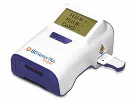
BD VERITOR™ SYSTEM FOR RAPID DETECTION OF SARS-COV-2 & FLU A+B* (EUA) TEST
From BD Veritor™ Plus System
A 3-in-1 antigen assay for COVID-19, Flu A and Flu B with test results in 15 minutes.1
The BD Veritor™ Plus System streamlines clinical and patient workflows by detecting three key respiratory viruses in one test, reducing the need for multiple sample collections. With multiple testing modes and workflow efficiencies, the analyzer empowers convenience and efficiency (1 kit = 30 tests).
*In the USA, the BD Veritor™ System for Rapid Detection of SARS-CoV-2 & Flu A+B has not been FDA cleared or approved but has been authorized by the FDA under an Emergency Use Authorization for use by authorized laboratories; use by laboratories certified under the CLIA, 42 U.S.C. §263a, that meet requirements to perform moderate, high, or waived complexity tests. The product is authorized for use at the Point of Care (POC), i.e., in patient care settings operating under a CLIA Certificate of Waiver, Certificate of Compliance, or Certificate of Accreditation.
This product has been authorized only for the detection of proteins from SARS-CoV-2, influenza A and influenza B, not for any other viruses or pathogens; and, in the USA, the emergency use of this product is only authorized for the duration of the declaration that circumstances exist justifying the authorization of emergency use of in vitro diagnostics for detection and/or diagnosis of COVID 19 under Section 564(b)(1) of the Federal Food, Drug and Cosmetic Act, 21 U.S.C. §360bbb-3(b)(1), unless the declaration is terminated or authorization is revoked sooner.
1. BD Veritor™ Plus System for Rapid Detection of SARS-CoV-2 and Flu A+B. Package insert. 500051910 Becton, Dickinson and Company.
STATUS FLU A&B
From LifeSign
Status Flu A & B is an in vitro rapid qualitative test that detects influenza type A and type B directly from nasal swab, nasopharyngeal swab, and nasopharyngeal aspirate/wash specimens obtained from patients with signs and symptoms of respiratory infection. It is intended to aid in the rapid differential diagnosis of Influenza A and B viral infections.
• CLIA waived *Innovative flip design with onboard sample extraction
• Premeasured developer solution capsule for increased accuracy and ease of use
• Flocked nasal swabs for improved patient comfort and superior specimen collection
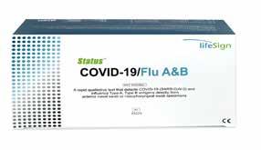
STATUS COVID-19/FLU A&B PANEL TEST
From LifeSign




A Rapid Immunoassay for the Simultaneous Direct Detection and Differential Diagnosis of SARS-CoV-2, Influenza Type A and Type B Antigen from Anterior Nasal and Nasopharyngeal swab specimens. Infections with these viruses may present similar symptoms. Can you tell them apart? WE CAN!
SCAN TO LEARN MORE
6107
SCAN TO LEARN MORE
Comprehensive toxicology menu now with 14 CLIA 1 categorized moderate complexity assays.

IMMTOX ™ 270 BENCHTOP ANALYZER
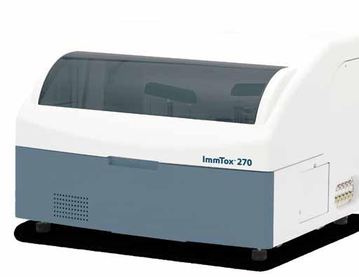
Toxicology screening solutions for physician offices, pain management, treatment centers and laboratories testing 200+ patient samples/mo.
MODERATE COMPLEXITY ASSAYS – FDA 510(K) CLEARED
6-acetylmorphine (6-AM Heroin metabolite)
Amphetamine
Barbiturates
Benzodiazepines
Benzoylecgonine (Cocaine metabolite)
Buprenorphine
Cannabinoids (THC)

EDDP (Methadone metabolite)
Fentanyl*
Methamphetamine
Opiates
Oxycodone
Phencyclidine (PCP)
Tramadol
Scan this QR code to view the ImmTox™ 270 product video
FLU AND RESPIRATORY
BE PREPARED FOR
RESPIRATORY SEASONS WITH THE OSOM® COVID-19 ANTIGEN RAPID TEST
From Sekisui Diagnostics
The OSOM® COVID-19 Antigen Rapid Test is a lateral flow immunoassay that detects the SARS-CoV-2 nucleocapsid protein with a nasal swab in only 15 minutes at the point-of-care. The test is intended to be used by healthcare professionals or operators on patients suspected of COVID-19 within the first 7 days of symptom onset. The clinical performance compares favorably against polymerase chain reaction methodology, with a positive percent agreement of 95.1% and a negative percent agreement of 97%.
OSOM® COVID-19 Antigen Rapid Test has not been FDA cleared or approved. It is authorized by FDA under an EUA for prescription use only. It has been authorized only for the detection of SARS-CoV-2 antigen, not for any other viruses or pathogens and is only authorized for the duration of the declaration that circumstances exist justifying the authorization of emergency use of in vitro diagnostics for detection and/or diagnosis of COVID-19 under Section 564(b)(1) of the Federal Food, Drug, and Cosmetic Act, 21 U.S.C S360bbb-3(b)(1), unless the authorization is terminated or revoked sooner.
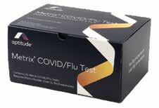
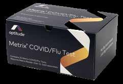
METRIX® COVID/FLU TEST RAPID MOLECULAR
TEST – PROVIDE LAB QUALITY RESULTS IN JUST 20 MINUTES
From Sekisui Diagnostics
• 3-in-1 Multiplex – Detects COVID-19, Flu A and Flu B from one swab, in one test
• Minimal Hands-On Time – Can be performed in a CLIA-waived setting in a few simple steps
• Convenient– No maintenance or calibration, ready for testing any time
• Accessible and Flexible – Suitable for any facility
• Exceptional Support – Experienced support teams and online training modules to help streamline your implementation



This product has not been FDA cleared or approved, but has been authorized for emergency use by FDA under an EUA; This product has been authorized only for the detection and differentiation of nucleic acid from SARS-CoV-2, Influenza A, and Influenza B, not for any other viruses or pathogens. The emergency use of this product is only authorized for the duration of the declaration that circumstances exist justifying the authorization of emergency use of in vitro diagnostics for detection and/or diagnosis of COVID-19, Flu A, and Flu B under Section 564(b) (1) of the Federal Food, Drug, and Cosmetic Act, 21 U.S.C. § 360bbb 3(b)(1), unless the declaration is terminated or authorization is revoked sooner.
COVID-19 OR FLU? STOP GUESSING. ONE SWAB,
ONE
TEST, RESULTS IN JUST 10 MINUTES
From Sekisui Diagnostics
From only one sample, the OSOM® Flu SARS-CoV-2 Combo Test simultaneously detects and differentiates between COVID-19, Flu A, and Flu B in just 10 minutes, allowing healthcare providers to make more informed decisions when treating patients who are symptomatic with viral infections that have similar symptoms, but different treatment protocols. Designed for point-of-care testing, the OSOM® Flu SARS-CoV-2 Combo Test empowers healthcare providers with the confidence necessary to initiate immediate treatment and isolation protocols at the very first patient visit.
This test has not been FDA cleared or approved. It is authorized by FDA under an EUA for use by authorized laboratories. It has been authorized only for the detection of proteins from SARS-CoV-2, influenza A and influenza B, not for any other viruses or pathogens and is only authorized for the duration of the declaration that circumstances exist justifying the authorization of emergency use of in vitro diagnostics for detection and/or diagnosis of COVID-19 under Section 564(b)(1) of the Federal Food, Drug, and Cosmetic Act, 21 U.S.C S360bbb-3(b)(1), unless the authorization is terminated or revoked sooner.
6112


FLU AND RESPIRATORY
CLIA WAIVED, POINT-OF-CARE, RSV TEST
from Sekisui Diagnostics
Test for both high-risk groups, younger children & elderly adults, for severe RSV infection using the OSOM® RSV Test. This test is a CLIAwaived, point-of-care test designed to detect Respiratory Syncytial Virus (RSV) using a painless anterior nasal swab in just 15 minutes. It is suitable for both children aged 6 months to 6 years and adults aged 60 and above, providing healthcare professionals with flexibility in diagnosing patients exhibiting symptoms of respiratory infections.
HEMATOLOGY
TURN SMALL PLACES INTO SMART SPACES
From Abbott
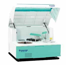
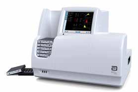
With reduced budgets, shrinking laboratory space and staffing challenges, many laboratories need a solution that lets them work smarter with less. The CELL-DYN Emerald 22 AL is a full performance, automated optical 5-part differential analyzer that delivers smarter results for small to midsize clinical laboratories.
• Compact Design
• Walkaway Functionality
• Ease Of Use
• Smart Safety Features



CLIA-WAIVED COMPLETE BLOOD COUNT CITOCBC
From CitoCBC
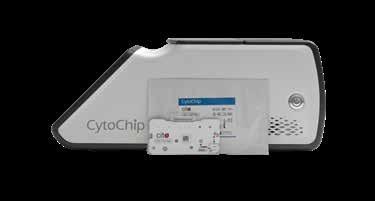
Cito means “quickly” in Latin, and CitoCBC® brings that efficiency to paitent care. FDA-cleared and CLIA-waived, it delivers lab-quality CBC results in minutes, reporting 16 parameters including a 5-part differential. Its cartridge-based design makes operation simple, while LIS connectivity and monthly QC ensure seamless workflow integration. For urgent care and primary care, CitoCBC is a true differentiator — enabling same-visit diagnostics that elevate patient care.


COLORECTAL CANCER SCREENING BEYOND FIT: WHAT’S CHANGING?
BY MATT BAKER, PHYSICIANS OFFICE RESOURCE STAFF WRITER
Colorectal cancer (CRC) remains one of the most preventable yet deadly cancers worldwide. Despite decades of advances in screening and early detection, CRC continues to be one of the leading causes of cancer-related deaths in the United States. For primary care physicians (PCPs), colorectal cancer screening represents one of the most impactful preventive services they can provide. Early detection dramatically improves outcomes, yet screening rates remain below optimal levels in many populations.
Historically, screening strategies have relied heavily on colonoscopy and stool-based tests such as the fecal immu-
nochemical test (FIT). While FIT has become a widely used and accessible screening method, rapid advances in diagnostic technologies are expanding the range of tools available to clinicians. New molecular tests, blood-based diagnostics, and imaging innovations are redefining the screening landscape and creating new opportunities for primary care physicians to identify disease earlier.
Understanding colorectal cancer, the populations at greatest risk, the evolution of screening technologies, and the emerging future of CRC detection is essential for clinicians working on the front lines of preventive care.
Understanding Colorectal Cancer
Colorectal cancer develops in the colon or rectum, which together make up the final portion of the gastrointestinal tract. Most colorectal cancers originate as noncancerous growths known as adenomatous polyps that form in the inner lining of the colon. Over time—often over a period of ten to fifteen years—these polyps may accumulate genetic mutations that transform them into malignant tumors. Because this progression typically occurs slowly, there is a substantial window of opportunity for screening programs to detect and remove precancerous lesions before cancer develops.
In its early stages, colorectal cancer frequently produces few or no symptoms. This silent progression is one of the main reasons screening is so critical. When symptoms do occur, they may include persistent changes in bowel habits such as diarrhea or constipation, narrowing of stool, rectal bleeding, blood in the stool, abdominal pain or cramping, unexplained weight loss, fatigue, or iron-deficiency anemia. Unfortunately, by the time these symptoms appear, the disease may already be advanced. Screening enables clinicians to detect both precancerous polyps and early-stage cancers when treatment is most effective and survival rates are highest.
The survival benefit of early detection is significant. When colorectal cancer is identified at a localized stage, five-year survival rates exceed 90 percent. However, when the disease has spread to distant organs, survival rates drop dramatically. This stark contrast underscores the importance of screening programs that identify disease before symptoms arise.
Who Is at Risk?
Colorectal cancer can develop in individuals without obvious risk factors, but certain characteristics significantly increase the likelihood of developing the disease. Age remains the most important risk factor. The majority of colorectal cancers occur in individuals over the age of 50. However, in recent years there has been a concerning increase in colorectal cancer incidence among younger adults. In response to this trend, major guideline bodies including the U.S. Preventive Services Task Force and the American Cancer Society now recommend that average-risk individuals begin screening at age 45 rather than 50.
Family history also plays a significant role in colorectal cancer risk. Individuals with a first-degree relative—such as a parent or sibling—who has been diagnosed with colorectal cancer or advanced adenomas face a higher risk of developing the disease themselves. In addition to familial clustering, several hereditary cancer syndromes dramatically elevate risk. Lynch syndrome, also known as hereditary nonpolyposis colorectal cancer, is the most common inherited cause of CRC and results from mutations in mismatch repair genes. Familial adenomatous polyposis (FAP), although less common, leads to the development of hundreds or thousands of colon polyps and carries an almost inevitable progression to cancer if untreated.
Lifestyle factors also contribute significantly to colorectal cancer risk. Diets high in processed or red meats have been associated with increased incidence, while diets rich in fruits, vegetables, and fiber appear to offer protective effects. Obesity, sedentary lifestyle, tobacco use, and excessive alcohol consumption have also been linked to higher CRC risk.
Additionally, certain medical conditions increase susceptibility, including inflammatory bowel diseases such as ulcerative colitis and Crohn’s disease, as well as type 2 diabetes.
Primary care physicians play a central role in identifying these risk factors through routine patient interactions. By recognizing individuals with elevated risk profiles, clinicians can initiate earlier screening, tailor screening intervals, and provide preventive counseling that may reduce overall risk.
The History and Evolution of Colorectal Cancer Screening
Screening for colorectal cancer has evolved significantly over the past several decades. Early screening strategies focused primarily on detecting hidden blood in stool, which can be a sign of colorectal tumors. The guaiac-based fecal occult blood test (gFOBT), introduced in the late twentieth century, became one of the first widely adopted population screening tools. This test used a chemical reaction to detect heme in stool samples and demonstrated the ability to reduce colorectal cancer mortality when used regularly.
However, gFOBT had several limitations. Patients were required to follow dietary restrictions prior to testing because certain foods could trigger false-positive results. The test also had relatively low sensitivity for detecting early-stage cancers and precancerous polyps. Despite these drawbacks, gFOBT represented a major milestone in preventive oncology and laid the groundwork for modern screening programs.
The development of colonoscopy transformed colorectal cancer detection and prevention. Unlike stool-based tests, colonoscopy allows direct visualization of the entire colon and rectum using a flexible endoscope. During the procedure, clinicians can identify and remove polyps before they progress to cancer. Because colonoscopy both detects existing tumors and prevents future cancers through polyp removal, it is widely considered the gold standard for colorectal cancer screening.
Despite its effectiveness, colonoscopy is not without barriers. The procedure requires bowel preparation, sedation, and access to specialized medical facilities. Some patients are reluctant to undergo invasive procedures, and limited availability of gastroenterology services in certain regions can restrict access. These challenges have contributed to ongoing interest in non-invasive screening alternatives.
The Emergence and Role of
FIT
The fecal immunochemical test (FIT) represented a significant advancement over earlier stool-based screening methods. Unlike gFOBT, FIT uses antibodies to detect human hemoglobin in stool samples. This specificity eliminates the need for dietary restrictions and improves test accuracy.
FIT offers several advantages that have made it a widely recommended screening option. It is inexpensive, non-invasive, and easy for patients to complete at home. Because the test requires only a single stool sample and no special preparation, patient adherence rates are often higher compared with colonoscopy-based screening programs. FIT is typically recommended on an annual basis for average-risk adults.
However, FIT also has limitations. The test primarily detects bleeding lesions, meaning that polyps or early cancers that are not actively bleeding may be missed. Additionally, because FIT must be repeated annually to maintain effectiveness, long-term patient adherence is critical. Positive results require follow-up colonoscopy to confirm the diagnosis and remove any detected lesions.
While FIT remains an important component of colorectal cancer screening programs, advances in diagnostic science are expanding screening options beyond FIT alone.
Screening Beyond FIT
Recent years have seen rapid innovation in colorectal cancer diagnostics, with new technologies designed to improve detection rates and increase patient participation in screening programs.
One of the most widely adopted innovations is multitarget stool DNA testing. These tests combine traditional FIT with molecular analysis of DNA biomarkers that are shed by cancerous or precancerous cells into stool. By detecting specific genetic mutations and epigenetic changes associated with colorectal cancer, multitarget stool DNA tests can identify tumors that may not yet be bleeding. Clinical studies have demonstrated higher sensitivity for detecting colorectal cancer compared with FIT alone. Because these tests are typically performed every three years rather than annually, they may also improve patient compliance with screening recommendations.
Blood-based screening tests represent another promising frontier in colorectal cancer detection. These tests analyze circulating tumor DNA or other biomarkers present in a patient’s bloodstream. The appeal of blood-based screening is significant, particularly within primary care settings. A simple blood draw performed during routine laboratory testing could potentially identify early-stage colorectal cancers without requiring stool collection or invasive procedures. Several bloodbased CRC screening tests have recently entered the market, and ongoing research continues to evaluate their performance characteristics.
Another alternative screening modality is CT colonography, sometimes referred to as virtual colonoscopy. This imaging-based technique uses specialized computed tomography to create detailed three-dimensional images of the colon. CT colonography is non-invasive and does not require sedation, although bowel preparation is still necessary. If suspicious lesions are identified, a traditional colonoscopy is required to remove polyps or obtain tissue samples.
Beyond these established methods, emerging research is exploring the use of advanced molecular biomarkers, including DNA methylation markers, microRNA signatures, and proteomic indicators of cancer development. Artificial intelligence is also being integrated into diagnostic workflows to improve the detection of polyps during colonoscopy and to enhance risk prediction models.
The Critical Role of Primary Care Physicians
Primary care physicians play a central role in colorectal cancer prevention and early detection. Numerous studies have demonstrated that physician recommendation is one of the strongest predictors of whether patients complete screening. Because PCPs maintain ongoing relationships with patients and coordinate preventive care, they are uniquely positioned to ensure screening guidelines are followed.
Routine primary care visits provide valuable opportunities to assess patient eligibility for screening, discuss available screening options, and address concerns that may prevent patients from participating in screening programs. Some patients may prefer non-invasive tests such as FIT or stool DNA testing, while others may opt for colonoscopy. Offering multiple screening options can significantly improve adherence.
In addition to initiating screening discussions, primary care physicians play an important role in ensuring appropriate follow-up. Positive results from stool-based or blood-based screening tests require timely colonoscopy to confirm the diagnosis and remove potentially precancerous lesions. Coordinating this follow-up care is critical to ensuring screening programs achieve their intended impact.
Primary care clinicians are also instrumental in addressing disparities in screening uptake. Socioeconomic barriers, limited healthcare access, and lack of patient awareness contribute to lower screening rates in certain populations. Through patient education and proactive outreach, PCPs can help close these gaps and improve population-level outcomes.
The Future of Colorectal Cancer Screening
The future of colorectal cancer screening is likely to involve increasingly personalized and technology-driven approaches. Advances in molecular diagnostics, artificial intelligence, and data analytics are expected to reshape how clinicians identify and monitor patients at risk.
Blood-based screening tests may become more widely adopted as evidence supporting their accuracy continues to grow. These tests could significantly improve screening participation among individuals who are reluctant to complete stool-based testing or undergo colonoscopy.
Risk-based screening models are also emerging. By integrating patient genetics, lifestyle factors, medical history, and biomarker data, clinicians may be able to tailor screening intervals and modalities to individual risk profiles. Such precision screening strategies could optimize resource utilization while improving early detection rates.
Artificial intelligence is also expected to play an increasing role in colorectal cancer prevention. AI-assisted colonoscopy systems are already improving adenoma detection rates by helping clinicians identify subtle lesions that might otherwise be missed. Digital health platforms may also enhance screening programs through automated reminders, remote monitoring, and population health analytics.
Conclusion
Colorectal cancer remains a significant public health challenge, yet it is one of the most preventable forms of cancer when effective screening programs are in place. Over the
past several decades, screening technologies have evolved from basic stool tests to advanced molecular diagnostics capable of detecting early-stage disease with increasing precision.
While the fecal immunochemical test continues to serve as an important screening tool, emerging technologies such as multitarget stool DNA tests, blood-based diagnostics, and advanced imaging modalities are expanding the options available to clinicians. These innovations have the potential to increase patient participation in screening programs and detect disease earlier.
For primary care physicians, the opportunity to impact colorectal cancer outcomes is substantial. By identifying patients at risk, initiating screening conversations, and offering a range of screening options beyond FIT, PCPs can play a critical role in reducing colorectal cancer incidence and mortality.
As diagnostic technologies continue to advance, integrating these tools into primary care workflows will be essential. Through proactive screening strategies and continued innovation, healthcare providers can move closer to the goal of preventing colorectal cancer before it becomes life-threatening.

- Move from Waived to Non-Waived Testing with the help of experts
- Expand testing, don’t worry about all the work- policies, fillable forms, e-signatures, calendar reminders included
- Remote access – Lab Directors and Consultants manage compliance efficiently, save time, and travel expenses
more information, call 888.534.2977 or email us at sales@myinspection.us Schedule a Free Demo Today!
STREP TESTS
BD VERITOR™ SYSTEM FOR RAPID DETECTION OF SARS-COV-2 & FLU A+B* (EUA) TEST
From
BD Veritor™ Plus System
A 3-in-1 antigen assay for COVID-19, Flu A and Flu B with test results in 15 minutes.1
The BD Veritor™ Plus System streamlines clinical and patient workflows by detecting three key respiratory viruses in one test, reducing the need for multiple sample collections. With multiple testing modes and workflow efficiencies, the analyzer empowers convenience and efficiency (1 kit = 30 tests).
*In the USA, the BD Veritor™ System for Rapid Detection of SARS-CoV-2 & Flu A+B has not been FDA cleared or approved but has been authorized by the FDA under an Emergency Use Authorization for use by authorized laboratories; use by laboratories certified under the CLIA, 42 U.S.C. §263a, that meet requirements to perform moderate, high, or waived complexity tests. The product is authorized for use at the Point of Care (POC), i.e., in patient care settings operating under a CLIA Certificate of Waiver, Certificate of Compliance, or Certificate of Accreditation.
This product has been authorized only for the detection of proteins from SARS-CoV-2, influenza A and influenza B, not for any other viruses or pathogens; and, in the USA, the emergency use of this product is only authorized for the duration of the declaration that circumstances exist justifying the authorization of emergency use of in vitro diagnostics for detection and/or diagnosis of COVID 19 under Section 564(b)(1) of the Federal Food, Drug and Cosmetic Act, 21 U.S.C. §360bbb-3(b)(1), unless the declaration is terminated or authorization is revoked sooner.
1. BD Veritor™ Plus System for Rapid Detection of SARS-CoV-2 and Flu A+B. Package insert. 500051910 Becton, Dickinson and Company.
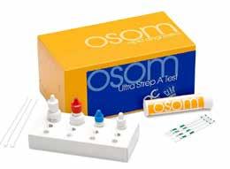
OSOM® ULTRA STREP A TEST
From Sekisui Diagnostics
The OSOM® Ultra Strep A test is a color immunochromatographic assay intended for the qualitative detection of Group A Streptococcus antigen directly from throat swab specimens. Shown to be not statistically different than single swab culture. Sensitivity 95.7% and 100% Specificity. Includes two additional test sticks for External QC. CLIA Waived.
ULTRASOUND
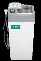
ETHOS AUTOMATED ULTRASOUND PROBE CLEANER DISINFECTOR

From CS Medical 6119

The Ethos Automated Ultrasound Probe Cleaner Disinfector is the first FDA-cleared device to provide both cleaning and high-level disinfection of ultrasound probes in one system. It’s fast and easy to use—simply place a soiled transvaginal, transrectal, or surface probe into the device, follow the prompts on the display, and a reprocessing report is printed at the end of the cycle. Ethos removes the unknowns of manual cleaning, giving healthcare professionals confidence and consistency with every cycle.
T H E Y A R E I N Y O U R W A I T I N G R O O M R
I G H T N O W .
1 O U T O F 3 H A S P A D A N D T H E Y P R O B A B L Y D O N ’ T K N O W I T . A
Healthy Artery
P E R I P H E R A L
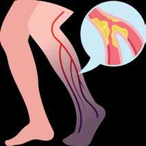
ARTERY DISEASE
Patients with Peripheral Artery Disease (PAD) who may be facing a heart attack, stroke, amputation, or even death within the next 5 years I N T R O D U C I N G
Diabetics Smokers Over age 65

Diseased Artery
Atherosclerosis
(PAD) is an often silent condition where narrowed arteries reduce blood flow to the legs, causing symptoms like leg pain, numbness, and slow-healing wounds.
DON’T LET PAD SNEAK UP ON YOU OR THESE PATIENTS.
50% report no symptoms, while those that do attribute their pain to arthritis or “old age”.

N o n - i n v a s i v e , p a t i e n t - f r i e n d l y t e s t
A C C U R A T E
A c c u r a c y e q u a l o r b e t t e r t h a n D o p p l e r A B I .
U s e f u l f o r d i a b e t i c s w i t h c a l c i f i e d a r t e r i e s
R E I M B U R S A B L E
r .
G r e a t R O I : t h e t y p i c a l i n t e r n i s t h a s 8 0 0 M e d i c a r e p a t i e n t s , p e r A C P
T
l e s s t h a n t w o m o n t h s .
C P T 9 3 9 2 3 ( A B I w / e x e r

THESE PATIENTS TRUST YOU TO FIND THEIR PAD
before they have a heart attack, stroke, or even die PAD also leads to significant disability and reduced quality of life.
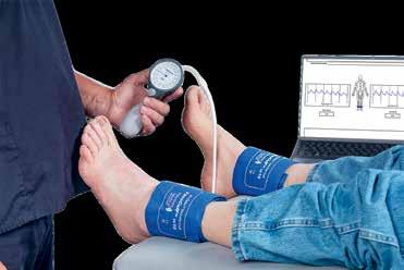

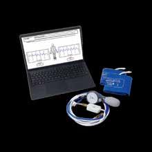
For over 45 years, Newman Medical has been a leader in vascular innovation The ABI-Q system continues that legacy with fast, accurate results you can trust
Scan for more info





9 Travel Destinations You Can't Miss in 2026
Whether you're craving pristine beaches or vibrant cities, these nine must-visit destinations around the world promise stunning scenery, rich culture, and unforgettable adventures for every type of traveler.
md-escapes.com
St. Thomas Ritz-Carlton Vienna
Four Seasons Orlando at Walt Disney World
Secret Bay Dominica · Garza Blanca Puerto Vallarta
Four Seasons Napa Valley · Four Seasons Las Vegas
Four Seasons Nashville
Four Seasons Gresham Palace, Budapest
RITZ-CARLTON ST. THOMAS
Escape to an island oasis surrounded by crystal blue waters, white sand and lush tropical greenery. A breathtaking 30 acre, oceanfront luxury hotel in St. Thomas, VI, The Ritz-Carlton, St. Thomas is a Caribbean marvel styled after traditional island architecture, but richly updated with all of the contemporary comforts for which the resorts of The Ritz-Carlton are known.

SCAN TO LEARN MORE


THE RITZ-CARLTON VIENNA
In a city renowned for its beauty and culture, The Ritz-Carlton, Vienna distinguishes itself with a unique blend of modern luxuries, fine dining, and spa experiences, all housed within four 19th-century palaces. Situated in the heart of downtown Vienna, along the iconic Ringstrasse and adjacent to the Stadtpark, our luxury hotel offers 200 elegant guestrooms, including 43 exquisite suites, allowing you to indulge in tasteful living, distinctive designs, and breathtaking views.

SCAN TO LEARN MORE
RITZ-CARLTON ST. THOMAS
THE RITZ-CARLTON VIENNA
FOUR SEASON ORLANDO AT WALT DISNEY WORLD
ORLANDO, FLORIDA
Discover an elevated escape with the perfect balance of fun and relaxation at our AAA Five Diamond, luxury Orlando Resort. Splash around with the family at Explorer Island water park, or unwind beneath swaying palms at Oasis adult-only pool while we entertain your young ones at our complimentary kids camp. Treat yourself to a soothing, post-park massage at The Spa, then toast to the nightly Walt Disney World® fireworks views over dinner at our Michelin-starred rooftop steakhouse Capa. SCAN TO LEARN MORE



SECRET BAY DOMINICA
Spectacularly sited on a clifftop promontory where the rainforest meets the sea on Dominica, the “Nature Island” of the Caribbean, Secret Bay is among the leading boutique resorts in the world and an acclaimed Relais & Châteaux property. The secluded six-star, all-villa resort consists of elegantly appointed, residential-style villas, each with a private plunge pool and dedicated villa host, on-call concierge, chefs and guides, access to a secret beach and transformative experiences curated specifically for each guest.

SCAN TO LEARN MORE
FOUR SEASONS ORLANDO AT WALT DISNEY WORLD
GARZA BLANCA PUERTO VALLARTA
All Inclusive Family Resort in Puerto Vallarta. Experience the best all-inclusive resort in Puerto Vallarta, where first-class amenities invite you to relax by infinity pools, indulge in gourmet dining, and rejuvenate at a world-class spa embraced by jungle and ocean views. Garza Blanca Puerto Vallarta combines breathtaking natural beauty, exceptional service, and upscale comfort for an unforgettable luxury family vacation.



FOUR SEASONS NAPA VALLEY
Welcome to Five-Star Luxury in California Wine Country. In the food and wine capital of North America, Four Seasons welcomes you to a bespoke luxury resort in Napa Valley, the heart of California wine country. Our Forbes FiveStar Hotel in Calistoga is set within its own world-class vineyard. Discover innovative and seasonal cuisine at Michelin-starred Auro, holistic spa rituals at Spa Talisa and thoughtfully personalized Four Seasons service. Raise a glass to all the best in life as you soak in magnificent views of Napa Valley.

SCAN TO LEARN MORE
GARZA BLANCA PUERTO VALLARTA
FOUR SEASONS
LAS VEGAS, NEVADA
As one of the only non-gaming and non-smoking hotels on The Strip, Four Seasons Hotel Las Vegas is a unique oasis in the heart of the action-packed sports and entertainment capital of the world. Offering Five Diamond luxury accommodations, acclaimed dining and a Forbes Five-Star spa, Four Seasons offers the best of both worlds: a resort retreat amid the famous energy of Las Vegas.



FOUR SEASONS NASHVILLE, TENNESSEE
Welcome to Four Seasons Hotel Nashville, a luxury hotel located in the heart of downtown’s vibrant SoBro neighborhood. This new social hub is just steps away from the city's iconic music, sports, and entertainment venues. Experience the rhythm of our lively restaurants and event spaces, the tranquility of our Spa, and the stunning views from our rooftop pool overlooking the Cumberland River and Riverfront Park. With the unmatched service of Four Seasons and warm Southern hospitality, we’ll inspire an authentic experience of Music City.

SCAN TO LEARN MORE

FOUR SEASONS GRESHAM PALACE BUDAPEST
Located in the heart of Budapest, our luxury art nouveau palace Hotel – the only Forbes Five-Star hotel in Hungary – embodies the spirit of Budapest’s Golden Era glam. From the moment you enter the iconic wrought iron peacock gates, pass through the lobby adorned with more than two million mosaic tiles illuminated with its striking chandelier, and let inspiration wash over you. Outside, views of the Danube and the picturesque Buda skyline stand before you. Spend the day exploring the sights, recharge in our Spa that harnesses the city’s heritage in wellness, then enjoy an innovative cocktail at our lobby bar MÚZSA.

SCAN TO LEARN MORE
Meet the Quadruple Aim in Diabetes Care with In-office HbA1c and uACR
Better outcomes. Lower costs. Better patient experience. Better clinician experience.

Diabetes management solutions at the point-of-care
Gain insights into your patient’s current status with real-time actionable results.
DCA Vantage® Analyzer
Rapid assessment of glycemic control and kidney health
HbA1c
• CLIA-waived
Albumin-to-creatinine ratio (ACR)
• CLIA Moderate Complexity
CLINITEK Status® Connect System
CLIA-waived analyzer for routine urinalysis, including kidney health
• CLINITEK® Microalbumin 2 Strip (ACR)
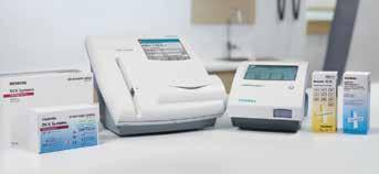
The Prevalence of Diabetes Among U.S. Adults is on the Rise1
Help your patients reverse the trend
Customize your patient consultations to enhance physician-patient partnership toward improved outcomes.

1. Rowley, William R

URINALYSIS IN PRIMARY CARE: AN UNDERRATED DIAGNOSTIC TOOL
BY ADAM IRVINE, PHYSICIANS OFFICE RESOURCE
Urinalysis is one of the oldest diagnostic tests in medicine and remains among the most versatile tools available to clinicians today. Despite advances in molecular diagnostics, imaging technologies, and complex laboratory assays, urinalysis continues to provide rapid, cost-effective, and clinically meaningful insights into a wide range of conditions. For primary care physicians—who often serve as the first point of contact for patients—urinalysis offers a unique opportunity to detect disease early, guide treatment decisions, and monitor chronic conditions.
Yet in modern clinical practice, urinalysis is sometimes overlooked or underutilized. While commonly associated with urinary tract infections (UTIs), its diagnostic potential extends far beyond this single application. Urinalysis can reveal clues
about kidney disease, metabolic disorders, systemic infections, liver dysfunction, and even early indicators of chronic illnesses such as diabetes and hypertension.
As healthcare continues shifting toward preventive medicine and earlier detection of disease, urinalysis deserves renewed attention as a powerful, accessible diagnostic tool in the primary care setting.
Understanding Urinalysis
Urinalysis is a diagnostic test that evaluates the physical, chemical, and microscopic characteristics of urine. Because urine is a filtrate of blood produced by the kidneys, it contains metabolic byproducts and biomarkers that reflect processes occurring throughout the body. Changes in urine composition can
therefore signal abnormalities in multiple organ systems.
A standard urinalysis typically consists of three components: physical examination, chemical analysis, and microscopic evaluation.
The physical examination assesses characteristics such as urine color, clarity, and odor. Normal urine is typically pale yellow and clear. Deviations from this appearance can indicate underlying pathology. Dark amber urine may suggest dehydration or liver disease, cloudy urine can be associated with infection or crystalluria, and reddish discoloration may indicate hematuria.
The chemical analysis is commonly performed using a dipstick test that evaluates several parameters simultaneously. These include pH, specific gravity, protein, glucose, ketones, bilirubin, urobilinogen, blood, leukocyte esterase, and nitrites. Each parameter provides specific clinical insights. For example, the presence of protein may indicate renal disease, glucose can signal hyperglycemia or diabetes, and leukocyte esterase combined with nitrites is suggestive of bacterial infection. The microscopic examination involves centrifuging urine and examining the sediment under a microscope. This analysis can identify red blood cells, white blood cells, epithelial cells, casts, crystals, bacteria, and yeast. These findings provide additional diagnostic clarity, helping differentiate between renal pathology, infection, and contamination.
Together, these three layers of analysis allow urinalysis to serve as a rapid screening tool that can reveal abnormalities across multiple physiological systems.
The Historical Roots of Urinalysis
Urinalysis has been practiced in some form for thousands of years and is among the earliest documented diagnostic techniques in medicine. Ancient physicians recognized that urine could provide valuable information about a patient’s health long before modern laboratory methods existed.
One of the earliest medical practices related to urinalysis was uroscopy, the visual examination of urine. Historical records from ancient Egypt, Greece, and India describe physicians assessing urine color, clarity, and sediment as indicators of disease. In medieval Europe, physicians often carried glass flasks known as “matulas,” which were used to inspect urine samples. The color and appearance of urine were compared against charts to diagnose various conditions.
Although many interpretations during these early centuries lacked scientific accuracy, the underlying concept—that urine reflects internal physiological processes—was fundamentally correct.
Scientific advances during the 17th and 18th centuries began transforming urinalysis into a more objective diagnostic tool. Early chemists discovered that urine from individuals with diabetes contained sugar, which could be detected by tasting the urine or observing fermentation reactions. The 19th century brought further refinement with the introduction of chemical testing methods capable of detecting protein,
glucose, and other substances. Microscopy also began playing a role in urine examination, enabling physicians to identify cells, crystals, and casts that reflected kidney pathology.
The 20th century marked a major turning point with the development of reagent strip testing, commonly known as dipstick testing. These strips allowed clinicians to assess multiple urine parameters simultaneously within minutes, making urinalysis accessible in physician offices, clinics, and hospitals.
Today, urinalysis continues to evolve with automated analyzers, digital microscopy, and emerging biomarker technologies that expand its diagnostic capabilities.
Evolution of Urinalysis in Modern Clinical Practice
Modern urinalysis combines traditional diagnostic principles with advanced laboratory technology. Automated urine analyzers now perform high-throughput testing with improved accuracy and reproducibility, reducing human error and standardizing results across clinical settings.
Automated systems can analyze urine chemistry using advanced reagent strip readers while also performing digital imaging of urine sediment. These technologies enable laboratories to identify cells, crystals, and microorganisms with greater precision than manual microscopy alone.
In addition, clinical laboratories increasingly incorporate reflex testing protocols, where abnormal dipstick findings automatically trigger microscopic examination or additional testing. For example, a positive leukocyte esterase or nitrite result may prompt microscopic analysis to confirm the presence of white blood cells or bacteria.
Point-of-care urinalysis has also become widely available in primary care practices, urgent care centers, and outpatient clinics. Rapid dipstick testing allows clinicians to obtain immediate diagnostic insights during a patient visit, facilitating faster decision-making and treatment initiation.
Despite these technological advancements, the core value of urinalysis remains unchanged: it provides a simple, noninvasive method for assessing a patient’s physiological status.
Why Urinalysis Remains an Underrated Diagnostic Tool
Although urinalysis is routinely used in clinical practice, its full diagnostic potential is often underappreciated. Many clinicians primarily associate urinalysis with diagnosing urinary tract infections, overlooking its broader applications in detecting systemic disease.
One reason urinalysis may be underutilized is the increasing availability of advanced diagnostic technologies. Molecular tests, imaging studies, and specialized laboratory assays have become integral components of modern medicine. While these tools provide valuable insights, they are often more expensive and less accessible than urinalysis.
Urinalysis, by contrast, is inexpensive, rapid, and widely available. Yet its ability to reveal early signs of disease remains significant.
For example, proteinuria detected through urinalysis can serve as an early indicator of chronic kidney disease (CKD). Identifying protein in urine before symptoms develop allows physicians to initiate interventions that may slow disease progression.
Similarly, the detection of microscopic hematuria can signal underlying pathology ranging from kidney stones to malignancy. Early identification enables timely evaluation and referral when necessary.
Urinalysis can also provide valuable clues about metabolic disorders. The presence of glucose and ketones in urine may indicate poorly controlled diabetes, while abnormal pH levels can suggest metabolic disturbances or predisposition to certain types of kidney stones.
Additionally, urinalysis plays a role in identifying systemic illnesses. Conditions such as lupus, vasculitis, and other autoimmune diseases often produce characteristic urine abnormalities before more obvious clinical manifestations occur.
Because urine reflects metabolic and renal processes, urinalysis effectively functions as a window into multiple organ systems. For primary care physicians responsible for early detection and disease management, this makes urinalysis particularly valuable.
Opportunities for Expanded Use in Primary Care
Primary care physicians are uniquely positioned to leverage urinalysis as a screening and monitoring tool across a broad range of conditions.
Routine urinalysis during annual wellness visits can provide baseline information about a patient’s renal and metabolic health. Changes in urine composition over time may reveal early disease processes that would otherwise remain undetected.
Patients with chronic conditions such as diabetes, hypertension, and chronic kidney disease particularly benefit from periodic urinalysis monitoring. Detecting worsening proteinuria, hematuria, or other abnormalities can guide treatment adjustments and referrals to specialists.
Urinalysis also plays an important role in evaluating nonspecific symptoms commonly encountered in primary care. Patients presenting with fatigue, abdominal pain, or edema may have underlying conditions that produce detectable urine abnormalities.
In addition, urinalysis remains a valuable tool in pediatric and geriatric populations, where symptoms of disease may be subtle or atypical. Early detection through routine testing can prevent complications and improve outcomes.
Expanding the use of urinalysis does not require complex
infrastructure. Most primary care clinics already have access to point-of-care dipstick testing, making implementation straightforward.
The Future of Urinalysis
As diagnostic medicine continues evolving, urinalysis is poised to benefit from new technologies that enhance its diagnostic capabilities.
One area of innovation involves urinary biomarkers—molecules in urine that can signal specific disease processes. Researchers are actively studying biomarkers associated with kidney injury, cancer, cardiovascular disease, and neurological disorders. These biomarkers may enable earlier detection of disease than traditional laboratory tests.
Another emerging field is digital urinalysis, which uses artificial intelligence and machine learning to analyze urine microscopy images. AI-powered systems can automatically identify cells, bacteria, and crystals, improving diagnostic accuracy while reducing laboratory workload.
Point-of-care testing technologies are also advancing rapidly. Portable analyzers and smartphone-integrated diagnostic platforms may soon allow clinicians—and potentially even patients—to perform sophisticated urine analyses outside traditional laboratory environments.
Additionally, advances in microfluidics and lab-on-a-chip technologies may enable multiplex urine testing capable of detecting dozens of biomarkers simultaneously from a single sample.
These innovations could transform urinalysis from a basic screening tool into a powerful platform for precision diagnostics.
Reaffirming the Value of Urinalysis in Primary Care
Despite the rapid expansion of advanced diagnostic technologies, urinalysis remains one of the most practical and informative tests available to clinicians. Its noninvasive nature, rapid turnaround time, and broad diagnostic scope make it particularly well suited for primary care settings.
For primary care physicians tasked with detecting disease early and managing diverse patient populations, urinalysis provides valuable insights that extend far beyond urinary tract infections. From identifying early kidney disease to uncovering metabolic and systemic conditions, the information contained in urine can guide clinical decision-making in meaningful ways.
As healthcare continues emphasizing preventive care and early diagnosis, re-embracing urinalysis as a frontline diagnostic tool may improve patient outcomes while reducing healthcare costs.
With ongoing technological advancements and the discovery of new urinary biomarkers, the future of urinalysis appears increasingly promising. For clinicians seeking efficient, accessible diagnostic tools, urinalysis represents not merely a routine test—but an underrecognized cornerstone of modern primary care diagnostics.
TOXICOLOGY
TOXICOLOGY SCREENING SIMPLIFIED ABBOTT’S IMMTOX 270 BENCHTOP ANALYZER NOW WITH 14 ASSAYS
CLIA CATEGORIZED AS MODERATE COMPLEXITY
From Abbott
The ImmTox270 benchtop analyzer offers comprehensive toxicology screening solutions for physician offices, treatment centers and independent laboratories.
Broad test menu with over 20 assays to choose from including 14 that are now available as moderately complex.
With complete laboratory solutions from consultation to licensure, and compliance the Abbott Clinical Laboratory Solutions team has you covered.
FIRST EVER FDA-CLEARED AND CATEGORIZED, POINT-OF-CARE FENTANYL TEST (<6 MINUTES)
From Carolina Liquid Chemistries
Carolina Liquid Chemistries Corp. introduces the Fentanyl Urine Detection Test on CLC’s next-generation RYAN™ immunofluorescent analyzer. This FDA cleared and CLIA categorized, moderately complex, compact, point-of-care system detects fentanyl qualitatively in human urine at a cutoff concentration of 1.0 ng/mL. The test can be run in < 6 minutes, produces a chartable result, and is interfaceable. For detailed information, visit carolinachemistries.com/products/fentanyl-urinedetect-on-clc-ryan-analyzer/.
URINALYSIS

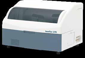

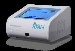
NEED MORE CONTROL OVER YOUR POINT-OF-CARE URINALYSIS TESTING PROCESS?
From Siemens Healthineers
CLINITEK Status ® Connect System simplifies wireless or wired connectivity and testing oversight in point-of-care urinalysis for improved risk management.
• Offers flexible connectivity solutions by integrating data directly to the LIS, EMR or via point-of-care data management software solutions
• Provides improved POC testing workflow efficiencies when interfaced to leading data management solutions
• Minimize transcription errors with the 2-D bar-code scanner
• Improves risk management through advanced operator control functions, prevents unauthorized use
• Drives compliance across testing sites with programmable QC protocols and QC lockout
• Auto-Checks* help to ensure the quality and accuracy of data while facilitating an enhanced interpretation of results.
*Only available when using Siemens test strips with IR or color bands

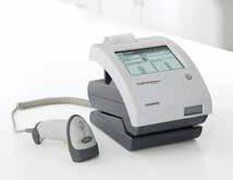
6124
SCAN TO LEARN MORE
WOMEN'S HEALTH
OSOM® BVBLUE®
From Sekisui Diagnostics
The OSOM® BVBLUE® detects elevated vaginal fluid sialidase activity, an enzyme produced by bacterial pathogens associated with bacterial vaginosis including Gardnerella, Bacteroides, Prevotella and Mobiluncus. OSOM® BVBLUE® is more sensitive than Amsel criteria providing physicians with a more accurate diagnosis to treat and minimize serious health consequences such as early spontaneous preterm births and miscarriage.


OSOM® TRICHOMONAS RAPID TEST
From Sekisui Diagnostics
The OSOM® Trichomonas Rapid Test is intended for the qualitative detection of Trichomonas vaginalis antigens from vaginal swabs or from the saline solution. The OSOM® Trichomonas Rapid Test is a CLIA-waived rapid test available today. OSOM® Trichomonas is more sensitive than wet mount due to the assay being able to detect viable and non-viable organisms which offers significant benefits to the patient and clinician alike.
ULTRA HCG COMBO TEST
From Sekisui Diagnostics
The OSOM® Ultra hCG Combo test is a simple immunoassay for the qualitative detection of human chorionic gonadotropin (hCG) in serum or urine for the early confirmation of pregnancy. Internal studies have confirmed that the OSOM® Ultra hCG Combo test does not have a false negative result from hCG variants providing physicians with a higher level of confidence.

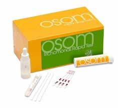


6125
6126
SCAN TO LEARN MORE
6127
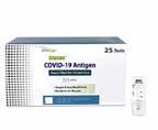


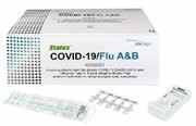

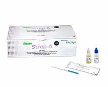
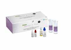


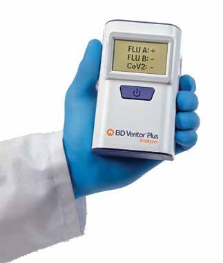
BD Veritor™ Plus System Your simple solution





Digital clarity:
• Unambiguous positive/negative display which may help to reduce result interpretation errors and streamline diagnosis.
Easy to use:
• No monthly calibration required, operated with single-button functionality.
Cost-effective:
• No service contracts, maintenance or calibration needed for device lifetime.
Rapid results:
• Delivers digitally read results in 15 minutes or less,** supporting timely clinical decisions.
Flexible workflow options:
• Walk Away mode: Built in timer allows you to perform other tasks throughout test development.
• Analyze Now mode: Enables immediate processing to support batch testing.†
• Compact, lightweight and portable— able to function without cords.
*In the USA, the BD Veritor™ System for Rapid Detection of SARS-CoV-2 & Flu A+B has not been FDA cleared or approved but has been authorized by the FDA under an Emergency Use Authorization for use by authorized laboratories; use by laboratories certified under the CLIA, 42 U.S.C. §263a, that meet requirements to perform moderate, high, or waived complexity tests. The product is authorized for use at the Point of Care (POC), i.e., in patient care settings operating under a CLIA Certificate of Waiver, Certificate of Compliance, or Certificate of Accreditation. This product has been authorized only for the detection of proteins from SARS-CoV-2, influenza A and influenza B, not for any other viruses or pathogens; and, in the USA, the emergency use of this product is only authorized for the duration of the declaration that circumstances exist justifying the authorization of emergency use of in vitro diagnostics for detection and/or diagnosis of COVID 19 under Section 564(b)(1) of the Federal Food, Drug and Cosmetic Act, 21 U.S.C. §360bbb-3(b)(1), unless the declaration is terminated or authorization is revoked sooner.
**Result processing times for each BD Veritor™ assay are 15 minutes for the SARS-CoV-2 and SARS-CoV-2 & Flu A+B assays, 10 minutes for Flu A+B and RSV assays, and 5 minutes for the Group A Strep assay.
†Any batch testing performed should be conducted at the discretion of the user, in accordance with local policies and clinical judgment.