The material in this eBook also appears in the print version of this title: ISBN: 978-1-26-012240-4, MHID: 1-26-012240-9.
eBook conversion by codeMantra Version 1.0
All trademarks are trademarks of their respective owners. Rather than put a trademark symbol after every occurrence of a trademarked name, we use names in an editorial fashion only, and to the benefit of the trademark owner, with no intention of infringement of the trademark. Where such designations appear in this book, they have been printed with initial caps.
McGraw-Hill Education eBooks are available at special quantity discounts to use as premiums and sales promotions or for use in corporate training programs. To contact a representative, please visit the Contact Us page at www.mhprofessional.com.
Notice
Medicine is an ever-changing science. As new research and clinical experience broaden our knowledge, changes in treatment and drug therapy are required. The authors and the publisher of this work have checked with sources believed to be reliable in their efforts to provide information that is complete and generally in accord with the standards accepted at the time of publication. However, in view of the possibility of human error or changes in medical sciences, neither the authors nor the publisher nor any other party who has been involved in the preparation or publication of this work warrants that the information contained herein is in every respect accurate or complete, and they disclaim all responsibility for any errors or omissions or for the results obtained from use of the information contained in this work. Readers are encouraged to confirm the
information contained herein with other sources. For example and in particular, readers are advised to check the product information sheet included in the package of each drug they plan to administer to be certain that the information contained in this work is accurate and that changes have not been made in the recommended dose or in the contraindications for administration. This recommendation is of particular importance in connection with new or infrequently used drugs.
TERMS OF USE
This is a copyrighted work and McGraw-Hill Education and its licensors reserve all rights in and to the work. Use of this work is subject to these terms. Except as permitted under the Copyright Act of 1976 and the right to store and retrieve one copy of the work, you may not decompile, disassemble, reverse engineer, reproduce, modify, create derivative works based upon, transmit, distribute, disseminate, sell, publish or sublicense the work or any part of it without McGraw-Hill Education’s prior consent. You may use the work for your own noncommercial and personal use; any other use of the work is strictly prohibited. Your right to use the work may be terminated if you fail to comply with these terms.
THE WORK IS PROVIDED “AS IS.” McGRAW-HILL EDUCATION AND ITS LICENSORS MAKE NO GUARANTEES OR WARRANTIES AS TO THE ACCURACY, ADEQUACY OR COMPLETENESS OF OR RESULTS TO BE OBTAINED FROM USING THE WORK, INCLUDING ANY INFORMATION THAT CAN BE ACCESSED THROUGH THE WORK VIA HYPERLINK OR OTHERWISE, AND EXPRESSLY DISCLAIM ANY WARRANTY, EXPRESS OR IMPLIED, INCLUDING BUT NOT LIMITED TO IMPLIED WARRANTIES OF MERCHANTABILITY OR FITNESS FOR A PARTICULAR PURPOSE. McGraw-Hill Education and its licensors do not warrant or guarantee that the functions contained in the work will meet your requirements or that its operation will be uninterrupted or error free. Neither McGraw-Hill Education nor its licensors shall be liable to you or anyone else for any inaccuracy, error or omission, regardless of cause, in the work or for any damages resulting therefrom. McGraw-Hill Education has no responsibility for the content of any information accessed through the work. Under no circumstances shall McGraw-Hill Education and/or its licensors be liable for any indirect, incidental, special, punitive, consequential or similar damages that result from the use of or inability to use the work, even if any of them has been
advised of the possibility of such damages. This limitation of liability shall apply to any claim or cause whatsoever whether such claim or cause arises in contract, tort or otherwise.
Dedication to
William Francis Ganong
William Francis (“Fran”) Ganong was an outstanding scientist, educator, and writer. He was completely dedicated to the field of physiology and medical education in general. Chairman of the Department of Physiology at the University of California, San Francisco, for many years, he received numerous teaching awards and loved working with medical students.
Over the course of 40 years and some 22 editions, he was the sole author of the best selling Review of Medical Physiology, and a co-author of 5 editions of Pathophysiology of Disease: An Introduction to Clinical Medicine. He was one of the “deans” of the Lange group of authors who produced concise medical text and review books that to this day remain extraordinarily popular in print and now in digital formats. Dr. Ganong made a gigantic impact on the education of countless medical students and clinicians.
A general physiologist par excellence and a neuroendocrine physiologist by subspecialty, Fran developed and maintained a rare understanding of the entire field of physiology. This allowed him to write each new edition (every 2 years!) of the Review of Medical Physiology as a sole author, a feat remarked on and admired whenever the book came up for discussion among physiologists. He was an excellent writer and far ahead of his time with his objective of distilling a complex subject into a concise presentation. Like his good friend, Dr. Jack Lange, founder of the Lange series of books, Fran took great pride in the many different translations of the Review of Medical Physiology and was always delighted to receive a copy of the new edition in any language.
He was a model author, organized, dedicated, and enthusiastic. His book was his pride and joy and like other best-selling authors, he would work on the next edition seemingly every day, updating references, rewriting as needed, and always ready and on time when the next edition was due to the publisher. He did the same with his other book, Pathophysiology of Disease: An Introduction to Clinical Medicine, a book that he worked on meticulously in the years following his formal retirement and appointment as an emeritus professor at UCSF.
Fran Ganong will always have a seat at the head table of the greats of the art of medical science education and communication. He died on December 23, 2007. All of us who knew him and worked with him miss him greatly.
About the Authors
KIM E. BARRETT
Kim Barrett received her PhD in biological chemistry from University College London in 1982. Following postdoctoral training at the National Institutes of Health, she joined the faculty at the University of California, San Diego, School of Medicine in 1985, rising to the rank of Professor of Medicine in 1996, and was named Distinguished Professor of Medicine in 2015. From 2006 to 2016, she also served the University as Dean of the Graduate Division. Her research interests focus on the physiology and pathophysiology of the intestinal epithelium, and how its function is altered by commensal, probiotic, and pathogenic bacteria as well as in specific disease states, such as inflammatory bowel diseases. She has published more than 250 articles, chapters, and reviews, and has received several honors for her research accomplishments including the Bowditch and Davenport Lectureships from the American Physiological Society (APS), the Bayliss-Starling Lectureship from The Physiological Society of the UK and Ireland, and the degree of Doctor of Medical Sciences, honoris causa, from Queens University, Belfast. She has been very active in scholarly editing, serving currently as the Editor-in-Chief of the Journal of Physiology. She is also a dedicated and award-winning instructor of medical, pharmacy, and graduate students, and has taught various topics in medical and systems physiology to
these groups for more than 30 years. Her efforts as a teacher and mentor were recognized with the Bodil M. Schmidt- Nielson Distinguished Mentor and Scientist Award from the APS in 2012, and she also served as the 86th APS President from 2013 to 2014. Her teaching experiences led her to author a prior volume (Gastrointestinal Physiology, McGraw-Hill, 2005; second edition published in 2014) and she was honored to have been invited to take over the helm of Ganong in 2007 for the 23rd and subsequent editions, including this one.
SUSAN M. BARMAN
Susan Barman received her PhD in physiology from Loyola University School of Medicine in Maywood, Illinois. Afterward she went to Michigan State University (MSU) where she is currently a Professor in the Department of Pharmacology/Toxicology and the Neuroscience Program. She is also Chair of the Institutional Animal Care and Use Committee and serves on the College of Human Medicine (CHM) Curriculum Development Group for medical school education. She has had a career-long interest in neural control of cardiorespiratory function with an emphasis on the characterization and origin of the naturally occurring discharges of sympathetic and phrenic nerves. She has published about 150 research articles, invited review articles, and book chapters. She was a recipient of a prestigious National Institutes of Health MERIT (Method to Extend Research in Time) Award. She is also a recipient of an MSU Outstanding University Woman Faculty Award, a CHM Distinguished Faculty Award, a Distinguished Service Award from the Association of Chairs of Departments of Physiology, and the Carl Ludwig Distinguished Lecture Award from the Neural Control of Autonomic Regulation section of the American Physiological Society (APS). She is also a Fellow of the APS and served as its 85th President. She has also served as a Councilor of APS and Chair of the Women in Physiology and Section Advisory Committees of the APS. She is also active in the Michigan Physiological Society, a chapter of the APS.
HEDDWEN L. BROOKS
Heddwen Brooks received her PhD from Imperial College, University of London and is a Professor in the Departments of Physiology and Pharmacology at the University of Arizona (UA). Dr. Brooks is a renal physiologist and is best known for her development of microarray technology to address in vivo signaling pathways involved in the hormonal regulation of renal function. Dr. Brooks’ many awards include the American Physiological Society (APS) Lazaro J. Mandel Young Investigator Award, which is for an individual demonstrating outstanding promise in epithelial or renal physiology. In 2009, Dr. Brooks received the APS Renal Young Investigator Award at the annual meeting of the Federation of American Societies for Experimental Biology. Dr. Brooks served as Chair of the APS Renal Section (2011–2014) and currently serves as Associate Editor for the American Journal of Physiology-Regulatory, Integrative and Comparative Physiology, and on the Editorial Board for the American Journal of Physiology-Renal Physiology (since 2001). Dr. Brooks has served on study sections of the National Institutes of Health, the American Heart Association, and served as a member of the Nephrology Merit Review Board for the Department of Veterans’ Affairs.
JASON X.-J. YUAN
Jason Yuan received his medical degree from Suzhou Medical College (Suzhou, China) in 1983, his doctoral degree in cardiovascular physiology from Peking Union Medical College (Beijing, China), and his postdoctoral training at the University of Maryland at Baltimore. He joined the faculty at the University of Maryland School of Medicine in 1993 and then moved to the University of California, San Diego in 1999, rising to the rank of Professor in 2013. His research interests center on pathogenic roles of membrane receptors and ion channels in pulmonary vascular disease. He has published more than 300 articles, reviews, editorials and chapters, and has edited or co-edited nine books. He has received several honors for his research accomplishments including the Cournand and Comroe Young Investigator Award, the Established Investigator Award and the Kenneth D. Bloch Memorial Lectureship from the American Heart Association; the Guggenheim Fellowship Award from the John Simon Guggenheim Memorial Foundation; the Estelle Grover Lectureship from the American Thoracic Society; and the Robert M. Berne Memorial Lectureship from The American Physiological Society. He is an elected Fellow of the American Association for the Advancement of Science and an elected Member of the American Society for Clinical Investigation and the Association of American Physicians. He has served on many advisory committees including Chair of the Respiratory Integrative Biology and Translational Research study section of the National Institutes of Health and Chair of the Pulmonary Circulation Assembly of the American Thoracic Society. He has also been very active in scholarly editing serving currently as the Editor-in-Chief of the journal Pulmonary Circulation and Associate Editor of the American Journal of Physiology-Cell Physiology. He is a leading editor of the Textbook of Pulmonary Vascular Disease (Springer, 2011).
39 Acidification of the Urine & Bicarbonate Excretion
Answers to Multiple Choice Questions
Index
Preface
It is difficult to believe that this preface signifies the fourth edition of Ganong’s Review of Medical Physiology that our author group has overseen, and the 26th edition overall of this important reference work aimed at medical and other health professional students. As always, we have tried to maintain the highest standards of excellence that were promulgated by the original author, Fran Ganong, over the 46 years where he served, remarkably, as the sole author of the textbook.
In this new edition, we have cast a fresh eye on the pedagogical approach taken in each chapter and section, and have focused particularly on including only material that is of the highest yield. We have thoroughly revised the learning objectives for every chapter, reorganized and updated the text to ensure that all objectives are clearly addressed in a logical order, aligned chapter summaries so that the take-home messages quickly address each learning objective in turn, and expanded the number of review questions so that readers also have the ability to check their understanding and retention of every objective covered. As a discipline evolves and new information emerges, there is a tendency simply to concatenate these concepts such that chapter structure degrades inevitably over time. With in-depth discussions amongst the author team and significant “spring-cleaning,” we believe we have freshened and simplified the volume while also making sure that important new developments are incorporated. We are immensely thankful to Erica Wehrwein, PhD, Assistant Professor of Physiology and an award-winning instructor at Michigan State University, who took on the task of reviewing the book as a whole and providing specific and detailed feedback to us on each chapter.
This new edition also welcomes a new member to the author team. We are delighted to have been able to recruit Jason X.-J. Yuan, MD, PhD, Professor of Medicine and Physiology as well as Chief of the Division of Translational and Regenerative Medicine and Associate Vice President for Translational Health Sciences at the University of Arizona, who has assumed responsibility for some
cell physiology and cardiovascular topics, as well as the respiratory physiology section. We are particularly excited to have a physician-scientist on the team, who can guide us overall to focus on material that is of most benefit to those preparing for a career incorporating patient care. We are most grateful for the past contributions of Scott Boitano, PhD, whose other obligations meant that he could no longer serve as an author.
We continue to be gratified by the many colleagues and students who contact us from all over the world to request clarification of material covered in the text, or to point out errors or omissions. We are especially grateful to Rajan Pandit, Lecturer in Physiology at Nepal Medical College, who has painstakingly offered dozens of suggestions for revision over the years. His efforts, and those of the many others whom we have not named, allow us to engage in a process of continual improvement. While, as always, any errors that remain in the book (inevitable in a complex project such as this) are the sole responsibility of the authorship team, we greatly value critical input, and urge readers once again to contact us with any suggestions or critiques. We thank you in advance both for such feedback, and also for your support of this new edition.
This edition is a revision of the original works of Dr. Francis Ganong.
SECTION I
Cellular & Molecular Basis for
Medical Physiology
Study of physiological system structure and function, as well as pathophysiological alterations, has its foundations in physical and chemical laws and the molecular and cellular makeup of each tissue and organ system.
Ganong’s Review of Medical Physiology is structured into seven sections. This first section provides an overview of the basic building blocks or bases that provide the important framework for human physiology. It is important to note here that the seven chapters in this initial section are not meant to provide an exhaustive understanding of biophysics, biochemistry, or cellular and molecular physiology; rather, they are to serve as a reminder of how the basic principles from these disciplines contribute to medical physiology discussed in later sections associated with physiological functions of organs and systems.
In the first two chapters of this section, the following basic building blocks are introduced and discussed: electrolytes; carbohydrates, lipids, and fatty acids; amino acids and proteins; and nucleic acids. Students are reminded of some of the basic principles and building blocks of biophysics and biochemistry and how they fit into the physiologic environment. Examples of direct clinical applications are provided in the clinical boxes to help bridge the gap between basic principles and human cell, tissue, and organ functions. These basic principles are followed up with a discussion of the generic cell and its components. It is important to realize the cell is the basic functional unit within the body, and it is the collection and fine-tuned interactions among and between these fundamental units that allow for proper tissue, organ, and organism function.
In the third to seventh chapters of this introductory section, we take a cellular
approach to lay a groundwork of understanding groups of cells that interact with many of the systems discussed in future chapters. The first group of cells presented contribute to inflammatory reactions in the body. These individual players, their coordinated behavior, and the net effects of the “open system” of inflammation in the body are discussed in detail. The second group of cells discussed are responsible for the excitatory responses in human physiological function and include both neuronal and muscle cells. A fundamental understanding of the inner workings of these cells, and how they are controlled by their neighboring cells, helps the student to understand their eventual integration into individual systems discussed in later sections.
This first section serves as an introduction, refresher, and quick source of material to best understand organ functions and systems physiology presented in the later sections. For detailed understanding of any of the chapters within this section, several excellent and current textbooks that provide more in-depth reviews of principles of biochemistry, biophysics, cell physiology, and muscle and neuronal physiology are provided as resources at the end of each individual chapter. Students are encouraged to visit these texts for a more thorough understanding of these basic principles.
CHAPTER 1
General Principles & Energy Production in Medical Physiology
OBJECTIVES
After studying this chapter, you should be able to:
Define functional units used in measuring physiological properties.
Define pH and buffering.
Understand electrolytes and define diffusion, osmosis, and tonicity.
Define and explain the significance of resting membrane potential.
Understand in general terms the basic building blocks of the cell (eg, nucleotides, amino acids, carbohydrates, and fatty acids) to cell metabolism, proliferation, and function.
Understand higher-order structures of the basic building blocks of the cell (eg, DNA, RNA, proteins, and lipids) to cell replication, proliferation, and signal transduction.
Understand the basic contributions of the basic building blocks of the cell to its structure, function, and energy balance.
INTRODUCTION
In unicellular organisms, all vital processes occur in a single cell. As the evolution of multicellular organisms progressed, various cell groups organized into tissues, and organs have taken over particular functions. In humans and other vertebrate animals, there are a number of specialized collections of cells that consist in organ systems serving for different functions. For example, a gastrointestinal system to digest and absorb food; a respiratory system to take up O2 and eliminate CO2; a urinary system to remove wastes; a cardiovascular system to distribute nutrients, O2, and the products of metabolism; a reproductive system to perpetuate the species; and nervous and endocrine systems to coordinate and integrate the functions of the other systems. This book is concerned with the way these systems function and the way each contributes to the functions of the body as a whole. This first chapter lays a foundation for the discussion of these organ systems with a review of basic biophysical and biochemical principles at the cellular level and the introduction of the molecular building blocks that contribute to cell physiological function within these organ systems.
GENERAL PRINCIPLES
THE BODY AS ORGANIZED “SOLUTIONS”
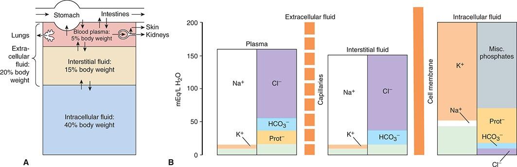
In the average young adult male, 18% of the body weight is protein and related substances, 7% is mineral, and 15% is fat. The remaining 60% is water. The distribution of the body water is shown in Figure 1–1A.
FIGURE 1–1 Organization of body fluids and electrolytes into compartments. A) Body fluids can be divided into intracellular and extracellular fluid compartments (ICF and ECF, respectively). Their contribution
to percentage body weight (based on a healthy young adult male; slight variations exist with age and gender) emphasizes the dominance of fluid makeup of the body. Transcellular fluids, which constitute a very small percentage of total body fluids, are not shown. Arrows represent fluid movement between compartments. B) Electrolytes and proteins are unequally distributed among the body fluids. This uneven distribution is crucial to physiology. Prot , protein, which tends to have a negative charge at physiologic pH.
The cells that make up the bodies of all but the simplest multicellular animals, both aquatic and terrestrial, exist in an “internal sea” of extracellular fluid (ECF) enclosed within the integument of the animal. From this fluid, the cells take up O2 and nutrients; into it, they discharge metabolic waste products. The ECF is more dilute than present-day seawater, but its composition closely resembles that of the primordial oceans in which, presumably, all life originated.
In animals with a closed vascular system, the ECF is divided into the interstitial fluid, the circulating blood plasma, and the lymph fluid that bridges these two domains. The interstitial fluid is the part of the ECF that is outside the vascular and lymph systems, bathing the cells. The plasma and the cellular elements of the blood, principally red blood cells, fill the vascular system, and together they constitute the total blood volume. About one-third of the total body water is extracellular; the remaining two-thirds is intracellular (intracellular fluid). Inappropriate compartmentalization of the body fluids can result in edema (Clinical Box 1–1).
CLINICAL BOX 1–1
Edema
Edema is the buildup of body fluids extracellularly or interstitially in tissues. The increased fluid is related to an increased leak from the blood and/or reduced removal by the lymph system. Edema is often observed in the feet, ankles, and legs, but can happen in many areas of the body in response to disease, including those of the heart, lung, liver, kidney, or thyroid.
THERAPEUTIC HIGHLIGHTS
The best treatment for edema includes reversing the underlying disorder. Thus, proper diagnosis of the cause of edema is the primary first step in therapy. More
general treatments include restricting dietary sodium to minimize fluid retention and using appropriate diuretic therapy.
The intracellular component of the body water accounts for about 40% of body weight and the extracellular component for about 20%. Approximately 25% of the extracellular component is in the vascular system (plasma = 5% of body weight) and 75% outside the blood vessels (interstitial fluid = 15% of body weight). The total blood volume is about 8% of body weight. Flow between these compartments is tightly regulated.
UNITS FOR MEASURING CONCENTRATION OF SOLUTES
In considering the effects of various physiologically important substances and the interactions among them, the number of molecules, electrical charges, or particles of a substance per unit volume of a particular body fluid are often more meaningful than simply the weight of the substance per unit volume. For this reason, physiological concentrations are frequently expressed in moles, equivalents, or osmoles.
Moles
A mole is the gram-molecular weight of a substance, that is, the molecular weight (MW) of the substance in grams. Each mole (mol) consists of 6 × 10²³ molecules. The millimole (mmol) is 1/1000 of a mole, and the micromole (µmol) is 1/1,000,000 of a mole. Thus, 1 mol of NaCl = 23 g + 35.5 g = 58.5 g and 1 mmol = 58.5 mg. The mole is the standard unit for expressing the amount of substances in the SI unit system.
The molecular weight of a substance is the ratio of the mass of one molecule of the substance to the mass of one-twelfth the mass of an atom of carbon-12. Because molecular weight is a ratio, it is dimensionless. The dalton (Da) is a unit of mass equal to one-twelfth the mass of an atom of carbon-12. The kilodalton (1 kDa = 1000 Da) is a useful unit for expressing the molecular mass of proteins. Thus, for example, one can speak of a 64-kDa protein or state that the molecular mass of the protein is 64,000 Da. However, because molecular weight is a dimensionless ratio, it is incorrect to say that the molecular weight of the protein
is 64 kDa.
Equivalents
The concept of electrical equivalence is important in physiology because many of the solutes in the body are in the form of charged particles. One equivalent (Eq) is 1 mol of an ionized substance divided by its valence. One mole of NaCl dissociates into 1 Eq of Na+ and 1 Eq of Cl . One equivalent of Na+ = 23 g, but 1 Eq of Ca2+ = 40 g ÷ 2 = 20 g. The milliequivalent (mEq) is 1/1000 of 1 Eq.
Electrical equivalence is not necessarily the same as chemical equivalence. A gram equivalent is the weight of a substance that is chemically equivalent to 8.0 g of oxygen. The normality (N) of a solution is the number of gram equivalents in 1 liter (L). A 1 N solution of hydrochloric acid (HCl) contains both H+ (1.0 g) and Cl (35.5 g) equivalents, = (1.0 g + 35.5 g)/L = 36.5 g/L.
WATER, ELECTROLYTES, & ACID/BASE
The water molecule (H2O) is an ideal solvent for physiological reactions. H2O has a dipole moment where oxygen slightly pulls away electrons from the hydrogen atoms and creates a charge separation that makes the molecule polar. This allows water to dissolve a variety of charged atoms and molecules. It also allows the H2O molecule to interact with other H2O molecules via hydrogen bonding. The resulting hydrogen bond network in water allows for several key properties relevant to physiology: (1) water has a high surface tension, (2) water has a high heat of vaporization and heat capacity, and (3) water has a high dielectric constant. In layman’s terms, H2O is an excellent biological fluid that serves as a solute; it provides optimal heat transfer and conduction of current.
Electrolytes (eg, NaCl) are molecules that dissociate in water to their cation (Na+) and anion (Cl ) equivalents. Because of the net charge on water molecules, these electrolytes tend not to reassociate in water. There are many important electrolytes in physiology, notably Na+, K+, Ca2+, Mg2+, Cl , and HCO3 . It is important to note that electrolytes and other charged compounds (eg, proteins) are unevenly distributed in the body fluids (Figure 1–1B). These separations play an important role in physiology, for example, in the establishment of membrane potential and generation of action potential.
pH & BUFFERING
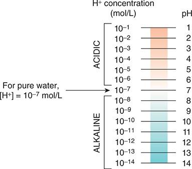
The maintenance of a stable hydrogen ion concentration ([H+]) in body fluids is essential to life. The pH of a solution is defined as the logarithm to the base 10 of the reciprocal of the H+, that is, the negative logarithm of the [H+]. The pH of water at 25°C, in which H+ and OH ions are present in equal numbers, is 7.0 (Figure 1–2). For each pH unit less than 7.0, the [H+] is increased 10-fold; for each pH unit above 7.0, it is decreased 10-fold. In the plasma of healthy individuals, pH is slightly alkaline, maintained in the narrow range of 7.35–7.45 (Clinical Box 1–2). Conversely, gastric fluid pH can be quite acidic (on the order of 3.0) and pancreatic secretions can be quite alkaline (on the order of 8.0). Enzymatic activity and protein structure are frequently sensitive to pH; in any given body or cellular compartment, pH is maintained to allow for maximal enzyme/protein efficiency.
FIGURE 1–2 Proton concentration and pH. Relative proton (H+) concentrations for solutions on a pH scale are shown.
Molecules that act as H+ donors in solution are considered acids, while those that tend to remove H+ from solutions are considered bases. Strong acids (eg, HCl) or bases (eg, NaOH) dissociate completely in water and thus can most change the [H+] in solution. In physiological compounds, most acids or bases are considered “weak,” that is, they contribute or remove relatively few H+ from
solution. Body pH is stabilized by the buffering capacity of the body fluids. A buffer is a substance that has the ability to bind or release H+ in solution, thus keeping the pH of the solution relatively constant despite the addition of considerable quantities of acid or base. Of course there are a number of buffers at work in biological fluids at any given time. All buffer pairs in a homogenous solution are in equilibrium with the same [H+]; this is known as the isohydric principle. One outcome of this principle is that by assaying a single buffer system, we can understand a great deal about all of the biological buffers in that system.
When acids are placed into solution, there is dissociation of some of the component acid (HA) into its proton (H+) and free acid (A ). This is frequently written as an equation:
According to the laws of mass action, a relationship for the dissociation can be defined mathematically as:
Acid–Base Balance and Disorders
Excesses of acid (acidosis) or base (alkalosis) exist when the blood or blood plasma is outside the normal pH range (7.35–7.45). Such changes impair the delivery of O2 to and removal of CO2 from tissues. There are a variety of conditions and diseases that can interfere with pH control in the body and cause blood pH to fall outside of healthy limits. Acid–base disorders that result from respiration to alter CO2 concentration are called respiratory acidosis and respiratory alkalosis. Respiratory acidosis is often caused by respiratory failure or ventilator failure, while respiratory alkalosis is caused by alveolar hyperventilation and often found in patients with chronic liver disease. Nonrespiratory disorders that affect HCO3 concentration are referred to as metabolic acidosis and metabolic alkalosis. Metabolic acidosis or alkalosis can be caused by electrolyte disturbances, severe vomiting or diarrhea, ingestion of certain drugs and toxins, kidney disease, and diseases that affect normal metabolism (eg, diabetes).
THERAPEUTIC HIGHLIGHTS
Proper treatments for acid–base disorders are dependent on correctly identifying the underlying causal process(es). This is especially true when mixed disorders are encountered. Treatment of respiratory acidosis should be initially targeted at restoring ventilation, whereas treatment for respiratory alkalosis is focused on the reversal of the primary causes (eg, alveolar hyperventilation associated with head injury and anxiety, hypoxemia due to peripheral chemoreceptor stimulation, pulmonary embolism, and edema).
Bicarbonate (via intravenous injection) is typically used as a treatment for acute metabolic acidosis. An adequate amount of a chloride salt can restore acid–base balance to normal over a matter of days for patients with a chloride-responsive metabolic alkalosis whereas chloride-resistant metabolic alkalosis requires treatment of the underlying disease.
where Ka is a constant, and the brackets represent concentrations of the individual species (elements). In layman’s terms, the product of the proton concentration ([H+]) and the free acid concentration ([A ]) divided by the bound acid concentration ([HA]) is a defined constant (K). This can be rearranged to read:
If the logarithm of each side is taken:
Both sides can be multiplied by –1 to yield:
This can be written in a more conventional form known as the HendersonHasselbalch equation:
This relatively simple equation is quite powerful. One thing that can be discerned right away is that the buffering capacity of a particular weak acid is
best when the pKa of that acid is equal to the pH of the solution, or when:
Similar equations can be set up for weak bases. An important buffer in the body is carbonic acid (H2CO3). Carbonic acid is a weak acid, and thus is only partly dissociated into H+ and HCO3 :
If H+ is added to a solution of carbonic acid, the equilibrium shifts to the left and most of the added H+ is removed from solution. If OH is added, H+ and OH combine, taking H+ out of solution. However, the decrease is countered by more dissociation of H2CO3, and the decline in H+ concentration is minimized.
A unique feature of HCO3 is the linkage between its buffering ability and the ability for the lungs to remove CO2 from the body. Other important biological buffers include phosphates and proteins.
DIFFUSION
The particles (molecules or atoms) of a substance dissolved in a solvent are in continuous random movement. Diffusion is the process by which a gas or a substance in a solution expands or moves from a region to another, because of the motion of its particles, to fill all the available volume. A given particle is equally likely to move into or out of an area in which it is present in high concentration. However, because there are more particles in the area of high concentration, the total number of particles moving to areas of lower concentration is greater; that is, there is a net flux of solute particles from areas of high concentration to areas of low concentration. The time required for equilibrium by diffusion is proportional to the square of the diffusion distance. The magnitude of the diffusing tendency from one region to another separated by a boundary (eg, cell membrane, blood-gas barrier) is directly proportional to the cross-sectional area across which diffusion is taking place and the concentration gradient, or chemical gradient, which is the difference in concentration of the diffusing substance divided by the thickness of the boundary (Fick’s law of diffusion). Thus,
where J is the net rate of diffusion, D is the diffusion coefficient, A is the area, and Δc/Δx is the concentration gradient. The minus sign indicates the direction of diffusion. When considering movement of molecules from a higher to a lower concentration, Δc/Δx is negative, so multiplying by –DA gives a positive value. The permeabilities of the boundaries across which diffusion occurs in the body vary, but diffusion is still a major force affecting the distribution of water and solutes.
OSMOSIS
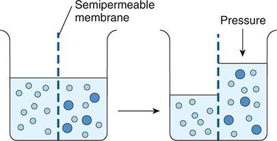
When a substance is dissolved in water, the concentration of water molecules in the solution is less than that in pure water, because the addition of solute to water results in a solution that occupies a greater volume than does the water alone. If the solution is placed on one side of a membrane that is permeable to water but not to the solute, and an equal volume of water is placed on the other, water molecules diffuse down their concentration (chemical) gradient into the solution (Figure 1–3). This process—the diffusion of solvent molecules into a region in which there is a higher concentration of a solute to which the membrane is impermeable—is called osmosis. It is an important factor in physiological processes. The tendency for movement of solvent molecules to a region of greater solute concentration can be prevented by applying pressure to the more concentrated solution. The pressure necessary to prevent solvent migration is the osmotic pressure of the solution.
Osmotic pressure—like vapor pressure lowering, freezing-point depression, and boiling-point elevation—depends on the number rather than the type of particles in a solution; that is, it is a fundamental colligative property of solutions. In an ideal solution, osmotic pressure (P) is related to temperature and volume in the same way as the pressure of a gas:
FIGURE 1–3 Diagrammatic representation of osmosis. Water molecules are represented by small open circles, and solute molecules by large solid circles. In the diagram on the left, water is placed on one side of a membrane permeable to water but not to solute, and an equal volume of a solution of the solute is placed on the other. Water molecules move down their concentration (chemical) gradient into the solution, and, as shown in the diagram on the right, the volume of the solution increases. As indicated by the arrow on the right, the osmotic pressure is the pressure that would have to be applied to prevent the movement of the water molecules.
where n is the number of particles, R is the gas constant, T is the absolute temperature, and V is the volume. If T is held constant, it is clear that the osmotic pressure is proportional to the number of particles in solution per unit volume of solution. For this reason, the concentration of osmotically active particles is usually expressed in osmoles. One osmole (Osm) equals the grammolecular weight of a substance divided by the number of freely moving particles that each molecule liberates in solution. For biological solutions, the milliosmole (mOsm; 1/1000 of 1 Osm) is more commonly used.
If a solute is a nonionizing compound such as glucose, the osmotic pressure is a function of the number of glucose molecules present. If the solute ionizes and forms an ideal solution, each ion is an osmotically active particle. For example, NaCl would dissociate into Na+ and Cl ions, so that each mole in solution would supply 2 Osm. One mole of Na2SO4 would dissociate into Na+, Na+, and SO4 2− supplying 3 Osm. However, the body fluids are not ideal solutions, and although the dissociation of strong electrolytes is complete, the number of
particles free to exert an osmotic effect is reduced owing to interactions between the ions. Thus, it is actually the effective concentration (activity) in the body fluids rather than the number of equivalents of an electrolyte in solution that determines its osmotic capacity. This is why, for example, 1 mmol of NaCl per liter in the body fluids contributes somewhat less than 2 mOsm of osmotically active particles per liter. The more concentrated the solution, the greater the deviation from an ideal solution.
The osmolal concentration of a substance in a fluid is measured by the degree to which it depresses the freezing point, with 1 mol of an ideal solution depressing the freezing point by 1.86°C. The number of milliosmoles per liter in a solution equals the freezing point depression divided by 0.00186. The osmolarity is the number of osmoles per liter of solution (eg, plasma), whereas the osmolality is the number of osmoles per kilogram of solvent. Therefore, osmolarity is affected by the volume of the various solutes in the solution and the temperature, while the osmolality is not. Osmotically active substances in the body are dissolved in water, and the density of water is 1, so osmolal concentrations can be expressed as osmoles per liter (Osm/L) of water. In this book, osmolal (rather than osmolar) concentrations are considered, and osmolality is expressed in milliosmoles per liter (of water).
Note that although a homogeneous solution contains osmotically active particles and can be said to have an osmotic pressure, it can exert an osmotic pressure only when it is in contact with another solution across a membrane permeable to the solvent but not to the solute.
OSMOLAL CONCENTRATION OF PLASMA: TONICITY
The freezing point of normal human plasma averages −0.54°C, which corresponds to an osmolal concentration in plasma of 290 mOsm/L. This is equivalent to an osmotic pressure against pure water of 7.3 atmospheres (atm). The osmolality might be expected to be higher than 290 mOsm/L, because the sum of all the cation and anion equivalents in plasma is over 300 mOsm/L. It is not this high because plasma is not an ideal solution and ionic interactions reduce the number of particles free to exert an osmotic effect. Except when there has been insufficient time after a sudden change in composition for equilibrium to occur, all fluid compartments of the body are in (or nearly in) osmotic equilibrium. The term tonicity is used to describe the osmolality of a solution
relative to plasma. Solutions that have the same osmolality as plasma are said to be isotonic; those with greater osmolality are hypertonic; and those with lesser osmolality are hypotonic. All solutions that are initially isosmotic with plasma (ie, that have the same actual osmotic pressure or freezing-point depression as plasma) would remain isotonic if it were not for the fact that some solutes diffuse into cells and others are metabolized. Thus, a 0.9% saline solution remains isotonic because there is no net movement of the osmotically active particles in the solution into cells and the particles are not metabolized. On the other hand, a 5% glucose solution is isotonic when initially infused intravenously, but glucose is metabolized, so the net effect is that of infusing a hypotonic solution.
It is important to note the relative contributions of the various plasma components to the total osmolal concentration of plasma. All but about 20 of the 290 mOsm in each liter of normal plasma are contributed by Na+ and its accompanying anions, principally Cl and HCO3 . Other cations and anions make a relatively small contribution. Although the concentration of the plasma proteins is large when expressed in grams per liter, they normally contribute less than 2 mOsm/L because of their very high molecular weights. The major nonelectrolytes of plasma are glucose and urea, which in the steady state are in equilibrium with cells. Their contributions to osmolality are normally about 5 mOsm/L each but can become quite large in hyperglycemia or uremia. The total plasma osmolality is important in assessing dehydration, overhydration, and other fluid and electrolyte abnormalities (Clinical Box 1–3).
NONIONIC DIFFUSION
Some weak acids and bases are quite soluble in cell membranes in the undissociated form, whereas they cannot cross membranes in the charged (ie, dissociated) form. Consequently, if molecules of the undissociated substance diffuse from one side of the membrane to the other and then dissociate, there is appreciable net movement of the undissociated substance from one side of the membrane to the other. This phenomenon is called nonionic diffusion.
DONNAN EFFECT
When an ion on one side of a membrane cannot diffuse through the membrane, the distribution of other ions to which the membrane is permeable is affected in a
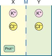
predictable way. For example, the negative charge of a nondiffusible anion hinders diffusion of the diffusible cations and favors diffusion of the diffusible anions. Consider the situation shown in Figure 1–4, in which the membrane (M) between compartment X and compartment Y is impermeable to charged proteins (Prot ) but freely permeable to K+ and Cl .
FIGURE 1–4 Equilibrium across a cell membrane. Diagram showing two compartments (X and Y) separated by the membrane (M). Charged K+ and Cl are distributed in both compartments, while charged protein (prot ) is only in X compartment.
CLINICAL BOX 1–3
Plasma Osmolarity & Disease
Unlike plant cells, which have rigid walls, animal cell membranes are flexible. Therefore, animal cells swell when exposed to extracellular hypotonicity and shrink when exposed to extracellular hypertonicity. Cells contain ion channels and pumps that can be activated to offset moderate changes in osmolarity; however, these can be overwhelmed under certain pathological conditions. Hyperosmolality can cause coma (hyperosmolar coma), a prolonged state of deep unconsciousness. Because of the predominant role of the major solutes and the deviation of plasma from an ideal solution, one can ordinarily approximate the plasma osmolarity within a few mOsm/L by using the following formula, in which the constants convert the clinical units to millimoles of solute per liter:
BUN is the blood urea nitrogen. The formula is also useful in calling attention to abnormally high concentrations of other solutes. An observed plasma osmolarity (measured by freezing-point depression) that greatly exceeds the value predicted by this formula indicates the presence of a foreign substance such as ethanol, mannitol (sometimes injected to shrink swollen cells osmotically), or poisons such as ethylene glycol (component of antifreeze) or methanol (alternative automotive fuel).
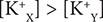
Assume that the concentrations of the anions (eg, Cl ) and of the cations (eg, K+) on the two sides are initially equal. Cl diffuses down its concentration gradient from Y to X, and some K+ moves with the negatively charged Cl because of its opposite charge. Therefore,
Furthermore, that is, more osmotically active particles are on side X (or compartment X) than on side Y (or compartment Y).
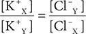
Donnan and Gibbs showed that in the presence of a nondiffusible ion, the diffusible ions distribute themselves so that at equilibrium their concentration ratios are equal:
Cross-multiplying,
This is the Gibbs–Donnan equation. It holds for any pair of cations and anions of the same valence.
The Donnan effect on the distribution of ions has three effects in the body introduced here and discussed below. First, because of charged proteins (Prot ) in cells, there are more osmotically active particles in cells than in interstitial or intercellular fluid, and because animal cells have flexible walls, osmosis would
make them swell and eventually rupture if it were not for the sodium-potassium adenosine triphosphatase (Na, K ATPase) pumping ions back out of cells. Thus, normal cell volume and pressure largely depend on Na, K ATPase, also known as the Na+/K+ pump. Second, because at equilibrium the distribution of permeant ions across the membrane (m, in the example shown in Figure 1–4) is asymmetric, an electrical difference exists across the membrane whose magnitude can be determined by the Nernst equation (see below). In the example used here (Figure 1–4), side X will be negative relative to side Y. The charges line up along the membrane, with the concentration gradient for Cl exactly balanced by the oppositely directed electrical gradient, and the same holds true for K+. Third, because there are more proteins in plasma than in interstitial fluid, there is a Donnan effect on ion movement across the capillary wall.
FORCES ACTING ON IONS
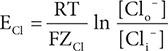
The forces acting across the cell membrane on each ion can be analyzed mathematically. Chloride ions (Cl ) are present in higher concentration in the ECF than in the cell interior, and they tend to diffuse along this concentration gradient into the cell. The interior of the cell is negative relative to the exterior, and chloride ions are pushed out of the cell along this electrical gradient. An equilibrium is reached between Cl influx and Cl efflux. The membrane potential at which this equilibrium exists is the equilibrium potential. Its magnitude can be calculated from the Nernst equation, as follows:
where
ECl = equilibrium potential for Cl
R = gas constant
T = absolute temperature
F = the Faraday number (number of coulombs per mole of charge)
ZCl = valence of Cl (–1)
[Clo ] = Cl concentration outside the cell
[Cli ] = Cl concentration inside the cell
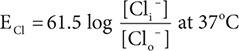
Converting from the natural log to the base 10 log and replacing some of the constants with numeric values holding temperature at 37°C, the equation becomes:
Note that in converting to the simplified expression the concentration ratio is reversed because the –1 valence of Cl has been removed from the expression.
The equilibrium potential for Cl (ECl) in the mammalian spinal neuron, calculated from the standard values listed in Table 1–1, is −70 mV, a value identical to the typical measured resting membrane potential of −70 mV. Therefore, no forces other than those represented by the chemical and electrical gradients need to be invoked to explain the distribution of Cl across the membrane.
A similar equilibrium potential can be calculated for K+ (EK; again, at 37°C):
where
EK = equilibrium potential for K+
ZK = valence of K+ (+1)
[Ko +] = K+ concentration outside the cell
[Ki +] = K+ concentration inside the cells R, T, and F as above
In this case, the concentration gradient is outward and the electrical gradient inward. In mammalian spinal motor neurons, EK is −90 mV (Table 1–1).
Because the resting membrane potential is −70 mV, there is somewhat more K+ in the neurons that can be accounted for by the electrical and chemical gradients.
TABLE 1–1 Concentration of some ions inside and outside mammalian spinal motor neurons.