Exosomes in Aesthetic Dermatology: Biological Basis, Clinical Evidence and Regulatory Considerations Part- II
Acute Adverse Reaction to Intralesional Hyaluronidase in Dermatologic Practice
Multimodal Management of Acne Scarring in Adolescents: Pathogenesis, Diagnosis and Treatment Strategies
Resolution of Acne and PostInflammatory Hyperpigmentation Using Combined Systemic, Topical and Peel-Based Therapy
Multimodal Management of Persistent Melasma: A Case Report
Combining Oral Antioxidant Therapy, Laser Toning and Autologous GrowthFactor Concentrate
Dr. Madhuri Agarwal MBBS, MD (SKIN & VD), DDV
Dr. Komal Jerath MD (Dermatology)
Dr. Jyoti Aneja MD (Dermatology)
Dr. Manu Singh Walia MD (Dermatology)
EXECUTIVE EDITOR & PUBLISHER
Dom Daniel
CORPORATE OFFICE
22, Shreeji Bhavan, 275-279, Samuel Street, Masjid Bunder (W), Mumbai-4000 03, INDIA.
EMAIL: theaestheticiansjournalindia@gmail.com
Website: www.theaestheticiansjournal.online
Printed, Published, Edited and Owned by Dom Daniel Printed at Swastik Printer, Gala No.9 & 10, Vishal Industrial Estate, Bhandup (West), Mumbai- 400078. Published at 22 Shreeji Bhavan, 275/279, Samuel Street, Masjid Bunder (West), Mumbai - 400003. India.
“The Aestheticians Journal” takes no responsibility for unsolicited photographs or material
ALL PHOTOGRAPHS, UNLESS OTHERWISE INDICATED, ARE USED FOR ILLUSTRATIVE PURPOSE ONLY.
Views expressed in this Journal are those of the contributors and not of the publisher. Reproduction in whole or in parts of texts or photography is prohibited. Manuscripts, Photographs and art are selected at the discretion of the publisher free of charge (advertising excluded). Whether published or not, no material will be returned and remains the property of the publishing house, which may make use of it as seen fit. This may include the withdrawal of publication rights to other publishing houses.
All rights reserved. Reproducing in any manner without prior written permission prohibited.
Published for the period of March 2026
Empowering Beauty: A Women’s Day Perspective in Aesthetic Dermatology
..Aesthetic dermatology combines scientific precision with artistic judgment. As patient expectations rise, dermatologists must enhance appearance while preserving individuality through strong anatomical knowledge, evidencebased treatments, ethical practice, and patient understanding.
As we celebrate International Women’s Day, this issue also acknowledges the important relationship between skin health, aesthetic confidence, and women’s wellbeing. Across cultures and age groups, women often seek aesthetic care not merely for appearance, but for the confidence and selfassurance that comes with feeling comfortable in their own skin. Thoughtful, responsible aesthetic practice therefore plays a meaningful role in supporting selfesteem while respecting natural beauty.
This issue explores advances in energy-based devices, injectables, and combination therapies. Emphasis is placed on patient selection, realistic goals, complication management, and effective communication to ensure safe outcomes and patient satisfaction. We also highlight the importance of holistic assessment. True aesthetic success lies not in isolated corrections but in achieving facial harmony and balanced rejuvenation. Subtle enhancements, when thoughtfully planned, can significantly improve confidence and quality of life.
On this Women’s Day, we also recognize the growing contribution of women within dermatology— clinicians, researchers, and innovators who continue to shape the future of aesthetic medicine with dedication and compassion.
As aesthetic dermatology continues to expand, continuous learning and ethical practice remain our strongest foundations. We hope this edition provides practical insights, stimulates critical thinking, and supports your growth in this exciting field.
We invite you to share your clinical experiences, innovations, and perspectives in upcoming issues as we collectively advance the standards of aesthetic care.
Hope you have a great read!
Thanks & Cheers
- Dom Daniel Executive Editor & Publisher
Dr. Deepthi Atmakuri, MD, DDVL
Editorial Board
Dr. Radhika Kopikar
MBBS, DDVL
Medical & Aesthetic Dermatologist
Founder Kopikar Dermatology
Mumbai
Dr. Sama Rais
MBBS, DNB (Dermatology)
Clinical and Aesthetic Dermatologist
Medical Head & Founder Derma Hub
Mumbai
Dr. Deepthi Atmakuri
MD, DDVL
Founder & Consultant Dermatologist
Clinica Derm
Hyderabad
Dr. Sanyogita Warang
MBBS, DDVL, DNB
Consultant Dermatologist
Mumbai
Dr. Neha Agrawal
MD (Dermatology)
Founder & Consultant Dermatologist
ECOS Clinic, Jaipur
Consultant Dermatologist
Jeevanrekha Hospital, Jaipur
Multimodal Management of Persistent Melasma: A Case Report
Melasma is a common acquired disorder of hyperpigmentation characterized by symmetric light to dark brown macules and patches that predominantly appear on sun exposed areas of the face, particularly the forehead, cheeks, and malar regions. The condition occurs more frequently in women and is especially prevalent in individuals with darker skin phototypes, commonly Fitzpatrick skin types III to V. Although medically benign, melasma can have a considerable psychosocial impact due to its chronic nature, cosmetic visibility, and tendency to recur, often negatively affecting quality of life.1
The exact etiology of melasma remains complex and multifactorial. Several triggering factors have been identified, including chronic exposure to ultraviolet radiation, hormonal influences, genetic
predisposition, and inflammatory processes within the skin. Pregnancy, oral contraceptive use, hormone replacement therapy, certain cosmetics, and photosensitizing medications have also been reported to precipitate or exacerbate the condition. Environmental factors, particularly ultraviolet exposure, play a crucial role in stimulating melanocyte activity and increasing melanin production, which contributes to the development and persistence of pigmentation.2,3
Traditionally, melasma was classified into epidermal, dermal, and mixed types based on the localization of melanin within the skin. However, advances in imaging techniques such as in vivo reflectance confocal microscopy have revealed that melanin distribution is often heterogeneous, with features of both epidermal and dermal involvement present in most
cases. This has led to the current understanding that melasma is rarely confined to a single layer of the skin and is better considered a dynamic and mixed pigmentary disorder.2 Histologically, the condition may demonstrate increased epidermal and dermal pigmentation, enlarged and hyperactive melanocytes, increased melanosome transfer, solar elastosis, and enhanced dermal vascularity. In some cases, a mild perivascular lymphohistiocytic infiltrate may also be observed.1
Recent research has further expanded the understanding of melasma pathogenesis. Rather than being solely a disorder of melanocytes, it is now considered the result of complex interactions between melanocytes, keratinocytes, dermal fibroblasts, and vascular endothelial cells. Factors such as vascular growth mediators, mast cells, oxidative stress, and molecular pathways including H19, inducible nitric oxide synthase, and Wnt signaling have also been implicated in disease development and persistence.3, 4 These findings highlight the multifactorial and dynamic nature of melasma and underscore the importance of treatment strategies that target multiple pathogenic pathways to achieve optimal clinical outcomes.
Case Report
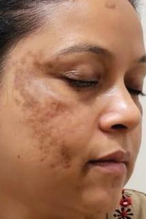
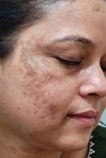
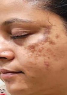
A 36 year old female presented with progressively increasing facial hyperpigmentation of approximately two years duration. The pigmentation was predominantly distributed over the bilateral cheeks, malar areas consistent with the clinical presentation of melasma. The
patient reported prior use of topical depigmenting agents and 3 Laser Toning sessions with minimal improvement. On examination, irregular, symmetric brown to dark brown macular pigmentation was observed over the malar and centrofacial regions. The lesions were non scaly and appeared more prominent in some areas and unchanged in some areas under Woods Lamp, suggesting a mixed epidermal and dermal pattern of melasma. Considering the chronicity and resistance to previous therapy, a multimodal treatment strategy was initiated to target different pathogenic mechanisms involved in melasma.
Treatment Protocol
A comprehensive multimodal treatment strategy was planned to address the multifactorial pathogenesis of melasma, including melanocyte hyperactivity, oxidative stress, and dermal alterations.
• Oral Antioxidant Therapy:
The patient was started on Red Orange Complex (Citrus sinensis 75 mg) with L-Glutathione 500mg once daily to help reduce oxidative stress, regulate melanogenesis, and provide systemic photoprotection. This internal approach was intended to complement procedural therapies and support overall pigment control.
• Laser Toning: The patient underwent three sessions of low fluence fractional laser toning at monthly intervals. The procedure was selected to gently target both epidermal and dermal melanin, facilitating gradual pigment clearance while minimizing the risk of post inflammatory hyperpigmentation.
The interval between sessions allowed adequate skin recovery and progressive improvement.
• Autologous Growth Factor Therapy (Face GFC): Three sessions were performed at monthly intervals to enhance dermal rejuvenation. Growth factors help stimulate cellular repair, improve skin quality, stabilising pigment incontinence and support better pigment regulation, thereby contributing to overall treatment outcomes.
• Topical therapy: The patient was prescribed a 10% tranexamic acid cream twice daily (morning and night) to suppress melanogenesis and reduce vascular-mediated pigmentation. A broad-spectrum SPF 50+ tinted sunscreen was advised for daily photoprotection to prevent UV- and visible light–induced pigment exacerbation.
• Therapeutic Goal: The combined protocol was designed to work synergistically by decreasing melanogenesis, reducing oxidative stress, improving dermal health, and enhancing overall skin tone uniformity and radiance.
Treatment Outcome and Follow-Up
After three months of treatment, a noticeable improvement in pigmentation was observed. There was a visible reduction in the intensity of hyperpigmented patches, particularly over the malar areas, along with better uniformity of skin tone. The skin appeared brighter with improved texture and luminosity. The patient expressed high satisfaction with the clinical outcome, indicating the effectiveness of the combined therapeutic approach in managing persistent melasma.
* Clinical Image Courtesy: Dr. Radhika Kopikar. Unauthorized use, reproduction, or distribution of this image is strictly prohibited without prior permission.
Future Course of Action
• Oral supplements to be continued for 3–6 months to support melanogenesis control and overall skin health.
• Topical tranexamic acid to be applied for 3–6 months to help inhibit melanocyte activation and reduce pigmentation.
• Strict photoprotection with a broad-spectrum sunscreen to prevent UV-induced melanogenesis and recurrence.
• Three additional sessions of low-fluence fractional Q-switched Nd:YAG laser planned at appropriate intervals to further improve pigmentation and enhance treatment outcomes.
Discussion
Melasma remains a challenging pigmentary disorder to manage because of its chronic, relapsing course and is often associated with significant psychosocial impact. Treatment outcomes can vary widely due to differences in clinical presentation, gender, skin phototype, and ethnicity. Because of its multifactorial etiology, effective management requires a multimodal therapeutic strategy that addresses key contributing factors such as photoprotection, inflammation, vascular components, pigmentation pathways, and hormonal influences.1,5
Recent research using histological evaluation, genetic studies, and biochemical analysis has identified
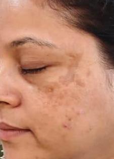
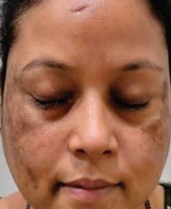
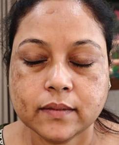
A. Right facial melasma
Figure 1:*
B. Left facial melasma
C. Frontal view showing bilateral malar melasma
Visible reduction in malar hyperpigmentation with improved skin tone uniformity and radiance after
additional mechanisms involved in the development of melasma, including oxidative stress pathways and inflammatory mediators. These insights have supported the development of newer topical therapies, particularly combination formulations containing tyrosinase inhibitors, antioxidants, and anti-inflammatory agents, which have demonstrated encouraging results in improving pigmentation and reducing recurrence. Although it is a benign condition, its visible nature and chronic course can significantly affect an individual’s quality of life.6 Procedural interventions have also emerged as useful adjuncts in the management of melasma. However, treatment remains difficult due to the chronic and relapsing nature of the condition and the variable response to available therapies.7
Management of melasma requires a comprehensive, multimodal approach tailored to the patient’s clinical presentation. Strict photoprotection remains the cornerstone of therapy, while topical agents such as tyrosinase inhibitors, antioxidants, and antiinflammatory formulations help reduce melanogenesis and control pigmentation. In recent years, procedural interventions including various laser and light-based therapies have gained attention as adjunctive options, using controlled thermal energy to target melanin and improve pigmentation. Although responses may vary, these modalities can enhance outcomes when combined with medical therapy and
appropriate maintenance strategies. Therefore, an individualized treatment plan integrating topical therapy, systemic support, procedural interventions, and long-term photoprotection is essential for achieving optimal and sustained clinical improvement.1
Red Orange Complex (ROC) has shown protective and corrective effects against ultraviolet induced skin damage. Studies indicate that ROC improves several UV related skin barrier parameters including erythema, melanin production, transepidermal water loss, skin elasticity, and wrinkle formation. It also suppresses inflammatory responses by inhibiting the mRNA expression of pro inflammatory cytokines such as interleukin 6 and tumor necrosis factor alpha. In addition, ROC enhances the expression of genes responsible for hyaluronic acid and collagen synthesis, thereby supporting dermal repair and improving skin structure. The formulation further reduces collagen degradation by downregulating matrix metalloproteinases. These protective actions are associated with inhibition of UV induced activation of signalling pathways such as c Jun NH2 terminal kinase and activator protein 1, which are involved in photoaging and pigmentation.8
A study by Nobile et al. demonstrated that Red Orange Complex extract, rich in anthocyanins, hydroxycinnamic acids, flavanones, and ascorbic acid, helps counteract the harmful effects of UV radiation and improves signs of skin aging.9
Laser toning has become an
increasingly used modality in the management of melasma, particularly in patients with darker skin types where aggressive laser parameters may worsen pigmentation. The technique utilizes a low-fluence, multipass delivery of a Q-switched 1064-nm laser administered over multiple treatment sessions. It works through the principle of subcellular selective photothermolysis, in which low levels of laser energy delivered in repeated passes gradually heat and disrupt melanosomes within melanocytes rather than destroying the entire cell. This controlled photothermal effect selectively fragments mature pigment particles while preserving the melanocyte cell membrane and nucleus, thereby minimizing cellular damage and reducing the risk of postinflammatory hyperpigmentation. Ultrastructural studies have further shown that laser toning decreases melanocyte dendricity, limiting the transfer of melanin to surrounding keratinocytes. With repeated sessions, this selective targeting of melanosomes results in gradual reduction of pigmentation while maintaining the integrity of surrounding tissues.10,11,12
Growth factor concentrate (GFC) therapy is emerging as a safe and promising addition to the therapeutic armamentarium for melasma. Similar to plateletbased therapies such as PRP, GFC provides a concentrated source of autologous growth factors that can influence pigmentation and support dermal repair. The mechanism of action involves platelet activation and degranulation, resulting in the release of several bioactive
growth factors including plateletderived growth factor (PDGF), epidermal growth factor (EGF), vascular endothelial growth factor (VEGF), fibroblast growth factor (FGF), insulin-like growth factor (IGF), and transforming growth factor-beta (TGF-β). These mediators stimulate fibroblast and keratinocyte proliferation, promote angiogenesis, and enhance collagen synthesis, thereby improving tissue regeneration and skin remodeling. In melasma, certain growth factors also modulate melanogenesis. TGF-β1 has been shown to inhibit melanin synthesis in a concentration-dependent manner by downregulating microphthalmia-associated. .. transcription factor (MITF) and reducing the expression of
References
1. Ogbechie-Godec, O. A., & Elbuluk, N. (2017). Melasma: an Up-to-Date Comprehensive Review. Dermatology and therapy, 7(3), 305–318. https://doi. org/10.1007/s13555-017-0194-1
2. Sarkar R, Bansal A, Ailawadi P. Future therapies in melasma: What lies ahead?. Indian J Dermatol Venereol Leprol 2020;86:8-17
3. Wang W-J, Wu T-Y, Tu Y-K, Kuo K-L, Tsai C-Y, Chie W-C. The optimal dose of oral tranexamic acid in melasma: A network meta-analysis. Indian J Dermatol Venereol Leprol 2023;89:189-94.
5. Sarkar R, Narayan R V, Vinay K, Lakhani R, Sinha S, Mysore V. et al. Prescribing practices of tranexamic acid for melasma: Delphi consensus from the Pigmentary Disorders Society. Indian J Dermatol Venereol Leprol. 2024;90:41-5. doi: 10.25259/IJDVL_1157_2022
6. Darshan Kumar R, Richa Sood, Prashant Tiwari. Melasma management: Unveiling recent breakthroughs through literature analysis. Health Sciences Review.
tyrosinase and tyrosinase-related proteins (TRP-1 and TRP-2), which are key enzymes in melanin production. Additionally, EGF can decrease melanocyte activity by suppressing prostaglandin E2 and tyrosinase activity. Through these combined effects, GFC therapy may help reduce pigmentation while simultaneously improving overall skin quality.13 Tranexamic acid, a fibrinolytic agent, reduces melanogenesis by inhibiting UV-induced plasmin activity in keratinocytes. By blocking the plasminogen–plasmin pathway, it decreases arachidonic acid and prostaglandin production, thereby limiting paracrine stimulation of melanocytes and pigment formation.14,15
Conclusion
This case highlights the value of a multimodal approach in managing persistent melasma. The combination of systemic antioxidant therapy, laser toning, autologous growth factor concentrate, and a consistent topical therapy including tranexamic acid and broadspectrum photoprotection may provide complementary benefits. Oral Red Orange Complex helps address oxidative stress and melanogenesis, laser toning gradually reduces epidermal and dermal pigment, and growth factor therapy supports dermal repair and skin quality. Together, these strategies can enhance pigmentation control, improve skin texture and radiance, and contribute to better overall treatment outcomes and patient satisfaction.
7. Cassiano D, Esposito ACC, Hassun K, Lima MMDA, Lima EVA, Miot LDB, et al. Histological changes in facial melasma after treatment with triple combination cream with or without oral tranexamic acid and/or microneedling: A randomised clinical trial. Indian J Dermatol Venereol Leprol 2022;88:761-70.
8. Kim YH, Lim CY, Jung JI, Kim TY, Kim EJ. Protective effects of red orange (Citrus sinensis [L.] Osbeck [Rutaceae]) extract against UVA-B radiation-induced photoaging in Skh:HR-2 mice. Nutr Res Pract. 2023;17(4):641-659. doi:10.4162/ nrp.2023.17.4.641
9. Nobile, V., Burioli, A., Yu, S., Zhifeng, S., Cestone, E., Insolia, V., Zaccaria, V., & Malfa, G. A. (2022). Photoprotective and Antiaging Effects of a Standardized Red Orange (Citrus sinensis (L.) Osbeck) Extract in Asian and Caucasian Subjects: A Randomized, Double-Blind, Controlled Study. Nutrients, 14(11), 2241. https://doi.org/10.3390/ nu14112241
10. Aurangabadkar SJ. Optimizing Q-switched lasers for melasma and acquired dermal melanoses. Indian J Dermatol Venereol Leprol 2019;85:10-17
11. Shah, S. D., & Aurangabadkar, S. J. (2019). Laser Toning in Melasma. Journal of
cutaneous and aesthetic surgery, 12(2), 76–84. https://doi.org/10.4103/JCAS. JCAS_179_18
12. Wu X, Cen Q, Lin X, Shang Y, Wang X, Zhang Z. Novel melasma therapy using combined low fluence and microsecond pulse Q-switched 1064-nm neodymiumdoped yttrium aluminium garnet laser. Scientific Reports. 2025;15:24596. doi:10.1038/s41598-025-10129-4
13. Sthalekar, B., Agarwal, M., Sharma, V., Patil, C. Y., & Desai, M. (2021). Prospective Study of Growth Factor Concentrate Therapy for Treatment of Melasma. Indian dermatology online journal, 12(4), 549–554. https://doi.org/10.4103/idoj. IDOJ_750_20
14. Agrawal M, Varma K, Kumar U, Bhargava S, Mahadik A, Agrawal V. Comparison of oral versus topical tranexamic acid for treatment of melasma [Internet]. IP Indian J Clin Exp Dermatol. 2023 [cited 2026 Feb 26];9(2):84-89. Available from: https://doi. org/10.18231/j.ijced.2023.015
15. Gaćina, K., & Krstanović Ćosić, A. (2023). THE USE OF TRANEXAMIC ACID IN DERMATOLOGY. Acta clinica Croatica, 62(2), 368–372. https://doi. org/10.20471/acc.2023.62.02.16
Exosomes in Aesthetic Dermatology: Biological Basis, Clinical Evidence and Regulatory Considerations Part- II
Dr. Sama Rais
MBBS, DNB (Dermatology)
Clinical and Aesthetic Dermatologist
Medical Head & Founder Derma Hub
Mumbai Exosome source
Several sources for isolation of exosomes have been tried across several studies. Below is a compilation of their reported
effects across studies including predominantly preclinical data, for skin rejuvenation and hair growth.
Table 1: Source-wise Evidence Mapping for MSC-Derived and Non-MSC Exosomes in Skin and Hair Applications (Conceptual evidence synthesis for research interpretation only)
Exosome Source
Placentaderived MSC exosomes
Adiposederived MSC (ADSC) exosomes
Hair folliclederived exosomes
Bone marrowderived MSC exosomes
Foreskinderived MSC exosomes
Primary Indications Evaluated
Skin rejuvenation; hair density improvement
Reported Biological / Clinical Effects
Increased collagen synthesis; wrinkle reduction; clinically reported increases in hair density
Study Design & Population Strength of Recommendation*
Reduced trans epidermal water loss; antiinflammatory effects
Evidence Interpretation
Summary
• No approved clinical pathway for allogeneic MSC exosomes in aesthetics.
• Highest relative certainty exists for placenta- and adiposederived MSC exosomes in shortterm skin rejuvenation and hair
Experimental and exploratory studies
Translational and clinical wound-healing studies
Experimental and early clinical studies
Not recommended outside research
Evidence supports antiinflammatory effects rather than cosmetic hair induction14, 15
Lack of controlled human trials; formulation variability12
Conditional (nonhair)
Hair effects are secondary; no evidence of follicular regeneration12
Weak
No direct evidence supporting hair growth outcomes12
density outcomes, though evidence remains non-definitive and heterogeneous.12, 13, 14
• Umbilical cord MSC exosomes demonstrate stronger alignment with inflammatory modulation than cosmetic hair induction, supporting potential use in educational or investigational inflammatory scalp contexts.14, 15
• Combination and non-MSC exosome products currently lack sufficient clinical validation and should be considered experimental.12
Table 2: Comparative Evidence: Platelet-Derived vs MSC-Derived Exosomes in Hair Regeneration (Conceptual evidence synthesis for research interpretation only)
Lower theoretical oncologic risk but no validated dosing or efficacy17
Experimental modality with mechanistic promise but no proven clinical efficacy
Key Regulatory Interpretation
• Platelet-derived exosomes demonstrate mechanistic plausibility for hair-cycle modulation but lack human efficacy data, precluding clinical claims.17
• No approved clinical pathway for allogeneic MSC exosomes in aesthetics.
• MSC-derived exosomes show broader biological activity and limited early clinical signals, but outcomes remain heterogeneous and nonstandardized.12, 13, 14
Transdermal Absorption of Exosomes in Dermatology
The intact stratum corneum represents a major barrier to trans epidermal penetration of extracellular vesicles, making stand-alone topical application of exosomes biologically inefficient for dermal delivery in most settings.13 General exosome literature notes poor stratum corneum transit due to size (40–100 nm). Consequently,
13
Promotion of anagen entry, follicular survival, perifollicular angiogenesis, and dermal support12, 13
Extensive in vitro and animal data demonstrating haircycle modulation across multiple MSC sources12, 13, 14
Limited but emerging clinical studies reporting increased hair density and thickness, particularly with adipose- and placenta-derived sources13, 14
Variable outcomes depending on MSC source, isolation method, and delivery protocol12, 13
Low–Moderate (early clinical + preclinical)
Conditional support for hair density improvement; durability and generalizability remain uncertain13, 14
Not approved for hair regeneration
Greater biologic complexity; long-term safety and standardization unresolved12
More mature evidence base, yet still investigational and non-approved
dermatologic studies have focused on delivery strategies that transiently disrupt the epidermal barrier to facilitate dermal access.13
Delivery Modalities Investigated
Exosomes have been administered:
• Topical emulsions
• Microneedling-assisted topical delivery
• Micro-needle radiofrequency (MNRF)
• Fractional laser–assisted application13
• Intradermal injections (Allogenic /autologous exosome products is not recommended outside IRB-approved clinical trials)
Among these, procedures that generate controlled microchannels consistently demonstrate superior delivery efficiency compared with topical-only approaches.13
Delivery Methods with the Strongest Evidence
The highest level of clinical evidence exists for fractional CO₂ laser followed by immediate topical exosome application. However, evidence does not establish clinical superiority or regulatory acceptability. A double-blind randomized split-face trial in acne scarring demonstrated greater clinical improvement on the exosome-treated side, accompanied by reduced erythema and shorter downtime.13 The study attributed these outcomes to laser-created micro-ablative columns that temporarily bypass the epidermal barrier, enabling dermal penetration of nanoscale vesicles.¹³
Microneedling followed by immediate topical exosome application has also shown clinical benefit. A PubMed-indexed case series using 0.5-mm microneedling reported improvements in skin texture and pore appearance without severe adverse events, with efficacy
attributed to microneedlinginduced microchannels facilitating deeper delivery.13
Dose Ranges
Experimental data demonstrate a dose-dependent relationship between exosome particle density and fibroblast proliferation, angiogenesis, and extracellular matrix remodelling, supporting concentrations near 1010 particles/mL as an experimental reference range rather than a validated clinical dose.18 Across published human studies, no universally standardized topical exosome dose has been established, and dosing is reported primarily as particle concentration and applied volume rather than mass-based units.13,19
Side effects
Possible side-effects and adverse events reported with exosomes used for aesthetic skin rejuvenation, from literature reviews are as follows:
• Delayed-onset granulomatous reactions presenting as erythematous, indurated papules or nodules at injection sites have been reported weeks to months after treatment, consistent with a foreign-body inflammatory response.20
• Persistent inflammatory cutaneous reactions, including erythema, swelling, nodules, hyperpigmentation, and scarring, have been observed following intradermal injection of unregulated exosome formulations.20 Treatments included corticosteroids (systemic and intralesional), laser therapy, and surgical removal, with incomplete resolution and residual scarring in all cases.20
• Ischemic necrosis with
subsequent scarring has been described after dermal injection of exosome products used offlabel, highlighting the potential for severe tissue compromise.21
• Risk of immune rejection and pathogen transmission, from allogenic or synthetic sources.22
• Mild, transient reactions such as erythema, oedema, and minor bleeding have been reported following topical or combined microneedlingbased exosome treatments, suggesting a comparatively more favourable short-term safety profile with superficial application.²³
Most severe complications appear to be associated with intradermal injection of unapproved or poorly regulated exosome products, while topical use after microneedling shows fewer reported adverse events, although long-term safety data remain limited. Intradermal injection of exosome products is not recommended outside IRBapproved clinical trials.
Current evidence does not demonstrate a confirmed longterm malignancy risk from exosome preparations used for anti-ageing, skin rejuvenation, or hair growth; however, definitive safety data are lacking. Theoretical oncogenic mechanisms include transfer of pro-proliferative microRNAs, activation of PI3K/AKT or MAPK signalling, promotion of angiogenesis, and immune modulation that could favour tumour microenvironments, particularly with allogeneic or poorly characterized products. Most concerns arise from preclinical oncology models, not aesthetic clinical studies. Long-
term, well-controlled human data are absent, warranting cautious, investigational use only.24, 25, 26
Regulatory and Technical Constraints in Clinical Exosome Use
Translation of exosome research into dermatological practice remains constrained by regulatory classification and unresolved technical limitations.¹
From a regulatory perspective, exosome products intended for therapeutic or diseasemodifying use are classified in the United States as unapproved biological products, requiring formal investigational authorization. Manufacturing processes involving cell culture expansion and vesicle isolation exceed definitions of minimal manipulation, thereby triggering biologics-level oversight and stringent compliance requirements. Regulatory authorities have further highlighted safety concerns related to sterility assurance, batch-to-batch consistency, and unsubstantiated therapeutic claims.
On the technical front, commonly used isolation methods, including ultracentrifugation and sizeexclusion chromatography, are associated with low yield, co-isolation of nonexosome components, and limited scalability.1 Biological heterogeneity related to donor cell source, culture conditions, and processing methods introduces variability in vesicle cargo, complicating reproducibility across experimental systems.1,3
Exosome stability is additionally challenged by enzymatic degradation and vesicle aggregation during storage, which may alter functional activity.1 Preclinical findings derived largely from murine skin models may not reliably predict biological behaviour in human skin because of structural and physiological differences.³
Regulatory and Evidence Caveats
No exosome-based product or delivery technique is approved by the US FDA or CDSCO for dermatologic or aesthetic indications. Reported outcomes are heterogeneous, productspecific, and not generalizable across formulations or suitable for routine clinical recommendation.
Informed consent form
Obtaining informed consent is
References
1. Wei H, Chen Q, Lin L, et al. Regulation of exosome production and cargo sorting. Int J Biol Sci. 2021;17(1):163–177.
2. Senoo M, Pinto F, Crum CP, McKeon F. p63 is essential for the proliferative potential of stem cells in stratified epithelia. Cell. 2007;129:523–536.
3. Xiong M, Zhang Q, Hu W, et al. The novel mechanisms and applications of exosomes in dermatology and cutaneous medical aesthetics. Pharmacol Res. 2021;166:105490.
4. Zhang B, et al. Topical application of mesenchymal stem cell exosomes alleviates imiquimod-induced psoriasislike inflammation. Int J Mol Sci. 2021;22(2).
5. Kim WS, Park BS, Sung JH. The woundhealing and antioxidant effects of adiposederived stem cells. Expert Opin Biol Ther. 2009;9(7):879–887.
6. Shabbir A, Cox A, Rodriguez-Menocal L, Salgado M, Van Badiavas E. Mesenchymal stem cell exosomes induce proliferation and migration of normal and chronic wound fibroblasts, and enhance
essential to ensure patients understand the experimental nature of exosome-based interventions and potential risks. Appropriate disclosure and regulatory oversight are required to safeguard patient welfare.27
Educational and Scientific Use Disclaimer
The information summarized herein is provided solely for scientific and educational purposes. The described findings do not constitute evidence of clinical safety, efficacy, or regulatory approval. Any clinical application outside approved investigational frameworks should be considered off-label and investigational, requiring ethical oversight and compliance.
Conclusion
No exosome-based product, source, dose, or delivery modality discussed herein is approved by regulatory authorities for dermatologic or aesthetic indications. References to disease models, molecular pathways, or biological effects are derived from preclinical or exploratory human studies and must not be interpreted as evidence of therapeutic intent, safety, or efficacy. Clinical use outside formally approved investigational frameworks carries regulatory, ethical, and medico-legal risk.
In summary, while exosome-based approaches represent a promising research frontier, widespread clinical adoption will depend on standardized manufacturing, robust clinical trials, and clearly defined regulatory pathways.
7. Chin T, Lee XE, Ng PY, et al. The role of cellular senescence in skin aging and agerelated skin pathologies. Front Physiol. 2023;14:1297637.
8. Rinnerthaler M, Bischof J, Streubel MK, et al. Oxidative stress in aging human skin. Biomolecules. 2015;5(2):545–589.
9. R H Mahmoud, E Peterson, E V. Badiavas, M Kaminer, Al E. Eber.Exosome: A Comprehensive Review for the Practicing Dermatologist. J Clin Aesthet Dermatol. 2025;18(4):33–40.
10. Tienda-Vázquez MA, Hanel JM, Márquez-Arteaga EM, Salgado-Álvarez AP, Scheckhuber CQ, Alanis-Gómez JR, Espinoza-Silva JI, Ramos-Kuri M, Hernández-Rosas F, Melchor-Martínez EM, Parra-Saldívar R. Exosomes: A Promising Strategy for Repair, Regeneration and Treatment of Skin Disorders. Cells. 2023 Jun 14;12(12):1625. doi: 10.3390/ cells12121625. PMID: 37371095; PMCID: PMC10296902.
11. Zhang Y, Liu Y, Liu H, Tang WH. Exosomes: biogenesis, biologic function and clinical potential. Cell Biosci. 2019 Feb 15;9:19. doi: 10.1186/s13578019-0282-2. PMID: 30815248; PMCID: PMC6377728.
12. Dal’Forno-Dini T, Birck MS, Rocha M, Bagatin E. Exploring the reality of exosomes in dermatology. An Bras Dermatol. 2025;100(1):121–130. doi:10.1016/j.abd.2024.09.002. PMID: 39562240; PMCID: PMC11745280.
13. Al Ameer MA, Alnajim AT, Al Ameer A, et al. Exosomes and hair regeneration: a systematic review of clinical evidence across alopecia types and exosome sources. Clin Cosmet Investig Dermatol. 2025;18:2215–2227. doi:10.2147/CCID. S543451. PMID: 409XXXXX.
14. Siriphanit R, Thanasarnaksorn W, Boonpethkaew S, et al. Comparative study of adipose- and umbilical cord-derived MSC exosomes in dermatologic models. Exp Dermatol. 2025;34(11):e70176. doi:10.1111/exd.70176. PMID: 41217063.
15. Huang YC, Chang CY, Huang CJ. Effectiveness of exosomes from different mesenchymal stem cells in psoriasis: murine study and meta-analysis. Biomedicines. 2025;13(9):2093. doi:10.3390/biomedicines13092093. PMID: 41007656; PMCID: PMC12467663.
16. Dal’Forno-Dini T, Bagatin E. Immunomodulatory and regenerative implications of exosomes in inflammatory skin disease. An Bras Dermatol. 2025. PMID: 39562240.
17. Lu C, Zhang Y, Li Y, et al. Plateletrich plasma-derived exosomes stimulate hair follicle growth through activation of the Wnt/β-catenin signaling pathway. Regenerative Therapy. 2025;24:1-12. doi:10.1016/j.reth.2025.01.0XX. PMID: 40487920.
18. Sun T, Li M, Liu Q, et al. Insights into optimizing exosome therapies for acute skin wound healing and other tissue repair. Front Med. 2024;18(2):258-284. doi:10.1007/s11684-023-1031-9. PMID: 38216854; PMCID: PMC11283324.
19. Bai G, Truong TM, Pathak GN, Benoit L, Rao B. Clinical applications of
exosomes in cosmetic dermatology. Skin Health Dis. 2024;4(6):e348. doi:10.1002/ ski2.348. PMID: 39624733; PMCID: PMC11608875.
20. Park KY. Adverse reactions following intradermal injection of exosome-based formulations: a case series. J Cosmet Dermatol. 2025 Oct;24(10):e70520. doi:10.1111/jocd.70520. PMID: 41097876; PMCID: PMC12528963.
21. AlBargawi S. Necrosis following dermal injection of lyophilized exosomes: a case report. J Cosmet Dermatol. 2025 Aug;24(8):e70387. doi:10.1111/ jocd.70387. PMID: 40820962; PMCID: PMC12359287.
22. Lener T, Gimona M, Aigner L, et al.Applying extracellular vesicles based therapeutics in clinical trials – an ISEV position paper. J Extracell Vesicles. 2015;4:30087.doi:10.3402/jev.v4.30087. PMID: 26725829; PMCID: PMC4698466..
23. Wan J, Yoon SE, Yi KH. The efficacy of combined exosome (Exodew) and microneedling treatment for facial pore reduction and skin texture improvement. Plast Reconstr Surg Glob Open. 2025
Jun 5;13(6):e6849. doi:10.1097/ GOX.0000000000006849. PMID: 40474926; PMCID: PMC12140750.
24. Kalluri R, LeBleu VS. The biology, function, and biomedical applications of exosomes. Science. 2020;367(6478):eaau6977. PMID: 32029601.
26. Tkach M, Théry C. Communication by extracellular vesicles: where we are and where we need to go. Cell. 2016;164(6):1226–1232. PMID: 26967288.
27. Wang L, Hu L, Zhou X, Xiong Z, Zhang C, Shehada HMA, et al. Author Correction: Exosomes secreted by human adipose mesenchymal stem cells promote scarless cutaneous repair by regulating extracellular matrix remodeling. Sci Rep. 2021;11:3245. doi: 10.1038/s41598021-82225-0.
Experiences as a Woman: Balancing Practice, Society and Family
1. What does it mean to you to be a "strong woman"?
Strong will, a positive outlook and compassion is my definition of a strong woman. These are the qualities built slowly, sometimes painfully through a journey that was tough and challenging and filled with struggles but that has also been the most rewarding and satisfying of my life. Strong will for me means not bowing down to pressure of any kind. It means sticking to your convictions even when the easier thing would be to bend. The positive outlook taught me something that took time to fully understand that women don't need to erase themselves to succeed but they do need to stop being timid. It is about being proud to be a woman fully and without apology. And compassion that is the most underrated of the three gave me the insight to recognise differences. It is about learning from the objectivity that men often bring while holding on fiercely to my own sensitivity. This combination I have come to believe is a distinct kind of intelligence.
2. How do you deal with situations where you feel your gender is being used against you?
There was a period in my life when professional setbacks and a serious personal crisis arrived almost simultaneously. Alongside all of that were the quieter, more insidious pressures such as the societal perceptions of what a woman physician should be, how she should carry herself, what she should tolerate how much space she was entitled to take up. I did not handle it gracefully every single day. Some days were genuinely hard. But I did not
Dr. Madhuri Agarwal MBBS, MD (SKIN & VD), DDV Founder & Medical Director Yavana Aesthetics Clinic Mumbai
give up and I kept getting up again and again. What pulled me through was the realisation that I did not have to face it alone. My family was there unconditionally and I learned that asking for help is not a concession. Some of the most enduring bonds of my life were formed in those difficult moments. I also learned and this one surprised me not to take everything so seriously. To find the moments worth enjoying even inside the hard seasons and that shift in perspective changed everything.
3. Do you believe there are areas where women are still underrepresented, and if so, which ones?
Yes and I think we do ourselves a disservice when we pretend otherwise to make it more comfortable. The underrepresentation is not always visible in numbers alone. Sometimes it is in who gets heard in a room, who is assumed to be the authority and who has to
earn that assumption repeatedly with the same credentials, the same experience standing right next to a male counterpart. In medicine, women have made extraordinary inroads. But leadership, research, the spaces where decisions get made about the future of the field those remain disproportionately occupied. And beyond the profession the expectations placed on women in their personal lives have not shrunk at the same pace that their professional ambitions have grown. That gap between what is expected of us at home and what we are reaching for outside it is still very real and very rarely acknowledged honestly.
4. How do you advocate for yourself and other women in your life?
I advocate about being aware about the full picture. Not just the achievements but the struggle behind them. I think one of the quiet harms we do to younger women is presenting only the polished version of success. They see the outcome and assume the path was cleaner than it was and then when their own path gets messy they think something has gone wrong with them specifically. My pursuit in this field was never only about achieving excellence in skills and techniques. It has always been about how much value I can add to the lives of the people who place their faith and trust in me. When I advocate for other women it comes from that same place. It's not transactional. It is about reflecting back to them what they're capable of before they can see it themselves.
5. What advice would you give to young women about navigating the world as a female?
Don't be timid but at the same time don't lose yourself in the pursuit of proving something. Be proud to be a woman. Learn from everyone around you especially from those who are different from you but never at the cost of your own sensitivity. That sensitivity is not a liability but your greatest asset. Cherish your family. Ask for help without shame. And find a way to not take everything so seriously because the moments worth smiling about are happening right alongside the hard ones and they deserve your attention too. I believe happiness is free so sprinkle it everywhere. The
best can always be better and that for me is not a burden but it is the whole point.
1. What does it mean to you to be a strong woman?
To me, being a strong woman means having the courage to dream beyond circumstances and the patience to build those dreams slowly. Strength is staying consistent through ups and downs without losing grace. It is balancing family and profession without guilt.
2. How do you deal with situations where you feel your gender is being used against you?
In my early years of introducing aesthetics and lasers in Amritsar, skepticism was real. Instead of reacting emotionally, I responded with knowledge and results.
Consistency and ethical practice gradually silence bias.
Today, I let my work speak louder than any prejudice.
3. Do you believe there are areas where women are still underrepresented, and if so, which ones?
Yes, women are still underrepresented in leadership roles, decision making bodies, and technology driven medical entrepreneurship.
In aesthetic dermatology too, ownership of large scale setups is often male dominated.
Financial independence and risk taking are areas where women are still catching up socially.
However, I see a positive shift with younger generations.
With the right mentorship, women can lead confidently in every sphere.
4. How do you advocate for yourself and other women in your life?
I advocate by leading through example.
Being the first aesthetic dermatologist in my city to introduce lasers and injectables was my way of creating space.
I support younger dermatologists with honest guidance and realistic expectations.
At home, I raised my sons to respect ambition in women.
Empowerment begins in daily
Dr. Komal Jerath
MD (Dermatology)
Cosmetic Dermatologist and Trichologist
Komal Skin and Laser Clinic
Amritsar, Punjab
conversations and professional integrity.
3. What advice would you give to young women about navigating the world as a female?
Do not wait for perfect support systems, start with what you have.
Build slowly if needed, even one machine at a time.
Invest in knowledge before luxury.
Balance is possible, but it requires discipline and clarity.
Most importantly, never let your background define the scale of your dreams.
1. What does it mean to you to be a “strong woman”?
To me, strength is composure.
It’s the ability to remain steady and rooted while building, nurturing and evolving often all at once. It’s ambition without apology and softness without compromise.
It’s showing up on difficult days with the same dignity as the easy ones.
Above all, it is selfbelief…knowing who you are and refusing to negotiate your values for convenience.
2. How do you navigate situations where your gender is used against you?
Competence has a way of neutralising bias.
When your work is meticulous and your standards are consistent, perception recalibrates on its own.
I don’t spend energy proving myself, I invest it in delivering excellence.
If someone still reduces you to gender, that reveals their framework, not your capacity.
My focus remains on substance, not narratives.
3. Are there areas where women remain underrepresented?
Yes, particularly in ownership, capital control and strategic decision-making.
We have visibility; what we need is proportionate authority.
When women shape structure and not just surface, systems evolve.
That is the shift we are still consciously building toward.
4. How do you advocate for yourself and other women?
By holding standards…professionally and personally.
By valuing my time, my expertise and my boundaries without guilt.
By not romanticising burnout as a badge of honour.
And by mentoring younger women with realism, not illusion.
When you normalise high standards, you quietly elevate the room.
Dr. Jyoti Aneja MD (Dermatology) Founder & Medical Director
La Grace Luxury Skin Clinic Mumbai
5. What advice would you offer young women navigating today’s world?
Cultivate competence before chasing visibility. Establish financial independence as a foundation, not an afterthought.
Choose partners, teams and friends who celebrate your ambition and wins rather than feel threatened by it.
Build a life that feels aligned privately before it appears impressive publicly.
And never allow external validation to become the measure of your worth.
1. What does it mean to you to be a “strong woman”?
To me, a strong woman is someone who remains consistent in her values while navigating multiple roles - with clarity and purpose. It’s showing up even on days when you’re tired, emotionally stretched, or doubting yourself and still doing what feels right. Being a strong woman doesn’t mean being loud, aggressive, or constantly proving something. It’s having the courage to set boundaries, to say no without guilt, and to stay kind without becoming small. For me, true strength lies in balancebetween ambition and empathy, discipline and compassion.
2. How do you deal with situations where you feel your gender is being used against you?
With experience, I have learned that the most effective response to bias is confidence rooted in competence. I address such situations with composure, clarity, and facts rather than defensiveness. While not every moment requires confrontation but when necessary, do address it respectfully but firmly. Choosing when to speak and how to respond is an essential part of professional growth.
3. Do you believe there are areas where women are still underrepresented, and if so, which ones?
Yes, absolutely. While women are increasingly present in many professions, we are still underrepresented in leadership roles, decisionmaking positions, and spaces where policies and systems are shaped.
In healthcare and medicine, while women form a substantial part of the workforce, fewer women hold top leadership or ownership roles. Representation matters - not just for equality, but because women bring a different perspective that leads to more
Dr. Manu
Singh Walia
MD (Dermatology) Founder & Chief Dermatologist, The Derma House, Mumbai
inclusive and ethical systems.
4. How do you advocate for yourself and other women in your life?
I advocate first by setting an example, by taking my own work seriously and expecting the same respect I give others. I speak up when something feels unfair, and I support other women by amplifying their voices, not competing with them. Progress is most sustainable when women uplift one another rather than compete.
5. What advice would you give to young women about navigating the world as a female?
Don’t rush to become someone else to fit in. Remember that your softness is not a weakness, and your ambition is not something to apologise for. Be patient with yourself as growth takes time.
Learn to trust your instincts, invest in your education and independence, and don’t be afraid to outgrow people or expectations. Most importantly, define success on your own terms.
Final word for Women’s Day
Women’s Day is not just a celebration of achievement, but a reminder of the responsibility we share, to create spaces where women are respected, represented, and supported. When women are empowered to lead with both intellect and empathy, society as a whole moves forward.
Acute Adverse Reaction to Intralesional Hyaluronidase in Dermatologic Practice
Dr. Deepthi Atmakuri MD,
DDVL
Founder & Consultant Dermatologist
Clinica Derm
Hyderabad Introduction
Hyaluronidase is an enzyme that degrades hyaluronic acid (HA), an essential component of the extracellular matrix responsible for maintaining tissue hydration and regulating the diffusion of substances through interstitial spaces. Because of its ability to break down HA and temporarily increase tissue permeability, hyaluronidase has long been used in medical practice as a spreading factor to enhance the dispersion and absorption of injected drugs. It has also been utilized to reduce tissue damage in cases of drug extravasation and to improve the effectiveness of local anesthetics in various surgical and pain management procedures. With the growing use of hyaluronic acid–based fillers in cosmetic dermatology and aesthetic practice, hyaluronidase has become an indispensable tool for clinicians. It is widely used to manage filler-related complications such as overcorrection, asymmetry, nodules, vascular compromise, and unsatisfactory cosmetic
outcomes. The enzyme acts as an endoglycosidase that cleaves the glycosidic bonds within HA, breaking the polymer into smaller fragments and facilitating its rapid resorption by the body. However, the effectiveness of hyaluronidase in dissolving fillers depends on several factors, including the concentration of HA, the degree of cross-linking, and the cohesivity or physical structure of the filler material. For optimal clinical outcomes, the enzyme must reach the filler and interact with the HA network. Therefore, correct injection technique and adequate dosing are essential. Ideally, hyaluronidase should be administered as close as possible to the filler deposit. Direct injection into the material can accelerate dissolution when the filler lies in the subcutaneous plane. In cases of suspected intravascular placement, however, injecting hyaluronidase around the affected vessel is generally sufficient because the enzyme diffuses through surrounding
Acute Adverse Reaction to Intralesional Hyaluronidase in Dermatologic
tissues. Since hyaluronidase is rapidly degraded and inactivated in the body, timely administration and appropriate dosage are critical for effective treatment. Although hyaluronidase is generally considered safe, adverse reactions can occur. The most commonly reported complications are allergic reactions, which are usually localized but may rarely become systemic. Most reactions represent immediate hypersensitivity, and therefore skin testing before treatment is often recommended. Nevertheless, delayed hypersensitivity responses may still occur even when test results are negative. As the use of dermal fillers continues to increase, a clear understanding of the mechanism, indications, and safety considerations of hyaluronidase is essential for clinicians performing aesthetic procedures.1,2
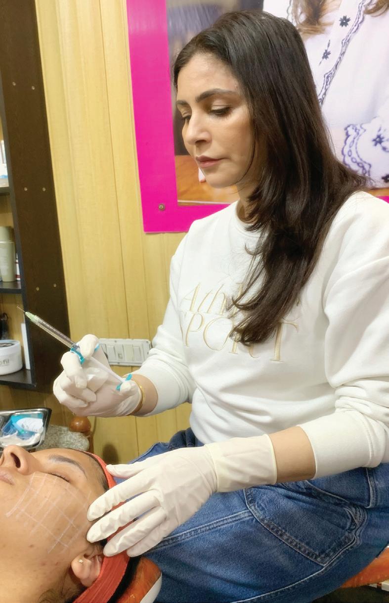
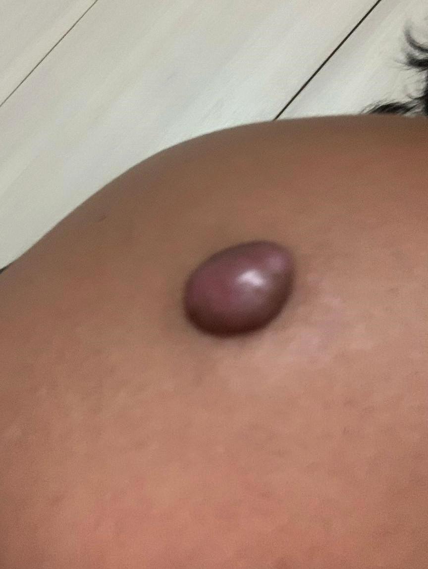
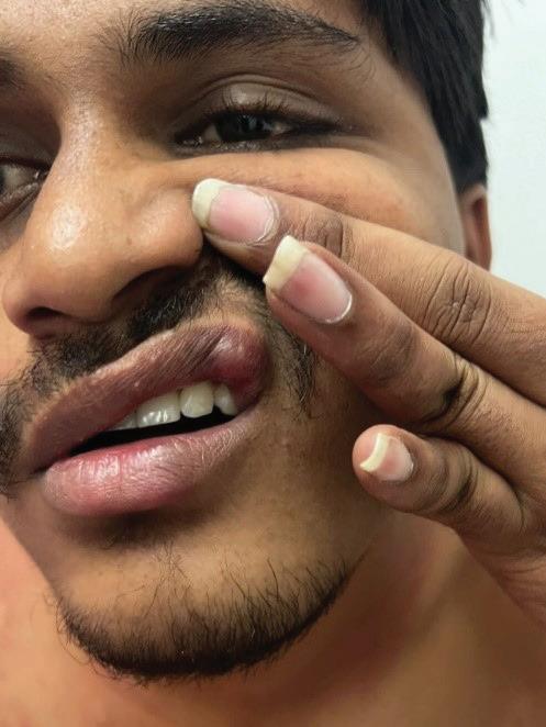
Case Report
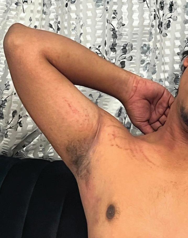
A 23-year-old male presented with a solitary keloid over the shoulder region. The lesion was firm, raised, and cosmetically concerning. The patient had no known comorbidities, drug allergies, or prior history of hypersensitivity reactions (Fig. 1). After counselling and informed consent, intralesional therapy was planned. The first treatment session was performed using triamcinolone acetonide (40 mg/ml) combined with hyaluronidase. A vial of hyaluronidase 1500 IU was diluted with 2 ml sterile water. Triamcinolone and the diluted hyaluronidase were mixed in a 1:1 ratio and injected intralesionally, with a total of 10 units administered. The
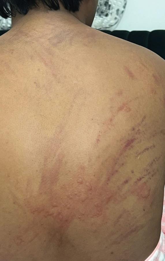
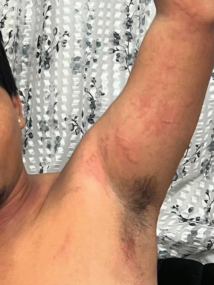
procedure was well tolerated. The patient returned after one month for the second session. Clinically, the keloid had become softer and more amenable to injection. The same dilution protocol was followed and a total of 14 units were injected. The patient was observed in the clinic for 20 minutes and was asymptomatic at the time of discharge. Approximately one hour later, the patient contacted the clinic reporting intense generalized itching, diffuse erythema, throat discomfort, hoarseness of voice, and itching in the genital region (Fig. 2A, 2B, 2C). He was immediately advised to visit the nearest hospital. On arrival, clinical evaluation suggested an acute urticarial reaction with angioedema. Vital parameters were stable. The patient was treated with intravenous hydrocortisone 100 mg and intravenous chlorpheniramine maleate. Adrenaline was kept ready in case of deterioration. The patient responded well to treatment, with reduction in rash and symptomatic improvement within a short period. Hoarseness of voice and throat discomfort gradually subsided. He was monitored for six hours and subsequently discharged in stable condition. The patient was prescribed oral corticosteroids and antihistamines for five days, as he continued to experience residual itching over the body and mild swelling with pruritus in the groin region. By day 1 following discharge, there was significant reduction in erythema and pruritus, with visible improvement in cutaneous wheals and swelling. The images below depict the clinical status on day 1 post-
Figure 1: Solitary keloid over the shoulder region at baseline prior to intralesional therapy
Figure 2 (a)
Figure 2 (b)
Figure 2 (A, B, C): Clinical presentation showing generalized erythema and urticarial rash associated with intense pruritus
Figure 3 (A, B, C): Posttreatment photograph demonstrating significant reduction in urticaria and edema after discharge
Mechanism of Action of Hyaluronidase
Hyaluronidase exerts its effects primarily through the degradation of hyaluronic acid (HA), a major glycosaminoglycan present in the extracellular matrix (ECM). HA is composed of repeating disaccharide units of N-acetylglucosamine and glucuronic acid linked by β-1,3 and β-1,4 glycosidic bonds. By cleaving these bonds, hyaluronidase reduces the size and structural integrity of HA polymers, leading to decreased viscosity of the interstitial matrix and increased tissue permeability. This property explains its long-standing use as a “spreading factor” that enhances the diffusion of injected drugs, facilitates the dispersion of local anesthetics, and aids in the management of hyaluronic acid–based dermal fillers.3
• Enzymatic degradation of hyaluronic acid
The primary mechanism involves enzymatic hydrolysis of glycosidic linkages within HA.
Hyaluronidases recognize HA through specialized substratebinding clefts that contain positively charged amino acids capable of interacting with the negatively charged HA molecule. Once bound, catalytic residues within the enzyme initiate cleavage of the glycosidic bonds, fragmenting the polymer into smaller oligosaccharides. In human and animal systems, different classes of hyaluronidases hydrolyze either β-1,4 or β-1,3 glycosidic bonds through a stepwise catalytic process involving proton transfer, nucleophilic attack, and the formation of transient intermediates. Water molecules then hydrolyze these intermediates, releasing smaller HA fragments and restoring the enzyme for subsequent catalytic cycles. Through repeated reactions, the highmolecular-weight HA polymer is progressively degraded into lowmolecular-weight fragments.3
Another class of enzymes can degrade HA through β-elimination reactions that generate unsaturated oligosaccharide products. Although the detailed catalytic pathways of these enzymes are still being investigated, they similarly result in fragmentation of the HA network and disruption of the extracellular matrix structure.3
• Non-enzymatic and oxidative degradation
In addition to direct enzymatic cleavage, HA can also undergo degradation through oxidative mechanisms. Reactive oxygen species (ROS), including hydroxyl radicals and other oxidative molecules generated during inflammation or tissue injury,
Figure 2 (c)
Figure 3 (a)
Figure 3 (b)
Figure 3 (c)
can attack the HA chain and induce depolymerization. These reactions break glycosidic bonds and produce heterogeneous oligosaccharide fragments. Increased oxidative stress may also alter the conformation or expression of hyaluronidase, indirectly enhancing enzymatic degradation of HA.3
• Clearance and metabolism of HA fragments
Following degradation, HA fragments are efficiently cleared through local and systemic mechanisms. In tissues such as skin, smaller fragments may undergo further enzymatic breakdown into basic monosaccharide components. A significant proportion enters lymphatic capillaries via interstitial fluid and is transported to regional lymph nodes, where lymphatic endothelial cells internalize and metabolize them. Remaining fragments eventually enter the bloodstream and are degraded primarily by liver endothelial cells. These multilevel clearance pathways ensure rapid removal of HA fragments and maintenance of tissue homeostasis.3
• Modulation of the extracellular matrix
Beyond simple hydrolysis, hyaluronidase significantly alters the physical and biological properties of the extracellular matrix. HA functions as a major structural component that provides hydration, viscosity, and mechanical stability to tissues. When hyaluronidase degrades HA, the ECM becomes less viscous and more porous, reducing tissue resistance and facilitating the movement of fluids, drugs, and cells through the interstitial space. This
structural remodeling explains the enzyme’s ability to enhance drug diffusion and to disperse accumulated hyaluronic acid fillers.3
• Cellular and signaling effects
Degradation of HA also generates biologically active fragments that influence cellular signaling. High-molecularweight HA generally maintains tissue stability and exhibits anti-inflammatory and antiangiogenic effects. In contrast, low-molecular-weight fragments produced after hyaluronidase activity can stimulate cellular proliferation, angiogenesis, and inflammatory responses. These fragments interact with several cell-surface receptors, including CD44, RHAMM, and Toll-like receptors (TLR2 and TLR4), triggering intracellular signaling pathways such as NFκB activation and the release of inflammatory mediators.3
Through these combined mechanisms—enzymatic hydrolysis, oxidative fragmentation, extracellular matrix remodeling, and modulation of cellular signaling— hyaluronidase plays an important role in regulating tissue permeability, inflammation, and matrix dynamics. These properties form the basis of its clinical applications in medicine and aesthetic dermatology.3
Side Effects of Hyaluronidase
Local injection of hyaluronidase may lead to adverse effects such as itching at the injection site and allergic reactions. The reported incidence of allergic responses ranges from approximately 0.05% to 0.69%, while urticaria and angioedema occur less frequently, in fewer than 0.1%
of cases. The likelihood of allergic reactions increases with higher doses, particularly when hyaluronidase is administered intravenously. When doses exceed 100,000 IU, the risk rises, and it has been reported to reach up to 31.3% at doses around 200,000 IU.1
Most allergic reactions associated with hyaluronidase are immediate hypersensitivity reactions (type I), mediated by immunoglobulin E. These reactions typically present within 1–2 hours as erythema and localized edema that do not respond to antibiotic therapy. Management generally includes systemic corticosteroids, antihistamines, and topical steroid preparations. Delayed hypersensitivity reactions (type IV), mediated by T cells, may also occur and can appear more than 24 hours after injection. In such situations, routine skin testing may fail to predict the reaction, as the result may remain negative within the usual 20-minute observation period.1
A skin test using approximately 3 IU of hyaluronidase is often recommended prior to treatment, although this may not always be practical in routine clinical settings. Importantly, there is generally no clear correlation between a patient’s general allergy history and their response to hyaluronidase. However, caution is advised because cross-reactivity may occur in individuals with allergies to bovine collagen or bee stings, depending on the enzyme’s source.1
Discussion
Hyaluronidase plays an important role in modern
Acute Adverse Reaction to Intralesional Hyaluronidase
clinical practice due to its ability to temporarily degrade hyaluronic acid and increase tissue permeability. By breaking down hyaluronic acid within the extracellular matrix, the enzyme facilitates the enhanced dispersion and systemic absorption of injected medications. This property has led to its use in combination with several therapeutic agents across different medical specialties. Examples include insulin for diabetes, beta interferons for multiple sclerosis, biotherapeutic agents in rheumatoid arthritis, immunoglobulin replacement therapy in primary immunodeficiency disorders, and monoclonal antibody therapies used in oncology. By improving drug diffusion and bioavailability, hyaluronidase can enhance therapeutic outcomes and allow more efficient subcutaneous delivery of medications. Several clinical indications for hyaluronidase have received regulatory approval. It is commonly used as an absorption and dispersion enhancer for injected drugs. It is also utilized in subcutaneous fluid administration for hydration therapy, a technique known as hypodermoclysis, and in subcutaneous urography to
References
1. Jung H. (2020). Hyaluronidase: An overview of its properties, applications, and side effects. Archives of plastic surgery, 47(4), 297–300. https://doi. org/10.5999/aps.2020.00752
2. Song, Y.-K., Kim, Y.-D., & Kim, J.-H. (2014). Acute allergic reaction caused by hyaluronidase used in the pain management: A case report and literature review. Anesthesia & Pain Medicine, 9 (3), 174–178.
improve the absorption and distribution of radiopaque contrast agents. In addition to these approved indications, hyaluronidase has a number of off-label applications. These include the management of medication extravasation from intravenous lines, use as an adjunct to local anesthetic eye blocks in ophthalmic surgery, and combination therapy in keloid management alongside cryosurgery, corticosteroids, and 5-fluorouracil. In aesthetic dermatology, hyaluronidase is widely used to reverse hyaluronic acid–based dermal fillers and to manage filler-related complications.4,5
Despite its benefits, certain limitations and precautions must be considered. One of the main concerns during filler correction is the potential loss of the desired cosmetic effect if excessive doses are used. In a retrospective review of patients with lower eyelid edema following hyaluronic acid filler injections, treatment with hyaluronidase resulted in improvement in all cases, although complete degradation of the filler occurred in a few patients, leading to loss of treatment effect. To minimize this risk, many clinicians recommend administering smaller doses over multiple sessions with reassessment between treatments, particularly in nonemergent situations such as noninflammatory nodules. The effectiveness of hyaluronidase is influenced by the physical characteristics of the filler, including its degree of cross-linking and resistance to enzymatic degradation. Higher doses may be required for more resistant products. In contrast, urgent complications such as vascular occlusion require prompt administration of large doses to restore perfusion and prevent tissue necrosis. Overall, hyaluronidase demonstrates a favorable safety profile, and hypersensitivity reactions remain uncommon in routine aesthetic practice.4,5
Conclusion
In conclusion, the use of hyaluronidase has expanded significantly in aesthetic and clinical dermatology, particularly for managing complications and correcting unsatisfactory outcomes following hyaluronic acid filler procedures. As its application becomes increasingly common, it is essential for clinicians to possess a clear understanding of its indications, mechanism of action, metabolism, and potential adverse effects. When used judiciously and with adequate expertise, hyaluronidase serves as a valuable tool for safely and effectively addressing a range of filler-related concerns.
4. Murray RL, Zafar Gondal A. Hyaluronidase. [Updated 2023 May 29]. In: StatPearls [Internet]. Treasure Island (FL): StatPearls Publishing; 2025 Jan-. Available from: https://www.ncbi.nlm.nih.gov/ books/NBK545163/
5. Kroumpouzos G, Treacy P. Hyaluronidase for Dermal Filler Complications: Review of Applications and Dosage Recommendations. JMIR Dermatol 2024;7:e50403 doi: 10.2196/50403PMID: 38231537PMCID: 10836581
Multimodal Management of Acne Scarring in Adolescents: Pathogenesis, Diagnosis and Treatment Strategies
Dr. Sanyogita Warang MBBS, DDVL, DNB Consultant Dermatologist
Mumbai Introduction
Acne scars are a common and frequently permanent sequelae of acne vulgaris, a chronic inflammatory disease of the pilosebaceous unit with a high global prevalence. Acne affects more than 90% of adolescents and persists into adulthood in approximately 12–14% of individuals, often resulting in considerable psychological, social and emotional burden. Lesions predominantly involve anatomical sites rich in pilosebaceous glands, including the face, chest, shoulders and upper back. Inflammatory acne lesions may resolve with residual scarring, the extent and severity of which are influenced by the magnitude and duration of inflammation, host immune response, genetic predisposition, and delays in initiating appropriate therapeutic intervention.1,2
The epidemiology of acne scarring remains incompletely characterized; however, available evidence consistently
demonstrates a strong association between acne severity and the likelihood of scar formation. Populationbased studies estimate that approximately 1% of individuals exhibit acne scars, with a smaller subset experiencing severe or disfiguring forms. Severe acne scarring is associated with long-term physical disfigurement and substantial psychosocial distress, particularly during adolescence and early adulthood, highlighting the importance of early identification, aggressive control of inflammation, and appropriate preventive strategies.1,2
The pathogenesis of acne scarring is intrinsically linked to the multifactorial mechanisms underlying acne vulgaris. Key contributing factors include increased sebum production, qualitative alterations in sebaceous lipid composition, androgen-mediated ............. sebaceous gland activity, follicular hyperkeratinization, and colonization of
the pilosebaceous unit by Cutibacterium acnes. Sebaceous gland lipids actively participate in inflammatory signaling pathways through interactions with nuclear receptors such as peroxisome proliferator-activated receptors, exerting both pro- and anti-inflammatory effects. Additionally, C. acnes activates keratinocytes and sebocytes via pattern recognition receptors, including toll-like receptors, CD14 and CD1 leading to activation of downstream transcription factors and the release of proinflammatory cytokines and chemokines. These inflammatory events promote infrainfundibular inflammation, follicular rupture, and perifollicular abscess formation, thereby triggering the cutaneous wound-healing response. Wound healing is a complex and tightly regulated biological process involving soluble mediators, extracellular matrix components, resident cutaneous cells and infiltrating immunoinflammatory cells. When the wound-healing cascade is dysregulated— particularly in the setting of prolonged or intense inflammation—abnormal scar formation ensues.2,3
The wound-healing process progresses through three overlapping phases: inflammation, granulation tissue formation, and matrix remodeling. Persistent inflammation has been identified as a critical determinant of acne scar development. Histopathologic studies reveal that individuals who develop scars exhibit more intense
and prolonged inflammatory infiltrates at the level of the pilosebaceous unit compared with those who heal without scarring. During the granulation phase, macrophage-derived growth factors stimulate fibroblast migration, proliferation and collagen synthesis, initially characterized by predominance of type III collagen. As healing progresses, matrix remodeling occurs through the coordinated activity of matrix metalloproteinases and their tissue inhibitors, which regulate collagen degradation and deposition. Disruption of this balance results in net collagen loss, producing atrophic scars or excessive collagen accumulation resulting in hypertrophic or keloid scars.2,3
Atrophic scars account for approximately 80–90% of acne scars and are further classified into icepick, boxcar, and rolling subtypes based on morphology and depth. These subtypes frequently coexist within the same individual, complicating clinical assessment and management. Less commonly, hypertrophic and keloid scars develop, particularly in individuals with darker skin phototypes. Post-inflammatory erythema and post-inflammatory hyperpigmentation frequently accompany acne scars and may accentuate their clinical appearance, further contributing to patient distress.1,2,3
Acne scars represent the cumulative outcome of sustained inflammation and aberrant wound healing following acne lesions. A comprehensive understanding
of the inflammatory, immunologic, and reparative mechanisms involved is essential for prevention, accurate classification and effective management.
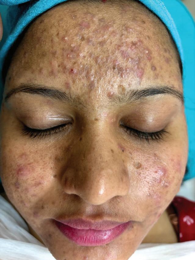
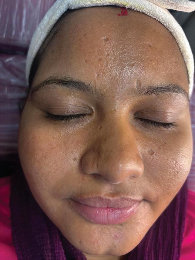
Case Report
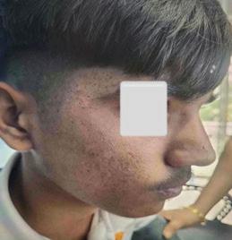
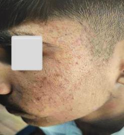
An 18-year-old male presented with a history of persistent acne vulgaris and visible post-acne scarring over both cheeks. Clinical examination revealed multiple atrophic acne scars associated with postinflammatory erythema and residual inflammatory papules. The patient reported recurrent acne episodes over several years and had received no prior structured dermatologic treatment.
A comprehensive multimodal regimen was initiated to control active disease and improve skin texture, consisting of topical retinoids to normalize follicular keratinization and enhance cellular turnover, oral antibiotics to reduce inflammation and suppress Cutibacterium acnes, and weekly topical glycolic acid peels to promote epidermal renewal and gradual refinement of scar texture. With regular follow-up and treatment adjustments based on response and tolerability, the patient showed progressive reduction in inflammatory lesions and overall improvement in skin texture. Stabilization of acne activity prevented further scar formation, while serial glycolic peels contributed to smoother skin and reduced post-inflammatory changes. The patient demonstrated good compliance and minimal adverse effects.
Before treatment
After treatment
Figure 1: Progressive clinical improvement in acne lesions and skin texture following multimodal therapy with topical retinoids, oral antibiotics, and serial glycolic acid peels
Diagnosis
Morphological evaluation is fundamental to the diagnosis and severity assessment of acne scarring and relies on a detailed analysis of scar morphology, including shape, depth, surface texture, size and distribution. Acne scars are categorized as atrophic, hypertrophic, or keloidal, with atrophic scars further subdivided into icepick, boxcar, and rolling types.
Visual inspection, palpation and standardized clinical photography are essential components of routine evaluation, enabling accurate baseline documentation and longitudinal comparison.3 Validated grading systems enhance objectivity and reproducibility. The Clinical Scale for the Evaluation of Acne Scars (ECCA) quantitatively grades severity by weighting scar morphology and extent.4 Similarly, the Global Acne Scarring Grading System integrates scar type, number, and severity into a composite score.5 Advanced imaging modalities further refine diagnostic precision. Three-dimensional imaging provides noninvasive, high-resolution topographic mapping of the skin surface, permitting accurate measurement of scar depth and volume.6 High-frequency ultrasound enables visualization of dermal and subdermal structures.7 Reflectance confocal
microscopy offers real-time in vivo imaging at near-histologic resolution.8 Laser Doppler imaging evaluates microvascular activity and post-inflammatory erythema within scar tissue.9 Together, clinical examination, standardized grading scales, and adjunctive imaging provide a comprehensive framework for acne scar assessment.
Treatment
Retinoids are fundamental in the management of acne scars, particularly atrophic variants, due to their multifaceted effects on epidermal turnover, dermal remodeling, and modulation of post-inflammatory repair processes. They stimulate fibroblast proliferation and enhance collagen synthesis, improving scar depth and texture. Topical retinoids— including tretinoin, adapalene, tazarotene and trifarotene— promote epidermal turnover, facilitate dermal remodeling, and enhance deposition of type I and III collagen. Their use is most effective when initiated early or incorporated into combination therapy.10
Oral antibiotics, particularly tetracycline-class agents such as doxycycline, minocycline and sarecycline, play an adjunctive role by reducing inflammatory lesions that contribute to scar formation. Their antiinflammatory properties include inhibition of cytokines, neutrophil chemotaxis, and matrix metalloproteinases. Their role is primarily preventive rather than directly remodeling established scars.11 Glycolic acid peels improve post-acne scars by promoting controlled
Multimodal Management of Acne Scarring in Adolescents: Pathogenesis,
keratolysis, enhancing epidermal turnover and stimulating dermal remodeling. They increase fibroblast proliferation and collagen synthesis, improving scar depth, texture and pigmentation.12 Other modalities include chemical peels, dermabrasion, laser resurfacing, microneedling, subcision, fillers, and surgical techniques. Hypertrophic scars and keloids respond to silicone gel, intralesional corticosteroids, cryotherapy, and pulsed dye laser.³ Treatment should be individualized based on scar type, skin phenotype, and patient preference.
Discussion
Acne scarring represents a persistent and clinically significant sequela of acne vulgaris, characterized by irreversible alterations in cutaneous architecture and substantial psychosocial
References
1. Connolly D, Vu HL, Mariwalla K, Saedi N. Acne Scarring-Pathogenesis, Evaluation, and Treatment Options. J Clin Aesthet Dermatol. 2017; 10(9):12-23.
2. Jfri A, Alajmi A, Alazemi M, Ladha MA. Acne Scars: An Update on Management. Skin Therapy Lett. 2022; 27(6):6-9.
3. Fabbrocini G, Annunziata MC, D'Arco V, et al. Acne scars: pathogenesis, classification and treatment. Dermatol Res Pract. 2010; 2010:893080. doi:10.1155/2010/893080.
4. Abdel Hay R, Shalaby K, Zaher H, et al. Interventions for acne scars. Cochrane Database Syst Rev. 2016; 4(4):CD011946. Published 2016 Apr 3. doi:10.1002/14651858.CD011946.pub2
5. Bae IH, Kwak JH, Na CH, Kim MS, Shin BS, Choi H. A Comprehensive Review of the Acne Grading Scale in 2023. Ann Dermatol. 2024; 36(2):65-73. doi:10.5021/ad.23.094
6. Petit L, Zugaj D, Bettoli V, et al.
morbidity. Atrophic scars account for approximately 75–90% of cases and occur nearly threefold more frequently than hypertrophic scars. Although acne is often perceived as self-limiting, reported rates of permanent scarring approach 95% among affected individuals, while prevalence in the general population ranges from 1% to 11%. Importantly, scarring is not restricted to severe acne, as mild-to-moderate disease also carries significant risk. Delayed treatment, lesion manipulation, male sex, androgen-mediated sebaceous activity, and genetic susceptibility increase scar risk. Dietary factors are increasingly recognized. Dairy products may exacerbate acne through insulin-like growth factor 1 and androgen precursors, while low-glycemic diets and adequate micronutrient intake may support improved outcomes. Regenerative and energybased modalities such as platelet-rich plasma, exosome therapies, and non-ablative lasers have shown promise. Non-ablative lasers offer gradual improvement with minimal downtime and reduced risk of post-inflammatory hyperpigmentation, particularly in darker phototypes.13,14
Conclusion
Acne scarring is a common dermatologic consequence of sustained inflammation, delayed treatment, behavioral factors, dietary influences, stress, and genetic predisposition. Early evidencebased intervention is critical to prevent permanent skin changes. Management with topical retinoids, oral antibiotics, and serial glycolic acid peels effectively controls active lesions, promotes epidermal renewal, minimizes scar formation, and improves both clinical outcomes and patient quality of life.
Validation of 3D skin imaging for objective repeatable quantification of severity of atrophic acne scarring. Skin Res Technol. 2018; 24(4):542-550. doi:10.1111/ srt.12464
7. Malinowska S, Jaguś D, Woźniak W, Mlosek RK. Usefulness of high-frequency ultrasound in the monitoring of laser treatment of acne scars. J Ultrason. 2021; 20(83):e279-e283. doi:10.15557/ JoU.2020.0049
8. Cretu S, Papachatzopoulou E, Dascalu M, Salavastru CM. The role of in vivo reflectance confocal microscopy for the management of acne: A systematic review. J Eur Acad Dermatol Venereol. 2023; 37(12):2428-2439. doi:10.1111/jdv.19327
9. Baran U, Li Y, Choi WJ, Kalkan G, Wang RK. High resolution imaging of acne lesion development and scarring in human facial skin using OCT-based microangiography. Lasers Surg Med. 2015; 47(3):231-238. doi:10.1002/lsm.22339
10. Issa N, Alexis A, Baldwin H, et al.
Recommendations to Improve Outcomes in Acne and Acne Sequelae: A Focus on Trifarotene and Other Retinoids. Dermatol Ther (Heidelb). 2025; 15(3):563-577. doi:10.1007/s13555-025-01344-y
11. Baldwin H. Oral Antibiotic Treatment Options for Acne Vulgaris. J Clin Aesthet Dermatol. 2020; 13(9):26-32.
12. Sharad J. Glycolic acid peel therapy - a current review. Clin Cosmet Investig Dermatol. 2013; 6:281-288. Published 2013 Nov 11. doi:10.2147/CCID.S34029
13. Etgu F, Sekerlisoy Tatar G. Risk Factors and Epidemiology of Acne Severity and Acne Scar Development: A Comprehensive Clinical Study. Dermatol Pract Concept. 2025; 15(4):e20256108. Published 2025 Oct 1. doi:10.5826/ dpc.1504a6108
14. Tahiliani S, Mysore V, Ganjoo A, et al. Practical Aspects of Acne Scar Management: ASAP 2024. Cureus. 2024; 16(3):e55897. Published 2024 Mar 10. doi:10.7759/cureus.55897
Resolution of Acne and PostInflammatory Hyperpigmentation Using Combined Systemic, Topical and PeelBased Therapy
Active acne vulgaris is a prevalent inflammatory disorder of the pilosebaceous unit, manifesting as comedones, papules, pustules, nodules, and cysts. The pathogenesis involves follicular hyperkeratinization, increased sebum production, Cutibacterium acnes colonization, and immune-mediated inflammation, which collectively contribute to the formation and persistence of active lesions. In individuals with darker skin phototypes (Fitzpatrick III–VI), these inflammatory lesions frequently predispose to postinflammatory hyperpigmentation (PIH), an acquired reactive hypermelanosis that develops following cutaneous injury or inflammation. PIH can emerge rapidly, often within days of lesion resolution, and may affect the epidermis, dermis, or both, leading to variable degrees of pigmentation that range from tan or brown to blue-grey discoloration.1, 2
Epidemiologically, acne-induced PIH is highly prevalent among adolescents and young adults, particularly those aged 15 to 30 years. Data from India indicate that over 70% of individuals with a history of acne develop hyperpigmentation, with females, patients experiencing prolonged or recurrent inflammatory lesions, and those with moderate to severe acne (grades II–III) being disproportionately affected. Individuals with darker skin phototypes are especially prone to intense and persistent pigmentary changes, which often persist longer than in lighter-skinned populations.1, 2
The development of PIH is influenced by a combination of endogenous and exogenous factors. Endogenous contributors include genetic predisposition, melanocyte hyperreactivity, metabolic or nutritional deficiencies such as iron or vitamin B12 deficiency,
seborrhea, atopic dermatitis, thyroid disorders, and hormonal fluctuations. Exogenous factors encompass ultraviolet radiation, mechanical trauma from lesion manipulation, aesthetic procedures including chemical peels and laser therapy, improper skincare, systemic medications such as contraceptives, frequent facial scrubbing, obesity, insulin resistance, and prolonged sun exposure. These factors, in combination with the severity and duration of active inflammatory lesions, dictate both the likelihood and extent of hyperpigmentation.1, 2
The pathophysiology of acneinduced post-inflammatory hyperpigmentation (PIH) involves two interrelated mechanisms: epidermal inflammation and pigmentary incontinence. During active acne, follicular inflammation triggers the release of inflammatory mediators such as cytokines, prostaglandins, leukotrienes, and neuropeptides. These mediators stimulate melanocyte proliferation, dendritic extension, and increased melanogenesis, leading to enhanced melanin transfer to keratinocytes and subsequent epidermal hyperpigmentation. In pigmentary incontinence, injury to basal keratinocytes results in the leakage of melanin into the dermis, where it is phagocytosed by macrophages, forming melanophages that cause dermal pigmentation. While epidermal pigmentation is typically transient, dermal pigmentation may persist for prolonged periods or become permanent. The severity and distribution of PIH correlate
with the intensity and duration of inflammation and are exacerbated by mechanical trauma such as squeezing or picking of lesions. Clinically, PIH presents as asymptomatic, irregular macules or patches corresponding to prior inflammatory acne lesions. Epidermal PIH appears tan to dark brown, whereas dermal PIH manifests as blue-grey discoloration; mixed patterns are common. Lesions often evolve from erythema to violaceous and brown pigmentation as inflammation resolves. Patients with severe, nodular, or persistent acne are at increased risk. Although PIH is generally reversible, its visibility and unpredictable course significantly impair psychosocial well-being, underscoring the importance of early and effective acne management.1,2
Case Report
A 27-year-old female presented with a history of persistent facial acne associated with residual pigmentation. On clinical examination at baseline, the patient exhibited active inflammatory acne lesions, predominantly involving the forehead, cheeks, and chin, comprising multiple erythematous papules, pustules, and few closed comedones. These lesions were accompanied by diffuse postinflammatory hyperpigmentation (PIH) in the form of irregular light- to dark-brown macules corresponding to the distribution of prior inflammatory lesions. The pigmentation appeared predominantly epidermal with areas of mixed depth, more pronounced over the cheeks and forehead. The skin texture
was uneven, with mild postacne textural irregularities, and seborrhea was evident. There was no evidence of hypertrophic scarring or keloidal tendency. Based on the clinical findings, a diagnosis of moderate inflammatory acne vulgaris with associated PIH was made. The patient was managed using a structured, multimodal therapeutic approach over a period of five months.
Systemic therapy included oral isotretinoin initiated at a dose of 20 mg once daily and continued throughout the treatment period under regular clinical and laboratory monitoring. To rapidly control inflammatory lesions and reduce Cutibacterium acnes–associated inflammation, azithromycin 500 mg was prescribed thrice weekly for two months. Topical therapy comprised ozenoxacin 2% cream applied twice daily for the first 15 days to address active bacterial involvement. This was followed by a maintenance regimen of topical clindamycin combined with adapalene once daily to prevent new lesion formation and promote comedolysis. A kojic acid–based depigmenting formulation was prescribed to target PIH. Procedural interventions included two sessions of Black Peel and one session of Yellow Peel at appropriate intervals to enhance acne resolution, improve skin texture, and accelerate pigment clearance. At the end of five months, the patient demonstrated complete resolution of active acne lesions with marked reduction in postinflammatory hyperpigmentation, improved skin tone uniformity, and smoother texture, without
any significant adverse effects. The patient reported high satisfaction with both cosmetic and clinical outcomes.
Before treatment
After treatment
Figure 1: Patient demonstrated complete resolution of active acne lesions with marked reduction in post-inflammatory hyperpigmentation, improved skin tone uniformity and smoother texture
Diagnosis
Acne-induced post-inflammatory hyperpigmentation (PIH) is diagnosed primarily on clinical grounds through a detailed patient history and meticulous cutaneous examination. Diagnosis is established by demonstrating a clear temporal and anatomical correlation between antecedent inflammatory acne lesions and residual hyperpigmented macules. Clinically, PIH presents as welldefined macular discoloration ranging from light brown to dark brown or greyish hues at sites of prior acne activity, with color variation reflecting the depth of melanin deposition. Adjunctive diagnostic tools may support clinical evaluation in selected cases. Wood’s lamp examination can help estimate whether pigmentation is predominantly epidermal or dermal, although its utility is limited in darker skin phototypes. Dermoscopy may further aid assessment by enhancing visualization of pigment patterns and lesion morphology. Skin biopsy is rarely required and is reserved for atypical, persistent, or diagnostically uncertain cases to confirm melanosis and its depth.2,3
Disease severity, pigmentation intensity, and distribution can be objectively assessed using validated instruments such as the PostAcne Hyperpigmentation Index (PAHPI), which allows standardized grading, documentation, and monitoring of treatment response. Active acne severity is commonly evaluated using the Global Acne Grading Scale due to its correlation with PIH risk.4 Psychosocial impact may be assessed using acne-specific quality-of-life questionnaires and self-esteem scales. Where available, noninvasive imaging modalities—including polarized photography, colorimetry, and reflectance-based techniques—provide quantitative assessment,
enabling a comprehensive and individualized diagnostic approach.2
Treatment
Topical therapy
Clindamycin is a lincosamide antibiotic that inhibits bacterial protein synthesis in Cutibacterium acnes, thereby reducing bacterial proliferation. Its anti-inflammatory properties decrease neutrophil chemotaxis and cytokine release, leading to a reduction in inflammatory papules and pustules in acne vulgaris.5
Adapalene is a third-generation topical retinoid that modulates keratinocyte differentiation and proliferation, normalizing follicular epithelial turnover. This prevents microcomedone formation, promotes comedolysis, and exerts anti-inflammatory effects. Additionally, by enhancing epidermal turnover, adapalene facilitates gradual resolution of post-inflammatory hyperpigmentation (PIH) associated with acne lesions.5
The combination of clindamycin and adapalene provides a synergistic effect, effectively managing both active acne lesions and acne-induced PIH, optimizing clinical outcomes and minimizing the risk of antibiotic resistance.5
Ozenoxacin 2% is a topical non-fluorinated quinolone antibiotic used for the treatment of acne vulgaris, exhibiting potent antimicrobial activity against Cutibacterium acnes and other Gram-positive organisms while also reducing pro-inflammatory cytokine production, including IL-6, IL8, and TNF-α, in keratinocytes and monocyte models. It
may also inhibit sebaceous lipid synthesis through suppression of mTORC1-related pathways, addressing sebum overproduction, a key factor in acne pathogenesis. Ozenoxacin 2% provides combined antimicrobial, anti-inflammatory, and sebostatic effects, making it a promising topical option for managing acne vulgaris.6
Kojic acid functions as a depigmenting agent by inhibiting the tyrosinase enzyme, thereby reducing melanin synthesis in melanocytes and preventing the development of postinflammatory hyperpigmentation (PIH) that often follows inflammatory acne lesions. By promoting epidermal turnover and accelerating the exfoliation of pigmented keratinocytes, kojic acid facilitates gradual fading of residual pigmentation and helps restore uniform skin tone, making it an effective adjunctive therapy for patients with acne-associated pigmentary changes.7
Other topical treatments for acne vulgaris are applied directly to affected skin to minimize systemic absorption and are mainly used for mild to moderate disease. Topical retinoids normalize follicular keratinization, reduce sebum production, prevent comedone formation, and improve hyperpigmentation; tretinoin is commonly used, while tazarotene is a newer option reserved for resistant cases and may cause irritation. Topical antibiotics such as erythromycin reduce Propionibacterium acnes and inflammation but should be used in combination to limit resistance. Combination therapies, including benzoyl peroxide, salicylic acid, niacinamide, and dapsone,
enhance efficacy through antibacterial, keratolytic, antiinflammatory, and sebumregulating actions, supporting effective topical management of acne vulgaris.6
Oral isotretinoin acts by markedly reducing sebaceous gland size and sebum production through modulation of nuclear retinoid receptors, leading to decreased lipid synthesis and turnover in sebocytes, which in turn limits the substrate available for Cutibacterium acnes proliferation and reduces follicular occlusion; it also normalizes keratinocyte differentiation, diminishes microcomedone formation, and exerts anti inflammatory effects by altering toll like receptor expression and reducing pro inflammatory cytokine signaling. Azithromycin, a macrolide antibiotic with high tissue penetration, inhibits bacterial protein synthesis in susceptible organisms, including C. acnes, thereby decreasing bacterial load within pilosebaceous units; it also possesses immunomodulatory properties that downregulate neutrophil chemotaxis and inflammatory mediator release, contributing to the reduction of inflammatory lesions in acne vulgaris.5
Other systemic antibiotics are used for moderate to severe inflammatory acne resistant to topical therapy or involving large areas. Agents such as erythromycin, clindamycin, fluoroquinolones, tetracyclines (doxycycline, minocycline, lymecycline), and co-trimoxazole reduce Propionibacterium acnes and inflammatory mediators, with efficacy depending on follicular penetration.
Tetracyclines are preferred for their antibacterial and antiinflammatory effects, good follicular penetration, tolerability, and lower resistance, while doxycycline and minocycline offer better lipid solubility and fewer gastrointestinal effects. By controlling inflammation, systemic antibiotics also help prevent and improve post-inflammatory hyperpigmentation.5
Black peel is used to treat postacne hyperpigmentation by promoting controlled exfoliation and increasing epidermal turnover, which removes pigmented keratinocytes and stimulates skin regeneration. Its antioxidant and antiinflammatory properties help reduce melanocyte activity and prevent further pigment deposition, resulting in lightened pigmentation, improved skin texture, and a more even complexion.8
Yellow peel is used to treat post-acne hyperpigmentation by promoting exfoliation and increasing epidermal turnover to remove pigmented cells and stimulate skin regeneration. Its antioxidant and antiinflammatory properties reduce melanocyte activity, prevent further pigmentation, and improve skin texture, restoring a more even complexion.9
Other chemical peels used in acne and post-acne hyperpigmentation management include salicylic acid, glycolic acid, lactic acid, mandelic acid, trichloroacetic acid, Jessner’s solution, and phenol, which act by promoting controlled exfoliation of the stratum corneum, enhancing epidermal
turnover, and facilitating the removal of pigmented or damaged keratinocytes, thereby improving skin texture, reducing pigmentation, and stimulating regeneration of rejuvenated epidermal layers.7
Procedural therapies are important in managing acne and its sequelae, particularly in patients with comedonal, nodulocystic, or scarred lesions. Manual comedone extraction, electrocauterization of macrocomedones, intralesional triamcinolone, and cyst or abscess drainage are used to directly treat persistent lesions. Laser and light-based therapies, including photodynamic therapy, have variable efficacy. Acne scars are managed based on type and depth, with superficial scars addressed by dermabrasion and deeper scars by dermal fillers, microneedling, ablative or non-ablative fractional lasers, dermarollers, radiofrequency, punch excision, punch elevation, or subcision. Hypertrophic scars and keloids are primarily treated with intralesional corticosteroids, with refractory cases managed using multimodal approaches such as cryotherapy, silicone gel sheeting, pulsed-dye laser, or surgical excision with perioperative corticosteroids and pressure dressings. These interventions are generally safe, though further highquality studies are needed to standardize treatment and optimize outcomes.8
Discussion
Acne vulgaris is a chronic inflammatory disorder of the pilosebaceous unit, clinically characterized by comedones, papules, pustules, nodules,
and, in severe forms, cysts. Its pathogenesis is multifactorial, involving increased sebum production, follicular hyperkeratinization, colonization by Cutibacterium acnes, and a dysregulated innate and adaptive inflammatory response. Genetic predisposition plays a significant role in determining disease severity, lesion distribution, and therapeutic response, as inherited variations influence sebaceous gland activity, keratinocyte differentiation, and immune signaling pathways, thereby increasing susceptibility to both active acne lesions and their long-term sequelae. Postinflammatory hyperpigmentation (PIH) is among the most common complications of acne, particularly affecting individuals with darker skin phototypes (Fitzpatrick IV–VI). Higher baseline melanin content and increased melanocyte reactivity in these populations predispose to more intense and persistent pigmentary alterations. PIH develops secondary to inflammatory injury involving the epidermis and dermis, resulting in excessive melanin production and abnormal pigment distribution. The severity and persistence of PIH correlate with the depth, intensity, and duration of inflammation, highlighting the importance of early, effective acne control to prevent pigmentary complications. Accurate differential diagnosis is essential to ensure appropriate management and minimize PIH risk. Conditions that may mimic acne include bacterial and Pityrosporum folliculitis, acne keloidalis nuchae, milia, miliaria rubra, syringomas, perioral dermatitis,
sebaceous hyperplasia, nevus comedonicus, papulopustular rosacea, keratosis pilaris, molluscum contagiosum, eruptive vellus hair cysts, steatocystoma multiplex, and verruca vulgaris. Drug-induced acneiform eruptions—commonly associated with corticosteroids, anabolic steroids, lithium, isoniazid, hormonal agents, and targeted oncologic therapies—present as suddenonset monomorphic papules in atypical distributions and resolve upon withdrawal of the offending agent. Rare genetic syndromes should be considered in atypical or treatment-resistant cases.10
Complications of acne include PIH, atrophic scarring, hypertrophic and keloid scars, persistent facial edema as in Morbihan disease, and rarely, dystrophic calcinosis cutis. Atrophic scars are classified as boxcar, ice pick, or rolling, while hypertrophic and keloid scars are distinguished by their growth pattern beyond the original lesion borders. The risk of both scarring and PIH is proportional to the severity and depth of inflammation, highlighting the importance of timely and targeted anti-inflammatory therapy.
Patient-centered care and skin hygiene play a pivotal role in preventing PIH. Patients should use gentle, non-comedogenic cleansers, moisturizers, and broad-spectrum sunscreens, avoiding harsh soaps, scrubs, and aggressive exfoliation that can exacerbate inflammation and trigger hyperpigmentation. Mechanical manipulation of lesions, such as squeezing or picking, should be strictly avoided to reduce the risk of both
scarring and PIH. Techniques such as patting the skin dry after cleansing minimize mechanical trauma and further reduce pigmentary complications.10
In genetically predisposed individuals, particularly those with darker skin types, acne lesions frequently lead to PIH if not managed promptly and effectively. Early recognition, accurate differential diagnosis, appropriate pharmacologic therapy, and consistent skin care are critical to reduce
References
1. Sangha AM. Managing Postinflammatory Hyperpigmentation in Patients with Acne. J Clin Aesthet Dermatol. 2021; 14(6 Suppl 1):S24-S26.
2. Thomas J, Shankar K, Pujara S, Sharma R, Pudukadan D, Sachdev M, et al. Consensus on management of acne-induced postinflammatory hyperpigmentation: an Indian perspective. Int J Res Dermatol 2021; 7:336-45.
3. Sharma R, Khunger N, Poddar S. Dermoscopy as a novel tool to examine and assess treatment response of acne scars: A pilot study in an Indian population. J Cutan Aesthet Surg. 2025; 18:187-91. doi: 10.25259/jcas_43_23.
4. Auffret N, Leccia MT, Ballanger F, Claudel JP, Dahan S, Dréno B. Acne-induced Post-inflammatory Hyperpigmentation: From Grading to Treatment. Acta Derm Venereol. 2025;
inflammatory damage, prevent post-acne hyperpigmentation, and optimize both clinical and cosmetic outcomes. Understanding the interplay between acne pathogenesis, inflammation, and pigmentary response is essential for devising comprehensive treatment strategies that address both active lesions and post-inflammatory sequelae.10
Conclusion
Active acne lesions can lead to post-inflammatory hyperpigmentation, particularly in darker skin types. Early targeted therapy including systemic isotretinoin and azithromycin, topical ozenoxacin, clindamycin–adapalene, kojic acid, and procedural interventions such as black and yellow Peels demonstrated significant efficacy in controlling active lesions, preventing new outbreaks, and accelerating resolution of post-acne hyperpigmentation, optimizing clinical and cosmetic outcomes.
105:adv42925. Published 2025 Apr 22. doi:10.2340/actadv.v105.42925.
5. Vasam M, Korutla S, Bohara RA. Acne vulgaris: A review of the pathophysiology, treatment, and recent nanotechnology based advances. Biochem Biophys Rep. 2023; 36:101578. Published 2023 Nov 23. doi:10.1016/j.bbrep.2023.101578
6. Tabara K, Tamura R, Nakamura A, et al. Anti-inflammatory effects of ozenoxacin, a topical quinolone antimicrobial agent. J Antibiot (Tokyo). 2020; 73(4):247-254. Doi: 10.1038/s41429-020-0278-5
7. Castillo DE, Keri JE. Chemical peels in the treatment of acne: patient selection and perspectives. Clin Cosmet Investig Dermatol. 2018; 11:365-372. Published 2018 Jul 16. doi:10.2147/CCID.S137788
8. Puri N, Kumar S, Kaur S, Brar BK. Comparative evaluation of therapeutic
efficacy and safety of black peel versus 25% trichloroacetic acid peel in mild to moderate acne vulgaris: a split face study. J Cutan Aesthet Surg. 2024; 17(1):11-18. doi:10.4103/JCAS.JCAS_69_23
9. Deda A, Hartman-Petrycka M, Gędoś M, Wojciechowska M, Wcisło-Dziadecka D. Cosmetic Benefits of Medium-Depth Chemical Peels for Moderate Acne Lesions and Atrophic Acne Scars: A Single-Arm Clinical Trial in Young Adults. J Clin Med. 2025; 14(23):8598. Published 2025 Dec 4. doi:10.3390/jcm14238598
10. Leung AK, Barankin B, Lam JM, Leong KF, Hon KL. Dermatology: how to manage acne vulgaris. Drugs Context. 2021; 10:2021-8-6. Published 2021 Oct 11. doi:10.7573/dic.2021-8-
Pincode:
OBSERVERSHIP IN AESTHETICS
3-Nights, 4-Days
Program Highlights* 2 Half-days
Lectures by DHA Certified Faculty 2 Half-days
Observership in Aesthetic Procedures with upto 6 DHA@CME Credits# CME Credits# with
Package to be announced shortly
Inclusions:
* 3-Nights 4-Days Stay, in a 4-STAR Hotel with breakfast and dinner, Lunch at Academy/Aesthetic Centre, To and fro - Hotel to Academy/Aesthetic Centre SIC◊
Exclusions:
Air fare to Dubai and back , airport transfer, Sight seeing etc.
@ DHA- Dubai Health Authority
# Proposed s/t confirmation
◊ SIC- Seat in Coach
Interested Drs kindly call for further details: +91