Clinical Insights and Management of a Rare Case of Tuberculosis Sclerosis: A Case Report
Dr. Sherofina W Nathanial
3rd Year Resident
Department of Dermatology
Mahatma Gandhi Memorial (MGM) Medical College
Indore, Madhya Pradesh
Dr. Sanjay Khare
MD (Dermatology)
Head of Department
Department of Dermatology
Mahatma Gandhi Memorial (MGM) Medical College
Indore, Madhya Pradesh
Introduction
Tuberous sclerosis complex (TSC) is a genetic disorder characterized by the development of benign tumors in various organs, including the skin. These tumors often manifest as distinctive patches or lesions on the skin's surface. The presence of these skin abnormalities, along with other clinical features such as brain or kidney tumors, helps clinicians in diagnosing tuberous sclerosis. Skin involvement in tuberous
sclerosis can present with a variety of manifestations, including facial shagreen patch (colored patch), hypomelanotic macules (white patch), and fibrous plaques. These skin findings play a crucial role in the recognition and diagnosis of tuberous sclerosis, guiding further evaluation and management strategies.1
Some manifestations may appear later in life, while others, such as cardiac rhabdomyomas, may occur prenatally and
spontaneously regress postnatal. Central nervous system (CNS) manifestations encompass epilepsy, cognitive impairments, behavioural issues, autism spectrum disorder, and attention-deficit/ hyperactivity disorder. These neurological complications significantly compromise patient quality of life and necessitate multidisciplinary management approaches.1,2
Tuberous sclerosis complex diagnosis relies predominantly on clinical assessment and differential diagnosis. Anamnesis, physical examination, and dermatological evaluation are key for confirmation. Treatment involves a multifaceted approach, incorporating immunosuppressive agents, antibiotics, and surgical intervention as warranted. The therapeutic objective is to mitigate the physical and neuropsychiatric manifestations of tuberous sclerosis.3 It is a rare, chronic genetic disorder that can significantly impact a patient's quality of life. Early detection and effective management are crucial for mitigating disease progression and enhancing patient wellbeing. Timely diagnosis and appropriate interventions
Clinical Insights and Management of a Rare Case of Tuberculosis Sclerosis: A Case Report
can prevent exacerbations of the condition, thereby promoting improved quality of life for individuals affected by tuberous sclerosis.3
Pathophysiology of tuberous sclerosis, a genetic disorder stemming from mutations in the TSC1 or TSC2 genes, disrupts crucial protein production vital for bodily functions, especially brain development. Typically, mutations in TSC2 lead to more severe symptoms. The proteins encoded by these genes, hamartin from TSC1 and tuberin from TSC2, act as tumor suppressors, regulating the mTOR pathway essential for cell growth and survival. Dysregulation of this pathway due to mutations in TSC1 or TSC2 triggers tumor formation, a hallmark of tuberous sclerosis.2 Clinical presentation of tuberous sclerosis involves a variety of dermatological, ocular, dental, and systemic manifestations. One of the key dermatological signs includes Shagreen patches, which are rough, raised areas of skin with a leathery texture, typically found on the lower back or buttocks. Another notable skin manifestation is adenoma sebaceum, presenting as small, flesh-colored or reddish
papules, particularly around the nose and cheeks, caused by the overgrowth of sebaceous glands. Additionally, individuals with TSC often exhibit hypomelanotic macules, which are light-colored patches of skin resulting from reduced melanin production or distribution. Ocular abnormalities associated with tuberous sclerosis include retinal hamartomas, appearing as discrete white patches on the retinal surface, though these usually have minimal impact on vision. Dental anomalies in tuberous sclerosis encompass dental pits and gingival fibromas, with the latter manifesting as nodular growths on the gums, buccal mucosa, and tongue. Beyond these physical manifestations, tuberous sclerosis can also lead to cognitivebehavioral impairments, renal complications, cardiac involvement, pulmonary issues, and epileptic seizures, reflecting the systemic nature of the disorder.3
Case Report
A 42-year-old female patient presents with a chronic history of cutaneous lesions affecting the face, back, and chest over the past three decades. The
Clinical Insights and Management of a Rare Case of Tuberculosis Sclerosis:
onset of skin lesions occurred during childhood, initially manifesting on the face and subsequently involving nail changes. Additionally, the patient experienced her first epileptic episode 15 years ago, with a recent occurrence noted 5 years ago. Concurrently, the patient has a medical history significant for tubo-ovarian abscess fibroid (AUBL). Clinical examination reveals characteristic signs of tuberous sclerosis, including adenoma sebaceum on the face, a shagreen patch on the back, and hypomelanotic macules on the chest and extensor surfaces. The initial treatment plan includes the administration of sirolimus, a medication compounded with petroleum jelly, which is applied topically to the face twice daily at a dosage of 3 mg. Surgery is being considered as a potential therapeutic option due to the non-contagious nature of the condition.
Diagnosis
The diagnosis of tuberous sclerosis is primarily based on clinical evaluation, with genetic testing serving as an adjunct, particularly for cases with familial implications. A thorough physical examination is crucial, with particular attention to dermatological signs such as multiple hypomelanotic macules, each at least 5 mm in diameter, more than two angiofibromas or a fibrous cephalic plaque, and the presence of over one ungual fibroma. Other key dermatological indicators include shagreen patches, confetti skin lesions, dental enamel pits, and retinal achromic patches. In addition to dermatological features, the examination should consider systemic manifestations like multiple
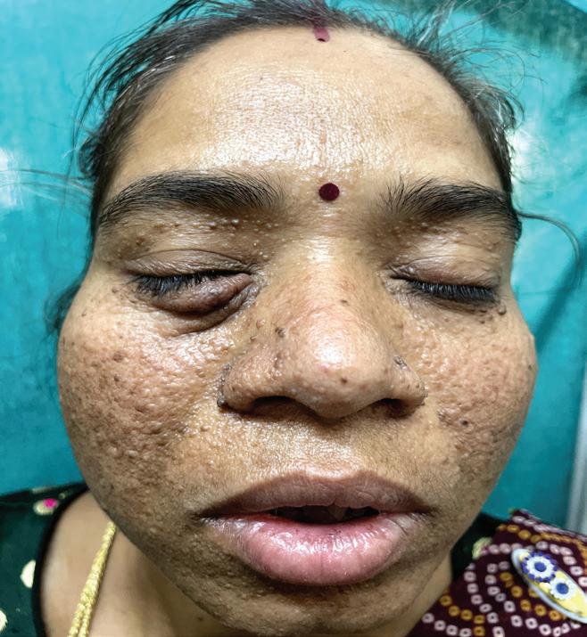
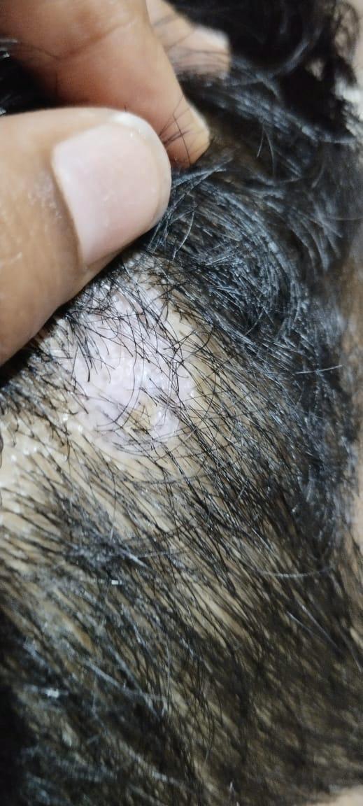
Figure 1: Adenoma sebaceum on the face
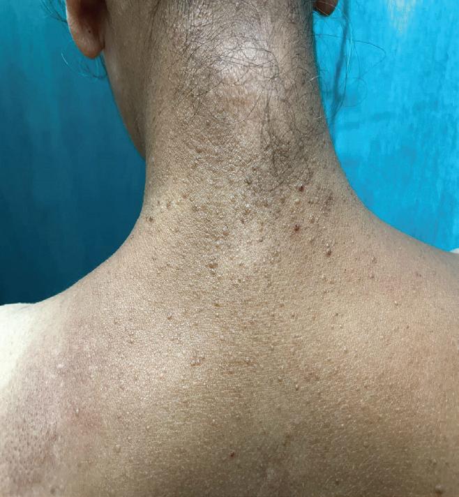
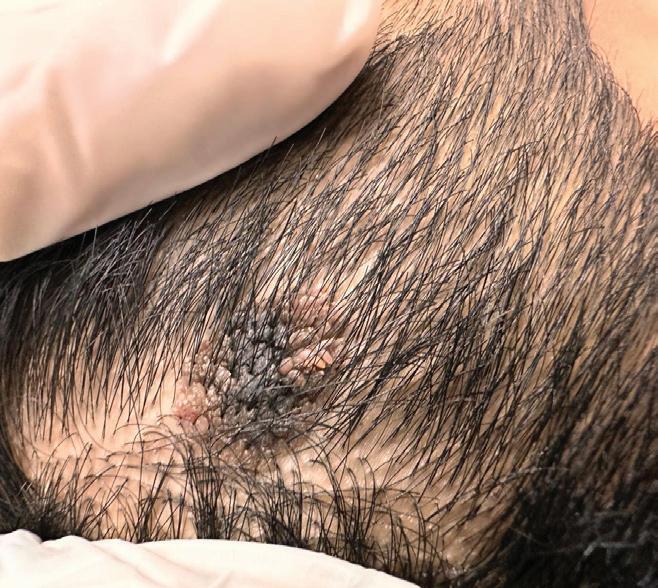
Figure 3: Adenoma sebaceum on the neck
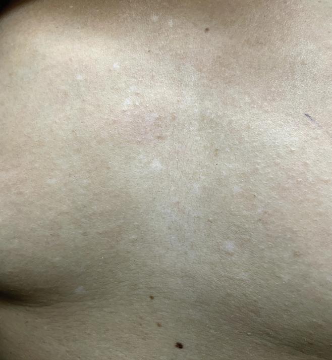
Figure 4: Hypomelanotic macules on the chest
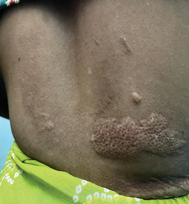
Figure 2: Shagreen patch on the back
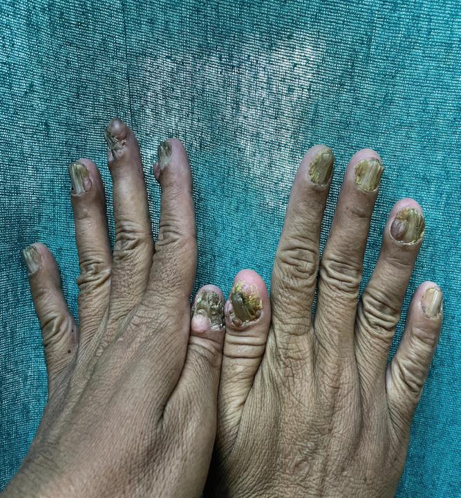
Figure 5: Koenen's tumor on hand
retinal hamartomas, subependymal nodules, cardiac rhabdomyomas, lymphangioleiomyomatosis, and multiple renal cysts, as these findings are integral to the accurate diagnosis of tuberous sclerosis and the initiation of appropriate management strategies. Wood's lamp examination is a valuable tool in this context, as it highlights pigmented lesions with distinct demarcation due to increased melanin absorption, while depigmented or hypopigmented areas, such as ash-leaf macules in tuberous sclerosis, are more easily identified under ultraviolet light, especially in individuals with lighter skin tones. Fundoscopic examination is also important in detecting retinal hamartomas, a common ocular manifestation in TSC. Imaging techniques such as echocardiography, MRI, or CT scans are instrumental in identifying cardiac rhabdomyomas, tubers, and subependymal tumors, respectively. Genetic testing, while not always necessary for diagnosis, can be helpful in identifying mutations in the TSC1 or TSC2 genes, confirming the diagnosis and assisting in family counseling.3
Treatment
Tuberous sclerosis is a chronic condition without a cure, but effective management is possible through a multidisciplinary approach tailored to the individual's symptoms. Treatment typically involves collaboration among various specialists, including pediatricians, neurologists, dermatologists, and others, to provide supportive and symptomatic care. Early intervention is crucial to ensure optimal development and improve the quality of life for those affected by tuberous sclerosis. Medications play a central role in managing tuberous sclerosis symptoms, addressing issues such as seizures and tumor growth. Anti-seizure drugs are prescribed based on the type of seizures, the patient's age, and the severity of symptoms. Vigabatrin, an FDAapproved medication, is used cautiously, particularly for infantile spasms. It works by increasing levels of gamma-aminobutyric acid (GABA) in the brain, helping to reduce seizure activity. Vigabatrin has shown efficacy in altering the course of seizures in infants with tuberous sclerosis, decreasing their risk and severity, although it
does not completely prevent epilepsy. Everolimus is another FDA-approved medication, specifically for certain tumors associated with tuberous sclerosis. In some cases, surgical interventions, such as embolization or resection, may be required to manage tumor growth. Overall, the management of tuberous sclerosis is focused on controlling symptoms and improving the patient's quality of life through a combination of medical and surgical treatments.4,5,6,7
Lymphangioleiomyomatosis predominantly affects young women of childbearing age, leading to speculation about the role of female hormones, particularly estrogen, in its pathogenesis. While this potential link has been widely considered, no definitive causal relationship between lymphangioleiomyomatosis and female hormones has been established. Despite this, clinicians have explored various interventions aimed at modulating estrogen production or its effects in the body, with varying degrees of effectiveness among patients. Among these interventions, medroxyprogesterone........


acetate has been considered as a treatment option. It is generally recommended that patients with lymphangioleiomyomatosis discontinue the use of estrogen-containing.......... medications and dietary supplements to avoid potential exacerbation of the condition. In the context of immunosuppressive therapy, a macrolide immunosuppressant agent, known for its inhibitory effects on the mechanistic target of rapamycin kinase (mTOR), plays a significant role. This agent reduces the responsiveness of T and B cells to interleukin-2 (IL-2) and received FDA approval in 1999 as an immunosuppressant for preventing organ rejection in patients aged 13 years and older undergoing renal transplantation, as well as for treating patients with lymphangioleiomyomatosis. Additionally, psychiatric conditions are commonly observed in patients with lymphangioleiomyomatosis, encompassing a wide range of disorders, including behavioral disorders like intermittent explosive disorder, oppositional defiant disorder, and selfinjurious behavior, as well as autism spectrum disorders, anxiety disorders, attention-
deficit/hyperactivity disorder (ADHD), and mood and thought disorders. On average, patients with lymphangioleiomyomatosis often present with dual diagnoses of two psychiatric conditions. Management of these psychiatric disorders typically involves pharmacotherapy, ............ utilizing antipsychotics, antidepressants, and anticonvulsants with moodstabilizing properties. This therapeutic approach generally leads to an improvement or stabilization of psychiatric symptoms, contributing to a better overall quality of life for patients.3,6
Surgical intervention may become necessary in tuberous sclerosis when growths cause complications, requiring the removal of problematic growths affecting various organs or body systems. One specific surgical technique, photocoagulation, utilizes laser technology to induce controlled thermal damage to the blood vessels supplying ocular tumors, helping to manage these growths effectively. Dermatological treatments also play a crucial role in managing the visible skin manifestations of
tuberous sclerosis, which can significantly impact a patient's mental wellbeing. Interventions such as cryoablation, laser skin resurfacing, excision, or skin grafts can help address these visible symptoms, improving the patient's quality of life. Additionally, skin lesions may be treated with mTOR inhibitors or other procedures like dermabrasion or surgical removal. Overall, treatment for tuberous sclerosis is highly individualized, often involving a combination of therapies to manage symptoms effectively and enhance the patient's overall well-being.7
Discussion
Tuberous sclerosis, also known as tuberous sclerosis, is a rare genetic disorder characterized by the formation of benign tumors, or hamartomas, in various organs throughout the body. These growths can develop in the brain, kidneys, heart, lungs, skin, and other tissues, leading to a wide range of symptoms and potential complications. The clinical manifestations of tuberous sclerosis can vary greatly among individuals. Common features include reddish papules on the face, the development of benign brain Clinical Insights and Management of a Rare Case
tumors known as cortical tubers, which may cause seizures, developmental delays, intellectual impairment, and behavioral issues. Cardiac involvement can manifest as cardiac rhabdomyomas, which, although often self-resolving, can lead to arrhythmias or heart failure. Other potential complications include pulmonary tumors, various skin abnormalities, and ocular issues such as retinal hamartomas. Diagnosing tuberous sclerosis can be challenging due to the wide range of symptoms and their variability among individuals. The diagnostic process typically involves a thorough clinical assessment, imaging studies like MRI or CT scans, and genetic testing to identify mutations in the TSC1 or TSC2 genes. Management of tuberous sclerosis focuses on controlling symptoms and preventing complications through a multidisciplinary approach. Dermatological manifestations may be treated with topical medications, laser therapy, or surgical removal. Seizures associated with cortical tubers are managed with antiseizure medications, such as vigabatrin, and immunosuppressant ......... medications, with surgery
Clinical Insights and Management of a Rare Case of Tuberculosis Sclerosis: A Case Report
considered in cases that do not respond to medication. Renal tumors are monitored regularly and may be treated with embolization, surgical resection, or medication. Cardiac rhabdomyomas generally resolve spontaneously but require monitoring for potential complications. Genetic counseling is recommended for affected individuals and their families to understand inheritance patterns and make informed decisions. The prognosis for individuals with tuberous sclerosis varies depending on the severity of symptoms and complications. While many individuals can lead fulfilling lives with appropriate management, severe neurological or renal involvement can significantly impact morbidity and mortality rates.1,2,6
Conclusion
The outlook for individuals with tuberous sclerosis varies depending on the severity of symptoms and complications. With appropriate care, many people with tuberous sclerosis can lead full lives, although severe issues involving the brain or kidneys can impact morbidity and life expectancy. Tuberous sclerosis is a complex medical
condition that demands comprehensive care and support from healthcare professionals, caregivers, and the affected individuals themselves. Managing tuberous sclerosis requires a multidisciplinary approach aimed at controlling symptoms and preventing complications. Treatment strategies often include antiseizure medications, surgical interventions for tumors, dermatological treatments, and genetic counseling for individuals and their families. By raising awareness, encouraging early diagnosis, and implementing personalized treatment plans, we can work toward improving the quality of life for those living with tuberous sclerosis.
References
1. Crino PB, Nathanson KL, Henske EP. The tuberous sclerosis complex. N Engl J Med. 2006 Sep 28;355(13):1345-56. [PubMed].
2. Baskin HJ., Jr The pathogenesis and imaging of the tuberous sclerosis complex. Pediatr Radiol. 2008;38(9):936.
3. mayoclinic.org/diseases-......... conditions/tuberous-sclerosis/ symptoms-causes/syc20365969
4. Randle S. Tuberous sclerosis complex: Management and prognosis. https://www.
A Case Report
Dr. Sujata Thakur MD
(Dermatology)
Consultant Dermatologist, Cosmetologist, Trichologist & Dermatosurgeon
Sanjeevani Clinic, Mumbai Introduction
Epidermal nevi (EN) are developmental abnormalities characterized by excessive growth of the epidermis and its associated adnexal structures. Lesions that prominently feature adnexal components such as sebaceous glands, hair follicles, or apocrine glands are termed "organoid," whereas those primarily composed of epidermal cells are referred to as "nonorganoid" or "keratinocytic" nevi. Epidermal nevi occur in approximately 1 to 3 per 1000 live births and typically arise sporadically, although familial cases are infrequent. About 60% of these nevi are evident at birth, with 95% manifesting by age 7. Variants of epidermal nevi include verrucous
epidermal nevus (VEN), nevus sebaceous, nevus comedonicus, and eccrine nevus.1
Verrucous epidermal nevus is the predominant variant among epidermal nevi, characterized by localized hyperplasia of keratinocytes originating from embryonic ectoderm. These lesions manifest as circumscribed hamartomatous growths, typically demonstrating progressive pigmentation and developing a rough, verrucous texture over time. The global incidence of verrucous epidermal nevus is estimated between 0.1% and 0.5%. Epidermal nevi originate from pluripotential germinative cells located in the basal layer of the embryonic epidermis. A
Verrucous Epidermal Nevus (VEN) Treated with Electrocautery: A Case Report
significant proportion of epidermal nevi are attributed to mosaicism, characterized by activating mutations in the FGFR3 gene within the human epidermis. These mutations arise postzygotically during early embryonic development. When such mutations occur very early in embryogenesis, they give rise to extensive epidermal nevi that may potentially involve additional organ systems. These lesions can manifest as unilateral or bilateral and typically follow the lines of Blaschko, reflecting the mosaic distribution of mutated cells.1
Mosaicism resulting from somatic mutations early in embryogenesis results in the formation of two distinct cell lines that migrate along developmental lines (Blaschko lines), producing patchy and linear abnormalities on the skin. Clinically, this presents as alternating bands of normal and affected skin following Blaschko's lines, termed type 1 segmental manifestation. Another type of segmental manifestation arises from a germ line mutation occurring after a post-zygotic mutation, leading to double inactivation of the gene.2
Epidermal nevi, particularly
the verrucous type, are hamartomatous lesions originating from embryonic ectoderm, exclusively composed of keratinocytes. They typically manifest as solitary or multiple unilateral linear plaques, adhering to the lines of Blaschko, reflecting their mosaic nature. Clinically, these lesions present as well-circumscribed, hyperpigmented, ............ papillomatous papules or plaques, commonly asymptomatic. Verrucous epidermal nevi can appear as localized small areas or generalized across larger regions of the body. In some cases, they may follow a swirled or linear pattern, often affecting the trunk, extremities, cervical region, and face. Systematized forms, also known as ichthyosis hystrix, exhibit multiple lesions in a swirl pattern along segmental and linear skin lines, such as V-shaped or circling patterns on the chest and linear distributions on the extremities.2
Papillomatous nevi can present as velvety soft lesions in newborns or hard keratotic, verruciform lesions in adolescence, varying in color from light to dark brown. Multiple lesions of verrucous epidermal
nevus may be associated with defects in other tissues, particularly the skeleton and central nervous system, leading to the application of 'epidermal nevus syndrome. Inflammatory Linear Verrucous Epidermal Nevus (ILVEN) is a rare variant predominantly observed in females, characterized by recurrent inflammatory conditions like chronic eczema or psoriasis. These conditions are typically unilateral and may cause severe itching. In very rare cases, malignant transformation into basal cell or squamous cell carcinomas has been documented.2
Case report
A 14-year-old patient weighing 49.8 kg presented with discomfort associated with a recently progressing verrucous epidermal nevus on the scalp over the past 2 months. There was no history of hypertension, diabetes mellitus, or known drug allergies. Based on the clinical appearance of the lesion, we made a diagnosis of verrucous epidermal nevus. We treated this patient with electrocautery along with pharmacological treatment of amorphous hydrogel wound dressing with colloidal silver to manage the skin lesions.
Verrucous Epidermal Nevus (VEN) Treated with Electrocautery: A Case Report
Additionally, a course of cefadroxil (500mg) three times daily for 5 days was prescribed to address potential bacterial infection. Symptomatic relief included analgesic and antipyretic tablets ibuprofen and paracetamol twice a daily for 5 days and omeprazole 20 mg capsules twice daily for 5 days to alleviate discomfort and manage gastrointestinal symptoms. Following completion of the treatment regimen, the patient's symptoms improved significantly.
Before treatment
After treatment
Figure 1: Circular inflammatory epidermal nevus on the scalp
Diagnosis
Diagnosing verrucous epidermal nevus requires a comprehensive approach encompassing clinical assessment, histopathological analysis, and potentially imaging studies to characterize the lesion thoroughly. Diagnosis of verrucous epidermal nevus relies primarily on clinical assessment, identifying localized overgrowths of keratinocytes on the skin that appear rough, verrucous, and occasionally pigmented. These lesions often conform to the lines of Blaschko, reflecting their mosaic distribution. They commonly appear on the trunk, extremities, cervical area, and face, with onset either congenitally or shortly after birth, evolving in texture and pigmentation over time. A thorough physical examination evaluates the size, distribution, and texture of the nevi. While histopathological confirmation via skin biopsy may be performed in atypical cases, it is typically unnecessary when clinical features are characteristic.3
Histopathologically, confirmation through skin biopsy is pivotal. Microscopic examination reveals typical features including hyperkeratosis, acanthosis (epidermal
hyperplasia), and papillomatosis characterized by finger-like projections of the epidermis into the dermis. Elongation of rete ridges and hyperpigmentation of the basal layer further support the diagnosis, particularly when a mosaic pattern is evident, indicative of segmental distribution.3
Imaging studies such as MRI or CT scans may be warranted in cases where there is suspicion of deeper tissue involvement or in the context of associated developmental anomalies, particularly in the evaluation of potential epidermal nevus syndrome. Genetic testing may occasionally be considered to identify underlying mosaic genetic mutations contributing to the pathogenesis of verrucous epidermal nevus, although it is not routinely performed for diagnosis.3
The diagnosis of verrucous epidermal nevus relies on integrating clinical observations with detailed histopathological examination to confirm the diagnosis accurately and guide optimal management strategies suited to each patient's specific clinical presentation and needs.
Treatment
Management of verrucous
Verrucous Epidermal Nevus (VEN) Treated with Electrocautery: A Case Report
epidermal nevus involves a multidisciplinary approach, collaborating with dermatologists, pediatricians, and other specialists as needed based on associated systemic findings. Treatment options are diverse and tailored to individual circumstances, encompassing topical therapies, cryotherapy, laser interventions, and surgical excision depending on lesion size, location, and clinical symptoms.
Treatment strategies for verrucous epidermal nevus are tailored based on lesion extent and patient impact. Observation suffices for asymptomatic cases, with periodic monitoring recommended. Topical therapies like keratolytic agents and retinoids help reduce lesion thickness and roughness. Surgical excision is reserved for symptomatic or cosmetically concerning lesions, aiming for complete removal with minimal scarring. Laser therapy is beneficial for smaller or sensitive areas, while cryotherapy, electrosurgery, and dermabrasion provide alternative options based on lesion size and location. Combination therapies may be employed for extensive nevi. Regular follow-up is crucial to monitor for recurrence or
progression of the nevus, ensuring comprehensive management aligned with individual patient needs.4
Oral retinoids
Oral retinoids, such as acitretin, have emerged as a therapeutic option for verrucous epidermal nevus, aiming to normalize keratinocyte differentiation and reduce epidermal hyperplasia through their vitamin A derivative mechanism. Their clinical effectiveness in treating verrucous epidermal nevus varies, often resulting in reduced lesion thickness and hyperkeratosis, which can significantly improve both appearance and symptoms over time. Treatment typically requires a prolonged course tailored to the severity of the condition and individual patient response, often spanning several months to achieve noticeable results. Additionally, oral retinoids may be used in conjunction with topical therapies (such as keratolytics or corticosteroids) or procedural interventions (like laser therapy) to enhance overall efficacy and therapeutic outcomes.4
Topical keratolytics
Topical keratolytics are integral in managing verrucous epidermal
nevus by targeting the hyperkeratosis that characterizes the condition. Through mechanisms such as softening and breaking down keratin, agents like salicylic acid, urea, and lactic acid help to reduce lesion thickness and scaling, thereby smoothing and improving the appearance of affected skin. These treatments are typically applied directly to the affected areas as part of a daily skincare routine, with the regimen tailored based on lesion severity and individual response. Their efficacy is enhanced when used in conjunction with other topical or procedural therapies, providing a comprehensive approach to managing verrucous epidermal nevus and improving patient outcomes. Regular monitoring by dermatologists ensures optimal treatment adjustment and minimization of potential side effects, promoting effective longterm management of the condition.5
Corticosteroid
Corticosteroids serve as a versatile treatment option for verrucous epidermal nevus, primarily addressing inflammation and associated symptoms. When applied topically, corticosteroids are
formulated as ointments, creams, or lotions tailored to the severity and location of lesions, exerting antiinflammatory effects directly on affected skin areas. For more resistant or thicker lesions, intralesional corticosteroid injections offer a concentrated dosage at the lesion site, facilitating deeper penetration and efficacy. These treatments mitigate redness, swelling, and itching typical of verrucous epidermal nevus, while also aiding in the softening and flattening of thickened skin, thereby enhancing the cosmetic appearance of affected areas. The use of corticosteroids in verrucous epidermal nevus management is often integrated with other therapies to optimize outcomes, with careful monitoring by dermatologists to manage potential side effects and ensure treatment efficacy over time.6
Laser therapy
Carbon dioxide (CO2) and erbium lasers are effective treatments for verrucous epidermal nevus. CO2 lasers vaporize and remove thickened skin layers with minimal damage to surrounding tissue, reducing lesion size and improving
Verrucous Epidermal Nevus (VEN) Treated with Electrocautery: A Case Report
cosmetic appearance. Erbium lasers, emitting light at 2940 nanometers, target superficial to moderately deep layers of thickened tissue with precision, minimizing collateral damage and enhancing skin texture. Both lasers offer tailored treatment options that significantly benefit patients by reducing lesion thickness and improving overall cosmetic outcomes. Their specific mechanisms and adjustable settings represent significant advancements in dermatological care for managing verrucous epidermal nevus.7,8
Cryotherapy
Cryotherapy is a widely utilized therapeutic method in managing verrucous epidermal nevus, primarily through the application of extreme cold to targeted lesions. By applying liquid nitrogen or another cryogen directly onto verrucous epidermal nevus lesions using a cryospray or cryoprobe, dermatologists freeze the affected skin cells. This freezing action induces necrosis, causing the abnormal keratinocytes responsible for the thickened, wart-like appearance to die off. As the treated lesions undergo natural processes of
sloughing off or absorption by the body, the overall size and thickness of verrucous epidermal nevus lesions are reduced. Depending on the size, location, and depth of the lesions, multiple cryotherapy sessions may be necessary. Dermatologists typically space these treatments to allow for adequate healing and to assess the effectiveness of the therapy in achieving smoother skin texture and improved cosmetic outcomes.9
Photodynamic therapy (PDT)
Photodynamic therapy has emerged as a promising treatment modality for verrucous epidermal nevus. By utilizing a photosensitizing agent that preferentially accumulates in abnormal cells within the epidermal nevus, Photodynamic therapy offers a targeted approach to treatment. Once applied to the skin and allowed to penetrate, the photosensitizing agent is activated by exposure to specific wavelengths of light. This activation triggers the production of reactive oxygen species, which selectively damage and destroy the abnormal cells while minimizing harm to surrounding healthy tissue.10
Verrucous Epidermal Nevus (VEN) Treated with Electrocautery: A Case Report
Adjuvant therapy
Hydrogel and colloidal silver cream each serve distinct yet complementary roles in the treatment of verrucous epidermal nevus. Hydrogel dressings maintain a moist environment that hydrates and soothes the affected skin, promoting natural healing processes and alleviating discomfort associated with dry, thickened lesions. The cooling effect of hydrogel further aids in reducing itching and irritation commonly experienced with verrucous epidermal nevus. In contrast, colloidal silver cream harnesses potent antimicrobial properties to prevent and treat secondary infections that may arise from scratching or irritation of verrucous epidermal nevus lesions. It also reduces inflammation and forms a protective barrier, safeguarding the lesions from external irritants and contaminants. Together, hydrogel and colloidal silver cream form a comprehensive skincare regimen that supports both symptomatic relief and therapeutic healing, enhancing overall management outcomes for verrucous epidermal nevus when used in combination with other treatment
modalities like cryotherapy or laser therapy.4
Electrofiguration ......... (electrosurgery or electrocautery)
Electrofulguration, also referred to as electrosurgery or electrocautery, offers a targeted approach to treating verrucous epidermal nevus through the precise application of high-frequency electrical current. This method works by generating heat at the tip of a fine electrode, which vaporizes and removes abnormal tissue, including the thickened layers characteristic of verrucous epidermal nevus. The procedure typically begins with the administration of a local anaesthetic to ensure patient comfort. The electrode is then carefully maneuvered over the nevus, delivering controlled electrical pulses to ablate the lesion. This process may need to be repeated to ensure thorough removal of the nevus tissue.4
Chemical peels
Chemical peels offer a versatile approach to managing verrucous epidermal nevus by effectively addressing its visible symptoms through controlled exfoliation and skin renewal. Superficial peels, utilizing gentle acids
like glycolic or salicylic acid, primarily target the surface layer of the skin (epidermis). These peels are beneficial for improving skin texture, reducing minor irregularities, and enhancing overall skin tone affected by verrucous epidermal nevus. Conversely, medium to deep peels utilize stronger acids such as Trichloroacetic acid (TCA) or phenol, enabling them to penetrate deeper into the skin layers. This deeper penetration makes them particularly suitable for treating thicker or more resistant verrucous epidermal nevus lesions. By inducing a more profound exfoliation and regeneration process, medium to deep peels aim to achieve substantial improvements in the appearance of verrucous epidermal nevus, including significant reduction in lesion thickness and enhanced skin smoothness.4
Discussion
Epidermal nevi are benign skin patches resulting from excessive growth of epidermal cells, typically appearing at birth or early childhood. They vary in size and type based on the specific epidermal cell involved. These nevi often conform to the lines of Blaschko, representing
embryonic skin cell migration patterns. Epidermal nevi involve other epidermal cell types like hair follicle, sweat gland, or sebaceous gland cells. For instance, nevus sebaceous is a common type, manifesting as waxy, yellow-orange patches on the scalp or face, often accompanied by hair loss (alopecia) within the affected area. These nevi can also become more pronounced and verrucous with age. While some individuals may only exhibit epidermal nevi without additional health issues, others may experience associated abnormalities in various body systems, such as the brain, eyes, or bones. When such systemic issues accompany epidermal nevi, the condition is termed an epidermal nevus syndrome, which encompasses several distinct syndromes characterized by the type of epidermal nevus present.4
Epidermal nevi, including verrucous epidermal nevus, can mimic several other skin conditions, necessitating careful consideration and differentiation for accurate diagnosis and management. Among the various syndromes and lesions that may resemble verrucous epidermal nevus, Sebaceous Nevus
Syndrome and Becker's Nevus Syndrome stand out due to their distinctive characteristics such as yellowish plaques or hyperpigmented patches with coarse hair growth, respectively. These syndromes differ in texture, coloration, and distribution from verrucous epidermal nevus. Other benign conditions like seborrheic keratosis and verruca vulgaris can also present with wart-like appearances but typically exhibit different textures and growth patterns. Cutaneous lesions such as epidermoid cysts lack the hyperkeratosis seen in verrucous epidermal nevus, while inflammatory conditions like lichen planus have distinct distribution patterns and may show signs of inflammation not typically seen in verrucous epidermal nevus. Genodermatoses like linear epidermal nevus and Incontinentia Pigmenti can resemble verrucous epidermal nevus but have unique features such as linear streaks or characteristic stages of progression. Lastly, neoplastic conditions such as Bowen's Disease can sometimes mimic verrucous epidermal nevus, necessitating biopsy for definitive diagnosis due to its
potential malignant nature. Clinicians must rely on clinical evaluation, historytaking, and sometimes biopsy to differentiate verrucous epidermal nevus from these various conditions, ensuring appropriate management and optimal outcomes for patients.4,5
Six different syndromes associated with epidermal nevi have been identified: Proteus syndrome, CHILD syndrome (congenital hemidysplasia with ichthyosiform nevus and limb defect), phakomatosis pigmentokeratotica, sebaceous nevus syndrome, Becker's nevus syndrome, and nevus comedonicus syndrome. Managing epidermal nevi presents significant challenges as no single ideal or universally accepted treatment has been established. Medical treatments such as corticosteroids under occlusion, corticosteroid injections, and topical tretinoin (vitamin A) cream may offer partial effectiveness. Oral retinoids are considered for widespread epidermal nevi but often require longterm therapy. Surgical excision is typically the treatment of choice for small
Verrucous Epidermal Nevus (VEN) Treated with Electrocautery: A Case Report
lesions, although superficial removal methods frequently result in recurrence. More aggressive approaches including dermabrasion, cryosurgery, electrosurgery, and laser surgery have been utilized, each with its own set of risks and potential benefits. Laser treatments, including CO2, long-pulsed Nd, and 585 nm pulsed dye lasers, have evolved with advancements in technology, improving precision and safety. Despite these advancements, recurrence of epidermal nevi remains possible months to years after treatment.1,2
Patients with epidermal nevi commonly present to dermatologic surgeons for evaluation and treatment. However, dentists who are vigilant in clinical examination can also contribute to diagnosing these lesions based on their appearance. A comprehensive multidisciplinary .............. assessment is essential to exclude associated neurologic, ocular, skeletal, and cardiovascular complications of epidermal syndromes. Following this evaluation, it is crucial for physicians to recommend the most appropriate and effective therapy for the
skin manifestations that prompted the patient's initial consultation.
Conclusion
In managing patients with verrucous epidermal nevus, significant challenges persist due to the condition's variable clinical course and the lack of a definitive treatment protocol. While current therapies encompass a range of modalities from topical agents to surgical interventions like laser therapy and excision, each approach carries inherent limitations, including recurrence risks and potential adverse effects. The evolving landscape of dermatological techniques, such as advanced laser technologies and targeted therapies, shows promise in improving treatment outcomes. However, achieving optimal results often requires a personalized, multidisciplinary approach to address both the cutaneous manifestations and potential systemic implications of verrucous epidermal nevus. Continued research and innovation are essential to overcoming these challenges and enhancing the management of verrucous epidermal nevus, ultimately aiming
to provide patients with effective, well-tolerated therapies that optimize longterm outcomes and quality of life.
References
1. Rogers M, McCrossin I, Commens C. Epidermal nevi and the epidermal nevus syndrome. A review of 131 cases. J Am Acad Dermatol. 1989;20(3):476-488. doi:10.1016/ s0190-9622(89) 70061-x.
2. Saini S, Agarwal S, Yadav C, Sharma S. Verrucous Epidermal Nevus with Unusual Presentations: A Case Series of 5 Cases. Indian J Dermatol. 2022;67(3):317. doi:10.4103/ ijd. ijd_880_21
3. Verzì AE, Lacarrubba F, Quattrocchi E, Micali G. Verrucous Epidermal Nevus: Dermoscopy, Reflectance Confocal Microscopy, and Histopathological Correlation. Dermatol Pract Concept. 2019;9(3): 230-231. Published 2019 Jul 31. doi:10.5826 / dpc.0903a16
4. Arora B, Singh Khinda VI, Bajaj N, Singh Brar G. Congenital epidermal nevus. Int J Clin Pediatr Dent. 2014;7(1):43-46. doi:10.5005/ jp-journals-10005-1232
5. Kerawala SR, Rizvi NU, Tabassum S. Bilateral Systematised Epidermolytic Epidermal Nevus: A case report. Sultan Qaboos Univ Med J. 2021;
21(1): e124-e126. doi:10.18295/ squmj.2021.21.01.018
6. Hong JK, Han HS, Yoo KH. Inflammatory linear verrucous epidermal naevus successfully treated with a combination of triple topical agents (corticosteroid, calcipotriol and 20% urea). Clin Exp Dermatol. 2021;46(5): 940942. doi:10.1111/ ced.14603.
7. Thual N, Chevallier JM, Vuillamie M, Tack B, Leroy D, Dompmartin A. Laser CO2 continu dans le traitement des hamartomes épidermiques verruqueux [CO2 laser therapy of verrucous epidermal nevus]. Ann Dermatol Venereol. 2006;133(2): 131-138. doi:10.1016/ s01519638(06) 70863-8.
8. Park JH, Hwang ES, Kim SN, Kye YC. Er:YAG laser treatment of verrucous epidermal nevi. Dermatol Surg. 2004;30(3):378381. doi:10.1111/j.15244725.2004.30104.x.
9. Panagiotopoulos A, Chasapi V, Nikolaou V, et al. Assessment of cryotherapy for the treatment of verrucous epidermal naevi. Acta Derm Venereol. 2009;89(3):292294. doi:10.2340/000155550610
10. Kim TI, Jeong KH, Shin MK. Verrucous epidermal nevus (VEN) successfully treated with indocyanine green (ICG) photodynamic therapy (PDT). JAAD Case Rep. 2015;1(5):312314. Published 2015 Aug 25. doi:10.1016/j.jdcr.2015.07.002
Verrucous Epidermal Nevus (VEN) Treated with Electrocautery: A Case Report
MUMBAI 202
MUMBAI 2025
1 Day Conference, Hands on Workshop and Exhibition
AESTHETICCON Mumbai 2025 is just the event for you with practical insights shared in the Conference, tips while training in the Hands on workshop and interaction
AESTHETICCON Mumbai 2025 is just the event for you with practical insights shared in the Conference, tips while training in the Hands on workshop and interaction with product manufacturers.
Spend the day catching up and meeting with your fellow Dermatologists colleagues.