22, SHREEJI BHAVAN, 275-279, SAMUEL STREET, MASJID BUNDER (W), MUMBAI-4000 03, INDIA.
EMAIL: info@indiancosmetologistjournal.com
WEBSITE: indiancosmetologistjournal.online
PRINTED, PUBLISHED, EDITED AND OWNED BY DOM DANIEL
PRINTED AT SWASTIK PRINTER, GALA NO.9 & 10, VISHAL INDUSTRIAL ESTATE, BHANDUP (WEST), MUMBAI400078.
PUBLISHED AT 22 SHREEJI BHAVAN, 275/279, SAMUEL STREET,
MASJID BUNDER (WEST), MUMBAI - 400003. INDIA.
“INDIAN COSMETOLOGIST JOURNAL” TAKES NO RESPONSIBILITY FOR UNSOLICITED PHOTOGRAPHS OR MATERIAL
ALL PHOTOGRAPHS, UNLESS OTHERWISE INDICATED, ARE USED FOR ILLUSTRATIVE PURPOSE ONLY.
VIEWS EXPRESSED IN THIS JOURNAL ARE THOSE OF THE CONTRIBUTORS AND NOT OF THE PUBLISHER. REPRODUCTION IN WHOLE OR IN PARTS OF TEXTS OR PHOTOGRAPHY IS PROHIBITED. MANUSCRIPTS, PHOTOGRAPHS AND ART ARE SELECTED AT THE DISCRETION OF THE PUBLISHER FREE OF CHARGE (ADVERTISING EXCLUDED). WHETHER PUBLISHED OR NOT, NO MATERIAL WILL BE RETURNED AND REMAINS THE PROPERTY OF THE PUBLISHING HOUSE, WHICH MAY MAKE USE OF IT AS SEEN FIT. THIS MAY INCLUDE THE WITHDRAWAL OF PUBLICATION RIGHTS TO OTHER PUBLISHING HOUSES.
ALL RIGHTS RESERVED. REPRODUCING IN ANY MANNER WITHOUT PRIOR WRITTEN PERMISSION PROHIBITED.
PUBLISHED FOR THE PERIOD OF OCTOBER -2025
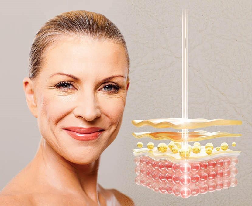
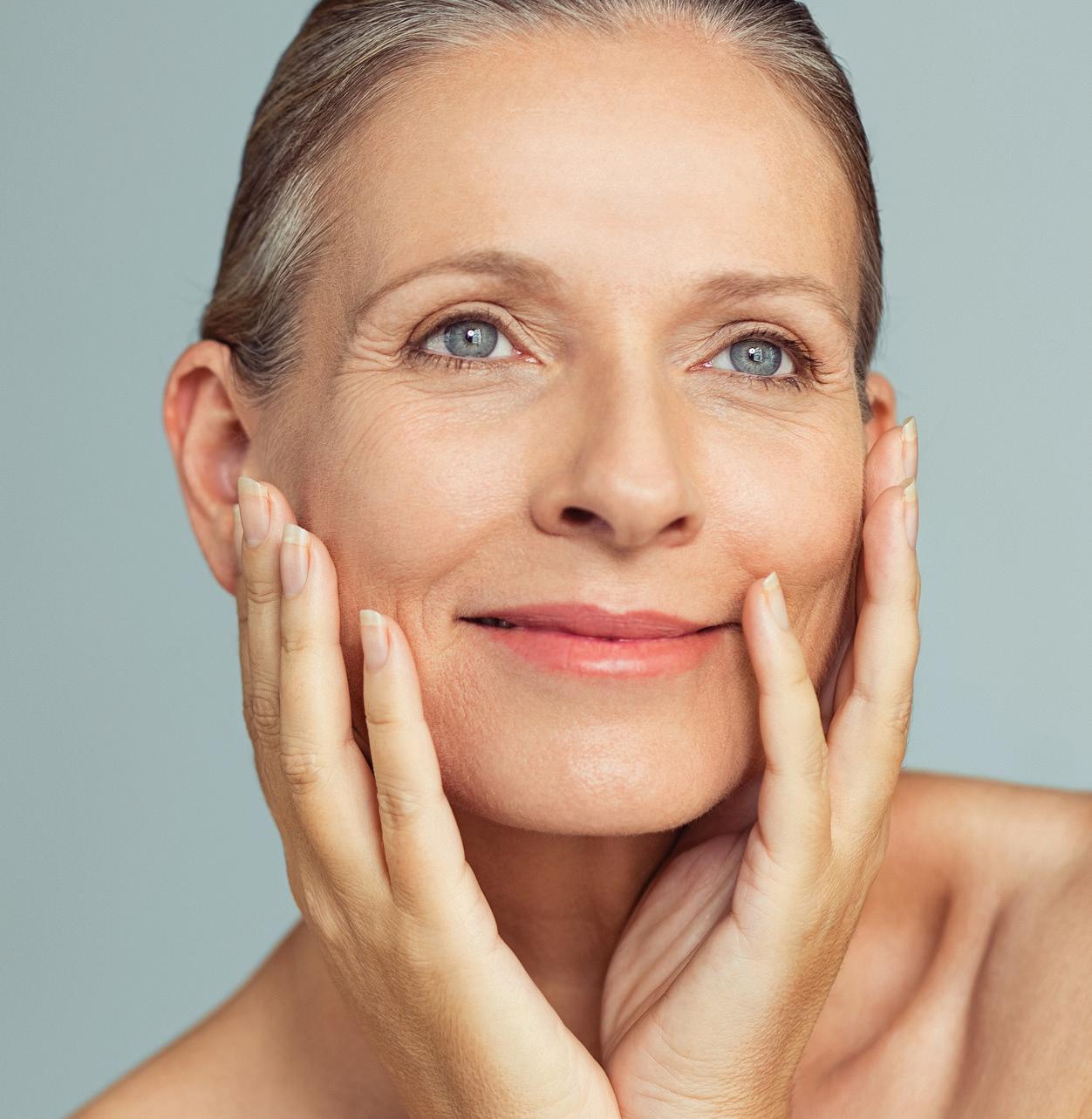
Exploring the Science of Skin: Aesthetic Insights for the Evolving Mind
Welcome to this issue, presenting a vibrant and thoughtfully assembled collection of insights and breakthroughs that are actively redefining the future landscape of skincare and the sophisticated art of aesthetic science.
THIS EDITION PRESENTS A CAREFULLY CURATED COLLECTION OF INSIGHTS THAT SEAMLESSLY INTEGRATE THE ART AND SCIENCE OF SKINCARE.
This edition presents a carefully curated collection of insights that seamlessly integrate the art and science of skincare. From the subtle surface characteristics of persistent milia to the complex biological processes that influence skin vitality, each article offers readers an opportunity for deeper understanding and discovery. “Unveiling Milia: What Lies beneath the Surface” provides a detailed examination of these small but persistent formations, encouraging a more nuanced and informed perspective. “Transepidermal Water Loss (TEWL): A Key Marker in Skin Health” underscores the vital importance of the skin barrier as the body primary defense mechanism, emphasizing its fundamental role in maintaining homeostasis and preserving dermal integrity. The feature on “The Science and Application of Hydrafacial Technology in Skin Rejuvenation” explores a holistic approach that combines mechanical exfoliation, hydration, and active ingredient infusion, highlighting its efficacy in promoting skin renewal and enhancing overall radiance. Finally, “Unlocking Skin Health: The Role of Epigenetics in Aging and Rejuvenation” offers a forward-looking perspective on the emerging science of gene-environment interactions, illuminating how these insights are shaping personalized, nextgeneration approaches to aging and rejuvenation.
Collectively, these features foster a deeper appreciation for the evolving interplay between scientific knowledge and aesthetic practice within the field of cosmetology.
- DOM DANIEL EXECUTIVE EDITOR & PUBLISHER
06
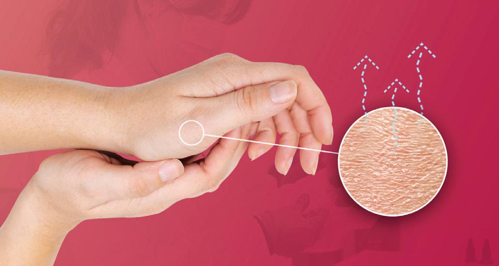
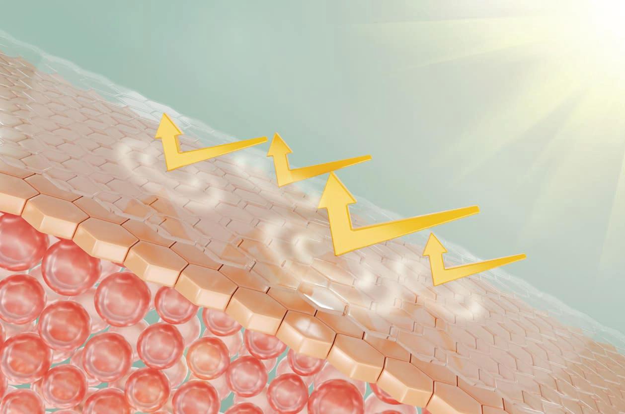
TRANSEPIDERMAL WATER LOSS (TEWL): A KEY MARKER IN SKIN HEALTH
18
UNLOCKING SKIN HEALTH: THE ROLE OF EPIGENETICS IN AGING AND REJUVENATION
THE SCIENCE AND APPLICATION OF HYDRAFACIAL TECHNOLOGY IN SKIN REJUVENATION.
UNVEILING MILIA: WHAT LIES BENEATH THE SURFACE 23
TRANSEPIDERMAL WATER LOSS
(TEWL): A KEY MARKER IN
SKIN HEALTH
INTRODUCTION
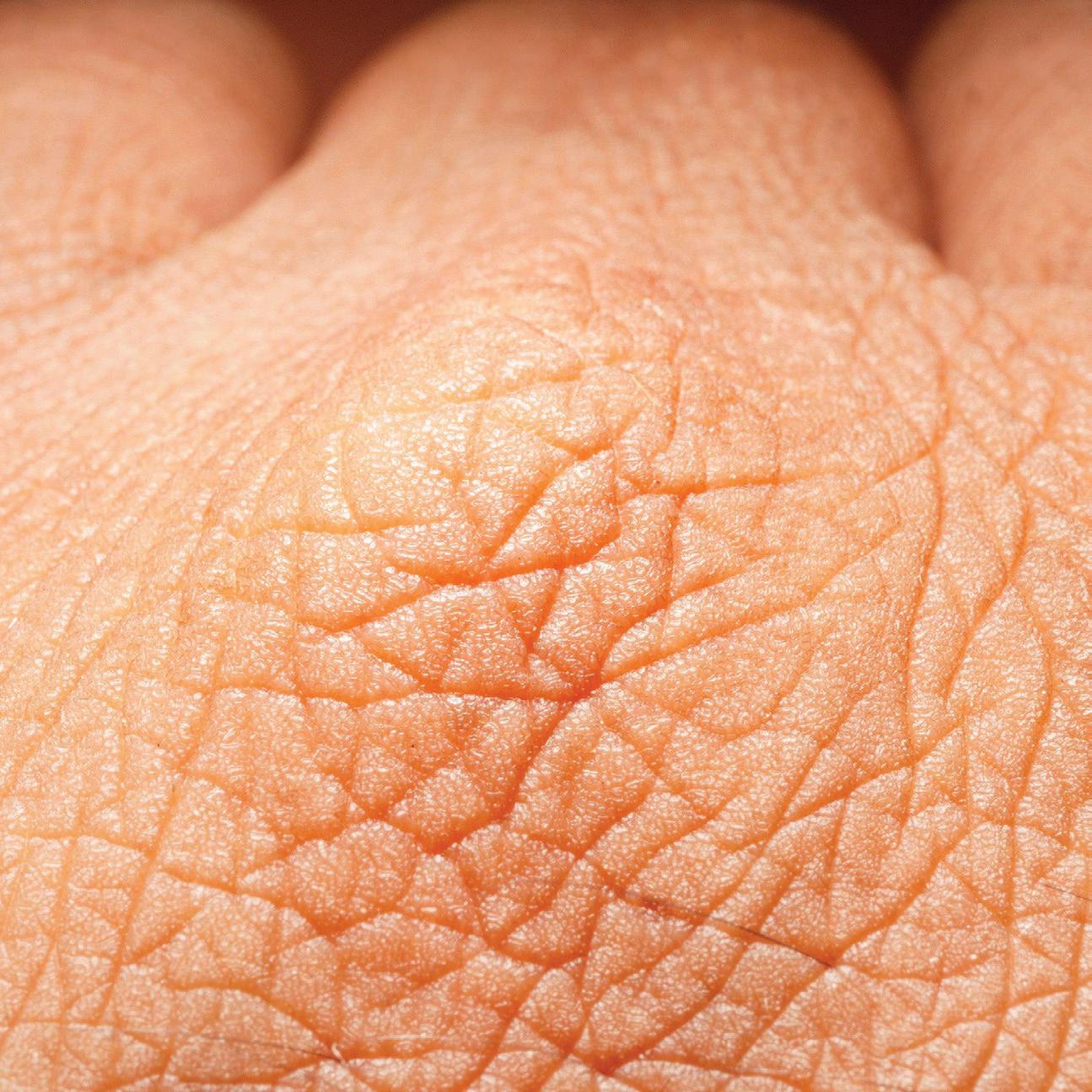
The skin, while seemingly smooth and resilient, is constantly engaged in a complex physiological process to preserve hydration, structural integrity, and barrier function. One of the most fundamental mechanisms in this system is Transepidermal Water Loss (TEWL), the passive diffusion of water from the deeper layers of the skin to the external environment through the stratum corneum. Although TEWL is a normal and essential process, it becomes clinically relevant when water
loss exceeds the skin’s natural ability to retain moisture. In such cases, the skin may present with signs of dehydration, dryness, sensitivity, and even premature ageing. TEWL is widely recognised in dermatological science as a measurable marker of skin barrier integrity. A compromised barrier, whether due to environmental factors such as low humidity, UV exposure, or pollution, inappropriate skincare practices, or age-related physiological changes, can significantly increase TEWL. This disruption weakens
the skin’s natural defence, resulting in a cascade of concerns including inflammation, irritation, and impaired skin function. Understanding Transepidermal Water Loss (TEWL) is essential for evaluating skin barrier integrity and guiding effective skincare strategies. As a key marker of epidermal function, TEWL informs both clinical and cosmetic dermatology in assessing skin health, identifying barrier disruption, and supporting targeted interventions to restore hydration and resilience.1, 2
AGE-RELATED
CHANGES AND TEWL
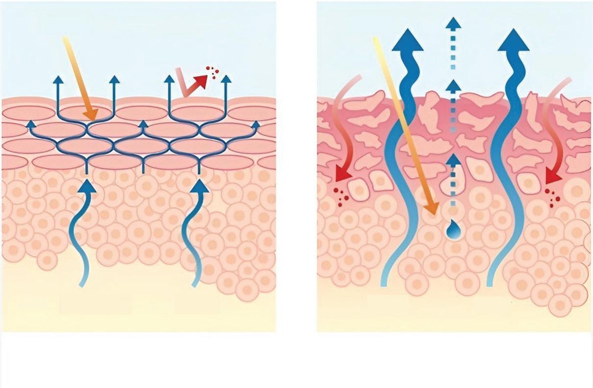
With advancing age, the skin undergoes intrinsic physiological alterations that impair its barrier function and increase transepidermal water loss (TEWL). Key changes include diminished lipid synthesis, reduced sebaceous gland activity, and slowed epidermal turnover, resulting in a thinner, more fragile stratum corneum. These factors compromise moisture retention, leading to xerosis, rough texture, and heightened susceptibility to irritation. Concurrent hormonal changes, particularly during menopause, exacerbate these effects by decreasing collagen production, epidermal thickness, and hydration levels. Additionally, environmental stressors and age-associated chronic low-grade inflammation (“inflammaging”) characterized by increased pro-inflammatory cytokines and senescent cell accumulation
further degrade collagen and elastin fibers, accelerating barrier dysfunction and visible signs of skin aging such as fine lines and loss of elasticity.1, 2
MECHANISM OF TRANSEPIDERMAL WATER LOSS (TEWL)
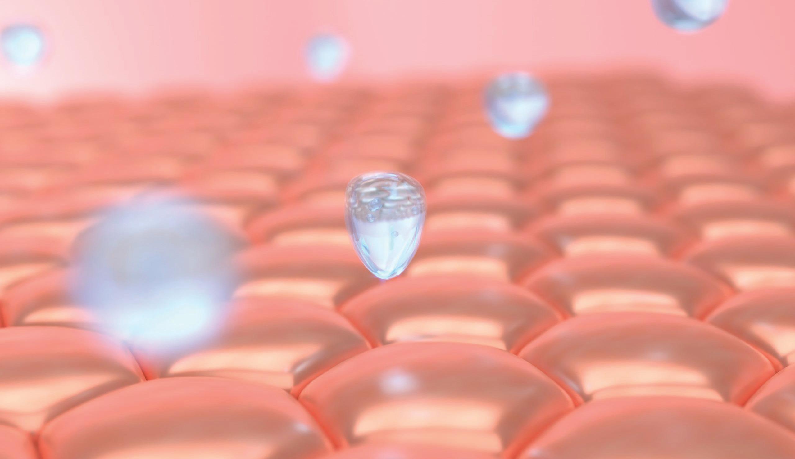
Transepidermal Water Loss (TEWL) is the passive diffusion of water from the dermis through the epidermis, culminating in evaporation from the stratum corneum (SC). The SC functions as the primary barrier, composed of anucleated corneocytes embedded in a lipid matrix rich in ceramides, cholesterol, and free fatty acids. Water transport is facilitated by osmotic gradients and aquaporins, particularly AQP3. Lamellar bodies secreted by keratinocytes at the stratum granulosum–SC interface release lipids that form lamellar bilayers, critical for barrier integrity. Natural moisturizing factors (NMFs) within corneocytes maintain hydration, while the acidic pH of the SC supports lipid processing and microbial defense. Disruption of this barrier, due to intrinsic or extrinsic factors, leads to increased TEWL, resulting in dehydration, enhanced permeability, and inflammation. Clinically, TEWL serves as a noninvasive biomarker for assessing skin barrier function and monitoring response to barrier-repair therapies.3
FACTORS CONTRIBUTING TO INCREASED TRANSEPIDERMAL WATER LOSS (TEWL)
Transepidermal water loss (TEWL) serves as a critical marker of skin barrier integrity. Elevated TEWL indicates a disruption in the epidermal barrier and is commonly associated with dryness, irritation and increased vulnerability to external stressors. Various environmental, intrinsic, extrinsic, and pathological factors can impair the structure and function of the stratum corneum, leading to increased water loss. Recognizing these contributing factors is essential for maintaining optimal skin barrier function and overall skin health.1, 4
1. Environmental Factors
➢ Low Relative Humidity: Reduced ambient moisture levels increase the vapor pressure gradient across the skin surface, promoting accelerated water evaporation and elevated TEWL.1, 4
➢ Thermal and Mechanical Stressors: Exposure to high temperatures, wind, and ultraviolet (UV) radiation leads to oxidative damage of epidermal lipids and structural proteins, compromising the integrity of the stratum corneum and increasing barrier permeability.1, 4
➢ Indoor Climate Control: Prolonged exposure to air conditioning and heating systems decreases environmental humidity, resulting in skin dehydration and increased TEWL.1, 4
2. Intrinsic Factors
➢ Genetic Variability: Individual differences in genes regulating epidermal lipid metabolism and keratinocyte function influence baseline barrier robustness and TEWL susceptibility.1, 4
➢ Aging: Age-related declines in epidermal lipid synthesis, sebaceous gland activity, and stratum corneum thickness impair barrier function, resulting in higher TEWL and skin dryness.1, 4
➢ Skin Phototype: Individuals with lighter skin phototypes have reduced melanin-mediated photoprotection, increasing vulnerability to UVinduced barrier disruption and subsequent water loss.1, 4
3. Extrinsic Factors
➢ Harsh Cleansing Practices: Frequent use of alkaline or surfactant-rich cleansers disrupts the acid mantle and lipid matrix, leading to increased permeability and TEWL.1, 4
➢ Over-exfoliation: Excessive mechanical or chemical exfoliation removes corneocytes and disrupts the protective barrier, facilitating greater transepidermal water diffusion.1, 4
➢ Topical Agents: Application of irritant substances or occlusive formulations can damage epidermal structures or interfere with normal desquamation, thereby modulating TEWL.1, 4
➢ Cosmetic Procedures: Interventions such as chemical peels, microdermabrasion, and laser therapies transiently impair barrier integrity, causing temporary elevations in TEWL.1, 4
4. Pathological Conditions
➢ Chronic Inflammatory Dermatoses:
Conditions such as seborrheic dermatitis and contact dermatitis are associated with disrupted barrier function and significantly elevated TEWL due to inflammatory cytokine activity and impaired lipid synthesis.
➢ Inflammation: Persistent cutaneous inflammation exacerbates barrier dysfunction, perpetuating a cycle of water loss and disease activity.1, 4
METHODS OF MEASURING TEWL
Transepidermal water loss (TEWL) is measured using non-invasive instruments designed to detect the gradient of water vapour evaporating from the skin surface. These devices provide accurate, real-time readings expressed in grams per square meter per hour (g/m²/h). This objective measurement allows clinicians and researchers to assess the skin’s barrier function with precision. TEWL measurement plays a crucial role not only in clinical practice but also in cosmetic product testing, helping evaluate the effectiveness of
barrier repair treatments and track skin recovery after procedures such as chemical peels or laser therapies.1
CLINICAL RELEVANCE OF TEWL
TEWL is a key indicator of skin barrier integrity and hydration status. Elevated TEWL reflects a compromised barrier, which can predispose the skin to dehydration, increased sensitivity, and irritation. Measuring TEWL is useful for monitoring changes in skin health and evaluating the effectiveness of interventions aimed at restoring and maintaining barrier function. It also plays an important role in the development and assessment of skincare formulations designed to improve moisture retention and support overall skin resilience.1
ENDOCANNABINOID SYSTEM AND SKIN BARRIER REGULATION
The ECS plays a pivotal role in maintaining skin homeostasis and modulating TEWL through several mechanisms:
• Barrier Function: Endocannabinoids such as anandamide (AEA) and 2-arachidonoylglycerol (2AG) activate CB1 and CB2 receptors on keratinocytes, influencing cellular differentiation and enhancing the structural integrity of the stratum corneum, thereby improving moisture retention.5
• Sebum Production: The ECS regulates sebaceous gland activity and lipid composition, contributing to the formation of a lipid-rich surface film that minimizes water loss.5
• Anti-inflammatory Action: ECS signaling modulates immune responses, reducing cutaneous inflammation that may otherwise impair barrier function and exacerbate TEWL.5
• Wound Healing: ECS involvement in skin repair mechanisms facilitates restoration of the epidermal barrier following injury, thereby supporting TEWL reduction.5
LIFESTYLE FACTORS INFLUENCING TEWL
Multiple lifestyle-related elements significantly impact TEWL and skin health:
• Nutrition: Adequate intake of essential fatty acids, vitamins (A, C, E), and antioxidants supports epidermal lipid synthesis and cellular repair mechanisms essential for maintaining barrier integrity.1.3
• Hydration: Sufficient water intake is necessary to sustain dermal hydration and elasticity, indirectly influencing TEWL levels.1.3
• Environmental Exposure: Chronic exposure to low humidity, UV radiation, wind, and pollutants
disrupts the lipid matrix and protein structure of the epidermis, leading to increased TEWL.1.3
• Skin Care Practices: Frequent use of alkaline soaps, hot water, and aggressive exfoliants can disrupt the skin’s acid mantle and lipid bilayer, compromising barrier function.1.3
• Stress and Sleep: Chronic psychological stress and sleep deprivation negatively impact the endocrine and immune systems, contributing to delayed skin barrier recovery and increased TEWL.1.3
• Microbiome Balance: A diverse and balanced skin microbiota plays a protective role by enhancing immune regulation, producing antimicrobial peptides and lipids, and maintaining barrier homeostasis all of which contribute to reduce TEWL.1.3
STRATEGIES FOR PREVENTING AND REDUCING TEWL
Effective management of TEWL requires a multifaceted approach combining topical therapies with supportive lifestyle modifications:
• Barrier Repair Moisturizers: Formulations containing physiological lipids—such as ceramides, cholesterol, and free fatty acids, aid in restoring the stratum corneum’s lipid matrix, reinforcing barrier integrity and reducing water loss.
• Humectants and Occlusives: Ingredients like glycerin and hyaluronic acid serve as humectants by attracting moisture, while occlusive agents (e.g., petrolatum, dimethicone) form a protective film to prevent evaporation.
• PH Maintenance: Topical products designed to preserve the skin’s natural acidic pH help maintain the acid mantle, promoting enzymatic activity essential for lipid processing and offering antimicrobial protection.
• Environmental and Behavioral Adjustments: Minimizing exposure to environmental stressors, avoiding over-washing, and selecting gentle, nonirritating skincare products are essential in preventing disruption of the epidermal barrier.
CONCLUSION
Transepidermal water loss is both a natural physiological process and a sensitive marker for the skin’s barrier health. Elevated TEWL indicates a compromised barrier that requires attention to prevent dehydration, inflammation, and premature aging. By understanding TEWL and its contributing factors, skincare professionals and individuals can adopt targeted interventions—ranging from advanced topical formulations to simple lifestyle changes—that restore and preserve the skin’s natural defences. Monitoring TEWL is invaluable in both clinical dermatology and cosmetic science, guiding the development of effective strategies for maintaining radiant, resilient skin.
REFERENCES
1. Jansen van Rensburg S, Franken A, Lodewykus Du Plessis J. Measurement of transepidermal water loss, stratum corneum hydration and skin surface pH in occupational settings: A review. Skin Res Technol. 2019; 25: 595–605. https://doi.org/10.1111/srt.12711
2. Green M, Kashetsky N, Feschuk A, Maibach HI. Transepidermal water loss (TEWL): Environment and pollution-A systematic review. Skin Health Dis. 2022; 2(2):e104. Published 2022 Feb 25. doi:10.1002/ski2.104.
3. Kottner J, Lichterfeld A, Blume-Peytavi U. Transepidermal water loss in young and aged healthy humans: a systematic review and meta-analysis. Arch Dermatol Res. 2013; 305(4):315-323. Doi: 10.1007/s00403-0121313-6.
4. Akdeniz M, Gabriel S, Lichterfeld-Kottner A, and Blume-Peytavi U, Kottner J. Transepidermal water loss in healthy adults: a systematic review and meta-analysis update. Br J Dermatol. 2018; 179(5):1049-1055. doi:10.1111/bjd.17025
5. Río CD, Millán E, García V, Appendino G, DeMesa J, Muñoz E. The endocannabinoid system of the skin. A potential approach for the treatment of skin disorders. Biochem Pharmacol. 2018; 157:122-133. doi:10.1016/j. bcp.2018.08.022
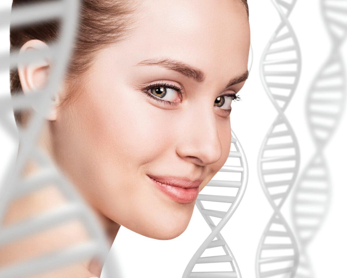
UNLOCKING SKIN HEALTH: THE ROLE OF EPIGENETICS IN AGING AND REJUVENATION
INTRODUCTION
Aging is increasingly understood through two distinct lenses: chronological aging which is measured simply by the passage of time and biological or epigenetic aging which reflects the functional state and visible condition of the skin. While chronological age counts the years lived, epigenetic age reveals how well the skin maintains its vitality, resilience, and appearance. This distinction is gaining significant relevance in aesthetic medicine as it offers a
more precise understanding of skin health. Epigenetics, the study of how gene expression is regulated without altering the underlying DNA sequence, is transforming the landscape of anti-aging science. It provides a framework to explain why individuals of the same chronological age can exhibit markedly different skin qualities. Crucially, epigenetic mechanisms demonstrate that gene activity is dynamic and responsive to environmental factors, lifestyle choices, and skincare interventions.
This evolving understanding highlights the important role lifestyle modifications and targeted skincare can play in influencing gene expression to slow or even reverse the biological aging process of the skin. The exploration of this concept is shaping a new era in aesthetic care, one focused on prevention, personalization, and scientifically guided therapies designed to optimize skin health and longevity.1
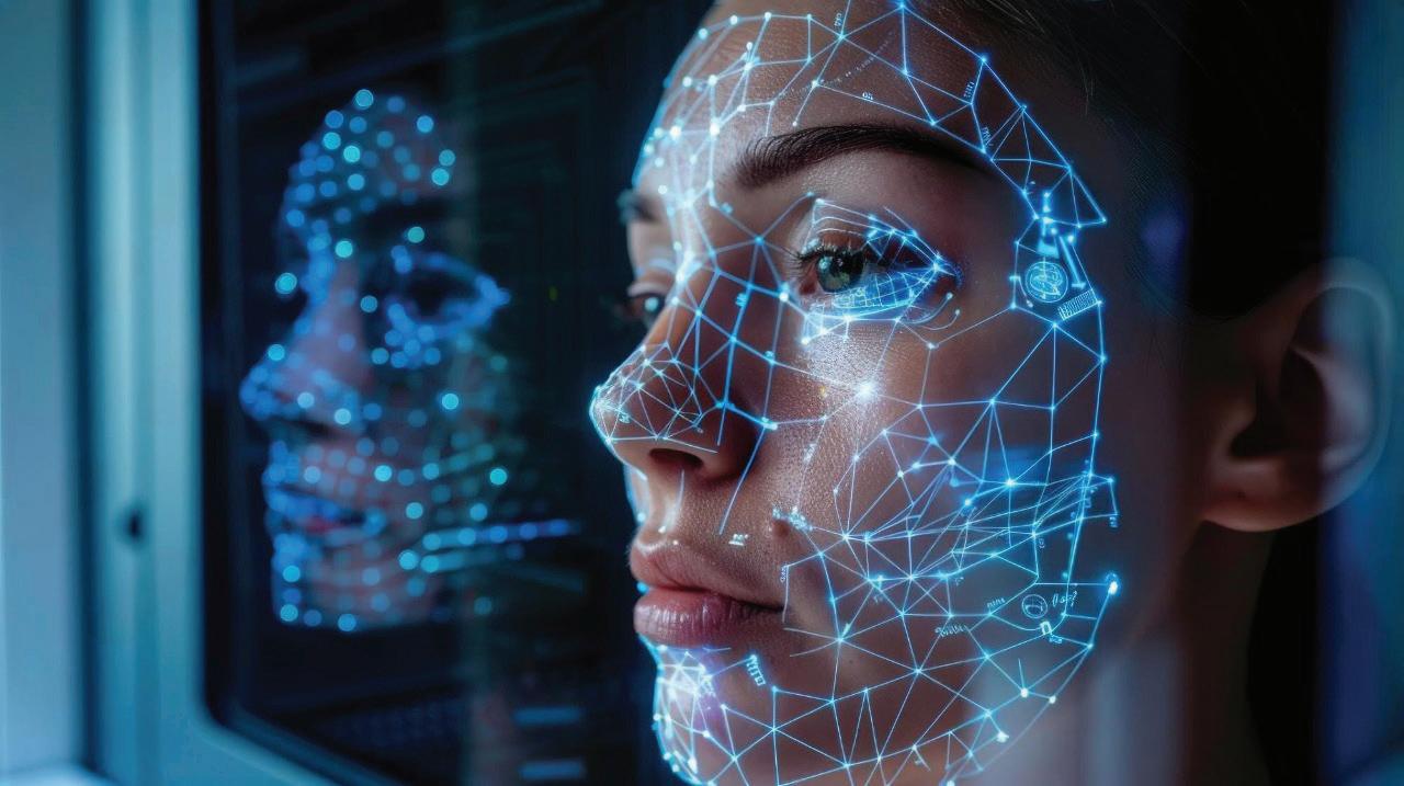
WHAT IS EPIGENETICS AND WHY DOES IT MATTER FOR SKIN?
Epigenetics refers to the science of how gene activity is regulated without altering the underlying DNA sequence. It plays a critical role in how skin functions, regenerates, and visibly ages. Unlike genetic mutations, epigenetic changes are reversible and influenced by both internal factors and external elements such as environmental exposure, skincare practices, and lifestyle habits. Mechanisms including DNA methylation, histone modifications, and non-coding RNAs contribute to maintaining skin homeostasis by controlling the balance between cell renewal, differentiation, and natural cell turnover. This process ensures the continuous regeneration of the epidermis, strengthens the skin barrier, and supports the behaviour of stem cells within the basal layer. When this regulation is disrupted, signs of premature aging and diminished skin repair can emerge. Advances in understanding these molecular pathways are reshaping how skin health is approached. Rather than focusing solely on surface concerns, modern skincare is beginning to address gene expression at the cellular level, enhancing how skin adapts, heals, and renews over time. Epigenetics provides a powerful link between science and beauty, laying the foundation for a more personalized and effective era of skin wellness.2
HOW EPIGENETICS DRIVES SKIN AGING
• Epigenetic Skin Aging Defined
Skin aging is increasingly understood as a consequence of epigenetic modifications influenced by environmental and lifestyle factors. These changes alter cellular behavior and function without modifying the underlying DNA sequence, a process termed epigenetic skin aging.1,2
• Triggers of Epigenetic Skin Aging
External insults such as ultraviolet (UV) radiation, pollution, tobacco smoke, poor nutrition, and chronic psychological stress induce chemical modifications to DNA and histone proteins. These epigenetic alterations dysregulate key genes responsible for maintaining skin homeostasis, leading to progressive damage.1,2
• Breakdown of Collagen and Elastin
Dermal fibroblasts, which synthesize collagen and elastin,
undergo epigenetically driven senescence. Shifts in DNA methylation patterns and microRNA-mediated suppression of collagen-producing enzymes result in decreased extracellular matrix (ECM) production and
increased matrix degradation. Consequently, skin firmness and elasticity deteriorate.1,2
• Reduced Skin Cell Turnover
Epigenetic modifications impair the proliferative and regenerative capacity of epidermal stem cells. This diminishes keratinocyte renewal, slowing epidermal turnover, causing accumulation of corneocytes, and leading to a dull, uneven skin surface.1,2
• Impaired Barrier Function
Keratinocytes, which form the epidermis’ protective outer layer, experience epigenetic dysregulation affecting gene expression of crucial barrier components. This results in compromised barrier integrity, increased transepidermal water loss (TEWL), heightened skin sensitivity, and susceptibility to environmental irritants.1,2
• Increased Pigmentation and Inflammation
Senescent cells, influenced by epigenetic changes, secrete pro-inflammatory cytokines and modulate melanin synthesis. This promotes hyperpigmentation, uneven skin tone, and chronic low-grade inflammation, which collectively accelerate visible signs of skin aging.1,2
• Visible Clinical Manifestations of Epigenetic Skin Aging1,2
➢ Fine lines and wrinkles
➢ Loss of skin elasticity and increased laxity
➢ Dullness and heterogeneous pigmentation
➢ Dryness and rough texture
➢ Hyperpigmentation and erythema
SKIN BARRIER AND EPIGENETIC REGULATION
The skin barrier is vital in aging, relying on the stratum corneum to prevent water loss and protect against the environment. Epigenetic mechanisms regulate key genes like filaggrin and ceramide enzymes. Disruption weakens the barrier, accelerating dryness, sensitivity, inflammation, and aging.3
• Role of the Stratum Corneum
The stratum corneum is the outermost epidermal layer, critical for preventing water loss and protecting against pathogens and environmental insults. Its strength and function depend heavily on the coordinated expression of genes encoding structural and functional proteins.3
• Epigenetic Control of Barrier-Related Genes
Key genes involved in skin barrier homeostasis include:
➢ Filaggrin — essential for aggregating keratin filaments and maintaining hydration.3
➢ Ceramide-synthesizing enzymes — vital for lipid matrix formation, critical for barrier integrity.3
Epigenetic mechanisms such as DNA methylation, histone modifications, and non-coding RNAs modulate the transcription of these genes. For instance, DNA methyltransferases regulate the activation or silencing of filaggrin and ceramide biosynthesis pathways, fine-tuning keratinocyte differentiation and barrier
formation.3
• Consequences of Epigenetic Disruption
Altered epigenetic regulation leads to decreased expression of barrier proteins, weakening the lipid matrix and impairing moisture retention. This results in increased TEWL, xerosis (dryness), heightened sensitivity, and inflammation — hallmark features of both intrinsic aging and dermatological conditions like atopic dermatitis.3
• Dynamic and Responsive Nature
Epigenetic regulation of the skin barrier is dynamic, responding to intrinsic factors such as chronological aging and extrinsic environmental influences. While this adaptability confers resilience, it also renders the barrier susceptible to epigenetic dysregulation, accelerating skin aging.3
The skin appearance and health are profoundly influenced by lifestyle choices that modulate gene expression through epigenetic mechanisms. Understanding these factors provides valuable insight into maintaining youthful, resilient skin. Below is a detailed overview of key lifestyle elements and their impact on skin aging.4
Nutrition
• Antioxidants such as vitamin C, polyphenols, and omega-3 fatty acids play a crucial role in activating anti-aging genetic pathways. These nutrients protect skin cells from oxidative damage and support collagen synthesis, thereby enhancing skin firmness and luminosity.4
• Conversely, diets high in processed foods and sugars have been associated with adverse methylation of collagen-related genes, inhibiting collagen production and accelerating structural decline, which manifests as sagging and diminished elasticity.4
Sun Exposure (UV Radiation)
• Ultraviolet radiation is the most significant environmental epigenetic stressor, promoting collagen degradation, irregular pigmentation, and cellular damage that contribute to premature skin aging.4
• Consistent application of broad-spectrum sunscreen, complemented by antioxidant-rich formulations, mitigates UV-induced epigenetic alterations, thereby preserving skin integrity and youthful appearance.4
Sleep and Circadian Rhythm
• Inadequate or disrupted sleep patterns interfere with circadian clock genes that regulate essential skin regeneration and repair processes.4
• Optimal night time rest facilitates peak cellular turnover and DNA repair mechanisms, substantiating the physiological basis for the concept of “beauty sleep” as vital for skin renewal.4
Stress
• Chronic psychological stress elevates systemic cortisol levels, which modulate skin immune responses and upregulate pro-inflammatory gene expression.4
• This cascade precipitates visible skin concerns, including dullness, acne flare-ups, and accelerated aging, highlighting the importance of effective stress management in skincare regimens.4
Pollution and Environmental Toxins
• Exposure to airborne pollutants induces oxidative stress and inflammatory responses via epigenetic modifications, compromising the skin barrier function.4
• The resultant effects often include increased sensitivity, uneven pigmentation, and a lackluster complexion, all indicative of premature cutaneous aging.4
Topical Skincare Agents
• Several active compounds influence gene expression to promote skin rejuvenation:
➢ Retinoids stimulate the transcription of collagenproducing genes, improving dermal structure and texture.4
➢ Niacinamide modulates inflammatory gene pathways, reducing redness and irritation.4
➢ Peptides act as signaling molecules that encourage skin cells to adopt a more youthful phenotype, enhancing overall skin vitality.4
CAN WE REVERSE EPIGENETIC SKIN AGING?
Epigenetic skin aging is reversible because the epigenome is highly adaptable. This means gene expression linked to skin aging can be modified through lifestyle habits and targeted skincare, reactivating youthful genes and restoring skin vitality. The idea of a “pro-aging” versus “pro-youth” gene environment explains this further. In a pro-aging state, certain epigenetic marks silence genes responsible for collagen production and skin repair, while a pro-youth environment encourages their activation. By nurturing a pro-youth environment, the skin can regain firmness, elasticity, and a radiant glow. Topical treatments and healthy habits help shift the skin’s gene expression toward youthfulness. For instance, specific epigenetic serums can reactivate silenced genes, promoting visible
improvements like reduced wrinkles and increased hydration. Together with protection from sun damage, balanced nutrition, and stress management, these strategies enhance the skin natural ability to renew and rejuvenate.5
EPIGENETIC SKINCARE
Epigenetic skincare represents a ground-breaking approach that targets gene expression to promote healthier, younger-looking skin. Key innovations include:
Epigenetic-targeting peptides that stimulate genes essential for skin renewal, collagen production, and improved texture.6
➢ Gene-specific antioxidant complexes that neutralize oxidative stress at the genetic level, protecting skin cells from environmental damage and slowing aging.6
➢ DNA repair enzymes incorporated in formulations help boost the skin natural ability to repair damage caused by UV exposure and other stressors.6
Many skincare products now use peptides,
antioxidants, and plant-derived compounds to support epigenetic actions, aiming to optimize gene expression linked to youthful skin. These ingredients work by stimulating collagen production, neutralizing oxidative damage, and promoting skin renewal at the genetic level, helping the skin look firmer, smoother, and more radiant.
CONCLUSION
Epigenetics has transformed understanding of skin aging by showing gene expression can be influenced by daily lifestyle habits as well as targeted skincare rather than being fixed by genetics. Choices such as proper nutrition, consistent sun protection, adequate sleep, effective stress management, and advanced topical agents including peptides, antioxidants, and DNA repair enzymes can help optimize skin gene activity. This dynamic approach allows skin to maintain youthful resilience, radiance, and repair capacity, making visible aging more modifiable. Embracing these science-backed strategies means beauty truly goes beyond DNA, empowering individuals to support skin health and slow the biological clock.
REFERENCES
1. Wang, K., Liu, H., Hu, Q. et al. Epigenetic regulation of aging: implications for interventions of aging and diseases. Sig Transduct Target Ther 7, 374 (2022). https://doi.org/10.1038/s41392-022-01211-8
2. Pozzo LD, Xu Z, Lin S, et al. Role of epigenetics in the regulation of skin aging and geroprotective intervention: A new sight. Biomed Pharmacother. 2024; 174:116592. doi:10.1016/j.biopha.2024.116592
3. Haykal D, Flament F, Mora P, Balooch G, Cartier H. Unlocking Longevity in Aesthetic Dermatology: Epigenetics, Aging, and Personalized Care. Int J Dermatol. Published online March 10, 2025. doi:10.1111/ ijd.17725
4. Alegría-Torres JA, Baccarelli A, Bollati V. Epigenetics and lifestyle. Epigenomics. 2011; 3(3):267-277. doi:10.2217/epi.11.22.
5. Yang JH, Petty CA, Dixon-McDougall T, et al. chemically induced reprogramming to reverse cellular aging. Aging (Albany NY). 2023; 15(13):5966-5989. doi:10.18632/aging.204896
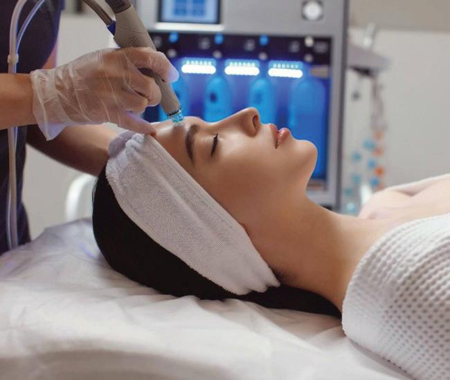
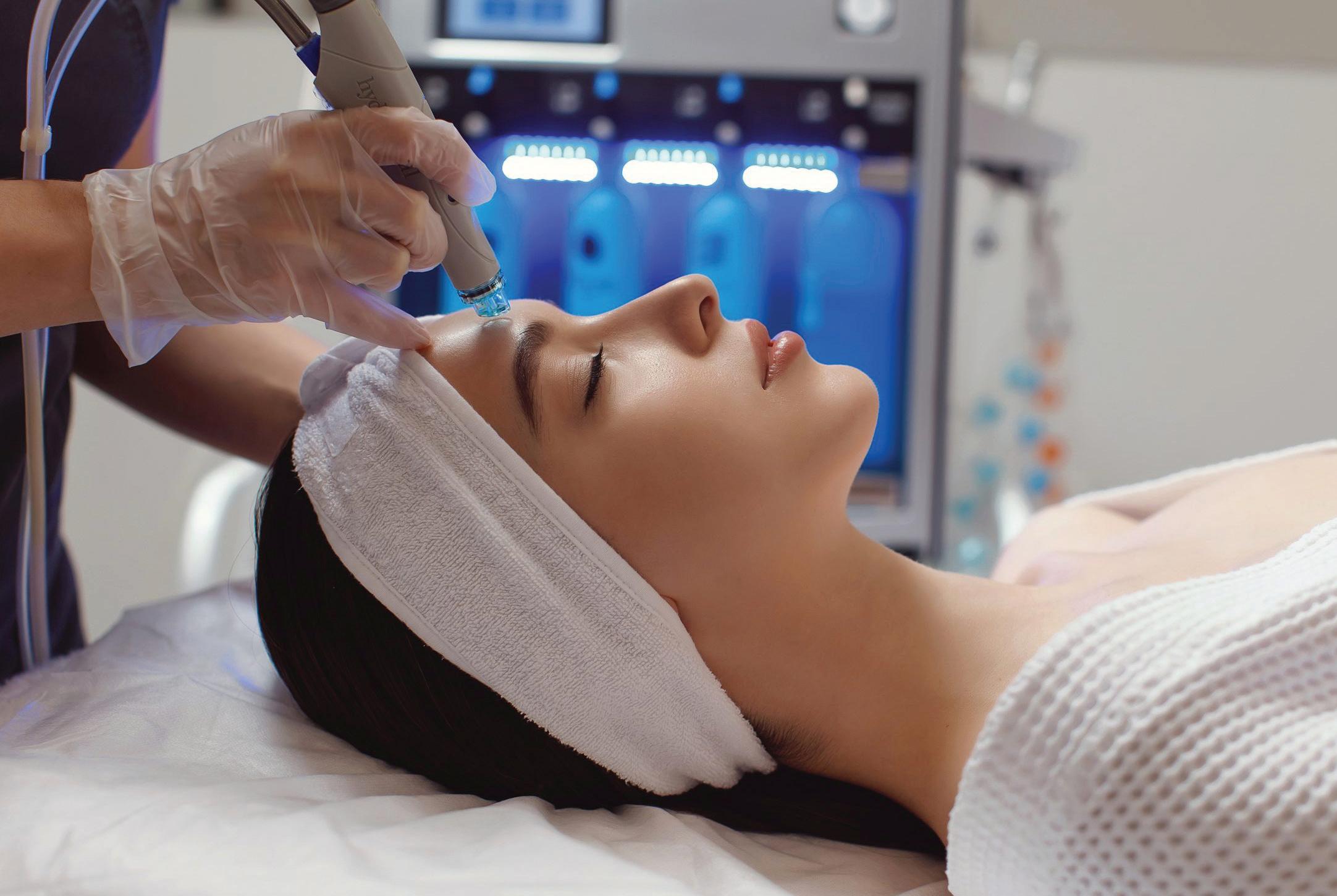
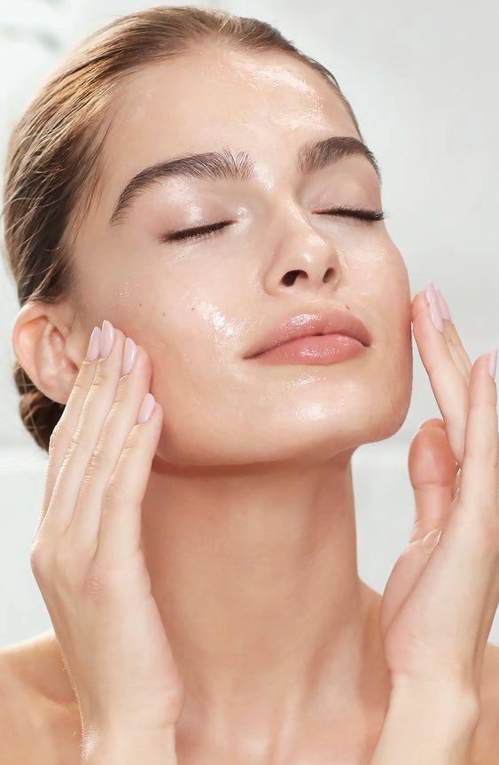
THE SCIENCE AND APPLICATION OF HYDRAFACIAL TECHNOLOGY IN SKIN REJUVENATION.
INTRODUCTION
Skin rejuvenation is more than just a beauty trend; it has become a modern skincare essential. As the understanding of skin health continues to evolve, the focus has shifted from surface-level fixes to treatments that restore the skin’s natural glow and vitality from within. True rejuvenation brings
back radiance, smooths texture, reduces pigmentation, softens fine lines, and supports the skin renewal process. With the growing demand for non-invasive yet effective treatments, beauty-conscious individuals are seeking skincare solutions that deliver visible results with minimal discomfort or downtime. In this space, HydraFacial
has emerged as a preferred choice. It seamlessly blends the luxury of a spa experience with advanced skincare technology, offering a treatment that deeply cleanses, exfoliates, removes impurities, and infuses hydration in one comprehensive session. HydraFacial stands out for its versatility. It addresses a wide range of skin concerns and is
suitable for all skin types and ages, whether the concern is dryness, dullness, congestion, uneven tone, or early signs of ageing. This article
explores the science behind HydraFacial, its step-by-step process, the key active ingredients it uses, how it compares with traditional facials, and the exciting innovations that are redefining the future of skin rejuvenation.1
METHODS FOR IMPROVING
SCAR
APPEARANCE
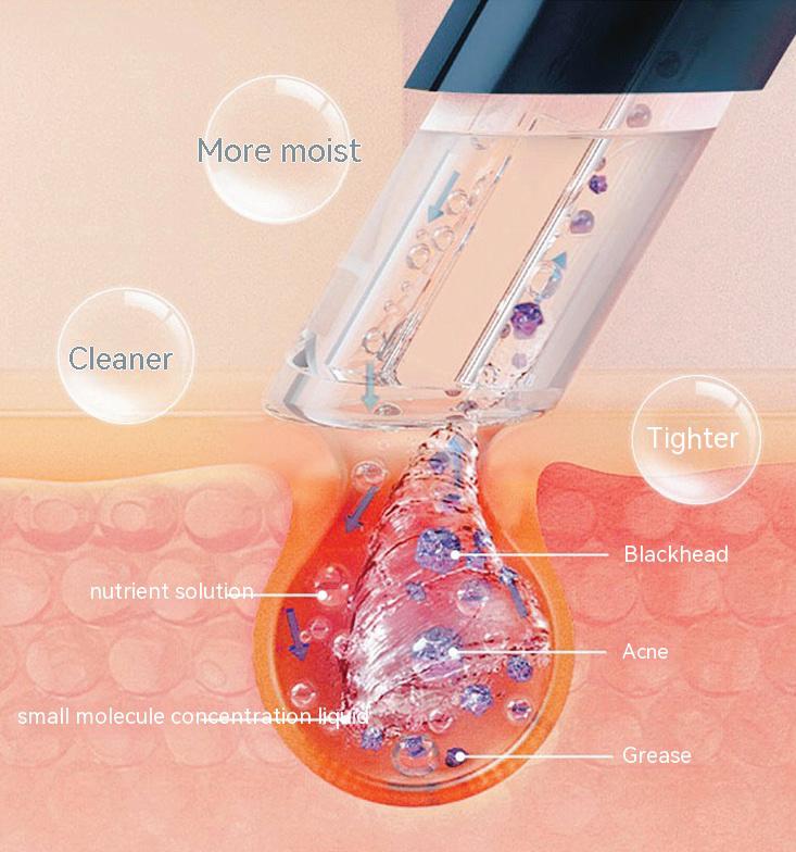
At the forefront of advanced skincare, HydraFacial distinguishes itself through its innovative Vortex Fusion Delivery System, a technology that redefines the way facial treatments interact with the skin. This system combines precisely controlled vacuum suction with a specially designed spiral tip, creating a vortex effect that effectively dislodges and removes impurities from the skin. The process gently exfoliates the surface, extracting dead skin cells, excess sebum, and environmental pollutants from deep within the pores, all without causing trauma, irritation, or damage to the skin barrier. Unlike traditional manual extractions, which can sometimes lead to redness, inflammation, or micro-tears, the HydraFacial system ensures a consistent and non-invasive experience, making it suitable for all skin types, including sensitive or reactive skin.1
What sets HydraFacial apart is its dual action mechanism. While the skin is being cleansed and exfoliated, it is simultaneously infused with custom formulated, nutrient rich serums. These include hydrating agents such as hyaluronic acid, antioxidant rich solutions, and peptides that
Key aspects include:
support collagen production and overall skin health. Because the skin is freshly exfoliated, it is more receptive to these active ingredients, allowing for deeper penetration and enhanced effectiveness. This comprehensive approach not only delivers immediate visible results, such as improved tone, texture, and radiance, but also contributes to long term improvements in skin hydration, elasticity, and clarity with regular treatments. Professionally administered, the HydraFacial is a clinically proven solution for individuals seeking a noninvasive, results driven treatment that bridges the gap between medical grade skincare and a luxurious facial experience.1
• Controlled Vacuum Pressure: Tailored to engage skin without damage, adapting to various sensitivities.
• Spiral Tip Design: Enhances the vortex flow, ensuring even exfoliation and serum distribution.
• Multi-Phase Delivery: Seamlessly combining cleansing, extraction, and hydration.
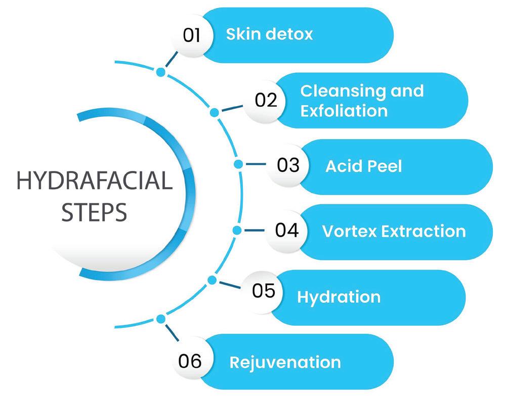
STEP-BY-STEP PROCESS OF A HYDRAFACIAL TREATMENT
HydraFacial combines advanced skincare technology with a systematic, non-invasive protocol to deliver immediate and long term skin health benefits. The treatment is carried out in three key steps, each designed to progressively restore, cleanse, and rejuvenate the skin.2
1. Skin Detox
The treatment begins with lymphatic drainage techniques to stimulate circulation and remove toxins. This detoxification step helps improve skin tone, reduce puffiness, and support the body natural elimination process, creating a clean foundation for the rest of the treatment.
2. Cleansing and Exfoliation
A gentle resurfacing applicator, paired with mild chemical exfoliates like glycolic and salicylic acids, is used to deeply cleanse the skin. This step removes dead skin cells, excess oil, and surface impurities preparing the skin for optimal absorption of active ingredients in the following phases. Skin is left smoother, brighter, and more receptive.
3. Acid Peel
A light, non-irritating acid peel is applied to loosen debris in pores without causing post-treatment flaking or redness. This step further exfoliates and softens sebum build up, targeting dullness, congestion, and uneven texture for a more refined look.
4. Vortex
Extraction
Using HydraFacial patented vortex suction technology, this step gently and painlessly removes blackheads,
whiteheads, and other impurities from the pores. Unlike traditional manual extractions, this method is non-invasive and minimizes the risk of redness or inflammation.
5. Hydration
Following extractions, the skin is infused with deeply hydrating serums rich in hyaluronic acid, antioxidants, and peptides. This blend restores moisture, soothes the skin, and enhances elasticity, leaving the complexion supple and replenished.
6. Rejuvenation
The final phase involves the application of protective and rejuvenating serums tailored to the individual skin needs. LED light therapy may also be used to stimulate collagen production, reduce redness, and support longterm skin health. The result is a radiant, revitalized appearance with a healthy glow.
KEY INGREDIENTS USED IN HYDRAFACIAL SERUMS
The science behind HydraFacial success lies in its potent serum formulations, which are specifically
designed to complement and enhance the mechanical aspects of the treatment. Common bioactive ingredients include:
• Antioxidants: Neutralize free radicals produced by UV radiation and pollution, preventing oxidative damage to skin cells which accelerates aging.3
• Hyaluronic Acid: A powerful humectant known for retaining moisture, thus plumping the skin and diminishing the appearance of fine lines.3
• Peptides: Short chains of amino acids that trigger collagen and elastin synthesis, improving skin elasticity and firmness.3
• Glycolic and Salicylic Acids: Exfoliating a gents that assist in removing dead skin cells and unblock pores, reducing acne and encouraging skin renewal.3
These ingredients address a spectrum of skin concerns such as dryness, early signs of aging, pigmentation irregularities, and acne, making HydraFacial a versatile treatment option.
COMPARATIVE ADVANTAGES OVER TRADITIONAL TREATMENTS
HydraFacial technology offers several advantages when compared to traditional skin rejuvenation methods:
• Gentleness and Safety: Unlike microdermabrasion or conventional chemical peels, HydraFacial avoids aggressive abrasion and harsh chemicals, making it suitable for sensitive and reactive skin.4
• Hydrating Effects: Most traditional treatments tend to dry the skin; by contrast, HydraFacial restores moisture during the process, reducing irritation and enhancing skin barrier integrity.4
• Customization: The ability
to tailor serums according to specific skin challenges allows for highly personalized treatments.4
• Minimal Downtime: Instant visible improvements with virtually no recovery time distinguish this procedure from invasive or more aggressive alternatives.
• All Skin Types: From dehydrated to oily, aging to acne-prone, HydraFacial can be safely used across all skin types and ages.4
CLINICAL BENEFITS AND APPLICATIONS
Clinical observations and patient reports affirm HydraFacial benefits for various aesthetic needs:
• Promotes improved skin texture and tone through effective exfoliation and increased cellular turnover.
• Enhances hydration, providing a plumper, more youthful complexion.
• Reduces visible fine lines and wrinkles by stimulating collagen renewal.
• Shrinks the appearance of enlarged pores and diminishes acne flare-ups.
• Mitigates pigmentation irregularities and redness.
• Provides an immediate radiant glow and long-term skin health improvements.
The treatment versatility means it can be integrated into maintenance skincare regimes or used to address specific conditions like acne, photo-damage, and mild to moderate signs of aging.
THE TREATMENT MAY NOT BE SUITABLE IN THE FOLLOWING CASES:
• Active skin issues: Ongoing bacterial, viral, or fungal infections, or flare-ups of acne, eczema, or rosacea.
• Pregnancy or nursing: Due to ingredients like salicylic and glycolic acids.
• Severe skin conditions: Includes cystic acne, advanced rosacea, or significant skin damage.
• Recent aesthetic treatments: Such as chemical peels, laser procedures, or injectable fillers.
• Ingredient sensitivities: Known allergies to acids or other components used in the treatment.
• Weakened skin barrier: Conditions like sunburn, open wounds, or irritated skin.
FUTURE OUTLOOK AND TECHNOLOGICAL INNOVATIONS
As skincare technology evolves, HydraFacial systems continue to advance, with emerging innovations including:
• AI-Driven Personalized Treatments: Leveraging artificial intelligence to analyse skin condition in real-time and adjust suction and serum delivery accordingly.
• Smart Diagnostics: Integration of advanced imaging methods to visualize subsurface damage and pore congestion invisible to the naked eye, guiding precise treatments.
• Combination Therapies: Fusion with complementary treatments such as LED phototherapy or radiofrequency to amplify rejuvenation and collagen stimulation effects.
• New Serum Formulations: Next-generation bioactive
CONCLUSION
tailored for enhanced cellular repair, pigmentation control, and barrier reinforcement.
These advancements promise even more tailored and effective skin rejuvenation outcomes, aligning with trends towards precision and noninvasive care.
HydraFacial technology stands at the intersection of science and skincare luxury, offering a sophisticated, noninvasive approach to skin rejuvenation that delivers deep cleansing, exfoliation, extraction, and nutrient infusion in a single session. Its patented Vortex-Fusion Delivery System provides controlled vacuum suction and vortex infusion of antioxidants, peptides, and hydrating agents, making it effective and gentle for all skin types. Compared to traditional methods, HydraFacial holistic approach results in immediate glow, reduced fine lines, improved texture, and long-term skin health with minimal downtime. As technology evolves, HydraFacial continues to advance toward smarter, more personalized treatments, shaping the future of skincare with innovation and precision.
REFERENCES
1. Storgard R, Mauricio-Lee J, Mauricio T, Zaiac M, Karnik J. Efficacy and Tolerability of HydraFacial Clarifying Treatment Series in the Treatment of Active Acne Vulgaris. J Clin Aesthet Dermatol. 2022; 15(12):42-46.
2. Razi S, Truong TM, Khan S, Sanabria B, Rao B. Hydradermabrasion through the lens of Line-Field Confocal Optical Coherence Tomography. Skin Res Technol. 2024; 30(4):e13684. doi:10.1111/srt.13684
3. Gold MH, Biron JA, Wilson A, Nelson DB. Efficacy and tolerability of a hyaluronic acid-based serum and a peptide-rich cream for the face and neck in subjects with photodamaged skin. J Cosmet Dermatol. 2022; 21(8):3458-3463. doi:10.1111/jocd.14981.
4. Freedman BM. Hydradermabrasion: an innovative modality for nonablative facial rejuvenation. J Cosmet Dermatol. 2008; 7(4):275-280. doi:10.1111/j.1473-2165.2008.00406.x
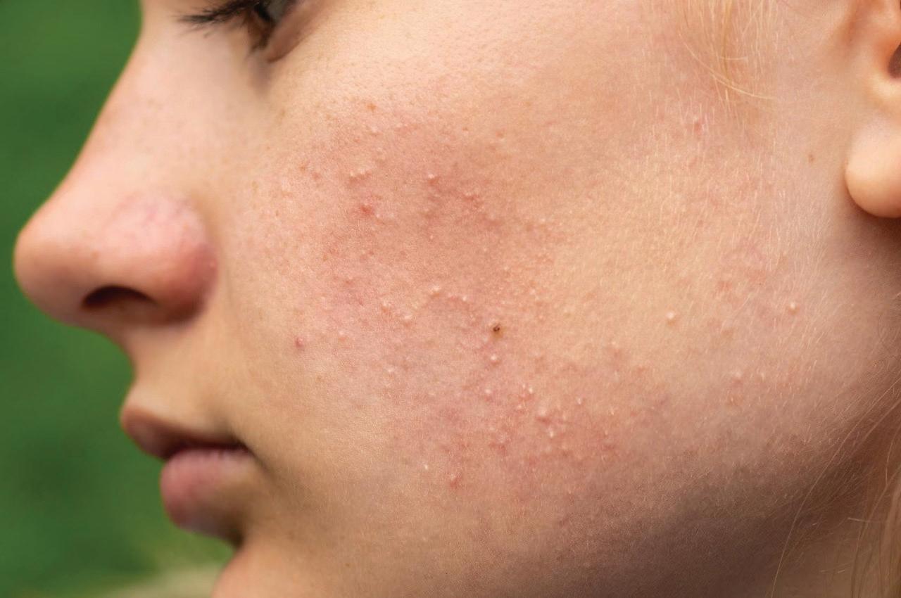
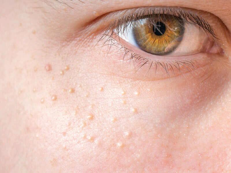
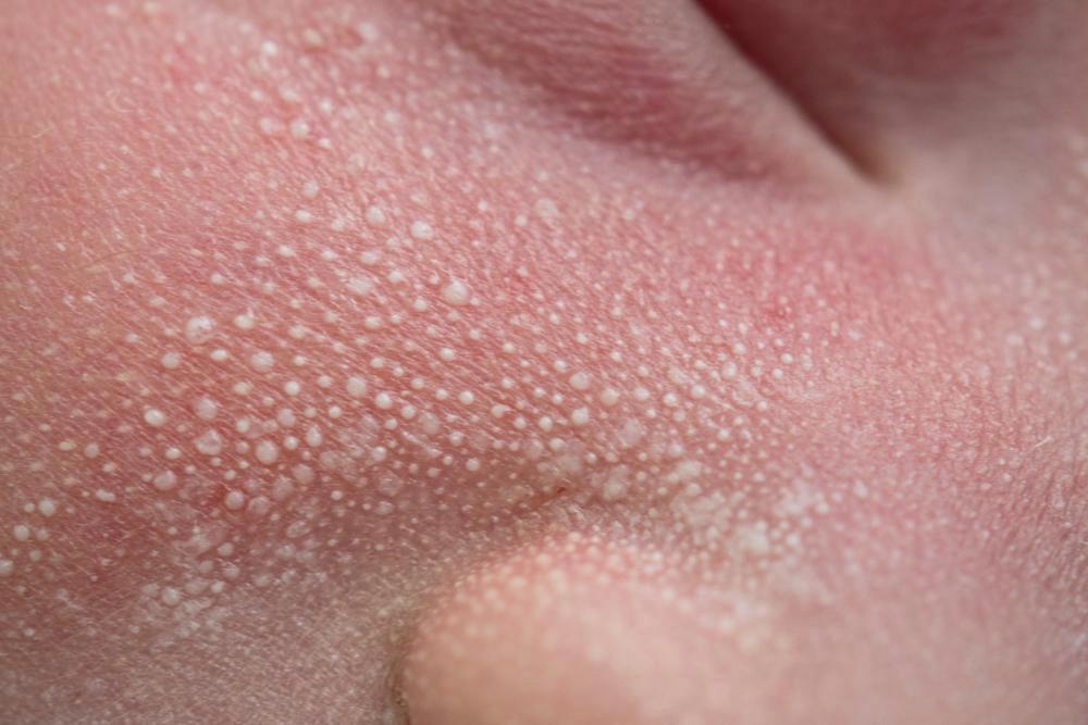
UNVEILING MILIA: WHAT LIES BENEATH THE SURFACE
INTRODUCTION
Tiny in size yet visibly distinct, milia are small, dome-shaped bumps that quietly settle on the skin, often appearing as pearly white or pale yellow spots. Most often found around the eyes, cheeks, nose, and chin, these keratin-filled cysts are frequently mistaken for whiteheads
or clogged pores. But unlike common breakouts, milia form beneath the skin surface and do not arise from oil or bacteria. They can appear at any age—from the softness of a newborn skin to the more mature canvas of adulthood. Though medically harmless, their presence can subtly alter the texture
of the skin, leading many to seek answers, gentle treatments, or aesthetic solutions. Understanding their quiet presence is the first step toward reclaiming a smoother, more radiant complexion.1
WHAT ARE MILIA?
Milia (singular: milium) are tiny, white or pearly bumps that quietly appear on the skin, often mistaken for whiteheads or blemishes. In reality, they are superficial keratin-filled cysts that form when dead skin cells, also known as keratin, become trapped just beneath the surface layer of the skin, creating a small, enclosed pocket. These miniature cysts are completely harmless. They lack infection or inflammation, but their firm, raised texture can still be a cosmetic concern, especially when they appear on more visible areas of the face. Measuring just 1 to 2 millimetres, milia are often described as resembling seed pearls resting under the skin. They may appear individually or in small clusters, adding an unexpected texture to otherwise smooth skin.
Although they may seem prominent, milia generally cause no discomfort. Itching, redness, or pain is typically absent.2
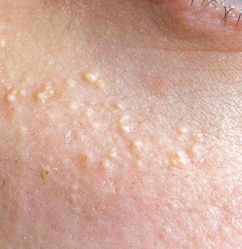
DIFFERENT TYPES OF MILIA
Milia present in diverse forms, each with distinct characteristics and implications. Recognizing these variations offers valuable insight into their nature and guides appropriate care or intervention.
Neonatal Milia
Often regarded as a hallmark of newborn skin, neonatal milia manifest as tiny, pearly white cysts typically clustered around the infant nose. These benign formations are present at birth and are transient by nature, resolving spontaneously within weeks without the need for treatment. Their appearance adds a delicate, almost ethereal charm to a newborn complexion.3
Primary Milia
Primary milia are small, keratin-filled cysts that commonly emerge on areas such as the eyelids, forehead, cheeks, and occasionally more intimate regions. Affecting both children and adults, these cysts arise independently of prior skin injury or disease. Though benign, primary milia in adults may persist for extended periods, often eliciting cosmetic concern due to their visible texture.3
Secondary Milia (Traumatic Milia)
Secondary milia develop as a sequela to skin trauma or disruption. They may arise following burns, inflammatory skin conditions, blistering, or prolonged ultraviolet exposure. Additionally, the use of occlusive or heavy topical formulations can contribute to their formation. These cysts represent a response to altered skin architecture, where keratin becomes entrapped beneath the surface during the healing process.3
Milia En Plaque
A relatively rare presentation, milia en plaque predominantly affects women between the fourth and sixth decades of life. It is characterized by clusters of milia set upon erythematous, elevated plaques, commonly located behind the ears, on the eyelids, cheeks, or along the jawline. This distinct pattern imparts a textured and persistent skin irregularity that often warrants professional evaluation and management.3
Multiple Eruptive Milia
This uncommon variant is defined by the rapid appearance of numerous milia over weeks or months, frequently affecting the face, upper arms, and upper abdomen. These lesions may be accompanied by mild pruritus, distinguishing them from other types. While alarming in their sudden onset, multiple eruptive milia remain benign and typically respond well to professional care.3
WHAT CAUSES MILIA?
• Milia appear when dead skin cells, primarily keratin, become trapped just beneath the skin surface, forming small, firm cysts that gently disrupt the skin smoothness. The skin naturally renews itself by shedding old cells and revealing
fresh, glowing ones. However, when this renewal cycle is interrupted, these dead cells can get encapsulated, leading to the formation of these delicate white bumps.4
• Skin trauma from injuries, burns, or too much sun exposure can damage the skin surface, slowing down the natural exfoliation process and encouraging milia to develop.4
• Prolonged use of steroid creams or ointments may thin the skin and affect its ability to regenerate properly, making it more vulnerable to milia formation.4
• Genetic factors also play a role; in some cases, the skin may be naturally more prone to trapping keratin, allowing these tiny cysts to appear more frequently.4
• Autoimmune conditions that alter normal skin function may further contribute to milia appearance.4
Beyond these, lifestyle and environmental influences like heavy skincare products that block pores, pollution, and sun damage can also tip the balance, trapping keratin beneath the skin and encouraging milia to surface. Understanding these triggers is key to managing and preventing milia while embracing healthy, radiant skin.
WHO IS MOST AFFECTED BY MILIA
• Adults with Dry Sun Damaged or Mature Skin
➢ Adults especially those with drier or sun-exposed skin tend to develop primary milia more frequently.5
➢ As the skin matures its natural renewal process slows allowing keratin to become trapped beneath the surface.5
➢ These bumps are commonly found on areas with thinner skin such as the temples forehead and eyelids.5
• People with a History of Skin Trauma or Irritation
➢ Secondary milia may develop after the skin experiences trauma like burns blisters rashes or prolonged sun exposure.5
➢ Damage to the skin barrier can hinder normal exfoliation causing keratin to become enclosed under the surface.5
➢ These types may also form during healing from inflammatory conditions or after resurfacing treatments.5
• Those Using Heavy or Occlusive Skincare Products
➢ Regular use of thick moisturiser balms or oil-based products especially around delicate areas can trap dead skin cells and contribute to milia.5
➢ Applying multiple layers of skincare without proper exfoliation may lead to clogged skin and reduced cell turnover.5
• Individuals with Genetic or Autoimmune Conditions
➢ Some genetic factors can interfere with the natural process of skin regeneration making certain people more prone to developing milia.5
➢ Autoimmune skin responses can also impact how skin heals increasing the chance of these cysts forming.5
• All Ages All Skin Types
➢ Milia can affect anyone regardless of gender age or skin tone.5
➢ Although hormonal changes are not a direct cause shifts in the skin during phases like pregnancy or menopause may influence their appearance.5
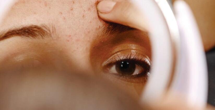
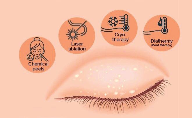
MANAGEMENT AND TREATMENT
Though milia are completely harmless and often fade over time, many seek treatment to restore smooth, even-toned skin especially when the bumps appear in visible or delicate areas such as around the eyes. While at-home remedies may seem appealing, professional care is always the safest and most effective route, particularly for persistent or sensitive cases.
➢ Manual Extraction
Performed by a trained skincare professional, this technique involves using a sterile needle or blade to gently puncture the skin surface, followed by the precise removal of the keratin-filled cyst with a comedone extractor. This method is particularly effective for isolated lesions, especially in facial areas.1,6
➢ Topical Retinoids
Topical retinoids such as tretinoin, adapalene, or tazarotene accelerate epidermal turnover and prevent the accumulation of keratin within pores. With consistent application, they can aid in the gradual resolution of existing milia and help prevent recurrence.1,6
➢ Chemical Peels
Superficial chemical peels containing glycolic acid or salicylic
acid provide gentle exfoliation that helps refine skin texture and promote cell turnover. These treatments are beneficial for reducing the frequency and appearance of milia over time.1,6
➢ Microdermabrasion
This non-invasive resurfacing procedure exfoliates the stratum corneum (outermost layer of the skin), removing dead skin cells and enhancing skin renewal. It may be used as a supportive therapy to improve texture and diminish superficial milia.1,6
➢ Laser Therapy
In cases where milia are resistant to conventional treatments or are more widespread, targeted laser therapy can be utilized. This precise modality allows for the removal of milia without compromising surrounding tissue and is typically reserved for more persistent lesions.1,6
Although milia are harmless, they can affect the skin appearance, especially in visible areas. Fortunately, a variety of professional treatments offer safe and effective solutions. Consulting a skincare expert ensures the right method is chosen to gently remove milia while preserving healthy skin, helping you achieve a smoother, clearer complexion.
SELF-CARE AND PREVENTION
Resist the urge to squeeze or pick at milia, as this can cause scarring or infection. Instead, adopt gentle skincare habits to keep your skin smooth and clear:
• Use gentle exfoliants like alpha hydroxy acids (AHAs) or beta hydroxy acids (BHAs) to help remove dead skin cells and prevent milia formation.
• Avoid heavy or greasy creams, especially around the eyes, to prevent clogged pores and buildup.
• Protect your skin daily with broad-spectrum sunscreen to prevent sun damage, which can slow skin renewal and increase milia risk.
• Use steroid creams sparingly and only under medical supervision, as long-term use can thin the skin and promote milia.
CONCLUSION
Milia may be small but their impact on skin texture can feel significant especially when they appear in noticeable areas. Recognizing their causes and who they affect most allows for a thoughtful approach to treatment and prevention. With professional care combined with gentle mindful skincare routines milia can be managed effectively without compromising your skin health. Remember patience and consistency are key to maintaining a clear smooth complexion because true beauty lies in nurturing your skin natural radiance.
REFERENCES
1. Gallardo Avila PP, Mendez MD. Milia. In: StatPearls. Treasure Island (FL): StatPearls Publishing; January 31, 2023.
2. Tharini G, Subashini M, Roshan SA, Prabhavathy D, Jayakumar S. Congenital hypotrichosis, eruptive milia, and palmoplantar pits: a case report with review of literature. Int J Trichology. 2012; 4(1):32-35. doi:10.4103/09747753.96086
3. Berk, David R. and Susan J. Bayliss. “Milia: a review and classification.” Journal of the American Academy of Dermatology 59 6 (2008): 1050-63.
4. Kurokawa I, Kakuno A, Tsubura A. Milia may originate from the outermost layers of the hair bulge of the outer root sheath: A case report. Oncol Lett. 2016; 12(6):5190-5192. doi:10.3892/ol.2016.5335
5. Patsatsi A, Uy CDC, Murrell DF. Multiple milia formation in blistering diseases. Int J Womens Dermatol. 2020; 6(3):199-202. Published 2020 Apr 1. doi:10.1016/j.ijwd.2020.03.045
6. Davis, DiAnne S. MD, MS*; Taylor, Mark B. MD*. Successful Treatment of Milia in Skin of Color (FST IV-VI) With Variable Short-Pulse Er:YAG Laser Vaporization. Dermatologic Surgery 46(12): p 1750-1751, December 2020. | DOI: 10.1097/DSS.0000000000002101