V..
1^
■m
«-
f-.m
.. -f, ifi.
¥
« 'i'
4-
*1 -
m-
I
•<1
i
.’s
-f
Jt-ir ’ V
' ’^4
4^ TW
^1 THE r^ .
t
‘'X
I •<
i ’'^
<M^
%
i
•I
4
■-' 'r.4,
ii
%,
1^
■#
e MURDOCH. INSTITUTE i
i.
;l^
I:
>»
?
■ e ■. 'ff
,<t. ^-T- . #•'"
;■
1^
for Research into Birth Defects Limited r >. ■■X-%
•t
%
* ;x
*r
-.
"V
-f^'
■■ !!}=■?!
rtl
<
■>m- .
~
? t
r». >, t*
r
’’Nf
,
-
t;
, •'-f ^
•III.
•»'.
%
■
1Fl
'-•'" * .1. '■
. ^
I
t
X
#'
>
*
r-i
"frt-
r .,»:■
1v
s
7
.1
:■
if ■ ■*.
.
■'i?'
f^.M
'iv
►
mi
?!
•4 •■%
Sf-
^ Annual Report 1990 ■
«»k. i‘ ■
1
>
?
8fn ■"
^
I
>i
« (!k/ .
>
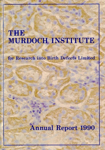
COVER Kidney tubules cut across and stained to show distribution of pyruvate dehydrogenase (PDH). The fine brown granules are mitochondria (cellular energy generators) full of PDH to supply the energy needed to reabsorb 99% of the water that is initially filtered by the kidney. The cell nuclei are stained purple.
The Murdoch Institute for Research into Birth Defects
a
4a
Royal Children’s Hospital, Flemington Road, Parkville, Victoria. 3052.
The Murdoch Institute is remarkable inter nationally among Institutes working on gene tic diseases for an even balance between fundamental research and clinical practice covering genetic disease in all age groups in the whole State of Victoria. This gives the Institute a particular style of which we are very proud — an innovative approach to clinical work and a practical start to basic research. It also provides special opportuni ties, for training free-thinking young clini cians and research workers. The Murdoch Institute is the major Australian centre for research on genetic diseases and for training clinical geneticists.
1
How you can support the work of the Institute
The high standing of the Institute within Australia is shown by its inclusion among the five Research Institutes supported by Block Grants from the National Health and Medi cal Research Council. Internationally its scientists and clinicians are in demand as speakers at major scientific meetings, on editorial boards of scientific journals and as authors of chapters in important books. Birth defects are very important to the Aus tralian community. Each year 5000 Austra lian couples feel the anguish of the birth of a baby with a serious defect. Australian tax payers provide over $2 billion annually to care for the survivors and still we cannot give all the care they and their families would like. Surely we all have a responsibility to reduce this personal and communal burden next decade. Only research and prompt implemen tation of the results of research can achieve this.
At the Murdoch Institute we are doing our best to bring about this reduction in birth defects — we need your understanding of our efforts, your voice to persuade the Government to continue and increase its support and your personal financial assistance. To maintain our research we need to raise $1 million every year — help us now with a donation and help us in the future by remembering us in your will.
DONATIONS ARE TAX DEDUCTIBLE
BEQUESTS How to help the Murdoch Institute in your will. The following is a suggested form for a bequest to the Murdoch Institute: I bequeath to the Murdoch Institute for Research into Birth Defects the sum of $ . . . (or part or all of residue of estate) to be applied for the purposes of the Institute. For further information contact the Business Manager,
The Murdoch Institute for Research into Birth Defects Royal Children’s Hospital Flemington Road PARKVILLE VICTORIA 3052 Telephone: (03) 345 5045
.yi r' -
I
MAJOR DONORS TO THE MURDOCH INSTITUTE
MAJOR DONORS TO THE MURDOCH INSTITUTE
FOUNDERS — Donors of $1 million or more
TRUSTEES — Donors of $25,000 or more
The Murdoch Family: Dame Elisabeth Murdoch Mr. Rupert Murdoch Mrs. Helen Handbury Mrs. Anne Kantor Mrs. Janet Calvert-Jones The late Sir Jack Brockhoff The Brockhoff Foundation The Scobie and Claire Mackinnon Trust
Arthur Andersen Foundation Coles Myer Limited H. & L. Hecht Trust J.B. Were & Son Charitable Foundation Mrs. Joan Roxburgh Mrs. M.L. Griffin National Australia Bank Limited Qantas Repco Corporation Limited (Ariadne) The Banks Trust The Ian Potter Foundation The late Mr. Clive Roxburgh The late Mrs. L.B. Quayle The News Corporation Limited The Percy Baxter Charitable Trust The Sidney Myer Fund
BENEFACTORS The Miller Foundation The Helen M. Schutt Trust
Donors of $250,000 or more
CORPORATE SPONSOR GROUP — Corporations undertaking substantial future support Amcor ANZ Banking Group Coles Myer Limited National Australia Bank Limited Qantas
•f
DONATIONS TO THE MURDOCH INSTITUTE Cruden Investments The Jack Brockhoff Foundation Estate of Clarice Ida Lean S. & C. MacKinnon Trust The Miller Foundation The Sidney Myer Fund Coles Myer Ltd The Banks Trust Friends of Murdoch Mr. & Mrs. K.L. Day (Retinitis pigmentosa) ANZ Bank The Morris Family Trust Amcor Commonwealth Bank Rothschild Australia The Pierce Armstrong Foundation Pacific Dunlop Comalco Clive & Jean Roxburgh (Uncle Bobs Club) The Arthur Andersen Foundation Sportscraft Consolidated P/L Prof. D. Banks Westpac Banking Corporation Mr. Grant Stephenson Nestle The Brash Charitable Foundation Mayne Nickless Ltd Dr. J.M. Gooch The Shell Company of Australia In memory of Amy Rochester Petra Foundation McMullin Unit Trust Mr. F.D. Ryan
509,375 275,000 133,826 100,000 50,000 30,000 25,000 20,000 20,000 10,000 7.500 5,000 5,000 5,000 5,000 5,000 5,000 5,000 4,100 4,000 2.500 2,396 2,000 1.500 1,500 1,000 1,000 1,000 1,000
980 620 600 600
Mrs. M. Brent Mr. & Mrs. S.F. Gooley Mr. G.E. Heeley In memory of Emma Ritchie Little Peoples Assoc, of Aust. Mrs. B. Burzak-Stefanowski Mr. E. Basarke Miss P.P. Kennedy S.F.S. & N.V. Peters M. & B. Campion Katherine 1. Behrend Mr. & Mrs. L. & 1. Barbieri Cadbury Schweppes Pty Ltd T.M. Bryant Arthur Robinson & Hedderwicks M.A. & D. Waldron Tay Creggan Social Services M. Reid R.E. Dowland Theresa Carey Mrs. G.A. Grimwade Lederman Foundation Pty Ltd Christine Bell C.F.K. Chan Mrs. C. Broome J.G. Hope Mrs. R. Heywood Dr. J. Rogers
Mr. George Erlichster
$ 500
500 500 430 400 300 250 250 250 200 200 150 150 100 100 100 100 50 50 50 50 50 50 50 35 35 20 20 10
The Institute thanks all those listed above and also Beckman Instruments (Australia) Pty Ltd for donating one Beckman Model 210A deluxe H.P.L.C. injector, value $2,518.00.
Chairman’s Report
INDEX Chairman’s Report............................... 1 Board of the Murdoch Institute......... 2 Finance Committee.............................. 4 Friends ofthe Murdoch Institute ...... 5 Director’s Report .................................. 6 Post Doctoral Fellows ......................... 11 Medical Research Fellows ................. 14 The Unravelling of a Genetic Disease 15 Victorian Clinical Genetics Service 22 Training Programmes.................... 25 Work in Progress ........................... 30 Murdoch Institute Lecture Series 1990..... .... 37 Staff Involvement in Scientific Community Activities....... 37 Editorial Boards 38 Postgraduate Degrees Awarded ............................. 38 Lectures and Seminars by Institute Staff.............. 38 Collaborations ............................................................ 40 Staff List — Murdoch Institute................................ 42 Staff List — Victorian Clinical Genetics Services 43 Olive Miller Protein Chemistry Laboratory......... 44 Studies of Pyruvate Dehydrogenase....................... 45 Centromere Structure ............................................... 47 The Scobie and Claire Mackinnon Trace Element Group .. 50 Metabolic Unit....................... 53
Tissue Culture Laboratory ... DNA Diagnostic Laboratory Epidemiology .......................... Cytogenetics Laboratory .... Clinical Projects ................... List of Publications —1990 ..
57 57 57 58 59 62
Mr Neil Walford
1990 was another very productive year in the life of the Murdoch Institute. Not only was the previous high level of progress and research achievement maintained, but there were other events which have important implications for the future. Perhaps the two most significant of these were the appointment of a number of additional high quality post doctoral scientists, and the gaining of considerable additional space by taking over the balance of the 10th floor at Royal Children’s Hospital. Once we have overcome the problems of funding and completing the fitting out of this area we will have the basic space and equipment facilities to meet our needs for some years ahead. It was with great regret that during the year we accepted the retirement of two of our foundation directors. Both represented the Royal Children’s Hospital on our Board. Dr Catchlove recently retired as the Hospital’s Chief Executive and is going into industry and Mrs Lewisohn has retired as President of the Hospital. We welcome Mrs Caroline Searby as a new director representing the Hospital. This country is in severe economic recession. The Institute is affected because the fund raising which is essential to our survival and growth becomes more difficult at such a time. We have made plans to raise substantial funds at an appropriate time in the future. Details are given in the Director’s Report. Fundraising is a matter which will always require our attention. This seems the proper time for me to give thanks to all those who did so generously support us during the year. I certainly hope that after studying the Annual Report, they will feel satisfied that their money is being well spent. Again, I wish to express my special thanks to our three major donors, the Murdoch family, the Brockhoff Foundation and the Scobie and Claire Mackinnon Trust. The Director of the Institute, Professor Danks, and I are very fortunate in being able to rely on the support and advice of the members of both the Board of Directors and of the Finance Committee. We extend our warm thanks to these ladies and gentlemen.
1
'■I:
4
Phelan
4 4
2
3
Friends of the Murdoch Institute
w
e were all very sad when Dame Patricia Mackinnon retired from the Board of the Institute after years of strong support hoth as Chairman of the Research Foundation and later as a founding Board member. Fortunately, her daughter Mrs Ann McFarling agreed to take her place on the Board. We were delighted when Ann indicated that she was willing to take on the task of setting up an auxiliary group to support the Institute financially and to make it better known in the community. The organisation, known as the Friends of the Murdoch Institute (FOM), was launched in March 1990 with the objective of run ning one or two major fundraising functions each year and undertaking a variety of activities to interest community members in the Institute. Soon afterwards the FOM spawned the Young FOM led by Julie Calvert. This younger group got away to such a quick start that it beat the parent organisation to running its first function — a car rally held on the Sunday that should have been a week after the football Grand Final. Unfortun ately, the postponement of the Grand Final by one week made it the morning after the Grand Final which proved not very popular with some of the young people whom the YFOM had hoped to
attract. None the less, they raised $4000 for which we are most grateful. They are still full of enthusiasm and intend to be run a bigger and grander car rally next year. Some of the young members of the Institute staff can certainly vouch for the fact that this year’s function was a success as well. The first function of the FOM was well worth waiting for. On November 30 Sotheby’s showroom in High Street Armadale was packed to its capacity by Murdoch Institute well-wishers attending a Christmas Tree Festival. Most of us had not been quite certain what a Christmas Tree Festival would be like, but it was indeed a great success with beautifully decorated trees dotted around the fioor and walls covered with Christmas wreaths and other decorations. Interior decorators, media personalities and other community notables con tributed by decorating a tree and these were auctioned by the very experienced senior auctioneer of Sotheby’s. The result was a contribution of over $30,000 to the funds of the Institute. We are all very much indebted to Ann McFarling, Anne Hayward and Davina Hanson who worked extra ordinarily hard to make this function such a success.
■v«
Ann McFarling and Anne Haywood preparing for the Christmas Tree Festival. 4
5
Director’s Report
T
/i
Professor David Banks
6
his has been a year of good, steady progress in all of our research groups. Each of the groups has been strengthened by new post doctoral scientists during the year and we have been very pleased with both the scientific calibre and the personal qualities of the young people who have joined us in these roles. Each has brought something in scientific knowledge, technical skills or personal qualities which has made a valuable contribution to the Institute. It is just as well that all of these new young people are good humoured, as are our existing staff, because all have had to work in even more cramped conditions than previously during 1990. During February and March 1991, we finally gained access to nearly all of the 10th floor for the use of the Institute and the Victorian Clinical Genetics Service. This will have the immediate effect of expanding our laboratory space for 1991 by about 10-15% and also allowing the establishment of some temporary offices in the north wing of the floor. A really substantial increase in usable space, for which we have waited so long, will not be felt until the latter part of 1992, at the earliest. This is because the alterations required to fit out the area for our use will have to be done in stages so that we can keep on working while the alterations are being done. In the present economic circumstances we may have to proceed more slowly. Because there have been so many delays in getting access to the additional space, we have deliberately postponed detailed planning, lest changes in staff or research objectives should render such detailed plans obsolete. Now that we have the access to this space, we must develop detailed plans quickly and efficiently. In doing so it has been important to review our overall policies about the type of research that we do and the way we go about it and to finally determine how we should cope with the cost of the rebuilding. Our research activities have developed over the years in close relationship with clinical work on patients with genetic diseases. The development of the Victorian Clinical Genetics Service as part of the Murdoch Institute and the continued close relationship with the Royal Children’s Hospital have all been part of this policy. After careful consideration with the senior scientists who make up my Executive Committee, we have concluded that there is every reason to expect this style of research to be as successful in the future as it has been in the past. We are convinced that it has been efficient to draw many of our research ideas from our clinical experience and we are confident we can continue to draw many important ideas from this in the future. As discussed in previous Reports, this does not mean allowing clinical needs to
1
4
dictate the style or priorities of our research. Rather, it recognises that observations made in individual patients with genetic diseases comprise an important source of new research hypotheses to be tested in laboratories. This reconsideration of our fundamental modus operand! may sound unduly introspective. How ever, it is important to recognise and consider the very powerful pressures that exist in the 1990s for human genetics groups to focus their attention upon the mapping of the human genome, studying genes because they lie adjacent to other known genes rather than because there is any particular interest in, or clue to, their function. Some have characterised our style of genetic research as physiological genetics because of its emphasis upon the functional roles of genes as revealed by analysis of the functional disturbances in patients with gene mutations. In essence we have decided not to bow to the dictates of fashion in genetic research, but to stick to our tried and proven approach. We use the new molecular skills exten sively in this basic approach, using the important new techniques of molecular analysis and gene mapping whenever appropriate. We see these techniques as valuable servants, rather than as masters of our research. This decision has important bearing upon decisions about laboratory development. It re affirms the importance of being in, or very close to, the Royal Children’s Hospital which is one of the world’s great sources of experience with genetic diseases. (If we were to change our policy to a genome mapping approach, we could relocate any where in Melbourne). There is no spare ground in the Royal Children’s Hospital site on which to erect a separate building which would give the Institute a more obvious identity, nor are we aware of any ground space available very close to the Hospital. In addition, the cost of establishing a new building of the size required would be considerably greater than the cost of reimbursing the Hospital for part of its cost in making the 10th floor available to us, then altering and refurbishing the area. We are most grateful to the Hospital for making this option available. We believe that the occupation of the entire 10th floor will give the Institute a sufficiently independent identity for our purpose. The other policy which is reaffirmed by our decisions about our style of research is the importance of having the Institute and the VCGS side by side. This will finally be achieved when the 10th floor alterations have been completed. Until then, we will still be six floors away from our cytogenetics colleagues and 15km from our laboratory which performs the newborn metabolic screening studies for all babies in Victoria. The main problem now confronting us is financial. An up-to-date costing of the alterations that we have to carry out on the 10th floor plus the amount that we have to reimburse the Hospital for making the space available is over $6 million — more than double the amount anticipated at the time of the original fundraising for the Institute in 1984/85. I have previously explained that meeting our current annual running costs consumes most of the interest earned on our invested capital. If we
were simply to use our invested capital for these alterations, we would find ourselves with an annual shortfall of over $600,000 for ever after wards. The Board has decided to mount a new fund raising appeal with a target of $5 million, to defray most of the costs of the building alterations. We are delighted that Mr Nobby Clark, who recently retired from the position of Chief General Manager of National Australia Bank, has agreed to lead this appeal. More information about this appeal will be found elsewhere in this Report. The time for launching this appeal, formally, is yet to be decided. Of course, we have continued to be active in fundraising to cover the ongoing costs of our research during 1990. Several additional com panies have joined our Corporate Sponsors Group, undertaking substantial commitments for several years into the future. A notable development has been the establishment of the Friends of the Murdoch Institute. This organisation, set up by Mrs Ann McFarling, comprises a group of enthusi astic and energetic people who have come together to raise money for the Murdoch Institute and, equally importantly, to raise the community awareness of the Institute. Very soon after their establishment a second group the Young Friends of the Murdoch Institute — was set up. Some details of the activities of these groups, which have already contributed over $30,000 to the Institute, and of their future plans are given elsewhere in this report. We are most grateful to Ann McFarling for this initiative and to Dame Patricia Mackinnon, our very dear friend and Ann’s mother, who is Patron of the Friends of the Murdoch Institute. Increasing public awareness of the Murdoch Institute is one of the objectives of the FOM. Activities like medical research depend upon the generosity of governments, individuals, corpor ations and charitable foundations. There is necessarily competition between many worthy activities going on in the community for the limited resources available. It is therefore critical for an institute like ours to ensure that as many members of the public as possible are aware of what we are trying to achieve and that we are succeeding. We must avoid exaggerated or unwarranted claims about the importance of our subject or the quality of our achievements and
Dr Hal Rauch
Dr Irma Dianzani 7
T
ensure that we never belittle competing causes in our endeavour to attract financial support. In most areas of medical research there is a second reason for public awareness. As science makes more progress and medical skills become more sophisticated and more complex, it is very important to keep the public well informed so that they can better understand the treatments that are being offered. It is particularly important in our field of genetics for there to be good public education because treatments that are directed at genes and inheritance seem to raise special and emotive concerns. Members of our staffare in demand as speakers to various organisations who are curious about the modern knowledge of genetics and we try to meet as many of these requests as we can. The pressures involved in maintaining funding for medical research, and the interest of the general public in “new breakthroughs” in medicine lead some doctors and scientists to release their new findings to the public media before they are properly scrutinized by scientific colleagues through scientific publications. We regard this as an unfortunate trend and try to avoid any such premature releases of our own findings. In this Annual Report we once again have a substantial section of Work in Progress in which the principal projects of each research groups are mentioned very briefly along with some of their more important findings. I will therefore say very little about the scientific work of the year in this section of the Report. However, I must draw attention to just a very few of our achievements. Dick Cotton has continued to refine his chemical cleavage method for detection of mutations and to explore the range of its applications in genetics and in the study of virus diseases in the com munity. We are grateful to the Victorian Health Promotion Foundation for its support of this work. His studies of the properties of the enzymes controlling the metabolism of phenylalanine, and at fault in children with phenylketonuria (PKU) are also progressing well. Henrik Dahl’s findings (along with Garry Brown) about two different genes encoding the enzyme pyruvate dehydrogenase, one on the X chromosome controlling the gene in most body cells and another on chromosome 4 controlling a specific form of the enzyme found only in sperm, have also attracted international attention. Henrik’s work is now
Dr Valery Chestkov 8
focusing upon the control of these two different genes during embryonic development. The detailed arrangement of DNA in the centromeres of chromosomes (discussed in some detail in last year’s Report) continues to be the main focus of the research in Choo’s group and their findings are also attracting the interest of scientists around the world who are studying the mechanisms which control the separation of pairs of chromosomes during formation of egg and sperm cells. The collaboration between Julian Mercer in the Institute and Jim Camakaris in the Department of Genetics at the University is proving particularly effective at the moment, combining the findings regarding copper transport genes in bacteria and various mammalian systems. Our work in enzymology and metabolism has changed most over the last 12 months. Naturally the replacement of Garry Brown by David Howells and Geoff Thompson has brought a new set of projects into the laboratory. Geoff Thompson’s work involves close collaboration with Mr James Pitt, a chemist expert in mass spectrometry, who works in the Department of Clinical Biochemistry. David Howells’ special interest in neurotrans mitters (chemical messengers in the brain) is getting underway, using as a model a strain of mice with a defect in one of these systems. The appointment of a new Director of Laboratory Biochemistry (Dr Adrian Herington) is also infiuencing the work in this laboratory and we are hoping for some interesting new collaborations in the near future. I have mentioned already the importance of the bright young post doctoral scientists who have joined our laboratory during 1990. They and their work are of sufficient interest to warrant a separate section in this Report. Young scientists who have completed their PhD studies, and especially those who have had a few years’ experience in another laboratory overseas, are at the height of the intellectual and technical power and bubbling with enthusiasm and energy. They bring to the Institute new ideas and new techniques from the places where they have done their training or had post doctoral experience. All this ensures a lively contribution to our activities. The particular scientists we have with us at the moment also bring attractive and interesting personalities. Part of this interest stems from their diverse origins in India, Hungary, United States, UK, Japan, Hong Kong and Poland. Five overseas visiting scientists worked in the Institute for part of 1990. Dr Hal Rauch of the University of Massachusetts in Amherst, Massa chusetts spent a short sabbatical here from November 1989 to March 1990 collaborating with Julian Mercer in the study of toxic milk mice which he had originally discovered. Dr Irma Dianzani from Turin in Italy spent 12 months in Dick Cotton’s laboratory making an excellent contribution to the development of the chemical cleavage method and to resolving the mutations in PKU. Lotte Hanson from Copenhagen worked with Henrik Dahl for 12 months and made good progress in analysing mutations in pyruvate dehydro genase. Valery Chestkov from the Institute of
Medical Genetics in Moscow spent six months in Dick Cotton’s laboratory gaining experience with the chemical cleavage method, and studying the form of phenylalanine hydroxylase present in human liver. Dr Hartmut Menger from Mainz, West Germany spent six months working with Agnes Bankier on computerised diagnostic systems. There have been few changes in the long-term staff of the Institute or the VCGS this year. The appointments of Geoff Thompson and David Howells in the enzymology/metabolism area became effective during this year, but were dis cussed last year. Geoff has an appointment shared between the Department of Paediatrics, University of Melbourne, in recognition of the Institute’s role in teaching genetics to medical students, and the VCGS. His time is shared between management of patients with inborn errors of metabolism and research on these conditions. David Howells is fully employed by the Institute, but his responsi bilities include supervision of some diagnostic work which the Institute carries out on behalf of the VCGS. Both came to us in 1989 from London, although Geoff is an Australian, trained originally in Adelaide. In June, Geoff, assisted by David and Dorothy Francis (Chief Dietitian, RCH) organised a very successful meeting of an Australian group inter ested in inborn errors of metabolism, at Burnham Beeches with Professor Jean-Marie Saudubray of Paris as guest speaker. Plans were made to formal ise this organisation, which will meet annually. We welcomed Dr Howard Slater as Head of the VCGS Cytogenetics Laboratory and farewelled Ms Margaret Leversha, the previous Scientist in Charge, who has gone to the UK to undertake studies for a PhD (see VCGS report). POSSUM has been an important factor in the life of the Murdoch Institute since its establishment. It was during our discussions of the Institute’s activities with Rupert Murdoch that the first contact with Computer Power Ltd was made from which emerged a mutually satisfying development of a top-grade system interfacing a personal com puter with a video disc player to give rapid and well controlled access to many thousand photographs illustrating the features of the birth defect syn dromes. The key to the success of POSSUM has been the effective interaction of Agnes Bankier from this Institute and John Marquet from Computer Power. Both are very energetic and imaginative in the development of their respective and complementary skills. It is pleasing to report that POSSUM is now clearly established throughout the Western world as the system most useful to clinicians faced with a difficult diagnosis of a patient born with multiple abnormalities. Of the original field of three com peting systems, only the London Dysmorphology Database is still widely used. It and POSSUM have evolved in different directions and are now largely complementary to one another. POSSUM, with its 25,000 photographs, is more useful to clinicians trying to make diagnoses. The London Dysmor phology Database has no pictures, but has a more extensive bibliography of publications on each
syndrome. It is therefore useful to research workers who are wishing to gain rapid access to all publications on any particular syndrome. Experts find that they need both systems. It is very gratifying to note that many recent papers on birth defect syndromes are now quoting the POSSUM Number of the syndrome as part of its definition just as papers on genetic diseases have for some years quoted the MIM (Mendelian Inheritance in Man Professor McKusick’s catalogue of genetic disorders) number. It is also becoming common practice to refer to a search of POSSUM in defending their claims that their new syndrome has not been described before. Work on Version 3.0 of POSSUM went smoothly and it was released on time at the end of January 1991. The designation Version 3.0 means an update which includes a new issue of the video disc. (Version 2.5, issued in December 1989, was an update of the verbal database only). For some time we, and Professor Jurgen Spranger of Mainz in Germany, have been plan ning to produce a sister system to POSSUM which will deal with the 350 hereditary disorders of bone growth, known as bone dysplasias. The diagnostic problem with these syndromes is very similar to that with the wider range of birth defect syndromes. The main difference is that the illustrations that are essential to assist the doctor making the diagnosis are of X-rays and, to a lesser extent, of bone micro scopic changes. Our video disc system is ideally suited for this purpose. Development of this system is running on schedule. As the Institute has grown larger and more complex, it has been necessary to move from an informal system of consultation with senior staff members to a more formal arrangement of executive advisory committees. We have found a two-tiered system to be most effective. The Group I Executive Committee comprises those members of the scientific staff who have a major stake in the future of the Institute and have already made a major contribution to its development to this point. These are Dick Cotton, Jim Camakaris, Choo, Henrik Dahl, Julian Mercer and Les Sheffield. The Group II Executive Committee adds to these people those who play important roles in the day-to-day Anne Ellis running of the Institute or VCGS (Business Manager), Barry Holt and Andrew Grimes (laboratory management), Ivan Francis (Newborn Screening Laboratory), Howard Slater (cytogenetics laboratory). Sue Forrest (DNA diagnostic laboratory), Ron Davidson, John Rogers and Agnes Bankier (clinical genetics), Geoff Thomp son and David Howells (enzymology/metabolism). Various individuals and groups have accepted responsibility for organising various activities/ services within the Institute — computing (Les Sheffield, Julian Mercer, Anne Ellis, Barry Holt, Ivan Francis), tissue culture (Dick Cotton, Mar jorie Crawford), graphics/photography (Julian Mercer, Anne Ellis, Kati Bromley), Library/ journals (Choo, Michelle Halden, Susan Taaffe), mouse laboratory (Julian Mercer), seminar pro grammes (Dick Cotton, Henrik Dahl, Geoff Thomp son, Ron Davidson). I am grateful for their help and expertise in these roles. 9
The burden of administrative work falling upon the shoulders of Anne Ellis and Barry Holt has become greater during 1990. In Barry’s case his very heavy involvement in the alterations of the 3rd floor new laboratories for the Research Foun dation has added a special burden. It has been necessary to recruit (jointly with the Research Foundation) part-time assistants for Anne in per sonnel matters (Debbie Zombolas) and accounting (Suzanne Nash). Andrew Grimes has been called upon to take over many of Barry’s laboratory management duties on the 10th floor and we are very grateful to him for the way in which he has managed to cope with this in addition to doing very effective research with Julian Mercer. These pres sures will become greater in the next two years as we are altering the 10th floor, especially the pres sures on Barry Holt. We have long anticipated that it will eventually be necessary for Barry to move to have a greater proportion of his time committed to the Institute (as opposed to the Research Founda tion). Mr Ivan Francis, the Head of VCGS Newborn Screening Laboratory, has continued to apply his personal expertise with computer systems to our needs in this area, and generally to collaborate with Barry Holt and Andrew Grimes in the labora tory management matters. It has been a year in which I have enjoyed par ticularly generous support from all levels of staff. Senior scientists and clinicians have contributed many important ideas to the organisation of the Institute and have pledged their support in many additional activities which will need to be under taken during the early part of 1991 as we plan new laboratories and office accommodation. And ways of maintaining our work around building activities and try even harder than usual to obtain additional research grants from a variety of sources, as well as raising the large amount of money needed for our capital development. As always, they have approached these tasks, additional to the job of maintaining high quality research, with loyalty and enthusiasm. We are also very fortunate in the calibre of our research assistants, technical staff and secretarial staff. A number of our research assistants are among the longest serving members of the Institute and demonstrate a great pride in both their work and their place in the organisation. We have a very able group of secretaries, most of whom have to work under quite difficult circumstances, each dealing with the needs of four or five doctors and/or scientists, in addition to some regular admini strative responsibilities. I have been most fortunate to have the assistance of Ms Davina Hanson and Mrs Miriam Davidson in our fundraising and promotional activities. Davina, who has generously donated her efforts, has made excellent progress in making lawyers, accountants and trust officers aware of the Insti tute and its work. In the long run our success will depend greatly upon the generosity of those who leave bequests. She has also played a major role in establishing the Friends of the Murdoch Institute. Miriam has shown a particular flair for attracting the interest of various community service groups. 10
in urban and rural areas and has streamlined our fundraising systems and records. She has played an important role in improving communication within the Institute, especially through “Murdocuments”, the fortnightly news bulletin she has established. I have already indicated the special roles played by Anne Ellis and Barry Holt and wish merely to add my deep gratitude to them. Finally, there is no way that this Institute could maintain its momentum without the efforts of the members of our Board and of our Finance and Investment Committee. In these two committees we have a wonderful resource of knowledge and experience in business, finance and in the organi sation of research and research institutes. I am fortunate to have such people available to provide advice. Especially, I depend upon the Chairman, Mr Neil Walford, and the Deputy Chairman and Chairman of the Finance Committee, Mr Laurie Cox, who never fail me when I am looking for advice. DAVID M. DANES
Post Doctoral Fellows f
^ he policy of recruiting a number of post docI toral fellows over the last 2 or 3 years has .A. served well the interests of the Institute and of the individuals concerned. As a young Institute trying to establish itself despite very cramped physical accommodation we made a deliberate decision to begin by recruiting post doctoral fellows rather than PhD students. In addition to a core group of very able senior scientists and career research assistants, every research institute needs the stimulation and liveliness which post doctoral fellows and PhD students bring. The typical career of a young person aiming to become established in biological science starts with an undergraduate degree in one of the major disciplines relevant to biology, followed by a final undergraduate year as a BSc Honours student (the qualifying step towards a PhD) in which they start to focus on the broad general area of their career — e.g. genetics, biochemistry, microbiology. Then comes a three year study for a PhD which is usually focussed on a very specific topic which may or may not become an abiding interest. This is followed by two or more periods as a post doctoral fellow each occupying two or three years. Most trainees will shift topics of research between PhD and post doctoral fellow ship, changing departments in the process and thereby giving themselves a broad background. It is usual for the four phases mentioned — under graduate, PhD, and two post doctoral fellowships to involve at least three departments and often four. One of the great strengths of Australian science has been the expectation that at least one of the periods of post doctoral fellowships should be undertaken overseas. This custom began as a response to our intellectual isolation, but has proved one of the great strengths of our system — long may it persist. A post doctoral fellow who has already worked in two or three departments in two or more countries brings to the next institution a range of
experience and considerable maturity. In return, we at the Murdoch Institute are able to offer post doctoral fellows who feel that they would like to develop a career in aspects of genetics related to human disease an opportunity to rub shoulders with the real practical applications of genetic knowledge in our genetic clinics while enhancing their basic scientific skills in our laboratories. Mrinal Bhave was born and educated in India taking her basic training in Dharwar and her PhD in Poona. Her PhD was in molecular aspects of plant genetics. She then had three short periods of post doctoral experience covering a total of four years in the Indian Council of Industrial Research, the US National Cancer Institute and the Depart ments of Plant Pathology and Vegetable Crops at the University of Florida. During the course of these latter fellowships she developed an interest in metal binding proteins, especially metallothioneins, which provided an excellent background in working with Julian Mercer on copper transport in mammals. She has been busy, along with Suzanne Rogers, analysing the genes concerned in copper transport in the bacterium E. coli, and comparing these processes with those found in mammals. Scott Garrett came to us from the University of South Dakota in the department of Dr. Frank Brady in the United States, one of the world’s experts in the chemistry of metallothioneins. Before moving to the department to do his PhD, Scott had his undergraduate training in the Uni versity of South Dakota! Now in his second year in Melbourne, Scott’s work is supported by a grant to Julian Mercer and Jim Camakaris from the Aus tralian Research Council. He is making a detailed analysis of the mechanisms of control of the mul tiple metallothionein genes found in sheep, seeking an explanation of the tendency of sheep to accumu late large amounts of copper in the liver. Rocco lannello had his undergraduate education at Monash University where he also undertook the
M ■>
Dr Mrinal Bhave
Dr Scott Garrett
Dr Rocco lannello
Dr Anna Michalska 11
!,
12
first part of his PhD studies with Dr Peter Jeffrey in the neurochemistry group in the Department of Biochemistry. This degree was completed in Sydney when Dr Jeffrey moved to the Children’s Medical Research Foundation. A post doctoral fellowship for two years at the University of California in San Francisco followed and during this period Rocco’s interest moved to the genetic mechanisms involved in embryonic cell differenti ation, a very appropriate background for the work he is now doing with Henrik Dahl’s group examin ing the developmental control of the enzyme pyruvate dehydrogenase, in the mouse embryo. His biochemical approach is giving information about the amount of enzyme, while Fumie Takakubo’s histochemical approach shows which cells are making the enzyme. Rocco has another connection his wife (Pina Puglieri) worked to the Institute in Jim Gamakaris’ group in the Department of Genetics for several years before they went to the United States. It is good to see her back in the building, working with George Kannourakis in Haematology Research. Anna Michalska has been working with Choo for two and a half years now and, applying the skills that she learned during her PhD studies in Adelaide following basic training in science in the University of Warsaw, to the production of trans genic mice in which the function of the metallothionein genes has been disrupted. This is a very ambitious and difficult project which has tested Anna’s patience and determination very severely. Although she has come up smiling, she has not yet produced the mice that she is wanting. Along the way she has made a number of technical improve ments in the procedures she is using and we are confident that the objective will soon be achieved. Adam Nagy was born in Hungary, but had his basic science education at Tufts University in Massachusetts and undertook his PhD at the Uni versity of Southern California in Los Angeles. There he worked in a very well respected molecular genetics group studying factors which control the expression of genes transferred into mammalian cells. Now he is working with Choo and Anna Michalska on homologous gene recombination and production of transgenic mice, and with Choo and Bryce Vissel in mapping the arrangement of the alpha satellite DNA around chromosome centro meres. The latter project has grasped his imagina tion and is now consuming most of his time.
Enzo Palombo is making his first venture outside his primary institution, having undertaken his first undergraduate studies and his PhD in the field of bacterial genetics at Lafrobe University. This has equipped him well for the Victorian Health Promo tion Foundation funded project being conducted by Dick Cotton and Dr Ruth Bishop from the Depart ment of Gastroenterology. The aim is to explore the value of Dick Cotton’s chemical cleavage method of detecting mutations in charting the evolution of rotavirus strains in the community, knowledge needed for the development of effective vaccines. Suzanne Rogers began her career as a medical laboratory technologist in cytology at the Institute of Medical and Veterinary Science in Adelaide and then at St Stephen’s Hospital in London. There she decided to proceed to PhD studies at the School of Biotechnology in the Polytechnic of central London. She made a molecular genetic study of the fungus, Penicillium crysogenum. From this work she moved to a post doctoral fellowship at the University of Kent where her work was on • bacterial molecular genetics. Both these studies equipped her well for joining Dr Barry Lee and Dr Jim Gamakaris in their analysis of mutations which alter the transport of copper in the bacterium E. coli. During her first two years in this work she was supported by an Australian Research Council grant. However, in 1989 we decided that the work should move more strongly towards using the knowledge of E.coli to understand mammalian copper transport and her work has since been supported by the Institute. Latterly Suzanne has been collaborating closely with Mrinal Bhave. Jenny Saleeba is also undertaking her first post doctoral fellowship after graduating with a PhD from the Department of Genetics in the University of Melbourne in 1989. Like Enzo Palombo and Zilla Wong, her work is supported by Dick Cotton’s Victorian Health Promotion Foundation Grant. Jenny is concentrating on the human applications of the CCM method and especially upon improve ment of the basic technique, endeavouring to eliminate the use of radioactivity, and to simplify the procedures. Peter Smooker is another of the more experienced post doctoral fellows to join the group. His graduate training and PhD were at Monash in the Department of Biochemistry, working with mito chondrial genes of yeast. He stayed on there for
Dr Adam Nagy
Dr Jenny Saleeba
Dr Enzo Palombo
Dr Peter Smooker
•f
f
another year as a post doctoral fellow before moving to the Max Planck Institute for Molecular Genetics in Berlin for 18 months. In Dick Cotton’s research group he has replaced David Howells, carrying on the work on the mutations in dihydropteridine reductase, and is making excel lent progress. The objective is to use the mutations to identify those parts of the molecule which are crucial to its function. Peter’s experience with expressing genes in various cell systems will be particularly useful in this study. Fumie Takakubo also comes with wide and varied experience. Trained initially in pharmacy in Tokyo, her Masters course, studying the effects of anti inflammatory drugs on chick embryo cartilage development, led to work on the teaching staff at the Tokyo Medical and Dental University for six years. There she studied the formation and differentiation of facial tissues in rat embryos, and she received a doctorate in dental science. Next, she worked with Gudron Moore in Bob Williamson’s molecular genetics group at St Mary’s Hospital in London, undertaking molecular analyses of certain rare types of cleft palate in humans and some experiments on rat embryos. In her present work in Henrik Dahl’s group she is using histochemical techniques to analyse the expression of pyruvate dehydrogenase during mouse development, with Rocco lannello. David Thorburn has come to us for the final years of his CJ Martin Fellowship. This is a prestigious award from the NH & MRC given to a small number of scientists each year to take them overseas for two years experience, and to support them for two further years on return. David’s undergraduate training and PhD were at the University of Sydney where he studied metabolism in red blood cells. He then spent two years in the very well known laboratory of Professor Ernest Beutler at the Scripps Clinic and Research Foundation in San Diego, before returning to Melbourne. He has been working with some enzyme assays of interest to our metabolic group and is now moving on to apply his skills of measurement of protein turnover to pyruvate dehydrogenase, working with Henrik Dahl. Zilla Wong is another person with extensive and international experience. Born and educated in Hong Kong, she had her tertiary training in biochemistry at the University of Sheffield and then worked for several years in Manchester
Dr Fumie Takakubo
before moving to the very well known Department of Genetics at the University of Leicester to work with Professor Alex Jeffreys FRS, the discoverer of DNA fingerprinting. She worked initially as a research assistant and then transferred to a PhD program which she completed shortly before moving to Melbourne. Her extensive hands-on experience with analysing DNA sequences equipped her well to be the person applying Dick Cotton’s chemical cleavage method to the study of the genetic evolu tion of Dengue virus, working at Monash Univer sity in the Department of Microbiology with Dr Peter Wright.
Dr David Thorburn 13
Medical Research Fellows
The Unravelling of a Genetic Disease: Energy, the Brain and Fertility
f
^
he contribution of medical graduates I training in clinical genetics to the Vic.^L torian Clinical Genetics Service is very substantial. Most of these trainees undertake a research project which contributes to the progress of the Institute. Those for whom research is a very major part of the training are generally supported by NH & MRC Medical Research Scholarships. In addition to these doctors training to be clinical geneticists, we also have some young doctors training in other paediatric specialties who wish to undertake a genetic laboratory study as part of their training. Some of these doctors have been supported by the Hospital’s Clinical Research Fellowships. John Christodoulou completed his training in clinical genetics and wound up his research project before departing for further experience at the Hospital for Sick Children in Toronto in May. He has subsequently been awarded a PhD for his thesis involving studies of malonyl CoA decarboxylase. His work has added substantially to knowledge of this interesting enzyme which is important in balancing formation and breakdown of fats in the body. His research was supported by an NH & MRC Scholarship. David Ravine is currently supported by NH & MRC to undertake a major research study of adult polycystic kidney disease (APKD). He is using gene tracking methods to study over 700 individuals who are at 50% risk of having inherited the APKD gene from an affected parent, a study which has kept him very busy for 3 years. He trained in paediatrics in Perth and Melbourne before starting his training in genetics. Now he needs intensive clinical experience during 1991 to complete his requirements for training as a clinical geneticist. Meredith Wilson split her training between Sydney and Melbourne and there has not been enough time for her to undertake a major research project during her two years here. However, she
has been very energetic in studying a small number of patients with rare metabolic diseases by stable isotope techniques and when this work is com pleted it will constitute a useful contribution to the understanding of these diseases. She achieved cer tification as a clinical geneticist at the end of 1990. Andrew Kornberg and David Mackey are train ing in paediatric neurology and paediatric ophthal mology, respectively. Both have been supported by RCH Clinical Research Fellowships. Andrew is continuing his training in the USA and David plans to do the same in July 1991. Andrew worked in our laboratory during 1989 and for the first half of 1990 analysing, by DNA tracking techniques, the early manifestations of neurofibromatosis. David is studying a rare, but very interesting, ophthalmological disease called Leber’s hereditary optic neuropathy which causes sudden onset of severe visual impairment in young adults. He has traced and studied all known affected families in Australia.
^
I
he cells in our body need energy for proper I function, e.g. muscle movement, heat proJL duction and the many other biological processes, including thinking. This energy is generated from the food that we eat, from which the body extracts the necessary components such as carbohydrate (sugars), protein (amino acids), and fat (fatty acids). These components are taken up by the different tissues and organs in our body and further broken down to generate the basic fuel that our body cells use. This basic fuel is a compound called ATP. Although most tissues and organs in our body can use sugars, amino acids and fatty acids for energy production, they do have certain individual preferences. This leads to a cooperation between the various cells in the body. The liver for example uses mainly fatty acids for its energy production, and it uses part of this energy to make a sugar called glucose which is then exported to the blood stream. From there it is taken
up by tissues like the muscles which mainly use glucose to generate the energy they need for contraction. Although muscles generate most of their energy from glucose they can, if necessary, use some of the alternative energy sources. However, this is not true of all tissues. The brain is unique and gets nearly all its energy from glucose. It is not able to utilise fatty acids or amino acids directly, because these compounds cannot get into the brain. It is interesting to note that during a crisis, e.g. if the body is starved, all the tissues in the body will minimise the use of glucose as an energy source and switch to alter native components, so that all the glucose is made available for the brain. Even under normal con ditions the brain uses more than 60% of all glucose metabolised in the body. It is therefore very important to understand how energy is generated from glucose, as defects in this reaction might lead to severe disorders.
ETucos^Stage 1. (glycolysis)
?iigars
Energy I l2 ATP unitsf mf
a
dim
Lactic Acid
Acetyl CoA
Stage 2. \ tnergy^X 36 ATP units
Krebs cycle /
1
Dr John Christodoulou 14
Dr David Ravine
Dr Meredith Wilson
Dr David Mackey
Fig. 1. Energy (ATP) is generated from sugars, protein or fat. Sugars are metabolised to pyruvate during which two ATP units are made. Pyruvate dehydrogenase (PDH) then converts the pyruvate to acetylCoA. This compound is also generated from protein and fat, and its further breakdown generates 36 ATP units in the Krebs cycle and oxidative phosphorylation reactions. 15
Glucose is broken down to produce energy in two stages. The first stage is called glycolysis. In this process the glucose is broken down to pyruvate and a related component, lactic acid (Figure 1). Although energy can be generated relatively quickly in this reaction, it is rather an inefficient way of generating ATP as only two ATP molecules are produced from each molecule of glucose. This is the process that dominates in muscles during sudden bursts of activity, e.g. during a short sprint. However, much more energy is derived when pyruvate or lactic acid is further utilized in a second stage which generates 36 ATP molecules per molecule of glucose (Figure 1). This reaction needs oxygen (that is why we breathe). The amount of energy a cell derives from glucose is therefore mainly controlled by the amount taken through stage 2 of its breakdown. The enzyme that controls the amount of pyruvate that enters stage 2 is called pyruvate dehydrogenase (PDH) (Figure 1). Its activity is normally regulated by the energy requirement of the cell, and it is a main target for several hormones, including insulin. Patients with defects in the PDH enzyme are not rare and their condition is usually very severe and often results in early death. However, unlike many other genetic diseases, the clinical symptoms in patients with PDH deficiency vary quite dramatically between individuals. In some patients there is a build up of lactic acid in the blood, which if it cannot be con trolled, will lead to early death. In other patients the levels of lactic acid appear almost normal. However, these patients have major brain abnor malities and are therefore severely mentally handicapped (Figure 2).
Fig. 2. A girl with mental re tardation caused by pyruvate dehydrogenase deficiency. The reasons for this broad clinical spectrum were not well understood when Dr Henrik Dahl and Dr Garry Brown decided to look at the genetic control of the PDH complex. The complex contains many copies of 7 different subunits. They focused their analysis on the so-called Eja subunit as this is the subunit that regulates the activity of this complex enzyme. It is also the subunit most often 16 Li,
defective in patients with PDH deficiency. Our analysis of this gene gave some rather surprising results. First of all, they and Ruth Brown showed that the EjU subunit found in all our body cells is coded for by a gene located on one of our sex chromo somes, the X chromosome. Men have one X chromo some (and a Y chromosome), whereas women have two X chromosomes. To compensate for the extra X chromosome copy, women only use one of the two X chromosomes and inactivate the other one (Figure 3). This X chromosome inactivation is a random process that occurs early during the development of the female embryo. Our knowledge about the metabolic requirements of the various tissues and organs in the body, the X chromosome location of the gene for PDH EjU subunit and the process of X chromosome inactivation in women enabled us to explain many of the features in patients with PDH deficiency. Deficient activity of PDH can cause illness because of accumulation of lactic acid in the blood and tissues of the body or because individual cells cannot generate enough energy to survive and function well. Cells like those of the brain which have a high energy requirement, but can use only glucose, are particularly likely to suffer a lack of energy. Organs like the liver, which can use sugars, fatty acids or amino acids are less vulnerable, but may contribute to a build up of lactic acid. Different genetic defects (mutations) may reduce the activity of an enzyme like PDH to different degrees. Some mutations may reduce activity to zero. Others may leave cells with 20% or 30% of their normal PDH activity. Because all cells in the male body have the same X chromosome the effect of a mutation in a gene on this chromosome will manifest itself in every cell of the body (Figure 3). We therefore predicted that males with a complete loss of PDH activity would not survive to be born. Our analysis has shown that all boys who were born alive had some detectable PDH activity. Their problem is not so much the lack of energy for brain function, but rather that there is not enough PDH enzyme in the body to clear from the blood the pyruvate and lactic acid produced in the muscles (Figure 4). In females the situation is different. Females are a mixture of two types of cells, some that use one X chromosome and some that use the other X chromosome (Figure 3). A female may survive with a very severe mutation in the PDH EjU subunit without suffering from lactic acid accumulation. This is because the lactic acid which builds up in those of her body cells that have very little PDH activity can diffuse through the tissues to those cells with normal PDH where it is then broken down further (Figure 4). However, in the brain those cells that express the mutant gene cannot generate enough ATP from glucose to meet their need for energy and ATP can not be transferred from the healthy cells to supply this need (Figure 4). The PDH deficient cells die or fail to carry out their normal functions. The pro portion of cells which express the normal gene or the mutant gene is dependent on the pattern of X chromosome inactivation. If a large number of cells in the brain express the mutant form there will be
FEMALES -r
K ■
««
m';
N
4
^
■f,
N
MUTANT CELL
Vl'
^-1
fi*'
i,
I.
■'■■x
■
a NORMAL CELL
MALES P: ax
X
M f '"asi
r ■
.-i .
I'X;
S% vX
'.'ii
-a '
■VS
•
'
'
MUTANT CELL
• -'i'
■! A
i v;:‘i
V'ii
MUTANT CELL
T
Fig. 3. The effect of X chromosome inactivation in females. Females have two X chromosomes (shown in red). Early in fetal development one of the X chromosomes contracts and becomes inactive. Females will have a mutant PDH E,a gene (M) on one of their X chromosomes and a normal PDH E,a gene (N) on the other. If the defective PDH E,a gene is on the active X chromosome the cell will be a mutant cell. However, if the normal PDH E,a gene is on the active X chromosome the cell will be normal. Males have only one Xchromosome (red) and a Y chromosome (blue). The Y chromosome does not contain a PDH E,a gene and all cells use the same X chromosome. So if the male has a mutant PDH EjU gene (M) all his cells will be defective. 17
males
females
Fig. 4. All males with PDH Efl deficiency have some residual PDH activity. The mutant PDH Eja gene (M) can generate enough energy to allow the cells to survive (although at a low energy level), but more pyruvate is produced than can be converted to acetylCoA by the defective enzyme. As a result pyruvate — and lactic acid — builds up. In females, cells expressing the mutant PDH E,a gene can have very low levels of PDH activity, lower than in males. Such cells will have problems generating enough energy from sugars. However, build up of lactic acid is normally not so serious, because the females also have normal cells expressing a normal PDH Efi gene (N). These normal cells can use the lactic acid from the blood, but they cannot export their ATP to the mutant cells.
.,..i.»-?».•
■r:-
► MALES
SPERMATOCYTE
'■-A,
4
► FEMALES Fig. 5. Spermatogenesis. 18
4
severe mental retardation. However, if a large proportion of cells in the brain express the normal gene, the woman might be nearly normal. The consequences of PDH deficiency in females are therefore highly variable. Before our studies, doctors had not realised that baby girls with PDH deficiency could be born with profound brain damage, but no lactic acid accumulation. During the analysis of the X chromosome-located PDH Eitt gene a related gene was found on chromo some 4. Although this gene had a very simple structure, it had the potential to code for a similar Eitt subunit. There was initially no indication that this PDH gene was being expressed in any of the tissues that were analysed. These tissues included brain, kidney, liver, skin fibroblasts and heart, but did not initially include one rather important cell type, namely sperm. Since Garry and Ruth Brown left the Institute in 1989, Henrik and his colleagues have analysed sperm production in some detail and with interesting results. Sperm, like the brain, use mainly sugars to produce the energy they require in order to survive and to swim towards the egg cell. The sperm is a very highly specialized cell. It is the smallest cell in the body and consists of extremely tightly packed chromosomes with an energy generating system and a tail for motility. Sperm are made continuously in adult testis. Spermatogenesis, the maturation of immature sperm cells to mature sperm is a complicated pro cess that last approximately 6 to 7 weeks (Figure 5). Early in spermatogenesis the immature sperm cells are called spermatocytes. They then go through a division called meiosis, in which the chromosome number is reduced from the diploid 46 (23 pairs) to the haploid number 23. When a sperm at fertiliza tion fuses with the egg cell (which also has 23 chromosomes) the normal diploid chromosome number of 46 is re-established. After the meiotic division, the haploid sperm is called a spermatid. The young spermatids continue through a matura tion period during which a number of changes take place. Most visible is the extremely tight packing of the chromosomes and the development of the characteristic sperm structure (Figure 5). All through this process the spermatogenic cells are connected to each other by cell bridges. As a result, spermatids can exchange components and, although they are haploid, they behave as if they were diploid cells. This phase ends with the breakdown of the cytoplasmic bridges and the release of a mature sperm cell. It appears that spermatogenic cells derive most of their energy from simple sugars (e.g. fructose and glucose) or derivatives thereof (e.g. lactic acid and pyruvate). Sperm cells have very limited storage capacity for such energy sources and are therefore dependent on the constant availability of these components. In the testis these components are presumably supplied by support cells, and the mature sperm will also find plenty of nutrition in the seminal fluid, which has an unusually high content of fructose and lactic acid. Previously, it was believed that sperm cells derived nearly all their energy from the relatively inefficient glyco lytic process, but it is now becoming clear that they generate the necessary ATP by further oxidation of
pyruvate and lactic acid. PDH is therefore a very important enzyme for sperm, as it is for the brain. Without PDH, sperm would presumably not be able to function properly and males would be infertile. However, the fact that the gene for PDH is located on the X chromosome creates some special pro blems for sperm cells. When the sperm go though the meiotic division and reduce the chromosome number from 46 to 23, they retain only one of the sex chromosomes (Figure 6). A sperm which eventually will produce a male baby carries the male Y chromosome, but no X chromosome. It will therefore have no X chromosomal PDH EjU gene. Even a sperm carrying an X chromosome (and which will produce a female) has a similar problem because the X chromosome is inactivated and its genes do not appear to be functioning. If sperm either lack the X chromosomal PDH EjU gene or do not use it, how can they then make this essential enzyme? There are a number of pos sibilities. In the early stages of spermatogenesis, when the cell has 46 chromosomes and the X chromosome is still fully functional, the PDH subunit, or a precursor to the subunit, could be made and stored for later use. Another possibility is that spermatogenic cells generate PDH from a gene on one of the 22 autosomes (non sex chromo somes). Autosomes, unlike the sex chromosome, are still functional in the haploid sperm cells. It was therefore obvious to ask whether the PDH gene on chromosome 4 would be expressed in sperm cells. We showed that this is indeed the case, and that this gene on chromosome 4 is exclusively expressed in sperm cells and not in any other body cells. In order to ensure that sperm produce enough energy, the body has developed this complicated system of turning on the chromosome 4 PDH EjU gene during spermatogenesis and then switching it off again when the X chromosome-located PDH EjO becomes available. This seems to be the case not only with PDH, but also with a number of other enzymes required for energy production. The sperm cells appear to have their own set of sperm-specific enzymes that are very similar, but not identical, to the ones found in other cells in the body. A similar system is found in mice as well as in humans and, presumably, in other mammals. One would expect this sperm-specific PDH Eja gene to be most active when the X chromosome is absent or inactivated, namely when the sperm cell is haploid. We have used a number of different methods to look into the timing of expression of the sperm-specific PDH Eja gene. When a gene is expressed, a copy of that specific region of the chromosome is first made. This copy is called a messenger RNA (mRNA) and it is a template for making a protein. Dr Fumie Takakubo has taken DNA probes that recognise only the mRNA coding for the sperm-specific PDH EjO subunit and has incubated them with tissue sections of mouse testis. These in situ hybridization experiments allow us to see which cells are making the mRNA (Figure 7). They show that the mRNA for the sperm-specific PDH Eja subunit first appears in the diploid spermatocyte, well before meiosis. The level of mRNA then decreases after meiosis. 19
DIPLOID (46 chromosomes)
HAPLOID (23 chromosomes ) f
MEIOSIS
T
i
Elongating Spermatid
1
0
2
1
Maturing Spermatid
Round Spermatid
Spermatocyte
+
4
3
+
5
+
6
7
weeks Fig. 6. When a spermatocyte goes through the meiotic division, the number of chromosomes are halved. The 22 pairs ofautosomes (represented by the green chromosome) are distributed equally between the sperm cells. The sex chromosomes, X and Y, are also passed on, but in such a way that a sperm cell can have a Y chromosome (blue) or an Xchromosome (red).
mmmmm
-■ a
't':
ii:
L
r
These unexpected results have been confirmed by Dr Rocco lannello who also has looked at the sperm-specific PDH Eja using specific DNA probes to detect mRNA. He has taken a slightly different approach and has examined the size and quantity of the mRNA in testis extracts at different stages of development. He finds that the sperm-specific PDH EjU mRNA initially appears at day 15 in mice, which is also the time when spermatocytes first appear in the mouse testis. Two mRNAs coding for the sperm specific PDH EjO subunit were seen. A longer form is synthesized from day 15 and increases in abundance with the age of the mouse. A shorter mRNA is first detected in 30 day old mice and is observed in all samples prepared from sexually mature mice. We do not know why there are two different mRNAs in mouse testis, since they appear to code for the same testis-specific PDH Eitt protein. They may vary quite significantly in efficiency of production of the PDH EiU subunit. Our results indicate that the sperm-specific PDH Eitt is mainly made before meiosis. It is known that some sperm-specific genes are expressed before meiosis, some after, some for a short period, and some all through spermatogenesis. We mentioned that sperm might have several potential options for making PDH EjU at a time when the X chromo some-located gene was absent or non functional. Our results indicate that they are not using just one of these options, but actually are using a com bination of them. Not only does a sperm express a sperm-specific PDH gene, but this gene is mainly active during the early phases of spermatogenesis. In addition, mRNA or protein are mainly, or exclusively, made before the meiotic division and stored for later use in the haploid sperm. Much more remains to be learned about the details of, and the reasons for, this system. The study of pyruvate dehydrogenase has therefore been very exciting and rewarding. It has given us important insight into the clinical effects in patients with PDH deficiency and has changed our view on how we can diagnose this disorder and how it affects the brain. Further studies of the X chromosomal gene should give us important information on how energy production in the brain develops and will be important for future treatment of PDH deficiency. Furthermore, our study has led to some very interesting discoveries about how sperm cells function. The PDH EjU system is an ideal model for studying how genes are expressed and regulated in spermatogenic cells, knowledge which is essential for a full understanding of sperm function and male infertility.
-r V-
.!
.V ; '■y-
•'"rr r
Fig. 7. Tissue sections of mouse testis. Panel A and B shows the staining of such a section. In panel A a lower magnification enables us to see the structure of the testis with the many tubules. The higher magnification in panel B shows a single tubule and allows the identification of the various cell types that is found in the testis. In panel C we have used an antibody to detect PDH Efi protein. The protein shows up as a brown stain. The cell nuclei have been stained with a blue dye in order to identify the cell types. In panel D the messenger RNA is detected by in situ hybridisation. Cells with PDHE,a messenger RNA are identified by having a black signal. 20
i'
21
Victorian Clinical Genetics Service he Victorian Clinical Genetics Service has enjoyed a year of smooth operation and gradual expansion of services. New clinics have been established in Albury/Wodonga, Traralgon and Hamilton and the number of patients wish ing to attend these clinics has increased rapidly, as has the workload in Geelong and Tasmania. We have also increased the range of DNA diagnostic tests we are offering. Finally, we are about to start our first training course for genetic counsellors. We owe Professor Ron Davidson a particular debt of gratitude for the way in which he has streamlined the organization of the various clinics and for the personal energy that he has invested in all of our activities, especially in the development of country clinics and in all aspects of training in clinical genetics. We were most fortunate that he chose to take early retirement from his position as Director of the Program in Human Genetics at McMaster University Medical Centre, Hamilton, Ontario and he joined us in June 1989 for a two year period. His special interest in country clinics and in training of personnel involved in genetic counselling in Canada matched exactly our needs. He has directed our clinic since December 1989. We will he very sad to see him leave at the end of June 1991 when he and his wife will return to Ontario to spend some time with their family. Country clinics make it easier for rural patients to take advantage of genetic services. Of course, this is true of all specialised medical services, but it is particularly important in genetics because it is often necessary to see a number of members of a family rather than just one patient. The opportun ity for close interaction with the doctors in the region and also for general educational activities within the rural community is also valuable. Nearly every country or interstate visit involves at least one lecture to regional medical practitioners or regional general interest public groups. These are important in encouraging intelligent use of genetic services. The size of the clinics in the country towns has increased so much that we are now including our Trainee Fellows in these clinics, enabling us to see more patients and broadening the scope of their experience. The demand for genetic advice in our main clinics within Melbourne is increasing steadily. Our busiest clinic is at the Royal Children’s Hospital and on most Tuesday afternoons we have four clinical geneticists plus two or three Trainee Fellows and a genetics registrar each seeing two or three couples. The meeting that follows the clinic is an important component of the training pro gramme for the younger doctors. Organising this clinic and the Tuesday morning clinic, in which we see an even larger number of patients for whom we I
22
are providing long term care, keeps our clinic co ordinator very busy. Our clinic at the Royal Women’s Hospital has also expanded and requires Dr Les Sheffield, Ron Davidson plus a Trainee Fellow and Mrs Ann Robertson, the clinic coordinator at that hospital, to cope with the number of patients referred for the once weekly clinic. Ann Robertson is kept very busy through the remainder of the week helping obstetricians to discuss problems like the risk of Down syndrome for an older women. Ann co ordinates a number of the diagnostic tests that are performed prenatally in the Hospital and provides important support to those women who decide to terminate pregnancies because of an abnormal result of a prenatal test indicating a serious disease in the fetus. At the Monash Medical Centre Ms Mary-Ann Young provides a similar service, organising a Thursday morning clinic which is run by Dr John Rogers and a Trainee Fellow in addition to MaryAnn. At that hospital Mary-Ann also provides an important liaison with our DNA diagnostic labora tory in which Mrs Janice Brasch and Miss Andrea Twomey perform tests for patients throughout (■Sb
ii ill
;■
,
Victoria for thalassaemias, for haemophilias and for myotonic dystrophy. Thalassaemias are serious hereditary anaemias which are prevalent in people from the Mediterranean region 09-thalassaemia) and in those from south-east Asia (a-thalassaemia). The haemophilias are serious inherited bleeding diseases and myotonic dystrophy is a progressive muscle disease which usually starts in mid-adult life. Couples seek prenatal diagnosis of thalas saemias and haemophilias, but in myotonic dystro phy it is mainly presymptomatic diagnosis in young adult family members which is requested in order to help these individuals make decisions about family planning. Our main DNA diagnostic laboratory is at the Murdoch Institute and during 1990 Dr Susan Forrest assumed charge of this laboratory. She came to Dick Cotton’s laboratory as the first Helen McPherson Schutt Fellow after making a major contribution to the analysis of the Duchenne muscular dystrophy gene during her PhD studies in Oxford. It soon became clear that she had just the right combination of skills to lead our DNA a strong knowledge and diagnostic work practical experience with recombinant DNA techniques, a calm and authoritative manner and good communication skills. Running this type of laboratory is a stressful activity unless you have the right temperament. Every doctor and every patient thinks that his or her test is more important than all the others. Dr Pam Dry, Mrs Michaela Balnaves and Mr Steven Nasioulas have formed an effective team in the laboratory, concentrating on Huntington’s disease, cystic
:
■ I'-
Ir
is
lilfia
A-' i
.... DNA — the basic material of life. Extracted in the DNA Diagnostic Laboratory.
Professor Ron Davidson
Dr Susan Forrest in DNA Diagnostic Laboratory
fibrosis and muscular dystrophy, respectively, al though each has also handled a small number of tests for other diseases in which prenatal diagnosis is more rarely demanded. Our largest laboratory is that dealing with cytogenetics with a staff of 8 full-time and 4 part-time scientists. We were sorry to see Ms Margaret Leversha leave the laboratory in July, but were glad to share her enthusiasm for her new life as a PhD student in the internationally renowned laboratory of Professor Malcolm Ferguson-Smith in Cambridge. There she plans to study the application of molecular genetic techniques to cytogenetics. At the end of her three years in the UK Margaret should return to Australia with a special set of abilities which will be much sought after. She had been with us for over 10 years, running the day-to-day activities of the laboratory for much of this time, and being in complete charge of the laboratory during the last year. Dr Howard Slater joined us in October as Head of the Cytogenetics Laboratory, coming from Inver ness, Scotland. He trained originally in bio chemistry and obtained a PhD in this field before switching to cytogenetics. We are also pleased to have somebody with the dual background since we believe that an interface between molecular gene tics and cytogenetics is an important way forward in the development of diagnostic tests and in research. We are particularly pleased that Dr Choo has developed a strong interest in the molecular aspects of cytogenetics and will provide an effective research interface with Howard and his colleagues in the service laboratory. We owe special gratitude to Mrs Anne Robertson who played an important role as acting Scientist in Charge during the period between Margaret Leversha’s departure and Howard Slater’s arrival. This type of interregnum period is never very easy to handle, but Anne carried off the role with great style and ability. The formal accreditation survey by the National Association of Testing Agencies (NATA) took place during this time and involved her in a great deal of work, documenting the various routines and systems of the laboratory. The standards of work in the laboratory were praised. Howard Slater is very lucky to have a deputy of Anne’s calibre. A new venture for the cytogenetics laboratory will commence in 1991 when Ms Vida Petrovic will exchange positions with Ms Yvonne Harney of the Manchester Regional Laboratory for a period of eight months. We hope that Mrs Melissa Curtis will exchange positions with a cytogeneticist in Vancouver, British Columbia for a similar period. We feel that these exchanges will bring consider able benefit to both laboratories, and to the scien tists concerned, and are grateful to the laborato ries mentioned for their willingness to take part in such arrangements. The diagnosis and long-term care of patients with metabolic diseases forms an important part of our service activity. Dr Geoff Thompson and Dr David Howells have made some changes in the way we deal with laboratory investigations and in some aspects of our collaboration with the Department 23
of Clinical Biochemistry. These are achieving a closer collaboration and have involved the intro duction of a number of loading tests, tests in which the patient’s metabolic processes are stressed a little by giving or withholding particular nutri ents. These require close co-operation of patient, parents, ward nurses, resident staff, and laboratory staff. Dr Meredith Wilson, one of our Trainee Fel lows who has been working particularly with metabolic diseases in 1990, has played an important part in establishing these methods. The stable iso tope tests which Geoff Thompson has developed for his research are proving valuable in refining the dietary treatment of some metabolic diseases. Ms Denise Kirby and Ms Effie Tsotsis, scientists in the Murdoch Institute metabolic laboratory, perform many diagnostic tests for VCGS patients. The remainder of the tests are carried out by Mr James Pitt, Ms Helen Crowle, Mr Peter Vervaart and Mr Peter Farrant in the metabolic laboratory of the Department of Clinical Biochemistry of the Royal Children’s Hospital, with some enzyme assays performed by Dr Drew Adams and Mr Russell Hawkins. Children and adults with hereditary bone dys plasias, the most frequent causes of dwarfing, comprise the other main group for whom we provide longterm care. In most of these conditions there is not a great deal of medical treatment required, but each family requires a great deal of advice and emo tional support. Over the years Dr John Rogers has developed a particular expertise and interest in these conditions as has Mrs Margaret Sahhar, our social worker. It is very difficult for parents to come to terms with the fact that their child will be dwarfed and equally difficult for a dwarfed child, and especially a dwarfed teenager, to accept his body shape. Our staff have provided a great deal of help to many families with these conditions. A minority of the conditions which cause dwarfing do require a great deal of orthopaedic surgery. Professor Bill Cole is a world authority on both the biochemical basis of these conditions and their surgical management. We are lucky to have him available to collaborate with us in their care. The Newborn Screening Laboratory has operated very efficiently during 1990. After many years of negotiation, the staff finally moved onto the VCGS payroll from July 1. We only wish we could wave a magic wand and move them to Parkville imme diately. Mr Ivan Francis continues a remarkable double life, running this laboratory and using his expertise with computers to develop information systems throughout the VCGS and the Murdoch Institute. The screening system for cystic fibrosis (CF) in troduced in January 1989 has caused some difficul ties. To detect most babies with CF (25 cases a year) it has been necessary to request second blood samples from 450 young babies each year, causing great anxiety to the parents. We have devised a method of replacing this second blood test by a DNA test on the original blood sample. This will reduce the number of couples who need to be contacted each year to 80-100 and will allow us to sort those with false positive results from the 25 whose babies really have CF within 2 days of making contact
li.
24
(compared with 10 days in the old system). This new system started in January 1991. The range of genetic diseases for which we know how to provide prenatal or presymptomatic diag nosis is increasing rapidly. There are also new methods of screening pregnant women to recognise some with special risks of having babies with birth defects. Although this work is costly in the labora tory and in the counselling required, the savings to the community are enormous. We hope that the State and Federal Governments will continue to recognise the long-term wisdom and financial benefits of this work and will progressively in crease their support. The economics imposed in Victoria for 1990/91 have prevented the escalation of the support needed and Murdoch Institute staff have been used to partially bridge the gap. This arrangement cannot continue into 1991/92 — if additional funds are not available then expansion of services will have to cease, and some existing services will have to be terminated.
•I*
Training programmes in the Murdoch Institute & Victorian Clinical Genetics Services
T TRAINING OF CLINICAL GENETICISTS History When the Clinical Genetics Services first began in the Royal Children’s Hospital in 1962 there was an established pattern of research units in various paediatric specialties, each of which provided the relevant clinical specialist service. David Danks initiated genetic research within the Clinical Research Unit headed by Dr. Howard Williams and a clinical genetics service in the outpatients clinic of Dr. Williams’ medical unit. In addition to genetic counselling the cross range of Service activities involved principally paediatric liver disease and inborn errors of metabolism. The Trainee Fellows associated with this work in the early years (Dr. Joan Hiller, Dr. John Barry, Dr. Arnold Smith) were training in general paediatrics or in paediatric liver disease. They also learnt about clinical genetics by apprenticeship. A formal Genetics Clinic was established at the Royal Children’s Hospital in the late 1960s and the Genetics Research Unit was formalised in 1973. Other clinics were later established at the Royal Women’s Hospital and then the Queen Victoria Fellows trained Name Leslie Sheffield
Dates 1974-76
David Sillence
1975-77
Eric Haan
1978
Agnes Bankier
1981-83
James McGill
1985-1987
Ed Wraith
1985-86
Alison Colley
1988
John Christodoulou
1986-90
Meredith Wilson
1989-90
In training David Ravine
1988-
Ian Alexander
1990-
t i
Medical Centre. From 1977 Dr. Arnold Smith took over the management of liver disease but did this work within the Genetics and Metabolic Diseases Unit until the 1980s when it finally moved over to the Department of Gastroenterology. Although Dr. John Rogers developed an interest in clinical genetics while working as a registrar in the Unit, formal training was arranged for him at the John Hopkins Hospital and Dr. Les Sheffield was the first formal Trainee in Genetics, joining the Unit in 1974. Dr. Rogers returned in 1976 and played an important role in bringing the clinical service to its present form over the intervening years. He was Director of the Clinic until 1989. Initially the training of clinical geneticists was of an apprenticeship type with attendance at university genetic lectures where possible and participation in hospital and genetics research unit meetings. More formal training sessions have been added in recent years. Most trainees com plemented their fellowship experience with a period of some years overseas in a specific sub specialty of genetics.
Current position Medical Geneticist Murdoch Institute Professor of Medical Genetics Children’s Hospital, Sydney Medical Geneticist Adelaide Children’s Hospital Medical Geneticist Murdoch Institute Medical Geneticist Brisbane Metabolic Disease Physician Manchester Children’s Hospital United Kingdom Medical Geneticist Newcastle Clinical Fellow Hospital for Sick Children, Toronto, Canada Medical Geneticist Children’s Hospital, Sydney Fellow Murdoch Institute Fellow Murdoch Institute 25
Current programme Today the programme has evolved to comprise a minimum of 3 years, as proscribed by the Human Genetics Society of Australasia (HGSA, see below), during which time the fellow gains experience, under the supervision of senior geneticists and scientists, in cytogenetics, biochemical genetics, molecular genetics and clinical genetics. The fellows typically attend genetic clinics at the Royal Children’s Hospital, Royal Women’s Hospital and the Monash Medical Centre, each hospital con ducting a genetics clinic once per week. In addition, the fellows now have the opportunity to participate in the outreach clinics in Albury/Wodonga, Traralgon, Hamilton/Horsham, Geelong and, depending on availability of travel funds, Tasmania. In these clinics, the fellows are assigned referred families. Before the family is seen, each is discussed with the supervising geneticist and the fellow has the responsibility to become familiar with the nature of the problem. The fellow then meets the family, obtains the pertinent medical and family history, examines the patient and other members of the family if indicated, and often provides the counselling. At the beginning of the fellow’s training programme, one of the clinical geneticists will be present for the entire encounter; as the fellow gains experience and confidence, more responsibility is assigned. Feedback occurs for each patient and there is a careful review by one of the senior staff of each consultation letter that the fellow writes. The genetic clinic patients may have birth defects caused by single gene abnormalities (dominant and recessive, the so-called Mendelian conditions), by the interaction of several poorly functioning genes (multigenetic or multifactorial conditions), and sometimes by environmental factors such as alcohol ingestion or other drugs taken by the mother during a pregnancy. Genetic advice may be sought because a couple is consanguineous (blood relatives) or belongs to an ethnic or racial group that has a relatively high frequency of some genetic disease (examples are thalassaemia or Mediterranean anaemia in people who come from countries around the Mediterranean Southern Italians, Greeks, Africans; Sickle cell disease in blacks; Tay-Sachs disease in Jews of Eastern European origin). Biochemical genetics is a unique component of clinical services primarily because it deals with a large number of patients with disorders of body chemistry, each of which requires a highly individ ualised management programme often including both diet and drug therapy. These infants and children can develop major chemical disturbances with something as minor as a common cold and emergency situations arise frequently. Providing for the special needs of these patients has long been a special interest of Professor Danks. Initially he was assisted by Dr. John Rogers, Dr. Arnold Smith and then Dr. Eric Haan. Dr. Haan gradually assumed full responsibility until he moved to Adelaide in 1985. After this Drs. Wraith, McGill, Christodoulou and Thompson successively per formed this role, backed up by Professor Danks. Now that Dr. Geoff Thompson is directing the work 26
we have started a formal 6 month rotation devoted exclusively to biochemical genetics for each Fel low. Dr. Meredith Wilson is the first to take this role. She receives the incoming calls from patients and referring doctors and has a major respon sibility for their ongoing care through consul tations on the wards and by seeing the patients regularly in our weekly clinics at the Royal Chil dren’s Hospital and other hospitals in the area. In addition. Dr. Wilson has a clinical laboratory re search project involving one group of these patients. The “rotation” in biochemical genetics will become a integral part of fellowship training for all current and future fellows. Patients and families with chromosome abnor malities are seen frequently in all of our clinics and our fellows gain experience with the common and the very rare disorders. In addition, each fellow spends 6 months participating in the twice weekly meetings with the senior laboratory cytogene ticists and two of the senior clinical geneticists, bringing clinical information to the laboratory staff and helping to provide feedback to the refer ring doctors on results that require interpretation or further investigations. With the arrival of the new director of cytogenetics at the Royal Child ren’s Hospital, we look forward to setting up a more formal training period for each fellow in cytogenetics similar to that in biochemical genetics. The fellow would spend time in clinical cytogenetics laboratories and participate in a research project, in addition to seeing the patients with chromosome abnormalities in at least the three Melbourne hospitals. A similar sort of “rotation” will soon commence in molecular genetics in response to the rapidly increasing numbers of patients and families using DNA diagnosis, the emergence of screening programmes (eg. cystic fibrosis) and the need for interpretation of new and often complex data. Didactic teaching Formal instruction in basic genetics is required by the HGSA. We attempt to tailor the course work to the background and educational experience of the individual trainees, most of whom undertake the Genetics-201 course at the University of Mel bourne. Special arrangements are made for other courses such as biostatistics, molecular biology etc. when deemed necessary. An intensive educational programme is ongoing within the genetics programme. Dr. Les Sheffield runs a weekly tutorial for each group of fellows over a period of one year on integrating genetic knowledge with clinical practice and application of numerical techniques to genetic counselling. Each patient seen by a fellow is discussed with at least one senior geneticist before the counselling session and then presented in detail, usually to a group consisting of other trainees, the co-ordinators, geneticists and other staffmembers (eg. obstetricians at the Royal Women’s Hospital and Monash Medical Centre). Special education rounds in medical genetics are held weekly on Friday at 9.00 am. There is a research seminar on Friday at 1.00 pm and a regular biochemical gene tics teaching session on Tuesday at 1.00 pm. We
recently initiated a monthly 2 hour session for ourfellows and co-ordinators designed to teach and improve general counselling skills. It is in a work shop format with emphasis on learner partici pation. Session leaders have included invited expert counsellors as well as our medical geneti cists and genetic social worker. This new component of the programme is being co-ordinated by Margaret Sahhar, Social Worker in Genetics.
to be publishable in an internationally refereed journal. Under discussion are the development of written and oral examinations to ensure that candidates have a high level of knowledge and proficiency in counselling skills, and mechanisms whereby train ing centres will be accredited for training.
Birth defects registry Fellows now spend one half day per week at the Health Department under the supervision of Dr. Leslie Sheffield, entering and interpreting the reports on infants and children with birth defects as submitted by the various institutions and doc tors in the State of Victoria. A new computerised system is being developed and our clinical and research expertise is being used to make it more accurate. Therefore it will be more useful to moni tor birth defects and for research into their causes. A major thrust is developing good relationships with doctors so to encourage notification.
As part of the training programme in general paediatrics at the Royal Children’s Hospital, one resident and one registrar are assigned to a com bined genetics/neurology rotation every 3 months. They both have the opportunity to attend the RCH Clinics and teaching sessions in much the same way as*the fellows but receive closer supervision by the geneticists. The resident, in particular, plays a major role in carrying out the complex diagnostic and treatment protocols for patients with bio chemical disorders. From time to time trainees in paediatrics and other medical specialties from other hospitals, both within Australia and around the world, come to the Murdoch Institute for elective rotations in genetics. These may be clinical, research or combinations of both. In January and February 1991, a paediatric resident from the University of Massachusetts, USA, will be with us.
Research Serious participation in research has always been an important and essential component of the education of a medical geneticist at the Murdoch Institute. Opportunities are open to our fellows in virtually all aspects of human genetics and our scientists welcome fellows to their laboratories and into their medical research programmes. Most trainees have completed M.D. or Ph.D. theses on their work. H.G.S.A. Certification of clinical geneticists in supervised training programmes by the HGSA was initiated in 1987 through the creation of a Board of Censors. The 3 year training programme has now been established and the rules for content of training are similar to those already in place at the Murdoch Institute and the VCGS, as described above. Of interest is the requirement for a log book: each trainee is expected to log 100 patients per year and at least 10 per year must be “written up as extended reports”. In addition to becoming familiar with all aspects of human genetics, each trainee is to undertake a research programme in clinical or laboratory genetics for an equivalent period of 6 months full time. It should be of such a standard as
■“I-
i-
i
Margaret Sahhar
GENETICAL EXPERIENCE FOR RESIDENT MEDICAL STAFF
TRAINING OF GENETIC COUNSELLORS: A PROBLEM-BASED EDUCATION PROGRAMME The following programme has been proposed and designed by Ron Davidson, Margaret Sahhar and Les Sheffield. It was presented to the Human Genetics Society of Australasia as part of the workshop on education at the July, 1990 Annual Meeting. It has been funded, in part, by a grant from the Myer Foundation of $30,000 per year for 2 years. Definition Genetic counsellors are individuals employed by hospitals, universities or genetic clinics, to work as a member of a team involved in a wide variety of genetic services, education and research. In their patient care activities, they function closely with and under the supervision of a clinical geneticist(s). The range of activities in which a genetic counsellor may participate includes the screening and accept ing of referrals for genetic counselling, the initial client assessment on the telephone or in person, obtaining a family history and drawing an appro priate pedigree, and providing the actual genetic counselling in conjunction with the geneticist and other members of the genetics team. Follow-up of counsellees to ascertain level of understanding and the need for further appointments, or possible referral to other specialists or community agencies, is also done by the counsellor. Teaching genetics to high school students, the public and other health care professionals, participation in research and a variety of administrative duties including co-ordin ation of services and the maintenance of the record system, are other areas in which the genetic counsellor participates. 27
This relatively new career in medical genetics has evolved as a result of the increasing demands for genetic services, particularly in such areas as screening newborns for genetic disorders, genetic screening of couples and/or pregnant women in relation to providing data on which reproductive decisions may be based, and rapidly developing new technologies, such as DNA diagnosis and ultrasonography.
I'
Current situation in Victoria Some of the activities described above are being carried out by our 3 currently employed genetic co-ordinators who come from backgrounds in nursing and midwifery. They have done remark ably well in learning genetics on the job and in acquiring both counselling and administrative skills, but are keen to have more formal training. Objectives of the course The overall goal is to prepare trainees from a wide variety of backgrounds and experiences for careers as genetic counsellors, using a problembased approach with emphasis on self instruction and small group tutorials. Specifically, they must acquire sufficient knowledge of the principles of inheritance and segregation analysis so that they can compute genetic risk assessments for patients and families; they must develop the skills to synthesise factual information and to develop the ability to communicate it clearly, non-directively and without personal bias; and they must develop the attitudes that will allow them to become sympathetic, sensitive, non-judgmental and non directive counsellors who recognise their personal limitations and who seek additional help and supervision whenever appropriate. Major problems in designing such a course The major issue is how to achieve the above goals in an education programme that will: 1. Accommodate counsellors/co-ordinators already employed 2. Be adaptable to a future full time, possible masters level course for new graduates of uni versities or community colleges 3. Take into account the potential large variation in background training and experience of those who will enrol in the course 4. Be feasible in most of the genetic centres in Australia that are likely to mount such a course. Why is a problem-based course the solution? Problem-based learning is flexible, it is team learning and clinical genetics is a team activity, it creates people who become life-long learners, it is an enjoyable approach to learning even though it is often hard work, and it is known to work! Prerequisites of problem-based learning The main requirement is the abandoning of many, if not most, of the cherished sacred cows of education; — Sequential learning — first you learn the basics and then you apply them to clinical pro blems. We will start with clinical problems and 28
the students will learn the basics as they solve them. Core curriculum taught in a lecture format — Core knowledge will be replaced by a set of core problems similar to those encountered in the genetic clinics. It is inefficient for students to learn on their own — they may be inefficient at the beginning but they quickly learn because if they do not, they quickly become hopelessly behind their peers. In addition, they are not really on their own both the tutors and their fellow students act as guides. Students are poor teachers of each other — in fact, the opposite is true. Students with a science background will have an advantage since the main chore is to learn genetics. This is nonsense. All of the learners will bring to the tutorials their own strengths and weaknesses, and will have the oppor tunity to identify the gaps in their knowledge and to find ways to fill the gaps. The format — how does it work? A set of core problems, most of which are actual clinical cases, are being prepared. Each one consists of a clinical situation that raises a large number of issues relating to genetics, counselling, ethics, the law and so on. Included for the students is a guide highlighting what we feel are important issues and a list of resources that will aid the students in researching appropriate data. Each problem also includes a guide for the tutor, again highlighting the important issues. The tutorial groups will consist of no more than 6 students and tutor and will be held once a week for 2-3 hours. The students work their way through the clinical situation literally sentence by sentence, in order to identify the issues raised by the particular pro blem. They then decide which issues are the most pertinent and will begin to discuss them. Between tutorials the ordinary responsibilities of each student will be covered by a relief co-ordinator so that an entire day is available to gather data from whatever resources are chosen. At the next tutorial, each student has the opportunity to present a synopsis of the week’s work. The flex ibility of this approach is remarkable: the student who is already familiar with the genetic aspects of a particular problem might decide to concentrate on the ethical issues or on the principles of breaking bad news to an individual or family. Other students could use the same problem to deal with the basic biological issues. In order to keep the students aware of their particular strengths and weaknesses, a number of evaluation procedures are ongoing. At the end of each tutorial, the students evaluate each other with the guidance of the tutor. The tutor also meets with each student individually every few months to review and discuss the students progress. Each student is also responsible for keeping a log which takes the form of a grid. Along one axis is the name of each problem and along the other axis are the specific goals of the programme. Each time a specific goal is dealt with in a specific problem, the student puts a mark in the appropriate square and
with the passage of time, the empty grids act as a guide to areas that need attention.
€»
Special counselling courses Expert counsellors in the Melbourne area pro vide opportunities for students to take usually a series of short courses emphasising the general principles of counselling and to which the students may bring problems from their own experiences. Our co-ordinators have already participated in such courses and have found them extremely valuable.
and there will very soon be a need for adding at least one more clinical geneticist to the staff of the VCGS to handle this new function, as well as the expanding patient load, particularly the outreach clinics mentioned above. We anticipate starting the course for our current co-ordinators early in 1991.
Field work As the students/co-ordinators gain knowledge and experience, they will be given increasing roles in the clinics under the supervision of the clinical geneticist. An extremely valuable component of the field work is the composing of the consultation letters, both to the families and to the referring doctors. These will be carefully reviewed and co signed by the clinical geneticist only after detailed discussion. Evaluation Ongoing or formative evaluation as part of each tutorial comprises input from self, peers and tutor. The preparation of letters and reports also con stitute a major proportion of the evaluation system, along with self administered quizzes in the form of multiple choice questions and modified essay questions. We anticipate that some sort of qualifying summative examination will be required for accreditation by the HGSA. The tutors and tutor training Tutoring will be a shared activity by the medical geneticists, senior laboratory personnel, post doctoral fellows, scientific and clinical, our genetic social worker and possibly other genetic coun sellors. Prior to functioning as a tutor, each will be required to participate in a tutor training work shop. The workshop is usually at least an entire day where the potential tutor is instructed in general principles of problem-based, self instruc tion education and then participates in simulated tutorial groups with expert tutors and experienced students. A major problem for most new tutors is learning not to teach. They must learn to trust the students and to guide them to sources of infor mation, resisting the almost overwhelming compul sion of most traditional teachers to provide infor mation on the spot. The philosophy is exemplified by an old Chinese proverb, “give me a fish and I will eat for a day; teach me how to fish and I will eat for a life time”. Costs of the programme The main cost in dollars will be the salary of an additional genetics co-ordinator/counsellor who will cover the currently employed counsellors at the three base hospitals for one day each week. That will give each of our current counsellors a day free from duties and away from the telephone to pursue the work necessary for participation in the tutorials. Managing the course will probably be about a half-time position for a clinical geneticist 29
enteritis, Dengue virus infection and AIDS. Dr Zilla Wong is working in Dr Peter Wright’s laboratory in the Department of Microbiology at Monash University applying the method to the Dengue virus and Dr Dale McPhee at Fairfield Hospital is also using the method in his analysis of genetic variation in the HIV virus which causes AIDS. Dr Choo was one of our own trainees who worked overseas and then returned to build up his own research group within the Institute. The principal focus of his work is upon the centromeres of chromosomes, attempting to find the DNA sequences in this specialised part of the chromo some which is responsible for its special function. Some readers will remember that Choo gave a detailed description of this work. in last year’s Annual Report. The centromere is a critical part of the chromosomes of all higher organisms. All organ isms that have paired chromosomes (i.e. all organisms more complex than viruses and bacteria) use centromeres to ensure that only one member of each chromosome pair ends up in a germ cell. The sequence of DNA which confers this special property on a particular region of the chromosome has been identified in yeast, but not yet in any other organism. It is anticipated that it will be a special DNA sequence embedded within the extensive region of repeated DNA sequences which make up most of the region around the centromere. Choo, Mr Bryce Vissel (a PhD student) and Dr Adam Nagy (Post doctoral Fellow) have made a great deal of progress in the last 12 months towards defining the detailed arrangement of the repeated DNA sequences around the centromeres of some human chromosomes. As one might expect, the arrangement is nearly identical within a chromosome pair, but differs somewhat from one pair to another. It is presumably this identity of the repeated sequences that allows the paired chromo somes to recognize one another when they come together in the early stages of cell division. The final step of cell division involves attachment of contractile spindle fibres to the centromere to pull the members of the pairs of chromosomes to the opposite poles of the cell, allowing daughter cells to form. Choo and his colleagues almost have enough information to define a small region in which the centromere itself must lie. This work has led Choo to take a detailed
Work in Progress f
he aim of this section, now appearing for the third time in our Annual Report, is to give an overview in non-technical terms of work of our main research groups. Those interested in details of the projects that are mentioned will find them later in this publication. Because most projects are spread over several years, there is inevitably some repetition of information made in last year’s Report. Dr Dick Cotton, the Deputy Director, leads the Olive Miller Protein Chemistry Research Group. Although protein chemistry does still describe the focus of this group, they now use molecular genetic techniques (analysis of DNA) extensively in their work. Indeed, careful analysis of the strengths and weaknesses of his own chemical cleavage method of analysing mutations in DNA now forms a major part of Dick’s work. A very detailed understanding of the enzymes which are required for the metabolism of the amino acid phenylalanine in the human body remains the major goal. Defects in these processes are respon sible for phenylketonuria (PKU), the most common treatable cause of mental retardation. Dick and Ian Jennings have been using two broad approaches to determining the parts of the phenylalanine hydro xylase (PAH) molecule which are critical for its ability to convert phenylalanine to another amino acid named tyrosine. Defects in PAH are the most frequent cause of PKU, but defects in another related enzyme (DHPR), which maintains supply of a cofactor needed by PAH, can also cause similar problems. The main method involves a very elegant use of the great specificity of antibodies. An antibody is produced against the substrate and then a second generation of antibodies is formed against those antibodies. These anti-idiotypic or substrate mimicking antibodies are enabling them to identify some parts of the active site of the enzyme, especially the part where the cofactor (tetrahydrobiopterin) binds. This work has taken them into wide ranging collaborations with Australian and overseas scientists who are interested in some related enzymes also recognised by the antibodies which are very important for the growth of tumour cells and are therefore targets for anti-tumour drugs. Their second approach involves analysing mutations found in PKU patients, affecting the PAH or DHPR genes and the consequent alter ations in function of the enzymes. The fact that the PKU patients have serious symptoms means that these particular mutations affect important parts of the enzyme molecule. Dr Susan Forrest started this work and showed an interesting pair of different mutations in one patient, before she I
i!
ii
iP
)!
30 I
moved on to head the DNA Diagnostic Laboratory in the VCGS. Ms Susan Ramus is carrying out this work on PAH and Dr Peter Smooker is performing similar studies on the DHPR gene and its protein. Genes produce single strands of protein, but these strands often join together with other iden tical strands to produce a larger molecule which actually constitutes the enzyme which is active in body cells. Dr Valery Chestkov, visiting from the Institute of Molecular Genetics in Moscow, has been studying this higher order structure of PAH within liver cells and has discovered some unex pected association with unknown molecules. He has also demonstrated that small amounts of PAH leak from liver cells into the blood plasma and can be detected there. The other major section of Dick’s work relates to improvement of his chemical cleavage method (CCM) of identifying gene mutations and deter mination of its range of applications. This work is supported by a Program Grant from the Victorian Health Promotion Foundation. Dr Jenny Saleeba is making a number of modifications to the method to improve its general application. Our labora tories and a number of overseas laboratories have shown it to be a very valuable method, but there is still room for improvement. Dr Enzo Palombo is working with Dr Ruth Bishop in the Department of Gastroenterology at the RCH to apply the CCM to analysis of the changes that go on in the rotavirus (an important cause of infantile diarrhoea) from time to time. All will be familiar with the way in which influenza reappears every few years as a fresh epidemic. This is because the virus has evolved through a number of changes in its DNA which alters some of its proteins enough to prevent recognition by the immune system of a person previously infected by and immune to, the earlier strain of virus. Documentation of changes of this type are critical in the development of effective vaccines against diseases like infantile gastro-
i Dr Dick Cotton
DrK.H.Choo
Dr Henrik Dahl
Dr Jim Camakaris
interest in the application of DNA techniques to the study of chromosome abnormalities in patients with birth defects. He collaborates closely with the cytogenetics laboratory and his methods of studying centromeres are already proving useful in identifying the origin of tiny fragments called marker chromosomes, which are sometimes found in human cells. These tiny fragments include the centromere and a few of the genes on either side. It is very difficult to tell from which chromosome they have arisen by cytogenetic methods. His other major aim is to produce a mouse which is unable to form metallothionein, a protein of considerable interest to our researchers studying trace elements like copper and zinc. Despite many years of work, the function of metallothionein is poorly defined. Unfortunately, production of these mice is proving very difficult. Each step in the process has been accomplished successfully on different occasions, but to date no experiment has had every step work satisfactorily. It is some consolation that other research groups around the city are having similar difficulties in applying this technique to other genes. Dr Henrik Dahl’s work in molecular genetics has focussed on the EjU subunit of the enzyme pyruvate dehydrogenase during the last 12 months. Pyruvate dehydrogenase is a very complex enzyme which combines proteins made by seven different genes. Situated within the mitochondria, the power houses of our cells, it controls the efficient release of energy from sugars and also plays a critical role in switching over to production of energy from fats. It is not surprising that this enzyme is subject to very complicated control systems, changing its function according to food intake, energy require ments and physical activity. It seems that the EjO subunit is the component most directly influenced by the control systems and the one which is most often at fault in patients who have a deficiency of the overall function of PDH. Purification of the EjU subunit by Garry Brown led to the isolation of the gene by Henrik. To our surprise this gene turned out to be on the X chromosome and there was a second surprise when another copy of the gene was found on chromosome 4. These findings have been reported in previous years. The chromosome 4 gene turned out to function only in sperm cells. In fact it makes good sense that there should be a separate gene for sperm cells because those sperm cells which are going to produce male offspring carry no X chromosome and could not use PDH to derive the energy that they need if the X chromosomal gene were the only one available. The major part of Henrik’s research is now focussed upon understanding the mechanisms which activate the gene on chromosome 4 during sperm development and also those which activate the X chromosomal gene in other body cells and not in sperm. He has assembled a very effective team to help him in this work. In addition to Mrs Wendy McGarry, who has worked with him for a number of years, he has Dr Rocco lannello and Dr Fumie Takakubo, two post doctoral fellows, analysing the activity of the two genes at different stages of mouse development and in different tissues. Rocco is applying direct biochemical analyses which are
I
31
good at measuring the quantity of the enzyme produced at various stages, whereas Fumie is using histochemical methods which allow one to identify which cells are producing the enzyme. Ms Beth McIntosh, an experienced research assistant, is analysing the DNA sequences around the PDH gene to determine which regions are responsible for various aspects of control of the gene. Mr James Fitzgerald, a PhD student, is working towards production of transgenic mice with mutations in the EjU subunit gene that is expressed in body cells (X chromosomal gene) and Dr David Thorburn is starting to study how the PDH EjU subunit is imported into the mitochondria, how they are later degraded and what determines the life-span of the molecules. In many of these studies good use will be made of the mutations which Ms Lotte Hansen identified in 7 of our patients with PDH deficiency. Using Dick Cotton’s CCM method she was able to pin-point the mutations in these patients and the location of these changes in the genes will eventually teach us a lot about the way in which the enzyme functions. There are two senior scientists in the Scobie and Claire Mackinnon Trace Element Research Group — Dr Julian Mercer who works at the Murdoch Institute and Dr Jim Camakaris who is Senior Lecturer in Human Genetics in the Department of Genetics at the University of Melbourne. Each has a slightly different, but complementary, approach to the problem of determining the various steps in transport of copper through the human body and identifying the genes responsible for these various steps. Jim Camakaris has taken a particular interest in the analysis of copper transport in various cultured cell systems. He has worked extensively with cultured cells from patients with Menkes disease, a genetic disorder producing the effects of copper deficiency in the body, and from brindled and blotchy mice, which seem to have a very similar disturbance of copper transport. In recent years he, and research students working with him, have developed methods of selecting cells in culture which have mutations disturbing the transport of copper as an alternative to cultivating cells from patients or animals with symptoms of disturbed copper transport. Chinese hamster ovary cells have proved particularly useful and also a special type of lymphocyte (white blood cell). Both these cell lines have the advantage of not producing metal lothionein, a ubiquitous copper binding protein which binds copper so readily that it tends to obscure the other copper binding proteins in which we are interested. Two different disturbances of copper transport have been defined in these cells through the work of two successive PhD students, Ms Janet Patten and Ms Jasmine Georgiou. More recently, Jim and his colleagues have been very successful in looking at disturbances of copper transport in the bacterium, E.coli. Bacteria have the advantage of replicating extremely rapidly, making them very amenable to sophisti cated genetic analysis. In quite a short period a PhD student and, subsequently, a post doctoral scientist (Dr Suzanne Rogers) working with Jim and Dr Barry Lee in the Department of Genetics
'f
■i'
32
that the gene at fault in the mouse is in a different chromosomal position to the genes encoding the two copper proteins mentioned. Careful studies conducted by Julian Mercer, Andrew Grimes and Mrs Jenny Paynter, in collaboration with Harold Rauch, have eliminated the possibility of muta tions affecting the control of these genes as the basic problem. To define more precisely the disturb ance of copper transport within liver cells in the toxic milk mice, Ms Sharon Gross has been study ing the uptake of copper isotope into cultured liver cells and the subsequent release of this labelled copper. The initial rate of uptake of copper and the initial rate of release seemed to be normal, but more prolonged studies are now being undertaken. Julian’s work on sheep with John Howell in Perth has focussed on changes in the expression of metallothionein and the caeruloplasmin genes in the livers of sheep which are loaded with excess zinc or copper, or subjected to zinc deficiency and/or fed plants which are suspected to contain liver toxins. Veterinarians treating sheep with copper toxicosis suspect that the copper overload is only part of this problem and that plant liver toxins may play an important role. Another study, funded for 1991-93 by the Aus tralian Research Council, involves collaboration with Dr Kevin Ward from CSIRO Division of Animal Production, Prospect, NSW. Dr Ward is interested in introducing genes into sheep embryos to modify the rate of growth or to allow animals to use the nutrients in their food more efficiently to produce better quality meat or wool. There is considerable advantage in putting the introduced gene under the control of a promoter (a genetic switch) which can be manipulated by minor additions of harmless chemicals to the diet. Metallothionein genes are strongly activated by zinc administration, a procedure suitable for this purpose. The previous studies of the multiple sheep metallothionein genes has allowed Julian to select those most suitable. Dr Scott Garrett, a post doctoral fellow from the United States, has been working on this project. Dr Les Sheffield is a geneticist and epidemi ologist who trained with us and subsequently at McMaster University in Canada before starting the Department of Medical Genetics in the Adelaide Children’s Hospital. He returned to Melbourne in 1985 to establish work in epidemi ology as well as joining in our clinical team.
have been able to define six different genes involved in copper transport. Dr Suzanne Rogers and Dr Mrinal Bhave (working with Dr Mercer) have sequenced one of these genes and are now working to insert this gene into human cells with genetic defects in copper transport hoping to observe at least partial correction of the defect. The study is now moving on to some of the other genes that have been shown to be involved in copper transport. This work is being assisted by some techniques, developed by Jim and Mr Rohan Farrell (a PhD student), for identifying and analysing copper binding proteins within cells. The quality of these analyses has been greatly enhanced by taking scrupulous care to avoid contact of the analytical systems with air because oxygen can alter quite drastically some of the important copper-binding proteins. Julian Mercer is taking a molecular genetic approach to the study of copper transport working particularly with the toxic milk mice discovered in the United States by Dr Harold Rauch, who spent a short sabbatical leave here early in the year, and with sheep, in collaboration with Professor John Howell of Murdoch University in Perth. Toxic milk mice accumulate high concentrations of copper in the liver and develop progressive liver disease in later life. The females produce pups that are copper deficient in-utero and become more severely deficient when fed on their mother’s milk, although they recover if they are fostered on to a normal mother. It was this characteristic w’hich gave the strain its name. Julian and Mr Andrew Grimes, using a variant of Rohan Farrell’s method of separation of copper binding proteins, showed one particular protein to be almost completely missing from the liver of toxic milk mice. Studies in normal mice that were overloaded with large amounts of copper to increase the copper levels in the liver did not give a similar result so it seemed possible that this protein may be the site of the genetic fault in these mice. Detailed studies of the protein, and of the gene which encodes it, are in progress. Originally it seemed possible that a mutation in the genes controlling the production of metallo thionein or caeruloplasmin, the principal copper protein produced by the liver and secreted into the blood stream, might be the cause of the problem in the toxic milk mice. Subsequently it was shown
1
1
V
\
I j
Dr Julian Mercer
Dr Les Sheffield
Dr Agnes Bankier
Dr Geoff Thompson
Unfortunately, the clinical workload has been excessive during all of these years and it is only in 1990 that Les has been able to put as much time into his epidemiological work as we had originally intended. For most of 1989 he had to take on the role of organising the RCH genetics clinic during Dr John Rogers’ illness and this made it very difficult to maintain any progress in his research. However, during 1990 he has made considerable progress. One of his major interests has been in developing an effective method of monitoring the drug intake of a large number of women during pregnancy in order to be able to assess the safety of currently used drugs and new drugs as they are developed. He found a very willing collaborator in Mr Ron Batagol, the chief pharmacist at the Royal Women’s Hospital. They, and Ms Helen McNeil, were able to demonstrate that the prescriptions recorded on computer through that pharmacy gave an accurate picture of the prescribed drugs actually taken by women attending the antenatal clinic. Unfortunately, the completion of his study coincided with a shift to prescribing drugs to antenatal patients through their own local pharmacies and he is now planning a new study which will use the collaboration of a large number of pharmacies throughout the community. In the course of this work a diary system has been developed which may prove the most valuable of all. Another major study has been of a group of conditions called chondrodysplasia punctata, because the growing ends of bones show speckled calcification within the cartilage. This condition had been considered to be very rare until Les Sheffield and David Danks described a quite common variant of the condition back in 1976. Les and Mrs Jane Halliday have gone a long way towards developing a new classification of the whole group of conditions. New evidence suggests that some forms of the condition may be caused by mutations in genes on the X-chromosome and this study is now moving into a phase of molecular analysis. Les has also been active in collaborating with the Victorian Congenital Malformations Register, acting as consultant in the classification of some of the more complex cases reported and interacting with them in the design of research projects, and as the clinical geneticist collaborating with the Huntington’s disease clinic at the Department of Psychiatry at the Royal Melbourne Hospital in providing presymptomatic diagnosis. Finally, he has continued his strong interest in methods of analysing various aspects of inheritance of single gene disorders through a collaborative project on haemophilia A, commenced when he was in Adelaide, and through co-operation with Dr Danuta Loesch from Latrobe University in analysing the unusual pattern of inheritance of the Fragile X syndrome, a common cause of mental retardation. His skills in study design and data analysis are always in demand. Recent examples include design of a trial of milk-free diet in infantile colic (with Dr David Hill, RCH Allergy Department) and of a
'\
33 il
study of the genetics of epilepsy (with Dr Sam Berkovic, Austin Hospital). Dr Agnes Bankier, the driving force behind the success of POSSUM, our computerised syndrome diagnostic system, has had another busy year. Version 3.0 of POSSUM was due for distribution in January 1991 and was distributed on time. It contains 7000 extra illustrations of patients with birth defect syndromes as well as six video clips increasing the proportion of syndromes that are now properly illustrated to 85%, and adding 150 new syndromes. She has a number of enthusiastic and competent assistants — Ms Sophie Mercer, Dr Christine Sanderson, Mr Yuri Kontrobarsky and Mr Daniel Leong (medical students), all working part time. In addition, she has made considerable progress towards developing a companion system to help doctors to diagnose hereditary bone disorders which cause dwarfing. Mr John Marquet, from Computer Power, has made a number of valuable modifications to adapt the POSSUM program to this new system and Dr Hartmut Menger, from Mainz, West Germany, visited the Institute during 1990 to assist in these plans. His supervisor. Professor Jurgen Spranger, is co-sponsor of the system and Professor David Sillence of Sydney is also helping. We are very pleased indeed with the progressive acceptance of POSSUM throughout the world. It is now accepted as the authoritative source of infor mation on birth defect syndromes. World experts on the subject are writing articles in which they have used POSSUM to help them analyse the occurrence of individual abnormalities in syn dromes, POSSUM Numbers are being quoted in articles about syndromes as part of the definition of the syndrome and a number of authors are citing a “negative POSSUM search” as evidence that what they are describing is a new syndrome. Dr Geoff Thompson trained in paediatrics and clinical biochemistry at the Adelaide Children’s Hospital and then worked in London for three years before joining the Institute in 1989. In 1990 he was appointed to take charge of the clinical management of patients with inborn errors of metabolism and to undertake research on these conditions. During his time in London he developed a special interest and expertise in the use of stable isotopes in monitoring metabolism in the body. He has begun studies of this type in Melbourne, working within the limitations of the less sophisticated equipment we have available here at present. Isotopes are different physical forms of chemical elements. For instance, hydrogen exists in its most abundant form with atomic weight of 1, and in a low abundance, stable, heavy form with an atomic weight of 2 (deuterium). It can be converted to an even heavier and unstable, radioactive isotope with an atomic weight of 3 (tritium). Similar series of isotopes exist for most elements. These isotopes can be incorporated into molecules which are important in human metabolism (e.g. into glucose or phenylalanine) and the presence of the isotope can be used as a label to track molecules as they are metabolised in the body or in laboratory
!
34 I!
experiments. Instruments for measuring radio activity are very sensitive and relatively cheap and so radioactive isotopes have been used extensively for laboratory experiments. However, the hazards of radioactivity limit their use in the whole body. The stable heavy isotopes are quite safe to admini ster to patients, but the instruments (mass spec trometers) used to measure them are less sensitive and considerably more expensive. Recently the price of stable isotopes has fallen and the sensi tivity of the instruments (mass spectrometers) has increased without comparable increase in price. These factors have made the use of stable isotopes in whole body experiments practicable. Many metabolic genetic diseases are treated by dietary modifications or by administration of chemicals which alter body metabolism. In a few instances these treatments are relatively simple and very effective. More often the treatment method is rather complicated and quite difficult to control to give a really good result. In these latter diseases doctors like Geoff Thompson are continu ally thinking of new modifications to diets or of other measures which may improve the control of the disease. An accurate method of assessing the effects of each treatment is important and the stable isotope studies in patients make this pos sible. During 1990 Geoff has shown that his methods can allow accurate identification of the cases of methylmalonic acidaemia in whom the metabolic problem is improved by large doses of vitamin B12 and can also assess the value of a nutritional additive called carnitine. He has also evaluated the use of synthetic diets in methyl malonic and propionic acidaemias, as well as the value of prolonged use of antibiotics which alter the intestinal bacteria in these conditions. These bacteria in the intestine form some of the meta bolites which cause the symptoms. In order to obtain the numbers of patients required to evaluate this last treatment, Geoff is organising a collabora tion with colleagues in Britain, France, Holland and the United States. Dr David Howells is also new to a position of responsibility in the Institute in 1990. He trained in London and did a PhD at the Institute of Child Health there before joining Dick Cotton’s group as a post doctoral fellow in 1989. Dr Garry Brown’s departure left an opening in the metabolic/ enzymology laboratory and David was appointed to fill this vacancy. He has taken over supervision of
the diagnostic enzyme and metabolite assays performed by Ms Denise Kirby and Ms Effie Tsotsis and is also developing his own particular interest in the disturbances of biopterin metabolism and in related disturbances of neurotransmitters in the brain. Neurotransmitters are the chemical messengers of the brain. The production of two groups of these neurotransmitters (dopamine and catecholamines; serotonin) depends upon the availability of tetrahydrobiopterin which is also required as a cofactor for phenylalanine hydroxylase, the enzyme studied so intensively in Dick Cotton’s group. Inadequate supply of tetrahydrobiopterin leads to a malignant form of PKU in which there are-particularly severe brain symptoms caused by the deficiency of the neurotransmitters. These can be treated if syn thetic forms of neurotransmitters are supplied in addition to the special diet used in other cases of PKU. It is not at all surprising that the patients who are unable to produce these neurotransmitters show a disturbance of brain function, but it is rather surprising that they show a progressive destruction of brain tissue. A strain of mice with a defect in one step in the production of tetrahydrobiopterin has been dis covered by a group in the United States and David is studying these mice to seek a more detailed understanding of the way in which these metabolic disturbances damage the brain. He has also developed a range of special diagnostic tests to apply to cerebrospinal fluid (the fluid which surrounds the brain and spinal cord) and we hope to discover some new inborn errors of neurotrans mitter metabolism through the application of these tests to patients with unusual movement disorders who come to our clinics. Dr Susan Forrest took over leadership of the DNA diagnostic group in the Victorian Clinical Genetics Service in July. Streamlining the diag nostic testing and reorganising some aspects of record keeping and reporting has occupied a large part of her time. She and her colleagues have set up direct testing for the common cystic fibrosis mutation as part of our newborn screening system and the PGR system for simultaneous detection of most of the deletions which can cause Duchenne muscular dystrophy. She was able to show that Dick Cotton’s CCM method can be used to pick up the single base change which causes al anti trypsin deficiency when we use this test in prenatal
Dr David Howells
Dr Malgorzata Schmidt
Dr Susan Forrest
diagnosis for one family. Dr Pam Dry went on to develop a very simple and rapid new method of diagnosing this particular disease, which is unusual because nearly all patients have the same mutation. (In most diseases different mutations are encountered in different families). The group has also set up a similar method for a defect in fat utilisation (MCAD deficiency), another condition in which one mutation is almost universal. Dr Malgorzata Schmidt has focussed all of her time on research during 1990, concentrating on analysis of various abnormalities of the X chromo some, especially the effects seen in patients who have part of their X chromosome translocated onto an autosome. She has developed a hypothesis which argues that some of the effects of these rearrangements are the result of a segment of the X chromosome being active in double dose, whereas both normal females and normal males normally utilise only one dose of each X chromosomal gene. Some of her findings are of particular relevance to the current world-wide interest in sex determining genes, indicating that several genes on the X and Y chromosomes, not just one gene on the Y, must be involved. She has also collaborated with researchers interstate and overseas in further mapping of genes around the region of the X chromosome which causes the fragile X syndrome, the second most common cause of mental retardation in our community. Dr John Christodoulou completed his studies of the enzyme malonyl CoA decarboxylase before moving to the Hospital for Sick Children in Toronto. He has been awarded a PhD for his thesis on the properties of this enzyme and of the altered form of the enzyme present in patients with enzyme deficiency. Scientists have known that there are two forms of malonyl CoA decarboxylase — one in the cytoplasm (cell fluid) and one found in the mitochondria (the energy generators of cells). The cytoplasmic form of the enzyme plays an important role in the formation and breakdown of fats in the body. The role of the mitochondrial form was unknown and is still not quite certain. John showed that the enzyme is present in peroxisomes as well as mitochondria. Both of these subcellular particles are involved in breaking down fats and his finding suggest that this new form of the enzyme may also play an important role in controlling the formation and breakdown of fats. Dr David Ravine is nearing the end of his large scale project on polycystic kidney disease. This dominantly inherited kidney disorder is a frequent cause of renal failure in mid-adult life and we estimate that there are about 3000 individuals in Victoria who have a 50% risk of developing the disease, and of passing it on to some of their children. David has assessed 700 relevant family members, organising ultrasound scans and using DNA methods to test who has inherited the causative gene and who has not. The clinical and laboratory work is nearly completed and he is now computing the results to determine the reliability of ultrasound scanning as a method of diagnosis at different ages during early adult life. His study has already produced a number of interesting results as by-products. For instance, he has found that a third 35
of the patients he has newly diagnosed with poly cystic kidney disease already have complications of the disease which require treatment, most frequently hypertension or kidney infection. It is therefore likely that careful assessment of family members can delay the onset of serious symptoms in many patients. He has also shown that modern treatment of renal failure has caused a marked increase in the life expectancy of patients with the disease. Dr Andrew Kornberg, a trainee in paediatric neurology, performed a research study on neuro fibromatosis during 1989/90. This is a fairly common dominantly inherited condition which is passed from parent to child. It causes large numbers of small lumps on nerves under the skin and in other parts of the body. Often these are quite harmless, but some may cause serious symptoms. In advising families about the risk of this condition, we have always been bothered about the possibility that a family member who has no features of the disease may nonetheless carry the gene. Some American clinicians with long experience of this disease claimed that nearly all patients over the age of 5 show some abnormalities which can be detected by careful examination. Now that the gene has been located (and isolated since the completion of Andrew’s project) it proved possible to use DNA tests to identify members of affected families who had inherited the gene causing the condition, but had never gone along to doctors with any symptoms. All of these patients who were over the age of 5 proved to have sufficient physical signs for a clinician to have diagnosed the disease. Although the number of patients studied is too small to be absolutely certain, it does seem that the American claim is probably correct. Dr David Mackey, a trainee in paediatric ophthalmology, is studying a rare ophthalmological disease called Leber’s hereditary optic neuropathy. Many years ago the peculiar pattern of inheritance of this condition was interpreted as showing so called maternal inheritance which is now known to be the consequence of mutations in the separate genetic material present in mito chondria which is passed on in egg cells, but not in sperm cells. David has identified all known families with this condition in Australia and has made molecular studies of these in collaboration with Dr Neil Howells of Galveston, Texas. The findings are showing that several different mutations, in different mitochondrial genes, can cause this condition.
Murdoch Institute Lecture Series — 1990 Professor J. Freisheim, Medical College of Ohio, Toledo, Ohio, USA. Molecular studies of human dihydrofolate reductase. Dr. B. Kemp, St. Vincent’s Institute of Medical Research. Regulation of protein kinases by pseudosubstrate prototypes. Dr. D. Woodcock, Cancer Institute. Host strain induced biases in cloning mammalian genes and transposable elements. Professor J. McC. Howell, School of Veterinary Science, Murdoch University. Cumulative Cu poisoning in animals with some thoughts on the disease in man. Dr, J. Warke, Department of Medicine, Royal Melbourne Hospital. Physiological and patho logical influences on circulating osteocalcin. Dr. D. Smyth, Department of Genetics, Monash University. Genes controlling human development in Arabidopsis. Dr. R.F. Anders, Walter & Eliza Hall Institute. Structure diversity in potential molecular vaccine antigens. Mr. G. O’Neal, “The Age”, Melbourne. Science, the media and the new biology. Dr. P. Beart, Department of Medicine, Austin Hospital. Glutamate: simple molecule, but complex receptors and roles in neurological disease. Dr. D. Phillips, Department of Biochemistry, LaTrobe University. Use of in vitro transcription to probe drug/DNA interactions. Dr. A.A. Azad, Biomolecular Engineering, CSIRO. Production of recombinant subunit vaccine against an immuno depressive viral disease. Dr. P. Wright, Department of Microbiology, Monash University. Expression of dengue virus genes.
Staff involvement in Australian and International Scientific Community Activities Dr. A. Bankier Convenor — 1st Meeting of the Dysmorphology Group of the HGSA, Melbourne. Member — Organising Committee for the 1991 HGSA Meeting Member — Scientific Subconunittee of the 1993 International Congress on Cleft Palate and other Craniofacial Anomalies. Dr. J. Camakaris Member — Radiation Advisory Committee, Health Department Victoria. Member — NH&MRC Assessors Panel. Member — ARC Assessors Panel. Dr. K.H. Choo Member — NH&MRC Assessors Panel. Dr. R.G.H. Cotton Member — NH&MRC Assigners Committee. Chairman — NH&MRC Regional Grants Com mittee. Professor D.M. Danks Deputy Chairman — Genetic Manipulation Advi sory Committee, Australian Government Member — Scientific Program Committee, 8th International Congress of Human Genetics. Member — International Organising Committee, Vth International Congress of Inborn Errors of Metabolism. Chairman — Expert Co-ordinating Committee on Genetic Services, Health Department Victoria. Congenital Malformations Sub Member committee, Consultative Council on Obstetric and Paediatric Mortality and Morbidity, Health Department Victoria. Chairman — Neonatal Metabolic Screening JointCommittee, Human Genetics Society of Austral asia and Australian College of Paediatrics. Member — Board of Censors in Clinical Genetics, Human Genetics Society of Australasia. Member — Committee of Review, Department of Chemical Pathology, Adelaide Children’s Hospital. Dr. H.-H.M. Dahl Member State Committee, Human Genetics Society of Australasia. Member — NH&MRC Regional Grants Committee. Member — NH&MRC Assessors Panel.
36
37
Dr. J.F.B. Mercer State Committee, Human Genetics Member Society of Australasia. Organising Committee, Human Member Genetics Society Conference, Melbourne 1991. Dr. J.G. Rogers Member — Drugs in Pregnancy — Subcommittee of the Australian Drug Evaluation Committee. Dr. L. J. Sheffield Australian Ionizing Radiation Member Advisory Council. Consultant — Non-ionizing Radiation Subcom mittee, Radiation Advisory Committee, Health Department Victoria. Member — Expert Co-ordinating Committee on Genetic Services, Health Department Victoria. Member — Congenital Malformations Subcom mittee, Consultative Council on Obstetric and Paediatric Mortality and Morbidity, Health Department Victoria. Chairperson — Prenatal Diagnosis Committee, Human Genetics Society of Australasia. Working Party on Genetic Counsel Convenor ling, Human Genetics Society of Australasia. Secretary — Board of Censors in Genetic Counsel ling, Human Genetics Society of Australasia. Member — Genetic Registery Working Party, Human Genetics Society of Australasia.
Editorial Boards Professor D.M. Danks American Journal of Medical Genetics Birth Defects Encyclopedia Brain Dysfunction European Journal of Pediatrics Genomics Journal of Trace Elements in Medicine Journal of Trace Elements and Electrolytes in Health and Disease Molecular Biology and Medicine (Associate Editor) Prenatal Diagnosis Dr. R.G.H. Cotton Pteridines
Postgraduate Degrees Awarded Doctor of Philosophy Dr. J. Christodoulou 38
Overseas and Australian Lectures and Seminars by Institute Staff Ms. M. Balnaves Human Genetics Society of Australasia, Perth — Cystic fibrosis: detection of deletion phe508. Dr. A. Bankier Eleventh David W. Smith Workshop on Malfor mations and Morphogenesis, Lexington, Ken tucky, USA — Dominantly inherited frontonasal dysplasia. Human Genetics Society of Australasia, Perth — The role of skin chromosome analysis; the dysmorphologist’s gene map. Annual Meeting of the American Human Genetic Society, Cincinatti, Ohio, USA — POSSUM display. International School of Genetics, Trieste, Italy — Invited faculty member. Workshops on genetic counselling and dysmorphology. Diploma of Psychological Medicine Course, Prince Henry’s Hospital — Lecture on the psychological aspects of genetic counselling. Regional Health Officers Inservice Training — Lec ture on approach to the dysmorphic child. Geelong Hospital — Clinical application of recent advances in human genetics. Shannon Park, Geelong — Lecture to staff and parents on DNA diagnosis of Duchenne muscular dystrophy. Mental Health Research Seminar — Lecture on POSSUM. Maternal and Child Health Nurses — Lectures in Knoxfield and Burwood on Victorian clinical gene tic counselling services. Nurses Midwifery training, St. Georges and Mercy Maternity hospitals — Lectures on genetic coun selling. Dr. J. Camakaris American Society for Microbiology, 90th Annual Meeting, Anaheim, USA — Invited speaker. Department of Biochemistry, California State Uni versity, Fullerton, USA — Invited lecturer. Australian Animal Technicians Association, Annual Meeting — Invited lecturer. Dr. K.H. Choo C.S.I.R.O., Parkville — Aetiology of chromosomal translocation and non-disjunction (Down syn drome) in man. Molecular Monash Medical Centre, Clayton aetiology of Robertsonian translocation and meiotic non-disjunction. Children’s Medical Research Foundation, Sydney — A) Targeted gene mutation by homologous recombination in ES cells and mice; B) Studies on Robertsonian translocation and Down syn drome.
Microbiology Department, Monash University — Molecular genetics of human diseases and gene therapy. American Society of Human Genetics, USA — Organisation of alpha DNA on human acrocentric chromosomes: implications for centromere structure and chromosomal exchange. American Society of Human Genetics, USA — A homologous subfamily of satellite III DNA on human chromosome 14 and 22. Microbiology Department, Monash University — Homologous recombination and the genetic manipulation of animals. St. Vincent’s Hospital, Melbourne — Molecular studies on Robertsonian translocations and chromosomal trisomies. Dr. R.G.H. Cotton Pteridine Workshop, St. Moritz. American Society of Human Genetics, Cincinatti, USA. Scripps Clinic, San Diego, USA. Department of Chemistry, University of Dundee, Scotland. MRC Genetics Unit, Edinburgh, Scotland. MRC Immunochemistry and Cellular Biology Units, Oxford, United Kingdom. Department of Pediatrics and Haematology, Children’s Hospital, Turin, Italy. Institute of Medical Genetics, Moscow, USSR. Department of Biochemistry, University of Mel bourne. Animal Health Laboratories, Geelong. Department of Biochemistry, LaTrobe University. Australian Neuromuscular Group. Department of Biochemistry and Queensland Institute of Medical Research, Brisbane, Queens land. Department of Clinical Immunology, Royal Perth Hospital, Western Australia. Dr. H.-H.M. Dahl Australian Society for Microbiology — DNA polymorphisms. Centre for Early Human Development, Monash Medical Centre — Pyruvate dehydrogenase Eja and sperm-specific gene expression. Cytogenetics Department, St. Vincent’s Hospital, Melbourne — Pyruvate dehydrogenase EiU: when gene mapping matters. Melbourne Society for Developmental Biology, Walter & Eliza Hall Institute, Melbourne — Expression of a testis-specific form of the pyruvate dehydrogenase Eja subunit. Integrated Medical Science Course, University of Melbourne — Application of DNA techniques in medicine. Department of Anatomy, Monash University — Pyruvate dehydrogenase and sperm-specific gene expression. Vth International Congress, Inborn Errors of Meta bolism, Asilomar, California, USA — Molecular basis of pyruvate dehydrogenase EjU deficiency. Division of Genetics and Department of Pediatrics, University of Colorado, Denver, USA — The human PDH EjU subunit — consequences of X-chromosomal location.
Australasian Inborn Errors of Metabolism, Vic toria — Molecular genetics of pyruvate dehydro genase deficiency. Royal Children’s Hospital, Melbourne — New developments in diagnosis of genetic diseases. Department of Pathology and Immunology, Monash Medical School, Alfred Hospital, Prahran — Pyruvate dehydrogenase: not just another boring metabolic enzyme. Human Genetics Society of Australasia Annual General Meeting, WA Detection of microsatellite and non-RFLP polymorphisms near the PDH EjU gene. Children’s Medical Research Foundation, Sydney — Pyruvate dehydrogenase. Biochemistry Department, LaTrobe University, Melbourne — 8 lectures to 3 year science students on inborn errors of metabolism. Professor D.M. Danks Vth International Congress on Inborn Errors of Metabolism, Asilomar, California. Australian Association of Clinical Biochemists — 2 lectures on prenatal diagnosis and molecular genetics. Baker Institute — DNA diagnostic tests. Biotechnology Course, Monash University — DNA diagnostic tests. Genetics for medical students. University of Mel bourne —11 lectures. Dr. S. Forrest Human Genetics Society of Australasia, Perth — Mutation detection in phenylalanine hydroxylase using the chemical cleavage of mismatch method. Department of Genetics and Developmental Bio logy, Monash University Methods of mutation detection in DNA. Department of Biochemistry, LaTrobe University — Mutation detection in genetic diseases: past, present and future. Dr. D.W. Howells Human Genetics Society of Australasia, Perth — Characterisation of the mutation in dihydropteridine reductase (DHPR) deficiency. Australasian Inborn Errors of Metabolism Confer ence, 1991, Sherbrook, Victoria Neurotransmitter metabolism — variant phenylketo nuria and other defects. Department of Chemical Pathology, Adelaide Chil dren’s Hospital Disorders of neurotransmitter metabolism — functional and analytical aspects. Ms. D.M. Kirby Australian Inborn Errors of Metabolism Confer ence, Sherbrook, Victoria Haemofiltration is rapidly effective in acute management of maple syrup urine disease. Mrs. C. McQuillan Prince Henry’s Hospital, Melbourne — Expression of human bone genes. Dr. J.F.B. Mercer Trace Elements in Man and Animals meeting, Dubrovnik, Yugoslavia — Expression of the 39
metallothionein and caeruloplasmin genes in toxic milk mouse. National Institute of Nutrition, Rome — Molecu lar studies of copper metabolism. Murdoch University, WA — Discussions on col laborative studies on copper poisoned sheep. Veterinary Biochemistry Course, University of Melbourne — Lectures on the application of recombinant DNA technology to the study of gene tic diseases (4 lectures). Dr. A. Michalska Genome Conference, Lome — Targeted mutation of ceruloplasmin gene in mouse. Department of Embryology, Warsaw University, Poland — Gene targeting by homologous recombi nation in mouse embryonic stem cells. C.E.N.G., Grenoble, France — Growth hormone transgenic mice and pigs. Dr. D. Ravine Radiology Meeting, Royal Children’s Hospital — Diagnostic criteria for autosomal dominant poly cystic kidney disease. Human Genetics Society of Australasia, Fremantle — The genetic knowledge of individuals born with a 1 in 2 risk of inheriting the autosomal dominant polycystic kidney disease gene. Murdoch Institute Research Meeting — The natural history of autosomal dominant polycystic kidney disease. Maternal and Child Health Seminar — Genetics. Royal Children’s Hospital Research Foundation — Penetrance of the autosomal dominant polycystic kidney disease gene. Melbourne Cystic Fibrosis Management Group — Cystic Fibrosis and Genetics: An update. Dr, M. Schmidt Human Genetics Society of Australasia, Perth — Functional disomies of the X chromosome are responsible for the cell selection and hence the X inactivation pattern in females with balanced X/autosome translocations. Human Genetics Society of Australasia, Perth — Duplications of the X chromosome in males: evidence that most parts of the X-chromosome can be active in two copies. Dr. L. J. Sheffield David Smith Meeting, Lexington, Kentucky — To lump or split chondrodysplasia punctata. Human (jenetics Society of Australasia, Perth — Ascertainment, bias and segregation frequencies in 54 large families with fragile X syndrome. Dr. G.N. Thompson Australasian Inborn Errors of Metabolism Confer ence, 1990. Sherbrook, Victoria — Sudden Infant Death Syndrome and Metabolic Disease. Australasian Inborn Errors of Metabolism Confer ence, 1990. Sherbrook, Victoria — Maternal phenylketonuria — dietary management. Vth International Congress of Inborn Errors of Metabolism, Asilomar, California — Carnitine deficiency does not impair propionate oxidation in vivo in methylmalonic and propionic acidaemias. 40
Vth International Congress of Inborn Errors of Metabolism, Asilomar, California- Stable isotope studies in propionic and methylmalonic acidaemia. Disorders of fat oxidation and organic acidurias, La Jolla, California — Stable isotope studies in organic acidaemias. Society for the Study of Inhorn Errors of Meta bolism, Birmingham — Cardiomyopathy as a com plication of methylmalonic acidaemia. Society for the Study of Inborn Errors of Meta bolism, Birmingham — Propionate clearance in methylmalonic acidaemia. Society for the Study of Inborn Errors of Meta bolism, Birmingham — Successful treatment of a PKU pregnancy by restricting phenylalanine intake to maintain plasma phenylalanine within the normal. Society for the Study of Inborn Errors of Meta bolism, Birmingham — Management and suc cessful outcome of pregnancy in tyrosinaemia type 11. Society for the Study of Inborn Errors of Meta bolism, Birmingham — In vivo methods of studying hepatic metabolism. Human Genetics Society of Australasia, Perth — Enzyme measurements in vivo: New techniques directed towards a functional understanding of metabolic disease. Annual Conference of Dietetic Association of Dynamics of protein Australia, Melbourne metabolism in health and disease.
Richmond, Virginia, USA Chromosome “painting” and in situ hybridisation studies on Robertsonian translocations.
I
I
Collaborations Dr. A. Bankier The Australian Family Physician; Syndrome of the month quiz. Articles on Syndromes of Prader-Willi, Fetal Alcohol, Marfan, Maternal PKU, X-linked ectodermal dysplasia, William syndrome, Apert syndrome, Beckwith-Wiedemann, CHARGE asso ciation, Zellweger, Meckel and Brachmann-de Lange syndromes. Geneticist to the ESSO Familial Adenomatous Polyposis Register. Institute of Molecular Genetics, Baylor College of Medicine, Houston, Texas: Using DNA from SmithMagenis patients to try and map the gene for Charcot-Marie-Tooth disease. John Burn, Newcastle-upon-Tyne: Mapping the gene for the di George syndrome. Dr. J. Camakaris Department of Genetics, University of Melbourne (Dr. B.T.O. Lee) — Molecular genetic analysis of copper transport in E.coli. Department of Biological Sciences, Birmingham MolecuUniversity, U.K. (Professor N. Brown) lar genetic analysis of copper resistance in E.coli. Department of Chemistry and Biochemistry, North-Western University, Illinois, U.S.A. — Molecular genetic analysis of copper resistance in E.coli. Dr. K.H. Choo Department of Human Genetics, Medical College of Virginia, Virginia Commonwealth University,
i
Dr. R.G.H. Cotton Institute du Cancer de Montreal (Dr. J.J. Olivet) — Use of pterin antiidiotype antibody in the cloning of human methyltetrahydrofolate synthetase. Institute for Experimental Haematologie, Munich (Dr. I. Ziegler) — Use of pterin antiidiotype antibody in the study of BH4 induced modulation of IL2 binding. Lafayette Clinic, Detroit, USA (Dr. D. Kuhn) — Use of antibody PH8 to study tryptophan hydroxylase phosphorylation. Department of Neurology, Zurich University Hospital (Dr. V. Chan-Palay) — Antibody PH8 in the study of Parkinsons and Alzheimers disease. Department of Biochemistry, University of Alberta, USA (Dr. Madsen) — Reaction of pterin antiidiotype antibody with phosphorylase. Department of Biochemistry, Medical College of Ohio, Toledo, Ohio (Professor J. Freisheim) — Use of antiidiotypic antibodies in the study of dihydrofolate reductase and folate transport. Department of Biochemistry, University of Liverpool, UK (Dr. M. Fisher) — Use of antibody PH7 to assay the phosphorylation state of rat phenylalanine hydroxylase. IX^partment of Biochemistry, University of Bergen, Norway (Dr. A. Doskeland) — Ligand effects on phosphorylation of phenylalanine hydroxylase. Department of Microbiology, Monash University, Melbourne (Dr. P. Wright) — Variation in Dengue virus. Fairfield Hospital, Melbourne (Dr. P. McPhee) — Variation in HIV virus. Institute of Medical Genetics, Moscow (Dr. V. Chestkov) — Analysis of human phenylalanine hydroxylase using a panel of monoclonal anti bodies. Institute of Medical Genetics, Moscow (Dr. V. Kalinin) — Polymorphisms of phenylalanine hydroxylase. Institute of Clinical Pediatrics, Turin, Italy (Drs. A. Ponzone and 0. Guardamagna) — Phenyl alanine metabolism in various forms of hyperphenylalanaemia. University of Michigan, Ann Arbor, USA (Dr. R. Matthews) — Use of pterin antiidiotype antibodies in the study of 5,10 methylene tetrahydrofolate reductase. Department of Clinical Chemistry, Kinderspital, Zurich, Switzerland (Dr. G. Schoeden) — Use of pterin antiidiotype antibodies in the study of GTP cyclohydrolase. Department of Medicine, University of Sydney (Dr. G. Halliday) — Antibody PH8 in the study of Alzheimers disease. St. Vincent’s Hospital Research Institute, Mel bourne (Dr. B. Kemp) — Structure function rela tionships of phenylalanine hydroxylase, protein kinase C and myosin light chain kinase. John Curtin School of Medical Research, Canberra (Dr. W. Armarego) — Expression and structure studies of dihydropteridine reductase.
Dr. H.-H.M. Dahl University of Oxford, United Kingdom (Dr. G.K. Brown), Mrs. R. Brwon) — Analysis of mutations in patients with PDH EjU deficiency. Department of Pathology, Royal Children’s Hospital (Dr. C.W. Chow) — Analysis of PDH EiU in developing brain. Dr. D.W. Howells Fairfield Hospital, Melbourne (Dr. S. Crowe), Dr. J. Boothman) — Neopterin and GTP metabolism in macrophages. Baylor College of Medicine, Houston, Texas, USA (Dr. F. Ledley) — Analysis of the structure of the human dihydropteridine reductase gene. Department of Clinical Chemistry, Princess Margaret Hospital, Perth (Dr. D. Wilson) — Development of assays for prenatal diagnosis of SSADH deficiency. Dr. J.F.B. Mercer School of Veterinary Studies, Murdoch University, WA (Professor J. McC. Howell) — Copper toxicosis in sheep. School of Veterinary Studies, University of Melbourne (Dr. I. Walker) — Sequencing of copper binding proteins. CSIRO Division of Animal Production, Prospect, NSW (Dr. K. Ward) — Expression of metallothio nein genes in normal and transgenic sheep. CSIRO Division of Animal Production, Prospect, NSW (Dr. C. Shanahan) — Analysis of the expres sion of MT isoforms in sheep tissues. Dr. D. Ravine Department of Nephrology and Radiology, Royal Melbourne Hospital — Polycystic kidney disease. Dr. M. Schmidt Nuffield Department of Clinical Medicine, John Radcliffe Hospital, Oxford, U.K. (Dr. K. Davies) — Cloning from Xq27. Cytogenetics Unit, Adelaide Children’s Hospital (Dr. G. Sutherland). Department of Cell Biology, Erasmus University, Rotterdam, The Netherlands (Dr. B. Oostra) — Cloning from Xq27. University of Lewis Pasteur, Strasburg (Dr. J-L. Mandel) — Genetic map of Xq27-28. Consiglio Nazionale delle Ricerch, Istituto di Genetica, Biochimica ed Evoluzionistica, Pavia, Italy (Dr. D. Toniolo) — Duplications of the X chromosome in males. Dr. L.J. Sheffield Pharmacy, Royal Women’s Hospital, Melbourne (Mr. R. Batagol) — Teratogenic effects of drugs. Department of Pediatrics, Emory University, Atlanta, Georgia, USA (Dr. G. Sherman) — Genetics of Haemophilia. Department of Psychology, LaTrobe University, Melbourne (Dr. D. Loesch)— Fragile X syndrome. Royal Adelaide Hospital (Dr. J. Braun, Mr. B. Duncan) — Genetics of Haemophilia. Victorian Perinatal Data Collection Unit (Dr. J. Lumley).
41
Dr. G.N. Thompson The Institute of Child Health, London, UK (Dr. J.V. Leonard) — Study of fat oxidation defects and of disorders of propionate metabolism. Hopital des Enfants Malades, Paris, France (Dr. J.L. Bresson, Professor J-M. Saudubray) — Study of disorders of propionate metabolism. Department of Paediatrics, University of Padua, Padova, Italy (Dr. A. Berlina) — Trial of Metro nidazole therapy in methylmalonic acidaemia. Clinica Pediatrica DeMarchi, Milan, Italy (Dr. R. Parini) — Trial of Metronidazole therapy in methylmalonic acidaemia. Division of Metabolism, Children’s Hospital of Philadelphia, Philadelphia, USA (Dr. G.T. Berry) — Protein metabolism during acute illness in maple syrup urine disease. Division of Genetics and Metabolism, Duke Uni versity Medical Center, Durham, North Carolina, USA (Dr. D.S. Millington) — Study of fat meta bolism and fat oxidation defects. Nutrition Research Group, Clinical Research Centre, Harrow, UK (Dr. D. Halliday) — Develop ment of stable isotope techniques. University Children’s Hospital, Dusseldorf, Ger many (Professor U. Wendel) — Odd chain fatty acid levels in disorders of propionate metabolism. Department of Chemical Pathology, Adelaide Children’s Hospital, Adelaide (Dr. D. Johnson, Dr. A. Poulos) — Stable isotope techniques in studying phytanic acid metabolism. Department of Paediatrics, Free University Hos pital Amsterdam, Amsterdam, The Netherlands (Dr. C. Jakobs) — In vivo studies in 4-hydroxybutyric aciduria and stable isotope analyses. Department of Child Health, Royal Children’s Hospital, Brisbane (Dr. J. McGill) — Analysis of mutations for MCAD deficiency in Sudden Infant Death Syndrome.
Staff List — Murdoch Institute Administration Scientific Director: David Danks, A.O., M.D., B.S., F.R.A.C.P. Deputy Scientific Director: Richard Cotton, B.Ag.Sci., Ph.D., D.Sc. Business Manager: Anne Ellis, B.Sc., B.Bus.(Acc.), A.S.A. Laboratory Manager: Barry Holt, B.App.Sci.(M.T.), A.A.I.M.L.S. Assistant Accountant: Sue Nash, B.Bus.(Acc.) Personnel Assistant: Debbie Zombolas Secretaries: Debbie Davis Kristine Yeomans Susan Taaffe Public Relations/Fund Raising: Miriam Davidson Davina Hanson, B.A., M.B.A. Photography/Design: Kati Bromley 42
Susan Ramus, B.Sc. Kim Spence, B.Sc.(Hons.) Effie Tsotsis, B.Sc. Jenny Tursi, M.Sc. Anthony Urban, B.Sc.(Hons.) Hayley Vogel, B.App.Sci.
Scientists (Senior) Richard Cotton, B.Ag.Sci., Ph.D., D.Sc. Jim Camakaris, B.Sc.(Hons.), Ph.D. K.H. Choo, B.Sc.(Hons.), Ph.D. Henrik Dahl, Ph.D. Julian Mercer, B.Sc.(Hons.), Ph.D. Les Sheffield, B.Med.Sci., M.B., B.S., M.Sc., D.C.H., F.R.A.C.P. Research Fellows Susan Forrest, B.Sc.(Hons.), D.Phil.(Oxon.) David Howells, B.Sc.(Hons.), Ph.D.(London) Malgorzata Schmidt, M.D., Ph.D. Geoffrey Thompson, M.B., B.S., M.D., Ph.D., F.R.A.C.P. Clinical Scientists Agnes Bankier, M.B., B.S., F.R.A.C.P. Ron Davidson, M.D., F.R.C.P.C., F.C.C.M.G., F.A.A.P. Postdoctoral Fellows Mrynal Bhave, M.Sc., Ph.D. Rocco lannello, B.Sc.(Hons.), Ph.D. Anna Michalska, M.Sc., Ph.D.(Adelaide) Adam Nagy, B.A., Ph.D. Suzanne Rogers, B.Sc.(Hons.), Ph.D. Jenny Saleeba, B.Sc.(Hons.), Ph.D. Peter Smooker, B.Sc.(Hons.), Ph.D. Fumie Takakubo, D.D.Sc. David Thorburn, B.Sc.(Hons.), Ph.D. (NHMRC C.J. Martin Fellow)
Social Worker SueMansie,S.R.N.,B.S.W. r
Ph.D. Scholars Leigh Ackland, M.Sc. John Christodoulou, M.B., B.S. (NH&MRC Medi cal Postgraduate Scholar) Rohan Farrell, B.Sc.(Hons.) David James Fitzgerald, B.Sc.(Hons.) Bryce Vissel, B.Pharm. M.D. Scholar David Ravine, M.B., B.S. (NH&MRC Medical Postgraduate Scholar) M.Sc. Scholar Jane Halliday, B.Sc.(Hons.) Technical Assistants Evelyn Boyer Mandy Baxter Sophie Gazeas Moira Graham Sharon Howlett
Scientific Officers and Research Assistants Daniel Chiu Tina Colgan, S.R.N. Marjorie Crawford, A.R.M.I.T. Judy Dodge, B.Sc.(Hons.), M.Sc. Pam Dry, B.Sc.(Hons.), Dip.Ed., Ph.D. Trevor Duke, M.B., B.S. Elizabeth Earle, A.A.I.M.L.S. Andrew Grimes, B.App.Sci. Sharon Gross, B.Sc., Grad.Dip.Diet Ian Jennings, B.Sc. Paul Kalitsis, B.Sc. Denise Kirby, B.Sc.(Hons.) Yuri Kontrobasky Paul Lockhart, B.Sc.(Hons.) Beth McIntosh, B.Sc. Wendy McGarry, B.App.Sci.(App.Biol.) Helen McNeil, M.I.Biol. Camille McQuillan, M.Sc. Sofia Mercer, S.R.N. Tamara Basque Jenny Paynter, B.Sc.(Hons.) Garry Pfeifer
Scientists — DNA Diagnosis Michaela Balnaves, B.Sc.(Hons.) Janice Brasch, B.Sc.(Hons.), M.Sc. Steven Nasioulas, B.Sc.(Hons.) Andrea Twomey, B.Sc.(Hons.) Cytogenetics Sue Dale, B.Sc.(Hons.) Julie Davies, B.Sc. Desiree Dusart, B.App.Sci. Dean Foster, B.Sc. Melissa Grey, B.Sc. Louise Hills, B.Sc. Rhonda Hutchinson, M.Sc. Margaret Leversha, B.Sc.(Hons.) Ralph Oertel, B.Sc. Vida Petrovic, B.Sc. Anne Robertson, B.Sc. Howard Slater, Ph.D. — Scientist-in-Charge Marie Thorpe, B.Sc. Cathryn Vaux, B.Sc. Lucille Voullaire, M.Sc. Neonatal Screening Laboratory Ivan Francis, B.Sc., Dip.Comp.Sci. in-Charge Leonard Bonaquisto, B.Sc.(Hons.) Karina Forshaw Maureen Ryan Nick Tzanakos, B.App.Chem.
Clinical Fellows Ian Alexander, B.Med.Sci., M.B., B.S., F.R.A.C.P. Lachlan McGregor, M.B., B.S. Christine Sanderson, M.B., B.S. Meredith Wilson, M.B., B.S., F.R.A.C.P. Visiting Fellows Valery Chestkov, Ph.D.(Moscow) Irma Dianzani, M.D.(Turin) Lotte Hanson, B.Sc.(Aarhus)
Clinical Fellows Ian Alexander, B.Med.Sci., M.B., B.S. John Christodoulou, M.B., B.S. David Ravine, M.B., B.S. Geoffrey Thompson, M.B., B.S., F.R.A.C.P., M.D., Ph.D. Meredith Wilson, M.B., B.S., F.R.A.C.P.
Victorian Clinical Genetics Services Board Members 1990 Mr. N. Walford, B.Com., F.C.A. — Chairman Mr. L.G. Cox, B.Com., A.A.S.A., F.S.I.A. — Vice-Chairman Dr. G.L. Barnes, M.D., Ch.B., F.R.A.C.P. Dr. B.R. Catchlove, M.B., B.S., F.R.A.C.P., F.R.A.C.M.A.,F.H.A. Professor D.M. Danks, A.O., M.D., B.S., F.R.A.C.P. Mr. J.S. Guest, A.M., O.B.E., V.R.D., B.Sc., M.B., B.S.,F.R.C.S.,F.R.A.C.S. Dr. J.G. Rogers, M.B., B.S., D.C.H., F.R.A.C.P. Mr. G.E. Heeley, B.Ec., F.A.S.A. Staff List: Clinical Geneticists David Danks, A.O., M.D., B.S., F.R.A.C.P. — Executive Director Ron Davidson, M.D., F.R.C.P.C., F.C.C.M.G., F.A.A.P. —Director, RCH Clinic Agnes Bankier, M.B., B.S., F.R.A.C.P. John Rogers, M.B., B.S., D.C.H., F.R.A.C.P. Les Sheffield, B.Med.Sci., M.B., B.S., M.Sc., D.C.H., F.R.A.C.P.
Scientist-
Co-ordinator — Royal Children’s Hospital Clinic Margaret Stebbing, S.R.N. (to 31st December 1990) Co-ordinator Royal Clinic Ann Robertson, S.R.N.
Women’s
Hospital
Co-ordinator — Monash Medical Centre Gene tics Services Mary-Ann Young, S.R.N. Social Worker Margaret Sahhar, B.A., Dip.Soc. Studies Business Manager Anne Ellis, B.Sc., B.Bus.(Acc.), A.S.A. Laboratory Manager Barry Holt, B.App.Sci.(M.T.), A.A.I.M.L.S. Secretaries Sharon Grosvenor Michelle Halden Administrative Assistant Jo Wells
43
Olive Miller Protein Chemistry Laboratory R.G.H. COTTON Structure function analysis of phenylalanine hydroxylase I.G. Jennings, V. Chestkov, R.G.H. Cotton This year has seen the finalisation of the charac terisation of the pterin mimicking (antiidiotype) antibody produced to assist in the localisation of amino acid residues involved in the catalytic site of phenylalanine hydroxylase (PH). We have also accumulated evidence that a 27 amino acid stretch of PH is the target of this antibody. As this stretch of amino acids when synthesised can bind a pterin substrate analogue, we feel certain we have local ised the pterin binding site of the enzyme. Thus we have provided the first evidence that the anti idiotype route to active site identification is a viable alternative to more traditional methods, most of which have substantial disadvantages. Publications: 16,17,18, 26,28, 29,67, 72, 73,83, 98
Molecular defects in phenylalanine hydroxylase resulting in phenylketonuria S. Forrest, H-H.M. Dahl, I. Dianzani, S. Ramus, R.G.H. Cotton A liver sample from a patient with phenylketonuria was obtained which enabled the messenger RNA to be studied directly. Using the chemical cleavage of mismatch method, two mutations were identified. The second mutation was not detected using the conventional method of annealing normal probe to the patient sample but was detected when the probe was made from the patient and annealed to normal DNA. The mismatch turned out to be a less reactive T.G. mismatch which is the subject of another study. In vitro mutagenesis studies need to be performed to deter mine the exact effects of the mutation on the function of the protein. In patients where no liver sample is available, individual exons of patients need to be screened for mutations. Studies were performed on an Italian population, screening for mutations in exons 7 and 8 (I. Dianzani). A new splicing mutation was identified in intron 7 as a result of these studies. This work is being continued (S. Ramus) by examining a population of untreated PKU patients to determine the correlation between their clinical phenotype and the types of mutations. Initial screening involves PCR amplification of exons 7 and 8 together, and also exon 12 and direct sequencing of the exons. There are 41 independent families in the study. Publications: 51, 70
Molecular defects causing dihydropteridine reductase deficiency D. Howells, P. Smooker, I. Dianzani, R.G.H. Cotton DHPR-deficiency is a variant of PKU resulting in severe neurological disorders. A study of the molecular defects in dihydropteridine reductase (DHPR) is continuing, with the chemical cleavage method employed in the search for mutations. David Howells and Irma Dianzani have left this program during the year, having identified three mutations in DHPR deficient patients. An under standing of the way in which mutations in this gene result in inactivation of the protein is emerging. Peter Smooker has now joined the group to continue this work. We are pursuing a program to identify as many DHPR mutations as possible, both for diagnostic purposes and to develop a characteristic pattern of the location of mutations in the DHPR gene. In order to correlate the mutations found in the DNA with the effects on protein function, some of the mutant proteins will be expressed in a bacterial system. This facilitates purification of large amounts of the mutant proteins and subsequent in vitro analysis of the enzymatic defect. By this route we aim to identify the regions of the DHPR protein which are critical for binding of substrate and cofactor and hence the enzymatic process Satisfactory expression of wild type protein has already been achieved. Pubications: 52, 79, 86
12
Development of the chemical cleavage of mismatch method J.A. Saleeba, I. Dianzani, H-H.M. Dahl, R.G.H. Cotton The international scientific community has shown considerable interest in the chemical cleavage of mismatch (CCM) method of mutation detection. With the view to a wider application attention has been given to technical improve ments of the method. ®^P is generally used for cleavage product detec tion because it is an isotope that provides easy detection of small amounts of reaction product. However the isotope has relatively high energy emissions and most users would be more comfort able with an isotope of lower energy emissions. Such an isotope also requires fewer precautions for its use, therefore expensive protective apparatus is
627 bp -►
3 4
2
537 bp
-4
. •
m-
22
'2:2'' ' sjfitii
liiifiiili
42r
i-90 bp
..'iiiil
m
Ian Jennings 44
I
not needed. Conversion to the lower energy isotope 35S was thus the first step in the improvement of the CCM and in the approach to a wider usage. The second aspect of the CCM that required at tention was that of background reduction. This has developed as a technical challenge with the use of more ambitious techniques, such as the use of longer probes to scan more DNA for mutations at one time. One reason for high background is the time elapsed between the labelling of 32P probes and the resolution of reaction products on a gel which can vary from 3 days to several weeks. With a high energy isotope such as 32P, probe degrada tion can be substantial over e-^en the minimum time period, causing the observed background signal. Thus attempts to adapt the CCM to 35S clearer detecdetection serves a second purpose tion of products from longer probes in addition to the ease and safety of handling. The application of the CCM to a wider range of functions requires the development of a faster tech nique. Progress has heemmade in shortening the time required for probe preparation. A combined approach of more faithful PCR product production and use of a simple column for removal of unincor porated nucleotides has removed the necessity for time consuming gel purification of probes. A strategy for using the CCM method to deter mine whether a patient is heterozygous or homo zygous at the site of a mutation was developed
If:'', ''
Detection of a fi-thalassaemia mutation by chemical cleavage of mismatches. Tracks 1 and 2 show detec tion using and tracks 3 and 4 show detection using The 627 bp probe band is cleaved to 537 bp and 90 bp bands in each case.
I. Dianzani). This involved making a probe from the patient and annealing it to PCR product from the patient. If reactivity is observed, the patient con tains both normal and mutant sequences and is heterozygous. If no reactivity is observed, both chromosomes are mutant, that is, the patient is homozygous mutant. Publications: 9,48, 49, 50,65, 66,69
Fingerprinting viruses using the chemical cleavage method E. Palombo, Z. Wong, R. Bishop, P. Wright (Monash University), D. McPhee (Fairfield Hospital), H-H.M. Dahl, R.G.H. Cotton This programme aims to examine the utility of the chemical cleavage method in the study of viral variation. Work started about half way through the year. The initial months were involved with deciding the regions to be analysed and becoming familiar with the technology.
Studies of Pyruvate H-H.M. DAHL
Analysis of the expression pattern of the testis-specific mouse PDH Eja gene in developing testis R. lannello, H-H.M. Dahl During spermatogenesis in human and mouse a testis-specific isoform of the PDH EjU subunit is expressed. This gene-product is coded for by an intronless gene, PDHA2, which in humans is located on chromosome 4 and in mouse on chromo some 19. We have started to look at the expression pattern in spermatogenic cells during mouse development. RNA has been isolated from mice testis at different times during development and analysed by 1) Northern blotting using a testisspecific PDH E^a probe, and 2) PCR amplification using testis-specific PDH EiU primers. These results have shown that the testis-specific PDH EjU isoform appears in maturing mice at day 15. This coincides with the appearance of diploid pachytene spermatogenic cells. Furthermore, a change in the size of the testis-specific PDH EjU mRNA was noted in mature mice. Such changes have been seen in other testis-specific mRNAs and is normally due to shortening of the poly (A) tail. When the poly (A) tail of mRNA is annealed to oligo(dT) it becomes sensitive to degradation with double-stranded specific RNaseH. We have done RNaseH protection studies on the different sized testis-specific PDH Eitt mRNAs, and preliminary data suggest that the variations in size are only partially due to changes in the length of the poly(A) tail. Publications: 8, 38
|!l
]
Analysis of the expression patterns of the somatic and testis-specific PDH E^a genes by in situ hybridisation F. Takakubo, H-H.M. Dahl We have previously shown that a testis-specific isoform of the PDH EiU exists in human and mouse, and that this gene is located on an autosome. In all other tissues the PDH EjU subunit is 45
coded for by a gene on the X chromosome. Sperm is dependent on PDH activity, because they generate the required energy by aerobic oxidation of sugars, especially fructose. It therefore makes biological sense to have a testis-specific PDH Eja isoform coded for on an autosome, because the X chromo some is either absent or apparently inactive in postmeitotic spermatogenic cells. To study the expres sion pattern of the testis-specific PDH EjU isoform we have initiated the analysis of mRNA and protein- in mouse testis by in situ hybridisation and immunostaining. Testis sections were prepared by cutting frozen testis samples on the cryostat. PDH EiU specific probes were labelled using biotinylated nucleotides and hybridised to tissue sections of human and mouse testis. Cells reacting positively were identified after development using the streptavidin/alkaline phosphatase system. Cells were then counterstained with Pyronin Y. The results show that the mRNA for the testis-specific PDH EiO iso form is transcribed in pre-meiotic spermatogenic cells.
Cloning and analysis of mouse PDH E^a cDNAs and genes J. Fitzgerald, W. McGarry, H-H.M. Dahl We have continued the characterisation of mouse PDH EiU genes. cDNA clones have been isolated from mouse liver, brain and testis libraries by screening with a human cDNA clone. Clones for the X chromosome linked somatic form and the chromosome 19 testis-specific form have been identi fied and partly sequenced. In addition we have isolated genomic clones containing the two iso forms of PDH Eitt. The results have shown that the mouse PDH Eja genes are highly homologous to the human genes, and that the testis-specific mouse PDH EjU gene, like the human counterpart, lacks introns. Furthermore, it appears that several different testis-specific PDH EjO mRNAs exist, and that they differ in the length of the untranslated 3‘ region. This was also noted with the human testisspecific PDH EjU DNA, but it is not clear if the use of different polyadenylation sites has a biological function. Comparison of the mouse and human PDH EiU genes will help us identify functionally important regions in the PDH Eitt subunits and in the promoter regions. In addition this should help us in making a transgenic mouse with PDH Eja deficiency. Such a mouse would be invaluable in studying the clinical and pathological presen tations seen in patients with PDH Eja deficiency and to study early brain development.
Analysis of the methylation status of the human PDH E^a genes in various 'i'lQQIIPQ
B, McIntosh, Dahl The transcription of the testis-specific and somatic forms of PDH Eja is tightly regulated. It appears that the testis-specific isoform is only expressed in certain stages of spermatogenesis at which time the expression of the somatic form of PDH EjU is switched off. Gene expression is often associated with changes in methylation pattern in or near the gene. Changes in the methylation pattern of genes can to some extent be measured by Southern blotting analysis of genomic DNA from 46
various tissues digested with the methylation sensi tive/insensitive restriction endonucleases Hpall/ MspI and cfoI/Hhal. Specific probes to the two isoforms of PDH Eja have been isolated and used to probe the filters. Preliminary results suggest that the gene for the somatic form of PDH EiU is partly methylated in most tissues. It appears to be highly methylated in testis, and less in heart and kidney.
the males are located near the 3’-end of the coding region. All males have residual PDH activity. Characterisation of mutations has given us an insight into functionally important regions of the PDH Eitt protein. Analysis of the mutations and the X chromosome inactivation pattern is essential in order to understand the effect of PDH EjO deficiency. Publications: 53, 54
Pulse-field mapping of the regions around the human PDH E^a genes
Analysis of the import into mitochondria and the turnover of PDH Eitt in normal and mutant fibroblasts
B. McIntosh, H-H.M. Dahl Pulse-field gel electrophoresis enables us to sep arate large DNA fragments and thereby to produce restriction enzyme maps of relatively long regions of the chromosomes. We have initiated such mapping around the X chromosome located PDHAl gene. High molecular weight human genomic DNA was isolated in agarose blocks and digested with the rare cutting restriction endo- nucleases Notl, Mlul, Nrul and Pvul. The fragments were separated using the CHEF pulse-field electrophoresis system, blotted and probed with a human PDH EjU cDNA probe. A long range restriction enzyme map will he useful in locating possible CpG islands, in defining regions near the gene involved in the regulation of gene expression, eg. DNAse hypersensitive regions, enhancers and dominant control regions (or locus activation regions). In addition, the region of the X chromosome where PDH A1 is located has attracted considerable attention because it is close to genes that are not inactivated during X chromosome inactivation in females. Some long range restric tion enzyme maps of regions of the short arm of the X chromosome have already been established. Our results will complement such analysis and further define the chromosome location of PDHAl and the nature of flanking genes.
Characterisation of mutations in patients with PDH Eitt deficiency L. Hansen, G. Brown, R. Brown, H-H.M. Dahl We have continued the molecular analysis of patients with PDH Eja deficiency. So far the mutations causing this disorder have been localised and characterised in six females with the cerebral form of PDH EiU deficiency and in two males with lethal lactic acidosis in the newborn period. The mutations were located by use of the chemical cleavage method for detection of mismatches and characterised by direct DNA sequence analysis of DNA obtained by amplification of cDNA regions from the patients using the polymerase chain reaction. The results have shown that several types of mutations are causing this disorder. We have found small (1-3 bp) and large (1.3 kb) deletions, and several different base substitutions (including a CpG dinucleotide). One mutation affects the depho sphorylation of the PDH EiU subunit. It is of interest to note that the mutations in all females analysed so far reside in a relative small segment of the coding region (amino acids 300 to 315). It has also been shown that the clinical and pathological presentations in these females are dependent on the X chromosome inactivation pattern. This has lead us to study the X chromosome inactivation patterns in females using the M27P probe. The mutations in
D. Thorburn, H-H.M. Dahl The subunits in the PDH complex are all coded for in the cell nucleus, translated in the cytoplasm and imported into the mitochondrion, where the com plex is assembled. The PDH EjU subunit has a typical amphiphilic mitochondrial import sequence. Before and during import into the mitochondrion the subunit is kept in an unfolded conformation, and the import sequence eventually cleaved by matrixlocalised proteases. In the matrix space the imported mitochondrial proteins are folded and protein complexes assembled with the involvement of so-called chaperone proteins. Mutations in the PDH EjU subunit affects the assembly of the PDH complex. If not properly incorporated into the PDH complex the mutant PDH EiU subunit is quickly degraded. This also affects other subunits in the com plex, especially the PDH EiP subunit. Using fibro blasts from normal individuals and patients with PDH alpha deficiency we are investigating the import and stability of the PDH EjO subunit in these cells.
Centromere Structure DR. K.H. CHOO
Identification of a homologous subfamily of satellite HI DNA on human chromosomes 14 and 22 K.H. Choo, E. Earle, C. McQuillan We have identified a new subfamily of human satellite III DNA that is repre sented on two different acro centric chromosomes. This DNA is composed of a basic tandemly repeated array of diverged 5base-pair monomer units of the sequence GGAAT or GGAGT. These monomers are organised into a 1.37-kilobase higher-order structure that is itself tandemly reiterated. Using a panel of som atic cell hybrids containing specific human chromosomes, this higher-order structure is demonstrated on chromosomes 14 and 22, but not on the remain ing acrocentric chromosomes. In situ hybridisation studies have localised the sequence to the proximal p-arm region of these chromosomes. Analysis by pulsed-field gel electrophoresis Figure 1.
(PFGE) reveals that 70-110 copies of the higherorder structure are tandemly organised on a chromosome into a major domain which appears to be flanked on both sides by non-tandemly repeated genomic DNA. In addition, some of the satellite III sequences are interspersed over a number of other PFGE fragments. This study provides fundamental knowledge on the structure and evolution of the acrocentric chromosomes, and extends our under standing of the complex process of interaction involving these chromosomes. Publication: 44
Characterisation of four different subfamilies of alpha satellite DNA that are common to human chromosome 13,14 and 21: Genomic distribution and sequence analysis B. Vissel, K.H. Choo We have identified and characterised four new alpha satellite sequences which are found on a subset of the human acrocentric chromosomes. Analysis of somatic cell hybrids carrying specific human chromosomes shows a unique “higher-order structure” which indicates that the four sequences belong to different subfamilies of alpha DNA. Under very high stringency of Southern hybrid isation conditions, all four subfamilies were detected on chromosomes 13,14 and 21, with 13 and 21 showing a greater sequence homology in com parison to chromosome 14. [An example of this is shown in Fig.l. The figure illustrates the analysis of somatic hybrid cell lines carrying human chromosome 13 (hybrids 1 and 2), chromosome 14 (hybrids 3 and 4), or chromosome 21 (hybrid 5), using EcoRI (Ec), Hindlll (HdlH), PstI (Ps) and Ncol (Nc). One of the alphoid subfamilies was used as probe. Note the similar patterns obtained with EcoRI, Hindlll and Ncol on all three chromosomes, and a different pattern for PstI on chromosome 14 compared to 13 and 21]. The complete sequence (approx. 12kb) of the four alphoid subfamilies was derived. Sequences of the individual 171bp monomers that constitute these four subfamilies were compared both within and
47
sequence will be further characterised in detail, including sequence analysis and mapping hy pulsed-field gel electrophoresis.
between the different clones. The results indicated that, at the level of their primary sequence, the four subfamilies are characterised by structures that are as unrelated to each other as the different alpha subfamilies from other chromosomes. How ever, homology comparisons between monomers of these clones indicate that at least a portion of the different clones may have arisen from a common ancestral alpha monomer. The data demonstrate that multiple distinct alpha subfamilies are main tained within the centromeres of these chromo somes and suggest that these subfamilies may have arisen as a result of, as well as contribute to, the stringent pairing and exchange of sequences along the length of the centromere. These studies should provide a better understanding of the structural organisation of the human centromeres and their role in determining chromosomal interactions both between homologues and non-homologues.
Structural organisation of the human centromeres A. Nagy, B. Vissel, K.H. Choo The molecular organisation of the human centro meres is very poorly understood. The only well defined sequence which resides in all the centro meres is the alpha satellite DNA. Although this DNA is made up of a simple 171bp sequence repeated tens of thousands of times within a centro mere, we now know that complex subfamilies of this DNA have evolved and that a centromere can carry as many as 7 of the different subfamilies (see Fig.2). Recent work on the centromere of yeast S.pombe by L. Clarke’s group has demonstrated a similar presence of a number of different families of tandemly repeated DNA which have been directly shown to be functionally important. In particular, they have identified a non-repetitive core region within the repeated cluster that is essential for full centromere activity. In our present study, we have used pulsed-field gel electrophoresis to obtain a preliminary map of the centromere of the acro centric chromosomes. Indication of a non-repeti tive region has been derived but this needs to be confirmed. This long-range map of the different alphoid subfamilies is also essential for the under standing of the precise manner by which the different non-homologous acrocentric chromo somes interact. Our data have indicated an identical long-range map for chromosomes 13 and 21, and that this similarity is partially shared by chromosome 14. These findings suggest that these
Identification of a new alphoid subfamily that is specific for human chromosome 14 B. Vissel, K.H. Choo We have identified a new alphoid subfamily (pTRA-54) which appears to be present on human chromosome 14 but not 13, 15, 21 and 22. Earlier, Waye et al have reported an alphoid sequence (p82H) that is specific for human chromosome 14. The pTRA-54 subfamily is different from this sequence by virtue of the different restriction patterns obtained with a niimber of different enzymes. Although we have not tested other non- acrocentric chromo somes for the presence of the pTRA-14 subfamily, given our present knowledge of the organisation of alpha DNA on the acrocentric chromosomes, it is imlikely that it will be found elsewhere. This
■ ★
a men rv a I
na □ I
VI
VI
m□ IV
VII
vm IV a m up no
V
I I II
-p13 -p12
_p11
X ft
XII
— Cen
XI
K
I
-q
Human Chromosome
Figure 2. 48
13
21
14
22
15
three non-homologous chromosomes participate in recombinational exchanges that lead to the homo genisation of the different alphoid subfamilies. Fre quent exchanges between chromosomes 13 and 21 may predispose these two chromosomes toward meiotic non-disjunction (see previous page).
The role of cen-pter satellite DNA in determining interactions between homologous and non-homologous acrocentric chromosomes leading to precise chromosomal pairing, Robertsonian translocations and non-disjunctions K.H. Choo, B. Vissel, A. Nagy, C. McQuillan, E. Earle Work including that described above and in earlier reports has allowed the formulation of a detailed molecular map of the cen-pter regions of the acrocentric chromosomes (Fig.2). Based on the map, we conclude that: 1) Each centromere is defined by a number of clearly defined alphoid sub families or microdomains (with at least 5,7,3,5 and 2 different microdomains on chromosomes 13, 14, 15, 21 and 22, respectively). These microdomains must impose a relatively stringent subregional pairing of the centromeres of two homologous chromo somes — a requirement which may be important for the functional operation of the centromere. 2) Pairing and exchange between chromosomes 13 and 14, or between 14 and 21 (eg. via the 13/14/21 common alphoid subfamily, and chromosome 14) would lead to the commonly observed t(13ql4q) and t(14q21q) Robertsonian translocation. 3) Chromo somes 13 and 21 share a very similar molecular organisation of the cen-pter region. Such a homo logy may allow the two chromosomes to undergo meiotic pairing (analogous to pairing of the X and Y pseudoautosomal regions), and predispose these two chromosomes to error in meiotic segregation and non-disjunction. Publication: 41
Chromosome “painting” and in situ hybridisation studies on Robertsonian translocations E. Earle, S. Dale, L.G. Shaffer and C. Jackson-Cook, J.E. Spence (Department of Human Genetics, Medical College of Virginia, Virginia Commonwealth University, U.S.A.), K.H. Choo Chromosome “painting” describes the technique of in situ hybridisation using a mixture of probes to highlight a specific chromosome or a subregion of a chromosome. We have already obtained a “paint” for each of the human chromosomes from the Department of Energy, USA, and have begun test ing optimal conditions for their use. In addition to these paints, we have also tested use of the different chromosome-specific satellite probes for painting specific regions of the chromosomes. This has been successful and some of the probes isolated by ourselves have been supplied to a number of other workers for use in the detection of marker chromosomes. In collaboration with L. Shaffer et al, we have obtained 20 different patients with de novo Robertsonian translocations and from both parents of each patient. Some of these cases have
already been examined by in situ hybridisation using the battery of centromeric and p-arm satellite DNA probes in our collection. However, interpretation of the results to pinpoint the exact site of the translocation is made difficult by poor knowledge of the precise relative locations of these probes on the chromosomes. We are currently studying the detailed long-range maps of these probes and the translocation breakpoint by pulsedfield gel electrophoresis. Publication: 43
Study of the function of metallothionein (MT) by targeted mutation of the gene in transgenic mice A. Michalska, A. Nagy, K.H. Choo Our aim is to replace the normal MT gene in a transgenic mouse with a defective one to study the effect of MT deficiency in the animal. Initial experi ments involve gene replacement in mouse embry onic stem (ES) cells using the double selection system of Thomas and Capecchi. We have been able to repeatedly achieve precise gene replacement in over 15 independent events. However, a number of the positive ES cell colonies have undergone differentiation despite their constant maintenance in the cell trophic factors, LIF. Subsequent work showed that maintenance of ES cells on STO cell feeder layer plus LIF is more efficient in preventing differentiation. Another problem relates to the karyotypic alteration of some of the positive colonies. The recognition of these major problems have allowed us to adopt a protocol involving rigourous testing before proceeding with re implantation of ES cells into foster blastocysts to produce chimeric animals.
Establishment of a double-decker gel system for pulsed-field gel electrophoresis A. Nagy, K.H. Choo Pulsed-field gel electrophoresis (PFGE) is capable of resolving large DNA molecules of up to two million nucleotides and is becoming a widely used technique in DNA studies. Three factors can potentially limit PFGE in a laboratory: 1. the cost of the apparatus, 2. reproducibility of electro phoretic conditions, and 3. unusually long electro phoretic time. To alleviate some of these problems we have tested the simultaneous electrophoresis of two gels in a double-decker setup using a Bio Rad Chef-Dr II apparatus. By employing a buffer flow rate of 0.5 1/min the upper gel is stabilised in a completely flat and horizontal position supported on top of the slightly protruding corners and edges of the bottom gel. Subsequent Southern hybridi sation showed no cross contamination between the two gels. Under the electrophoretic conditions used, running double-decker gels resulted in a 6% reduction in the distance travelled by the DNA in comparison to the use of a single gel. We have noted that the two gels in the double-decker gave identical DNA resolution, making it feasible to perform accurate linkage mapping of adjacent genetic loci by overlaying the two resulting Southern autoradiograms and searching for com mon bands. This contrasts with a 5% difference between gels electrophoresed under the same 49
conditions in two separate sets of Chef-Dr II apparatus. Placing a dialysis membrane (not routinely used by us) between the gels had no effect, whereas a sheet of plastic (overhead trans parency) deformed the gel tracks in the upper gel significantly. This double-decker procedure there fore offers the advantage of doubling our PFGE capacity, and provides us with gels that can be directly compared to each other. Publication: 92
The Scobie and Claire Mackinnon Trace Element Group J.F.B. MERCER, J. CAMAKARIS
Carbonic anhydrase is absent in the liver of the toxic milk mouse J.F.B. Mercer, A. Grimes, M. Bhave, J. Paynter, I. Walker (Melbourne University) Using blots of total liver proteins probed with radioactive nickel or copper, we detected a Cu/Ni-binding protein which was absent in the liver of the tx mouse. We purified the protein using Ni-chelate and ion-exchange chromatography. Amino acid sequencing demonstrated that the N-terminus was blocked, so cyanogen bromide peptides were obtained and the sequence of these identified the protein as carbonic anhydrase III. Furthermore, we found that there were two types of carbonic anhydrase present, one identical to the published form and the other a novel form. The question we are attempting to establish is whether the reduction in CAIII in the tx liver is due simply to the high copper levels (ie. a secondary defect) or whether carbonic anhydrase III has a previously unsuspected role in copper metabolism. We are currently isolating cDNA clones for both forms and this will enable us to perform detailed molecular analysis of the CAIII species in the mutant and normal.
Production of brindled/toxic double mutants
18 days a pronounced MT peak was also evident but at 15 days, the copper was more widely distributed. We hope to use animals in the 10-15 day age group to compare the copper distribution between normal and mutant in more detail. This may reveal a copper component which differs between the two.
Copper transport studies in the Toxic Milk Mouse S. Gross, H. Rauch, J.F.B. Mercer Previous studies on copper transport in normal mice had been carried out with female mice. For these studies we decided to use male mice, and surprisingly this required modification of the conditions to produce satisfactory cultures. The mutant hepatocytes in culture were examined histochemically. The cells are enlarged and abnor mal nuclei are present. Nevertheless, they appear relatively healthy in culture and we are concen trating on using younger mice where these changes are minimal. The hepatocytes from mutants of 55-75 days of age contain about 8 fold more copper than the normal. We are carrying out a number of accumulation and efflux studies on these hepato cytes, but as yet the results are not consistent enough to establish any differences between the normal and mutant. To avoid problems with copper binding, attempts were made to block the accumu lated metallothionein in the tx hepatocytes with silver. However, the silver proved to be too toxic for reliable use.
Histocbemical analysis of tbe tissues of tbe Toxic Milk Mouse J.F.B. Mercer, H. Vogel, S. Gazeas, S. Howlett, J. McC. Howell (Murdoch University, WA) We have begun a detailed histological analysis of the changes in the liver and other tissues of the tx mouse in collaboration with Professor Howell, since he has had a wealth of experience in the histology of the copper-poisoned sheep. So far we have found very abnormal hepatic cells in the mutant, with enlarged cells and bizarre nuclei. Most of the copper appears to be cytoplasmic and diffuse, in contrast with the copper poisoned sheep where the copper is located in particles, presumed to be lysosomes. We are now continuing this analysis with more animals of different ages and different tissues.
1
S. Gazeas, S. Howlett, H. Rauch, J.F.B. Mercer As a way of exploring the interaction between the two known mutations which affect copper metabolism, we have produced animals which carry both the brindled and tx mutations. The brindled/tx males (the brindled mutation is X-linked), are much smaller than the brindled males and cannot be saved by copper injection. This indicates that the interaction of the two mutations produces a more profound copper deficiency. Further study has not been possible since the double mutants do not survive beyond about 12 days of age.
Interaction of zinc and copper and metallothionein in copper accumulation by sheep J. Paynter, J.F.B. Mercer, J. McC. Howell (Murdoch University, WA) In this series of experiments sheep were treated with doses of zinc, copper or a mixture of both. The animals are housed and treated at the Murdoch University, and the workers there are involved in the detailed analysis of the histology of the tissues. Our role in this project is to determine the metallo thionein gene response to the various treatments. Zinc dosing produced a marked induction of MT in the liver which did not occur in the copper treated animals. The accumulated copper following treat ment did not induce the mRNA. In the kidneys, zinc accumulated in the zinc treated animal, but the mRNA response was not pronounced. Copper treat ment produced a variable response which did not result in MT induction. The jejunum produced some odd results, with marked MT induction in response to zinc in some samples but not others. Overall the full data has not yet been analysed so conclusions have not been formulated.
Analysis of the expression of MT isoforms in sheep tissues J. Paynter, C. Shanahan (CSIRO, Sydney), J.F.B. Mercer One of our interests has been to try and explain why sheep and humans have so many different metallothioneins, and whether they may have different functions. As part of their production of transgenic sheep containing genes regulated by
Gel filtration analysis of copper distribution in the normal and mutant liver
H. Vogel, J.F.B. Mercer Analysis of the copper distribution in liver extracts of the tx mouse demonstrated that the majority of the copper in the mutant liver was associated with metallothionein, which is con sistent with the high levels of metallothionein mRNA we have found in the tx liver. In an attempt to detect non-MT Cu-binding proteins which may differ between mutant and normal, we analysed younger animals prior to the pronounced copper accumulation. In the 20 day postnatal animals, the mutants had already accumulated more copper than the normal and a pronounced MT-Cu peak was present which was not seen in the normal. At 50
II
Brindled female mice, showing mottled coats
our MT-Ia promoter, the group in CSIRO collabor ated on a detailed analysis of normal, zinc loaded and transgenic sheep. This enabled us to analyse a wide range of tissues from the sheep, and to deter mine the relative expression of the different iso forms of MT. The overall conclusion is that in all tissues the MT-Ia and MT-II genes are expressed at the highest levels. MT-Ic and MT-Ib are poorly expressed, and may have little physiological sig nificance. The promoters of these genes may however be useful for directing low level expres sion of certain genes in transgenic animals.
Zinc transport in human cells L. Ackland, H. McArdle Until recently, little has been understood about the way the metals zinc and copper are taken into cells. We have discovered a new transport mech anism for zinc in human skin fibroblasts. Our results show that zinc uptake into cells depends on the presence of the potassium gradient across the cell membrane. Agents which abolish the potas sium gradient also prevent zinc uptake. We propose that there is a zinc/potassium counter-transport system in which zinc enters the cell in exchange for intracellular potassium. The counter-transport system is different from the only other character ised zinc transport mechanism, that of red blood cells, where zinc is taken into cells as a bicar bonate/chloride ion complex. Publication: 27
Zinc-binding proteins in human and mouse cells L. Ackland, A. Grimes, J.F.B. Mercer We have developed a method for detecting cellular proteins which bind zinc. The method involves extracting either whole cells or tissues with detergents, separating the proteins out by size, then transferring them to a filter where they can be probed with radiolabeled zinc the “Western blot” procedure. In this way, we have found a number of intracellular zinc-binding proteins in cultured human skin cells and in mouse liver, gut, blood and pancreas. Some of the proteins appear to be specific zinc-containing enzymes. The composi tion and function of the remaining proteins, however, is a matter for speculation, as currently little is known about cellular zinc metabolism. We have used this method to detect zinc-binding proteins in the cell membranes, which were obtained from extraction of cells with Triton X-114. These previously unidentified proteins may be involved in the transport of zinc into cells. The method was also applied to look for altered zinc binding proteins in cells from patients with acrodermatitis enteropathica, an inherited zincdeficiency disease. As yet, no differences between the cells from patients and from controls have been detected. Likewise we have not found any abnor malities in tissues from the lethal milk mouse, which has a disorder of zinc metabolism.
Brindled male (left), brindled/tx male (right). 51
Expression of sheep metallothioneins in Chinese hamster ovary (CHO) cells S. Garrett, P. Lockhart, T. Pasque, A. Bruzanniti, J. Camakaris, J.F.B. Mercer. To further our analysis of the role of metallothio neins (MTs) in the copper toxicosis of sheep we have investigated a cell-culture system which will allow us to examine the effects of expression of sheep MT genes in a cell line (CHO) which does not contain MTs. Comparison of CHO clones contain ing the sheep genes with CHO clones containing mouse MT or active hamster MT genes will allow us to determine if sheep MTs confer some unusual cellular handling of copper or zinc. We were surprised to find that the MT gene (whether from sheep or mouse) was expressed in only a small proportion of the cells that expressed a marker gene that was physically linked and cotransfected. Indeed the transfectability of CHO cells with MT genes seems unusually low (1 in 100,000). We are currently investigating the sheep MT-la and clones expressing the mouse MT-1 genes. These clones are resistant up to 40pM cadmium whereas IpM cadmium will kill the parental cells. Two sheep clones that we are looking closely at, CLl and CL2, contain multiple sheep MT-la genes, as evidenced by Southern blots. Dot blot analysis shows the mRNA levels to be 3240 and 1470 copies per cell respectively. Zinc induction was unable to increase these levels. Currently we are also assessing the MT protein levels in these cells. Copper uptake studies indicate that CL2 accumu lates a greater amount of radiolabeled copper and efHuxes the same proportion as the parental cells do. Thus, there appears to be a higher turnover of copper within this clone. Later, the effect of MTs on the distribution of copper within various fractions of the cell lysates will be determined. We hope these experiments will yield clues to the copper handling in sheep and also answer some basic biological questions about metallothionein, as the biological roles of MT are poorly under stood. One of the MT genes in the sheep is not expressed at all even though it has a normal gene structure. One possibility is that it is too close to the other MT genes within the cluster to be efficiently expressed. We have isolated the MT-lb gene from its neighbouring MT genes and transfected it into CHO cells to see if this would be sufficient for expression. Paul Lockhart, a B.Sc. Honours student, has been working on this aspect. To date we have not been able to express the lb gene in CHO cells and so the reason that the lb gene is not expressed in the sheep may not relate to “gene crowding”. This work is funded in part by a grant from the Australian Research Council.
Immunoassay for metallothioneins A.R. Ward, R. Farrell, J.F.B. Mercer, J. Camakaris. An enzyme-linked immunosorbent assay (ELISA) technique has been developed for the detection and measurement of minute amounts of the metal binding protein, metallothionein. Although the antibody used for this assay was raised against rat metallothionein type I, it has been shown to cross52
react very efficiently with metallothioneins of the mouse, sheep and human species. It detects metallothioneins types I and II and is thus expected to provide a very sensitive tool for measuring total metallothionein levels across a number of species. The technique has been used to positively iden tify column fraction peaks, previously only in ferred to be metallothionein on the basis of elution volume and copper binding ability. Cultured lympho cytes derived from normal human and Menkes disease patients in addition to cells thought not to express metallothionein, have been assayed.
the progressicve increase in tracellular copper concentrations which occur when external Cu is elevated without the toxicity observed in normal cells suggesting the possibility of intracellular Cu sequestration. Analysis of ®‘‘Cu labelled proteins in SD-PR has shown that most of the Cu is associated with a 30kd protein. There is no evidence of reactivation of “silent” metallothionein genes nor is there any evidence of increased levels pf glutathione. The nature of the 30kd protein is currently being investigated.
Studies on plasmid-mediated copper-resistance in Escherichia coli
Copper-binding proteins in lymphocytes
B.T.O. Lee*, T. Williams*, L. Chuang*, S. Rogers* (*Genetics Department, University of Melbourne), J. Camakaris * Copper-resistance in E.coli is mediated by a plasmid, PRJ1004, which carries a copper-resist ance determinant, pco. Copper metabolism and/or resistance involves both chromosomal and plasmid encoded genes and must be carefully regulated to maintain homeostasis. Sequence analysis of pco supports the view that the resistance mechanism is regulated by a two-component “signal-trans duction” system involving a sensor (probably on the cell membrane) and a regulator. We have isolated, sequenced and expressed the regulator which is encoded by the pcoR gene. We have also cloned two copper-inducible promoters from pco and determined their induction kinetics. The induction of pco promoters by Cu also occurs in the absence of pcoR which indicates there is an equivalent chromosomal regulator, cutR. We have used the promoter pco clones as “reporter” constructs in attempts to isolate cutR mutants and mutants with defective sensors. Mutants with the appropriate phenotype have been isolated and partially characterised. The kinetics for Cu induction of the pco pro moters display linearity at low Cu concentrations, in contrast to induction in the well characterised mercury resistance system which shows an expo nential response. Southern blot analysis has revealed possible homologies between chromosomally encoded Cu transport genes and genes on pco. Such homologies support the notion that plasmid genes involved in Cu resistance have evolved from chromosomal elements. This work is funded in part by a grant from the Australian Research Council.
R. Farrell, J. Camakaris. Previous studies had revealed that during cell lysis procedures, *^Cu redistributes to Cu binding sites on metallothionein. Dithiothreitol (which is usually added to column buffers to prevent metallothionein oxidation) aids this redistribution, as we have found the redistribution to be minimal if dithiothreitol is omitted and FPLC columns are run under anaerobic conditions. However anaerobic conditions need to be monitored as partial oxidation of metallothionein leads to its elution in later column fractions whereas complete oxidation leads to polymerisation and elution with the void volume peak. Following short labelling periods of lymphocytes with ^'‘Cu (eg. 30 minutes), ®‘'Cu is associated with an approximately 70kd protein, a 30kd protein, metallothionein, and a small amount is associated with glutathione. After a 6 hour “chase” period most *^Cu is found to be associated with the 30kd protein, a slightly smaller amount is on metallo thionein and there is no evidence of the 70kd peak. This behaviour is similar in normal and Menkes lymphocytes although Menkes lymphocytes have more ®'*Cu associated with the metallothionein peak.
Copper-resistant variants of cultured Chinese Hamster Ovary (CHO) cells J. Georgiou, A. Bruzanniti, J. Camakaris SD-CI3 is a copper-resistant mutant CHO cell line which is resistant to 35pg/ml Cu and has an additional chromosome Prolonged culturing of this cell line in the absence of selective concentrations of copper resulted in the enrichment of a mutant population with a lesser degree of copperresistance. The latter mutants, SD-PR, are resis tant to 20pg/ml Cu and have lost the additional chromosome. These findings may be explained if SD-CI3 has two mutations causing copper-resist ance, one associated with the extra chromosome. More detailed studies have shown that SD-CI3 cells have reduced levels of intracellular Cu, when compared to parental cells, following growth in medium with elevated Cu. In medium with no added Cu, SD-CI3 cells grow more slowly than normal cells. Normal growth rates are restored by the addition of Cu to the medium. These data suggest that the mutation which results in high level resistance is associated with exclusion of Cu leading to Cu dependence which is manifest in normal medium. By contrast SD-PR cells tolerate
Copper transport homeostasis in Escherichia coli !
S. Rogers*, M. Bhave, J. Mercer, J. Camakaris, B.T.O. Lee* (*Genetics Department, University of Melbourne) Copper transport is being studied in E.coli as this organism provides a useful model system which is amenable to genetic and molecular analysis. CutE mutants are affected in a key step in Cu transport (probably storage/carrier functions). Previous work had identified a 2.4kb fragment which com pletely complements cutE mutants. We have sequenced this DNA and by using a series of sub-clones identified a reading frame of 512 amino acids which complements the phenotype of cutE
mutants. The putative copper transport protein shows no significant homology to other previously described proteins. A putative copper-binding region has been identified and further studies will be aimed at expressing the cutE protein and identifying its role in copper transport. This work is funded in part by a grant from the Australian Research Council.
Metabolic Unit D.W. HOWELLS, G.N. THOMPSON
Clinical trial of Metronidazole therapy in methylmalonic acidaemia G.N. Thompson, J.V. Leonard (London), J-M. Saudubray (Paris), A. Burlina (Padova), R. Parini (Milan), J.J. Pitt (Biochemistry). Methylmalonic acidaemia is a condition charac terised by build-up of methylmalonic acid resulting in failure to thrive, vomiting and, without treat ment, early death. We have recently shown that a substantial amount of methylmalonic acid arises from bacterial metabolism in the gut. This source can be significantly reduced by administration of the antibiotic Metronidazole. While this therapy appears promising, its value has not been confirmed by wider clinical testing. A collaborative clinical trial is now in progress and is examining the clinical response to metronidazole therapy in about 30 children with methylmalonic acidaemia in centres in Italy, France, England and Melbourne. The trial is being co-ordinated through Melbourne and is expected to be completed towards the end of 1991. Publications: 114,117
Trial of amino acid supplementation and carnitine treatment in methylmalonic acidaemia using stable isotope techniques M.J. Wilson, G.N. Thompson, J.J. Pitt (Biochemistry), D. Halliday (London), D. W. Howells, D.E.M. Francis (Dietetics) One of the principal forms of treatment in methylmalonic acidaemia is to restrict the dietary intake of the amino acids that can be broken-down to methylmalonic acid. It is still uncertain whether there is any benefit in supplementing the diet with the remaining amino acids which can be handled normally. The study is using a number of precise stable isotope techniques to examine the fate of amino acids which are normally and abnormally handled in methylmalonic acidaemia and to examine the production of propionic acid (the major precursor to methylmalonic acid) prior to and during specific amino acid supplementation. This trial forms part of a wider clinical and research appraisal aimed at improving the overall therapy of methylmalonic acidaemia. A similar approach has been used to test the value of another controversial therapy in methyl malonic acidaemia, namely that of carnitine supplementation. Careful kinetic measurements failed to show any benefit from this therapy. As a result of this study the unnecessary use of car nitine in children in our clinic has been ceased. Publication: 109 53
Detection, diagnosis and management of inborn errors of metabolism G.N. Thompson, D. W. Howells, J.J. Pitt (Biochemistry), H. Kroll (Biochemistry), M.J. Wilson, D.M. Kirby, E. Tsotsis, D.M. Danks, D.E.M. Francis (Dietetics) An important part of the diagnostic evaluation of children with suspected and proven inborn errors of metabolism has been the monitoring of their response to various physiological challenges such as administration of specific food stuffs and fasting. In recent years these diagnostic approaches have been used with increasing success in certain metabolic centres around the world with particular approaches having been devised for detecting metabolic disorders in children with hypoglycaemia and with lactic acidosis. Develop ment of new assays by the Department of Bio chemistry has allowed significant advances in our approach to these clinical presentations in particular. The increased use of these procedures has substantially increased the work load in the Department of Biochemistry. Within the Murdoch Institute there has been a need to up-grade the monitoring of sample handling by the laboratory, of the routine diag nostic analytical procedures used in patient management and of result reporting facilities. The laboratory also continues to develop new assays for clinical and diagnostic use and to expand the scope of current procedures. The clinical care of metabolic patients has been restructured now that two consultants with specific skills in metabolic disease are available. Outpatient clinics are conducted weekly and clinical fellows in training are assigned specifically to the Metabolic Unit. This role has been filled initially by Dr. Meredith Wilson.
Sudden Infant Death Syndrome and Metabolic Disease G.N. Thompson, J.J. McGill (Brisbane), J.J. Pitt (Biochemistry), D. W. Howells, T. Basque It has recently been demonstrated that a small percentage of cases previously classified as Sudden Infant Death Syndrome (SIDS) are due to specific inborn errors of metabolism. This is particularly significant as these disorders have a high likeli hood of recurring again within the same family and are generally easily treatable. The incidence of these disorders in SIDS appears to vary quite widely in different populations, so it has become important to ascertain their frequency in Australian com munities. Two approaches are being used. Firstly, organic acid analyses have been undertaken in body fluids from children who have died of SIDS, as yet without positive identification in the initial 30 cases. Secondly, mutation analysis for medium chain acyl-CoA dehydrogenase (MCAD) deficiency is being applied to newborn screening samples from SIDS victims (J.J. McGill). This method detects one particular mutation which appears to be present in the majority of cases of MCAD deficiency, which is the metabolic disorders most frequently suspected in SIDS. This new technique has also been set up within our own laboratories for rapid diagnosis of children with MCAD deficiency. 54
Analysis of urinary pterins in patients with classical and variant forms of phenylketonuria D. W. Howells, DM. Kirby All children in Australia are screened for phenyl ketonuria shortly after hirth. In most affected patients the raised phenylalanine concentrations occur because of deficiency of phenylalanine hydro xylase. These children are readily treated by re stricting the dietary intake of phenylalanine. A small proportion of children have a more severe disorder that cannot be treated by dietary restriction alone, and if untreated, develop rapidly progressive neuro degeneration. These children have defects in the synthesis and recycling of tetrahydrobiopterin. Tetrahydrobiopterin is the cofactor for hydroxylation of phenylalanine to tyrosine, hence the phenyl ketonuria, but tetrahydrobiopterin is also the co factor for hydroxylation of tyrosine to L-dopa and tryptophan to 5-hydroxytryptophan. These last two reactions are the rate limiting steps in the synthesis of the neurotransmitters dopamine and serotonin. Deficiency of these neurotransmitters is believed to contribute to the severe neurological disorder seen in affected children. A new HPLC system with electro chemical and fluorescence detection of urine pterins has been established to measure the concentrations of tetrahydrobiopterin and its breakdown products dihydrobiopterin and biopterin, and neopterin (one of the early intermediates in tetrahydrobiopterin syn thetic pathway). The pattern of these compounds excreted in urine is characteristic of each of the tetrahydrobiopterin synthetic and recycling defects, and different from the pattern seen in phenylalanine hydroxylase deficiency. This screening procedure is offered to all children born with PKU in Australia. The technique should enable early treatment and better monitoring of children with variant PKU, factors which are essential to good outcome.
Searching for inborn errors of monoamine neurotransmitter metabolism in children with neurological disease D. W. Howells, D.M. Kirby, I.J. Hopkins (Neurology), G.N. Thompson, D.M. Danks In a substantial number of children with dis orders of brain function that affect movement, mood and sleep pattern the cause of the defect is unknown. Study of the variant forms of phenyl ketonuria (PKU) has demonstrated that reduced availability of the hydroxylase cofactor, tetra hydrobiopterin, and subsequent reduced synthesis of the neurotransmitters, dopamine and serotonin, can produce such a group of symptoms. Other defects of dopamine, noradrenaline or serotonin synthesis, reduced availability of the aromatic amino acid substrates, impaired release of these neurotransmitters from storage vesicles, receptor insensitivity or perturbed development (or de struction) of the neuronal pathways themselves, are also potential primary defects in these dis orders. To be able to usefully investigate this problem, an integrated approach that addresses as many of these mechanisms as possible is required. We have established new techniques, using high
performance liquid chromatography with electro chemical and fluorescence detection, for analysis of metabolites of dopamine, noradrenaline and serotonin, and of tetrahydrobiopterin and its metabolites in cerebrospinal fluid. In children with primary defects in synthesis of these neurotrans mitters, or tetrahydrobiopterin, the relative proportions of particular metabolites will be altered. However, this will not be the case for children with defects of neurotransmitter storage, release or receptor binding. Monitoring the child’s response to drugs that specifically affect each of these steps will help pinpoint the defect. It is hoped that this research will lead to more specific treatment of children in whom inborn errors are found.
Dopamine and serotonin synthesis and neuronal development in GTP-cyclohydrolase deficient mice D. W. Howells, E. Tsotsis, J.D. McDonald (University of Wisconsin) The GTP-cyclohydrolase deficient mouse provides a model for the variant forms of phenylketonuria seen in man. These disorders are characterised by a progressive neurodegeneration which can be halted, but not reversed, by prompt neurotransmitter replacement therapy. Pharmacological disruption of serotonin synthesis has been shown to impair development of serotonergic neurons. In man, GTP-cyclohydrolase deficiency reduces synthesis of serotonin ajid dopamine. If these mice lack the ability to synthesise these neurotransmitters, neural development in utero may be impaired. Immunohistochemical mapping of the development of dopaminergic and serotonergic neurons in fetal mouse brain will determine whether synthesis of the associated neurotransmitters is an important signal for neuronal development. This will tell us whether impaired development of serotonergic and dopaminergic neurons plays a role in the degenerative disorder in man and help us decide whether prenatal intervention with co-factor replacement is likely to be of value in affected pregnancies.
Neopterin and GTP metabolism in macrophages D. W. Howells, J. Boothman, S. Crowe (Fairfield Hospital) In most cells, neopterins are by-products of the synthesis of tetrahydrobiopterin. Macrophages produce neopterins with very little concomitant synthesis of tetrahydrobiopterin, and upon stimu lation by gamma-interferon, neopterin synthesis increases dramatically. This phenomenon has been used extensively as a marker of macrophage activation during infections and during episodes of rejection and graft versus host disease in trans plant patients. However, nothing is known of the biological function of this neopterin production. One possible role of neopterin production might be removal of its precursor GTP and thus reduction of nucleic acid synthesis to prevent intracellular parasites establishing themselves within macro phages. To test this hypothesis we are investi gating the metabolism of neopterin, GTP, and
nucleic acids in macrophages maintained in culture either in a basal condition or infected or stimulated by gamma-interferon. If proved correct this hypothesis has important implications for our understanding of macrophage function within the immune systems.
Defects in fatty acid oxidation G.N. Thompson, D. W Howells, J.J. Pitt (Biochemistry), D.E.M. Francis (Dietetics), A. Adams (Biochemistry) The most active area in delineation of new inborn errors of metabolism in the last decade has been in the area of the metabolism of dietary and body fats. These conditions generally present with hypoglycaemia although abnormalities in muscle, cardiac and liver function are common. A strategy has been developed using loading tests of various types of fat, responses to fasting, urine metabol ite analysis and DNA studies. This diagnostic approach has identified a niunber of children with various disorders of fat metabolism. These con ditions are easily treatable but without treat ment can have fatal consequences.
Advances in treatment of maple syrup urine disease G.N. Thompson, D.E.M. Francis (Dietetics), D. W. Howells, D.M. Kirby, W. W. Butt (Intensive Care Unit) Maple syrup urine disease results from a defect in metabolism of branch chain amino acids. The massive acciunulation of these compounds which occurs in the absence of treatment or as a result of relatively mild changes in protein metabolism (such as during infection), may be rapidly fatal. A new “unwell” regime has been developed to aid treatment of children with MSUD at times when they are at risk of becoming unwell. This treatment has now been used over 2 years in one child and 6 months in another. It has been dramatically effective in preventing increases in branch chain amino acids and, therefore, morbidity and hospital admissions. At initial presentation, neonates with maple syrup urine disease may sustain brain damage because of the increased levels of branch chain amino acids. Conventional therapies take 2-3 days to clear the branch chain amino acid accumu lation. In collaboration with the Intensive Care Unit, continuous venous haemofiltration has been used to treat one neonate with maple syrup urine disease. Substantial reductions in branch chain amino acid levels were obtained within 8 hours with resultant rapid clinical improvement. This new approach to treatment is likely to be adopted as the management of choice in children with maple syrup urine disease in the neonatal period. Publications: 110, 111, 112
Management and prenatal diagnosis of 4-hydroxybutyric aciduria D. Thorburn, D.W. Howells, G.N. Thompson, C. Jakobs (Amsterdam), G. Wilson (Perth), A. Hockey (Perth) 4-hydroxybutyric aciduria is a rare defect in the breakdown pathway of the neurotransmitter. 55
GABA. Major features clinically are mental retardation and epilepsy. Vigabatrin is a new anti epileptic which inhibits the breakdown of GABA and so offers the potential to decrease metabolite production in this disorder. A clinical double blind cross-over trial of this medication has been undertaken in a child with 4-hydroxybutyric aciduria with clear-cut benefits being shown during the treatment period. Further trials are planned in a second child. Detailed metabolite analysis in CVS and urine is being undertaken in collaboration with the Free University, Amster dam, to monitor the biochemical affects of this treatment. One of these families also wishes to have further children and has requested prenatal diagnosis. Such testing has never before been performed and our laboratory has undertaken development of the assay of the deficient enzyme, succinic semialdehyde dehydrogenase, in chori onic villus tissue and amniocites. Development so far suggests that prenatal diagnosis should be possible.
X-Chromosomes M. SCHMIDT
Mapping of the region Xq28 P. Kalitsis, D. Du Sart, M. Schmidt We have characterised a balanced X-auto some translocation (X;9)(q28;q21) associated with preferential activity of the translocated X chromo some. Southern analysis of fibroblasts after restriction with methyl-sensitive enzymes demon strated that the region distal to the break point was active in two copies. This region encompasses at least three constitutive genes (GdX, P3 and G6PD) and one tissue-specific gene (F8), yet the phenotype of the patient was relatively unremarkable — mild mental retardation and a few minor congenital anomalies appear to be the only clinical manifest ations of the functional disomy for the distal Xq28. Mapping of the translocation break point with 16 single copy probes allowed to establish the follow DXS52 — ing order of loci in Xq28: Xcen-DXS305 — RCP/GCP — GdX — P3 — DXS33 — DXS15 G6PD — F8C — DXYS64. This reveals the previously unknown orientation of the loci (RCP/ GCP) — DXYS64 with respect to the centromere.
Characterisation of pathogenetic factors in balanced X-autosome translocations M. Schmidt, D. Du Sart, P. Kalitsis Further work on the reported and newly identi fied cases of balanced X-autosome translocations allowed establishment of an updated and complete record of these chromosome aberrations. It was found that partial functional disomy X is an intrinsic characteristic and the major pathogenetic factor in these rearrangements, and that it accounts for both cell selection and the clinical manifestations. In 9 cases we compared the X inactivation pattern evaluated by cytogenetic methods with that revealed by the analysis of DNA methylation. The latter approach proved to be objective and more accurate. 56
Duplications of the X chromosome in males: Evidence that most parts of the X chromosome can he active in two copies M. Schmidt, D. Du Sort, P. Kalitsis, M. Leversha, S. Dale, L. Sheffield, D. Toniolo Since our work on balanced X-autosome trans locations, we became aware of the necessity to characterise the conditions resulting from a partial functional disomy X. Thus, we analysed two duplications of the X chromosome in male patients using chromosome replication and DNA methylation patterns as determinants of the functional status of the duplicated segments. In both cases the large duplicated regions: Xql2-q22 and Xq26.3-qter were not inactivated. A review of previously reported male cases revealed that these duplica tions were also not subject to inactivation. Taken together the duplications considered cover almost the entire X chromosome except the pericentromeric region and Xq25-26. Thus, most regions of the X chromosome can be present in two functional copies without lethal consequences. However, these conditions are invariably associated with profound mental impairment and with specific syndromes of multiple congenital anomalies, depending on the region involved. It is necessary to characterise these syndromes better, both on a clinical and molecular level because they shed light onto a variety of specific functions of the X chromosome. For example, duplications involving the region Xp22.1-p21.3 result in sex reversal, which indicates that the effects of the testis determining gene on the Y chromosome can he overridden by a double dose of the sequences from Xq22.1-p21.3. Irrespective of the location and extent of the duplicated region, the resulting functional disomy puts a cell into a strong biological disadvantage, as it is evident from a complete elimination of the cells with the active dup(X) in heterozygous females. Publication: 105
Cloning of sequences from the region Xq27 M. Schmidt, P. Kalitsis, H-H.M. Dahl The region Xq27 encompasses the locus FRAXA which is responsible for the most common form of heritable mental retardation. Cloning of sequences from this region leads to the construction of a detailed physical map and eventually to identi fication of the locus. We have obtained a fraction of randomly sheared DNA fragments from a cell line with the whole human X chromosome in a mouse background. This fraction has been highly enriched in the sequences mapping within 10Mb around FRAXA by sub tractive hybridisation to DNA from a cell line containing del(X)(q27.1q27.3) in a mouse back ground, followed by recovery of the sequences hybridisable to a cell line which contains the distal part of Xq in a hamster background. The resulting material has been amplified using PCR primed on the previously ligated oligo nucleotide linkers. The PCR product has been ligated to a plasmid vector and the library is now to be screened for the presence of sequences that (i) are specific of Xq27 and map between the break points in the cell lines B17 and Cy-37. The latter
two cell lines have been previously characterised, in collaborative work including our team, and they overlap the locus FRAXA. Yet another such cell line — GLC5, has been identified in our laboratory more recently.
Tissue Culture Laboratory M. CRAWFORD, R. DE FAZIO, T. PASQUE, K. SPENCE, R.G.H. COTTON This year Tissue Culture has spent a great deal of time transferring our records onto computer and getting the programmes up and running (with the invaluable assistance of Ivan Francis). There is still a lot more work to he done but all the basic information is there and available to any inter ested parties. We received about 300 biopsies and cell lines during the year, a similar number to last year. Of these, 44 were for cytogenetic studies, 74 for Orthopaedic Research and 45 for the Metabolic laboratory. The Tissue Culture laboratory continued to be involved in some research projects including the detection of a mutation involved in MCAD deficiency and the isolation and mapping of the DHPR gene. We also did some collaborative work with St. Vincent’s Hospital producing monoclonal antibodies against various protein kinase C pep tides.
DNA Diagnostic Laboratory S.M. FORREST, H-H.M. DAHL
Development of DNA diagnostic tests S.M. Forrest, M.E. Balnaves, P.J. Dry, S. Nasioulas, T. Pasque, H.-H.M. Dahl, D.M. Danks; J. Brasch, A. Tioomey, D. Bowden (Monash Medical Centre) The DNA Diagnostic Laboratories are essen tially service laboratories offering DNA tests to families with inherited genetic disorders. Most tests are requests for prenatal or presymptomatic diagnosis but the tests can also be applied in a small number of diseases for initial diagnosis of a symptomatic patient or even to newborn screening for a genetic disease. Molecular genetic technology is still evolving at a very rapid rate and it is necessary to change methods frequently and to introduce tests of new diseases. PCR amplification has revolutionised many of the testing methods allowing each scientist to handle more tests than was previously possible. Searching for the deletions which are frequently present in Duchenne and Becker muscular dystrophy was very time consuming by Southern gel analysis, but can be accomplished much more quickly with a PCR-based detection which can identify deletions in any of nine different regions of a gene in one procedure. This method has been introduced successfully during the last 12 months. Determination of carrier status for females in families with deletions is still difficult and we are evaluating two different methods of assessing the dosage of a deleted sequence.
The cloning of the cystic fibrosis (CF) gene and demonstration that one particular 3 base pair deletion is present in a large percentage of CF affected individuals has simplified testing for this disease. We have developed a simply method of detecting this 3 base pair deletion in PCR amplified DNA and are now able to use this test in a majority of prenatal diagnoses, falling back on linkage tests only when other mutations are present. Aunts, uncles, brothers and sisters of CF individuals have always been anxious, about the chance of having an affected child and have not always been reassured by the relatively low calculated risks of 1 in 200 or 1 in 150. Linkage tests were able to determine whether the relative had inherited the mutant gene, but did not help in assessing the chance of a CF gene in the spouse. Useful testing can now be offered in those families with the common mutation. We have also developed a method of using the DNA test for the common mutation to streamline the newborn screening programme for CF. Pre viously we had to recall about 450 babies each year for second blood tests and then perform a sweat test on about 100 babies in order to pick up 25 with CF. This rather drawn out procedure caused serious anxiety in the parents of babies who turned out not to have the disease. By replacing the second blood test with a DNA test performed on the original blood sample, we can reduce the number of couples recalled to under 100 and give final results within two days. Two requests for prenatal diagnosis of a-l-antitrypsin deficiency encouraged development of two new methods of recognising the single base change which is present in nearly all cases of this disease. First, we were able to show that Dr. Cotton’s chemical cleavage of mismatch method could be applied quickly and satisfactorily. Then an even more rapid method was developed using a DNA sequence with a single mismatch to initiate the PCR reactions so that the DNA amplified from the normal chromosome would be cleaved by a restriction enzyme which could not cut the DNA amplified from the chromosome carrying the mutation. The same type of method has been set up to test for a common mutation in medium chain acyl-CoA dehydrogenase deficiency — a disorder which can cause serious hypoglycaemia and may be relatively common. The test is useful in diagnosing babies with the disease, as well as for prenatal diagnosis. A variety of PCR-based methods for detecting the more common mutations encountered in Mel bourne families of P-thalassaemia are being evaluated and we hope that we will be able to switch to direct mutation detection instead of the more cumbersome linkage analysis currently used.
Epidemiology L. SHEFFIELD Study of the effects of drugs on the fetus during pregnancy L. Sheffield, J. Dodge, H. McNeil, R. Batagol (Royal Women’s Hospital). Analysis is now complete on 402 women who 57
were interviewed for this study and the results showed that a 94% level of agreement between drug prescriptions recorded in the Pharmacy computerized records and drugs taken. However in addition to this 47% of all drugs taken were self prescribed, over-the-counter, medication and 11% were obtained from outside General Practitioners. We have commenced a diary system for pregnant patients to monitor over-the-counter medication and expect to apply this methodology shortly to a case control study of birth defects.
Clinical, radiological and biochemical features of chondrodysplasia punctata L. Sheffield, J. Holliday, D. Danks, J. Rogers, F. Jensen, (Monash Medical Centre) A. Poulos, (Adelaide Children’s Hospital) D. Dusart, M. Schmidt The clinical part of the study is complete and it is gratifying to find that the major clinical type recognised is the symmetrical type of chondro dysplasia punctata described by the Genetics Research Unit in 1976. This type of chondro dysplasia punctata is not well understood in the literature despite being the commonest type. Patients are often misclassified as having a more serious type or else no diagnosis is made. Our work should help others to identify correctly this con dition. Overseas work with chromosomal trans locations suggested that some of these cases may be due to an abnormality on the short arm of the X chromosome and we are attempting to determine whether a defect in this region is the underlying cause for some or all of our symmetrical cases. If we were able to find such a defect it would satisfy the original aim of the work which was to find biochemical or biological markers for the con dition.
Randomised controlled trial of vitamins and folic acid in the prevention of neural tube defects T. Colgan, L. Sheffield, A. Robertson, H. McNeil This study has continued with the Murdoch Institute a participating centre for the MRC Trial coordinated in Britain. The organisers are very keen to continue this study until there is a definitive answer.
Studies of the inheritance of fragile X syndrome D. Loesch (LaTrobe University), L. Sheffield Dr. D. Loesch has collected family data and conducted tests on a number of families with the Fragile X syndrome. Many of these families were initially referred from our Cytogenetics Depart ment. Last year it was realised that some of the findings relating to the apparent unusual inherit ance of Fragile X were actually due to an ascer tainment bias. We have extended this information to try and apply segregation analysis to Fragile X taking into account the peculiarities of ascertain ment. We aim to determine the penetrance of mental retardation in both males and females. 58
Investigation of prenatal diagnosis J. Holliday, L. Sheffield, D. Hanks, J. Lumley, J. Yates (Victorian Prenatal Data Collection Unit) Complete follow up of pregnancy outcome has been done for 1988 and 1989 is nearly complete. A study is being carried out to compare the characteristics of women age 37 and over who have amniocentesis compared with a group of similar age who do not have prenatal diagnosis. Both projects involve close cooperation with the Vic torian Perinatal Data Collection Unit.
Molecular diagnosis of Huntington’s disease P. Dry, L. Sheffield, S. Mansie, S. Forrest, with E. Chiu (University of Melbourne) The predictive testing for Huntington’s disease has completed its first year quite successfully. The counselling crisis of multiple interviews has gone to plan and there have been 50 sessions conducted for 22 patients. The molecular aspects have also been as expected although because many of the families do not have the ideal number of relatives available the analysis has been quite complex involving testing many people in a family with multiple probes. Often the final analysis has to be by combinations of many probes (haplotypes) rather than the more simple analysis of looking at one probe.
Victorian Congenital Malformation Register L. Sheffield, M. Wilson, I. Alexander in conjunction with J. Lumley and J. Yates (Victorian Prenatal Data Collection Unit) The main focus of this year’s activities has continued to be increasing the proportion of babies with congenital malformations notified to the register by feed back and contact with the referring doctors and maternal and child health nurses. In addition we have attempted to increase the accuracy of the register. A major new source of cases and confirmation of accuracy of us has been implemented with the cases provided by the Department of Orthopaedics, Royal Children’s Hospital. This department codes their diagnosis of every patient seen and has allowed us to access this diagnostic coding for use in the register. It is hoped that other departments who have similar diagnostic categories will also be able to provide information in this way.
Cytogenetics Laboratory The diagnosis of chromosomal abnormalities in the routine laboratory makes use of special staining techniques that allows identification of the chromosomes through a pattern of bands that is reflective of the underlying biochemistry of the chromosome. Coupled with more recent techniques that cause elongation of the chromosomes this banding pattern is revealed in increasing subtlety. The result is that chromosomal abnormalities involving smaller lengths of the chromosomes are being detected. In this laboratory we have detected
a number of these smaller abnormalities involving deletion or duplication of chromosome material. In 1989 5 patients, with a similar chromosome deletion (17pl2), on clinical review, were noted to have particular behavioural and clinical features. This study has since been published thus assisting in the definition of a new but common chromo somal syndrome. These patients will now be included in a detailed clinical and molecular study in the USA. Two other groups of patients with a different but equally subtle chromosome abnor mality involving the short arm of chromosome 8 are currently being reviewed. These studies involve chromosomal abnor malities that apparently occur relatively frequently. Many of the chromosome abnormalites that are detected in the laboratory are unique. Some of these also have clinical or scientific interest. In 1990 two of these cases were published. One involved a deletion of the short arm of chromosome 4 where a special staining method was used to help define a small deletion; and the other involved a patient with mosaicism for two forms of ring 13 which may have been the cause of her developing rhabdomyo sarcoma. Publications: 7,64,120
The importance of further cytogenetic and molecular investigation of acrocentric variants: Justification by presentation of a case [t(8;14)(q24;pll)] L. Hills, E. Earle, M. Wilson, V. Petrovic, L.E. Voullaire, M. Leversha, D.M. Hanks, K.H. Choo A moderately retarded 18 year old man was found on routine chromosome analysis to have an unusual short arm on one chromosome 14. With GTL-banding this chromosome showed an enlarged short arm with no evident secondary constriction. Negative CBG-banding of the short arm suggested the possibility of a translocation involving euchromatin. Interpretation of the abnormality as an unbalanced translocation relied on chromosome analysis using GTL-, CBG-, and Ag-NOR-banding of the proband’s phenotypically normal mother who was found to carry a balanced translocation involving chromosomes 8 and 14. In situ hybridi sation of sequences known to map to the short arm of chromosome 14 confirmed the interpretation and established the breakpoint within pll. The patient whose karyotype is 46,XY,-14,Kder(14)t(8;14) (q24.1;pll), is trisomic for the terminal end of the long arm of chromosome 8. We have compared the patient’s clinical features with those reported in patients trisomic for this region. This study demonstrates the importance of using a number of different banding techniques in conjunction with in situ hybridisation for the investigation of morphologically unusual acrocentric short arm variants seen in a routine laboratory. Publication: 78
Clinical Projects POSSUM A. Bankier, J. Marquet (Computer Power Group), C. Sanderson, S. Mercer, D. Chiu, Y. Kontrobarsky We have continued to make pleasing progress with the development and distribution of the POSSUM program. Our major effort has been the production of the 3rd laserdisk, with the addition of 7000 images and 5 videoclips from our own patients and contribu tions from almost 600 collegues world-wide. Sofia Mercer who joined our team this year had the onerous task of correspondence and computer “clerking” of the images. Some 200 new syndromes have been added and existing syndromes revised and updated. Last year we developed a new trait dictionary and recoded all the published syn dromes. This year all the patient files have also been recoded by Dr. Chris Sanderson who joined the team on a part time basis. John Marquet has also made some elegant software modifications. POSSUM has become well established and is quoted increasingly as a reference source in medical publications. Although formal reviews have not yet been published, we have been told that the reviews (in press) are highly favourable. POSSUM is now installed in 220 Pediatric Centres in 34 countries, a pleasing result for a 5 year project.
OSSUM H. Menger, J. Spranger (Mainz, Germany), J. Marquet (Computer Power Group), A. Ellis, M. Davidson, S. Mercer, D.M. Danks, A. Bankier Collaborative work on a “brother system” to POSSUM began some years ago but 1990 is the year when project development really began. OSSUM uses the POSSSUM “shell” with some modi fications and is a system designed to help doctors diagnose bone growth problems in their patients. Dr. Hartmut Menger joined us for 6 months from Mainz, Germany. In his time here he completed the database of some 380 skeletal dysplasia syndromes. The laserdisk incorporated illustrations from the collections of Drs Jurgen Spranger, Kazamere Kozlowski, David Sillence and our own skeletal dysplasia library, with contributions from some
Dr Hartmut Menger 59
other centres. With the production of a user manual and promotion brochure, we are planning the commmercial launch of OSSUM at a number of major meetings in Europe and the United States.
Sleep Disorders in the Prader-Willi Syndrome C. Bearce, G. Warne, A. Bankier Sleep disturbance is a well recognised feature of the Prader-Willi syndrome, a condition character ised by obesity, insatiable appetite, mental retard ation and hypogonadism. The disturbance includes day-time somnolence and napping and cataplexy and may be attributed to hypothalamic dys function, hypotonia of the oropharynx and gross obesity-related hypoventilation. There are 38 patients with this syndrome attending this hospital. By careful dietary regime many have achieved a weight in the upper limits of the normal range and they are not grossly obese. We have obtained from Dr. Suzanne Cassidy (Tuscon, Arizona) a sleep study questionaire applied to markedly obese Prader-Willi patients. Our study aims to investigate with the same questionaire sleep patterns of less and non-obese Prader-Willi patients in an attempt to clarify the role of obesity in the cause of sleep disturbance.
Leber’s Hereditary Optic Neuropathy in Australia D. Mackey This study is an analysis of the prevalence and phenotypic features of visual loss in all Australian families with LHON in association with DNA testing for the mitochondrial mutation. With the help of a research grant from OPSM, a genealogical research assistant was employed to help extract records from Birth Deaths and Marriages Registries. This has allowed us to trace the families of every patient known to have LHON in Australia. All the organisations for the blind and guide dogs in the six states were contacted. They were able to find all clients who may possibly have LHON. No client has declined to get in contact so far. Every individual provided their family tree and then registry records were used to trace the origins of each family back before arrival in Australia. From the original matriarchs we have traced every female line descendant (which is how the condition is transmitted) and most male line descendants. We have established that there are 8 families in Australia with LHON and several individuals without an obvious family history. Total in pedimale 1132, female 1165. Total in blind — grees male 168, female 43 (about half are alive). From select members of each family blood was taken for DNA analysis. We found 4 of the families, including a very large Tasmanian family, to have the 11778 GA mutation of Wallace. Four families did not have this mutation. Blood from the latter families has been sent to USA for testing of a recently identified locus. The incidence of visual loss has been found to be much lower in Australian families than quoted in the literature 20% of 50% of males. Patients are still being seen to establish the clinical features and laboratory work is con tinuing looking for variations in mutant DNA. 60
Place of ultrasound and DNA analysis in early diagnosis of polycystic kidney disease ' D. Ravine, S.M. Forrest, L. Sheffield, D. Banks, R. Walker*, R. Gibson*, P. Kincaid-Smith* (*Royal Melbourne Hospital). 1990 saw completion of the clinical data col lection and DNA analysis required for this study. Clinical, ultrasound and DNA results from several hundred members born with a 1 in 2 risk of having inherited the gene responsible for polycystic kidney disease are now being analysed. Early results indicate that the project is going to yield considerably more data than had been expected when the original planning of the study was done. In addition to assessment of the place of ultrasound and DNA analysis in the early diagnosis of this condition, useful data has been collected and published on treatable, yet untreated, complica tions found in people with previously undiagnosed polycystic kidney disease. Sixty-eight people were newly diagnosed as a direct result of involvement in this study, one-third of whom had a significant untreated complication of this condition. They have been referred to nephrologists for asessment and follow-up. As the study participants were only a small sample of the Victorian population at high-risk of having this condition, it is clear that there are many similar undiagnosed treatable complications that may result in premature death or serious disability. The study has been done in collaboration with the Department of Nephrology and Department of Radiology at the Royal Melbourne Hospital. To assist with the assessment of ultrasound as a diagnostic tool for early detection of polycystic kidney disease, the Department of Nephrology has undertaken an additional study to determine the frequency of cystic changes in normal kidneys. 1991 will see the completion of both these studies.
Neurofibromatosis (NFl) — Do some family members inherit the gene without developing clinical effects A. Kornberg (Department of Neurology), H.H-M. Dahl Neurofibromatosis (NFl) is dominantly inherited. When it occurs in a child whose parents have no clinical features and no family history of the condition, a new mutation is generally assumed and a very low risk in future pregnancies is given. Can we be sure that neither parent carries the mutant gene? Riccardi and others have claimed that the mutant gene always produces clinical effects in persons over 5 years of age and would agree with the advice mentioned. Families with NFl were identified through the VCGS and Melbourne neurologists seeking families in which several teenage or young adult offspring of affected individuals were available. Among 51 such off spring, 29 showed clinical features satisfying the accepted criteria for diagnosis and 22 did not. Using gene tracking by gene linking, all of the affected individuals were shown to have inherited the mutant gene which was not tracked to any of those without clinical evidence of NFl. The results support the claims of the experienced clinicians. 61
List of Publications -1990 In Press in Previous Reports, Now Published 1. BANKIER A. — Approach to the Dysmorphic Child. In: Practical Paediatrics, second edition. M.J. Robinson ed, Churchill Livingstone, Melbourne. 45-54,1990. 2. BANKIER A. and SHEFFIELD L. — Piebaldism-Waadenburg syndrome. Am J Med Genet 37:600-601,1990. 3. BROWN G.K. — Genes and Chromosomes. In: Practical Paediatrics, second edition. M.J. Rohinson ed, Churchill Livingstone, Melbourne. 26-32,1990. Molecular Genetics. ^In: Practical 4. BROWN G.K. Paediatrics, second edition. M.J. Robinson ed, Churchill Livingstone, Melbourne. 33-40,1990. 5. CHOW C.W., McKELVIE P.A., ANDERSON McD.R., PHELAN E.M.D., KLUG G.L. and ROGERS J.G. — Autosomal recessive hydrocephalus with third ventricle obstruction. Am J. Med Genet 35:310-313,1990. 6. COLE W.G., CHOW C.W., ROGERS J.G. and BATEMAN The clinical features of three babies with J.F. osteogenesis imperfecta resulting from the substitution of glycine by arginine in the proal(I) chain of type I procollagen. J Med Genet 27:228-235,1990. 7. COLLEY A.F., ROGERS J.G., LEVERSHA M. and VOULLAIRE L.E. — Five new cases demonstrating the behavioural manifestation of chromosomal deletion 17pl2 [Smith-Magenis syndrome]. J Paediatr Child Health 26:17-21,1990. 8. DAHL H-H.M., BROWN R.M., HUTCHISON W.M., MARAGOS C. and BROWN G.K. — A testis-specific form of the human pyruvate dehydrogenase E,a subunit is coded for by an intronless gene on chromosome 4. Genomics 8:225-232,1990. 9. DAHL H-H.M., LAMANDE S.R., COTTON R.G.H., COLE W.G. and BATEMAN J.F. — A rapid chemical cleavage method for the detection and localization of base changes in RNA and DNA. Proc of the UCLA Symposia on Molecular and Cellular Biology; Biotechnology and Human Genetic Predisposition to Disease. (Cantor, Caskey, Hood, Kamely, Omenn eds), Alan R Liss Inc, New York. 126:209-218,1990. 10. DAHL H-H.M., NASIOULAS S., MARSCHALL M., DRY P.J., DANKS D.M., SHEFFIELD L.J., DODGE J. and FORREST S.M. — Diagnosis of genetic diseases using DNA methods. Today’s Life Science 2:6-10,1990. 11. DANKS D.M. — Copper-induced dystonia secondary to cholestatic liver disease. Lancet 335:410,1990. 12. DANKS D.M. — Genetic Disorders of Copper Transport — Menkes’ disease. Occipital Horn Syndrome and Wilson’s Disease. Proc of Second Meeting of International Society of Trace Element Research in Humans, Hiroshi Tomita, Springer-Verlag, Tokyo. 271-276,1990. 13. DANKS D.M. and ROGERS J.G. — Birth defects. In: Practical Paediatrics, second edition, M.J. Robinson ed, Churchill Livingstone, Melbourne. 19-25,1990. 14. DANKS D.M., METZ G., SEWELL R. and PREWETT E.J. — Wilson’s disease as a cause of cirrhosis in adults with no neurological abnormalities. Brit Med J 301:331-332,1990. 15. ENDO F., TANOUE A., KITANO A., ARATA J., DANKS D.M., LAPIERE C.M., SEI Y., WEDMAN S.K. and MATSUDA 1. — Biochemical basis of prolidase deficiency: polypeptide and RNA phenotypes and the relation to clinical phenotypes. J Clin Invest 85:162-169,1990. 16. GREEN A.K., COTTON R.G.H., JENNINGS 1. and FISHER M.J. — Experimental determination of the phosphorylation state of phenylalanine hydroxylase. Biochem J 265:563-568,1990. 17. HALLIDAY G.M., LI Y.W., BLUMBERGS P.C., JOH J.H., COTTON R.G.H., HOWE P.R.C., BLESSING W.W. and GEFFEN L.B. Neuropathology of immunohistochemically-identified neurons in Parkinson’s disease. Ann Neurol 27:373-385,1990. 18. HUGHES J.L., POULOS A., ROBERTSON E., CHOW C.W., SHEFFIELD L.J., CHRISTODOULOU J. and CARTER R.F. — Pathology of hepatic peroxisomes and
62
mitochondria in patients with peroxisomal disorders. Virchows Archiv A Pathol Anat 416:255-264,1990. 19. JENNINGS LG. and COTTON R.G.H. — Structural similarities amongst enzyme pterin binding sites as demonstrated by a monoclonal anti-idiotypic antibody. J Biol Chem 265:1885-1889,1990. 20. McARDLE H.J., GROSS S.M., DANKS D.M. and WEDD A.G. — The role of albumin’s copper binding site in copper uptake by mouse hepatocytes. Am J Physiol G988-991,1990. 21. McARDLE H.J., KYRIAKOU P., GRIMES A., MERCER J.F.B. and DANKS D.M. — The effect of D-penicillamine on metallothionein mRNA levels and copper distribution in mouse hepatocytes. Chem-Biol Interactions 75:315-324, 1990 22. OOSTRA B.A., HUPKES P.E., PERDON L.F., van BARKER C.A., BENNEKOM E., HALLEY D.J.J., SCHMIDT M., DU SART D., SMITS A., WIERINGA B. and van OOST B.A. — New polymorphic DNA marker close to the fragile site FRAXA. Genomics 6:129-132,1990. 23. ROGERS J.G. — Genetic Counselling. In: Practical Paediatrics, second edition, M.J. Robinson ed, Churchill Livingstone, Melbourne. 55-58,1990. 24. SHEFFIELD L.J. — Prenatal Diagnosis. In: Practical Paediatrics, second edition, M.J. Robinson ed, Churchill Livingstone, Melbourne. 41-44,1990. 25. WRAITH J.E., BANKIER A., CHOW C.W., DANKS D.M. and SARDHARWALLA I. — Gelophysic dysplasia. Am J Med Genet 35:153-156,1990. 26. ZAMOTRINSKY A.V., JENNINGS I.G., COTTON R.G.H. and CHESTKOV V.V. — Demonstration of phenylalanine hydroxylase antigen in human platelets. Immunochemical identification. Biokhimya 55:760-765,1990.
Accepted for Publication Since 1989 Report 27. ACKLAND M.L. and McARDLE H.J. — The significance of zinc-binding ligands in the uptake of zinc by human fibroblasts. J Cell Physiol (in press). 28. BAKER K.G., HALLIDAY G.M., HALASZ P., HORNUNG J-P., GEFFEN L.B., COTTON R.G.H. and TORK, 1. — Cytoarchitecture of serotonin-synthesizing neurons in the pontine tegmentum of the human brain. Synapse (in press). 29. BAKER K.G., HALLIDAY G.M., HORNUNG J-P., GEFFEN L.B., COTTON R.G.H. and TORK 1. — Distri bution, morphology and number of monoaminesynthesizing and substance P-containing neurons in the human dorsal raphe nucleus. Neuro Sci (in press). 30. BANKIER A. — Annotation: Approach to the dysmorphic child. J Paediatr Child Health 26:69-70,1990. Genetic Counselling/Antenatal 31. BANKIER A. Diagnosis. In Clinical Genetics Practical Applications, ed M. Rohinson. P.G. Publishing, Singapore (in press). 32. BANKIER A. — Genetics and epidemiology of tracheooesophageal fistula. In: Oesophageal Atresia, ChapmanHall (in press). 33. BANKIER A. — Syndrome identification. In: Atlas of Pediatric Oral Medicine and Oral Pathology, ChapmanHall (in press). 34. BANKIER A., DUKE J., SILLENCE D. and FORTUNE D. — Fibrochondrogenesis in twins. Am J Med Genet 38:95-98, 1991. 35. BEDO D. and WEBB G.C. — Localization of 5S RNA genes in polytene chromosomes of Lucilia cuprina (Diptera: Calliphoridae). Genome 33:941-943,1990. 36. BOWLING F.G., McGILL J.J. and DANKS D.M. — Newborn screening for cystic fibrosis. Use of ?F508 mutation. Lancet 335:925-926,1990. 37. BROWN R.M., FRASER N.J. and BROWN G.K. — Differential methylation of the hypervariable locus DXS255 on active and inactive X chromosomes correlates with the expression of a human X-linked gene. Genomics 7:215-221,1990.
38. BROWN R.M., DAHL H-H.M. and BROWN G.K. — Pyruvate dehydrogenase Eja subunit genes in the mouse. Mapping and comparison with the human homologues. Somatic Cell and Molecular Genetics 16:487-492,1990. 39. CHEN J.D., DICKINSON, P., GREY, P., CONSTABLE I., SHEFFIELD L. and DENTON M.J. Nonallelic mutations and X-linked retinitis pigmentosa. Clin Genet 35:338-342,1989. 40. CHEN J.D., HALLIDAY F.J., KEITH G., SHEFFIELD L, DICKINSON P., GRAY R., CONSTABLE I. and DENTON M. — Linkage heterogeneity between X-linked retinitis pigmentosa and a map of 10 RFLP loci. Am J Hum Genet 45:401-411,1989. 41. CHOO K.H. — A review on the role of acrocentric cen-pter satellite DNA in Robertsonian translocation and chromo somal nondisjunction. Mol Biol Med 7:437-449,1990. 42. CHOO K.H. and VISSEL B. — Four distinct alpha satellite subfamilies shared by human chromosomes 13, 14 and 21. Nucl Acids Res Vol.19/2 (in press). 43. CHOO K.H., EARLE E. and BROWN R.M. — In situ hybridisation of chromosomes. In “Methods in Molecular Biology — Molecular Biology in Medicine, ed. C. Mathew, Vol.7, Humana Press, U.S.A., 1990. 44. CHOO K.H., EARLE E. and McQUILLAN C. — A homologous subfamily of satellite III DNA on human chromosomes 14 and 22. Nucl Acids Res 18:5641-5648,1990. 45. CHOO K.H., EARLE E., VISSEL B. and FILBY R.G. — Identification of two distinct subfamilies of alpha satellite DNA that are highly specific for human chromosome 15. Genomics 7:143-151,1990. 46. CHRISTODOULOU J., LOUGHNAN P. and BANKIER A. — Ring chromosome 22 karyotype in a patient with Opitz (BBBG) syndrome. Am J Med Genet 37:422-424,1990. 47. COLE W.G., CHIODO A.A., LAMANDE S.R., JANECO R., RAMIREZ F., DAHL H-H.M., CHAN D. and BATEMAN J.F. — Deletion of 36 amino acids from the pro-al(III) chain of type III procollagen in a patient with Ehlers-Danlos syndrome type IV. J Biol Chem 265:17070-17077,1990. 48. COTTON R.G.H. — Detection of single base changes in nucleic acid. In: Advances in Genome Biology: Unfolding The Genome, R. Verma ed, JAI (in press). 49. COTTON R.G.H. — Detecting mutations by chemical cleavage of mismatch. Today’s Life Science, October 1989. 50. COTTON R.G.H. — Detection of mutation in DNA and RNA by chemical cleavage. In: Molecular Biology in Medicine, Methods in Molecular Biology, C. Mathews ed, Humana Press, Vol.l. (in press). 51. COTTON R.G.H. — Heterogeneity of phenylketonuria at the clinical, protein and DNA levels. J Inher Metab Dis 13:739-751,1990. 52. COTTON R.G.H. — Molecular defects in dihydropteridine reductase deficiency. Proc of the 9th International Sym posium, Pteridines and Folic Acid Derivatives, Chemical, Biological and Clinical Aspects, Zurich, Switzerland, September 3-8,1989, 389-392,1990 53. DAHL H-H.M., HUTCHISON W., GUO Z., FORREST S. and HANSEN L. — Analysis of polymorphisms in the human X-linked pyruvate dehydrogenase E,a gene. Hum Genet (in press). 54. DAHL H-H.M., MARAGOS C., BROWN R.M., HANSEN L. and BROWN G.K. — Pyruvate dehydrogenase deficiency caused by deletion of a 7 base pair repeat sequence in the EjU gene. Am J Hum Genet 47:286-293,1990. 55. DANKS D.M. — Copper and liver disease. Eur J Pediatr (in press). 56. DANKS D.M. — Copper deficiency of the skin. In: Biochemistry and Physiology of the Skin, second edition, L.A. Goldsmith ed, Oxford University Press, Rochester, New York (in press). 57. DANKS D.M. — Disorders of copper metabolism. In: Tbe Principles and Practice of Medical Genetics, second edition, A.E.H. Emery, D.L. Rimoin eds, Churchill Livingston, Edinburgh, 1990, pp.1771-1782. 58. DANKS D.M. — Disorders of copper transport; The Occipital Horn Syndrome and Menkes’ disease. In: Extracellular Matrix and Inheritable Disorders of Connective Tissue, Royce PM, Steinmann B eds, Alan R Liss (in press). 59. DANKS D.M. — DNA diagnostic tests; expanding role and evolving techniques. Med J Aust, 154:3-7,1991. 60. DANKS D.M. — Menkes’ disease. In: Inborn Metabolic Diseases. Diagnosis and Treatment, J. Fernandes, J-M. Saudubray, K. Tada eds, Springer-Verlag, Berlin. 515-521, 1990. 61. DANKS D.M. — Prenatal and presymptomatic diagnosis for genetic diseases. In: A Textbook of Preventive
Medicine. J.J. McNeil, R.W.F. King G.L., Jennings, J.W. Fowles eds, Edward Arnold Pty Ltd. 45-53,1990. 62. DANKS D.M. — Some thoughts on the Human Genome Project. Med J Aust 152:488,1990. 63. DANKS D.M. and BROWN G.K. — Inborn errors of metabolism in the neonate. In: Textbook of Neonatology, second edition. N.R.C. Robinson ed, Springer-Verlag (in press). 64. DAVIES J., VOULLAIRE L. AND BANKIER A. — Interstitial deletion of the band 4pl5.3 defined by sequential replication banding. Ann Genet 33:92-95,1990. 65. DIANZANI I., FORREST S.M., CAMASCHELLA C., SAGLIO G., PONZONE A. and COTTON R.G.H. — Screening for mutations in the phenylalanine hydroxylase gene in Italian PKU patients using the chemical cleavage method: a new splice mutation. Am J Hum Genet (in press). 66. DIANZANI I., FORREST S.M., CAMASCHELLA C., GOTTARDI E. and COTTON R.G.H. — Heterozygotes identification by chemical cleavage of mismatch. Am J Hum Genet (in press). 67. DOSKELAND A.P., VINTERMYR O.K., FLATMARK T., COTTON R.G.H. and DOSKELAND S.O. — Modulation by ligands of the phosphorylation state of phenylalanine 4-monoxygenase in intact hepatocytes. Amino Acids Chemistry, Biology and Medicine. G. Lubec, G.A. Rosenthal eds. EBSCO Scientific Publisher 867-874,1990. 68. DU SART D., KALITSIS P. and SCHMIDT M. — Noninactivation of a portion of Xq28 in a balanced X-autosome translocation. Am J Med Genet (in press). 69. FORREST S. and COTTON R.G.H. — Methods of detection of single base substitutions in clinical genetic practice. Molec Biol Med 7:451-459,1990. 70. FORREST S.M., DAHL H-H.M., HOWELLS D.W., DIANZANI I. and COTTON R.G.H. — Mutation detection in phenylketonuria using the chemical cleavage of mismatch method: Importance of using probes from both normal and patient samples. Am J Hum Genet (in press). 71. FRASER N.J., BROWN R.M. and BROWN G.K. — Differential methylation of the hypervariable locus DXS255 on active and inactive X chromosomes correlates with the expression of a human X-linked gene. Genomics (in press). 72. HALLIDAY G.M., BLUMBERGS P.C., COTTON R.G.H., BLESSING W.E. and GEFFEN L.B. — Loss of brainstem serotonin and substance P-containing neurons in Parkinson’s disease. Brain Res (in press). 73. HALLIDAY G.M., COTTON R.G.H., TORK I., BLUMBERGS P.C., BLESSING W.W. and GEFFEN L.B. — Serotonergic neurons in Parkinson’s disease using antibody PH8 to phenylalanine hydroxylase. Proc Workshop on unconjugated pteridines and biogenic Eunines, St. Moritz, March 1990 (in press). 74. HALLIDAY J., SHEFFIELD, L., DANKS D. and LUMLEY J. — Complete follow-up in assessing fetal losses after chorion villus sampling. Lancet i:1156,1990. 75. HANSEN L.L., BROWN G.K., KIRBY D.M. and DAHL H-H.M. — Characterisation of the mutations in three patients with pyruvate dehydrogenase Ela deficiency. J Inher Metab Dis (in press). 76. HARVEY S., LEAPER P. and BANKIER B. — Charge Association: Clinical spectrum and developmental outcome. Am J Med Genet (in press). 77. HAYASAKA K., NARISAWA K., OURA T., OGAWA E. and DAHL H-H.M. Restriction fragment length polymorphisms detected with a dihydropteridine reductase cDNA probe among Japanese, (in press). 78. HILLS L., EARLE E., WILSON M., PETROVIC V., VOULLARIE L.E., LEVERSHA M., DANKS D.M. and CHOO K.H. — The importance of further cytogenetic and molecular investigation of acrocentric variants: Justification by presentation of a case [t(8;14)(q24;pll)]. Hum Genet (in press). 79. HOWELLS D.W., DAHL H-H.M., FORREST S.M. and COTTON R.G.H. — Characterisation of the mutation in dihydropteridine reductase deficiency. In: Chemistry and Biology of Pteridines, H-Ch. Curtius, B. Ghish, N. Blau eds., Walter de Gruyter, Berlin, p511-514,1989. 80. HOWELLS D.W., FORREST S.M., DAHL H-H.M. and COTTON R.G.H. Insertion of an extra codon for threonine is a cause of dihydropteridine reductase deficiency. Am J Hum Genet 47:279-285,1990. 81. HOWELLS D.W., FORREST S.M., DAHL H-H.M. and COTTON R.G.H. — Mutation detection in dihydro pteridine reductase and phenylalanine hydroxylase deficiencies. In: Unconjugated pterins and related biogenic amines. H-Ch. Curtius, N. Blau, R.A. Levine eds., Walter de Gruyter, Berlin, 1990 (in press).
63
82. HOWELLS D.W., STROBEL S., SMITH L, LEVINSKY Central nervous system R.J. and HYLAND K. involvement in the erythrophagocytic disorders of infancy: The role of cerebrospinal fluid neopterins in their differential diagnosis and clinical management. Paed Res 28:116-119,1990. Pteridine 83. JENNINGS 1. and COTTON R.G.H. mimicking antibodies. Proc of the 9th International Symposium on Pteridines and Folic Acid Derivatives, Chemical, Biological and Clinical Aspects, Zurich, Switzerland, Walter de Gruyter, Berlin, New York. 84. LEE B.T.O., BROWN N.L., ROGERS S., BERGEMANN A., CAMAKARIS J. and ROUGH D.A. — Bacterial resistance to copper in the environment: copper resistance in E.coli as a model system. In: Metal Speciation in the Environment. S. Gucer and J.A.C. Broekaert eds, Springer-Verlag, Berlin (in press). ‘In vivo’ 85. LEONARD J.V. and THOMPSON G.N. techniques for studying hepatic metabolism. J Inher Metab Dis (in press). 86. LIPSON A.H., EARL J.W., WILCKEN B., YU J.S., O’HALLORAN M. and COTTON R.G.H. — Successful treatment of dihydropteridine reductase deficiency: With an interesting effect of 5-hydroxytryptophan deficiency on sleep patterns. J Inher Metab Dis (in press). 87. McARDLE and ERLICH R. — Copper uptake and transfer to the mouse fetus during pregnancy. J Nutr (in press). 88. McARDLE H.J., MERCER J.F.B., SARGESON A.M. AND DANKS D.M. — The effect of cellular copper on copper uptake and metallothionein and ceruloplasmin mRNA levels in mouse hepatocytes. J Nutr 120:1370-1376,1990. 89. MERCER J.F.B., GRIMES A., DANKS D.M. and RAUCH H. — Hepatic ceruloplasmin gene expression is unaltered in the toxic milk mouse. J Nutr (in press). 90. MIHALIK S.J., MOSER H.W., WATKINS P.A., DANKS Peroxisomal D.M., POULOS A. and RHEAD W.J. L-pipecolic acid oxidation is deficient in liver from Zellweger syndrome patients. Pediatric Research (in press). 91. MIKAMI H., MATSUBARA Y., KAYASAKA K., NARISAWA M., ORINATA M., WATANABE A., HAGINOYA K., MIYABAYASHI S., TADA K. and DAHL H-H.M. Molecular analysis of dihydropteridine reductase deficiency and restoration of the enzyme activity by gene transfer. J Inher Metab Dis 13:787-791,1990. 92. NAGY A. and CHOO K.H. Pulsed field gel electrophoresis using a double-decker gel system. Nucl Acids Res 18:5317-18,1990. 93. OOSTRA B.A., MAJOOR-KRAKAUER D.F., van BAKKER J.O., HELEM E., CALLEN D.F., SCHMIDT M and van OOST B.A. — Mapping of a new RFLP marker RNl (DXS369) close to the fragile site FRAXA on Zq27-q28. Am J Med Genet (in press). 94. PACY P.J., THOMPSON G.N. and HALLIDAY D. — Measurement of whole body protein turnover in insulin dependent (type 1) diabetic patients during insulin withdrawal and infusion: Comparison of [13C]leucine and [2H5]phenylalanine methodologies. Clin Sci (in press) 95. PAYNTER J.A., CAMAKARIS J. and MERCER J.F.B. — Analysis of hepatic copper, zinc, metallothionein and metallothionein-lA in developing sheep. Eur J Biochem 190:149-154,1990. 96. PHILLIPS M., CAMAKARIS J. and DANKS D.M. — A com parison of phenotype and copper distribution in blotchy and brindled mutant mice and in nutritionally copper deficient controls. Biol Trace Element Res (in press). 97. PITT J.J., BROWN G.K., CLIFT V. and CHRISTODOULOU J. — Atypical pyroglutamic aciduria: possible role of paracetamol. J Inher Metab Dis 13:755-757,1990. 98. RATNAM S., RATNAM M., COTTON R.G.H., JENNINGS LG. and FREISHEIM J.H. — Anti-idiotypic antibodies elicited by pterin recognize active site epitopes in dihydrofolate reductase and dihydropteridine reductase. Arch Biochem Biophys 275:344-353,1989. 99. RAVINE D., WALKER R., GIBSON R., SHEFFIELD L., KINCAID-SMITH P. and DANKS D. Treatable complications in undiagnosed cases of autosomal dominant polycystic kidney disease. Lancet 337:127-129,1991. 100. RAVINE D., McGREGOR L., WALKER R. and SHEFFIELD L. — Genetic knowledge of individuals born with a 1 in 2 risk of autosomal dominant polycystic kidney disease. Med J Aust (in press). 101. SCHMIDT M. and DU SART D. — Functional disomies of the X chromosome are responsible for the cell selection and hence the X inactivation pattern in females with balanced X-autosome translocations. A review of 122 cases. Am J Med Genet (in press). 102. SCHMIDT M. and MIGEON B. — Asynchronous replication
64
of homologous loci on human active and inactive X chromosomes. Proc Natl Acad Sci 87:3685-3689, 1990. 103. SCHMIDT M., CERTOMA A., DU SART D., KALITSIS P., LEVERSHA M., FOWLER K., SHEFFIELD L., JACK 1. and DANKS D.M. — Unusual X chromosome inactivation in a mentally retarded girl with an interstitial deletion Xq27: Implications for the fragile X syndrome. Hum Genet 84:347-352,1990. 104. SCHMIDT M., DU SART D., KALITSIS P., FRASER N., LEVERSHA M., VOULLAIRE L., FOSTER D., DAVIES J., HILLS L., PETROVIC V. and HUTCHINSON R. — X chromosome inactivation in fibroblasts of mentally retarded female carriers of the fragile site Xq27.3. Am J Med Genet (in press). 105. SCHMIDT M., DU SART D., KALITSIS P., LEVERSHA M., DALE S., SHEFFIELD L. and TONIOLO D. — Duplications of the X chromosome in males: Evidence that most parts of the X chromosome can be active in two copies. Hum Genet (in press). 106. SUTHERS G.K., HYLAND V.J., CALLEN D.F., OBERLE I., ROCCHI M., THOMAS N.S., MORRIS C.P., SCHWARTZ C.E., SCHMIDT M., ROPERS H.H., BAKER E., OOSTRA B.A., DAHL N., WILSON P.J., HOPWOOD J.J. and SUTHERLAND G.R. — Physical mapping of new DNA probes near the fragile X mutation (FRAXA) by using a panel of cell lines. Am J Hum Genet 47:187-195,1990. 107. THOMPSON G.N. and CHALMERS R.A. — Changes in urinary metabolite excretion during fasting in disorders of propionate metabolism. Pediatr Res 27:413-6,1990. 108. THOMPSON G.N. and HALLIDAY D — Significant phenylalanine hydroxylation in vivo in patients with classical phenylketonuria. J Clin Invest 86:317-22,1990. 109. THOMPSON G.N., BRESSON J-L., BONNEFONT J-P., WALTER J.H., READ M.A., SAUDUBRAY J-M., LEONARD J.V. and HALLIDAY D. — A simple isotopic technique for assessing vitamin responsiveness in vivo in propionic acidaemia. J Inher Metab Dis 13:349-51,1990. no. THOMPSON G.N., BRESSON J.L., PACY P.J., BONNEFONT J.P., WALTER J.H., LEONARD J.V., SAUDUBRAY J.M. and HALLIDAY D. — Protein and leucine metabolism in maple syrup urine disease. In: Stable isotopes in paediatric nutritional and metabolic research, T.E. Chapman, R. Berger, D.J. Reijngoud, A. Okken (eds). Andover, Hampshire. Intercept 271-277,1990. 111. THOMPSON G.N., BRESSON J.L., WALTER J.H., BONNEFONT J.P., PACY P.J., SAUDUBRAY J.M., LEONARD J.V. and HALLIDAY D. — Protein and leucine metabolism in maple syrup urine disease. Am J Physiol 258:E654-60,1990. 112. THOMPSON G.N., BUTT W.W., SHANN F.A., KIRBY D.M., HENNING R.D., HOWELLS D.W. and OSBORNE A. — Continuous venovenous haemofiltration in the management of acute decompensation in inborn errors of metabolism. J Pediatr (in press). 113. THOMPSON G.N., CHALMERS R.A. and HALLIDAY D. — The contribution of protein catabolism to metabolic decompensation in 3-hydroxy 3-methylglutaric aciduria. EurJ Pediatr 149:346-50,1990. 114. THOMPSON G.N., CHALMERS R.A., WALTER J.H., BRESSON J.L., LYONNET S.L., REED P.J., SAUDUBRAY J.M., LEONARD J.V. and HALLIDAY D. — The use of metronidazole in management of methylmalonic and propionic acidaemias. Eur J Pediatr 149:792-797,1990. 115. THOMPSON G.N., PACY P.J., WATTS R.W.E. and HALLIDAY D. — Protein metabolism in phenylketonuria and Lesch-Nyhan syndrome. Pediatr Res 28:240-246,1990. 116. THOMPSON G.N., WALTER J.H., BRESSON J.L., BONNEFONT J.P., SAUDUBRAY J.M., LEONARD J.V. and HALLIDAY D — [13C]propionate oxidation in vivo in disorders of propionate metabolism. Eur J Pediatr 149:408-411 1990 117. THOMPSON G.N., WALTER J.H., BRESSON J.L., FORD G.C., BONNEFONT J.P., CHALMERS R., SAUDUBRAY J.M., LEONARD J.V. and HALLIDAY D. — Sources of propionate production in inborn errors of propionate metabolism. Metabolism 39:1133-7,1990. 118. THOMPSON G.N., WALTER J.H., LEONARD J.V. and HALLIDAY D. — In vivo enzyme activity in inborn errors of metabolism. Metabolism 39:799-807,1990. 119. TREBLE N.J., JANSEN F.O., ROGERS, J.G., COLE W.G. and BANKIER A. — Development of the hip in multiple epiphyseal dysplasia; natural history and susceptibility to premature osteoarthritis. J Bone Joint Surg (in press). 120. VOULLAIRE L.E., PETROVIC V., SHEFFIELD L.J. and CAMPBELL P. — Two forms of ring 13 in a child with rhabdomyosarcoma. Am J Med Genet (in press).
The Murdoch Institute for Research into Birth Defects Limited Directors’ Report The directors have pleasure in submitting their report for the year ended 31 December 1990. 1. DIRECTORS The names and relevant details of the directors of the company in office at the date of this report are: Dr. G.L. Barnes, M.D., Ch.B., F.R.A.C.P. Dr. Barnes is the Director of the Department of Gastroenterology, Royal Children’s Hospital. He represents the Hospital on the Institute’s Board. Dr. R.F. Bishop, Ph.D., D.Sc. Dr. Bishop is the Chief Executive of the Royal Children’s Hospital Research Foundation. Mrs. J. Calvert-Jones Mrs. Calvert-Jones is the Chairman of the Herald and Weekly Times Limited and represents Cruden Investments Pty Ltd on the Institute’s Board. She is a member of the Council of the University of Melbourne. Dr. R.G.H. Cotton, B.Ag.Sci., Ph.D., D.Sc. Dr. Cotton is Deputy Scientific Director of the Institute. Mr. L.G. Cox, B.Com., A.A.S.A., F.S.I.A. Mr. Cox is Vice-Chairman of the Board of the Institute and the Chairman of the Finance Committee. He is the Chairman of the Australian Stock Exchange Limited. Professor D.M. Danks, A.O., M.D., B.S., F.R.A.C.P. Professor Danks is the Scientific Director of the Institute and the Executive Director of the Victorian Clinical Genetics Services. He holds the Chair of Paediatric Research at the University of Melbourne. Mr. J.A. Fitzgerald Mr. Fitzgerald is the Managing Director of International Public Relations Pty. Ltd., Australia’s largest public relations company. Mr. J.S. Guest, A.M., O.B.E., V.R.D., B.Sc., M.B., B.S., F.R.C.S., F.R.A.C.S. Mr. Guest is a distinguished Melbourne surgeon and the Chairman of the Jack Brockhoff Foundation. Mr. W.H. Hodgson Mr. Hodgson is a Director of various public and private companies. Mrs. 1. McFarling Mrs. McFarling is a successful public relations advisor. Professor P.D. Phelan, B.Sc., M.D., B.S., F.R.A.C.P. Professor Phelan is the Stevenson Professor of Paediatrics at the University of Melbourne and a distinguished thoracic physician. Professor G.B. Ryan, M.D., B.S., Ph.D., F.R.C.P.A., F.R.A.C.P. Professor Ryan is Dean of the Faculty of Medicine, Dentistry and Health Sciences, University of Melbourne. Mrs. C. Searby Mrs. Searby is a member of the Board of Management of the Royal Children’s Hospital and a Vice-President. Mr. N. Walford, B.Com., F.C.A. Mr. Walford is the Chairman of the Institute’s Board. He is a Director of various companies. 2. At the date of this report, and since the date of the previous report, no director has declared any interest in any contract or proposed contract with the company. 3. The principal activities of the Institute during the course of the financial year were to promote and undertake medical research into the understanding, prevention and treatment of birth defects. 4. The Institute is a company limited by guarantee. As such it has no share capital, and no dividends are paid.
5.
The net surplus of the Institute for the last financial year was $821,998 (1989 $366,604). No provision is required for taxation as the company is exempt from Income Tax. 6. Review of operations This has been a year of good, steady progress in the research groups of the Murdoch Institute. Each of the groups has been strengthened by the appointment of new post-doctoral scientists during the year. The research activities of the Institute continue to develop in close relationship with clinical work on patients with genetic diseases. The development of the Victorian Clinical Genetics Service and the continued close association with the Royal Child ren’s Hospital are part of this policy. There is every reason to expect this style of research to be as successful in the future as it has been in the past. It recognises that observations made in individual patients with genetic diseases comprise an important source of new research hypotheses to be tested in laboratories. Five overseas visiting scientists worked in the Institute for part of 1990 and made excellent con tributions to the development of projects. There were very few changes to the long-term staff during this year. POSSUM, the computer system developed at the Institute to assist with recognition of birth defect syndromes is now clearly established throughout the Western world as the most useful system for clinicians when faced with a difficult diagnosis of a patient born with multiple abnormalities. A sister system to deal with the 350 hereditary disorders of bone growth is being developed at the Institute and it is expected to be ready for release in late 1991. Access to almost all of the 10th floor of the main building of the Royal Children’s Hospital is expected by March 1991. The cost of the required alterations has been estimated at over $6 million and to meet most of the costs the Board has decided to mount a new fundraising appeal with a target of $5 million. The time for formally launching this appeal is yet to be decided. 7. There were no significant changes in the state of the company’s affairs. 8. There were no significant post balance date events other than the signing of an agreement on 7 March 1991 to pay the Royal Children’s Hospital $1,450,000 for occupancy of the 10th floor of the Royal Children’s Hospital, payable when the North Wing is made avail able. 9. Since the end of the previous financial year, no director of the company has received or become entitled to receive a benefit other than: a) A benefit included in the aggregate amount of emoluments received or due and receivable by directors shown in the accounts; or b) the fixed salary of a full time employee of the company or of a related corporation, by reason of a contract made by the company or a related corporation with a director or with a firm of which he is a member, or with a company in which he has a substantial financial interest. By Order of the Board
R. NEIL WALFORD (Director)
LAURENCE G. COX (Director) Melbourne, 17th April 1991.
The Murdoch Institute for Research into Birth Defects Limited The Murdoch Institute for Research into Birth Defects Limited
Balance Sheet at 31 December 1990
Auditors’ Report to the members of the Murdoch Institute for Research into Birth Defects Limited
Note
We have audited the accompanying accounts set out on pages 2-8 in accordance with Australian Auditing Standards. As an audit procedure it was not practicable to extend our examination of donations heyond the accounting for amounts received as shown in the hooks and records of the company. Subject to this, in our opinion, the accounts are properly drawn up in accordance with the provisions of the Companies (Victoria) Code so as to give a true and fair view of: 1. 2.
The state of affairs of the company at the 31 December 1990 and of the surplus of the company for the year ended on that date; The other matters required by Section 269 of that Code to be dealt with in the accounts;
and are in accordance with Statements of Accounting Concepts, applicable Approved Accounting Standards and applicable Australian Accounting Standards.
1990 $
1989 $
10,704 1,585,991
14,136 803,870
l,596,6gj
818,006
7,644,871
7,590,405
9,241,566
8,408,411
256,619 162,717 145,222
248,228 97,954 75,000 131,982
564,558
553,164
Special Purpose Fund
763
1,000
TOTAL NON-CURRENT LIABILITIES
763
1,000
565,321
554,164
NET ASSETS
8,676,245
7,854,247
MEMBERSHIP FUNDS
8,676,245
7,854,247
CURRENT ASSETS Cash Investments
2
TOTAL CURRENT ASSETS TOTAL NON-CURRENT ASSETS Investments
2
TOTAL ASSETS CURRENT LIABILITIES Creditors & Borrowings Accrued Expenses Grants in Advance Provision for Long Service Leave TOTAL CURRENT LIABILITIES
3 4
// /
KPMG PEAT MARWICK
/C
NON-CURRENT LIABILITIES
R. DOUGLAS — PARTNER Chartered Accountants Melbourne, 17th April 1991.
TOTAL LIABILITIES
Statement of Income and Expenditure for the year ended 31 December 1990
1990 $ 821,998
1989 $ 366,604
Operating Surplus /(Deficit) after income tax
821,998
366,604
Accumulated funds at beginning of the financial year
7,854,247
7,487,643
Total available for appropriation
8,676,245
7,854,247
8,676,245
7,854,247
Note Operating Surplus /(Deficit)
5
Income Tax attributable to Operating Surplus/(Deficit)
1
Transfer to reserves Accumulated Funds at end of the Financial Year
The accompanying notes form part of these accounts.
The accompanying notes form part of these accounts.
The Murdoch Institute for Research into Birth Defects Limited
The Murdoch Institute for Research into Birth Defects Limited
Statement of Sources and Applications of Funds Year ended 31st December 1990
Notes to and forming part of the accounts Year ended 31 December 1990 Note
1990 $
1989 $
SOURCES OF FUNDS Inflow of funds Less Outflow from operations
5
3,944,736 3,109,498
2,971,040 2,572,042
FUNDS FROM OPERATIONS
6
835,238
398,998
REDUCTION IN ASSETS Current assets Non-current assets
7 7
3,432
1,109 3,856,663
3,432
3,857,772
73,154
80,412 1,000
73,154
81,412
911,824
4,338,182
INCREASE IN LIABILITIES Current liabilities Non-current liabilities
7 7
TOTAL SOURCES OF FUNDS
1,
STATEMENT OF ACCOUNTING POLICIES 1.1 Basis of preparation of the financial statements (a) The Murdoch Institute for Research into Birth Defects Limited is classified by the Australian Taxation Office as a scientific institution and is exempt from income tax under Section 23(e) of the Income Tax Assessment Act. (b) The financial statements have been prepared in accordance with the historical cost accounting convention. Applicable approved accounting standards, Australian Account ing Standards and the disclosure requirements of Schedule 7 of the Companies (Victoria) Code have been adopted as the basis for preparing the financial statements. 1.2 Investments Investments are stated at cost. Market value of investments is disclosed in Note 2. Any diminution of investments is only recognised in the financial statements if the directors consider it to be a permerant change in the investments’ underlying value. 1.3 Equipment The Murdoch Institute adopts the policy of writing off plant and equipment acquired for research and development activities in the year of purchase, against profit. This is a consistent policy with similar research institutes. As no future benefit will be derived from this plant and equipment, the prudent approach under ASRB 1011 is to write off the asset 100% in the year of purchase. This accounting treatment is consistent with that of the prior year. The amount of the write off was $156,974. 1.4 Employee benefits The amounts expected to be paid to employees for their entitlements to long service and annual leave are accrued annually at current wage rates. Contributions to employee superannuation funds are charged against operating profit.
APPLICATIONS OF FUNDS INCREASE IN ASSETS Current assets Non-current assets
REDUCTION IN LIABILITIES Current liabilities Non-current liabilities
TOTAL APPLICATIONS OF FUNDS
8 8
2.
782,121 54,466
4,259,643
836,587
4,259,643
75,000 237
78,539
75,237
78,539
911,824
4,338,182
1990 $
1989 $
918,522 667,469
776,056 27,814
1,585,991
803,870
2,100,206
2,053,033 16,000
2,100,206
2,069,033
GOVERNMENT BONDS — Unlisted
3,259,578
3,075,000
INTEREST IN TRUSTS
2,285,087
2,446,372
TOTAL
7,644,871
7,590,405
TOTAL INVESTMENTS
9,230,862
8,394,275
TOTAL MARKET VALUE OF INVESTMENTS OTHER SHORT TERM DEPOSIT SHARES GOVERNMENT BONDS INTEREST IN TRUSTS
669,346 918,522 1,907,993 3,417,387 2,163,698
27,815 776,056 2,347,900 3,221,035 2,526,415
9,076,946
8,899,221
INVESTMENTS AT COST CURRENT — Short Term Deposit — Other TOTAL
8 8
NON-CURRENT SHARES — Listed on a prescribed stock exchange — Unlisted
Notes to and forming part of the accounts
The Murdoch Institute for Research into Birth Defects Limited
3.
CREDITORS & BORROWINGS Royal Children’s Hospital
1990 $
1989 $
256,619
248,228
The Murdoch Institute for Research into Birth Defects Limited
2)
(This is a suspense account which is used for payments to creditors. The hospital pays the creditors on behalf of the Institute). 4.
ACCRUED EXPENSES Salaries & Wages Annual Leave Superannuation
67,314 95,404 162,718
INCREASE IN LIABILITIES 3) Current liabilities — Creditors & Borrowings — Accrued Expenses — Grants in advance
45,235 44,032 8,687
After charging Salaries & Wages Payroll oncosts Lab consumables Equipment & Furnishing Equipment maintenance Travel Central Services and Administration
7.
FUNDS FROM OPERATIONS Operating Surplus Add non-fund items: — Provision for Long Service Leave
SOURCES OF FUNDS REDUCTION IN ASSETS 1) Current Assets — Cash — Accrued Income
5,412 75,000 80,412
97,954 Non-current liabilities — Special Purpose Fund
1,000
1,000 '-916,440 579,237 1,244,964 747,926 193,830 218,780 43,559
840,346 105,454 790,037 468,426 226,393 353,803 186,581
3,944,736
2,971,040
'^,785,808 265,945 461,361 156,974 60,181 97,455 295,014
1,510,298 157,370 425,838 150,938 32,178 82,592 245,323
3,122,738
2,604,436
8.
821,998
366,604
13,240
32,394
835,238
398,998
967 142
3,432
1,109
Non-Current Assets — Investments
REDUCTION IN LIABILITIES 1) Current Liabilities — Creditors & borrowings — Grants in Advance
782,121
54,466
2)
78,539
Non-Current Liabilities — Special purpose Fund
REMUNERATION OF DIRECTORS REMUNERATION Amounts received or due and receivable from the company by directors of the company. Number of directors whose remuneration was within the following bands $0 —$9,999
$15,000 —$19,999 $75,000 —$79,999 $85,000 —$89,999 AUDITORS’ REMUNERATION Amounts received or due and receivable by auditors for: — Auditing the accounts — Other services
4,259,643
75,000 75,000
10.
3,432
APPLICATION OF FUNDS INCREASE IN ASSETS 1) Current Assets — Investments 2)
9. 6.
8,391 64,763
OPERATING SURPLUS BEFORE INCOME TAX HAS BEEN DETERMINED After crediting Grants — NH & MRC Grants — Other Donations Interest from Investments Dividends Net Gain Sale of Investments Income — Other
1989 $ 3,856,663
73,154 4)
5.
Non-current Assets — Investments
1990 $
78,539
237
101,874
77,485
No 12 1
No 14 1
1
3,600
3,330
3,600
3,330
The Murdoch Institute for Research into Birth Defects Limited Acknowledgements
11.
12.
13.
SUPERANNUATION BENEFITS Superannuation contributions paid in respect of directors. The directors believe that the provision of full particulars would be unreasonable.
1990
1989 $
8,218
7,114
The Murdoch Institute for Research into Birth Defects Limited acknowledges the following donations: Overseas travel by:
CAPITAL EXPENDITURE COMMITMENTS The Institute has agreed to pay the Royal Children’s Hospital $1,450,000 to occupy for its use and that of the Victorian Clinical Genetics Service, the whole of the 10th Floor of the Royal Children’s Hospital, payable when the North Wing is made available. This is expected to be during March 1991. RELATED PARTY DISCLOSURES Directors: The names of each person holding the position of Director of the Murdoch Institute for Research into Birth Defects Limited during the financial year are Dr. G.L. Barnes, Dr. R.F. Bishop, Mrs. J. Calvert-Jones, Dr. R.G.H. Cotton, Mr. L.G. Cox, Professor D.M. Danks, Mr. J.A. Fitzgerald, Mr. J.S. Guest, Mr. W.H. Hodgson, Mrs. 1. McFarling, Professor P.D. Phelan, Professor G.B. Ryan and Mr. N. Walford. Dr. B.R. Catchlove and Mrs. P.M. Lewisohn retired as Directors during the year.
THE SPIRIT OFAUSTRALIA Typesetting by Wilke Directories.
1
WILKE DIRECTORIES Color reproduction by Wilke Color.
WILKE COLOR PRINTERS Statement by Directors 1.
2.
In the opinion of the directors of the Murdoch Institute for Research into Birth Defects Limited: (a)
The accompanying Statement of Income and Expenditure is drawn up so as to give a true and fair view of the surplus of the company for the year ended 31 December, 1990.
(b)
The accompanying Balance Sheet is drawn up so as to give a true and fair view of the state of affairs of the company as at 31 December, 1990.
(c)
As at the date of this statement, there are reasonable grounds to believe that the company will be able to pay its debts as and when they fall due.
Paper by Dalton Fine Paper.
FINE PAPER Plates supplied by Kodak (Australasia) Pty. Ltd.
The company’s accounts have been made out in accordance with applicable Approved Accounting Standards and applicable Australian Accounting Standards.
By Order of the Board Cover lamination by All-Kotes (Aust) Pty. Ltd. R. NEIL WALFORD (Director)
LAURENCE G. COX (Director) Melbourne, 17th April 1991.
^rflHEKcilES Printing by Brownhall Printing Pty. Ltd.
—...y'
■1^:
‘m*-
;
: 'f •V
-3»W
d
M
»
1
it
m
%
.- trC'-
I
%/»■
.-,1
<1 f
iip j
.^
te- %
1
f
a
.* nv l.>-