aI I:'
4
1 'w f
Ki
■M
% "
MURD H INSTITUTE *
msm
f
i m
■
4^#
m.
^
*
m
SI
P
a ■»■
B' IT
^-
...
1
W-'
Si
. m
■■''.is
S‘-,
n.!3
I
m
%
w t
rS:,
■A •■
5»
-■
m. *
4
»s
IK
K.
■•i..
mr Ai
Si
.*
;te
m
mm
-ffm ■« 1
H w
Ci m
la
r:
m
i
Sfe
m -■Mr ■■
* 4*
.'.is
mm .. '^4
a
’m:
ardi iiti^birtn infects mm Annual Re(jrl 1^1
\ *m
v„i;,..;
■.mrm-
,,
m.
. . <4 m
■
m
m
m
..'.fcV'4
Kf.-
»#
I-.,
A
A
w.
-f*
^
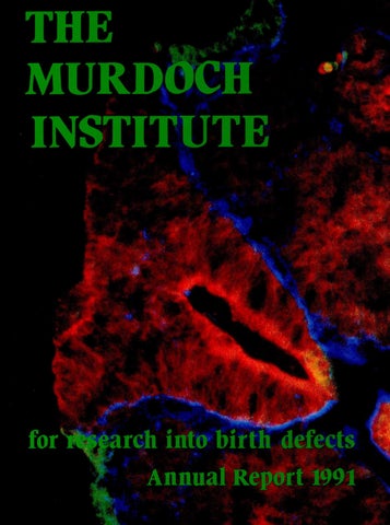
COVER Developing spinal cord of an early quail embryo with an open neural tube defect, labelled by triple immunofluorescence for L-CAM(green), A-CAM(red) and proteoglycan (blue). This shows that neural tissue expressing A-CAMhas not become internalized, and has failed to separate from the L-CAM expressing epidermis of the skin.
The Murdoch Institute for Research into Birth Defects
The Murdoch Institute is an independent research institute devoted to the investi gation and treatment of children with serious birth abnormalities, and to relevant basic research. The Murdoch Institute is renowned for a very close integration of fundamental research on genetic diseases with provision of genetic services to the whole population of Victoria. It’s scientific publications have attracted attention world-wide. The network of genetic services it has established has been used as a model within Australia and overseas. Doctors trained in the Institute lead the genetic services of most States.
The Murdoch Institute for Research into Birth Defects Limited Royal Children’s Hospital Flemington Road PARKVILLE VICTORIA 3052 ACN 006 566 972 Postal address: Post Office Box 1100 PARKVILLE VIC 3052 Telephone: (03)345 5045 Fascimile: (03) 3481391
m m:■vv-.
>
The Murdoch Institute was established in 1986 after generous supporters, led by the Murdoch family and the late Sir Jack Brockhoff, donated $10 million to set-up a new research institute devoted to the study of genetic diseases. In the intervening 6 years the Institute has made important scientific discoveries, developed a very high quality network of genetic services throughout Victoria and won a reputation as the leading genetics research institute and training centre in Australasia. All of this has been achieved by staff working in grossly over crowded conditions. Now, at last, the Institute has access to sufficient space to develop the facilities needed for the future. All that is needed is generous support from all those who want to see the scourge of birth defects diminished in our society.
■'
Birth defects kill or maim 1 in 50 babies. The strain on families is enormous. The cost to the Australian community exceeds $2,000,000,000 a year. Research into the genetic causes of birth defects is starting to reduce these figures. The Murdoch Institute needs your support every year to maintain the momentum of research into these genetic conditions. The Institute needs special support in 1992 as it tackles the last major stage in its establishment — raising $6.5 million to provide the laboratories and other facilities essential for its full development. Would you please give generously now. All donations are tax deductible. If you cannot give now, consider helping The Murdoch Institute in your will. The following is a suggested form for a bequest to The Murdoch Institute. I bequeath to The Murdoch Institute for Research into Birth Defects the sum of $ ... (or part or all of residue of estate) to be applied for the purposes of the Institute. For further information contact the Business Manager,
The Murdoch Institute for Research into Birth Defects Royal Children’s Hospital Flemington Road Parkville Victoria 3052 AUSTRALIA Telephone: (03) 345 5045 Fax: (03) 3481391
MAJOR DONORS TO THE MURDOCH INSTITUTE FOUNDERS — Donors of $1 million or more The Murdoch Family: Dame Elisabeth Murdoch Mr. Rupert Murdoch Mrs. Helen Handbury Mrs. Anne Kantor Mrs. Janet Calvert-Jones The late Sir Jack Brockhoff The Brockhoff Foundation The Scobie and Claire Mackinnon Trust
BENEFACTORS — Donors of $250,000 or more The Miller Foundation The Helen M. Schutt Trust
MAJOR DONORS TO THE MURDOCH INSTITUTE TRUSTEES — Donors of $25,000 or more Arthur Andersen Foundation Coles Myer Limited H. & L. Hecht Trust J.B. Were & Son Charitable Foundation Mrs. Joan Roxburgh Mrs. M.L. Griffin National Australia Bank Limited Qantas Repco Corporation Limited (Ariadne) The Banks Trust The Friends of the Murdoch Institute The Ian Potter Foundation The late Mr. Clive Roxburgh The late Mrs. L.B. Quayle The Morris Family Trust The News Corporation Limited The Percy Baxter Charitable Trust The Sidney Myer Fund
CORPORATE SPONSOR GROUP — Corporations undertaking substantial future support National Australia Bank Limited Qantas
$
DONATIONS TO THE MURDOCH INSTITUTE 1991 $
The Jack Brockhoff Foundation Dame Elisabeth Murdoch (Building Fund) The Scobie and Claire Mackinnon Trust Mrs Jean Roxburgh (Uncle Bob’s Club) The Miller Foundation Friends of the Murdoch Institute Coles Myer Ltd National Australia Bank Rotary Club of Balwyn The Arthur Andersen Foundation Amcor Ltd County Nat West Australia Morris Family Trust J B Were & Sons Charitable Fund Uncle Bob’s Club Scientific Hospital Supplies Ltd The Annie Danks Trust Muscular Dystrophy Association of Tasmania Prof D M Danks Medos Company Pty Ltd Riverside Lions Club The Petra Foundation The Brash Charitable Foundation Dr J M Gooch Mayne Nickless Ltd Mr Grant Stephenson The William Angliss (Victoria) Charitable Fund In memory of Melanie Rachel Peach McMullin Unit Trust Mr F D Ryan Melbourne Airport
300,000 100,000 100,000 65,000 50,000 25,430 25,000 20,000 7,200 6,000 5,000 5,000 5,000 5,000 3,900 3,275 2,000 2,000 1,790 1,500 1,200 1,100 1,100 1,000 1,000 1,000 1,000 960 700 650 515
In memory of Cameron McCarthy Mr & Mrs S F Gooley Mr G E Heeley In memory of Shaun Martin Lady Dorothy Darvall Soroptimist International of Melbourne In memory of Sarah Brueton Ms K Bromley Dr A Bankier Mr & Mrs L Barbieri Mr & Mrs B Thompson Mr S Poole Mrs C Arcaro Mr P W Botchie Mr & Mrs M A Campion Mr & Mrs E A Douglass Mr&MrsWLHirth Mrs M. Kinsey Mrs Knight Mrs P M A Lewisohn Dame Patricia Mackinnon Mr & Mrs L R Mills Strathcona Baptist Girls Grammar School In memory of Stephanie Waldron In memory of Emma Ritchie Mr & Mrs R S Brenton Mrs Cartwright Mrs G A Grimwade Mr J M N Hutchinson Dr LJ Sheffield Mr D Cockram Mr & Mrs J W Capes Mr & Mrs Kennedy Dr M L Cunningham The Institute thanks all those listed above for their generous support of research into the causes and treatment of genetic diseases and other birth defects.
502 500 500 455 250 200 175 160 150 150 150 110 100 100 100 100 100 100 100 100 100 100 100 100 75 50 50 50 50 50 25 20 20 10
Chairman’s Report
I
.
INDEX 1 Chairman’s Report............................................. Committees of the Murdoch Institute ............ 2 Selwyn Smith Medical Research Prize, 1991.. 3 Director’s Report ................................................ 4 Building Development Appeal......................... 8 Studying Biochemical Pathways in Children: Stable Isotope Mass Spectrometry.............. 11 OSSUM, brother of POSSUM.......................... 14 Work in Progress ................................................ 16 Post-Doctoral Fellows....................................... 23 Clinical Fellows.................................................. 24 Victorian Clinical Genetics Service............... 25 Genetic Counsellors/Coordinators................. 28 The Olive Miller Protein Chemistry Research Group......... 29 Studies of Pyruvate Dehydrogenase........ .... 30 Human Centromere and Down Syndrome 32 The Scobie and Claire Mackinnon Trace Element Group .. 35 Metabolic Unit................................................................... 39 Embryology Group ........................................................... 41 Epidemiology ..................................................................... 43 44 Mitochondrial Respiratory Chain Disease.................. DNA Diagnostic Laboratory........................................... 45 Tissue Culture.................................................................... 45 DNA and Autosomal Dominant Polycystic Kidney Disease .......................................... 45 List of Publications —1991 ............................................. 47 Murdoch Institute Lecture Series —1991..................... 50 Staff Involvement in Scientific Community Activities 50 Editorial Boards ................................................................ 51 Postgraduate Degrees Awarded .............. 51 Lectures and Seminars by Institute Staff 51 Collaborations ............................................ 53 Staff List Murdoch Institute............................... 54 Staff List Victorian Clinical Genetics Services 55
Mr Neil Walford
believe that there are few who might study this Annual Report without experiencing a keen awareness of the purpose, excitement and endeavour that permeates the activities of one of Australia’s major medical research institutes, one that is of world standing in its field. One can readily appreciate from the pages of the Report that our halls are graced by a very dis tinguished group of scientists. We can but admire their sense of mission, their force of character and their commitment. For them no great money for tunes but instead the greater reward of penetrating the frontiers of knowledge and of making vital con tributions for the benefit of their fellow men. During the year, despite the payment of $1.45 million to acquire the rest of the 10th floor of the Royal Children’s Hospital, we managed to main tain intact the corpus of our investment fund. It is vital that we do so for the income from it is an essential component in the revenue needed to cover our day to day operations. Our other major source of revenue is the block grant we receive from the NHMRC. I am delighted to report that after the recent quinquennial review the grant, slightly increased, has been renewed for a further five years. It is a great comfort to us and I feel sure to our friends and donors that we have been able to attract some of the best brains from the city, both to manage our finances and to help raise funds for the expansion of our activities. Our need for this assistance will never be greater than in 1992. Some time later this year under the dis tinguished chairmanship of Mr. Nobby Clark, we shall be launching an appeal to raise a substantial sum to enable us to refurbish and equip the 10th floor of the Royal Children’s Hospital which we have recently acquired. A successful appeal will consolidate the gains we have made to date and ensure that in the years ahead there is no abatement to our forward momentum. 1992 is a milestone year for the Murdoch Institute. In 1992 we welcomed three new directors. They were Mr. Ivor Davies, the new Chief Executive of the Royal Children’s Hospital, Dr. James Angus, Deputy Director of the Baker Institute and Chair man of the Grants Committee of the NHMRC, and also representing the NHMRC, Dr. Peter Gray, Secretary of its Medical Research Committee. I thank them and the other ladies and gentlemen of the Board for generously giving of their time during the past year and for the help and support they have given to me and the Director. And finally, but very important, my profound thanks to our donors past and present. They are our very life blood and as you may see from the accounts, the regular annual donations that keep coming in regardless of major appeals, are an essential part of our total revenue. 1
The Murdoch Institute Scientific Director: Professor D.M. Banks Deputy Scientific Director: Dr. R.G.H. Cotton
BOARD OF DIRECTORS
BUILDING AND DEVELOPMENT
Mr. N. Walford, Chairman Mr. L.G. Cox, Vice Chairman
APPEAL COMMITTEE
Dr. J. Angus Dr. G.L. Barnes Mrs. J. Calvert-Jones Dr. R.G.H. Cotton Professor D.M. Banks Mr. I. Davies Mr. J.A. Fitzgerald Dr. P.M. Gray Mr. J.S. Guest Mr. W.H. Hodgson Mrs. A. McFarling Professor P.D. Phelan Professor G.B. Ryan
Mr. L.G. Cox Mr. D. Craig Mrs. A. Cronin Professor D.M. Banks Mr. J. Fitzgerald Mr. P. Griffin Mr. M. Handbury Mr. G.E. Heeley Mrs. A. Insley Mr. N. Miller Mr. D.E. Meikeljohn Dr. M. Robinson Mr. N. Walford
FINANCE COMMITTEE Mr. L.G. Cox, Chairman
VICTORIAN CLINICAL GENETICS
Mr. C.P. Abbott
BOARD OF DIRECTORS Mr. N. Walford, Chairman
Mr. D.E. Meikeljohn Mr. F.D. Ryan
SERVICES
Dr. G.L. Barnes Mr. L.G. Cox Professor D.M. Banks Dr. J. De Campo Mr. J.S. Guest Mr. G.E. Heeley
2
cc
Mr. N. Clark, Chairman Sir Gordon Allard
Mrs. C. Searby
Mr. D.T. Craig Mr. P.J. Griffin Mr. G.E. Heeley
Selwyn Smith Medical Research Prize, 1991
i|j^ IP
Dr Richard Cotton
r Richard Cotton, Deputy Director of the Murdoch Institute for Research into Birth Defects, Royal Children’s Hospital, Parkville, has been awarded the Selwyn Smith Medical Research Prize for 1991 in recog nition of his outstanding research work during the past three years. Dr Cotton has devised a novel and extremely elegant procedure for the identification of mutations in genes. Such mutations are the causes of many birth defects and cancers. DNA of genes typically occurs in a double stranded form with precise matching of the component bases along the entire length of the DNA strands. When a mutation is present there is a mismatch between the strands. Dr Cotton recognised that regions of DNA that are mismatched are chemically less stable. Thus, when isolated mutant DNA is present as a double strand with normal DNA, chemical treatment will cause breaks in the strands where mismatches of bases occur as a result of mutations. It is possible to detect these breaks. The great advantage of this technical achievement is that DNA can be rapidly inspected for mutations in known genes without the need for laborious sequencing of the entire DNA sequence. Being able to specify the precise changes in genes that result from mutations is a vitally important goal of genetic research. This work will have a wide ranging impact on research into viruses, birth defects and oncogenes.” The Selwyn Smith Medical Research Prize is awarded by the Faculty of Medicine, Dentistry and Health Sciences each third year “to the candidate from amongst those applying for the prize whose original research has made, in the opinion of the committee, the most important contribution to medicine since the last award of the prize”. Regular readers of this Report will be aware of the discovery for which Dick Cotton has received this award and will share our pleasure in this recognition of his work. Dick has worked in the Institute since 1968, except for a period of overseas experience in 1970-73 during which he worked with Professor Cesar Milstein in Cambridge, performing studies which set the stage for the discovery of the methods of making monoclonal antibodies. His major work has been on the enzymes involved in phenylketonuria (PKU) and he is known as one of the world authorities on this subject. The work for which he was awarded the Prize was started during sabbatical leave in Oxford. A previous sabbatical in a different department in Oxford in 1980 was also very productive, leading to new methods of using embryonic carcinoma cell lines to analyse some events in embryonic development. Dick’s research has always been notable for technical innovation.
3
Director’s Report
Professor David Banks
4 !
^ I he two most special events of 1991 were the I Quinquennial Review of our NH & MRC -1. Block Grant and the acquisition of the remainder of the 10th floor of the Royal Children’s Hospital building for the use of the Murdoch Institute and the Victorian Clinical Genetics Service. The process of the quinquennial review involved submission of written documents, describing the last 5 years’ research and our plans for the next 5 years in April, followed by a defence of these achievements and plans before a Site Visit Committee in August. The outcome of the Review was pleasing, with renewal of our Block Grant for a further 5 years, 1992-96, and a modest increase in the value of the grant. This whole process was a major undertaking for all senior staffmembers taking up their full attention for the equivalent of two to three weeks over a six month period. Fortunately, the process of reviewing the last 5 years’ work is a very valuable exercise in its own right, as is the careful preparation of future plans. Indeed we plan to organise our own review process by colleagues chosen for special expertise in our areas of work in two or three years time. We are grateful to the members of the Site Visit Committee for the effort they put into the review and the consideration they showed for our work — Professor Nick Saunders (Newcastle) (Chairman), Professor Frank Gibson (ANU), Professor David Valle (Johns Hopkins Hospital, USA), Professor Ron Trent (Sydney) and Dr Nick Martin (Brisbane). As part of the review process Dick Cotton and Henrik Dahl applied for recognition by the NH & MRC as Research Fellows and we were very pleased that they were appointed at the levels of Principal Research Fellow and Senior Research Fellow, respectively. Dr Andy Choo’s Senior Research Fellowship was also reviewed and renewed for a further 5 years. All three were assessed by the Fellowships Committee of the NH & MRC as well as by the Site Visit Committee. The NH & MRC controls very carefully the awarding of Research Fellowships which carry a personal undertaking of tenure, for successive periods of five years, from the NH & MRC itself. All other scientists supported by NH & MRC grants have only whatever tenure the institution in which they are working can offer. In 1992 our Block Grant will be for $1,067,000, an increase of 12 per cent over the 1991 figure. Unfortunately, the effect of this increase is not as great as it might appear because there have been substantial increases in research salaries during this year as a flow on from decisions of the Academic Staff Tribunal. However, any increase in
funds is welcome in the present economic climate. These salary increases were long overdue. Academic and research salaries had fallen far behind the rest of the community over the last 5-10 years. A coincident restructuring of the salary scales has gone some way towards establishing a better career structure for research scientists, improving two serious anomalies that have existed for the last 10 years — namely a low starting salary for postdoctoral scientists and a low ceiling to the salary to which high quality research assistants can aspire. However, the new scales have also created some new anomalies. In March the Hospital vacated the newborn nursery in Ward 10 West, the bedrooms in 10 East used by living-in mothers (previously by Resident Medical Officers), the Resident Medical Officers’ Lounge and a number of laboratories in the western part of the southern block. This gave the Institute and the VCGS access to the whole of the 10th floor, except for some small areas still occupied until the end of 1992 by the offices of the Department of Gastroenterology. When we are able to alter and refurbish this area it will provide very efficient accommodation for at least a 50% increase in research activity, plus adequate space for the offices, clinics and labora tories of the VCGS. To finance these alterations, we will need to raise substantial new capital. Our plans for these alterations and for a Building Appeal during 1992 are described in detail later in this Report. In the meantime we have carried out a number of minor modifications to make our working conditions tolerable for the next few years. Thanks to the skill of our Laboratory Manager, Mr Barry Holt, the changes were achieved with little interruption to work and at low cost. Dr Andy Choo’s group is now occupying a satisfactory laboratory at the western end of the south block and their move has allowed Dr Henrik Dahl’s group and the DNA diagnostic group to have adequate space in the existing DNA laboratory. Don Newgreen’s embryology group has been accommodated in the laboratory that was previously used by the Department of Paediatrics. A very clever alteration in 10 West has produced a remarkably efficient newborn screening labora tory, and some minor changes have converted the Resident Medical Officers’ Lounge into a spacious seminar room and tearoom. Installation of tele-
Ivan Francis Head, Neonatal Screening Laboratory
phones and adequate lighting, and a coat of light-coloured paint have transformed the rather dingy bedrooms and corridors of 10 East into an adequate set of offices for scientists and some clinical researchers. Although these arrangements are adequate for the time being, we must look ahead and reorganise the 10th Floor to maximise its capacity for the future expansion of our activities. The move of the Newborn Metabolic Screening Laboratory was particularly welcome. We have been planning for this move for over 10 years and I am most grateful to Mr Ivan Francis and his col leagues for their tolerance of the delays. Their closer integration with our other laboratory activi ties is already bringing benefits in the few months since they moved, especially in the cystic fibrosis program, and we are all enjoying the constant access we now have to Ivan’s expertise in comput ing. An unexpected by-product of the move is that we have apparently saved the Parkville Post Office from closure! The more than 1000 samples which come through the mail to the laboratory each week has made a dramatic difference to the workload of this small office. This year I have decided to refrain from mention ing the progress of our science in my Report, preferring to deal with this in the section entitled “Research in Progress”. Of course, I will still comment on some highlights of external recogni tion of our science and scientists. The development of a second computer/videodisc information system to assist in the diagnosis of birth defect syndromes certainly warrants men tion. It also allows me the opportunity of boasting that POSSUM, the system we released in 1987, is now in use in 280 hospitals in 42 countries. OSSUM, a system based upon the logic and programs of POSSUM, is designed to assist geneticists and radiologists in diagnosing bone genetic disorders of bone growth dysplasias which cause short stature and/or disturbance of body proportions. There are more than 350 of these conditions and distinction between them requires considerable expertise. The 4000 X-rays on the OSSUM videodisc and the search strategy it offers will assist doctors greatly. The production of OSSUM has been a collaborative effort between Melbourne, Sydney and Mainz in Germany. The new system was very warmly received when displayed at the International Congress of Human Genetics in Washington in October and we already have a number of orders in hand even though the system will not be ready for release until late January, 1992. Dr Agnes Bankier and Mr John Marquet of Computer Power, who created POSSUM, have worked together again to develop OSSUM. This time the Murdoch Institute owns the product and will be paying a royalty to Computer Power for the use of their programs. POSSUM is owned and marketed by Computer Power, with the Institute receiving a royalty. More details about OSSUM can be found in a section elsewhere in this Report. In October, we sent ten scientists and clinicians to the Eighth International Congress of Human Genetics in Washington, our biggest ever contin5
r gent attending an overseas meeting. The meeting itself achieved a record attendance of 5,700 delegates, almost exactly double the number registered for the previous International Congress in Berlin in 1986. These numbers give some measure of the rapid expansion of this discipline world-wide. Dr Agnes Bankier and Dr Max Robinson (Marketing Manager of OSSUM) were kept very busy displaying OSSUM to many enthusiastic geneticists. They shared a booth with POSSUM which also continues to attract much attention. Agnes Bankier’s position as an expert in computerised information systems was recognized by the organisers of the Congress who asked her to plan and direct a Workshop on this subject. Another honour for an Institute staff member was the invitation of Dr David Ravine, a Clinical Genetics Fellow, to give one of the 45 minute talks in a Symposium on the practical application of DNA diagnostic tests in the community. His study of DNA tests in polycystic kidney disease has produced results which were of great interest to the large audience present at this session. David found that most family members were very pleased to undergo presymptomatic testing, an experience which contrasted with the anxiety generated by the availability of presymptomatic diagnosis for Huntington’s disease reported in another paper in the Symposium. The other Institute staff members were attending the Congress as speakers in Workshops, Chairmen of Scientific Sessions or to present papers or posters describing their work. In November, colleagues in Sydney were hosts to an International Symposium on Newborn Metabolic Screening. This Symposium brought to Australia some members of an international group of experts on pteridine and folate metabolism which meets every alternate year and Dick Cotton therefore arranged for this group to meet in Cairns straight after the Sydney meeting. The Cairns meeting was an unqualified success with a very good attendance of the world’s top scientists in this field. Ian Jennings, David Howells and Susan Ramus assisted Dick in the organisation. Congratulations also to Dick Cotton on two other recent successes. John Wiley and Son, a US based international publisher of medical textbooks and journals, chose the International Congress of Human Genetics as the occasion for announcing
IS*
il Professor Ron Davidson 6
the establishment of a new journal called Human Mutation, which will be jointly edited by Dick Cotton and Dr Haig Kazazian from Johns Hopkins Hospital. In December the University of Mel bourne announced the award of the Selwyn Smith Memorial prize for medical research to Dick Cotton in recognition of his valuable contributions to medical knowledge over the last decade. Dr Andy Choo has recently been asked to edit a book on In-situ Hybridization Techniques which will be published by the prestigious molecular genetics journal. Nucleic Acids Research. In January 1991 we welcomed Dr Don Newgreen. He came to us from Sydney to establish a new research group in embryology and his work is described in “Work in Progress”. In June we welcomed Dr Jack Insley and Mrs Anne Insley from Birmingham UK. Jack recently took early retirement from his post as a clinical geneticist and we have quickly learnt to appreciate his fund of knowledge and experience and his delightful sense of humour. Anne has also joined the staff, assisting part-time in our fundraising activities. We are delighted that they are able to extend their initial one year visit for a further 6 months until the end of 1992. The idea of encouraging very experienced clinical geneticists to come here for a year or two each, began because of a shortage of young, fully-trained clinical geneticists ready for career appointments. Ron Davidson and Jack Insley have given so much during their periods in this role, that it is tempting to try to continue this system for much longer. In November Dr Renee Martin, a world’s expert on sperm chromosomes, came to us from Calgary, Canada for six months’ sabbatical leave. Her interest in learning more about DNA techniques and Andy Choo’s need to examine sperm chromo somes in his projects create a productive two-way exchange. In June we farewelled Professor Ron Davidson and Mrs Miriam Davidson after two years in the Institute. Ron made a very important contribution to the development of the Victorian Clinical Gene tics Service, in which he was Clinical Director in the last 18 months of his stay. He will he particu larly remembered for the introduction of a training course for genetic counsellors and for establishing country clinics. Miriam made valuable contri butions to fundraising and public relations within the Institute and outside. Both left behind many warm friendships. Two scientists. Dr David Howells and Dr Malgorzata Schmidt left us at the end of the year. David came to us as a postdoctoral fellow, stayed on to take charge of the enzymology/metabolism laboratory and leaves to further his interests in neuroscience research at the Austin Hospital. Malgorzata came to a dual role in the diagnostic cytogenetics laboratory and in cytogenetic research, but proved more interested in the latter. Her interest in the special behaviour of the human X chromosome makes it logical for her to join Professor Jenny Graves at LaTrobe University, who is a world’s expert on this subject. Les Sheffield has taken over the supervision of the genetic counsellor training course which was
started by Ron Davidson, and Jack Insley has also joined in this. In addition, Les has conducted a very popular series of teaching sessions on basic genetics at 8.00 a.m. on Friday mornings. Next year we plan to expand this series to cover a wider range of subjects using all the senior staffas teachers. Also notable during 1991 has been the establishment of a Social Club, the committee of which has organised several well attended and enjoyable functions after work on Fridays and also the Institute Christmas Party. Self-organised activities like this are of importance to the morale of the Institute. Within the Institute administration we have shifted more responsibility for budgeting to group leaders. Quick feedback of monthly expenditure on laboratory chemicals, plus publicising the costs of some of the more expensive chemicals, has reduced expenditure on these items. Discussions between group leaders in the Executive Committees are proving fruitful and are proving important in implementing our plans for the next 5 years. During the year Dr Ruth Bishop, the Chief Executive of the Research Foundation, organised a review of the administration of the Foundation. While this review came up with some interesting observations and suggestions, I feel concerned that the outcome will be to diminish the independence of the Research Foundation from the Hospital. I believe that it is important that research in organisations as big as the Royal Children’s Hospital should be allowed to set its own priorities independent of the clinical and financial priorities of the Hospital, determined primarily by the quality of the scientists and of their research proposals without too much concern about the immediate clinical application of the results. The great importance that we place upon this indepen dence was a major reason for establishing the Murdoch Institute and we will certainly be making sure that our independence is maintained. Of course, we want a very close working relationship with the Hospital and Research Foundation, and with their clinicians and scientists, but this needs to be a relationship like that between friendly and respecting adults, not a relationship like that between a parent and young child. One flow on from the new arrangements in the
Research Foundation has been an ending of the arrangements hy which Mrs Anne Cronin (Ellis), our Business Manager, and her stalf, handled the Foundation’s accounting and personnel manage ment, and Mr Barry Holt acted as Laboratory Manager for both organisations. Both had found the double load excessive in the last few years, and this was especially the case for Barry during the conversion of the 3rd Floor laboratories for the Foundation. I am delighted that the Institute now has the undivided attention of these two very able people on whom I rely heavily. I want to thank them for the cheerful way in which they have coped with the special pressures they have experienced during 1991. Special thanks also to Andrew Grimes and Ivan Francis who helped out during Barry’s period of illness and at other times when Founda tion duties were keeping him away from the 10th Floor. I am fortunate in having a very helpful and understanding Chairman of the Board (Mr Neil Walford) who is always available and willing when I need wise counsel. We all felt for, and admired, Neil as he coped so wonderfully with the long terminal illness of his wife, Patsy, and marvelled at her ability to remain so cheerful and interested in others during the many months of pain and discomfort. The Institute lost a very special good friend when Patsy died. I also lean heavily on Mr Laurie Cox (Deputy Chairman and Chairman of the Finance and Investment Committee) for guidance and now we have another strong and wise supporter in Mr Nobby Clark, Chairman of The Building Appeal. All of these people are supported by Board and Committee members (listed elsewhere) who give generously of their knowledge and energy. The Friends of The Murdoch Institute have gone on to further successes this year, under the leadership of Mrs Anne McFarling, Mrs Anne Hayward and Ms Davina Hanson, staging a successful Fashion Parade at Diamaru during its opening week and a Derby Eve Party at 333 Collins Street. The Friends contributed $50,000 to the Institute during their first year of activity, a very laudable achievement, and have made many new friends for the Institute. We look forward to their continued success.
7
Building Development Appeal Our last big task — to provide satisfactory accommodation for the Institute
F
involved giving an undertaking to provide the ^ rom the very beginning of the planning for space required to house the Service. our new Research Institute we identified the provision of satisfactory laboratory and In arranging appropriate accommodation for the office accommodation as very high priority. Institute and VCGS we first needed to choose the best location. It was clear that it would be best to Of course our top priority had to be the fund remain within the Royal Children’s Hospital, pro raising drive to give us sufficient money to estab vided that sufficient space could be made available lish the Institute as an independent organisation. in a way which would give the Institute a clear and By mid 1986 we had promises of donations totalling $10 million from the Murdoch family, the late Sir separate identity. Originally the Hospital had offered the use of three quarters of the tenth floor Jack Brockhoff and many other generous sup of their building, rent free. The need for additional porters. The launching of the Institute was there space to accommodate the VCGS led to further fore assured and was formalised by the Prime Minister at a Dinner in the Great Hall of the negotiations and the Hospital agreed to provide National Gallery of Victoria in February 1987. Then we were able to turn our attention to enhancing our stand ing as a research institute, develop ing our own clinical service organi sation and acquiring sufficient laboratory and office accommo dation for efficient development of the combined research and service activities. To our delight the National Health and Medical Research Council awarded us a Block Grant in 1987 and by July 1988 the Vic torian Clinical Genetics Service had been established as a sub sidiary of the Murdoch Institute financed by the Health Depart ment of Victoria. The NH&MRC decision affirmed the quality of our The difficulties of overcrowded laboratories science and the calibre of our lead ing scientists. The establishment of the VCGS was the culmination 'loo 0 o a of negotiations with several col laborating hospitals, especially the Royal Children’s Hospital, and the Victorian Government. These were very important steps towards our ultimate goal of close integration of basic research and clinical service, both recognised nationally and internationally for their high quality. Achieving these goals so quickly brought some costs — the high expectations created by the early award of a Block Grant forced us to increase the amount of our original capital committed to the recurrent costs of research, and the bargain struck with the Victorian Government over the funding of the VCGS Our goal—modern well-equipped laboratories
1
8
the entire tenth floor in return for a payment of $1,450,000 and an undertaking to pay an annual fee to cover the cost of all services — such as electricity, gas, cleaning, library facilities. For tunately the Victorian Government agreed to pay this annual service charge. Our location within the Hospital ensures access to high quality paediatric services and facilities for the child patients of the VCGS and gives Hospital patients access to the skills of our geneticists. Our research has benefited from close contact with a wide range of clinical problems and there is a mutual advantage in the interaction between our scientists and those in the Hospital and Research
Foundation. The location of the Hospital within the “Parkville Strip” provides the best scientific environment available in Australia. Unfortunately the Hospital’s plans for construc tion of a new building on Flemington Road, and for other major alterations, were delayed nearly three years so that we Anally gained access to the remainder of the tenth floor in March 1991. In the period since 1984 the cost of building alterations has escalated greatly and we now find ourselves facing a total expenditure of $6.5 million to cover the cost of purchasing the tenth floor and altering it to provide the laboratory accommodation, clinic rooms and offices that we need. The alterations that we have planned will give enough accommoda tion for a substantial increase in our THE MURDOCH INSTITUTE Plan 1. basic research work and clinical Currant activities, sufficient to carry our twin organisations through the next decade or longer. We have designed • m . W . Ji....•... : •.....s.... ■ rooms of an efficient size laid out so as la ■ ® i [S to encourage interaction between different research groups, with a minimum of unproductive corridor In u«« space. The need to keep our labora tories working while the alterations The Scoble and Claire Mackinnon are made complicates the process, but Trace Element Research Group we have planned a way of completing the task in three stages over two years. We intend to locate most of the research laboratories in the south The Olive Miller Protein Chemistry Group block of the tenth floor, to use the link THE JACK BROCKHOFF RESEARCH LABORATORIES between the north and south blocks for service laboratories and some general housekeeping facilities and to fill the north block with clinical rooms and offices, other service THE MURDOCH INSTITUTE laboratories, research offices, admini Plan 2. strative offices and general facili Proposed Administration ties such a tea room and conference Support Services Conference Rooms Staff Amenities Library Research Offices rooms. The type of biochemical and mole cular research which our scientists undertake requires many items of sophisticated equipment. This equip Clinic Clinic Offices Cytogenetics Laboratory ment needs constant-voltage electri city, constant-pressure water supply E5 and piped pure water. These services Laboratory Support Screening Laboratories are available only in the south block and Computer of the building and this dictates the ‘he Scoble and Claire Mackinnon Trace Element Research Group The Olive Miller Protein Chemistry Group arrangement just described. Within the south block we aim to give each research group its own identity by providing separate labora tories. Collaboration between groups i will be encouraged by having shared facilities and flexibility to cope with future fluctuations in the size of groups will also be incorporated. For B. TISSUE CULTURE LABORATORY tunately the south block of the tenth C. EMBRYOLOGY LABORATORY floor was added to the building only D. DNA LABORATORY (DR. CHOO) in 1975/76 and the laboratory layout E. INSTRUMENT ROOMS F. DNA LABORATORY (DR. DAHL) was designed by our own Labora G. DIAGNOSTIC DNA LABORATORY (VCGS) tory Manager, Mr Barry Holt. Con H. ENZYME/METABOLISM LABORATORY sequently existing laboratories will
1
»/Llbrary
1
P
Offices Laboratories Conference/Library
11
i
9
need only minimal alteration and the main cost will come from converting offices and seminar rooms into laboratories. The unfortunate feature of the south wing is that a great deal of asbestos was used within the ceiling space and the cost of the removal of this asbestos will be very substantial. We will need to accommodate our genetics clinics within our new space, rather than using Hospital clinic areas, as well as providing offices for our clinicians. We want to have the offices of clinicians and scientists close together to encour age interchange of ideas, but we also have to ensure that the waiting areas for patients do not obstruct access to the offices. To achieve all of this without wasting too much space on clinical rooms which will be used infrequently, we will change the organisation of our clinics from two clinics a week, each staffed by six or seven doctors, to four clinics a week, each attended by three or four doctors. These clinical facilities and offices for clinicians and scientists will fill the north west wing of the floor. The office area for scientists will be planned so that each research group has a large room, shared by junior staff, adjacent to a small office for the group leader and another small room in which several members of a group can get together to discuss plans or results. Within the administrative area in the north east wing there will be a larger room which will serve as our board room when needed and will be used more frequently as a meeting room for larger groups of staff who need to get together to debate plans or results. We have learned that it is very important to have a number of rooms in which groups of people can talk together. These rooms are also useful when individuals need to spread out a large number of sheets of results in order to collate them. The north east wing will house the administrative area, the seminar room and tea room, the board room, a small library and reading area plus accommoda tion for the epidemiology group and the cyto genetics laboratory. During our period of cramped existence we learned the importance of having an adequate tea room and a separate seminar room. A good tea room is an important meeting place for researchers and creates opportunities for discussion between people doing quite different types of work. We use our seminar room quite frequently for meetings of
i
up to 50 people and we have an occasional need to accommodate a bigger audience. By locating the seminar room next to the tea room, separated by a folding door, we can satisfy all of these require ments. The link between the north and south wings will accommodate the newborn screening laboratory of the VCGS, the Laboratory Manager’s office and store rooms. We intend to pay careful attention to the layout of the two lift lobbies on the tenth floor. We want visitors who arrive in the lifts to be immediately aware that they are in the Murdoch Institute and VCGS and to find it easy to identify the location of the section that they are seeking to visit. Barry Holt has put a great deal of effort into drawing up plans for the most efficient utilisation of the space and has tried to do this in an economical way. Nonetheless, the quantity sur veyor’s estimate of cost is $5 million. Adding this figure to the $1.45 million already paid to the Hospital for the use of the tenth floor brings the target of our Building Appeal to $6.5 million. We also need some major items of new equip ment. For instance, the work on stable isotopes described elsewhere in the Report can proceed only very slowly with our present instrument which is nearly 10 years old. A new and more sensitive instrument will have much greater capacity, but will cost $225,000. We have a State Government grant of $50,000 towards its cost (to pay for the clinical service aspect of its use), but need a generous donor to provide the remaining $175,000. The Building Appeal will be launched with a series of functions at the Institute during the first two weeks of May 1992. Mr Nobby Clark is leading an Appeal Committee, the core group of which is made up of business men who have been our friends and advisors from the beginning — Mr Neil Walford (Chairman of the Board), Mr Laurie Cox (Chairman of the Finance Committee), Mr John Fitzgerald, Mr Peter Griffin, Mr David Craig, Mr David Meiklejohn, Mr Geoff Heeley, Mr Noel Miller — and a new friend. Sir Gordon Allard. Our Patron, Dame Elisabeth Murdoch has set us on the way towards our ambitious target in a charac teristically generous manner with a personal donation of $100,000.
Studying Biochemical Pathways in Children: Stable Isotope Mass Spectrometry
I
n order to function normally, the body needs to convert simple chemicals such as sugars, fats and protein components into the many complex substances required for building and maintenance of the body. Each of the many steps in these con version processes is controlled by an enzyme (catalyst). Over 500 enzymes have been identified in man, and each is manufactured in response to a specific genetic code; that is, all these processes are under genetic control. Faults in most of these genetic coding systems have been identified and are known collectively as inborn errors of metabo lism. These faults may cause a wide range of symptoms, but in all cases the symptoms are related either to an abnormal accumulation of the chemicals that are normally processed by the enzyme, or to a deficiency of the chemicals normally produced by the affected biochemical pathway (Figure 1). The outcome of these inborn errors is generally poor, and many affected children die in the first few days of life. Over the past 20 years substantial advances have been made in the treatment of these conditions, leading in some cases to a virtually normal life and in many others to a worthwhile survival beyond infancy.
condition is untreated, death in early infancy. Even with treatment the outcome of these con ditions has been very poor. Most children who survived did so with mental retardation and the risk of developing a range of major complications. Understanding of the conditions had largely been gained from laboratory studies of tissues in cell culture. These studies suggested that most propio nate came from protein and that a low protein diet should have been an effective form of treatment. Although these diets were of some benefit, it was clear that the treatment was not working as well as it should have. What was needed was a way of measuring the sources of propionate in the whole body to determine their relative importance.
propionic acid carboxylase enzyme 1 f
i methylmaionic acid mical A mutase enzyme
I t
succinic acid I Figure 2
Figure 1 The value of stable isotope mass spectrometry in the investigation of inborn errors of metabolism is best explained by demonstrating its application in a specific group of inborn errors, in this case the methylmalonic and propionic acidaemias. These conditions are caused by faults in the enzymes responsible for the breakdown of an acid called propionate (Figure 2). The normal products of these pathways can be manufactured in other ways, but the accumulation of propionate leads to serious toxic effects on energy metabolism. This presents itself with rapid onset of symptoms of lethargy, decreased consciousness and, if the 10
The great difficulty in performing whole body studies is the exceedingly complicated interaction of the many biochemical systems coexisting within the human body. This particular problem has been overcome in the past by studying biochemical pathways in cells isolated in tissue culture, where the environment can be modified to focus on the pathways of interest. While this approach has been very fruitful, it does not allow us to assess the function of biochemical pathways under the con ditions that prevail in real life. Developments in the technique of stable isotope mass spectrometry have now given us a way of focussing on individual biochemical systems within the body. Dr Geoff 11
labelled beam
protein components fgut bacteria
I
45% 30%
t propionic ac'^*
detector
beam
carboxylase enzyme
t
ion source
methylmalonic acid
succinic acid Figure 3 Thompson, initially with colleagues in London and Paris and more recently in the Murdoch Institute, has applied this technique in children with methyl malonic and propionic acidaemias to measure exact ly the amount of propionate that was being produced by the whole body. Once this was known the relative contribution of components of protein to this pro duction rate was measured. Rather than contributing the majority of propionate produced, protein proved to account for only 40-50% of the total. It had been known for some time that certain fats stored in the body and certain bacteria that reside in the gut could produce some propionate, hut the amounts produced were thought to be insignifi cant. The new findings meant that these sources of propionate now had to be more closely scrutinised. The stable isotope studies were repeated under conditions chosen to eliminate these alternative sources individually. First, antibiotics were given to sterilise the gut and remove the gut bacteria capable of producing propionate. This resulted in a 30% fall in propionate production, implicating bacteria as a significant source of propionate. Next, large amounts of glucose were given to prevent fat break down. This also reduced propionate production, by about 25%. It was then possible to construct a diagram of the relative contributions of major com ponents of propionate production (Figure 3). This understanding has now made it possible to focus the treatment of methylmalonic and propionic acidaemias more accurately. Further studies of this type should be able to improve the understanding and management of a wide range of inborn errors of metabolism. 12
The secret of the stable isotope mass spectrometry approach is the use of a tracer (a labelled substance) to effectively isolate components of a particular biochemical system within the whole body. These tracers employ stable isotope labels. Stable isotopes are non-radioactive variants of basic elements, such as hydrogen and carbon, that form the structural backbones of virtually all of the chemicals in our bodies. Stable isotopes are naturally present in small quantities in our food and in our bodies, which means that their use in research studies is most unlikely to cause ill effect. This supposition has now been verified after extensive injection and ingestion of stable isotopes in man over many years. Measurement of stable isotope labels needs to be very precise for these studies to be possible, and it is only quite recently that the technique of mass spectrometry has developed sufficiently to ensure success of the approach. The mass spectrometer (Figure 4) works on the principle that a chemical containing a stable isotope label will be slightly heavier than its unlabelled counterpart. The chemical of interest is purified and vaporised before entering the mass spectrometer. Upon entry it is converted to an ion beam, which is then exposed to electric or magnetic fields which bend the beam of heavier, labelled ions more than the beam of lighter, unlabelled ions. The separated ion beams can then be quantitated in the detector. Sophisticated and expensive computer software is needed to handle the data produced, especially for the whole body biochemical studies described above. State-of-the-art instruments are now pre sented as modular packages, as shown in Figure 4.
Figure 4 The application of these methods is quite labour intensive, both in performing the studies in children, and in analytical time. However, for the child being studied, the techniques are relatively non-invasive, and most tolerate the procedure with little discomfort. A child with methylmalonic acidaemia is shown undertaking a stable isotope study in Figure 5. The recent developments in stable isotope mass spectrometry and in the logistics of whole body biochemical studies are not only theoretically exciting, but have now produced results which were unattainable with earlier methods. This expanding field has already become an integral component of the Murdoch Institute research pro gram and we are confident that it will contribute much to our understanding and management of inborn errors of metabolism in the future.
Figure 5
13
OSSUM, brother of POSSUM
I
^or some years we have realised that bone dysplasias, hereditary disorders of bone growth which cause dwarfing and/or distort body proportions, would be a very appropriate subject for a computer/videodisc information system. This is because correct diagnosis depends upon instant recognition of characteristic X-ray changes in bones, characteristic physical features visible in the patient or characteristic changes seen when the cartilage at the growing ends of the bones is examined under a microscope. Since there are more than 350 different bone dysplasias it is difficult to remember all of these characteristic appearances. Even a large textbook has difficulty accommodating sufficient pictures. Often it is the combination of changes in different bones in the body, or even the way in which these changes evolve during childhood, which is diagnostic. A computer can help to sort out these combinations. The logic which we developed and used successfully in POSSUM is ideally suited for this purpose. A doctor can enter a list of features seen in his patient or their X-rays and then interrogate the computer database to find out which of the known bone dysplasias show the same features.
The program written by Mr John Marquet of Computer Power is so speedy that a search can be achieved in a few seconds. The doctor can then examine photographs of patients. X-rays or microscopic changes in cartilage and these are selected by the computer from the videodisc on which the images are stored. The computer displays a table of all of the different categories of pictures or X-rays that are available and of the various stages of childhood for which illustrations can be offered. A few simple keystrokes enable the operator to select the particular illustrations that would be most useful. To produce OSSUM Dr Agnes Bankier teamed up with Professor Jurgen Spranger of Mainz in Ger many, author of the best known textbook on bone dysplasias. Professor David Sillence of Sydney, a world expert on the microscopic changes in bone dysplasias, and Professor Kazimierz Kozlowzki of Sydney, another world expert on the X-ray changes. Dr Hartmut Menger from Professor Spranger’s department worked very hard in coding the descrip tions of the known bone dysplasias and in selecting X-rays from collections in Germany, Melbourne and Sydney. Ms Sofia Mercer has played a valuable
coordinating role in management of the illustra tions and Mrs Christina Marquet wrote the user’s manual. John Marquet made the modifications required in the operating program. Agnes and her associates have spent countless hours checking all the entries and writing commentaries on each syndrome and the illustrations of each case. We have assembled over 4000 illustrations for Version 1.0 of OSSUM and these illustrate most of the bone dysplasias. However, there are still many gaps to be filled before we have each condition ideally illustrated with pictures of all relevant bones at each phase of childhood. We are offering those individuals who purchase Version 1 a free copy of the first update, to be known as Version 2, which we anticipate producing in 1993, and will be seeking their assistance by providing illustrations of their own cases. OSSUM was first displayed at the International Congress of Human Genetics in Washington in October 1991 and we already had a number of orders for the system before the release date in February 1992. This is very pleasing and represents a much quicker acceptance than POSSUM received when it was first released. Of course the reputation that POSSUM has achieved has made it easier to sell OSSUM. POSSUM itself is now in use in over 280 hospitals in 42 countries. It was gratifying to find that nearly all of our POSSUM users purchased the most recent videodisc update (Version 3.0, released in May 1991). POSSUM is owned and marketed by Computer Power who provided the capital required for its development as well as the skills in programming. OSSUM is owned by the Institute itself and we will do the marketing. We have been assisted by an
Australian Government Discretionary Grant for Industrial Research and Development. Dr Max Robinson, recently retired from a senior position in the Department of Paediatrics, is Marketing Man ager for OSSUM, Mrs Anne Cronin is Business Manager and Dr Bankier is overall Project Leader. With OSSUM we will pay Computer Power a royalty for use of their programs, instead of receiving a royalty as we did with POSSUM. The Institute has provided some financial support to the departments of Professor Spranger and Pro fessor Sillence. We are hopeful that OSSUM will be successful financially as well as medically and that the proceeds of its sales will support new research in the Institute. Our graphic artist, Ms Kati Bromley, has produced an eye catching brochure with the text printed over a collage of X-rays characteristic of different bone dysplasias. Unfortunately, the subtle visual effect cannot be reproduced in this Report, but it certainly catches the attention of doctors walking by our display booth at a conference. We are also working with several of the world’s experts on these conditions to modify OSSUM so that they can use it to catalogue their undiagnosed cases, making it a research system as well as a diagnostic aide.
9
u
li.
« m M wr
m % % %
wolw & ftjssum
L
m OSSUM at the Radiological Congress of North America, Chicago, December 1991
OSSUM at the Radiological Congress of North America, Chicago, December 1991
ii;l
14
15
Work in Progress
I
n this section we aim to give our readers a brief overview of the work that is going on currently in the Institute. We aim to make this section comprehensible for our friends who are not scien tists and we welcome criticism if we are not achiev ing this degree of clarity. If our scientific col leagues find this section too light-weight then they can turn to the detailed descriptions of individual projects at the end of this Report. Each section is preceded by a list of the staff of the group con cerned. Dr Cotton’s Group — The Olive Miller Protein Chemistry Research Group Postdoctoral Fellows:
Phillip Dickson Enzo Palombo Jenny Saleeba Peter Smooker Zilla Wong
Scientific Officer:
Ian Jennings
Research Assistants:
George Makris Gary Pfeifer Susan Ramus
Dick Cotton, our senior scientist since 1968, is widely acknowledged as one of the world’s experts on the enzymes (biological catalysts), which are at fault in patients with phenylketonuria (PKU). In recent years he has become widely known for developing a chemical cleavage method (CCM) of identifying the mutations (faults) in genes which are responsible for genetic diseases. PKU is one of the most common treatable causes of mental retardation. All newborn babies are tested for PKU soon after birth so that a special diet which can prevent mental retardation can be introduced before the brain has become damaged. This screening, carried out in the Newborn Metabolic Screening Laboratory of the Victorian Clinical Genetics Service (VCGS), and subsequent
Jenny Saleeba 16
Ian Jennings
dietary treatment, carried out in the Metabolic Clinic of the VCGS, saves five or six Victorian babies from developing mental retardation each year. Although the dietary treatment is effective it is not perfect. We hope that at some time in the future it will be possible to replace the faulty gene or to treat patients with a small part of the enzyme which has been found to be critical for its function. In most patients with PKU the fault is in an enzyme called phenylalanime hydroxylase (PAH), but some years ago we discovered that defects in another related enzyme (DHPR) can produce an even more severe form of PKU. Ian Jennings, Peter Smooker, Phillip Dickson and Susan Ramus are working with Dick (and in the past David Howells) on these two enzymes studying the detailed structure of the enzymes themselves and analysing the mutations found in the genes controlling these enzymes in patients with PKU. Both these approaches are helping to identify the critical parts of the enzyme molecules and good progress is being made. The segment of the PAH enzyme which is essential for activity seems to be quite small, although another segment at one end of the mole cule may play a role in controlling the level of activity. The combined efforts of David Howells, Peter Smooker and Jenny Saleeba have identified the region of the DHPR molecule which is respon sible for its activity. The approach that Ian Jennings is using involves making antibodies which recognise just the functionally crucial parts of the PAH molecule. These antibodies turn out to have useful properties for other scientists overseas who are studying other related biochemical processes. They, and anti bodies isolated earlier, are being used in col laborative studies on topics as widely separated as the mapping of nerves in the brain and the analysis of the nutritional requirements of cancer cells. Phillip Dickson joined the group in mid year to analyse the way PAH works and has started by
Enzo Palombo
Peter Smooker
initiating test tube production of the enzyme for easier study, and is already making good progress. Dick’s work on identifying gene mutations is driven by the need for a quick and sensitive method of identifying the subtle changes which are the most common form of mutation causing genetic diseases. A gene can be likened to a paragraph of a very large instruction manual and mutations are comparable to typographical errors affecting a single letter which may alter the sense of the whole paragraph (disease-causing mutation) or may just cause the reader a little difficulty in following the sense of the paragraph (minor genetic differences between individuals). His chemical cleavage method (CMM) is one of four or five techniques developed over the last five years to speed up the process of identifying these mutations. The Vic torian Health Promotion Foundation recognised the potential of this method and awarded a pro gram grant for 1990-1992 which has allowed Dick to employ three post-doctoral scientists (Jenny Saleeba, Enzo Palombo and Zilla Wong) to study the' application of the method to human mutations and to mutations causing changes in rotavirus (an important cause of infantile diarrhoea), Dengue virus and HIV (the virus causing AIDS). George Makris is starting to apply it to finding mutations in retinitis pigmentosa. The method has also been applied to other human genes by other scientists in the Institute. Our experience, and that published by other laboratories overseas, suggests that the CCM is particularly useful when a scientist needs to identify all mutations within a particular gene. Some of the alternate methods are a little simpler to use when finding just some of the mutations that can cause a disease is sufficient. Dr Andy Choo’s Group Postdoctoral Fellows:
Adam Nagy Bryce Vissel
Scientific Officers:
Elizabeth Earle Anna Michalska
Research Assistant:
Paul Kalitsis
PhD Scholar:
Camille McQuillan
Andy Choo undertook post-graduate training in protein chemistry with Dick Cotton, and in molecular genetics in two outstanding overseas laboratories before returning to build up his own research group within the Institute over the last seven years. His current research on the molecular structure of the centromeres of human chromosomes is addressing one of the big unanswered fundamental questions of biology. Centromeres are specialised regions of chromosomes which control the ability of pairs of chromosomes to recognise one another, then separate and move to opposite poles of cells during the special form of cell division which produces egg or sperm cells. (Our genetic system uses pairs of genes to control each characteristic. Our sexual reproductive process uses the separa tion of pairs of chromosomes to insure that each offspring receives one of each pair of genes from
Camille McQuillan and Andy Choo each parent.) Several major research groups around the world are actively seeking to understand how the centromere controls this process. We cannot tell whether Andy’s group or one of the others will finally discover the secret of the process, but we do know that he has already made important contri butions towards the discovery. Errors in the separation of chromosomes are particularly frequent with chromosomes 21 and 13 in humans, leading to trisomy 21 (Down syndrome) or trisomy 13. Andy Choo recognised certain advantages in studying these error-prone chromo somes and his findings to-date are supporting this choice. Andy’s team is small by American standards, but quite substantial for a small Institute like ours — two post-doctoral scientists (Adam Nagy and Bryce Vissel), two research assistants (Elizabeth Earle and Paul Kalitsis) and one PhD student (Camille McQuillan), plus another one PhD student in 1992. The group collaborates closely with the Cytogenetics Laboratory of tbe VCGS. Andy has a second interest in making mouse models of human diseases by disrupting the relevant gene in a mouse embryo. Anna Michalska works with him, using the tendency of genes that are injected into cells to insert within the corresponding gene in the cell to disrupt a target gene. This is a difficult technique, but gradual progress is being made towards producing a mouse which is unable to make a metallothionein, an important metal binding protein. Dr Dahl’s Group Postdoctoral Fellows:
Rocco lanello Fumie Takakubo
Scientific Officer:
Wendy Hutchison
PhD Scholar:
Jamie Fitzgerald
Henrik Dahl received his university education in Denmark, trained in molecular genetics in Holland and the UK, and joined the Institute in 1984 as a senior molecular geneticist, after a period in the biotechnology industry. Henrik has recently been working on an enzyme called pyruvate dehydrogenase (PDH) which is very important in allowing cells to generate energy from glucose and other carbohydrates. PDH is a very complex enzyme controlled by at least seven 17
different genes and it has to be able to respond to many different chemical signals from within the cells in which it is operating. This responsiveness depends on one of the subunits called EjO so his work has focussed upon this subunit. When he isolated the gene encoding this subunit, we had two surprises — that the gene which is operative in most body cells lies on the X-chromosome and that there is a second gene on chromosome 4 which functions only in sperm cells. The location of the main gene on the X-chromosome, and the fact that the brain has a specific need to produce energy from glucose (rather than other fuels), provided an explanation for some unusual features of the clinical illness caused by deficiency of PDH. The need for a second gene to use in sperm made sense because half of all sperm have a Y-chromosome, but no X-chromosome, and would not be able to produce energy if the X-chromosomal gene was the only one. Recently the work of the group has focussed particularly upon the factors which control the activity of the two different PDH genes during the formation of sperm and the control of the X-chromosomal gene in the early stages of brain development in the mouse. This has involved careful analysis of the amounts of enzyme present in brain and sperm cells at different stages of their development and also analysis of the regions of the DNA adjacent to the gene which are involved in switching the gene on and off. Henrik has two post-doctoral scientists working with him who are particularly well suited to this work. Fumie Takakubo has extensive experience in mouse embry ology and in the techniques of identifying enzymes and the messenger molecules which encode them within individual cells. Rocco lannello has particular experience in identifying those regions of DNA which control the activity of genes. One interesting finding in regard to the sperm enzyme is that the gene is active for only a brief period of sperm cell development, producing messenger RNA and the enzyme protein which are stored until needed for sperm motility. Motile sperm cells do not seem to make new enzyme protein, but rather to activate the enzyme that they have stored. Rocco is well on the way to identifying substances in the testis which interact with the switching region of the DNA to turn on the sperm specific gene.
Much of the work cannot be done in humans and it has therefore been necessary to do many experi ments with mouse tissues. Jamie Fitzgerald has characterised the mouse PDH EjU gene. He is now looking in detail at the expresson pattern of the sperm PDH Eja gene.
The Scobie and Clare Mackinnon Trace Element Research Group
Embryology Group
Heads:
Jim Camakaris Julian Mercer
Postdoctoral Fellows:
Mrinal Bhave Suzanne Rogers
Scientific Officers:
Andrew Grimes Sharon Gross
Research Assistants:
Angela Bruzanniti Paul Lockhart Jenny Paynter
PhD Scholars:
Rohan Farrell Leigh Ackland
Don Newgreen
Head:
Research Assistants: Richard Kerr Joseph Minichiello Don Newgreen joined the Institute at the beginning of 1991 to establish an embryology research group. Don had his university education and post graduate training in the Department of Zoology at the University of Melbourne and then worked in Germany, France and Britain for the next twelve years, returning to Australia four years ago. While working in Europe he established an international reputation for discovering some factors which influence the movement of neural crest cells in chick embryos. Neural crest cells play a key role in controlling the development of many parts of the body. They travel quite long distances (relatively speaking) through the developing tissues of the embryo. This makes it somewhat easier to study the chemical factors which influence these cells — to be more accurate it makes this task a little less difficult! Don has developed some elegant techniques which allow him to make analyses on just a few hundred cells. This is crucial when studying the tiny structures present in early embryos. Naturally it took a few months to get his laboratory set up and to recruit the assistants that he was needing. He is now developing an efficient team with Richard Kerr and Joseph Minichiello. Interesting results are starting to appear. In due course we hope that he may team up with one of the molecular geneticists in the Institute for it is very likely that some phases of his work will need DNA techniques. We are also hoping that observations on babies with birth defects will provide useful clues to help his studies.
Jim Camakaris and Julian Mercer lead the research in this group. Jim is Senior Lecturer in Human Genetics in the Department of Genetics at the University of Melbourne, a position he took up after working with us in the days of the Genetics Research Unit as a post-doctoral scientist. Julian has been with the Institute since 1979 starting as a molecular geneticist tackling a number of different research problems before focussing on genes involved in copper transport in the last few years. The aim of this group is to work out how copper, a very toxic metal, is carried through cells to places where it is essential for survival. Jim’s original training in microbiology has led him to concentrate on cell biology techniques in the study of copper transport. Recent collaboration with Dr Barry Lee of the Department of Genetics and Suzanne Rogers (a post-doctoral scientist) has led to a description of six different mutations in the colon bacterium, E.coli, each of which seems to interfere with copper transport in a different way. Now one of these genes has been isolated and is being studied in considerable detail. Work towards isolating a second gene is in progress and plans are in hand for tackling the remaining genes. A com plete description of the copper transport processes in E.coli would be of considerable interest in its own right, but our particular interest lies in a hope that some of the steps in copper transport will be very similar in E.coli and in mammals. We are hopeful that experiments involving the intro duction of human DNA into the mutant E.coli strains may allow us to discover genes encoding human copper transport proteins. Mrinal Bhave,
I ....
:1J1
V Fumie Takakubo 18
Jamie Fitzgerald
Don Newgreen
!
Rohan Farrell
Leigh Ackland
another post-doctoral scientist, working with Julian Mercer, is doing this work. Recognising that there may be difficulties in persuading genes from humans to correct genetic faults in an organism so different as E.coli, Dr Camakaris, Dr Shen and research students have isolated and characterised a number of mammalian cells which have mutations which render them more resistant than usual to the toxic effects of copper. Further work will be directed towards trying to isolate mutations which render cells more sensitive to copper toxicity than normal. The cells chosen for this work have been Chinese hamster ovary cells and a particular type of mouse and human lymphocyte (white blood cell). Increased levels of two different proteins which bind copper have been observed in copper resistant ceil lines. We are starting to purify and analyse these pro teins. If we can isolate copper sensitive cell lines then we will try to correct this sensitivity with human genes, expecting that these genes code for proteins involved in copper transport. Julian Mercer has already had some success isolating and identifying a protein which is altered in a strain of mice which accumulate abnormal amounts of copper in the liver. These are known as toxic milk mice. The protein which is greatly reduced in amount in the liver of these animals proved to be carbonic anhydrase, a well known zinc containing enzyme. Our most recent results suggest that the levels are reduced because the excess copper accumulating in the liver interferes with the ability of zinc to stimulate the production of this enzyme — a finding of considerable general interest although probably not the basic explana tion of the toxic milk problem in the mice. Collaborating with Professor John Howell in Perth and with Dr Kevin Ward at the CSIRO Division of Animal Production in Prospect, NSW, Julian Mercer is studying a number of aspects of the function of the metal binding protein metallothionein. We think that this protein plays an important role in storage of zinc in cells, but that its function in relation to copper is mainly to pro tect cells against copper toxicity. It is clear that these genes are controlled rather differently in sheep compared to humans, rats or mice and the studies with Dr Ward involve observing the effects of interchanging the metallothioneins between sheep and mice. Late in 1991 an opportunity arose for Julian Mercer to join in work which may achieve isolation of the gene at fault in Menkes’ disease by taking nine months sabbatical leave at the University of Michigan in Ann Arbor. Scientists there have access to cells from a girl who died of Menkes’ disease which was caused by an exchange of genetic material between her X-chromosome and another chromosome. This exchange almost certainly dis rupted the gene which is involved in Menkes’ disease. This situation offers modern molecular geneticists a special advantage in isolating the gene concerned and the group in Ann Arbor are world leaders in the techniques required. Departure of the scientist leading this work provided an opportunity for Julian to step into this role. We are very hopeful that his work may finally identify the basic cause of 19
1.
Menkes’ disease. This would be very gratifying because we were the ones who originally showed the role of copper deficiency in Menkes’ disease back in 1971. Epidemiology Group Head:
Les Sheffield
Research Assistants: Tina Colgan Helen McNeil PhD Scholar:
Jane Halliday
Les Sheffield trained in genetics here and then in epidemiology at McMaster University in Canada. He is a very active clinical geneticist and leads our research in epidemiology — the science of dis covering causes of disease by observing the distri bution of birth defects within the community. He takes responsibility for our clinical activities in the Royal Women’s Hospital and this has led him into an active collaboration with the Chief Pharmacist of that Hospital, Mr Ron Batagol, about the possible roles of drugs taken during pregnancy in causing birth defects. Some aspects of this problem are being studied within the Royal Women’s Hospital itself. Observations on a much larger number of pregnancies are required to evaluate the effects of most drugs. They are testing a very interesting idea of recruiting large numbers of pharmacists throughout the community to assist in this study. The uniformity of the computer pro grams used for pharmacy records is an advantage, as is Les Sheffield’s involvement in the Victorian Congenital Malformations Register. A pilot study is underway to determine whether enough women can be persuaded to enrol in this study through their local pharmacies. Les has another interesting project which he has been pursuing for several years, regarding a group of bone conditions called chondrodysplasia punctata, a name which signifies spotty calcifica tion of the cartilage at the growing ends of bones. His interest in this condition began when he was training in clinical genetics in the mid-1970’s and continued interest in the condition within the Institute has resulted in the world’s largest collection of cases. Recent research has pointed to some genes on the X-chromosome and some biochemical disturbances of cartilage development as causes in different cases. The possible X-chromosomal causes are being studied at present.
POSSUM/OSSUM Group
Metabolism IEnzymology Group
Agnes Bankier Anne Cronin Sofia Mercer Max Robinson
Ian Alexander David Howells Geoff Thompson David Thorburn
Agnes Bankier continues to devote her energies to the development of computerised syndrome diagnostic systems in addition to her clinical activities which include heading our clinic at the Monash Medical Centre. The POSSUM system, which she and Mr John Marquet of Computer Power brought to fruition, has now passed through Versions 1.0, 1.5, 2.0, 2.5 to Version 3.0, distributed in early 1991. More than 280 hospitals around the world use POSSUM and this number is growing at the rate of about five a month. It is encouraging to find that nearly all users are purchasing the annual updates. This is essential if this system is to remain financially viable. Every second update involves a new videodisc which is expensive to produce. Elsewhere in this Report the new brother to POSSUM, named OSSUM, is described in detail. It will be launched on the international market in January 1992. In the market-place, internationally, there is direct competition between two computerised our POSSUM and SYNDROME, systems developed at the Hospital for Sick Children in London and marketed by Oxford University Press. SYNDROME provides a more detailed biblio graphic back-up to its database, but does not operate as quickly as POSSUM and contains no pictures. This really limits its capability. None theless it is popular with geneticists who are experts in syndrome diagnosis. Most of these people use both systems. POSSUM appeals to paediatricians who need a little more assistance in making a diagnosis of a birth defect syndrome and to trainees learning about these conditions.
Scientific Officer:
Denise Kirby
Research Assistants: Kay Seller Effie Tsotsis PhD Scholar:
Janice Fletcher
Geoff Thompson is now established as leader of our clinical work on inborn errors of metabolism, genetic disturbances of the body’s chemical exchange system. He trained in paediatrics and clinical biochemistry at the Adelaide Children’s Hospital and then had further experience in London. Apart from his busy life looking after the large number of patients who suffer from inborn errors of metabolism, Geoff has two particular interests. His first interest is in using stable isotopes to measure the performance of chemical reactions within the whole human person to give additional infor mation beyond that obtained by conventional biochemical assays performed on pieces of tissue or cultured cells. This work is described in some detail elsewhere in this report. The main stable isotope project at the moment is an analysis of ketone body production in medium chain acyl-CoA dehydrogenase deficiency, a disease in which an inability to breakdown fats leads to lowering of blood glucose and a failure to produce ketones, chemicals which normal people can use as an alternative to glucose to produce energy. Dr Janice Fletcher, an NHMRC Post graduate Scholar from Sydney, is performing this study, working with rats as a model for the disease. Geoff’s second major interest is in the burgeon ing field of genetic defects of energy productions in cells caused by faults in the mitochondria, the energy generators of cells. It came as a consider able surprise when scientists showed quite recently that these tiny organelles within cells have their own genes quite separate from the genes in the nucleus of a cell. Mitochondrial genes are passed on only in the egg and not in sperm. We are setting up a major new project to study these diseases. Despite the fact that we are starting several years behind three or four very active research groups
around the world, we believe that we have certain special advantages, and can compete successfully. We have some novel ideas about the types of disease that might be caused in this way, some special skills in identifying mutations in genes and some new and exciting proposals for identifying mitochondria which are not generating energy as efficiently as they should. A team of our scientists, coordinated by Dick Cotton, comprising Henrik Dahl, Geoff Thompson and David Thorburn, will collaborate in this work. Ian Alexander, one of our trainee clinical geneti cists, has been very actively involved in the initial stage of this project, but will drop out of it when he moves overseas for further experience during 1992. Dr David Howells joined the Institute as a post doctoral fellow in Dick Cotton’s group in 1989 then later took responsibility as senior scientist in our enzymology/metabolism laboratory. He had quite long experience in this type of biochemical analysis before doing his PhD in London and handled this role well. His own personal interest in research related to the groups of neurotransmitters (chemical messengers in the brain) which are disturbed in phenylketonuria (PKU). David made some inter esting progress in developing methods for analysing these neurotransmitters in patients with unusual neurological diseases and also in studying a strain of mice with a genetic defect which leads to under production of some of these neurotransmitters. Unfortunately it was difficult to see how to give David the opportunity of expanding this research in the next few years and so he moved at the end of 1991 to join a neuroscience research group at the Austin Hospital. We wish him well there and will watch his progress with interest. Dr Malgorzata Schmidt Dr Malgorzata Schmidt was trained in medicine and cytogenetics in Poland, spent a post-doctoral period in Johns Hopkins Hospital and has been in the Institute for the last five years. Her interest is particularly in some of the unusual features of the X-chromosome, especially the way in which only one or other X-chromosome remains functionally active in each cell of a normal female. The process of inactivation of the other X-chromosome plays quite an important part in the way in which abnormalities of the X-chromosome bring about disease. Malgorzata has made good use of unusual observations made in our cytogenetics laboratory
v
Les Sheffield 20
Sofia Mercer and Agnes Bankier
David Thorburn
Geoff Thompson
Malgorzata Schmidt 21
i
to analyse both the process of inactivation and some diseases due to faults in the X-chromosome. These interests overlap with those of Professor Jenny Graves in the Department of Genetics at La Trobe University and it seems sensible for Malgor-
zata to further develop her work in collaboration with Jenny. Together they have won an NH&MRC grant for this work and Malgorzata moved to La Trobe University in at the beginning of 1992.
Post-Doctoral Fellows
P
ost-Doctoral Fellows contribute a great deal to the life and energy of any research institute. These are young scientists who have recently completed their formal training — in Australia, three years as an undergraduate science student, a year (BSc Hons) of transition from learning in lectures to performing research, then three years of full time research as a PhD student. Some have come to us direct from their PhD studies, others have spent one or two periods of two or three years each in other laboratories in Australia or overseas. These people are at the most productive stage of their careers — fully equipped to do their work and full of enthusiasm to find an area of research in which to make a career. We have been very fortunate with the group of post-docs we recruited at the beginning of 1990. After the inevitable delay of starting up a new project each has been making good progress with interesting results during 1991. There have been two post-docs in each of five major areas of research in the Institute during the year. The projects of these groups have been outlined within Work in Progress. Adam Nagy and Bryce Vissel have been working with Andy Choo in the study of the DNA arrange ments in the centromeres of human chromosomes. Adam came to us after undertaking a PhD in Los Angeles and Bryce completed a PhD in the Institute in 1990. They have made important contributions to the work of this group. Fumie Takakubo and Rocco lannello have worked together with Henrik Dahl studying the development of pyruvate dehydrogenase (PDH) in mouse brain and testis. Fumie came to us from Tokyo where she had trained in both dentistry and embryology. Rocco obtained his PhD at Monash University and then had post-doctoral experience at the University of California in San Francisco. Fumie has focussed her attention upon analysing the expression of the PDH gene in cells by histochemical methods working with mouse sperm and with brain cells from mouse embryos. Rocco has focussed his attention on the factors which inter-
li :!;
22
act with the PDH gene in sperm to determine when the enzyme will be produced. Peter Smooker has worked with Dick Cotton on the enzyme dihydropteridine reductase which is at fault in some rare cases of phenylketonuria (PKU). He took over this work from David Howells and has continued to collaborate with David and also with Jenny Saleeba. He also did his PhD at Monash and then had post-doctoral experience in Germany before joining us. Jenny Saleeba came to us straight from a PhD at the Department of Genetics at the University of Melbourne and has worked with Dick Cotton to improve various aspects of the chemical cleavage method for detecting mutations, as well as collabo rating in studies of DHPR and phenylalanine hydroxylase. Enzo Palombo and Zilla Wong applied the chemical cleavage method to analyse genetic variation in the in two important viruses. Like Jenny Saleeba, they have been supported by a Victorian Health Promotion Foundation grant. Enzo has worked with Dr Ruth Bishop in the Department of Gastroenterology studying rota viruses isolated from different outbreaks of infantile diarrhoea. Zilla Wong, working with Dr Peter Wright at Monash University, is analysing genetic variation in the virus responsible for Dengue fever, an uncommon but very serious infectious disease. Both of these young people are undertaking their first post-doctoral positions. Mrinal Bhave and Suzanne Rogers have both been working on projects in the copper group, Mrinal at the Institute and Suzanne in the Department of Genetics at the University. They have collaborated together to determine the details of the genetic disturbances of copper transport in the bacterium E.coli which were discovered in the Department of Genetics. In addition, Mrinal has been trying to apply the knowledge gained from this analysis to the task of identifying the steps involved in copper transport in humans. Both of these scientists are quite experienced, having undertaken other post-doctoral positions before joining us.
23
Victorian Clinical Genetics Service
Clinical Fellows
M
edical graduates who are training in clinical genetics make up an important group within the staff of the Institute and Victorian Clinical Genetics Service. Most split their three or four year training period between a major research project and involvement in the dayto-day clinical work of the VCGS. A few come to us for just one or other of these two sectors of their training. The Institute has been the leader in training of clinical geneticists in Australia and until about three years ago was the only place in Australia that could offer training. This brought a number of very able young doctors to work with us, but it has been frustrating because most of these people were being trained for career positions in other States. Now that there are more people in permanent posi tions around Australia, some of our trainees are likely to return to us for career positions. We speak of returning because we encourage all these young doctors to seek further experience overseas before taking up career appointments. David Ravine is near the completion of his training, although he will need a little more time in supervised clinical practice overseas to completely fulfil the requirements of the Human Genetics Society of Australasia. This situation has developed because his research project took longer than intended The large scale of his study of adult polycystic kidney disease has proved rewarding because he has produced some notable results, some of which have already attracted considerable inter national attention. David will move to a position in Cardiff, UK, in April 1992. Ian Alexander came to us in July 1990 after obtaining a PhD in molecular genetics at the Garvan Institute in Sydney. He is a very able and highly organised person who completed his basic paediatric training in Sydney before his research training and then came to us with a very clear intention of working hard at clinical genetics to
Janice Fletcher
i
complete this aspect of his training requirement as quickly as possible. He has proved to be just as competent and willing in the clinic as he is in the laboratory. We anticipate that Ian will develop a very interesting and exciting career in the future and hope that he will choose to return to the Institute after overseas experience. Despite a very heavy clinical load Ian has still found time to join with Geoff Thompson and David Thorburn in developing some exciting proposals regarding a new research activity on mitochondrial diseases. He played a very active part in developing the molecular genetic approach to this work. Because he will be moving off overseas for further training in mid-1992, he realised that his input to this project had to be limited to planning and we are therefore seeking a post-doctoral molecular geneticist to join the project, supervised by Henrik Dahl and Dick Cotton. In July 1992 Ian will move to Seattle to work with Dr Dusty Miller, one of the world’s leaders in the development of viruses capable of delivering genes into hiunan cells for gene therapy. Janice Fletcher came to Melbourne to train in the management of metabolic diseases and to undertake a major research project on the use of stable isotopes in the study of inborn errors of metabolism. After a few months she decided that she wanted to concentrate the whole of her effort on the research project. Her project on the use of stable isotopes to measure the production of ketones in certain metabolic diseases is a difficult one involving some difficult synthetic chemistry as well as learning a great deal about stable isotopes and mass spectrometry. She is tackling this task energetically and is making good progress.
A
t long last we have all of the laboratory components of the Victorian Clinical Genetics Service within the Royal Children’s Hospital building and most of them are on the tenth floor. The newborn metabolic screening laboratory (headed by Mr Ivan Francis) moved in from Mont Park Pathology Centre in September 1991, ending more than eleven years of discussion and negotiation about this move. The effect upon the efficiency of interaction of the laboratory with clinicians and other laboratories has been so great that we wonder how we ever managed with the laboratories so far apart. Now the six floors separation from our cytogenetics colleagues on the fourth floor of the Hospital seems a long way. We will have to wait for success in our Building Appeal before we can move them up to the tenth floor. For the sake of new readers of this Annual Report it is necessary to give a brief description of the make-up and function of the Victorian Clinical Genetics Service (VCGS). The VCGS is a subsidiary of the Murdoch Institute, financed by the Victorian Government through the Health Department, to provide a wide range of diagnostic and counselling services for genetic diseases to people of all ages throughout the whole State of Victoria. The headquarters is in the Murdoch Institute where we have two clinical coordinators and an assistant acting as the focal point for organising our various clinics and services. Four clinical geneticists, one specialist in metabolic diseases and three trainee fellows employed by the Service have their offices as this location and we also have a cytogenetics (chromo some) laboratory, a metabolic laboratory, a DNA laboratory and a trace element laboratory perform ing diagnostic tests required in various genetic diseases. We hold two clinics weekly at the Royal Children’s Hospital. Each is attended by six to eight doctors. At one clinic we see couples referred
Jack Insley
Sue Mansie
for diagnosis or counselling with regard to a genetic disease. In the other clinic we provide long term care for a small groups of patients with genetic disorders with which we have special experience. The VCGS also runs weekly genetic counselling clinics at the Royal Women’s Hospital and Monash Medical Centre, monthly clinics at the Royal Victorian Eye and Ear Hospital, alternate monthly clinics in Geelong and quarterly clinics in Tas mania, Albury-Wodonga, Traralgon, Hamilton and Horsham. The VCGS has a second DNA diagnostic laboratory at the Monash Medical Centre and collaborates closely with cytogenetic laboratories at the Royal Women’s Hospital and Monash Medi cal Centre which perform prenatal diagnostic tests. The demand for genetic services is increasing steadily. This is partly because the general public is becoming more aware of genetic diseases, but especially because progress in DNA technology is increasing the number of genetic diseases for which we have prenatal tests that can be applied early in pregnancy or pre-symptomatic tests which are useful in some adult onset diseases. Each new test that is introduced puts additional pressure on the DNA diagnostic laboratories and brings additional counselling work to be done by our clinical geneticists and genetic counsellors. When the VCGS was first set up in 1988, we were gratified by the additional resources that the Victorian Government made available. We enjoyed a small further expansion of the resources in 1989, but have been in a stand-still situation ever since. This is limiting the number of the possible DNA tests that we have been able to make available to Victorian families and has also made it impossible for us to cope with the demand for genetic counselling. The increase in demand for genetic services has happened all over the world and has created a shortage of trained clinical geneticists. We would have had a serious deficiency in our staffing over the last two and a half years had it not been for the fortunate availability of Professor Ron Davidson from June 1989 to June 1991 and Dr Jack Insley from June 1991 until late in 1992. The independent decisions of these two very able clinical gene ticists to take early retirement from their positions in Hamilton, Canada, and Birmingham, UK, respectively, worked to our great advantage. Each brought a depth of experience and maturity to our clinical service plus a strong enthusiasm for developing country clinics, which was just what we needed at the time. Jack Insley initially joined us for a twelve month period, but has agreed to extend his stay for a further six months. Before Ron Davidson left Melbourne he set up a
I!
24
25
■r
training program for genetic counsellors and this program has been continued by Dr Les Sheffield and Jack Insley. Based on a tutor-guided self teaching system developed in Ron’s alma mater, the McMaster University in Canada, the program has been supported by the Sydney Myer Fund. This grant allowed us to recruit an extra genetic counsellor (Mrs Margaret Olsen) who can relieve the other genetic counsellors to allow them time for study. Genetic counsellors have been used extensively in the United States and Canada for over a decade, and in these countries there are special graduate training programs for these people. In the Aust ralian setting, where the number of genetic coun sellors required is not great enough to warrant a university course, we plan to recruit individuals with a background in nursing, science degree in genetics, medical social work, or psychology and then provide in their background by in-service training. The type of role that they fulfil is well illustrated by the work undertaken by Mrs Ann Robertson at the Royal Women’s Hospital and Ms Mary-Anne Young at the Monash Medical Centre. Both of these women are nurses with extensive experience in midwifery and they spend a large part of their time counselling older pregnant women who are concerned about the risk of Down syndrome, pregnant women in whom ultrasound has revealed an unexpected foetal abnormality and obstetric patients in whom there is a concern about a foetal abnormality. Another of our genetic coun sellors, Mrs Sue Mansie, has a background in both nursing and medical social work and supervises the counselling of family members at risk of Hunting ton’s disease. Margaret Olsen has developed a special involvement with cystic fibrosis families since we commenced newborn screening for this disease. Genetic counsellors can deal with quite a large proportion of the couples who seek genetic coun selling, especially those with conditions which are relatively frequent and easily diagnosed. This allows the clinical geneticists to concentrate on the more difficult diagnostic problems in which a depth of medical experience is necessary. In neonatal screening work the main change during 1991 was in the tests for cystic fibrosis. Introduction of a DNA method of further evalu ating the initial positive results has proved very successful. Previously the system was very cumber some, causing a great deal of anxiety to the 450 couples from whose babies we needed to request a second blood sample. Now we are able to do a second test, a direct DNA test for the mutation that is most commonly the cause of the disease, on these 450 samples and we need to call in for a final sweat test only about 80 babies a year, 25 of whom turn out to have the disease. Initially these follow-up tests were performed in the DNA laboratory, but they have now been taken over by staff in the neonatal screening laboratory who now have the satisfaction of seeing the testing procedure through to its conclusion. Another major technical advance is altering our method of testing for another very important disease. Fragile-X syndrome is second only to 26
Down syndrome among the most frequent causes of mental retardation. It causes severe retardation in males and mild retardation in females. It is even more important to diagnose than Down syndrome because other women in the family can be warned that they have a risk of having affected sons. In the past we used a quite cumbersome chromosome test which was also insensitive, failing to recognise quite a large proportion of women who carry the gene fault. Now research in four groups around the world, including that of Professor Grant Suther land in Adelaide, has isolated the gene involved in this condition and has revealed that all patients can be detected by a single type of DNA test. This DNA test is both quicker and more definitive than the old chromosome test. As with most new tests some minor technical difficulties were encountered, but it is now running fairly smoothly in our cytogenetics laboratory and we will soon be able to replace the chromosome test. Our cytogenetics laboratory, headed by Dr Howard Slater, is a very busy one, performing more than two-thirds of all tests, other than prenatal tests, in Victoria. Most of this work is financed by patient fees, but reductions in pathology schedules are making it very difficult to maintain this volume of work. The problem that our DNA laboratory (headed by Dr Sue Forrest) has in keeping up with the rapid development of tests for more and more genetic diseases has been mentioned already. Fortunately other technical advances have speeded up the procedures involved in DNA tests. If it were not for these improvements we would be hopelessly over whelmed by the demand for tests. The working conditions for molecular gene ticists at the Murdoch Institute have been improved considerably since more space became available. On the other hand, new problems have developed for our colleagues at the Monash Medical Centre who have had to move into new laboratories which are not really as satisfactory as the one they originally occupied. We have been given a clear understanding that these arrange ments are temporary, and we hope that they will not last too long. Dr Don Bowden, who supervises this laboratory, has moved his own research laboratory to the Monash Medical Centre from the Monash University campus and this will allow him to have a much closer involvement in the day-today working of the diagnostic laboratory.
When we prepared our service agreement with the Health Department in 1991 they requested that we should include an objective of developing a Five Year Plan for expansion of the genetic service in Victoria and we, in return, requested that this be considered promptly. Our current resources fall far below the level recommended in other western
countries and below the level accepted by the Australian Health Ministers’ Conference some years ago. We are hopeful that the importance of these preventive services will be recognised and that additional resources will be made available over the next two or three years.
f I
Howard Slater 27
The Olive Miller Protein Chemistry Research Group
Genetic Counsellors/Coordinators
Molecular defects causing dihydropteridine reductase deficiency.
R.G.H. COTTON
I
n last year’s Report we talked at some length about the program of training for genetic coun sellors which we were developing. This year it may be of some interest to hear about the people who are actually occupying these positions in the VCGS at present and the roles that they play. Mrs Jo Wells is the overall coordinator of our clinical activities. She works at the Murdoch Institute, supported by Ms Michelle Halden, a very able secretary. Together they deal with dozens of enquiries about genetic problems each week, booking appointments for about 15 or 20 patients a week at our various clinics, especially those at the Institute, in Tasmania and in country areas. Other calls which require immediate answers are passed on to the “on-call geneticist”. Each day one of the clinical geneticists is on call and makes certain of being accessible within the Institute to answer telephone enquiries. Jo’s role needs considerable skills in organisation, personal efficiency and great tact in dealing with anxious individuals on the telephone. Jo’s background experience was in secretarial work and raising a family, followed by seven years of part time work in our genetics coordinator’s office during which she has built up a fund of experience. Mrs Margaret Olsen came to us in 1991 to provide additional support in our three main City clinics and particularly to enable the coordinators at these hospitals enough free time to do the personal study required for the training program in genetic counselling. Her background was as a senior science teacher in secondary schools and in raising a family. She has coped well with the rather substantial change from school teaching and is settling into the role well. She assists Jo and Michelle in the clinic organisation, but also spends a considerable amount of her time dealing directly with patients over several specific conditions — for instance, she deals with all enquiries regarding newborn screening for cystic fibrosis. She also relieves Ann Robertson and MaryAnne Young in the way mentioned above. Mrs Ann Robertson is clinic coordinator at the Royal Women’s Hospital where there is a very big demand for her skills especially in relation to prenatal tests performed on older women for the detection of Down syndrome and for supporting pregnant women in whom foetal abnormalities are found by these tests or by ultrasound scans. It is a busy and demanding role which calls upon Ann’s considerable resources developed by a long period of experience in midwifery, especially in the obstetrics aspects of prenatal diagnosis. Ms Mary-Anne Young also has a background in midwifery with a particular involvement in IVF counselling before she joined us. She is our clinic 28
iL:^
Structure-function analysis of phenylalanine hydroxylase.
coordinator at the Monash Medical Centre where our genetics clinic works very closely with the Foetal Diagnostic Unit. She is also involved with many paediatric patients and some adult patients with genetic diseases, as well as providing an important liaison with the DNA diagnostic laboratory. Mrs Sue Mansie has been employed on a grant from the Victorian Health Promotion Foundation to provide the counselling needed in association with presymptomatic diagnosis for Huntington’s disease. She has worked mainly with Dr Edmond Chiu at the Department of Psychiatry at the Royal Melbourne Hospital and at St Vincent’s Hospital where the principal clinics for Huntington’s disease are held. Sue has a background in both nursing and medical social work and this has equipped her well for very difficult counselling tasks that arise in connection with this disease. It, more than any other genetic disease, seems to cause families stresses which quite often go beyond the limits of what individual family members can handle. Although not a genetic counsellor Mrs Margaret Sahhar, our medical social worker, is intimately involved in the genetic counselling activities of the VCGS, working with, and supporting, the genetic counsellors. She has played an important part in developing their training program.
I
I. G. Jennings, P. Dickson, B. Kemp (St. Vincent’s I.M.R.), R.G.H. Cotton. This program focussed on the 27 amino acid peptide which we had identified as the pterin binding site of phenylalanine hydroxylase, the data being published in PNAS during the year. The short term aim is to determine which residues are responsible for binding. As truncation at each end and cleavage in the centre leads to loss of binding, it is thought that at least several amino acids along the length of the peptide are responsible. This situation makes delineation of those crucial amino acids a longer task. Expression studies commenced during the year ensuring copious quantities of readily available enzyme for structural and other studies. These studies included a search for the smallest fragment of the enzyme which retained enzyme activity. The regulation achieved by phosphorylation/ dephosphorylation of phenylalanine hydroxylase is not understood in molecular terms. A start was made using synthetic N terminal regulatory pep tide which was found to inhibit substrate binding to the enzyme.
Molecular defects in phenylalanine hydroxylase resulting in phenylketonuria. S. Ramus, I. Dianzani (Italy), S. Forrest, D. Pitt, R.G.H. Cotton. The search for mutations in PKU continued with the focus moving to a cohort of untreated mentally retarded patients to determine the correlation between their clinical phenotypes and the types of mutations. Initial screening involved sequencing short exons in regions where either the common mutations occurred or where a number of
J
Mary-Anne Young
Jo Wells
/
P. Smooker, I. Dianzani (Italy), D. Howells, R.G.H. Cotton. Mutations in dihydropteridine reductase are responsible for a rare form of PKU and we have heen studying this enzyme and its defects for a number of years. Using the chemical cleavage method to screen mRNA from cell lines derived from patients with DHPR-deficiency at least 12 different mutations have heen defined so far. This work was presented at the International Congress of Human Genetics in Washington. The initial expectation that our study would find mutations in active site amino acids has only been partially realized. Several of these mutants have been expressed and studied. The mutations found so far cluster near the C terminus of the protein.
Development of the chemical cleavage of mismatch method. J. Saleeba, H-H.M. Dahl, R.G.H. Cotton. Work on the development of the chemical cleavage method (CCM) continued during the year. Particular efforts were concentrated on making both CCM and mutation detection simpler. The development of CCM-to an unlahelled mode was a major achievement and should allow even wider use, particularly in those countries where radio active isotopes are difficult to obtain. A start was made on developing single step methods of mutation detection.
Fingerprinting viruses with the chemical cleavage method.
E. Palombo, R. Bishop (Royal Children’s Hospital Research Foundation), Z. Wong, B. Lin, P. Wright (Monash University), D. McPhee (Fairfield Hospital), H-H.M. Dahl, R.G.H. Cotton. Using the chemical cleavage method excellent difference patterns have been obtained between strains of rotavirus, Dengue virus and HIV. Rotavirus strains col K. lected at the Children’s Hospital during the year were examined. Several interesting variants were detected and are currently I "5' being characterized. Clear cut patterns characteristic of their geographic origin have Direct sequencing of the phenylalanine hydroxylase gene from the PCR products of been found associated 6 PKU patients. All A tracks are run next to each other then G’s, C’s and T’s, so that with a number of mutant bands are easily detected. The arrow shows a heterozygous mutation in the Dengue virus variants first patient, as there are 2 bands in the same position. The extra band in the T track means this patient has a C changed to a Ton one chromosome, and the normal C on analysed. Progress in the HIV project has the other.
A
A
mutations were expected. Several novel mutations were described and many known mutations were found in the patients. The year finished with an assessment of strategies which used the chemical cleavage method and which could screen for mutations with a minimum of effort.
G
C
T
29
been inhibited by lack of properly characterized isolates, however, interesting differences have been found between patients before and after treatment with AZT.
Screening for mutations causing retinitis pigmentosa. G. Makris, M. Loughnan (Eye & Ear Hospital), S. Forrest, R.G.H. Cotton. A grant from the Retinitis Pigmentosa Association allowed a start on this project during the year. We aim to develop the most efficient method for screeing the rhodopsin gene for mutations. Initially at least this involves the chemical cleavage method.
Studies of Pyruvate Dehydrogenase H-H.M. DAHL
Studies on the structure and function of two testis specific PDH E^a mRNAs. J. Fitzgerald, R.C. lannello, H-H.M. Dahl. We have shown that two size classes of testis specific PDH EiU mRNA are present in adult mouse and human testis. In mice, the smaller size messenger is only seen after sexual maturation. Sequence analysis of cDNA clones and RNAse protection experiments have indicated that these mRNAs differ in the length of the 3' untranslated region. Primer extension studies have also suggested some heterogeneity at the 5' ends. The reason for having two mRNAs is not clear, but studies of other testis-specific genes have suggested that the mRNAs might differ in stability or trans lation efficiency. Further analysis of the structurefunction relationship between the two testisspecific PDH Eitt mRNAs in spermatogenic cells is needed. Spermatogenic cells isolated from mouse testis were fractionated on BSA gradients in a modified Sta-put chamber. The enriched cell populations are being analysed. Oligonucleotide primers that distinguish between the two mouse mRNAs are being used in polymerase chain reaction analysis of cDNA made from fractionated spermatogenic cells. mRNA from these cell fractions are also being analysed by Northern blotting and probed with a testis-specific PDH EiO probe. In sexually mature mice the 1.7 kb transcript is found only in the round spermatid fraction, whereas the 2.0 kb transcript is detected in pachytene spermatocytes. This data is being correlated to PDH EjU subunit expression in the fractionated spermatogenic cells as determined by immunostaining. In addition, we are analysing mRNA in polysomal gradients from purified spermatogenic cells to determine which transcript is translated in which cell type. We have established that EjU transcripts are present in pachytene cells and round spermatids. In order to determine whether EjU is actively 30
transcribed in only pachytenes or in both cell types, we plan to perform a series of nuclear run-off experiments. Briefly, nuclei are isolated from fractionated spermatogenic cells, then trans cription is allowed to proceed in the presence of a radiolabel. The resulting population of labelled transcripts are then used to probe a Southern gel containing only Eja DNA. These experiments will indicate whether EjU is only transcribed in pachytenes and stored for later use in round spermatids or whether it is actively transcribed in both cell types.
Mutations in patients with PDH E^a deficiency. H-H.M. Dahl, F. Takakubo, L. Hansen, G.K. Brown (Uni. of Oxford, UK). Understanding the function and interraction of the PDH subunits, and the pathology of PDH deficiency necessitates the characterisation of mutations in the PDH complex. Studies of PDH EjU mRNA and protein in cultured fibroblasts from patients with severe pyruvate dehydrogenase deficiency have revealed considerable hetero geneity. Using the chemical cleavage method and direct sequencing of DNA fragments obtained by the poly merase chain reaction, we have now characterised the mutations in 6 females and 3 males with PDH Eitt deficiency. Recently we have detected the same mutation in 3 females with PDH EjU deficiency. This mutation is an amino acid substitution (R302C). The clinical presentation of these 3 females (a mother, her daughter and an unrelated female) ranges from lactic acidosis with severe neurological dysfunction and death in infancy to mild moderate mental retardation and epilepsy in adult life. The mother of the severely affected patient is the first person in which a mutation in the PDH EjU gene has been defined in a heterozygous female who was suffici ently mildly affected to allow survival to adulthood and reproduction. Another female was shown to have a 5 base pair duplication (R305ins). All females suffer from the “cerebral” form of PDH deficiency. We have also characterised the mutation in a male with the severe form of neonatal lactic acidosis: he had a base change that resulted in an amino acid substitution (F205L).
Gene regulation of PDH E^a in mouse R.C. lannello, H-H.M. Dahl. During spermatogenesis, the regulatory mech anisms which co-ordinate the expression of genes coding for metabolic enzymes is not only important for the production of energy, but also necessary to accommodate changes in enzyme substrate affinities during sperm maturation. The testis-specific PDH Eitt is a central enzyme involved throughout this process. The expression of the autosomal gene which codes for this enzyme is precisely controlled and occurs initially during rhe pre-meiotic phase of spermatogenesis. Approximately 1 kb upstream of the testisspecific EjU gene has been sequenced and is cur rently being analysed for important elements. Vec tors have been constructed which contain various
y
4
deletion fragments of the EjU promoter and have been ligated to a bacterial gene, chloramphenical acetyltransferase (CAT). These promoter-CAT constructs will be introduced into mice and important regulatory regions of the promoter will be identified by assaying for the relative expression of CAT driven in transgenic mice by individual promoter cassettes. Recent footprinting experiments have already identified four regions of the promoter which bind putative transcription factors and may therefore be important in the regulation of the EiU gene in testis. One of these regions has been identified as a binding site for the Spl transcription factor. This factor has already been demonstrated to play an important role in the transcriptional regulation of many genes and in particular those which lack TATA or CCAAT binding sites. Interestingly, all four regions are bound by nuclear proteins present in both testis and brain, however, footprinting profiles show differences in DNase hypersensitive sites within or in close proximity of the regions to which these factors bind. Since the testis-specific EiO gene is not expressed in brain, differences in the hypersensitive sites may reflect differences in the transcriptional regulation of this gene in these two tissues. Gel-shift assays are currently being conducted to determine if the regions identified by footprinting analysis are binding sites for the same factors present in both testis and brain or whether they are sites which are bound by nuclear factors not common to both tissues but which recognise the same binding region on the promoter.
Transcriptional expression of the testis-specific PDH E^a gene in mouse. R.C. lannello, H-H.M. Dahl. In somatic cells, the expression of PDH EjU in both human and mouse has been shown to be derived from X-linked genes whereas the auto somal variants are transcribed exclusively in a testis-specific fashion. Since the X-chromosome in spermatogenic cells has been reported to he inactivated at the meiotic prophase stage and absent in haploid spermatids carrying the Y-chromosome, these “autosomally-derived” testis specific isoforms may have evolved in order to provide a mechanism to compensate for the loss of their X-linked counterparts. Spermatogenesis can be grouped into three main stages. Firstly, the proliferation of type A sperm atogonia to produce both type A and B cells. Type B cells then differentiate into primary sperm atocytes. Secondly, the meiotic progression of spermatocytes to produce round spermatids and finally the transition of round spermatids into mature sperm. These morphological changes in germ cells are accompanied by the stage specific expression of a number of testis-specific isogenes. In order to determine at what stage during spermatogenesis the testis-specific isoform of PDH EjU is expressed, we examined its mRNA distribu tion in mice testis during sexual development. Analysis of mRNA with a testis-specific EjU revealed that significant expression of the EjU tran script coincided with the stage in which early germ Cells begin to differentiate into pachytene cells.
This was confirmed by the detection of the testisspecific transcript in purified pachytene sperm atocytes. However, transcriptional activation of this gene is likely to occur at an earlier stage of spermatogenesis since low but detectable levels of the testis-specific EjU mRNA was also observed at the leptotene/zygotene stage of spermatocyte differentiation. During the transition from pachytene to ahploid spermatids there is a reduction in the size of the testis Eitt mRNA. The larger transcript is pre dominantly found in spermatocytes whereas the shorter transcript for spermatids. Analysis of both transcripts revealed that the difference in their size was not a result of a shortening of the poly A tail but likely to be due to the alternate use of two polyadenylation signals. Finally, by analysing polysomes from sexually immature and adult mouse testis, we have been able to determine that the testis variant of the Eitt gene is transcribed and translated during the meiotic prophase of spermatogenesis and not under any apparent translational control.
Analysis of pyruvate dehydrogenase Eitt subunit expression in the central nervous system in developing mouse embryos F. Takakubo, E. Tsotsis, H-H.M. Dahl. The purpose of this study is to elucidate the expression pattern of PDH EjU in embryos, especially in the developing brain, in order to understand variable clinical presentation in PDH deficiency. Mouse embryos between embryonic day 9 (EO) and 18 (E18) were used. Reverse transcriptase-PCR experiments showed that the X-linked form of the PDH EiU gene was expressed during embryogenesis. Expression of the testis-specific PDH EjU gene was not observed. Quantitative PCR experi ments revealed that relative amounts of PDH EiU mRNAs (per cell) in E9 and Ell brains were more than half of that in the adult brain. It increased as the brain develops but stayed at lower level than that in the adult brain up to E15. The enzyme activity (per cell) relative to the value in adult brain was less than 1% in E9 brain and approximately 16% in Ell brain. It showed a dramatic increase to approximately 160% in E15 brain. These results suggest that the dramatic increase in the enzyme activity during embryonic brain development is possibly attributed to post-transcriptional regulation. PDH EjU protein existed abundantly in embryonic brains, spinal corders and some of nerve tracts and ganglions. We detected variable expression in different regions of developing brains by immunoperoxidase staining and some of these regions were positive with an antibody against neurofilament polypeptide. Our results show that the PDH EjU subunit in embryogenesis is coded for by the X-linked form of the PDH Eitt gene. The increase of PDH EjU enzyme activity in the developing brain coincides with the establishment of the allantoic placenta and the initiation of proliferation and differentiation. Some cells which express high PDH EjU levels in the central nervous system synthesize neuro 31
filament polypeptide. The aerobic energy production by the PDH complex may be necessary to support development of these neural precursor cells. The regions in developing brains which are most sensitive to PDH Eja deficiency from clinical observations appeared to be those which express high PDH EjU levels in normal brain development. These results to a large extent explain the pathological presentation in the PDH EiU deficient patients with congenital brain malformations. The mechanism of activation of the PDH complex in embryonic brains has been under investigation.
The expression pattern of the pyruvate dehydrogenase E^a suhunit genes during spermatogenesis in adult mouse. F. Takakubo, H-H.M. Dahl. The expression patterns of the testis-specific and somatic forms of the pyruvate dehydrogenase (PDH) EjU subunit genes were examined in adult mouse testis by in situ hybridization with specific cDNA probes and by immunostaining. A considerable increase in the mRNA level of the testis-specific PDH EjU gene was observed in spermatocytes at the pachytene stage. The expression gradually decreased in spermatids as spermiogenesis progressed (especially after step 11) and it was not detectable in residual bodies. Transcripts of the testis-specific PDH EjU gene were not identified in non-germinal Leydig and Sertoli cells. In contrast, the expression of the somatic form of the PDH EjU gene was detected in spermatogonia, Leydig cells and Sertoli cells at a low level. Transcripts of the somatic form of the PDH EjU gene were not identified in other types of germ cells in adult mouse testis. Immunostaining with a PDH EjU spcific antibody showed that the synthesis of PDH EjU protein was drammatically increased in primary spermatocytes, and that PDH EjU protein existed abundantly in pachytene spermatocytes. The amount of PDH EjU protein remained at a high level throughout sperm iogenesis, however it declined remarkably in epididymal spermatozoa. Leydig cells, Sertoli cells and spermatogonia had low levels of PDH EjU protein. These results suggest that: 1) the transcription switch from the somatic form of the PDH EjU gene to the testis-specific PDH EiU gene occurs during the first meiotic prophase of spermatogenesis in adult mouse testis; 2) PDH EiU protein coded for by the testis-sepcific PDH EjU gene is involved in the development of spermatogenic cells especially after first meiotic prophase until the end of sperm iogenesis in the testis.
32
Human Centromere and Down Syndrome K.H.A. CHOO
A chromosome 14-specific human satellite III DNA subfamily that shows variable presence on different chromosomes 14. K.H.A. Choo, E. Earle, B. Vissel, P. Kalitsis. We have identified a new subfamily of satellite III DNA (pTRS-63), which, by a combination of in situ hybridisation to human metaphase chromo somes and analysis of a panel of somatic cell hybrids, is shown to be specific for human chromo some 14. This DNA has a basic 5-bp repeating unit of diverged GGAAT which is tandemly repeated and organised into either one of two distinct higher-order structures of 5kb (designated L form) or 4.8kb (designated S form). In addition, a third (Z) form representing no detectable levels of this satellite III subfamily is found. Results from five somatic cell hybrid lines and a number of informative human individuals suggest that on any one chromosome 14, only one of the three forms may exist. Subchromosomally, this sequence has been mapped to the pll region, and is distal to the domain occupied by another previously described satellite III subfamily (pTRS-47) found on chromo some 14. The pTRS-63 sequence described adds to the understanding of the structural organisation of the short arm of human chromosome 14 and has been proved useful for the investigation of the molecular aetiology of the frequently occurring t(14q21q) Robertsonian translocations (see below).
4
I
I
Sequence analysis of five different alpha satellite DNA subfamilies present on chromosome 21. B. Vissel, K.H.A. Choo. The complete sequence of five different alpha satellite DNA subfamilies (pTRA-1, pTRA-2, pTRA-4, pTRA-7 and alphaRI; refer to Nagy et al. below) was determined and compared. This repre sented a total of greater than 12 kilobases of sequence information. The results indicate that, at the level of their primary sequence, the five alpha subfamilies are characterised by structures that are as unrelated to each other as the different alpha subfamilies from other chromosomes. Sequence comparisons between monomers of these clones indicated the possibility that pTRA-2, -4 and -1 may have arisen from a common ancestral alpha sequence. The evolution of multiple alpha subfamilies within a single centro mere suggests that unequal exchange mechanisms may be restricted to limited domains. This may in turn contribute to relatively stringent pairing of sequences along the length of the centromere of these chromosomes.
)
Identification of an alpha satellite/ satellite 3 junction sequence that is common on chromosomes 13,14 and 21.
Comparison of total cellular DNA, mRNA, and rRNA levels between normals and Down syndrome patients.
B. Vissel, A. Nagy, K.H.A. Choo. We have identified a clone containing two dis tinct components: (1) a previously unreported sub family of human satellite 3 (pTR9-s3; 1485 bp); and (2) an alpha satellite sequence (pTR9-alpha; 250 bp), containing 1.5 copies of the 171 bp alphoid unit with an average 88.4% homology to a previously reported alpha satellite consensus sequence. The two components are separated by two direct re peats of 9 bp. Use of Polymerase Chain Reaction (PCR) to amplify across the junction between pTRS-s3 and pTR9-alpha established direct linkage of these two sequences in total human genomic DNA and DNA derived from somatic cell hybrids carrying human chromosomes 13, 14 or 21. Southern analysis of somatic cell hybrids at high stringency revealed a common structure of the pTR9-s3 sequence on chromosomes 13, 14 and 21 but not 15 and 22. This sequence should be useful for study of the structural organisation of these chromosomes, and the mechanism of their involve ment in Robertsonian translocation.
C. McQuillan, K.H.A. Choo. Two recent studies have provided reasons to suspect that the absolute cellular RNA and/or DNA levels in Down syndrome (DS) may be unusual compared to normals. We have extended these two studies by directly quantitating and comparing the total cellular mRNA, rRNA, and DNA levels in fibroblast and lymphocyte cells derived from normal and DS individuals. The assay methods used, which allowed us to present the various nucleic acid levels in picogram per cell, did not reveal any significant difference between the two groups. We can therefore rule out the above two suggestions as the underlying causes for the phenotypic expression of Down Syndrome.
Common long-range centromere organization of human chromosomes 13 and 21 that is partially shared by chromosome 14. A. Nagy, B. Vissel, K.H.A. Choo. The centromeres of human chromosomes 13, 14 and 21 are characterized by a number of clearly defined alpha satellite DNA subfamilies: (1) pTRA-1, pTRA-2, pTRA-4 and pTRA-7, which are simultaneously present on all three chromosomes; (2) a-RI-680, which is found on chromosomes 13 and 21; and (3) a-XT, which is found on chromosome 14 (also 22). Pulsed field gel electrophoresis was used to establish the long-range organization of these alpha satellite DNA subfamilies (and two peri centric satellite 3 DNA sequences: pTRS-2 and pTRS-47) using somatic cell hybrids. Restriction maps covering 3,000-5,000 kilobases of the centro meric regions of chromosomes 13, 14 and 21 were obtained. Chromosomes 13 and 21 gave an identical map order of pTRS-2 99 pTRA-1 99 pTRA-4 99 pTRA-2 99 pTRA-7 99 a-RI-680, whereas chromo some 14 gave a partially similar map order of pTRS-47 99 pTRA-1 99 pTRA-4 99 pTRA-2 99 pTRA-7 99 a-XT. No detectable interspersion of the different satellite DNA subfamilies was seen. The results provided evidence for the co-existence and co-evolution of multiple distinct alpha satellite DNA subfamilies/domains on a single chromosome. They indicated the close evolutionary origin of the centromeres of the three chromosomes, partic ularly those of chromosomes 13 and 21. The extensive molecular similarities between these three chromosomes suggested opportunities for these chromosomes to interact in ways that may predispose them to meiotic nondisjunction or Robertsonian translocation. The derived longrange maps should form the basis for the identi fication of functional region(s) within the centro meres of these chromosomes.
Absence of satellite III DNA in the centromere and proximal q arm region of human chromosome 14: Analysis of a 14p-variant. E. Earle, L.E. Voullaire, L. Hills, H. Slater, K.H.A. Choo. Cytogenetic staining methods and molecular probes derived from the centromere and p arm of chromosome 14 were used to investigate the structural properties of a chromosome 14 variant. Results of GTL, CBG, Ag-NOR and non-banded staining of chromosomes suggested the complete absence of the p arm and possibly a large part of the centromere. A negative in situ hybridisation result using a satellite III probe confirmed the absence of the p arm, whereas the detection of normal amounts of alpha satellite DNA indicated the retention of the centromeric domain. This study established the natural occurrence of an acro centric variant lacking a p arm. It demonstrated that satellite III DNA is not essential for normal centromeric activity, and has allowed us to exclude the presence of this satellite DNA within the centromere and proximal q arm region of the human chromosome 14.
1 ■;!, f
Gene targeting in transgenic mice. A. Michalska, K.H.A. Choo. The overall aim of this study is to establish the technique of gene targeting by homologous recom- bination in embryonic stem cells as a way of producing tailor-made mouse mutants. In our initial experiments, we will study the biological functions of the metallothionein genes. During the past year, after several bad patches of experimental problems, we finally have evidence that we are getting this extremely difficult technique under control. The major hurdle proved to be the choice of suitable lines of embryonic stem cells, and we have now obtained some from Oliver Smithies’ laboratory. These cells have now been successfully “targeted”, in which one or both the normal mouse metallothionein (MT-1 and MT-II) genes have been replaced by their defective counterparts. Some of the targeted cells have been returned to mouse blastocysts to produce chimeric animals. This time, we are confident that we will obtain the desired transgenic animal.
; .'ii
I'::
ill
33
Identification of DNA sequences flanking the breakpoint of human t(14q21q) Robertsonian translocations. E. Earle, L.G. Shaffer, P. Kalitsis, C. McQuillan, S. Dale, K.H.A. Choo. We have employed molecular probes and in situ hybridisation to investigate the DNA sequences flanking the breakpoint of a group of t(14q21q) Robertsonian translocations. In all the families studied, the probands were patients with Down syndrome who carried a de novo t(14q21q) trans location. The DNA probes used were: two alphoid sequences, alphaRI and alphaXT, which are specific for the centromeres of chromosomes 13 and 21, and 14 and 22, respectively; a satellite III sequence, pTRS-47, which is specific for the proxi mal pll region of chromosomes 14 and 22; and a newly defined satellite III DNA, pTRS-63, which is specific for the distal pll region of chromosome 14. The two alphoid probes detected approximately the same amount of autoradiographic signal on the translocated chromosomes as that expected for chromosomes 14 and 21 of the originating parent, suggesting no loss of these centromeric sequences during the translocation events. Results with the two satellite III probes indicated that the domain corresponding to pTRS-47 was retained in the translocated chromosomes, whereas the domain for pTRS-63 was lost. These results have allowed us to place the translocation breakpoint between the pTRS-47 and pTRS-63 domains within the pll region of chromosome 14.
The Scobie and Claire Mackinnon Trace Element Group J.F.B. MERCER, J. CAMARAKIS
Molecular biology of the Menkes gene. J.F.B. Mercer, J. Livingston, J. Paynter, T. Glover. A female Menkes patient discovered by Dr. J. Higgins has a translocation which presumably disrupts the Menkes gene. Molecular analysis and cytogenetic localization of the breakpoint was per formed in Dr. Tom Glover’s laboratory. Depart ment of Human Genetics, University of Michigan, Ann Arbor. The translocation is at the expected locus on the X chromosome. The use of pulse field gels and a PGK probe has shown that the Menkes gene probably resides within about 300kb of the PGK gene. We are currently investigating YAC clones, which include the PGK gene to see if they cross the break point. Once overlapping YACs are obtained these will be used to see if they correct the Menkes phenotype in culture. If they do then this will strongly suggest that the clone includes part of the Menkes gene. The challenge will then be to
>1
p-arm Satm — api---------— ap4__ __ __ '-'-ap2--
— ap7^
100 kb
i
21
alpha Rl
alpha Rl
..9■ '
pTRS -63 pTRS-47
PTRS-47 alphaXT
alpha XT
aXT--
q-arm
:
13
14
1
HUMAN CENTROMERE
I 14
t (14q21q)
Distribution ofprobes on chromosomes 14 and 21 before and after translocation. 34
n
21
Long-range restriction maps of the centromeres of human chromosomes 13,14 and 21. 35
identify the Menkes gene from all the candidate genes on the YAC. Northern blots of RNA and Southern blots of DNA, from Menkes and normal cell lines, are being prepared in readiness to be screened with candidate DNA sequences (thought to contain the Menkes gene).
Regulation of Carbonic Anhydrase III mRNA in the liver of the toxic milk mouse. J. Paynter, J.F.B. Mercer. Previous work had established that the protein carbonic anhydrase III was greatly reduced in the livers of Toxic Milk mice. The mRNA levels of CAIII were studied in the normal and tx mouse in order to determine whether the reduction was the primary genetic defect or a secondary effect caused by the high hepatic Cu levels. Total extract dot blots were used for this study. In normal mice CAIII mRNA was low at 15 days, peaked at 60 days and levels are higher in normal male mice than normal female. Both male and female adult tx mice have depressed CAIII mRNA to about 10-20% of normal by 150 days. Normal mice loaded with copper by intraperitoneal injection over two days were found to have CAIII mRNA levels that decreased with increasing hepatic copper levels and is almost undetectable at copper levels above 200pg/g dry weight. This mirrors what is seen in the tx mice that have very high copper levels and have little or no CAIII mRNA. These results suggest a role of CAIII in copper metabolism. Transcription assays will be performed to see if copper is acting directly by turning off the CAIII genes or by reducing the stability of the CAIII mRNA.
Carbonic anhydrase III isoforms in mouse liver. M. Bhave, J. Paynter, J.F.B. Mercer. Two forms of carbonic anhydrase III were pre viously identified in the liver of the normal mouse (DL strain). Both are reduced in the liver of the tx mouse. One is identical to the published muscle sequence in the mouse and the other is a novel form. Attempts were made to isolate cDNA clones encoding both forms. We isolated several cDNA clones from a commercial (balb/c) mouse liver library and found that they matched the published muscle sequence with only a few minor changes in the coding and noncoding regions. Six differences were revealed in the coding region, four of which led to amino acid changes (at positions 9, 86, 127 and 147). The altered amino acids have been reported to exist at the corresponding positions in the rat and human CAIII cDNAs but not in the mouse CAIII. However, these cDNAs correspond more closely to the published form. Several attempts to isolate cDNA clones of the novel protein have so far been unsuccessful. It is likely that there is a high degree of variation in both DNA and amino acid sequence of the novel peptide. The cDNA sequence of CAIII in the DL strain of mice is currently being confirmed to see if there is a strain variation in the amino acid sequence. 36
Analysis of copper distribution in the livers of normal and Toxic Milk Mice. A. Grimes, J.F.B. Mercer Using trace amounts of radioactive copper (64-Cu) is complicated by the problem of the high levels of endogenous copper inducing metallothionein. Metallothionein hinds the 64-Cu and prevents analysis of other copper binding proteins. In our studies of the tx mouse using 64-Cu we have avoided this problem by using mice in the age range of 10-15 days, an age when the tx mouse has yet to accumulate sufficient copper to induce metallothionein. We have demonstrated that the tx mouse has impaired ability to incorporate copper into ceruloplasmin and has reduced amounts of CAUL Analysis of the soluble and membrane liver fractions on these mice.has revealed that the tx mouse has less membrane-associated copper com pared to a normal mouse. Mice heterozygous for the tx mutation have intermediate levels. Our future studies will therefore look at the membrane proteins, to hopefully reveal the nature of this difference.
Copper Transport Studies in the Toxic Milk Mouse. S.M. Gross, J.F.B. Mercer. Comparative copper transport studies are continuing between the toxic milk mouse and the normal mouse. At a young adult age (55 - 77 days), isolated hepatocytes from both animal types act as healthy primary cultured cells under specific conditions. This is in contrast to histochemical findings, where the mutant liver has large cells with abnormal nuclei. Results obtained from accumulation and efflux experiments seem to vary greatly between different batches of radioactive copper, making interpretation of the results very difficult. However, by expressing the toxic results as percentage of normal, one can combine the results of different experiments, giving one major trend. Copper uptake in toxic hepatocytes is reduced when compared with normal hepatocytes. This is in contrast to whole-animal experiments, which suggest copper uptake is greater in the toxic animal. It has heen suggested that this difference may be due to elevated levels of metallothionein present in the cultured hepatocyte system, but not present in the whole animal. Studies are being made on metallothionein levels in primary cultured hepato cytes and whole liver extract, of both the toxic and normal mouse. Elevated metallothionein levels in culture conditions may mask any real differences that exist between the two animal types. However, further studies are needed to confirm this.
Histochemical Analysis of the tissues of the Toxic Milk Mouse. J.F.B. Mercer, S. Gazeas, S. Howlett, H. Vogel, J.McC. Howell (Murdoch University, W.A) In collaboration with John Howell, studies are being made on the histochemical status and metal distribution within the major organs and blood of the toxic and normal mouse. Both young weaning and old adult mice are being studied. Initial
findings indicate that in the adult tx, cells within the liver are enlarged with very abnormal nuclei. The distribution of copper is cytoplasmic and diffuse rather than particulate, as seen in the sheep. Data is incomplete, and more animals need to be studied.
Investigation of Novel Copper Chelators in Toxic milk Mice.
4*
S.M. Gross, S. Gazeas, S. Howlett, A. Sargeson, J.F.B. Mercer. Initial studies on some novel copper cage chelating agents, diamsar and N3S3, in primary cultured hepatocytes (Dr. H. J. McArdle), showed that the chelators effectively removed copper from the cells. We were hopeful therefore that these ' cage chelators would prove to he effective as decoppering agents in the whole animal. Prelimi nary studies showed that both copper and zinc were removed via the urine when animals were injected with diamsar but zinc was excreted faster than copper. However, the hepatic levels of copper and zinc remained unchanged. The N3S3 reagent which is inherently more specific for copper did not appear to be effective at removing either copper or zinc at either the whole animal level or at the liver or kidney level. We presume the diamsar chelator does not even reach the hepatocytes before being filled with extra cellular metal ions and that the N3S3 does not compete effectively for the metal ions over the natural chelators such as metallo thionein and ceruloplasmin.
Production of a brindled/toxic double mutant. S. Howlett, S. Gazeas, S.M. Gross, J.F.B. Mercer Two known copper mutations have been used to produce a double mutant strain of mice, specifically the brindled and tx mutations. We have found that the male double mutant is severely affected, and cannot be rescued by copper. The female br/ -I- ,tx/tx mutant is also severely affected and will die before 15 days, but can be rescued by copper. In fact we have one female double mutant which has had relatively normal pups. Initial studies suggest that copper is elevated in the kidney and liver of the double mutant male. But this copper elevation only occurs in the kidney of the brindled male, brindled female and brindled/ toxic female. We are continuing to collect data and hope this will tell us more about the interaction of the two mutations.
Studies on Zinc distribution and metallothionein in the lethal milk mouse. L. Ackland, J.F.B. Mercer. Newborn pups (normal or mutant) which are nursed on adult females which have the lethal milk mutation die as a result of zinc deficiency. This observation suggests that the maternal milk is zinc-deficient, as milk is the only source of nutri ents for the pups at this time. Using radiolabelled zinc in trace amounts, we have shown that there is impaired transport of zinc from the mutant mother
to the pup via the milk, due to a 50% decrease in the level of zinc in the milk relative to the control mouse. In the fostered adult lethal milk mice we could not find any obvious abnormalities in zinc distribution throughout the body, following oral administration of radiolabelled zinc. We conclude that the major effect of the lethal milk mutation is the production of zinc-deficient milk. There is evidence that, in the mutant liver, the cellular protein metallothionein responds differently to induction by zinc. This is possibly a secondary effect of the mutation. We would like to find out if this difference in response is present in the gut as well as the liver, since the gut is very important in zinc homeostasis. An important step has heen to develop a technique for obtaining metallothionein messenger RNA preparations from the gut. This was not easy because of the potential sample degradation by endonucleases present in the gut. If we confirm that there is abnormal zinc induction of metallothionein in the mutant animal, we plan to investigate the mechanism for this. Gene amplification or methylation of the metallo thionein gene are possible mechanisms which could account for the observations.
Factors involved in the regulation of zinc uptake in human fibroblasts. L. Ackland, H.J. McArdle (University of Dundee, Scotland) In previous work we identified a new transport mechanism by which human skin cells take up zinc. This was a zinc-potassium countertransport system, which moves zinc into the cell in exchange for potassium moving out. We have now investig ated factors which regulate this process. The most significant of these seems to be calcium. Calcium has a very important role in regulating many cellular processes, especially as a second messenger. We found that in human skin fibroblasts, zinc uptake was reduced in the absence of calcium and stimulated with increasing extracellular calcium concentrations. Calcium ionophores, which in crease the permeability of the membrane to cal cium also enhanced zinc uptake, suggesting that calcium influx underlies this regulatory effect. Zinc accumulation did not appear to he sensitive to other second messenger processes apart from cal cium.
Interaction of zinc and copper and metallothionein in copper accumulation by sheep. P. Lockhart, J. Paynter, J.F.B. Mercer, J. McC. Howell (Murdoch University, WA) Our role in this project is to determine the metallothionein (MT) gene response to the various treatments in sheep. The first experiment explored the interaction of zinc or copper on MT gene expression. It was found that hepatic zinc levels of 100/ig/g (dry weight) produce a marked induction of MT mRNA in the liver (and to some extent in the kidney). Copper was found not to induce sheep MT. The first experiment explored the interaction of zinc and/or copper on MT gene expression. Zinc dosing produced a marked induction of MT mRNA 37
in the liver (and to some extent in the kidney) which did not occur in the Cu treated animals. It was also found that little induction of MT mRNA will occur until the zinc level rises above 100/^g/g dry weight in the tissue. The second experiment looked at the interaction of heliotrope and/or zinc on MT gene expression. This proved somewhat difficult as the levels of mRNA in the sheep were low, but this is not surprising as zinc levels were low not only in the control group but also in the zinc treated animals. It was surprising, however that the hepatic MT protein was elevated approximately 3 fold in the zinc plus heliotrope groups despite the low zinc levels. Presumably the reaction between heliotrope and MT protein in the liver has resulted in modified MT. It appears the protein has increased stability, is unable to bind zinc, but is detectable by the ^“Hg assay utilized to quantitate the protein. The failure of the protein to hind zinc prevents a rise in hepatic zinc and the elevation of MT mRNA associated with this. In the kidney MT protein is elevated to a greater extent than liver (approximately 10 fold). Both zinc and MT mRNA are elevated 2 fold. These results suggest heliotrope may causes an induction of MT by an acute phase response. The third experiment investigates the interaction of zinc and/or copper and heliotrope on MT gene expression. The samples are currently being analysed for MT mRNA. When this work is completed and the whole data analysed we hope to formulate some con clusions on the mechanisms involved in copper metaholism in sheep.
Structure and organization of genes involved in copper homeostasis in E.coli. M. Bhave, S. Rogers*, J.F.B. Mercer, J. Camakaris*, B.T.O. Lee* (*Dept. of Genetics, University of Melbourne). Previous work involving isolation, subcloning, sequencing and localization of the cutE gene involved in copper transport/storage made a major contribution to understanding the copper homeo stasis in E.coli. Further work has led to the isolation of the defective cutE gene from the mutant strain by Polymerase Chain Reaction, its sequencing and identification of the mutation as a single hut major amino acid change, from glu to lys. Sequencing of the region downstream of the cutE reading frame suggested the presence of a “repetitive palindromic extragenic sequence” that might be involved in the regulation of the cutE gene. The region immedi ately upstream of the cutE reading frame has revealed the presence of an extremely acidic open reading frame that could make a 33 kd protein. Efforts are being made to see if this open reading frame in some way regulates the expression of cutE gene. Additional genes involved in Cu homeostasis in E.coli will be analysed in a similar fashion as and when they are isolated. An attempt is also being made to see whether the various copper-sensitive E.coli mutants can also he used for functional complementation by mammalian cDNAs, and thus to directly isolate mammalian genes with similar functions. 38
Characterisation of copper uptake and efflux in the copper-resistant Chinese Hamster Ovary (CHO) cell line, ScCl2. L.J. Bailey, A. Bruzzaniti, J. Camakaris. The copper-resistant CHO cell line ScCl2 accumulates reduced levels of copper relative to the parental cell line, Kl, in copper supplemented media. The LD50 of ScCl2 is 236/iM Cu compared to U2^lM Cu for Kl. Using the radioactive copper isotope “Cu, kinetic studies were carried out to determine whether there is altered uptake or effiux in ScCl2. “Cu effiux was shown to be normal in this mutant. Rate of “Cu uptake was estimated over one minute periods. Data obtained fitted the Michaelis Menten equation, and demonstrated the existence of at least two “Cu uptake systems in CHO cells. One system is constitutive and is a relatively high affinity and low capacity Cu uptake system, whilst there is also a Cu inducible (or activated) Cu uptake system which has a relatively low affinity and high capacity for “Cu uptake. The mutation in ScCl2 appears to affect the constitutive system, which has an apparent Km of 90/iM for ScCl2 compared to 30/iM for CHO Kl.
Expression of sheep and mouse metallothionein (MT) genes in Chinese Hamster Ovary (CHO) cells. S. Garrett, P. Lockhart, J. Camakaris, J.F.B. Mercer. CHO cells transfected with the sheep MT-la gene or the mouse MT-1 gene were used to assess the involvement of MT in copper metabolism and to provide information as to the possible involvement of MT in Cu toxicosis in sheep. The parental CHO cells express extremely low levels of MT as their MT genes are methylated. Metallothionein over-expression only had moderate effects on the resistance to copper, increasing the LD50 of the parental cells from 130yuM Cu to 250//M Cu in cell lines expressing high levels of MT. In contrast the LD50 for cadmium increased over a 50 fold range in the MT “over-expressers”. “Cu uptake was increased 2-3 fold only in clones expressing a significant basal level of MT. Effiux of the accumulated “Cu uptake was increased such that the percentage Cu effluxed was the same as that of parental cells. This is consistent with increase in a Cu pool which turns over at the same rate as total Cu in MT non expressing parental cells.
Studies on copper transport mutants in Escherichia coli. S. Rogers*, B.T.O. Lee*, W. Woods*, C. Hawkins* (*Department of Genetics, University of Melbourne), J. Camakaris. The cutE gene had been cloned and sequenced in previous studies. It codes for a putative copper binding protein containing 512 amino acids. Fur ther studies have shown that transcription of cutE induced by Cu and the level of cutE gene trans cription is elevated in cells carrying a mutation in the cutE gene (probably due to the increased copper levels in cutE mutants). The cutE
I n
protein may also be involved in zinc handling as cutE mutants have increased levels of zinc and are zinc sensitive (as well as being copper-sensitive). The wild type cutE gene complements the pheno type of copper and zinc sensitivity. A plasmid has been constructed which involves the fusion of the cutE protein to the maltose bind ing protein encoded on the pMAL vector. Prelimi nary experiments indicate the presence of a 98 Kd IPTG inducible fusion protein on SDS gels. As the construct was selected in cutE mutant cells in the presence of Cu and IPTG, this is indicative of a fusion protein which is able to transport copper. A DNA clone has been isolated which appears to complement the copper-sensitive phenotype of cells with a mutation in the cutD gene. Further studies will be directed towards characterising the com plementing DNA.
Isolation of Cu-resistant mutants of a mouse T-lymphoma cell line. R.A. Farrell, J. Camakaris. Isolation of Cu-resistant mutants, which are presumably altered in some aspect of Cu transport, provide a useful system to investigate the steps involved in normal intracellular Cu transport. W7 mouse lymphocytes, which produce little metallothionein protein, provide a convenient model to investigate intracellular Cu transport in mammal ian cells. Using EMS mutagenesis followed by selection on agar-tissue culture media a number of Cu-resistant mutants of W7 have been isolated. Three of these cell lines have been chosen for further investiga tion. Preliminary studies indicate that these cells accumulate lower levels of intracellular Cu when compared to the parental cell line. One minute ®^Cu uptake experiments have shown that W7 lymphocytes possess a high affinity Cu uptake system (approximately 2/iM). Studies of ®^Cu uptake in the three Cu-resistant cell lines indicate that Cu resistance in all three mutant lines results from an alteration in the kinetics of Cu uptake. Identification of an altered, or missing, Cu-binding protein should lead to identification of the processes involved in Cu uptake.
Identification of a 100 kDalton copper-binding protein in mammalian cultured lymphocytes. R.A. Farrell, J. Camakaris. Previously FPLC gel filtration studies have shown that ®'‘Cu labelled cell extracts which con tain high levels of the heavy metal-binding protein, metallothionein (MT), may be subject to an artefactual redistribution of ®'‘Cu to the MT peak. This effect has largely been reduced by the development of an anaerobic buffer system and the use of cell lines which produce little or no MT protein. The mouse lymphoma cell line W7, which pro duces little MT protein, provides a useful system to investigate the processes involved in normal intracellular Cu transport. The use of the pulse/ chase ®'‘Cu labelling coupled with FPLC gel infiltration analysis of protein extracts, allows us
to study the steps involved in normal intracellular Cu transport. Using these techniques a 100 kDal ton, putative Cu-inducible, Cu-binding protein has been identified which may play a role early in intra cellular Cu-transport. Strategies, including 2-dimensional electro phoresis and ®'‘Cu labelled Western blots, are currently being developed in order to isolate and purify this protein.
Studies on copper distribution in CHO cells. P. Shen, J. Camakaris. To identify Cu binding proteins in a Cu-resistant mutant cell line of Chinese Hamster Ovary (CHO) cells, both the mutant cell line SDPR and wild type cell line Kl were grown in low copper media (0.1 ;Ug/ml) and in high copper media (12 juglml). FPLC gel-filtration was used to analyse the distribution of ®^Cu amongst Cu binding proteins in the cultured cells. Modification to the methods of cell harvest before gel-filtration has been made by changing the EDTA wash to a histidine wash since ®‘‘Cu-EDTA creates artefacts. A number of time points were selected to investigate the ®^Cu distribution amongst Cu binding proteins in SDPR and Kl. At early times (2 hr) after “Cu labelling the cells (in vivo), copper was mainly bound to 30 Kd and 100 Kd proteins, and the amount of these Cu binding proteins was significantly greater in SDPR than Kl. After 4 hr and 12 hr of “Cu labelling there is a substantial increase in a small molecular weight ligand (referred to as P60) in addition to the increase in the 30 Kd and 100 Kd proteins. This P60 ligand in SDPR pregrown in 12 /zg/ml Cu was increased approximately 2-fold relative to that in Kl pregrown in 12 fig/ral Cu. Furthermore, after 20 hr labelling, Cu was mainly bound to low molecular weight ligands, P60 and glutathione. These data indicate that the 30 Kd, 100 Kd and POO ligands could be responsible for Cu resistance in the mutant cell line SDPR. The altered levels of 30 Kd, 100 Kd, POO and glutathione in SDPR may be due to a regulatory mutation or the mutation may be resulting in overexpression of one of these ligands which results in “Cu channeling” to the others. A time course kinetics study demonstrated that the uptake of Cu by SDPR and Kl only increases in the early time (before 4 hr) and then remain relatively unchanged. This suggests Cu uptake is carefully regulated to avoid accumulation of toxic levels of Cu.
■
(
■!
jj;
(
Metabolic Unit G.N. THOMPSON, D.W. HOWELLS
Detection, diagnosis and management of inborn errors of metabolism. G.N. Thompson, D. W. Howells, J.J. Pitt (Biochemistry), A. Adams (Biochemistry), J.M. Fletcher, D.R. Thorburn, D.M. Kirby, E. Tsotsis, I. Alexander, D.M. Danks, D.E.M. Francis (Dietetics) The research of the Metabolic Unit is based heavily upon the patients who present to the hospital with metabolic disease. Principal com39
ponents of this clinical care are the diagnostic services provided by the Murdoch Institute Enzymology and Metabolic Laboratory, and the metabolic screening, mass spectrometry and enzymology services provided by the Department of Biochemistry, Royal Children’s Hospital. In each of these areas the initial investigation of patients is often taken further in developing a research approach to unanswered questions that may arise. The Murdoch Institute acquired a new amino acid analyser this year for diagnosis, management and research assessment of patients. This facility replaces an aging machine and has the advantage of interfacing software requirements with other laboratory equipment. The development of load tests, where admini stration of specific food stuffs and other physiolo gical challenges are used to screen for metabolic abnormalities, has continued with the Department of Biochemistry. Expansion of their enzymological facilities, particularly in the areas of fatty acid and ketone metabolism, will aid the diagnostic approach to clinical problems which have attracted our research interest (see below).
Sudden Infant Death Syndrome and Metabolic Disease. J.J. McGill (Brisbane), G.N. Thompson, J.J. Pitt (Biochemistry), P. Campbell (Anatomical Pathology) A small percentage of cases previously classified as Sudden Infant Death Syndrome (SIDS) has been shown to be due to specific inborn errors of metabolism. Whilst small in number these dis orders may be important in SIDS in that they have a high risk of recurrence and are potentially treatable. The incidence of the inborn errors responsible (particularly fat oxidation defects) varies widely in different populations. We have looked for the common mutation for medium chain acyl-CoA dehydrogenase (MCAD) deficiency in Guthrie blood spots specimens from 708 children who died of SIDS. This condition was the first and apparently most frequent to be linked to SIDS. In this Australian population we found no increase in the incidence of MCAD deficiency, suggesting that this is an uncommon explanation for SIDS in Australia. We are continuing a programme of organic acid analysis in samples from SIDS patients to define the frequency of other metabolic disorders in this group.
Clinical trial of Metronidazole therapy in methylmalonic acidaemia. G.N. Thompson, J. V. Leonard (London), J-M. Saudubray (Paris), A. Burlina (Padova), R. Parini (Milan), J.J. Pitt (Biochemistry) Methylmalonic acidaemia is a condition charac terised by build-up of methylmalonic acid resulting in failure to thrive, vomiting and, without treat ment, early death. We have recently shown that a substantial amount of methylmalonic acid arises from bacterial metabolism in the gut. This source can be significantly reduced by administration of the antibiotic Metronidazole. While this therapy 40
appears promising, its value has not been con firmed by wider clinical testing. We are continuing a collaborative trial to examine the clinical response to Metronidazole therapy in about 30 children with methylmalonic acidaemia in centres in Italy, France, England and Melbourne. This trial is being coordinated through Melbourne.
New therapies and new complications in methylmalonic acidaemia. J.M. Fletcher, M.J. Wilson, G.N. Thompson, J.J. Pitt (Biochemistry), D.E.M. Francis (Dietetics). Methylmalonic acidaemia is the most common of the clinically important organic acidopathies. As understanding and treatment of this condition have advanced, affected children have achieved greater longevity. This has been a major factor in the recent recognition of a growing number of complications of this and other metabolic con ditions. In the group of patients within our unit we have noted renal failure and basal ganglia infarction, as previously described, and have also recognised cardiomyopathy as a likely further complication. With groups in Europe and the USA, we have this year described the occurrence of pancreatitis in children with methylmalonic acidaemia. Methylmalonic acidaemia is a useful model for study of mechanisms by which pan creatitis occurs. We have also found that the early institution of parenteral nutrition is a useful way of managing this complication. Our work with amino acid supplementation described in last year’s report is continuing as part of a wider clinical and research appraisal aimed at improving the overall therapy of methylmalonic acidaemia.
Defects in fatty acid oxidation the role of carnitine. G.N. Thompson, D. Millington (North Carolina), D. Halliday (London), J. V. Leonard (London) Carnitine deficiency has long been recognised as a complication of most defects in fatty acid oxidation. The likely mechanism for this deficiency is through increased renal loss of carnitine. The exact degree of carnitine depletion and its physio logical significance have not yet been absolutely delineated. We have used carnitine labelled with stable isotopes to quantitate production of carni tine in normal subjects and in those with defects in fatty acid oxidation. Preliminary results suggest that there is substantial endogenous synthesis of carnitine still taking place in these subjects, probably sufficient to sustain adequate metabolism of fats. Of course these patients have difficulty with fat oxidation in areas unrelated to carnitine, and we plan to develop techniques again based on stable isotopes to measure these effects.
Stable isotope studies of ketone metabolism. J.M. Fletcher, J.J. Pitt (Biochemistry), D. W. Howells, G.N. Thompson The Krebs cycle forms the hub of cellular energy metabolism and so is involved either directly or
indirectly in many inborn errors of metabolism. Closely related to the Krebs cycle are the kinetics of ketone metabolism. We are developing further the previously described models of studying ketone metabolism in vivo using stable isotopes. These methods will be initially applied and verified in animal models of defects in fatty acid oxidation. Subsequently we hope to apply the techniques to help develop understanding and diagnosis of defects in ketone metabolism, fatty acid oxidation and the mitochondrial respiratory chain.
a uniform cell type, however, since they can originate from different tissues of the foetus. When the morphology of amniocyte cell lines was studied, it was found that all cells with low SSAD activity were fibroblast-like in appearance. Nonfibroblastic cells had uniformly high SSAD activity and should therefore provide a reliable cell type for prenatal diagnosis of the enzyme defect.
Management of 4-hydroxybutyric aciduria.
D. F. NEWGREEN
D. W. Howells, G.N. Thompson, C. Jakobs (Amsterdam) 4-Hydroxybutyric aciduria is a rare condition caused by a defect in the breakdown of the neurotransmitter GABA. The deficient enzyme is succinic semi-aldehyde dehydrogenase (SSAD). The major symptoms are mental retardation and epilepsy. We conducted a double blind cross-over trial in a 10 year old SSAD-deficient boy using a new anti-epileptic drug, Vigabatrin, which has been suggested for treatment of this disorder. The concentration of GABA in cerebrospinal fluid increased two-fold in the treatment period com pared to a placebo period. The concentration of 4-hydroxybutyrate, which is thought to be the toxic metabolite in this disorder, showed only a small decrease however, from 660 nmol/1 (600 times the normal control mean) to 553 nmol/1 on Vigabatrin. Assessments of the patient by clinicians, parents and teachers all suggested a significant improve ment in behaviour and concentration during the treatment period. Despite the fact that Vigabatrin therapy produced only a moderate improvement in biochemical parameters, it appears worthy of further study for ameliorating the clinical symptoms of SSAD-deficiency.
Prenatal diagnosis of succinic semi-aldehyde dehydrogenase deficiency. D.R. Thorburn, G.N. Thompson, D. W. Howells Succinic semi-aldehyde dehydrogenase (SSAD) is present in very low activity in cells, and cannot be assayed readily using conventional spectrophotometric or radiometric methods. We have established a more sensitive fluorometric assay for SSAD in order to provide pre- and post-natal diagnosis of the enzyme defect. Normal ranges of SSAD activity were established in transformed lymphoblasts and in lymphocytes from adults and children. SSAD-deficient patients had less than 5% of the normal enzyme activity, and parents of these patients had activities of 25% to 50% of normal, consistent with their being heterozygous for the defect. Most enzymatic prenatal diagnoses are done on chorionic villus sample cells, but SSAD activity was found to be absent from these cells. Normal amniocytes showed a hroad range of SSAD activities, including some cell lines with almost zero activity. This observation would normally preclude such cells from being used for prenatal diagnosis, since a low SSAD activity could be due to a normal or affected foetus. Amniocytes are not
Embryology Group Isolation and characterization of morpbogenetically active proteoglycans. R. Kerr, D.F. Newgreen, J. Minichiello. Neural crest morphogenesis, which builds up facial and neural tissues, involves cell migration over long distances, a morphogenetic tactic used by many other cells. (Note that 0.1mm qualifies as a long distance in early embryos.) Previous work in animal models had established that the course of this cell migration is in part controlled by the distribution of adhesive extracellular matrix mole cules like fibronectin and collagen. However these results also indicated that further molecules must exist that infiuence cell migration by reducing cell adhesion. Indirect evidence placed this inhibitory activity in the proteoglycan class. We have there fore used the general properties of proteoglycans to isolate and assay the particular embryonic proteo glycan from chick embryo tissues. The sulphaterich carbohydrate chains and leucine-rich core proteins of proteoglycans can both he exploited to introduce radioactive labels to the molecule, while the very high negative charge enables these molecules to be harvested on positively charged chromatography matrices. Defined segments of avian embryos were cul tured on semi-solid nutrient medium which con tained the radioactive cataholites, ®H leucine and ^^864. After 12-18 hours, tissue was harvested then subjected to chaotropic extraction. Components were separated by ion exchange chromatography on a positive matrix (DEAE) under dissociative conditions at neutral pH. A linear salt gradient was then used to elute bound material from the ion exchange matrix, in order least-charged to mostcharged. The elution profile was monitored by references to ®H or content. Each peak was analysed using SDS polyacryl amide gel electrophoresis (SDS PAGE) or com posite agarose/polyacrylamide gel electrophoresis (CA/PAGE). The 27ms peak (highly charged) contained two slightly overlapping components which were visualized as discrete major and minor bands after CA/PAGE. These molecules were too large to enter 3% polyacrylamide gels, which is typical of large aggregating proteoglycans. More over the larger and major component was sensitive to chondroitinase ABC and AC, indicating a chondroitin-4/6-sulphate proteoglycan. This also bound to collagen IV, but not to fibronectin or laminin in ligand binding blots. In functional assays, the material in peak A reduced neural crest cell adhesion to substrates of
'i'
41
fibronectin plus collagen IV. These results there fore go part way to defining a large, highly charged chondroitin sulphate proteoglycan as a negative control mechanism for cell adhesion in the neural crest morphogenesis system, and further work will aim to characterize it more fully.
Alterations in cell adhesion and extracellular matrix molecules in neural tube defects. D.F. Newgreen, J. Minichiello. The formation of the neural tube, the forerunner of the brain and spinal cord, is a crucial event in early embryonic development in all vertebrates, including humans. In essence, this process involves the population of pre-neural cells shifting from an exposed surface position to an internal position, the moulding of this group into tubular form, and its separation from adjacent cell groups. Neural tube defects (NTDs) involve alterations in the size and form of the neural tissue and, in severe NTD, central nervous tissue fails to separate from the adjacent epidermis of the skin and remains in an external, exposed position. This leads to gross structural and functional impairment of the nervous system. The mechanisms of neural tube formation are not completely understood, but in this and other systems, the distribution of molecules in the cell surface which enable cells to adhere to each other (termed cell adhesion molecules or CAMs) is of fundamental importance. Many different CAMs are known, and each particular type can bind only to a specific complementary type. In some cases.
the complementary molecule is of the same type, and such “monkey grip” binding is termed homophilic. In other cases, the complementary CAM is of different structure, and this “lock and key” binding is termed heterophilic. Consequently, pos session of complementary CAMs confers on cells the ability to cohere as stable groups and also to maintain group identity in the presence of other cells defined by CAMs of different, non-complementary types. In addition the placement of CAMs on the surface of individual cells, for example whether diffusely distributed or gathered into foci, influences the shape of cells. Thus, these molecules which mediate cell-to-cell adhesion, while not being solely responsible, can control the shape of cells and their multicellular assemblage, and hence can in part dictate the mechanics of organ for mation. As well as these CAMs, extracellular matrix (ECM) cells also secrete macromolecules which form a scaffolding around embryonic structures, delineating the new borders between forming and separating organs, such as between the neural tube and epidermis. Several ECM molecules also serve as substrates for cell attach ment, via specific cell surface molecules analogous to heterophilic CAMs, while others antagonize this function. These cell-to-ECM adhesions affect cell shape and polarity and also enable and guide the relative movements of cells and tissues which are the hallmark of embryonic events such as neural tube formation. Neural tube formation, especially at spinal levels, is similar in avian and human embryos, and NTDs occur spontaneously relatively frequently in T
f
i
t €
I
a*
» 0
9
SI
4
1
«
1 **
(.0
’.IHir*;#
'■-Iv,'?
A
» «|4» t ^
4?,'
m--
f
.*
i •
Scanning electron micrograph of an open neural tube defect in an early quail embryo, equivalent to the most severe spina bifida lesion in humans. A short region of the neural plate remains open (ONP) like a book, exposed on the outer surface, while adjacent regions form an apparently normal spinal cord. Scale = 0.1mm. 42
43
avian embryos, and these appear likewise similar to such defects in humans. Using the avian system as a model, we therefore studied the distribution of various CAMs and ECM molecules at the stage of neural tube formation using multiple labelling immunocytochemical techniques. In 23 cases of NTD examined in detail, the CAM and ECM distribution in the ventral neural tissue and adjacent notochord (the primary axial tissue, later incorporated into the vertebrae) and somites (the forerunners of the vertebrae and back muscu lature) was indistinguishable from that of normal embryos. Moreover, the epidermis always showed normal distribution of a homophilic CAM termed LCAM, in that this molecule was accurately restricted to the epidermis and was not expressed in adjacent or even contiguous neural tissue. In contrast a related homophilic molecule termed ACAM was found aberrantly co-distributed with LCAM in the epidermis, as well as in neural tissue where it is normally found. Likewise, an unrelated CAM termed NCAM, which is normally increas ingly expressed in neural tissue and decreasingly present in epidermal tissue, remained strongly expressed in the epidermis at and near the site of NTDs. In addition, around NTD sites, the ECM molecules fibronectin, laminin and chondroitin sulphate proteoglycan, were malpositioned, failing to be laid down on schedule between the separating neural and epidermal tissues. In severe NTDs these molecules often appeared within these tissues as well as in their normal position along their outer borders. These observations suggest, as a working hypothesis, that NTDs frequently involve the overlapping spatial expression of CAMs which are normally distributed in mutually exclusive zones. This could blur the functional adhesive borders between neural and epidermal cells, leading to failure of tissue separation as well as errors in cell shape and polarity. In addition, malpositioning of ECM may result from faulty cell polarization around the junction of dorsal neural tissue and adjacent epidermis, and this could act reciprocally to further deform cell and organ form.
Epidemiology L.J. SHEFFIELD
Study of the effects of drugs on the fetus during pregnancy. L. Sheffield, H. McNeil, J. Halliday, R. Batagol (Royal Women’s Hospital). During this year the prescribing system changed in Victoria and as a result obtaining records from hospital pharmacies using their computerized system became more difficult. For this reason a selfreport diary was introduced at the Royal Women’s Hospital and its usefulness evaluated. A pilot study was begun in community pharmacies which incor porates the diary system with a combination of computerized prescription records. In this way it is hoped to enrol a large proportion of the pregnant women in Victoria and record their drug intake during pregnancy utilising the computer facilities 44
Investigation of prenatal diagnosis.
of a localised pharmacy. The pilot study involves 20 pharmacies spread throughout Melbourne. Investigation of the feasibility of using the com puterized record keeping system is proceeding.
J. Holliday, L. Sheffield, D. Banks, J. Lumley, J. Yates (Victorian Prenatal Data Collection Unit). The complete follow up of pregnancy outcome for women having CVS in 1988 and 1989 has been done. A study is being carried out to compare women aged 37 and over who have amniocentesis or CVS with a group of women of similar age who do not have amniocentesis. The comparison will be of indications for the procedures and social, obstetric and perinatal factors.
Clinical, radiological and biochemical features of chondrodysplasia punctata. L. Sheffield, J. Holliday, D. Banks, J. Rogers, F. Jensen, (Monash Medical Centre), M. Schmidt, A. Holloway, J. Sadowski, (Tufts University, Boston). Mild chondrodysplasia punctata and Binders syndrome appear to be very similar. Further work has been carried in an attempt to delineate any differences between the two conditions. On the laboratory side the work has moved into two areas. Firstly we are looking at the possibility that some cases may be due to a deletion on the tip of the short arm of the X-chromosome. To investigate this hypothesis we are assembling and gaining experience with probes that map to this area and will screen patients from whom we have collected DNA systematically. Secondly we are continuing to investigate the role of Vitamin K metabolism in chondrodysplasia punctata in conjunction with Dr. J. Sadowski (Boston).
«•
Mitochondrial Respiratory Chain Disease R.G.H. COTTON, H-H.M. DAHL
Detection of mitochondrial DNA mutations. I.E. Alexander, R.G.H. Cotton, H-H.M. Dahl, S. Marzuki (Monash). Most of the respiratory chain mutations that have been detected to date have been in the mitochondrial DNA rather than the nuclear genome. This is in part because the mitochondrial genome is easier to screen, due to its small size (16.6 kb) and high information density, but also because of its high mutation rate, which is about ten times that of the nuclear genome. Mutations reported to be present in different disease states include point mutations (in both protein-coding and tRNAcoding genes), deletions, insertions and depletion of mitochondrial DNA. We have been concen trating on developing methods for detection of described and novel mutations in the mitochon drial genome. Common point mutations such as those causing MERRF (myoclonic epilepsy with ragged red fibres) and MELAS (mitochondrial encephalopathy, lactic acidosis and stroke-like episodes) can be detected by PCR amplification and restriction enzyme analysis of existing or intro duced sites. The high copy number of mito chondrial DNA (2-10 genomes per mitochondrion and several thousand mitochondria per cell) makes feasible the use of Southern blotting on unamplified DNA prepared from small tissue samples for detection of deletions. The CCM method and limited sequencing are also being investigated for detection of novel mutations.
Randomised controlled trial of vitamins and folic acid in the prevention of neural tube defects. T. Colgan, L. Sheffield, A. Robertson, H. McNeil, (in collaboration with theMRC study on folic acid and other vitamins in the prevention of neural tube defects), MRC Multi Centre Study. This study has now concluded. Results showed that for couples who have had a child with a neural tube defect, folic acid supplementation during pregnancy reduces the risk of such defects recur ring. As soon as the results became known we changed the supplementation of all the pregnant women in the study over to folic acid. The infants born to the women in the study will be followed.
Studies of the Fragile X-syndrome and X-linked disease. D. Loesch (LaTrobe University), L. Sheffield, J. Halliday. An explanation of the findings in the Fragile X-syndrome was developed which took into account ascertainment of initial cases. Some work was done on segregation analysis but this has ceased whilst the new information about the molecular basis of Fragile X-syndrome is assimilated. At present we are evaluating the place of segregation analysis and other statistical analysis methods in the face of the new molecular data. A collaborative study is being carried out together with Dr. S. Sherman which is aimed to determine the recurrence risk from Fragile X-syndrome by studying pregnancies which undergo prenatal diagnosis.
I.
Enzymology of respiratory chain disease. f
D.R. Thorburn, D. Kirby. In the past we have done these enzyme assays on fibroblasts, since skin biopsy is a safe and minor procedure. The tissue-specific nature of many mitochondrial defects suggests that muscle is now a more appropriate tissue to analyse, so we are in the process of switching our assays over to muscle biopsies. Each of the five enzyme complexes of the respiratory chain is composed of multiple peptide subunits, and the complexes are membrane-
associated, meaning that the enzyme assays are subject to problems of lability and hydrophobicity. The availability of new analogues of coenzyme Q has meant that these assays may now be able to be made more reliable than was possible previously. We have therefore spent considerable time modify ing our existing assays, and establishing normal reference ranges for each enzyme. Alternative approaches for assessing mitochondrial viability are also being investigated.
Mitochondrial protein synthesis. D.R. Thorburn. The mitochondrial genome encodes 13 proteins, all of which are components of the respiratory chain. Approximately another 50 proteins of the respiratory chain are encoded by the nucleus and imported from the cytosol. Mitochondrial (but not nuclear) protein synthesis can be inhibited by chloramphenicol, while the reverse is true for cycloheximide. By using pulse-chase studies with ®®S-methionine, we can thus manipulate isolated mitochondria or intact cultured cells in order to study synthesis, import and turnover of respiratory chain proteins. We are currently developing these techniques in order to study the molecular con sequences of certain respiratory chain mutations in patient tissues or cell lines.
Cofactor therapy of respiratory chain disease. G.N. Thompson. There have been numerous reports of metabolic therapies for respiratory chain disease. Some of these have claimed spectacular clinical improve ments, but a large number of patients have shown little response. Various therapeutic agents have been used at different doses. The aim of therapy is to increase mitochondrial ATP production, either by bypassing a metabolic block or by stabilising ' components of the respiratory chain. We are evaluating the use of a therapeutic cocktail in selected patients diagnosed at an early stage of their disease, when therapy is most likely to be beneficial. The cocktail contains ubiquinone (a co enzyme involved in transferring electrons between respiratory chain complexes), riboflavin (which is converted to a cofactor of complex I), ascorbate and menadione. The latter two compounds can donate electrons directly to cytochrome C, by passing earlier blocks in the respiratory chain.
Electron microscopy and enzyme histochemistry of muscle and liver. C. W. Chow (Department of Anatomical Pathology), X. Dennett (University of Melbourne), G.N. Thompson. Morphological investigation of muscle has been valuable in the investigation of mitochondrial abnormalities. Enzyme histochemistry allows analysis of the distribution between muscle fibres of respiratory chain enzyme activities, and the “ragged red fibres” (accumulations of abnormal mitochondria) detected by these methods are one of the classical hallmarks of mitochondrial disease. Liver structure has been less well documented, but 45
a recent review of liver biopsies over a 14 year period at the Royal Children’s Hospital has demonstrated a range of unusual features in liver from children who were later proven to have mitochondrial disorders. Enzyme histochemistry had not been performed on these liver biopsies, and it is possible that diagnostic abnormalities similar to the ragged red fibre appearance would be demonstrated. Developmental work is proceeding to adapt these techniques to liver biopsies and to establish experience with control samples.
DNA Diagnostic Laboratory S.M. FORREST The DNA Diagnostic Laboratory at the Murdoch Institute this year has been staffed by Dr. Susan Forrest, Mrs. Michaela Balnaves and Mr. Steven Nasioulas. Tbe main new test this year has been newborn screening for cystic fibrosis, performed by Michaela Balnaves. The introduction of this simple DNA test has reduced the blood samples required from two to one and substantially decreased the number of families that need to be contacted with regard to a possible diagnosis of cystic fibrosis. It is a far more efficient screening system. More recently, as the Newborn Screening Laboratory has moved to the Murdoch Institute, some of the staff have been trained in the DNA techniques, and from January 1992 the Newborn Screening Laboratory will again be entirely responsible for CF screening. In particular this year there has been a dramatic increase in the number of requests for CF carrier testing, mainly in families where an affected child already exists. This has put an extra strain on the laboratory as this was not a service originally anticipated as one that would be offered. The Huntington’s disease predictive testing program was run by Steven Nasioulas this year and 20 consultands have been successfully analysed. This can turn out to be a very time consuming process, requiring the performance of a number of different tests if there is a lack of family members available. With the development of the PCR technology, which allows multiple copies of small DNA fragments to be produced and easily analysed, most of the testing in the DNA laboratory has turned from Southern analysis which takes on average 2 weeks for a result, to PCR which takes 1-2 days. This greatly enhances both the efficiency and the number of samples which can be analysed in a week. PCR technology can be applied to either direct gene detection or to linked DNA markers. Recently highly informative DNA markers known as CA repeats have been discovered which seem to be located evenly throughout the genome. We have added testing for these markers in to our system. We are also attempting to isolate new CA markers around the genes we mainly test for, such as Huntington’s disease, to speed up the analysis of families. 46
disease as well as an increased longevity when compared with PKDl. In PKDl, DNA-linkage studies have shown that ultrasound diagnosis has a high sensitivity, failing to detect only a small minority of young adults with the PKDl gene. At the level of diagnosis, it has been shown in suitable families that DNA diagnosis of PKD has a higher sensitivity than ultrasound. However at present, the practical application of DNA-linkage analysis in PKD is complex, time-consuming, labour intensive and therefore expensive. Improve ments in DNA technology, particularly the intro duction of PCR-hased techniques, are speeding up and simplifying laboratory aspects. Despite these
Tissue Culture M. CRAWFORD, T. PASQUE, K. SPENCE The number of samples received for tbe year was similar to 1990-91. 250 skin fibroblast lines were received, 48 of these being established cell lines from other laboratories, the rest biopsies. We again established cell lines for Orthopaedic Research (80) and Cytogenetics (86). In addition, we received 47 human lymphoblast lines and approximately 40 cell lines of other types for various projects being carried out in the Institute. There were also numerous experimental cell lines stored during the year including embryo stem cells and clones producing monoclonal antibodies of interest. We continued adding data to our computer database and although there is still more to be added, the system is operational. The bank of cell lines stored in liquid nitrogen continued to expand. The updating of records for computer entry is now almost complete. The laboratory staff were again involved in some research projects. Marjorie Crawford continued her work producing monoclonal antibodies together with Dick Cotton who is collaborating with a research group at St. Vincent’s Hospital. The mapping of the DHPR gene was the project in which Tamara Pasque was involved. Working with David Howells and Henrik Dahl and using sequencing techniques she helped in experiments aimed at establishing the intron-exon boundaries of the gene.
technical improvements, genetic heterogeneity is making DNA diagnosis of PKD impractical for most families. Because of heterogeneity, there must first be an estimate of the likelihood that the family in question has the PKDl genotype before any linkage-derived risk estimates with PKDllinked markers are made for individuals in that family. This has introduced a complexity that limits the ability of linkage studies to contribute to the assessment of most individuals beyond the information already provided by imaging. This situation may change once the genes for PKD have been identified and direct testing for the mutations within these genes becomes possible.
DNA and Autosomal Dominant Polycystic Kidney Disease D. RAVINE The localisation of a gene for autosomal dominant polycystic kidney disease (PKD) on the short arm of chromosome 16 (PKDl) introduced DNA-linkage analysis in PKD as both a research and a diagnostic tool. In research, DNA-linkage could be used as the basis of a “reverse genetics’ approach to finding the PKDl gene and could also be used to search for evidence of other genes causing PKD. Diagnostically, DNA markers close to the PKDl site could be used to trace through families the chromosome carrying the PKDl gene. By this means, predictive testing for PKDl became possible. DNA-linkage studies have revealed genetic heterogeneity, with at least two genetic varieties present (currently described as PKDl and nonPKDl). Families with non-PKDl, recognised by the presence of independent inheritance of the PKD gene and PKDl-linked markers, are being increas ingly identified. The prevalence of non-PKDl is now estimated to be 15%. DNA-linkage studies have also allowed better definition of the pheno typic expressions of PKDl and non-PKDl. NonPKDl has a milder clinical course with a lower prevalence of hypertension and end-stage renal
I
47
List of Publications -1991 In press 1990 report, now published ACKLAND, M.L. and McARDLE, H.J. — The significance of zinc-binding ligands in the uptake of zinc by human fibroblasts. J Cell Physiol. 145:409-413,1990. BAKER, K.G., HALLIDAY, G.M. HALASZ, P., HORNUNG, J.-P., GEFFEN, L.B., COTTON, R.G.H. and TORK, I. — Cytoarchitecture of serotonin-synthesizing neurons in the pontine tegmentum of the human brain. Synapse 7:301-320, 1991. BAKER, K.G., HALLIDAY, G.M., HORNUNG, J.-P., GEFFEN, L.B., COTTON, R.G.H. and TORK, 1. — Distribution, morphology and number of monoaminesyntgesizing and substance P-containing neurons in the human dorsal raphe nucleus. Neuro Sci 42:757-775,1991. BANKIER, A. — Clinical Genetics. In: Paediatric Problems in Tropical Countries, M.J. Robinson and E.L. Lee eds. P.G. Publishing, Singapore, 2nd Ed. pp26-31,1991. BANKIER, A., BRADY, J. and AULDIST, A. — Epidemiology and Genetics. In: Oesophageal Atresia, S.W. Beasley, N.A. Myers, A.W. Auldist eds. Chapman-Hall, ppl9-30,1991. CHOO, K.H., BROWN, R. and EARLE, E. — In situ hybridisation of chromosomes. In: Methods in Molecular Biology, Vol.9: Protocols in Human Molecular Genetics, C. Mathew, ed. Humana Press, Clifton, NJ. pp233-254,1991. COTTON, R.G.H. — Detection of mutation in DNA and RNA by chemical cleavage. In: Molecular Biology in Medicine, Methods in Molecular Biology, C. Mathews ed., Humana Press, Vol 1, pp39-49,1991. COTTON, R.G.H. — Detection of single base changes in nucleic acid. In: Advances in Genome Biology: Unfolding The Genome, R. Verma ed. JAI Press pp253-300,1991. DAHL, H.-H.M., HUTCHISON, W., GUO, Z., FORREST, S. and HANSEN, L. — Polymorphisms in the human X-linked pyruvate dehydrogenase EjU gene. Hum Genet. 87:49-53, 1991. BANKS, D.M. — Copper and liver disease. Eur J Pediatr 150:142-148,1991. BANKS, D.M. Copper deficiency of the skin. In: Biochemistry and Physiology of the Skin, 2nd Ed., L.A. Goldsmith ed. Oxford University Press, N.Y. New York, Vol II, ppl351-1364,1991. DIANZANI, L, FORREST, S.M., CAMASCHELLA,C., GOTTARDI, E. and COTTON, R.G.H. — Heterozygotes and homozygotes: discrimination by chemical cleavage of mismatch. Am J Hum Genet 48:423-424,1991. DIANZANI, L, FORREST, S.M., CAMASCHELLA, C., SAGLIO, G., PONZONE, A and COTTON, R.G.H. — Screening for mutations in the phenylalanine hydroxylase gene from Italian patients with phenylketonuria by using the chemical cleavage method: a new splice mutation. Am J Hum Genet 48:631-636,1991. DU SART, D., KALLITSIS, P.and SCHMIDT, M. — Noninactivation of a portion of Xq28 in a balanced X-autosome translocation. Am J Med Genet 42:156-160,1992. FORREST, S.M., DAHL, H-H.M., HOWELLS, D.W., DIANZANI, 1. and COTTON, R.G.H. — Mutation detection in phenylketonuria using the chemical cleavage of mismatch method: importance of using probes from both normal and patient samples. Am J Hum Genet 49:175-183, 1991. FRASER, N.J., BROWN, R.M. and BROWN, G.K. — Differential methylation of the hypervariable locus DXS255 on active and inactive X-chromosomes correlates with the expression of a human X-linked gene. Genomics 7:215-221, 1990. HALLIDAY, G.M., BLUMBERGS, P.C., COTTON, R.G.H., BLESSING, W.E. and GEFFEN, L.B. — Loss of brainstem serotin and substance P-containing neurons in Parkinson’s disease. Brain Res 510:104-107,1990. 48
HALLIDAY, G.M., COTTON, R.G.H., TORK, L., BLUMBERGS, P.C., BLESSING, W.W. and GEFFEN, L.B. — Sertonergic neurons in Parkinson’s disease using antibody PH8 to phenylalanine hydroxylase. In: Pterins and Biogenic Amines in Neurology, Pediatrics and Immunology, C.Blau and R. Levine, eds., Lakeshore Publishing Company, PP283-290,1991. HANSEN, L.L.,BROWN, G.K., KIRBY, D.M. and DAHL, H.-H.M. — Characterisation of the mutations in three patients with pyruvate dehydrogenase Eja deficiency. J Inher Metab Dis 14:140-151,1991. HARLEY, V.R., CHAN, D., ROGERS, J.G. and COLE, W.G. — Marfan syndrome: Absence of type I or III collagen struc tural defects in 25 patients. J Inher Metab Dis 13:219-226, 1990 HARVEY, S., LEAPER, P. and BANKIER, A. Charge Association: Clinical spectrum and developmental outcome. Am J Med Genet 39:48-55,1991. HAYASAKA, K., NARISAWA, K., OURA, T., OGAWA, E. and DAHL, H.-H.M. — Restriction fragment length poly morphisms detected with a dihydropteridine reductase cDNA probe among Japanese. J Inher Metab Dis 13:873-878, 1990. HILLS, L. EARLE, E., WILSON, M., PETROVIC, V., VOUL LAIRE, L.E., LEVERSHA, M. BANKS, D.M. and CHOO, K.H. — The importance of further cytogenetic and mol ecular investigation of acrocentric variants: Justification by presentation of a case (t(8;14)(q24;pll)). Hum Genet 87:173-176,1991. HOWELLS, D.W., FORREST, S.M., DAHL, H-H.M., and COTTON, R.G.H. — Mutation detection in dihydropteridine reductase and phenylalanine hydroxylase deficiencies. In: Unconjugated pterins and related biogenic amines, H-Ch. Curtius, N.Blau, R.A. levine eds. Walter de Gruyter, Berlin, PP183-194,1991. JENNINGS, I. and COTTON, R.G.H. — Pteridine mimicing antibodies. Proc 9th International Symposium on Pteridines and Folic Acid Derivatives, Chemical, Biological and Clinical Aspects, Zurich, Switzerland, Walter de Gruyter, Berlin, New york, pp222-237,1990. LEE, B.T.O., BROWN, N.L., ROGERS, S., BERGEMANN,A., CAMAKARIS, J. and ROUGH, D.A. Bacterial resistance to copper in the environment: copper resistance in E.coli as a model system. In: Metal Speciation in the Environment. S.G. & J.A.C. Broekaert, ed.. Springer-Verlag, pp 625-632,1990. LEONARD, J.V. and THOMPSON, G.N. — “In vivo” techniques for studying hepatic metabolism. J Inher Metab Dis 14:546-553,1991 LIPSON, A.H. EARL, J.W., WILCKEN, B., YU, J.S., O’HALLORAN, M. and COTTON, R.G.H. — Succesful treatment of dihydropteridine reductase deficiency: With an interesting effect of 5-hydroxytryptophan deficiency on sleep patterns. J Inher Metab Dis 14:49-53,1991. McARDLE, H.J. and ERLICH, R. — Copper uptake and transfer to the mouse fetus during pregnancy. J Nutr 121:208-214,1991. OOSTRA, B.A., MAJOOR-KRAKAUER, D.F., VAN BARKER, J.O., HEMEL, E., CALLEN, D.F.,and SCHMIDT, M. — Mapping of a new RFlp marker RNl (DXS 369) close to the fragile site FRAXA on Zq27-q28. Am J Med Genet 38:332-335,1991. PACY, P.J., THOMPSON, G.N. and HALLIDAY, D. ~ Measurement of whole body protein turnover in insulin dependent (type 1) diabetic patients during insulin withdrawal and infusion: Comparison of ['®C]leucine and pHs]phenylalanine methodologies. Clin Sci 80:345-352,1991. PHILLIPS, M., CAMAKARIS, J. and BANKS, D.M. — A comparison of phenotype and copper distribution in blotchy and brindled mutant mice and in nutritionally copper deficient controls. Biol Trace Element Res 29:11-29,1991. RAVINE, D., MCGREGOR, L., WALKER, R. and SHEFFIELD, L. — Genetic knowledge of individuals borm with a 1 in 2 risk of autosomal dominant polycystic kidney disease. Med J Aust 154:689-691,1991.
SCHMIDT, M. and DU SART, D. — Functional disomies of the X chromosome are responsible for the cell selection and hence the X-inactivation pattern in females with balanced X-autosome translocations. A review of 122 cases. Am J Med Genet 42:161-169,1992. SCHMIDT, M., DU SART, D., KALITSIS, P., FRASER, N., LEVERSHA, M., VOULLAIRE, L., FOSTER, D., DAVIES, J., HILLS, L., PETROVIC, V. and HUTCHINSON, R. — X-chromosome inactivation in fibroblasts of mentally retarded female carriers of the fragile site Xq27.3. Am J Med Genet 38:411-415,1991. SCHMIDT, M., DU SART, D., KALITSIS, P., LEVERSHA, M., DALE, S., SHEFFIELD, L. and TONIOLO, D. — Duplications of the X-chromosome in males: Evidence that most parts of the X-chromosome can be active in two copies. Hum Genet 86:519-521,1991. THOMPSON, G.N., BUTT, W.W., SHANN, F.A., KIRBY, D.M., HENNING, R.D., HOWELLS, D.W. and OSBORNE, A. Continuous venovenous haemofiltration in the management of acute decompensation in inborn errors of metabolism. J Pediatr 118:879-884,1991. TREBLE, N.J., JANSEN, F.O., ROGERS, J.G., COLE, W.G. and BANKIER, A. — Development of the hip in multiple epiphyseal dysplasia; natural history and susceptibility to premature osteroarthritis. J. Bone Joint Surg. VOULLAIRE, L.E., PETROVIC, V., SHEFFIELD, L.J. and CAMPBELL, P. — Two forms of ring 13 in a child with rhabdomyosarcoma. Am J Med Genet 39:285-287,1991.
Accepted for Publication or Published during 1991 ACKLAND, M.L. and MERCER, J.F.B. — The murine mutation lethal milk results in production of zinc deficient milk. J Nutr (in press). ALLAN, G.L., CAMAKARIS, J. and LEGGE, G.J.F. — Elemental analysis of normal and Menkes’ fibroblast cells with the SPMB. Nuclear Instruments & Methods in Physics Research, B54:175-179,1991. AUSTIN, L. and BURGESS, A.W. Stimulation of myoblast proliferation in culture by leukaemia inhibitory factor and other cytokines. J Neurol Sci 101:193-197,1991. BALNAVES, M.E., NASIOULAS, S., DAHL, H.H-M. and FORREST, S. — Direct PCR from CVS and blood lysates for detection of cystic fibrosis and Duchenne muscular dystrophy deletions. Nucl Acids Res 19:1155,1991. BRESSON J-L. and THOMPSON, G.N. — Interet des isotopes stables pour I’etude des maladies hereditaires du metabolisme. In: Saudubray J-M, ed. Maladies metaboliques, Paris, Doin editeurs, pp54-62,1991. BROWN, N.L., LEE, B.T.O., CAMAKARIS, J., WILLIAMS, T., MORBY, A.P., PARKHILL, J. and ROUCH, D.A. — Bacterial resistances to mercury and copper. J Cellular Biochem 46:106-114,1991. CHOO, K.H.A., EARLE, E., VISSEL, B. and KALITSIS, P. — A chromosome 14-specific human satellite III DNA subfamily that shows variable presence on different chromosomes 14. Am J Hum Genet (in press). CHOO, K.H., VISSEL, B., NAGY, A., EARLE, E. and KALITSIS, P. — A survey of the genomic distribution of alpha satellite DNA on all the human chromosomes, and derivation of a new consensus sequence. Nucl Acids Res 19:1179-1182,1991. COTTON, R.G.H. — The detection of mutations in DNA. Current Opinion in Biotechnology, Feb. 1992. CRAWFORD, M. and COTTON, R.G.H. — Viability of skin biopsies stored at —70 C. Am J Hum Genet (in press). BANKS, D.M. — DNA diagnostic tests: expanding role and evolving techniques. Med J Aust 154:3-7,1991. BANKS, D.M. — Doctors and genetic manipulation. MJA 155*732-735 1991 BANKS, D.M. — Ethics Committees. Lancet 337:369,1991.
BANKS, D.M. — Maternal serum screening for neural tube defects and Down’s syndrome. MJA 155:279,1991. DIANZANI, I., CAMASCHELLA, C., SAGLIO, G., FORREST, S.M., RAMUS, S. and COTTON, R.G.H. — Simultaneous screening for B thalassemia mutations by chemical cleavage of mismatch. Genomics 11:48-53,1991. DIANZANI, I., RAMUS, S., COTTON, R.G.H. and CAMASCHELLA, C. — A spontaneous mutation causing unstable Hb Hammersmith: detection of the B 42 T- > C change by CCM and direct sequencing. Brit J Jaem 79:127-128,1991. DRY, P. — Rapid detection of alpha-l-antitrypsin deficiency by analysis of a PCR induced Taq I restriction site. Hum Genet 87:742-744,1991. EARLE, E., SHAFFER, L., KALITSIS, P., McQUILLAN, C., DALE, S. and CHOO, K.H.A. — Identification of DNA sequences flanking the breakpoint of human t(14q21q) Robertsonian translocations. Am J Hum Genet (in press). EARLE, E., VOULLAIRE, L., HILL, L., SLATER, H. and CHOO, K.H.A. — Absence of satellite III DNA in the pericentric, proximal q arm region of human chromosome 14: analysis of a 14p- variant. Cytogenet Cell Genet (in press). FITZGERALD, J., HUTCHISON, W.M. and DAHL, H.H-M. — Isolation and characterisation of the mouse PDH El? genes. Biochem Biophys Acta (in press). FORREST, S.M., DRY, P.J. and COTTON, R.G.H. — Use of the chemical cleavage of mismatch method for prenatal diagnosis of alpha-l-antitrypsin deficiency. Prenatal Diag (in press). FOWLER, K.J., MITRANGAS, K. and DZIADEK, M. — In vitro production of Reichert’s membrane by mouse embryo-derived parietal endoderm cell lines. Exp Cell Res 191:194-204,1990. FRANCIS, D., KIRBY, D. and THOMPSON, G. — Maternal tyrosinaemia II, Management and successful outcome. Eur J Pediatr 151:196-199,1992. HIRST, M.C., ROCHE, A., FLINT, T.J., MacKINNON, R.N., BASSETT, J., NAKAHORI, Y., WATSON, J., BELL, M., PATTERSON, M., BOYD, Y., THOMAS, N., KNIGHT, S., WARREN, S., HORSCAYLA, M., SCHMIDT, M. and al, e. — Linear order of new and established DNA markers around the fragile site at Xq27.3. Genomics 10:243-250,1991. HOKAMA, T. and ROGERS, J.G. — Williams syndrome affecting one dizygotic twin. Acta Paediatr Jpn 33:678-680, 1991. HOWE, A., WEBSTER, W., LIPSON, A., HALLIDAY, J.L. and SHEFFIELD, L. — Binder’s syndrome due to prenatal vitamin K deficiency: A theory of pathogenesis. Aust Dental J (in press). HOWELL, N., BINDOFF, L.A., McCULLOUGH, D.A., KUBACKA, J., POULTON, J., MACKEY, D., TAYLOR, L. and TURNBULL, D.M. Leber hereditary optic neuropathy: identification of the same mitochondrial NDl mutation in six pedigrees. Am J Hum Genet 49:939-950,1991. HOWELLS, D.W., JAKOBS, C., KOK, R., WRENNALL, J. and THOMPSON, G.N. — Vigabatrin therapy in succinic semialdehyde dehydrogenase deficiency. Molec Neuropharm (in press). HUTCHINSON, R., WILSON, M. and VOULLAIRE, L.E. — Distal 8p deletion (8p23.1->8pter): a common deletion? J Med Genet (in press). lANNELLO, R.C. and DAHL, H-H.M. — Transcriptional expression of a testis-specific variant of the mouse pyruvate dehydrogenase E,a subunit. Biol Rep (in press). lANNELLO, R.C., FITZGERALD, J. and DAHL, H-H.M. — The mouse testis-specific PDH EjU subunit: Transcriptional expression and interaction of the proximal promoter with putative transcription factors. In: Mammalian Sex chromosomes and Sex-Determining Genes, K. Reed and J. Graves, eds., Harwood Acad Publ (in press). JENNINGS, LG., KEMP, B.E. and COTTON, R.G.H. — Enzyme active site localization with monoclonal antiidiotype antibodies: phenylalanine hydroxylase. PNAS 88'5734-5738 1991 LOESCH, d’.Z., hay, D.A. and SHEFFIELD, L.J. — Fragile X family expressing digital abnormalities, cleft lip and palate, epilepsy and unusual features. Am J Med Genet (in press). MATSUBARA Y., NARISAWA, K., TADA, K., IKEDA, H., YE-QI, Y., BANKS, D.M., GREEN, A. and McCABE, E. — Prevalence of K329E mutation in medium-chain acyl-CoA dehydrogenase gene determined from Guthrie cards. Lancet 338:552-553,1991. 49
McARDLE, H.J. and DANKS, D.M. — Secretion of copper 64 into breast milk following intravenous injection in a human subject. J Trace Elem Exp Med 4:81-84,1991. McQuillan, C. and CHOO, K.H. — Comparison of total cellular DNA, mRNA and rRNA levels between normals and Down syndrome patients. J Inher Metab Dis (in press). MERCER J.F.B., GRIMES, A. DANKS, D.M. and RAUCH, H. — Hepatic ceruloplasmin gene expression is unaltered in the toxic milk mouse. J Nutr 121:894-899,1991. MERCER, F.B., STEVENSON, T., WAKE, S., MITROPOULOS, G., CAMAKARIS, J. and DANKS, D.M. — Development variation in copper, zinc and metallothionein mRNA in brindled mutant and nutritionally copper deficient mice. Biochim Biophys Acta 1097:205-211,1991. MERCER, J.F.B., GRIMES, A. and RAUCH, H. — Hepatic metallothionein gene expression in the toxic milk mouse. J Nutr (in press). MRC vitamin study research group — Prevention of neural tube defects: Results of the medical research council vitamin study. Lancet 338:131-137,1991. NEWGREEN, D. — Establishment of the form of the peripheral nervous system. In “Development, Regeneration and Plasticity of the Autonomic Nervous System”, (e. (G. Burnstock, ed.) Horwood Academic Publications, pp. (in press). NEWGREEN, D.F., POWELL, M.E. and NIEHORSTER, L. — Adhesion, spreading and migration of quail embryo epidermal ectoderm, neural tube and neural crest cells on fibronectin, laminin, vitronectin and collagen type I. Dev Biol (in press). PETROVIC, V., NASIOULAS, S., CHOW, C.W., VOULLAIRE, L., SCHMIDT, M. and DAHL, H-H.M. — Minute Y-chromosome derived marker in a child with gonadoblastoma: cytogenetic and DNA studies. J Med Genet (in press). PITT, D.B. and DANKS, D.M. — The natural history of untreated phenylketonuria over 20 years. J Paediatr Child Health 27:189-190,1991. PITT, D.B. and O’DAY, J. — Phenylketonuria does not cause cataracts. Eur J Pediatr 150:661-664,1991. PONZONE, A., GUARDAMAGNA, O., FERRARIS, S., FERRERO, G.T., DIANZANI, 1. and COTTON, R.G.H. — Tetrahydrobiopterin load test in hyperphenylalaninemia. PedRes 30:435-438,1991. RAVINE, D., FRANCIS, R.I. and DANKS, D.M. — Non-specific elevation of immunoreactive trypsinogen in sick infants. Eur J Pediatr (in press). ROBINSON, H.P., DE CRESPIGNY, L., NGU, A.C., ROBERTSON, A. and HALLIDAY, J.L. — Transabdominal chorion villus sampling: a safe and reliable procedure. Aust NZ J Obstet Gynaecol 31:22-25,1991. ROGERS, J.G. — Genetic Counselling. In “Orthopaedic Management in Childhood”. (C. P.F. Williams W.G., ed.) Chapman & Hall Medical, pp.5-7,1991.
ROGERS, S., BHAVE, M., MERCER, J., CAMAKARIS, J. and LEE, B.T.O. — Cloning and characterization of cutE, a gene involved in copper transport in Escherichia coli. J Bact 173:6742-6748,1991. SALEEBA, J., RAMUS, S. and COTTON, R.G.H. — Complete mutation detection using unlahelled chemical cleavage. Human Mutation (in press). SALEEBA, J.A. and COTTON, R.G.H. — “S-labelled probes improve detection of mismatched base pairs by chemical cleavage. NAR 19:1712,1991. SALEEBA, J.A. and COTTON, R.G.H. — Chemical cleavage of mismatch to detect point mutations. Methods in Enzym Recombinant DNA, 1991 (in press). SHEFFIELD, L.J., HALLIDAY, J. and JENSEN, F. — Maxillonasal dysplasia (Binder’s syndrome) and chondro dysplasia punctata. J Med Genet 28:503,1991. SHERMAN, S.L. and SHEFFIELD, L.J. — Collaborative prospective study of the Fragile X-syndrome one year progress report. Am J Med Genet (in press). TAKAKUBO, F. and DAHL, H.H-M. — The expression pattern of the pyruvate dehydrogenase EiU subunit genes during spermatogenesis in adult mice. Exp Cell Res (in press). TAN, S-S., PRIETO, A.L., NEWGREEN, D.F., CROSSIN, K.L. and EDELMAN, G.M. — Cytotactin expression in somites after dorsal neural tube and neural crest ablation in chicken embryos. Proc Natl Acad Sci 88:6398-6402,1991. THOMPSON, G.N. Inborn errors of propionate metabolism: methylmalonic and propionic acidaemias. J Paediatr Child Health (in press). THOMPSON, G., FRANCIS, D. and HALLIDAY, D. — Acute illness in maple syrup urine disease: dynamics of protein metabolism and implications for management. J Pediatr 119:35-41,1991. THOMPSON, G., FRANCIS, D., KIRBY, D. and COMPTON, R. — Pregnancy in phenylketonuria: dietary treatment and maternal plasma phenylalanine concen tration. Arch Dis Child (in press). THOMPSON, G.N. and HALLIDAY, D. — Protein metabolism in pregnancy. Eur J Clin Nutr (in press). THORBURN, D.R. and BEUTLER, E. — The loss of enzyme activity from erythroid cells during maturation. In: Red Blood Cell Aging, Magnani, M. and De Flora, A., eds.. Plenum Press, New York, ppl5-27,1991. VAUX, C., SHEFFIELD, L., KEITH, C.G. and VOULLAIRE, L. — Evidence that Reiger syndrome maps to 4q25 or 4q27. J Med Genet 39:285-287,1991. VISSEL, B. and CHOO, K.H. — Evolution of multiple alpha satellite subfamilies in the centromeres of human chromosomes 13,14 and 21. J Mol Evol (in press). VISSEL, B. and CHOO, K.H. — Four distinct alpha satellite subfamilies shared by human chromosomes 13, 14 and 21. Nucl Acids Res 19:271-277,1991.
Murdoch Institute Lecture Series 1991 Dr. S.S. Tan, Department of Anatomy, University of Melbourne. Expression of lacZ and L-CAM transgenes in mice. Professor G. Sutherland, Department of Cyto genetics, Adelaide Children’s Hospital. Molecular genetics of the Fragile X. Professor F. Beck, Howard Florey Institute. Growth factors in rat development. Dr. P. Hudson, CSIRO. Antibody engineering. Dr. I. Kola, Monash Medical Centre. Genes, Emhrogenesis and Down syndrome. Dr. J. Gorman, Biomolecular Research Institute, Parkville. Development of diagnostic strategies for paramyxoviruses: A protein structure-function approach. Professor J. Graves, Department of Genetics, LaTrobe University. Sex chromosomes and sex determination in mammals. Professor A. Clarke, Plant Cell Biology Research Centre, University of Melbourne. Self and non-self recognition in flowering plants. Dr. K. Ward, Division of Animal Production, CSIRO, Blacktown, NSW. Rewriting evolution: Modification of domestic animal biochemistry by genetic engineering. Dr. J. Angus, Baker Institute. Vascular re activity: A pharmacological view of vessel calibre.
Staff Involvement in Australian and International Scientific Community Activities Dr. A. Bankier Member — Organising Committee for the 1991 HGSA Meeting. Member — Scientific Subcommittee of the 1993 International Congress on Cleft Palate and other Craniofacial Anomalies. Dr. K.H.A. Choo Member — NHMRC Assessors Panel. Referee for Journals — Genomics, Gene. Dr. R.G.H. Cotton Member — NH&MRC Assigner’s Committee. Member — NH&MRC Regional Grants Committee. Initiator and Coorganizer of a Workshop “Muta tion Detection” Eynsham, U.K. May 1991. Chairman, 5th International Conference on Pteridines and related biogenic amines and folates, Cairns, November 1991. Member — NHMRC Program Grant Committee Review, Adelaide Children’s Hospital. Member Advisory Committee 10th International Symposium on the Chemistry and Biology of Pteridines, Alabama, March 1993. Professor D.M. Danks Deputy Chairman — Genetic Manipulation Ad visory Committee, Australian Government. Member — Scientific Program Committee, 8th International Congress of Human Genetics. Member — International Organising Committee, Vth International Congress of Inborn Errors of Metabolism. Chairman Expert Co-ordinating Committee on Genetic Services, Health Department Victoria. Member Congenital Malformations Sub committee, Consultative Council on Obstetric and Paediatric Mortality and Morbidity, Health Department Victoria. Chairman Neonatal Metabolic Screening JointCommittee, Human Genetics Society of Aust ralasia and Australian College of Paediatrics. Member — Board of Censors in Clinical Genetics, Human Genetics Society of Australasia. Dr. H-H.M. Dahl Member — State Committee, Human Genetics Society of Australasia. Member — NHMRC Regional Grants Committee. Member — NHMRC Assessors Panel. Unit Organiser — Advanced Study Unit on Mole-
60
51
cular Genetics and Genetic Diseases, Department of Paediatrics, University of Melbourne. Organiser — Scientific Program, Annual Meeting of the Human Genetics Society of Australasia, 1991. Member — Biosafety (Recombinant DNA) Commit tee, Monash Medical Centre. Honorary Treasurer — Human Genetics Society of Australasia. Member — Expert Co-ordinating Committee on Genetic Services, Health Department Victoria.
Dr. R.G.H. Cotton Pteridines Human Mutation (Co-editor and Initiator)
Dr. J.F.B. Mercer Member — State Committee, Human Genetics Society of Australasia. Member — Organising Committee, Human Gene tics Society Conference, Melbourne 1991.
Doctor of Philosophy Dr. B. Vissel
Dr. J.G. Rogers Member — Drugs in Pregnancy — Sub-committee of the Australian Drug Evaluation Committee. Dr. L.J. Sheffield Member Australian Ionizing Radiation Advisory Council. Consultant — Non-ionizing Radiation Sub committee, Radiation Advisory Committee, Health Department Victoria. Member — Expert Co-ordinating Committee on Genetic Services, Health Department Victoria. Member Congenital Malformations Sub committee, Consultative Council on Obstetric and Paediatric Mortality and Morbidity, Health Department Victoria. Chairperson — Prenatal Diagnosis Committee, Human Genetics Society of Australasia. Secretary — Board of Censors for Genetic Coun selling, Human Genetics Society of Australasia. Member — Genetic Registry Working Party, Human Genetics Society of Australasia. Chairperson — Organising Committee, Human Genetics Society of Australasia Annual Con ference 1991. Mrs. M. Sahhar Member — Board of Censors for Genetic Counsel ling, Human Genetics Society of Australasia. Member — Social Issues Committee, Human Gene tics Society of Australasia. Dr. D.R. Thorburn Member Metabolism and Metabolic Disease Special Interest Group, Australian Society of Biochemistry and Molecular Biology.
Editorial Boards Professor D.M. Danks American Journal of Medical Genetics Birth Defects Encyclopedia European Journal of Pediatrics Genomics Journal of Trace Elements in Medicine Molecular Biology and Medicine (Associate Editor) Prenatal Diagnosis 52
Dr. H-H.M. Dahl Human Mutation
Postgraduate Degrees Awarded Overseas and Australian Lectures and Seminars by Institute Staff Dr. LA. Alexander Australasian Inborn Errors of Metabolism Conference, 1991, Sydney, NSW — Mutation detection in the mitochondrial genome. Dr, J. Camakaris Invited speaker at the 35th Annual Conference of the Australian Society for Biochemistry and Molecular Biology, Canberra, July 1991. Chairman of symposium on membrane trans port, Annual Conference of the Australian Society for Biochemistry and Molecular Biology, Can berra, July 1991. Dr. K.H.A. Choo Human Genetics Society of Australasia, Mel bourne. International Congress of Human Genetics, Washington — Invited speaker. Microbiology Department — Monash University. Lome Genome Conference, Victoria — Invited speaker. International Genome Science Meeting, Adelaide — Invited speaker. Dr. R.G.H. Cotton Mutation Detection Workshop Eynsham, U.K. Department of Biochemistry, Charing Cross Hos pital, Medical School, London, U.K. MRC Immunochemistry Unit, Oxford, U.K. MRC Molecular Genetics Institute, Oxford, U.K. Department of Haematology, University of Turin, Italy. Federation and European Biochemical Societies Advanced Course, Patras, Greece. (Invited Speaker). McFarlane Burnet Centre for Medical Research, Melbourne. Department of Anatomy, Monash University. Baker Institute, Melbourne. 5th International Conference of Pteridines and Related Biogenic Amines and Folates, Cairns.
Dr. H-H.M. Dahl Annual Conference, Australian Society for Bio chemistry and Molecular Biology, Canberra, Chairman on session on Molecular Biology of Inherited Diseases Pyruvate dehydrogenase deficiency: Implications of X-chromosome location on energy production in the brain Guest speaker. Annual General Meeting, Human Genetics Society of Australasia — Sperm: All you ever wanted to know, but were afraid to ask. Professor D.M. Danks Albert Hogan Memorial Lecturer, University of Missouri, Columbus, Mo. Dr. S.M. Forrest Haematology DNA Workshop, Department of Applied Biology, RMIT: “DNA technology and haemostasis”. Victorian Society of Pathology and Experimental Medicine: “Detection of deletions and single base changes in DNA diagnostics”. Department of Obstetrics and Gynaecology, Mon ash University: “Fetal diagnostic applications of recent advances in recombinant DNA technology”. ASBMR, 35th Annual Conference, Canberra; “Mutation detection methods and their application to inherited diseases.”. Advanced Intensive Care Nursing Training, 2 lectures; “From DNA to disease”. Dr. D.W. Howells Free University Hospital, Amsterdam, continuing education program — Pterin and neurotransmitter metabolism in PKU and it variants. 5th International Pteridine Workshop, Cairns — Putative substrate binding sites in human dihydropteridine reductase. Mr. I. Jennings 5th International Conference on Pterins and related biogenic amines and folates, Cairns; “Locat ion of the pterin binding site on phenylalanine hydroxylase using a pterin mimicking antibody”. Dr. D. Newgreen Physiology Department, Melbourne University; Cell adhesion molecules in tissue development. Zoology Department, Latrobe University; The embryonic neural crest and the development of the peripheral nervous system. Centre for Early Human Development, Monash Medical Centre; Role of cell adhesion in cell migration. Discipline of Anatomy, Newcastle University; Control of neural crest cell morphogenesis. Genetics Department, Melbourne University; Adhesive events in cell migration. Anatomy Department, Melbourne University; Early development of the enteric nervous system. Neuroscience Seminar Group; Neural tube defects. Developmental Biology Society; Control of directional cell movement. Zoology Department, Melbourne University; Con trol of early development (4 lectures). Paediatric I.C.U.; Organogenesis and birth malfor mations (4 lectures)
Ms. S. Ramus 13th Annual Conference on the Organisation and Expression of the Genome, Lome, Victoria; Mutation detection in hiunan phenylalanine hydroxylase. Human Genetics Society of Australasia, 15th Annual Scientific Meeting, Melbourne; Mutation detection in human phenylalanine hydroxylase. 5th International Conference on Pterin and related biogenic amines and folates, Cairns; Study of mutations in untreated PKU patients. Dr. L. Sheflfield Human Genetics Society of Australasia — Mutation and Genetic fitness in haemophilia A in South Australia. International Congress of Human Genetics, Washington, Genetic Education in Australia — Mutation and genetic fitness of haemophilia in South Australia — Practical aspects of segregation analysis. Biological consequences of low level radiation (Health Department Victoria) — Epidemiology of genetic effects and how do you know if your cancer was caused by radiation? University of Melbourne — Lectures on teratology to medical students and science students. Royal Women’s Hospital — Lectures to trainee midwives on genetics. Victorian Psychiatry Training Program — Genetics and development psychiatry. Dr. P. Smooker 5th International Conference on Pterin and related biogenic amines and folates, Cairns; Analysis of mutation in human DHPR deficient patients. International Congress of Human Genetics, Wash ington; Dihydropteridine reductase deficiency: identification of natural mutations and analysis by recombinant expression and in vitro protein studies. Dr. D.R, Thorburn Annual Conference, Australian Society for Bio chemistry and Molecular Biology; Limited pro teolysis of hexokinase by a thiol protease during reticulocyte maturation. Australasian Inborn Errors of Metabolism Con ference; Prenatal diagnosis of succinic semi alde hyde dehydrogenase deficiency — a fluorometric assay. Dr. G. Thompson Society for Inherited Metabolic Disease, Santa Fe, USA; Protein metabolism in maple syrup urine disease — implications for management. Australasian Society for Inborn Errors of Metabo lism, Sydney; Metabolic causes of cardiomyopathy. Australasian Society for Inborn Errors of Metabolism, Sydney; Tryptophan and serotonin metabolism in familial erythrophagocytic lymphohistiocytosis. Australasian Society for Inborn Errors of Metabo lism, Sydney; Vigabatrin therapy in succinic semi aldehyde dehydrogenase deficiency. Royal Alexandra Hospital for Children, Sydney — Grand Round; Metabolic disorders in children with cardiomyopathy. 53
Collaborations Dr. J. Camakaris Department of Genetics, University of Melbourne (Dr. B.T.O. Lee) — Molecular genetic analysis of copper transport in E.coli. Department of Biological Sciences, Birmingham University, U.K. (Professor N. Brown) — Molecular genetic analysis of copper resistance in E.coli. Department of Chemistry and Biochemistry, North-Western University, Illinois, U.S.A. — Molecular genetic analysis of copper resistance in E.coli. Dr. K.H.A. Choo Department of Human Genetics, Medical College of Virginia, Virginia Commonwealth University, Richmond, Virginia, U.S.A. (Dr. C. Jackson-Cook) — Molecular analysis of 20 multiplex Down syndrome families. Department of Molecular Medicine, School of Medicine, University of Auckland, New Zealand (Dr. G.W. Krissansen) Cosmid cloning and analysis of the integrin B7 subunit gene. Institute for Molecular Genetics, Baylor College of Medicine, Houston, U.S.A. (Dr. L.G. Shaffer) — Identification of junction sequence in 14;21 Robertsonian translocations. Department of Genetics, Hospital for Sick Chil dren, Toronto, Ontario, Canada (Dr. Lap Chee Tsui and Mr. H. Heng) — High resolution mapping by in situ hybridisation of free chromatin fibres in inter phase cells. Dr. R.G.H. Cotton Institute of Clinical Pediatrics, Turin, Italy (Dr. A. Ponzone) — Tetrahydrobiopterin metabolism. St. Vincent’s Hospital Institute for Medical Research (Dr. B. Kemp) Structure function relationships of phenylalanine hydroxylase and protein kinase C. Department of Medicine, University of Sydney (Dr. G. Halliday) — Antibody PH8 in the study of Alzheimers disease. Lafayette Clinic, Detroit, USA (Dr. D. Kuhn) — Use of antibody PH8 to study tryptophan hydroxy lase phosphorylation. Department of Neurology, Zurich University Hospital (Dr. V. ChanPalay) — Antibody PH8 in the study of Parkinsons and Alzheimers disease. Department of Biochemistry, Medical College of Ohio, Toledo, Ohio (Professor J. Freisheim) — Use of antiidiotypic antibodies in the study of dihydro folate reductase and folate transport. Department of Microbiology, Monash University, Melbourne (Dr. P. Wright) — Variation in Dengue virus. Fairfield Hospital, Melbourne (Dr. P. McPhee) — Variation in HIV virus. John Curtin School of Medical Research, Canberra (Dr. W. Armarego) — Expression and structure studies of dihydropteridine reductase. Department of Chemistry, LaTrobe University (Dr. R. Brownlee) — NMR structure of a pterin binding peptide. 54
Dr. H-H.M. Dahl University of Oxford, United Kingdom (Dr. G.K. Brown, Mrs. R. Brown) — Analysis of mutations in patients with PDH EjU deficiency. Dr. S.M. Forrest Eye and Ear Hospital, Melbourne (Dr. M. Loughnan) — Mutation detection in Retinitis Pigmentosa. Dr. J.F.B. Mercer School of Veterinary Studies, Murdoch University, W.A. (Professor J. McC. Howell) — Copper toxicosis in sheep. ANU (Professor A. Sargeson) — Investigation of novel copper chelators in toxic milk mouse. CSIRO Division of Animal Production, Prospect, NSW (Dr. K. Wand) — Expression of metallothionein genes in normal and transgenic sheep. Department of Human Genetics, University of Michigan, Ann Arbor, Michigan, U.S.A. (Dr. T. Glover) — Cloning the Menkes gene. Mitochondrial Respiratory Chain Project Department of Anatomical Pathology, Royal Children’s Hospital (Dr. C.W. Chow) — Liver histo chemistry and electron microscopy in respiratory chain disease. State Neuropathology Service, Department of Path ology, University of Melbourne (Dr. X. Dennett) — Muscle enzyme histochemistry in respiratory chain disease. Department of Biochemistry, Monash University (Dr. S. Marzuki) — Analysis of mitochondrial DNA deletions. Dr. D. Ravine Department of Nephrology and Radiology, Royal Melbourne Hospital — Polycystic kidney disease. Dr. L.J. Sheffield Pharmacy, Royal Women’s Hospital, Melbourne (Mr. R. Batagol) — Teratogenic effects of drugs. Department of Pediatrics, Emory University, Atlanta, Georgia, U.S.A. (Dr. G. Sherman) — Genetics of X-linked disease and recurrence risk of Fragile X. Department of Psychology, LaTrobe University, Melbourne (Dr. D. Loesch) — Fragile X-syndrome. Victorian Perinatal Data Collection Unit (Dr. J. Lumley). Department of Allergy, Royal Children’s Hospital (Dr. D. Hill) — Treatment of colic. Tufts University, Boston, USA (Dr. J. Sadowski) — Vitamin K and Osteocalcin measurements. Dr. G.N. Thompson The Institute of Child Health, London, UK (Dr. J.V. Leonard) — Study of fat oxidation defects and of disorders of propionate metabolism. Hopital des Enfants Malades, Paris, France (Dr. J.L. Bresson, Professor J-M. Saudubray) — Study of disorders of propionate metabolism. Department of Paediatrics, University of Padua, Padova, Italy (Dr. A. Berlina) — Trial of Metro nidazole therapy in methylmalonic acidaemia;
Clinica Pediatrica DeMarchi, Milan, Italy (Dr. R. Parini) — Trial of Metronidazole therapy in methylmalonic acidaemia. Division of Genetics and Metaholism, Duke Uniersity Medical Center, Durham, North Carolina, USA (Dr. D.S. Millington) — Study of fat metabo lism and fat oxidation defects. Nutrition Research Group, Clinical Research Centre, Harrow, UK (Dr. D. Halliday) — Develop ment of stable isotope techniques. University Children’s Hospital, Dusseldorf, Ger many (Professor U. Wendel) Odd chain fatty acid levels in disorders of propionate metabolism. Department of Chemical Pathology, Adelaide Children’s Hospital, Adelaide (Dr. D. Johnson, Dr. A. Poulos) — Stable isotope techniques in studying phytanic acid metabolism. Department of Paediatrics, Free University Hos pital Amsterdam, Amsterdam, The Netherlands (Dr. C. Jakobs) In vivo studies in 4-hydroxybutyric aciduria and stable isotope analyses. Department of Child Health, Royal Children’s Hospital, Brisbane (Dr. J. McGill) — Analysis of mutations for MCAD deficiency in Sudden Infant Death Syndrome.
Staff Murdoch Institute Scientific Director David Danks, A.O., M.D., B.S., F.R.A.C.P. Deputy Scientific Director Richard Cotton, B.Ag.Sci., Ph.D., D.Sc. Business Manager Anne Cronin, B.Sc., B.Bus.(Acc.), A.S.A. Laboratory Manager Barry Holt, B.App.Sci.(M.T.), A.A.I.M.L.S. Scientists (Senior) Richard Cotton, B.Ag.Sci., Ph.D., D.Sc. Jim Camakaris, B.Sc.(Hons.), Ph.D. K.H. Andy Choo, B.Sc.(Hons.), Ph.D. Henrik Dahl, Ph.D. Julian Mercer, B.Sc.(Hons.), Ph.D. Donald Newgreen, B.Sc.(Hons.), Ph.D. Les Sheffield, B.Med.Sci., M.B., B.S., M.Sc., D.C.H., F.R.A.C.P. Visiting Scientists Viacheslav Kalinin, Ph.D., D.Sc.(Moscow) Renee Martin, Ph.D., F.C.C.M.G.(Calgary, Canada) Research Fellows Susan Forrest, B.Sc.(Hons.), D.Phil.(Oxon.) David Howells, B.Sc.(Hons.), Ph.D.(London) Malgorzata Schmidt, M.D., Ph.D. Geoffrey Thompson, M.B., B.S., M.D., Ph.D., F.R.A.C.P.
Clinical Scientists Agnes Bankier, M.B., B.S., F.R.A.C.P. Jack Insley, B.A., M.B., B.Chir., D.C.H., M.R.C.P. F.R.C.P.(E). Postdoctoral Fellows Mrinal Bhave, M.Sc., Ph.D. Phillip Dickson, B.Sc.(Hons.), Ph.D. Rocco lannello, B.Sc.(Hons.), Ph.D. Anna Michalska, M.Sc., Ph.D.(Adelaide) Adam Nagy, B.A., Ph.D. Suzanne Rogers, B.Sc.(Hons.), Ph.D. Jenny Saleeba, B.Sc.(Hons.), Ph.D. Peter Smooker, B.Sc.(Hons.), Ph.D. Fumie Takakubo, D.D.Sc. David Thorburn, B.Sc.(Hons.), Ph.D. (NHMRC C.J. Martin Fellow) Bryce Vissel, B.Pharm., Ph.D. Clinical Fellows Ian Alexander, B.Med.Sci., Ph.D., M.B., B.S., F.R.A.C.P. David Ravine, M.B., B.S. Catherine Rose, M.B., B.S. Scientific Officers and Research Assistants Daniel Chiu Tina Colgan, S.R.N. Marjorie Crawford, A.R.M.I.T. Elizabeth Earle, A.A.I.M.L.S. Andrew Grimes, B.App.Sci. Sharon Gross, B.Sc., Grad.Dip.Diet Andrew Holloway, B.Sc.(Hons.) Wendy Hutchison, B.App.Sci.(App.Biol.) Ian Jennings, B.Sc. Paul Kalitsis, B.Sc. Richard Kerr, B.Sc.(Hons.) Denise Kirby, B.Sc.(Hons.) Paul Lockhart, B.Sc.(Hons.) Helen McNeil, M.I.Biol. George Makris, B.Sc.(Hons.) Sofia Mercer, S.R.N. Joseph Minichiello, M.Sc. Tamara Pasque, B.App.Sci. Jenny Paynter, B.Sc.(Hons.) Garry Pfeifer Susan Ramus, B.Sc. Kaye Seller, B.App.Sci. Kim Spence, B.Sc.(Hons.) Effie Tsotsis, B.Sc. Social Worker Sue Mansie, S.R.N., B.S.W. Ph.D. Scholars Leigh Ackland, M.Sc. Rohan Farrell, B.Sc.(Hons.) David James Fitzgerald, B.Sc.(Hons.) Janice Fletcher, B.Sc., M.B., B.S.(NHMRC Medical Postgraduate Scholar) Jane Halliday, B.Sc.(Hons.) Camille McQuillan, M.Sc. M.D. Scholar David Ravine, M.B., B.S. (NHMRC Medical Postgraduate Scholar) 55
Technical Assistants Evelyn Boyer Mandy Baxter Sophie Gazeas Moira Graham Sharon Howlett Administration Assistant Accountant: Sue Nash, B.Bus.(Acc.) Personnel Assistant: Debbie Zombolas Secretaries: Debbie Davis Kristine Yeomans Susan Taaffe Fundraising Executive/OSSUM Marketing Manager Maxwell Rohinson, M.B., B.S., M.R.A.C.P., F.R.A.C.P. Fundraising Assistant Anne Insley Photography/Graphic Design Kati Bromley
Victorian Clinical Genetics Services
Visiting Cytogeneticists Yvonne Harney, B.Sc.(Hons.), Ph.D. Pearl Wong, B.Sc. Neonatal Screening Laboratory Ivan Francis, B.Sc., Dip.Comp.Sci. Leonard Bonaquisto, B.Sc.(Hons.) Karina Forshaw Maureen Ryan Nick Tzanakos, B.App.Chem. Genetic Clinic Co-ordinators Margaret Olsen, Dip. App.Biol. Ann Robertson, S.R.N. Jo Wells Mary-Ann Young, S.R.N. Social Worker Margaret Sahhar, B.A., Dip.Soc. Studies Business Manager Anne Cronin, B.Sc., B.Bas.(Acc.), A.S.A. Laboratory Manager Barry Holt, B.App.Sci.(M.T.), A.A.I.M.L.S.
Clinical Geneticists Agnes Bankier, M.B., B.S., F.R.A.C.P. Jack Insley, B.A., M.B., B.Chir., D.C.H., M.R.C.P., F.R.C.P.(E). John Rogers, M.B., B.S., D.C.H., F.R.A.C.P. Les Sheffield, B.Med.Sci., M.B., B.S., M.Sc., D.C.H., F.R.A.C.P.
Administrative Assistant Michelle Halden
Clinical Fellows Ian Alexander, B.Med.Sci., M.B., B.S., Ph.D. David Ravine, M.B., B.S. Scientists — DNA Diagnosis Michaela Balnaves, B.Sc.(Hons.) Janice Brasch, B.Sc.(Hons.), M.Sc. Steven Nasioulas, B.Sc.(Hons.) Andrea Twomey, B.Sc.(Hons.) Scientists-Cytogenetics Howard Slater, B.Sc., Ph.D., Dip.R.C.Path. Melissa Curtis, B.Sc. Sue Dale, B.Sc.(Hons.) Julie Davies, B.Sc. Desiree Dusart, B.App.Sci. Louise Hills, B.Sc.
THE MURDOCH INSTITUTE FOR RESEARCH INTO BIRTH DEFECTS LIMITED AND ITS CONTROLLED ENTITIES A.C.N.006 566 972 Directors’ Report
Laboratory Assistant Bozena Jezierski
Executive Director David Danks, A.O., M.D., B.S., F.R.A.C.P.
Metabolic Physician Geoffrey Thompson, M.B., B.S., F.R.A.C.P., M.D., Ph.D.
56
Ralph Oertel, B.Sc. Vida Petrovic, B.Sc. Anne Robertson, B.Sc. Marie Thorpe, B.Sc.(Hons.) Cathryn Vaux, B.Sc. Lucille Voullaire, M.Sc. Kathryn Wraight, B.Sc.
Secretary Sharon Grosvenor
i
1
The Directors present their report together with the accounts of the Murdoch Institute for Research into Birth Defects Limited (the Company) and the consolidated accounts of the economic entity, being the Company and its controlled entities, for the year ended 31 December, 1991 and the auditors’ report thereon. Directors Directors of the Company in office at the date of this report: Dr. J.A. Angus, B.Sc.(Hons), Ph.D. Dr. Angus is the Deputy Director of the Baker Medical Research Institute. He is Chairman of the Grants Committee and member of the MRC of the NHMRC. He represents the NHMRC on the Institute’s Board. Dr. G.L. Barnes, M.D., Ch.B., F.R.A.C.P. Dr. Barnes is the Director of the Department of Gastroenter ology, Royal Children’s Hospital. He represents the Hospital on the Institute’s Board. Mrs. J. Calvert-Jones Mrs. Calvert-Jones is the Chairman of the Herald and Weekly Times Limited and represents Cruden Investments Pty Ltd on the Institute’s Board. She is a member of the Council of the University of Melbourne and Chairman of the Advisory Council for Children with Impaired Hearing (Vic). Dr. R.G.H. Cotton, B.Ag.Sci., Ph.D., D.Sc. Dr. Cotton is Deputy Scientific Director of the Institute. He acts for the NHMRC in the Assigners and Regional Grants Inter viewing Committees. Mr. L.G. Cox, B.Com., A.S.A., F.S.I.A. Mr. Cox is Vice-Chairman of the Institute and the Chairman of the Finance Committee. He is the Chairman of the Australian Stock Exchange Limited and of Potter Warburg Limited. Professor D.M. Danks, A.O., M.D., B.S., F.R.A.C.P. Professor Danks is tbe Scientific Director of the Institute and the Executive Director of the Victorian Clinical Genetics Services. He holds the Chair of Paediatric Research at the University of Melbourne. Mr. I. Davies, B.H.A., F.A.I.M., F.C.H.S.E. Mr. Davies has been Chief Executive Officer, Royal Children’s Hospital since July 1991. He was formerly the Chief Executive Officer of the Princess Margaret Hospital for Children in Perth. Mr. J.A. Fitzgerald Mr. Fitzgerald is the Chairman of International Public Relations Pty. Ltd., Australia’s largest public relations company. He is corporate affairs adviser to some of Australia’s largest corporations. Dr. P.M. Gray, B.Sc.(Hons), Ph.D. Dr. Gray is Secretary of the NHMRC’s Medical Research Com mittee and has extensive experience of the various aspects of the Council’s activities. He represents the NHMRC on the Board. Mr. J.S. Guest, A.M., O.B.E., V.R.D., B.Sc., M.B., B.S., F.R.C.S.,F.R.A.C.S. Mr. Guest is a distinguished Melbourne surgeon and the Chairman of the Jack Brockhoff Foundation. Mr. W.H. Hodgson Mr. Hodgson is a Director of various public and private companies. He was the Deputy Managing Director of the National Australia Bank Limited, an appointment which completed a long career with the Bank. Mrs. I. McFarling Mrs. McFarling is a successful public relations advisor. She is the President of the Friends of the Murdoch Institute, the successful Fundraising Auxiliary of the Institute set up by Mrs. McFarling 2 years ago. Professor P.D. Phelan, B.Sc., M.D., B.S., F.R.A.C.P. Professor Phelan is the Stevenson Professor of Paediatrics at the University of Melbourne and a distinguished thoracic physician. Professor G.B. Ryan, M.D., B.S., Ph.D., F.R.C.P.A., F.R.A.C.P. Professor Ryan is Dean of the Faculty of Medicine, Dentistry and Health Sciences, University of Melbourne. Mrs. C. Searby Mrs. Searby is a member of the Board of Management of the Royal Children’s Hospital and a Vice-President. She is the Chairman of the Royal Children’s Hospital Research Founda tion, Chairman of the Science and Humanities Committee, Museum of Victorian and a Member of the Felton Bequest Committee. Mr. N. Walford, B.Com., F.C.A. Mr. Walford is the Chairman of the Institute’s Board. He is the chairman of Electrolux Pty Ltd and a director of various other companies.
Principal Activities 'The principal activities of the economic entity during the course of the financial year were to promote and undertake medical research into the understanding, prevention and treatment of birth defects, and to provide services for the diagnosis and treat ment of genetic diseases and other birth defects. Dividends The Company is a company limited by guarantee. As such it has no share capital, and no dividends are paid. Consolidated Net Surplus of the Economic Entity The consolidated net surplus of the economic entity for the last financial year was $206,543. No provision is required for taxation as the Company and its controlled entity are exempt from Income Tax. Review of Operations During 1991 the Quinquennial review of the Institute’s Block Grant from the National Health and Medical Research Council was held. The outcome was successful. The Grant was renewed for a further 5 years, 1992-%, with a modest increase in the value of the grant. For 1992 it will be $1,067,000, an increase of 12% over the 1991 figure. Substantial increases in research salaries occurred this year as a flow on from the decisions of the Federal Industrial Court on academic salaries. In March the Institute and the Victorian Clinical Genetics Services gained access to the whole of the 10th floor of the main RCH building. Minor refurbishments have been made to enable occupancy of the area. These are considered adequate and will suffice until it becomes possible to embark on the substantial alterations required for maximum utilisation of the space. The Neonatal Metabolic Screening Laboratory relocated during the year to the Institute and the closer integration with other laboratory activities is proving beneficial. The research activities of the Institute continue to develop from the close relationship with the Victorian Clinical Genetics Services and the clinical work with patients with genetic diseases. Development of OSSUM, a computer/videodisc system for the diagnosis of hereditary disorders of bone growth, was successfully completed towards the end of the year. The product will be ready for release in late January 1992. POSSUM, the system from which it was derived, is now in use in 270 hospitals in 36 countries. Significant Changes in the State of Affairs There were no significant changes in the state of the economic entity’s affairs other than the payment on the 7 March 1991 of $1,450,000 to the Royal Children’s Hospital for occupancy of the 10th floor of the Hospital’s building. Likely Future Developments and Expected Results The cost of the alterations to the 10th floor of the main building of the Hospital have been estimated at over $6 million. To meet most of these costs the Board has decided to mount a new fundraising appeal to be launched during 1992 to raise additional capital. The target of the Appeal is $5 million. Events Subsequent to Balance Date There has not arisen in the interval between the end of the financial year and the date of this report any item, transaction or event of a material and unusual nature likely, in the opinion of the Directors of the Company, to affect significantly the operations of the economic entity, the results of those operations, or the state of affairs of the economic entity, in subsequent financial years. Directors ’ Interests and Benefits Since the end of the previous financial year, no Director of the Company has received or become entitled to receive any benefit (other than a benefit included in the aggregate amount of remuneration received or due and receivable by Directors shown in the consolidated accounts) because of a contract made by the Company, its controlled entities, or a related body corporate with the Director or with an entity of which the Director is a member, or with an entity in which the Director has a substantial interest. Signed in accordance with a resolution of Directors:
R. NEIL WALFORD (Dirgctdr)
c--c_
'
LAUREFICE G. cox (Director) Melbourne, 8th April, 1992.
THE MURDOCH INSTITUTE FOR RESEARCH INTO BIRTH DEFECTS LIMITED AND ITS CONTROLLED ENTITIES
THE MURDOCH INSTITUTE FOR RESEARCH INTO BIRTH DEFECTS LIMITED AND ITS CONTROLLED ENTITIES
A.C.N.006 566 972
A.C.N.006566972
Profit and Loss Accounts for the year ended 31 December 1991
Balance Sheets as at 31 December 1991
Note
Operating Profit
2
Income Tax attributable to Operating Profit
1
Consolidated
Note
Chief Entity
1991 $
1991 $
1990 $
206,543
301,628
821,998
CURRENT LIABILITIES Creditors & Borrowings Accrued Expenses Grants in Advance Provision for Long Service Leave
8 9
Consolidated
Chief Entity
1991 $
1991
1990
833,970 395,810 15,343 240,387
613,468 233,867 15,343 148,457
256,619 162,717
564,558
145,222
Operating Profit after Income Tax
206,543
301,628
821,998
TOTAL CURRENT LIABILITIES
1,485,510
1,011,135
Accumulated funds at beginning of the financial year
8,676,243
8,676,243
7,854,245
NON-CURRENT LIABILITIES Special Purpose Funds
195,670
9,009
TOTAL NON-CURRENT LIABILITIES
195,670
9,009
TOTAL LIABILITIES
1,681,180
1,020,144
564,558
NET ASSETS
8,873,947
8,978,635
8,677,008
8,771,183 102,764
8,875,871 102,764
8,676,243 765
8,873,947
8,978,635
8,677,008
Adjustment to opening accumulated funds due to the adoption of AASB 1024: Consolidated Accounts
(9,603)
Total available for appropriation
8,873,183
Aggregate of amounts transferred to reserves
3
Accumulated funds at end of the financial year
8,977,871
102,000
102,000
8,771,183
8,875,871
8,676,243
8,676,243
To be read in conjunction with the attached notes.
MEMBERSHIP FUNDS Accumulated Funds Reserves TOTAL MEMBERSHIP FUNDS
Notes to and Forming Part of the Accounts Year ended 31 December 1991
Balance Sheets as at 31 December 1991 Note
Consolidated
Chief Entity
1991 $
1991
1990
100,074 72,500 278,919 288,199 17,831
- 21,515 72,500 " 41,657' 68,485 17,831
, ’ 10,704
757,523
221,988
1,596,695
1,305,000 8,355,409 137,195
1,305,000 8,355,409 116,382
7,644,871
TOTAL NON-CURRENT ASSETS
9,797,604
9,776,791
7,644,871
TOTAL ASSETS
10,555,127
9,998,779
9,241,566
CURRENT ASSETS Cash Deferred Expenditure Investments Debtors Inventories
4 5 6
TOTAL CURRENT ASSETS
NON-CURRENT ASSETS Deferred Expenditure Investments Plant & Equipment
3
4 5 7
1,585,991
1,
STATEMENT OF SIGNIFICANT ACCOUNTING POLICIES The principal accounting policies adopted by the Murdoch Institute for Research into Birth Defects Limited and its controlled entities are stated in order to assist in a general understanding of these accounts. These policies have been consistently applied except as otherwise indicated. The accounts have been drawn up in accordance with Schedule 5 to the Corporations Regulations, Statements of Accounting Concepts and applicable Accounting Standards. The Murdoch Institute for Research into Birth Defects Limited is classified by the Australian Taxation Office as a scientific institution and is exempt from income tax under Section 23(e) of the Income Tax Assessment Act. The Victorian Clinical Genetics Services Limited is classified as a public benevolent institution and is exempt from income tax under Section 23(e) of the Income Tax Assessment Act. Basis of Accounting The financial statements have been prepared in accordance with the historical cost accounting convention and except where stated do not take into account current valuations of non-current assets. Principles of Consolidation The consolidated financial statements combine the financial statements of the Murdoch Institute for Research into Birth Defects Limited for the twelve months ended 31 December 1991 and its controlled entity Victorian Clinical Genetics Services Limited for the six months ended 31 December 1991. Accordingly there are no comparative consolidated figures. The effects of all transactions between both entities have been eliminated in full. This is the first time such consolidated statements have been presented due to the introduction of AASB 1024: Consolidated Accounts.
THE MURDOCH INSTITUTE FOR RESEARCH INTO BIRTH DEFECTS LIMITED AND ITS CONTROLLED ENTITIES A.C.N.006 566972
THE MURDOCH INSTITUTE FOR RESEARCH INTO BIRTH DEFECTS LIMITED AND ITS CONTROLLED ENTITIES A.C.N.006566 972
Investments Investments are stated at cost. Market value of investments is disclosed in Note 5. Any diminution of investments is only recognised in the financial statements if the directors consider it to be a permanant change in the investments’ underlying value.
Consolidated 3.
Plant and Equipment Acquisition Items of plant and equipment are capitalised at historical cost and depreciated over their estimated useful lives commencing from the time each item is ready for use. This is a change from prior years when the cost of equipment was expensed. The change in policy has resulted in an operating profit for 1991 which is $173,369 greater than would have been obtained if the previous method had been used.
Employee Entitlements The amounts expected to be paid to employees for their pro-rata entitlements to long service and annual leave are accrued annually at current wage rates.
2.
OPERATING PROFIT Operating Profit before Income Tax has been determined after: (a) Crediting as Revenue Grants —NHMRC Grants^ HD V=< Grants — Other Donations Interest Dividends Net gain sale of investments (1) Income — other
4.
Chief Entity 1991
1990 $
1,014,065 684,788 579,717 748,222 902,486 222,305 193,019 417,846
1,014,065
916,440
579,717 748,222 902,486 222,305 193,019 300,842
579,237 1,244,964 747,926 193,830 218,780 43,559
4,762,448
3,960,656
3,944,736
2,255,923 541,459
2,255,922 541,459
2,051,753 461,361 156,974
115,587 56,032 90,434 14,536 790,459 339,724 58,374 293,377
115,587 56,032 90,434 14,536 234,694 56,987 293,377
290,773
4,555,905
3,659,028
3,122,738
(1) Proceeds from sale of investments less Costs of investments
11,532,253 11,339,234
11,532,253 11,339,234
Net gain sale of investments
193,019
193,019
(b) Charging as Expense Salaries and Wages Laboratory consumables Equipment and Furnishings Refurbishment Costs Equipment maintenance Travel Clinical Research Patient Care Services Central Services and Administration Depreciation OSSUM project
60,181 97,455 4,241
1991
1990
$ 100,000 2,764
$ 100,000 2,764
$
Balance at end of year
102,764
102,764
100,000
100,000
100,000
100,000
Balance at end of year
Deferred Expenditure Items of expenditure having a benefit or relationship to more than one accounting period are amortised over the periods to which they relate.
Consolidated 1991 $
1991 RESERVES Building Development Fund Social Work Fund
MO VEMENTS IN RESER VES BUILDING DEVELOPMENT FUND Balance at beginning of year Transfer from accumulated funds (Donation Dame Elisabeth Murdoch)
Depreciation The company has adopted the policy of depreciating plant and equipment in accordance with AASB 1021 for the first time this financial period. The straight line method is used. The amount of depreciation charged for the period was $56,987.
5.
Chief Entity
765 765
SOCIAL WORK FUND Balance at beginning of year Transfer from accumulated funds (Annie Danks Trust) Balance at end of year
764 2,000
764 2,000
765
2,764
2,764
765
DEFERRED EXPENDITURE CURRENT Rent in advance to Royal Children’s Hospital
72,500
72,500
NON-CURRENT Rent in advance to Royal Children’s Hospital
1,305,000
1,305,000
278,582 337
'' 41,320 337
918,522 667,469
278,919
41,657
1,585,991
2,056,686
2,056,686
2,100,206
2,056,686
2,056,686
2,100,206
3,721,849
3,721,849
3,259,578
3,721,849
3,721,849
3,259,578
2,576,874
2,576,874
2,285,087
8,355,409
8,355,409
7,644,871
8,634,328
8,397,066
9,230,862
TOTAL MARKET VALUE OF LISTED INVESTMENTS Shares 2,393,963 Short Term Deposit 278,582 Other 337
2,393,963 41,320
1,907,992 918,522 66 9,346
INVESTMENTS AT COST CURRENT — Short Term Deposit — Other
NON-CURRENT Shares — Listed on a prescribed stock exchange — Unlisted
GOVERNMENT BONDS — Listed on a prescribed stock exchange — Unlisted
INTEREST IN TRUSTS
TOTAL INVESTMENTS
337
THE MURDOCH INSTITUTE FOR RESEARCH INTO BIRTH DEFECTS LIMITED AND ITS CONTROLLED ENTITIES
THE MURDOCH INSTITUTE FOR RESEARCH INTO BIRTH DEFECTS LIMITED AND ITS CONTROLLED ENTITIES
A.C.N.006566 972
A.C.N.006566972
6. 7.
8.
INVENTORIES Raw materials and stores — at cost PLANT AND EQUIPMENT Plant and Equipment — at cost Accumulated depreciation
CREDITORS & BORROWINGS Royal Children’s Hospital
Consolidated
Consolidated 1991 $
1991 $
17,831
17,831
Chief Entity
201,120 63,925
173,369 56,987
137,195
116,382
833,970
1991 1990 $
613,468
Non-current Assets — Deferred Expenditure — Investments — Plant & Equipment
256,619
This is a suspense account which is used for payments to creditors. The hospital pays the creditors on behalf of the Institute. 9.
ACCRUED EXPENSES Salaries & Wages Annual Leave
147,357 248,453
93,582 140,285
67,314 95,404
395,810
233,867
162,718
Reduction in Assets Current Assets — Cash — Investments
1991 $
1990 $
4,762,448 4,411,969
3,944,736 3,109,498
350,479
835,238
3,432
Non-current Liabilities — Special Purpose Fund
8,391 64,763
825,787
73,154
195,670 2,679,008
APPLICATIONS OF FUNDS Increase in Assets Current Assets — Cash — Deferred Expenditure — Investments — Debtors — Inventories
911,824
54,466
75,000
Non-current Liabilities — Special Purpose Fund
237 2,679,008
911,824
Operating Profit before income tax Add Provision for Long Service Leave Provision for depreciation
206,543 95,165 58,374
821,998 13,240
360,082 9,603
835,238
Less Adjustment to opening accumulated funds
350,479
835,238
Consolidated 1991 $ 8,000
1991 $ 4,000
1990
8,000
4,000
3,600
76,018
76,018
85,656
Number of directors of the holding company whose total income falls within the following bands:
No.
No.
$0 —$9,999 $70,000 —$79,999
16 1
12 1
11. REMUNERATION OF AUDITORS Amounts received or due and receivable by auditors for Auditing the accounts Other services
1,307,072
577,351 233,093 15,343
2,211,108 Reduction in Liabilities Current Liabilities — Grants in Advance
Non-current Assets Increase in Liabilities Current Liabilities — Creditors & Borrowings — Accrued Expenses — Grants in Advance
54,466
1990 figures represent the chief entity only, 1991 figures the economic entity.
3,432 1,307,072
1,305,000 710,539 195,569
(1) Reconciliaton of operating profit before income tax with funds from operations.
Consolidated 10. SOURCES AND APPLICATIONS OF FUNDS SOURCES OF FUNDS Funds from Operations (1) Inflow from operations (refer Note 2) Less Outflow from operations
1990 $
Chief Entity 3,600
12. DIRECTORS’ INCOME Total income received or receivable by directors of the company, excluding amounts included under retirement payments. ■i
The individual remuneration received by the sixteen directors in the band $ 0 — $9,999 was nil. 89,370 72,500
782,121 288,199 17,831 467,900
782,121
Directors of the holding company in office at any time during the year. Dr. J.A. Angus Mr. L. G. Cox Mr. J. S. Guest Dr. G.L. Barnes Professor D.M. Danks Mr. W.H. Hodgson Dr. R.F. Bishop Mr. J. Davies Mrs. I. McFarling Mr. J. Fitzgerald Mrs. J. Calvert-Jones Professor P.D. Phelan Dr. R.G.H. Cotton Dr. P.M. Gray Professor G.B. Ryan
Mrs. C. Searby Mr. N. Walford
THE MURDOCH INSTITUTE FOR RESEARCH INTO BIRTH DEFECTS LIMITED AND ITS CONTROLLED ENTITIES A.C.N.006566972
Consolidated 1991 13. SUPERANNUATION BENEFITS Superannuation contributions paid in respect of directors. The directors believe that the provision of full particulars would be unreasonable.
Acknowledgements
Chief Entity 1991 $
1990 $
12,939
8,218
The Murdoch Institute for Research into Birth Defects Limited acknowledges the following donations: Overseas travel by:
12,939
14. LIABILITY OF MEMBERS In accordance with the Articles of Association, in the event of the Company being wound up, members may be called upon to make a subscription not exceeding ten dollars. As at 31 December 1991 the number of members of the company is 80.
THE SPIRIT OFAUSTRALIA Typesetting by Wilke Directories.
STATEMENT BY DIRECTORS 1.
2.
In the opinion of the Directors of the Murdoch Institute for Research into Birth Defects Limited: (a) the financial statements set out on pages 2 to 8 are drawn up so as to give a true and fair view of the results for the financial year ended 31 December 1991, and the states of affairs at 31 December, 1991, of the company and the economic entity; (b) the consolidated accounts have been made out in accordance with Divisions 4A and 4B of Part 3.6 of the Corporations Law; and (c) at the date of this statement, there are reasonable grounds to believe that the company will be able to pay its debts as and when they fall due. The financial statements have been made out in accordance with Statements of Accounting Concepts and applicable Accounting Standards.
WILKE DIRECTORIES Color reproduction by Wilke Color.
Dated at Melbourne this 8th day of April, 1992. Signed in accordance with a resolution of the Directors:
u
PRINTERS
-----
R. NEIL WALFORD (Director)
LAURENCE G. COX (Director)
Paper by Dalton Fine Paper.
FINE PAPER
INDEPENDENT AUDITORS’ REPORT TO THE MEMBERS OF THE MURDOCH INSTITUTE FOR RESEARCH INTO BIRTH DEFECTS LIMITED Scope: We have audited the financial statements of the Murdoch Institute for Research into Birth Defects Limited for the financial year ended 31 December 1991, consisting of the profit and loss accounts, balance sheets, accompanying notes, and statement by directors set out on pages 2 to 8. The financial statements comprise the accounts of the company and the consolidated accounts of the economic entity, being the company and its controlled entities. The company’s directors are responsible for the preparation and presentation of the financial statements and the information they contain. We have conducted an independent audit of these financial statements in order to express an opinion on them to the members of the company. Our audit has been conducted in accordance with Australian Auditing Standards to provide reasonable assurances as to whether the financial statements are free of material misstatement. Our procedures included examination, on a test basis, of evidence supporting the amounts and other disclosures in the financial statements, and the evaluation of accounting policies and significant accounting estimates. These procedures have been undertaken to form an opinion as to whether, in all material respects, the financial statements are presented fairly in accordance with Australian accounting concepts and standards and statutory requirements so as to present a view which is consistent with our understanding of the company’s and the economic entity’s financial position and the results of their operations. The audit opinion expressed in this report has been formed on the above basis. Audit Opinion: In our opinion, the financial statements of the Murdoch Institute for Research into Birth Defects Limited are properly drawn up: (a) so as to give a true and fair view of: i) the state of affairs of the Company and the economic entity at 31 December 1991 and the results of the Company and the economic entity for the financial year ended on that date; and ii) the other matters required by Divisions 4, 4A and 4B of Part 3.6 of the Corporations Law to be dealt with in the financial statements; (b) in accordance with the provisions of the Corporations Law; and (c) in accordance with Statements of Accounting Concepts and applicable Accounting Standards. Dated at Melbourne this 8th day of April, 1992.
KPMG Peat Marwick Chartered Accountants
R. DOUGLAS — Partner
Plates supplied by Kodak (Australasia) Pty. Ltd.
Cover lamination by All-Kotes (Aust) Pty. Ltd.
^AHiKcHES Printing by Brownhall Printing
h M
i
i
i
J
-5
■mm
m
r
■