THE MURDOCH lN(§TlWtE %' #
m M* a 1 »
1
♦ ,,
;l
t
U
m
*
»
» .'t
T-
mm
m
%'■
1i^<
‘.m
m
m
m
!•
m
t
V
M '\
m
W
, % f0-
Mt I
m
,3S
%
/
m'
% w.'
m
for research into birth defects Annual Report 1992
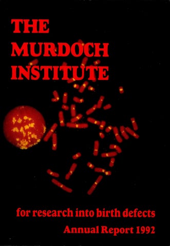
COVER Non-dividing cell (on left) and chromosomes spread from a dividing cell, stained for a-satellite and satellite 3 DNA which “lights up ” the centromeres in all chromosomes except one. This one chromosome is the abnormal chromosome 10 discussed on page 27.
I
The Murdoch Institute for Research into Birth Defects
The Murdoch Institute is devoted to advanc ing knowledge about genetic diseases and other types of birth defects so that prevention may become possible in the future. The basic research of the Institute finds practical expressions through its clinical arm, the Victorian Clinical Genetics Service, which provides a network of services throughout Victoria available to persons of all ages. The close integration of research and clinical service has provided a rich source of research ideas and prompt implementation of new knowledge.
The Murdoch Institute for Research into Birth Defects Limited Royal Children’s Hospital Flemington Road PARKVILLE VICTORIA 3052 ACN 006 566 972 Postal address: Post Office Box 1100 PARKVILLE VIC 3052 Telephone: (03)345 5045 Facsimile: (03) 3481391
During its short history of seven years the Murdoch Institute has made important scientific discoveries, achieved recognition as one of Australia’s top medical research centres, the premier training centre of clinical geneticists, and established a network of clinical services which is serving as a model to other countries and states. The generous founding gifts of the Murdoch family, the late Sir Jack Brockholf and other supporters set the Institute on a sound footing. Many of these generous friends still continue their support, but the Institute desperately needs further supporters to complete the final stage of its establishment — alteration of space purchased at the Royal Children’s Hospital to expand its laboratories and clinical facilities to the size required.
I I
I
>3
ms
a-
Birth defects kill or maim 1 in 50 babies. The strain on families is enormous. The cost to the Australian community exceeds $2,000,000,000 a year. Research into the genetic causes of birth defects is starting to reduce these figures.
Would you please give generously now. All donations are tax deductible. If you cannot give now, consider helping The Murdoch Institute through a bequest.
'i'
I
The Murdoch Institute needs your support every year to maintain the momentum of research into these genetic conditions.
The following is a suggested form for a bequest to The Murdoch Institute. I bequeath to The Murdoch Institute for Research into Birth Defects the sum of $ . . . (or part or all of residue of estate) to be applied for the purposes of the Institute.
The Institute needs special support in 1993 to complete its process of establishment by developing laboratories and other essential facilities, at a cost of over $5 million.
For further information contact the Business Manager,
mt . 4
K
‘^1
U
I I
The Murdoch Institute for Research into Birth Defects Royal Children’s Hospital Flemington Road, Parkville, Victoria 3052 AUSTRALIA Telephone: (03) 345 5045 Fax: (03) 3481391
L
s
m
aIf
'5l
MAJOR DONORS TO THE MURDOCH INSTITUTE
MAJOR DONORS TO THE MURDOCH INSTITUTE
FOUNDERS — Donors of $1 million or more
TRUSTEES — Donors of $25,000 or more
The Murdoch Family: Dame Elisabeth Murdoch Mr. Rupert Murdoch Mrs. Helen Handbury Mrs. Anne Kantor Mrs. Janet Calvert-Jones The late Sir Jack Brockhoff The Brockholf Foundation The Scobie and Claire Mackinnon Trust
Arthur Andersen Foundation Coles Myer Limited H. & L. Hecht Trust J.B. Were & Son Charitable Foundation Mrs. Joan Roxburgh Mrs. M.L. Griffin National Australia Bank Limited Qantas Repco Corporation Limited (Ariadne) The Banks Trust The Friends of the Murdoch Institute The Ian Potter Foundation The late Mr. Clive Roxburgh The late Mrs. L.B. Quayle The Morris Family Trust The News Corporation Limited The Percy Baxter Charitable Trust The Sidney Myer Fund
BENEFACTORS — Donors of $250,000 or more The Miller Foundation The Helen M. Schutt Trust I
■/«
CORPORATE SPONSOR GROUP — Corporations undertaking substantial future support 'iS
m
National Australia Bank Limited Qantas
■i!
m
DONATIONS TO THE MURDOCH INSTITUTE 1992 General The Jack Brockhoff Foundation Cruden Investments The Scobie & Claire Mackinnon Trust The Miller Foundation Mrs. J. Roxburgh Friends of the Murdoch Institute L.E.W. Charitable Fund H.& L. Hecht Trust National Australia Bank The Morris Family Trust Estate of the late D. Scott J.B. Were & Son Charitable Fund NEWS Limited Uncle Bob’s Club County Nat West Professor D.M. Danks Mr. Grant Stephenson Anon Dr. J.M. Gooch The Kimberley Foundation Mayne Nickless Ltd. Tambo Country Deli & Cheeses The William Angliss (Victoria) Charitable Fund McMullin Nominees Mr. F.D. Ryan Mr. & Mrs. S.F. Gooley Mr. G.E. Heeley Mr. N.R. Clark The Certus Club Mr. & Mrs. T.C. Crawford Little Peoples Association Mr. & Mrs. K. J. Redman Mr. & Mrs. L. Barbieri
$
■t
325,000.00 250,000.00 100,000.00 50,000.00 40,000.00 16,000.00 12,500.00 10,000.00 10,000.00 10,000.00 5,000.00 5,000.00 5,000.00 4,000.00 1,500.00 1,357.32 1,300.00 1,000.00 1,000.00 1,000.00 1,000.00 1,000.00 1,000.00 800.00 600.00 600.00 500.00 500.00 500.00 401.10 400.00 200.00 150.00
*
i ■5 A' .:.v-
-i
3 ife
$
Mr. & Mrs. M.J. Martin Mr. & Mrs. B. Ritchie Dame Patrician Mackinnon E.P. Gilligan Heidi Dietzsch Karen & Neil Greening L. Mesee Lady Dorothea Darvall Mr. & Mrs. Elliott Mr. & Mrs. K.C. Troon P.G. & J.E. Mayes R. & J. Griffiths S.J. & J.E. Broom T. J. & F.M. McMahon Therese Bryant Thomas B. Cottrell Pty. Ltd. Robyna Calisthenic College KMart Dominic lemma Friends of Michael Austin O’Donnell G.D. & N.E. Bush I.J. & S.J. McKenzie J. Bramwell Lt. Col. W.H.A. Becke M. Hargreaves Mr. & Mrs. L.R. Mills Miss J. Gardner Mr. & Mrs. Saunders Mr. & Mrs. Symons Mr. & Mrs. Weatherly Mr. Timothy Thompson Mrs. Melanie Kent Mrs. Michelle Collins Mrs. Tania Howell Ms. Katheryn Symons N.P. Derwent & M.J. O’Donnell P.A. & J.M. Huskins
P. J. & T.M. Neil
3
105.00 100.00 100.00 100.00 100.00 100.00 100.00 100.00 100.00 100.00 100.00 100.00 100.00 100.00 100.00 100.00 80.00 70.00 50.00 50.00 50.00 50.00 50.00 50.00 50.00 50.00 50.00 50.00 50.00 50.00 50.00 50.00 50.00 50.00 50.00 50.00 50.00 50.00
INDEX
$
P.S. & A.M. Saunders Paul & Liz McGuire Peter Saunders R.M. Valentine & Associates Roma Dawson Royal North Shore Hospital Susan Saunders T. J. & S.L. O’Donnell M.A. Flaherty M.B. & C.E. Lithgow Mrs. Y. Dunt O. Greenwood P.D. & B. Edwards Paul & Veronica Cracknell Stephen & Michelle Pearse A.C.&V.A. Walsh J.P. & S.G. Campbell — Plumbing M.J. & D.J. Watkins Mrs. M.J. Dwyer P.J. Huskins Betty D. Sommerville M. O’Donnell The Maiolo Family Building Appeal Donations Health Department Victoria National Australia Bank The Danks Trust Commonwealth Bank Pacific Dunlop The Sir Donald & Lady Trescowthick Fund Colonial Mutual Mr. & Mrs. R.J. Walker Metal Manufacturers The Institute thanks all those listed above for their generous support.
50.00 50.00 50.00 50.00 50.00 50.00 50.00 50.00 40.00 40.00 40.00 30.00 30.00 30.00 30.00 25.00 25.00 25.00 25.00 25.00 20.00
10.00 10.00 50,000.00 35,000.00 20,000.00 5,000.00 5,000.00 5,000.00 1,000.00 1,000.00 1,000.00
s
';y.V
1
I
Chairman’s Report.............................................. 1 Committees of the Murdoch Institute ............ 2 Director’s Report................................................ 3 Identification of the gene which is affected in Menkes disease................................................ 8 Targeted mutations in mice ............................. 11 Research in Progress ......................................... 14 Victorian Clinical Genetics Service............... 22 Olive Miller Protein Laboratory ..................... 24 Studies of Pyruvate Dehydrogenase............... 25 Human Centromere and Down Syndrome..... 27 The Scohie and Claire Mackinnon Trace Element Group .. 30 Embryology Group ...................................................... 33 Mitochondrial Respiratory Chain Disease............. 35 Epidemiology ................................................................ 36 Clinical Projects .......................................................... 37 Tissue Culture Laboratory......................................... 37 List of Publications —1992 ........................................ 38 Murdoch Institute Lecture Series —1992 ............... 42 Staff Involvement in Australian and International Scientific Community Activities........................... 42 Editorial Boards ........................................... ............... 43 Overseas and Australian Lectures and Seminars by Institute Staff...................................................... 44 Collaborations .............................................................. 45 International Visitors ................................................. 46 Staff List — Murdoch Institute................................. 47 Staff List — Victorian Clinical Genetics Services . 48
Chairman’s Report
I
Mr Neil Walford
n many fields of research one must accept that there will be long periods of endeavour and only the occasional exciting discovery. When success comes it is very important not just for the new knowledge it reveals but also because it pro vides the vital reward and stimulus that helps the scientist to keep going ahead. As you may read in Professor Banks’ detailed and fascinating Director’s Report for 1992, that year was marked by several important scientific achievements and other events all of which serve to underline the fact that the Murdoch Institute is a significant player on the world scene in its chosen field. I should like to offer congratulations from me and the Board to Julian Mercer and Andy Choo on their research findings and to Dick Cotton on his Pharmacia LKB Biotechnology Medal. In my report last year I referred to the need to raise a substantial sum to refurbish and equip the 10th floor of the Children’s Hospital which we had only recently acquired. We need several millions of dollars. Our Appeal Chairman Mr. Nobby Clark and the Committee members, ably assisted by Dr. Max Robinson have worked hard in a difficult economic climate. This year we shall be applying ourselves to the task again because we have a desperate need to get ahead with at least part of the planned alterations. As our Director Professor Banks has stated in his own report, he is to retire about the middle of 1995. A great deal of time and thought have gone into planning the search for a suitable successor. For one thing, we shall have to look world wide as there are few who have the special skills and back ground required. Such a person would normally have to give long notice to his present employer. Applications will close on 1st July 1993. At the same time we are planning a scientific meeting to honour Professor Banks on his retire ment. I am delighted to tell you that several of the biggest names in the world in genetics and medicine have accepted my invitation to attend. This is a great compliment to David Banks. And now there are those whom I should like to thank. I should like to place on record our special and happy relationships with both the Royal Chil dren’s Hospital and the University of Melbourne. To my colleagues on the Board my very warm thanks. I should like to make special mention to Mr. Laurie Cox, Deputy Chairman and Chairman of the Finance Committee, Mr. Nobby Clark, Chair man of the Appeal Committee, Mrs. Ann McFarling. Chairman of Friends of the Murdoch Institute, and Mrs. Anne Cronin, Business Manager. And finally my profound thanks to our donors past and present.
1 ■w
si If
ISI ;Si
K IS‘
The Murdoch Institute
tl
Scientific Director: Professor D.M. Banks
I
Deputy Scientific Director: Dr. R.G.H. Cotton
Director’s Report
sI r %
Si
II
'S' iI BOARD OF DIRECTORS Mr. N. Walford, Chairman
BUILDING AND DEVELOPMENT APPEAL COMMITTEE
Mr. L.G. Cox, Vice Chairman
Mr. N. Clark, Chairman Sir Gordon Allard
■'i’l
Professor J. Angus Dr. G.L. Barnes Mrs. J. Calvert-Jones
Mr. L.G. Cox
Dr. R.G.H. Cotton Professor D.M. Banks
Mrs. A. Cronin Professor D.M. Banks
Mr. I. Davies
Mr. J. Fitzgerald
Mr. J.A. Fitzgerald
Mr. P. Griffin
Mr. J.S. Guest Mr. W.H. Hodgson
Mr. M. Handbury
Mrs. A. McFarling Professor P.D. Phelan Professor A. J. Pittard Professor G.B. Ryan
Mr. D. Craig
Mr. G.E. Heeley Mr. N. Miller Mr. D.E. Meikeljohn Dr. M. Robinson Mr.N.Walford
& ■s;
'S I; II t s.f iI •s-
I I
1, mI
i
'■I'
MS
MI :I
Mrs. C. Searby
I i; II ■li Si
FINANCE COMMITTEE Mr. L.G. Cox
VICTORIAN CLINICAL GENETICS
Mr. C.P. Abbott
SERVICES . BOARD OF DIRECTORS
Mr. D.T. Craig
Mr. N. Walford, Chairman .
Mr. P.J. Griffin
Dr. G.L. Barnes Mr. L.G. Cox
Mr. G.E. Heeley Mr. D.E. Meikeljohn Mr. F.D. Ryan
Mr. J.S. Guest , Mr. G.E. Heeley
m
s
5-
m
M
m
If
m
I
111
11S' f; t
\ ■I: I't
i'l
V §
fI
If
IE:
*
1
II f f I "I I
A’
$
I' II h J"
g:
Iisi' 2
Pi
ii:
Professor D.M. Banks Dr. J. De Campo
m5*a
i j
Si cS'i '-I?
I
Professor David Banks
^ I his year two scientific achievements.stand I out as the special events of the year along -1- with a further recognition of Dick Cotton’s record in research. On the other side of the ledger we have had a frustrating year in our endeavours to raise the money needed for building alterations. Julian Mercer’s major role in the isolation of the gene at fault in Menkes disease provided a very exciting finish to the year. Results obtained in November were published in Nature Genetics in January 1993, an indication of the worldwide interest in this finding. Three composite research groups, each involving scientists from two different institutions, reached the same conclusions within a few weeks and it was fitting that Nature Genetics agreed to publish all three papers in the same issue. It would have been unfair for any of the groups to have been given precedence by such a small margin after more than three years of intensive research. For us it was the culmination of 21 years of research starting when we showed that the symp toms of Menkes disease could be explained by deficiency of copper at the crucial intracellular sites where it is essential. Our involvement in the actual race to isolate the gene started only late in 1991 when Julian Mercer was invited to lead a group of scientists at the University of Michigan who had been engaged in this task for over two years. During a seven month sabbatical in Ann Arbor and a frantic few months after he returned, Julian led the laboratory work on this project through the final stages to a successful conclusion. This work is described in more detail in one of the major presentations in this report and Julian will be speaking about his work at our Annual General Meeting. As well as acknowledging Julian’s special contribution I want to commend the special efforts of Andrew Grimes, Jenny Paynter, Paul Lockhart and Mrinal Bhave through the frantic weeks of work before and since Julian’s return. Andrew and Mrinal collaborated to lead the work of the group in Julian’s absence and Dr Peter Dorling, on sabbatical leave from Perth, provided some useful guidance. " ; Credit for the second exciting scientific achieve ment of the year goes to Anna Michalska and Andy Choo. For many years most scientists interested in trace metals in humans and animals have believed that a protein called metallothionein plays an im portant role in maintaining a supply of zinc within cells and protecting cells against the toxic effects of copper, but nb-one has ever managed to prove either of these beliefs. This unusual small protein was first identified through an apparent role in pre venting toxic effects of cadmium over 30 years ago. Many other roles have been proposed since. 3
We have always felt that discovery of a patient or a laboratory animal unable to make metallothionein would provide a great new way of learning more about its function, but did not hold out much hope of finding such a patient or animal, because all species have at least two pairs of metallothionein genes and a mutation which interfered with both genes would be unlikely to occur. Then a tech nique of deliberately disrupting the function of a gene in a fertilised mouse egg cell was discovered and Andy Choo thought it would be possible to adapt this technique to knock out both the metallothionein genes present in mice, since these genes lie side by side on the same chromosome. We knew that the technique of gene disruption by homologous recombination in transgenic mice (to give its full title) was technically demanding, but we did not quite realise how difficult it would prove. In fact, it took five years of Anna Michalska’s efforts to achieve the final result of producing mice with both copies of both metallothionein genes disrupted. To our surprise these mice develop, grow and re produce quite normally and susceptibility to the toxic effects of cadmium is the only abnormality that we have so far identified. We anticipate that we will find some more subtle deficiency effects in mice on a relatively low intake of zinc or toxic effects on a higher than usual intake of copper for a prolonged period. An increased rate of ageing or an increased frequency of cancers in older animals are other possible effects. Much of this further work will be carried out in the trace element group while Anna turns to other genes of interest within the Institute. This year Dick Cotton was awarded the Pharmacia LKB Biotechnology Medal by the Australian Society of Biochemistry and Molecular Biology. This medal is awarded to “an Australian biochemist or molec ular biologist for distinguished contributions to the field of Biochemistry and Molecular Biology”. The recipient of the medal is required to lecture on his particular area of research in major centres in Australia and New Zealand. During the year the Howard Hughes Institutes of USA offered a number of Senior Scientist Awards to Australian medical researchers, ostensibly to those who have recently established their independence. Andy Choo was selected into a short list, but unfortunately, was not finally chosen. We were surprised to hear that most of the awards went to long established senior scientists, but were
proud that Andy had come close in competition with scientists 10 years older. The difficulties experienced in raising enough money to alter the Tenth Floor to suit our require ments are not surprising in the current financial situation in Victoria. However, it is frustrating to work in crowded laboratories, or share a small office which was built as a Resident Medical Officer’s bedroom when we have large empty areas awaiting alteration to make them useable. Mr Nobby Clark and his colleagues on our Fund^ raising Committee, ably assisted by Dr Max Robin son, have worked very hard indeed and we are most grateful for their efforts. It is just the wrong time to raise the large amount of money that we need. A number of factors interacted together to make 1992 a difficult year in which to balance our income and expenditure. The fall in interest rates and the financial recession reduced the income on our invested funds despite the outstanding performance of our investment managers. A long overdue increase in academic and scientific salaries finally arrived in 1991 and 1992, so we did not feel the benefit of the reduced rate of inflation. Unfavourable exchange rates raised the cost of imported laboratory consum ables and the spread of molecular genetic techniques through all our laboratories increased the expendi ture necessary on consumables. These techniques bring great advantages to our research, but the reagents are much more expensive than those used in conventional biochemistry. Fortunately, a series of cost control measures were successful and we did balance income and expenditure. I want to thank Mr Keith Ince and his associates at County Australia, our Investment Managers, for achieving a return on investment which outstripped all similar funds in the calendar year 1992, and Mr Laurie Cox and his colleagues on our Finance and Investment Committee for their wise selection and guidance of the Fund Managers. Nonetheless we did have to cope with a lower contribution to our cash flow from this source in 1992. During these times of financial pressure we are especially grateful to our longstanding supporters, the Murdoch Family, the Brockhoff Foundation, the Scobie and Claire Mackinnon Trust, and the Miller Foundation who continue to fulfil past promises of support to the Institute with annual donations. Without these donations we would be in great difficulties in the present circumstances. I am particularly pleased that the Directors of the
"H'
f
I ! 3
t 3
I I
I
■I
.1
Mr. N.R. (Nobby) Clark Chairman, Appeal Committee 4
Dr. Max Robinson
Brockhoff Foundation have decided to take a special interest in Dr Andy Choo’s research on the centromeres of human chromosomes. We also have a further reason to thank a very special friend of the Institute, Mrs Joan Roxburgh, for the wonderful support that she has given our Trace Element Research Group during the past eighteen months. Mrs Roxburgh and her late husband, Clive, were associated with the Uncle Bob’s Club from its very beginning and developed a strong interest in our work at the time of our establishment. They provided the finance necessary to computerise our clinical records system and interest from a Fund that they established at that time continues to support the ongoing entry of records into the system. Since Mr Roxburgh’s death, Mrs Roxburgh has continued her interest in our work. Twice in the last two years she has contacted me to offer further support. On the first occasion a visit to the Institute identified an interest in the work that our Trace Element Group was doing on toxic milk mice as a model of Wilson’s disease. The second occasion coincided with the surge of work in isolating the Menkes gene and an injection of additional funds was particularly valuable to us at this time. The Friends of the Murdoch Institute have also continued their valuable work for the Institute, making us better known in the community in Melbourne and raising funds for our work. A very successful function during the race season raised a substantial sum and attracted the interest of a pleasing number of people. We are very grateful to Mrs Ann McFarling and her colleagues. A need for careful control of expenditure proved a valuable factor in improving a number of systems within the Institute for which Mrs Anne Cronin, our Business Manager, and Mr Barry Holt, our Laboratory Manager, deserve special credit. Much credit also to the leaders of the various research and diagnostic laboratories who have cooperated so well with the systems that have been introduced. Principal responsibility for staff management and budgetary control has been devolved to the leaders of research groups and heads of diagnostic laboratories, and more effective interactions with and between these people have been established. We now have two levels of executive committees to advise me. Executive 1 comprises Mrs Cronin and the seven heads of the major research groups. Executive 2 adds the heads of diagnostic laboratories, the Laboratory Manager and his deputy, and senior clinicians. Executive 1 meets three times a month and Executive 2 once a month. They are each proving valuable for scientific and administrative discussions. Laboratory heads receive accounts of each month’s expenditure soon after the month’s end and all scientists are kept informed about the costs of individual reagents they use, especially the more expensive ones. I want to thank all the senior personnel of the Institute for the great cooperation they have shown in 1992.1 am also grateful to the whole of the staff for the way they have joined in the drive to contain our costs of operation. I believe that this cooperation has also achieved greater collabora tion and interaction between research groups.
Discussing these matters brings to mind the very useful role that the Social Club that was set up in 1991 is playing in the affairs of the Institute. Those who have been leading this activity deserve com mendation for the good judgement they have shown in the number and style of social functions that they have organised. They have helped staff members to know one another better and enjoy working together more without interfering with the primary focus of getting on with the research. People and their personalities are critical to the success of a small research institute like ours. Dr Jack Insley and his wife Anne were two people who made a particular contribution to the Institute. Jack came for 12 months to succeed Ron Davidson as a visiting senior clinical geneticist and for tunately stayed 18 months. Anne took over a parttime role in fundraising and in public relation activities, both external and internal, from Ron Davidson’s wife, Miriam. This role included the production of our alternate weekly internal news letter “Murdocuments” with Margaret Olsen. Jack proved to be a very able and wise clinical geneticist. He played an important role in our clinics, and especially in further developing the country clinics which Ron Davidson had started. Anne helped considerably in our fundraising activities, especially in organising our records. These formal roles were very valuable, but almost more valuable were the informal role they played as what I would like to call a wise “uncle and aunt” in our “family”. Following Jack Insley’s departure we have need for an additional experienced clinical geneticist in 1993 and thereafter. Very satisfactory arrange ments have been made, which are described in the Clinical Services Report. One component of these arrangements is a decision by Les Sheffield to change the balance of his appointment from principally research with some clinical activity to principally clinical with some research activity. This will inevitably affect our future research activities in epidemiology. Agnes Bankier has assumed responsibility for the overall organisation of our clinics in 1993, but we do not expect this to reduce her ability to direct the work on POSSUM and OSSUM. She has stronger support in this work now that Dr Cathy Rose is able to spend more time with us each week. Anne Insley’s role would have been difficult to fill had it not been for some good fortune in recruiting
Dr. Jack Insley 5
!■
Mrs Lee Jackson as my secretary during 1992. Lee has quickly adapted to life in the Institute and brought excellent qualities to the role of Director’s Secretary, encouraging and organising the secre tarial activities within the Institute and interact ing effectively with all Institute staff. She is able to give Max Robinson the help he needs with fund raising and, with Pauline McGrath and Margaret Olsen, she is maintaining the production of Murdocuments as a valuable medium of communication within the Institute. Our association with Ron Davidson continues and he has assisted in demonstrating OSSUM at interna tional meetings in North America and Europe. OSSUM had a successful first year of distribu tion. It was released on time in April 1992 and sales for the year to December 31 were almost exactly in line with budget 47 units in total. There were some substantial deviations from our expectations in the detail of the sales. The number of POSSUM owners who purchased OSSUM exceeded our ex pectations and we made less impact on paediatric radiologists than we had hoped. Sales were better in USA than expected and we sold no systems at all in Japan. Agnes Bankier and Max Robinson are ad justing their marketing approaches to deal with these deficiencies in the coming year. We are grateful for the advice given by colleagues at AMRAD (Australian Medical Research & Deve lopment Corporation) when we were developing marketing plans for OSSUM and at other times when we have needed to consider patent protection for research findings of potential commercial value. We are very pleased to be a Member Institute of this young and energetic biotechnology development company. Further development of the OSSUM system has been carried out during 1992 including a careful review of all syndrome descriptions and illustrations by Dr Peter Turnpenny, a young English clinical geneticist with a special interest in bone dysplasias who spent three months in Melbourne to carry out this review. We are delighted to have recruited the collaboration of Professor Pierre Maroteaux of Paris, one of the most eminent authorities on bone dysplasias, to assist in the further development of this system and we are also grateful to Professor Andres Giedion of Zurich who is providing a critical review of the system. We aim to produce Version 2 of OSSUM at the end of 1993 or early in 1994 with a much better standard of illustration by
Dr. Renee Martin 6
Dr. Peter Dorling
X-rays. There were some disappointments about the technical quality of some of the X-rays that we had to use in Version 1. POSSUM continues to sell steadily with a total number of systems in use around the world reach ing 324 by the end of 1992. The fame of Agnes Bankier and John Marquet (Computer Power) in developing such systems has also spread and they have been consulted by several different groups who wish to develop computer/video disc diag nostic systems in other areas of medicine. The most detailed consultations was with a group in Ottawa wishing to develop a system to assist obstetric ultrasonologists to recognise fetal malformations at about 18-20 weeks gestation. The obstetrician concerned and the head of the computer service from his hospital spent a week in Melbourne work ing with Agnes and John and they are going to get ahead with a pilot study to determine the feas ibility of their project. Our involvement will be as consultants. • In mid year we were sorry to farewell Dr Renee Martin who had spent six months sabbatical leave in Andy Choo’s laboratory. She is one of the world’s experts on chromosome analysis in human sperm and many other aspects of sperm function and we all gained from her knowledge and infec tious enthusiasm. Dr Peter Dorling, from the School of Veterinary Science of the Murdoch University in Perth spent a similar period of sabbatical leave in the Trace Element laboratory. He has a fund of knowledge about the toxicology of chemicals found in native Australian plants and insects and their effects on introduced species like sheep. Some of these toxins interact with copper when this element accumulates in the liver of sheep. The overlap with our interest in copper accumulation in hmnans, sheep and mice provided a basis for useful collaboration. It was good to have Peter’s experience, and cheerful per sonality in the laboratory during Julian’s absence. We were sad to see Peter and Alison leave in June. Later in the year we were pleased to welcome Dr Eli-Anne Kvittigen from Oslo to spend a three month sabbatical working with Henrik Dahl in the group studying mitochondrial DNA. Like Renee Martin, Eli-Anne was particularly anxious to gain hands-on experience with DNA techniques. She is expert in both the clinical and laboratory aspects of inborn errors of metabolism and came to Mel bourne because we could offer exposure to work in that field and in molecular genetics. Late in the year Dr Eileen Treacy joined us, for a year or more, coming from Professor Charles Scriver’s biochemical genetics group in Montreal to which she will return to take up a career in the management of inborn errors of metabolism and related research. She has received a prestigious Canadian Travelling Fellowship to allow her to obtain additional clinical experience and under take relevant research. We are fortunate to have four other very bright young trainee clinical geneticists from overseas doing research projects in the Institute in 1993, each supported by external grants. More will be said about their activities in next year’s report, but we are already feeling a benefit from their presence.
During 19921 have been busy with some activities outside the Institute which form part of the general responsibility to the development of genetics in Australasia. I have been Chairman of a National Health & Medical Research Council Working Party on the prevention of birth defects and I spent two weeks in Auckland as the David Nathan Visiting Professor advising on the development of genetics services in New Zealand. When visiting another country in this way one has easier access to senior government officials than in one’s own country. Nonetheless I hope that our Working Party may be able to persuade the Australian Government to see the development of a really effective network of genetic services throughout Australia as a high priority. Our report will demonstrate that it is now possible to reduce the number of babies born each year in Australia with severe life threatening or chronically disabling birth defects by about 15% at a very affordable cost. Dick Cotton is also quite heavily involved in general public activities in relation to science, through NH&MRC Committees and as Editor of the journal Human Mutation, which has just com pleted a successful first year of publication. Henrik Dahl and Jim Camakaris also served on NH&MRC Grants Committees in 1992, and all senior staff are involved in reviewing applications for research grants for the NH&MRC and other research fund ing bodies. While discussing my own activities it is probably time to point out that I will be retiring in mid 1995, a time chosen so that my successor will have at least twelve months in the position before the next five yearly review by the NH&MRC. An Appoint ments Committee has been established by the Institute and the University of Melbourne and advertisements will be placed early in 1993. I have mentioned already the excellent support I have received from senior scientists and laboratory heads during 1992 and the special roles that Anne Cronin and Barry Holt have played in financial management. I want also to acknowledge the many other ways in which Anne and Barry help me and
to give special thanks to Ivan Francis, the Head of the Newborn Screening Laboratory. We are most fortunate that, in addition to running a very efficient laboratory and providing additional general help in laboratory management matters, Ivan has great expertise with computers and has acted as our computer systems expert since 1988. He has worked particularly hard over the last six months to network all our computers, both IBM PC’s and Macintosh. Nothing ever seems too much trouble for Ivan. I also want to thank our Computer Committee (Les Sheffield, Julian Mercer, Anne Cronin and Barry Holt) for working with Ivan to ensure the best use of computer facilities. My day-to-day work is made much easier by Lee Jackson, my very capable, tolerant and cheerful secretary. Beyond the day-to-day interactions within the Institute I am particularly fortunate to have wonder ful support from Mr Neil Walford, the Chairman of our Board, and from other Board Members, from Mr Laurie Cox, Deputy Chairman of the Board and Chairman of the Finance and Investment Committee and members of this Committee, and from Mr Nobby Clark, Chairman of the Building Appeal Committee, and the other members of that Committee. Neil and Laurie are always available to give wise advice and the frequent helpful telephone messages and cuttings from newspapers which they send to me indicate that our Institute is always in their minds. They really do provide me with the most wonderful support and I look forward with great confidence to the special role that they will play during the difficult phase of transition to my suc cessor. This will be a period in which the Institute will need very strong guidance from the Board to ensure that sight is not lost of its real objectives and need for independence. I never cease to be amazed by Nobby Clark’s energy and efficiency. We are most grateful for the time and effort that he has put into approaching potential donors on our behalf despite a very taxing array of responsibilities in the corporate sector.
Human Mutation Editorial ^ I he Murdoch Institute is now also the home I of the Human Mutation Editorial Office. -1- Human Mutation is the new research journal for which the Deputy Director, Dick Cotton, is the Editor along with Haig. H. Kazazian Jr. at the Johns Hopkins University in Baltimore. Haig is well know for his expertise in haemoglobin disorders. The Editorial Office is funded by the publisher and staffed by Editorial Assistant Julie Cross who performs a role of communication between the 64 international communicating editors, the authors and the publishers. Some occasional whip-cracking is necessary to fulfil the aim of rapid publication and to keep to schedule.
The average time from acceptance to publication of manuscripts in the Journal has been 15 weeks. In the twelve months since its inception, 130 manu scripts had been submitted to the Journal altogether, 90 of which have already been accepted for publica tion. Manuscripts have been received from nineteen countries. The first issue of the 6-issue first volume was published in June 1992 and the first issue of Volume 2 should appear on the bookshelves early in 1993. The Journal is published by Wiley-Liss, a division of John Wiley and Sons, Inc. of New York.
7
Identification of the gene which is affected in Menkes disease
M
enkes disease is a fatal genetic disease. The gene which is affected is on the Xchromosome, so like colour blindness and haemophilia (bleeding disorder), almost every patient is a boy. Patients with the disease have a wide range of defects, including mental retardation and neurological problems, connective tissue and blood vessel defects, and characteristic brittle hair. Although the disease was first recognized by John Menkes (hence the name), the reason for the disease was not recognized until David Banks noted the resemblance between the steely wool of copper deficient sheep and other features of copper deficient animals and patients with Menkes disease. He realized that this disease is a genetic disorder which results in a profound copper deficiency. Since this realization, about 20 years ago, many groups interested in the disease and in the biochemistry of copper have been working hard on trying to find the gene and the protein it codes for. It has been a long twenty years, with much hard work, but finally this year we and two other groups have managed to achieve this goal. It is an important discovery, not only for families affected by Menkes disease, but for the whole field of copper biochemistry. How was the gene isolated and why did it take until now? The success was based on two factors: the first is the rapid increase in new technologies of gene manipulation and the second is the occurrence of a unique female patient with Menkes disease. The female Menkes patient was first described in Michigan in US, and her disease is caused by a break in one of her X-chromosome, which infact disrupts the Menkes gene. Thus, the break or trans location could be used as a marker for the position of the Menkes gene. Previous localizations had only narrowed it down to a region which might contain hundreds of genes and searching for the
Dr. Julian Mercer 8
right gene would have been too slow and may not have succeeded at all. The three research groups who isolated the gene all made use of cell lines from this one patient, and how her cells travelled around the world to the different labs is quite an interesting story in its own right. We had tried to obtain the cell lines to attempt to find the gene ourselves, but, they were never sent to us. The cells were, however, passed on to Tom Glover at the University of Michigan, Ann Arbor. His lab did some important preliminary work and showed that the break in the chromosome was close to another gene which could be used as a marker. Soon after this, Tom Glover invited Julian Mercer to join him on a sabbatical to help with the cloning of the gene. We did not know then that three other teams were busily engaged upon the cloning. Two labs in San Francisco combined forces, one lab in Denmark joined with one in Oxford, and one in Italy combined with one in France. During Julian’s time in Ann Arbor, progress was rapid, but we began to hear disturbing (and sometimes misleading reports about the progress of the other groups). Each group took a slightly different approach, and three includ ing us, managed to succeed. One team isolated the wrong gene, which demonstrates the dangers in this type of activity: they worked just as hard but missed out on the ultimate prize. All groups made use of the new technology of so called YAC (yeast artificial chromosome) clone libraries. These are very large pieces of human DNA which can be grown in yeast, because they are attached to a yeast artificial chromosome. We isolated some of these clones using the marker gene that Tom showed to be close to the break in the female Menkes patient. An outline of the process is shown in Fig 1. Then using another new technique, known as FISH, in which the clones DNA can be labelled with a fluorescent marker and bound to chromosomes from the patient, we showed that one of the YAC clones included the broken region of the patients X-chromosome (Figs 2 and 3). This told us that the Menkes gene would probably be included in this YAC clone, since we thought that the break was actually in the gene (Phase 1, Fig 1). The next part (Phase 2 in Fig 1) of the process involved breaking up the large YAC clone into small pieces and finding which of these came from the break region, again using the FISH technique. We found such a piece and then used this to search through many other gene libraries, to find the protein coding sequences (so called cDNA clones) that came from this region. Eventually we found some cDNA clones and these turned out to be part of the Menkes gene.
We have used these to examine some of the cell lines from our Menkes patients and in many of them the gene does not seem to work at all. In some patients, there is probably protein produced but it is likely to be defective. We hope to find out the exact reason for the defect, and this may help us design more rapid prenatal diagnosis of Menkes disease. The gene structure shows that it produces a very interesting protein, and this information is the start of a major increase in our understanding how the body handles copper. The protein appears to be a molecular pump that moves copper from one part of the cell to the other (Fig 4). It looks very much like some proteins found in bacteria, which pump toxic metals out of the cell. In Menkes disease, we think that the pump does not work correctly, or is missing, so the copper cannot be properly absorbed by the gut, and this results in the fatal copper deficiency.
There is much interesting detailed work to he done trying to understand exactly how this protein works. In addition, we think that there may be a number of related proteins in the body, which perhaps move copper in different ways, or move other metals. Some other genetic diseases may be due to defects in these other metal carriers. One possibility is Wilsons disease in which copper transport in the liver is faulty. It seems likely that the discovery of the Menkes gene will be the start of a lot more studies which should help us understand copper biochemistry but also a number of other diseases and possibly the transport of other metals. Ultimately we hope to be able to answer important practical questions like — “Is copper deficiency an important factor in common diseases like osteo porosis (bone softening) in older people and coronary artery disease?”. We think the answer will be “yes” and that our research will contribute to preventing these conditions.
Phase 2
1
t
'i -P iK
-PGK
This crossed break
lambda which crosses breakpoint
Yac subcloned and contig established
Fig 1. Strategy for the isolation of the Menkes gene. YAC clones containing the marker gene (PGK) were isolated. One crossing the breakpoint was found using the FISH technique (phase 1). This was broken into small pieces and these were used to find the piece that crossed the break (phase 2). This piece was used to isolate the coding sequences of the gene.
Figure 1
X
4 #1
X
derX ✓
X
\
der2 Figure 2a
Figure 2b
Fig 2. Chromosomes from a normal girl (a) and from the patient with the translocation (b) labelled using the FISH technique. The fluorescent signal from the YAC clone appears orange, and the X-chromosomes are detected with another fluorescent marker which gives a green signal. In the patient’s cells three orange signals were detected showing that the YAC crossed the breakpoint. Three signals are detected because the patient’s cells contain three chromosomes which will bind to the probe: the normal X, the derivative X and the derivative 3 (see Fig 3). 9
Targeted mutations in mice derX
M
Figure 3
Figure 4
Fig 3. Showing the chromosomes involved in the patient’s cells. The cells contain normal X, and a normal 2. The chromosome breaks (indicated by the black lines) which led to the Menkes disease formed a derivative X (der X), which contains part of the chromosome 2, and a derivative 2 (der 2) which contains part of the X. When a YAC clone which crosses the break was labelled with the orange fluorescent dye and bound to the chromosomes, it detected the normal X, the der X and the der 2 (actualpicture is shown in Fig 2).
Fig 4. Model of the Menkes protein — a copper pump. The DNA sequence of the gene would translate to form a protein like the one shown here. The copper binding regions are shown in yellow, the pink rectangle represents a cell membrane that the protein loops through. Part of the protein includes a molecular motor (ATPase) which drives the copper through the channel.
ost often the reason for studying human genetic diseases is to develop a method of early diagnosis or treatment for the patients concerned. However, the study of genetic diseases has also taught us a great deal about the normal functions of various proteins in the body. This is because the disturbance that is produced when a faulty gene interferes with production of a particular protein can give information about the role of that protein in the body which cannot be obtained by studying healthy people. Unfortunately the ability to work out a function of a protein in this way is sometimes limited because there are only certain types of experiments that can be carried out on human patients. People have tried to get around this problem by finding mice that have the same genetic fault as is encountered in a human disease. Despite the existence of extensive catalo gues of both human genetic diseases and mutations in mice very few pairs have been matched up. Another limitation of this type of research is that we can only study proteins which have been found to be at fault in a human genetic disease. There are still many proteins in the body whose functions are not fully understood. These are proteins in which we have never yet found genetic faults in human patients or in mice. During the past decade various human genes have been introduced into mice by injection of the appropriate piece of DNA into the nucleus of the fertilised egg. The resulting animals transmit the introduced gene faithfully into future generations, making a large number of animals available for research studies. These animals make the human protein, but there are many problems in using this method of research. The mice make both the mouse protein and the human protein. It is difficult to control the amount of human protein made because a variable number of copies of the gene are taken up into the chromosomes of the mouse. Because the gene is not inserted in its normal position, it may
Dr. Anna Michalska 10 1.
not come under the normal range of genetic con trols. The introduced gene may have been inserted in the middle of an important gene and this may produce an unwanted disturbance of function.
Chimeric mouse carrying the gene targeting event. Recently research has developed a new approach which avoids most of these problems. However, it is a much more difficult technique to set up in the laboratory. It is described as gene replacement or gene knockout. The intention is to produce a mouse which is unable to make the protein of interest, just as a patient with a genetic disease may be unable to form a particular protein in the body. The difference is that the scientist can choose which gene he wants to knock out. A copy of the gene that is to be studied is made and a fault is introduced into this gene copy which will destroy its function. Special techniques are used to introduce the engineered DNA into mouse embryonic stem cells which can be cultivated in the laboratory (see diagrams). In a small number of cells, the engineered DNA exchanges with the normal gene on one of the pair of chromosomes. These cultured cells can then be introduced into a developing mouse embryo which can then be im planted into the uterus of a receptive female. This produces a baby mouse which has some clusters of cells which have been derived from the cells that were manipulated in the test tube. Some of these altered cells will be in the testis or ovary and therefore some of the progeny of these mice will come from an egg cell or sperm cell which carries the disrupted gene. These second generation progeny will have the disrupted gene on one chromo some in every cell. This gene targeting leaves the genetic make up of the animal quite normal except for the knock out of the function of one copy of the gene of interest. Because this altered gene can be inherited stably through generations it is possible to breed mice 11
TIT
EARLY
REMOVE
EMBRYO
INNER CELL MASS
GROW ES CELLS IN A CULTURE DISH
INTRODUCE
SELECT
MUTATED GENES
FOR CELLS CARRYING MUTATION
MICROINJECT
REIMPLANT
12
CHIMERIC MOUSE
who have the gene of interest knocked out on both chromosomes. Dr Andy Choo first decided to set up gene targeting five years ago. He chose the protein metallothionein which is of interest to researchers in our trace element group because it is believed to be involved in the use of trace elements in the body, especially copper. Although it is an abundant protein in the human body there is no clear understanding of its function. Many different functions are claimed. We believed that a mouse that was unable to make metallothionein would allow progress to be made towards understanding how copper and zinc are transported within cells of the body. There are actually two pairs of genes, known as metallothioneins I and II (or MTI and MTII) which are so close together on the same chromosome that Andy felt he could make a single piece of DNA which would replace both genes and stop the pro duction of both proteins. This proved correct. It soon became clear that the technique was even more difficult than we had anticipated. Dr Anna Michalska finally mastered it after spending three months in the laboratory of Dr Oliver Smithies, one of the world’s experts on this technique, in the United States. At last she and Andy have now achieved their goal of producing the desired animals. They have bred many animals which have both copies of MTI and MTII knocked out and, to our surprise, they seem quite healthy. They grow normally, breed normally and their progeny are healthy. MT is thought to play an important role in embryo development and in sperm, probably as a reservoir for zinc which is required for a number of important cell processes. We are therefore rather surprised that there were no developmental problems in these animals and that adults of both sexes are normally fertile. MT was originally discovered because of its ability to bind the toxic metal cadmium and has always been proposed to play an important part in preventing toxic effects of heavy metals like cadmium, copper and mercury in the body. We therefore injected some of the MT deficient mice with cadmium and these animals became quite ill within
15 hours whereas similarly treated normal mice were not affected at all. It does seem that MT has an impor tant role in detoxification, at least of cadmium. Now that we have these transgenic mice there will be a number of experiments to be done in our trace element group and others to be arranged by collaboration with experts around the world who have special interests in the claimed functions of metallothionein. In our own laboratory we will be particularly looking at the role of metallothionein as a reservoir for zinc and its possible role in detoxifying excess copper. This will involve experiments in which we raise the mice on a diet that has a reduced content of zinc to see whether this stress brings out some zinc deficiency effects which do not occur in normal mice. We might also expect that mice with no metal lothionein will have difficulty living on a high intake of copper. The absence of severe features of zinc deficiency or copper .toxicity on normal diet is probably tell ing us that there are other proteins in body cells which can also play the same type of role as metal lothionein, but we may find that metallothionein becomes essential when the animal has to cope with a deficient supply or an excessive intake. Other claimed roles of metallothionein are in the immune system and in mopping up toxic chemicals called free radicals which are produced in cells when the oxygen that is so essential for our life interacts with cellular proteins. These free radicals are thought to play a role in the development of cancers and also in ageing. We will obviously let some of these mice grow very old and see what illnesses they develop at that stage of life. While our trace elements group is doing these sort of experiments and other collaborators around the world are also undertaking other studies Anna Michalska and Andy Choo are getting on with pro ducing mice which are unable to produce caerulo plasmin, a copper containing protein which is thought to play an important role in the transport of copper in the bloodstream. These animals will give us further opportunities to understand how the body uses copper.
MUTATED MOUSE
13
Research in Progress
I
n this section we try to provide a brief overview of the work that is currently going on in the Institute, written for the non-scientists. Our scientist colleagues may find the treatment of topics in this section rather superficial. Conversely, the short reports of research projects in the latter part of the report are intended for our scientific colleagues, and may be rather difficult for lay readers to understand. Dr Cotton’s Group — The Olive Miller Protein Research Group Postdoctoral Fellows: Phillip Dickson (to June) Enzo Palombo Jenny Saleeba (to January) Peter Smooker Rima Youil (from April) Scientific Officer:
Ian Jennings
Research Assistants: Marita Black (from May) George Makris Susan Ramus Tamara Pasque Dick Cotton, our Deputy Scientific Director, is widely acknowledged as a world’s expert in the study of the enzymes (biological catalysts) which can be at fault in patients with phenylketonuria (PKU) and for developing a method of detecting the mutations (faults) in genes which cause genetic diseases. PKU is a fairly common and treatable cause of mental retardation and one of the diseases for which our Newborn Screening Laboratory tests every baby in Victoria. Most cases are caused by defects in the gene encoding an enzyme we call PAH. The results of dietary treatment are very good provided this is started within the first couple of weeks after birth, but it is not a perfect method of treatment. We believe that it may be possible in the future to introduce the normal gene which is lacking in these patients into some cells in the body to provide a permanent treatment, or to produce a simple form of the enzyme which could be administered by injection intermittently (e.g. once a month). Ian Jennings is making progress in identifying the smallest fragment, or fragments, of the enzyme which can perform its catalytic action. A complementary approach involves identifying the places within the gene where mutations which cause PKU occur, argu ing that mutations which prevent the enzyme work ing are likely to be affecting essential parts (Susan Ramus and Jenny Saleeba). Susan has also defined the mutations in PAH in a group of patients born before treatment of PKU was available (traced by Dr David Pitt). In some patients the type of mutation correlates well with the degree of retardation, but there are some exceptions. 14
Olive Miller Protein Research Group There is a rare form of PKU in which the fault lies in a different gene which produces an enzyme which we call DHPR. Peter Smooker has been studying the mutations in this gene, collecting samples from most of the patients known around the world. Few of the mutations have been found in more than one family — a common situation in rare genetic diseases. Tamara Pasque is analysing the arrangement of coding sequences within the DHPR gene. Phillip Dickson worked in the group for just twelve months as a postdoctoral fellow and started to analyse the actual physical shape of the PAH enzyme molecule to understand exactly how it acts as a catalyst. In mid year he moved to a career position in the Department of Biochemistry at the University of Melbourne where he is continuing his collaboration with Dick Cotton. The rapid method of detecting mutations in genes which Dick Cotton developed when on sabbatical leave in 1988 has been used in numerous laboratories around the world with considerable success. Its big advantage over rival methods is that it can survey the whole of a small gene, or a large fragment of a larger gene, in one experiment whereas some of the other methods may take 10 or 20 bites to cover this much of the gene. The disadvantage is that it is a multi-step procedure and uses rather toxic chemicals and therefore must be performed in a fairly sophisti cated laboratory. Jenny Saleeba made an exhaustive search for other, less toxic, chemicals which might be used, but without success. Rima Youil is studying en zymes which might have the properties required and this approach seems to be proving more successful. The problem is like identifying a single letter error in a chapter of typescript. The standard method (gene sequencing), which goes through the entire chapter (gene) checking one letter at a time.
is slow and expensive. Dick’s method lines up the two print-outs in parallel, matching them letter for letter. It ignores the letters which match and “sees” only the one position in the whole chapter where the two letters differ. All methods of mutation detection, including the chemical cleavage method, are more easily applied to samples of tissues in which the gene is actively expressed (making its protein product). This is because these tissues contain a reasonable number of messenger RNA (mRNA) molecules which are transcribed from the gene itself and then trans lated within the cell to produce the protein. In this mRNA all of the segments of the gene which encode the information for making the protein (exons) have been joined together into one long message. In the genes themselves these coding sequences are separated by blocks of non-coding DNA (introns). The mutations that cause disease are usually in the coding sequences, and only rarely in the introns, so the presence of the introns makes it harder to find the mutations. Unfortunately, the PAH gene is expressed only in liver cells and not in white blood cells or other easily accessible body cells. The same is true in reti nitis pigmentosa (an inherited cause of blindness) which has also been under study in Dick’s research group. Recently scientists overseas discovered that body cells that are not actively expressing a gene still make one or two molecules of mRNA from these genes and a very sensitive new technique, called PCR, can find these few molecules and make many additional copies from them. Susan Ramus has been using this approach to look at mutations in PKU and George Makris has been applying the method to retinitis pigmentosa patients. Susan has won an NH&MRC Post-Doctoral Scholarship to undertake PhD studies on this subject over the next three years. The development of the chemical cleavage method was supported by a grant from the Victorian Health Promotion Foundation which also financed an evaluation of this method in a niunber of viral infections. The work on rotavirus and Dengue virus was quite successful and will continue in the collaborating virus laboratories, after ceasing in Dick’s laboratory. The work on mutations causing human disease will continue on through 1993. Dr Andy Choo’s Group — supported by the Brockhoff Foundation Postdoctoral Fellows: Clara Gaff (from August) Adam Nagy (to July) Helen Trowell (from June) Bryce Vissel (to February) Scientific Officers:
Elizabeth Earle Anna Michalska
Research Assistant:
Paul Kalitsis
PhD Scholars:
Desiree Dusart Camille McQuillan
The main research activity in Andy Choo’s group is the detailed analysis of the structure of the centromeres of human chromosomes 13, 14 and 21. Most of the group are involved in this project.
working in close collaboration so that individual contributions are hard to separate in this non technical outline. A second project involves deve lopment of techniques for knocking out the func tion of specific chosen genes in a transgenic mouse — this is Anna Michalska’s project.
Dr. Andy Choo’s Group In the cell divisions that occur in body cells each chromosome doubles and then the two daughter chromosomes separate from one another, one going into each daughter cell. The movement of the daughter chromosomes to the opposite poles of the original cell is driven by spindle fibres which attach to the centromeres. In the special cell divisions which produce egg and sperm, the two members of each pair of chromosomes come together before being separated into the two cells that are formed. The centromeres play a crucial role in allowing pairs of chromosomes to recognise one another and in providing a site of attachment for the spindle fibres. It is clear that there must be special DNA sequences within the centromere which confer these properties, but these have not been identified. Andy has wisely chosen chromosomes 13, 14 and 21 because they are the chromosomes most often involved in errors of chromosomes sorting into egg or sperm cells. This error proneness may make it easier to recognise the essential normal processes. Important progress has been made. Careful analysis of the way in which blocks of repetitive DNA are arranged within the centromere has already indicated why chromosome 13 and 21 are frequently involved in failure of separation during cell division and why chromosomes 13 and 14 or 13 and 21 are often involved in errors which join two chromosomes together (translocation). Andy has always believed that there must be some more specialised DNA sequences buried in amongst this mass of repetitive sequences which confer the special properties of the centromere. During the year the group isolated one particular sequence at the junction point between two types of repetitive sequences which seems to be impor tant in the function of a centromere. Two other research groups have come forward with slightly different candidate sequences and it is not yet clear which, if any, of these claims is correct. In order to assess his own candidate and the other candidates more thoroughly, Andy and his 15
colleagues have now turned their attention to identifying the proteins which bind to the centro mere region during cell division. Some candidate proteins had already been discovered as a by-product of research on certain auto-immune diseases. Our researchers have now added at least three further proteins which have a specific ability to bind to the centromere sequences and hope to understand these proteins and their function in much greater detail within the next year or two. The recent great success of the gene knock out project as applied to the protein metallothionein is described in the Director’s Report and as a feature topic. We anticipate that the next similar experi ment with the protein caeruloplasmin will proceed much more quickly now that the basic techniques have been mastered. Dr Henrik Dahl’s Group and the Metabolism IEnzymology Group After several years of intensive work on an enzyme called pyruvate dehydrogenase (PDH) which is important in production of energy within cells, Henrik Dahl’s interests have shifted during 1992 to the genes encoding a series of enzymes called the electron transport chain (ETC) which is even more critical in the production of energy within each cell. This substantial new area of research on “mitochon drial diseases” has been put together through a collaboration between Henrik Dahl and some of his associates, David Thorburn and some of his staff from the Enzymology Laboratory and Geoff Thompson, a clinician working with metabolic diseases. Ian Alexander was also actively involved in planning this work before he went overseas in mid 1992 and Dick Cotton has also played an active part.
the maximum amount of energy that can be obtained from the breakdown of sugars. Without its function this capacity would be reduced to less than 20%. PDH is a very complex enzyme controlled by at least seven genes, but most of the patients with genetic deficiency of PDH have faults in the Ela subunit. Henrik and colleagues isolated the genes controlling this subunit several years ago and found that there is one gene on the X-chromosome which controls the enzyme in all general body cells and a different gene on chromosome 4 which controls the Ela subunit in sperm. The work on PDH during 1992 concentrated upon identifying the precise muta tions in patients with PDH deficiency and studying the mechanisms which control the activity of the two genes. Some interesting control mechanisms for the gene on chromosome 4, which seem to be shared with other genes that are activated in sperm, were identified by Rocco lannello and analysed. He, Fumie Takakubo and Jamie Fitzgerald were all involved in analysing various aspects of gene ex pression in mouse brain and testis. Jamie isolated both genes from the mouse and analysed the pro perties and control mechanisms. The studies of gene mutations in patients by Fumie, Wendy Hutchison and Lotta Hansen have shown that while the disease can be inherited from a mildly affected mother, or an unaffected mother who has a small proportion of cells containing a mutation, it is generally the result of a new muta tion in the egg or sperm cell. Once the mutation is identified, prenatal diagnosis can be offered to avoid the small risk of another affected child. 1
Work on pyruvate dehydrogenase Head:
Henrik Dahl
Postdoctoral Fellows: Rocco lannello (to September) Fumie Takakubo Scientific Officer:
Wendy Hutchison
Visiting Scientist:
Lotta Hansen
PhD Scholar:
Jamie Fitzgerald
Pyruvate dehydrogenase (PDH) is an enzyme which plays a key role in allowing cells to extract
carry out its activities. Energy production involves the coordinated action of hundreds of enzymes including PDH. Of particular importance is the very complex integrated set of enzymes called the electron transport chain (ETC). The ETC is of particular interest to geneticists because its assembly is controlled by two different sets of genes. Ever since the existence of genes was first postulated it has been assumed that they reside in the nucleus of the cell and are passed on to progeny in the nuclei of the egg and sperm. The whole set of genes in the nuclei of human cells is called the human genome. Quite recently scientists discovered that mitochondria have their own tiny genome which is passed on by the egg alone, because the part of the sperm which penetrates into the egg at fertilisation contains no mitochondrial (or rarely one or two mitochondria). The tiny mitochondrial genome encodes only 13 of the many subunits that make up the ETC, but, nonetheless, mutations in this genome are responsible for quite a large number of genetic diseases in humans. Often these genetic diseases develop in adult life because errors accu mulate in the mitochondrial genome over the years. Some are inherited through the mother. Because most of the subunits of the ETC are con trolled by genes in the nucleus we also see genetic diseases of energy production which are inherited in the usual way. Genetic diseases due to defects in the ETC have been recognised only in the last 10 or 12 years and the range of such diseases is still being determined. Involvement of muscle, heart muscle and brain is common and most diseases involve disturbances of function in several different body organs. However, there are some very specific single organ diseases, such as Leber’s hereditary optic neuropathy, a con dition which causes the sudden onset of blindness in young men and, less frequently, women. It can be caused by any of several very specific defects in the mitochondrial genome and shows maternal inheritance through many generations. More recently maternal inheritance of deafness has been identified as a result of a mitochondrial mutation. We believe there is still a great deal to be learned about the range of disease caused by mutations affecting the ETC, especially in children, and even more to be learned about the way in which these mutations behave during embryonic development and the remainder of life.
Metabolic!Enzymology Group
Although we are entering this field of research a little behind some other groups around the world we believe that our scientists have some very interest ing new hypotheses to test and that we may still be able to make a very significant contribution to knowledge. In particular, we plan two novel tech nical approaches, one of which is already yielding useful results. Other metabolic projects Senior scientist:
David Thorburn
Clinician
Geoff Thompson
Research Assistants: Effie Tsostis Kay Sellers PhD Scholar:
Janice Fletcher
Our longstanding interest in the development of methods of diagnosis and treatment of metabolic diseases continues with a switch of emphasis to diseases related to the deficiencies of cellular energy supply. These studies involve collaboration with clinical teams working with metabolic diseases in other States and with biochemists in the Hospital Department of Clinical Biochemistry (especially James Pitt) and staff of other sections of the Department of Pathology (especially Dr CW Chow). Geoff Thompson has continued his work using stable isotopes to measure the efficiency of chemical reactions in the whole patient which was described in detail in last year’s Annual Report. Studies have been performed in individual patients with different rare inborn errors of metabolism, but analysis of the samples stored from these studies has been delayed while awaiting the purchase, then the installation and validation, of a new mass spectrometer. These samples will be processed in the early months of 1993 and the results are awaited with interest. The new mass spectrometer is proving very satisfactory and will expand the capacity for this type of work. Dr Janice Fletcher, a NH&MRC Postgraduate Scholar from Sydney, was able to complete the laboratory analyses required for her project on the breakdown of fat in patients with a genetic defect (MCAD deficiency) in this process. The results are still being assembled for publication. They appear to contradict one widely held belief about one of the main biochemical changes seen in these patients during acute episodes of illness. Janice has returned to Sydney to complete her training in genetics and the management of metabolic diseases.
Work on Mitochondrial defects: Senior scientists:
Henrik Dahl David Thorburn
Clinician:
Geoff Thompson ■.i:
Postdoctoral Fellow: Rozanne Blok (Helen Schutt Fellow) (from June) Scientific Officer:
Denise Kirby
Research Assistants: Effie Tsostis Kay Sellers
Dr. Henrik Dahl
5^
Mitochondria are the power houses of our cells, generating all the energy that each cell needs to
A’*..
mm Kaye Seller with the new Amino Acid Analyser 17
16
The Scobie and Clare Mackinnon Trace Element Research Group Heads:
Jim Camakaris Julian Mercer
Postdoctoral Fellows: Mrinal Bhave (to November) Suzanne Rogers Scientific Officers:
Andrew Grimes Sharon Gross
Research Assistants: Paul Lockhart Jenny Paynter Cathy Economou Technical Assistant: Leanne Bailey PhD Scholars:
i!
Scobie and Claire Mackinnon Trace Element Group
I".
This research activity is split between a group within the Institute itself, headed by Julian Mercer, and a collaborating research group in the Department of Genetics at the University of Melbourne, led by Jim Camakaris. The overall aim is to understand fully the way in which copper is carried around the body to the cells which need it and, within cells, to the specific locations where it is required, without damaging the cell components that it contacts. This problem exists because copper is both an essential trace element and a toxic metal. Activities within Julian Mercer’s group have been dominated by the cloning of the gene at fault in Menkes disease which is mentioned in the Director’s Report and described in more detail in a special article elsewhere in this Report. During the early part of Julian’s period in Michi gan (from November 1991 to about May 1992) the involvement of staff in Melbourne in this project was limited to preparation of DNA and RNA from past patients with Menkes disease (Jenny Paynter) in order to be ready to look for mutations when a candidate gene was found. In the last couple of months before Julian’s return in August and for the remainder of the year the full effort of the whole group was thrown into the final stages of isolation of the gene and the studies needed to prove that it was the right gene. 18
L
Rohan Farrell Leigh Ackland
Two other projects made good progress towards completion in the first half of the year. One involves inserting genes which encode metallothionein (a metal binding protein) from sheep and mice into cultured Chinese hamster ovary cells, chosen because they do not make their own metallothio nein. In cells that are making the mouse metallo thionein most of the copper is bound to this protein. However, in cells that are making the sheep metallo thionein, most of the copper is bound to a different copper binding substance called glutathione. This may be providing a lead towards understanding the peculiar way in which copper accumulates in the liver of sheep with very little bound to metallothio nein. In other species, most excess copper is found in the liver attached to metallothionein. Paul Lock hart took over this work when Scott Garrett returned to the US. A study of copper binding proteins in the liver of toxic milk mice (mice that develop abnormal copper accumulation in the liver) had shown a great reduction in the level of a protein called carbonic anhydrase III in the liver, raising the possibility that a genetic deficiency of this protein might be responsible for the effects seen in the animal. This seemed unlikely for a number of rea sons, especially because the carbonic anhydrases are zinc enzymes, not copper enzymes. Andrew Grimes’ and Jenny Paynter’s studies have demon strated that the reduction in the production of the enzyme is in response to the excessive accumula tion of copper, not the cause of it. In the course of this work we identified a novel genetic form of this protein which is of great interest to a world’s expert on this enzyme who happens to work in the Institute in Michigan where Julian did the work on Menkes disease. A collaborative project is continuing in his laboratory. Dr Peter Dorling from the School of Veterinary Science at Murdoch University in Perth spent a six month sabbatical in the Trace Element Group. He is a veterinary toxicologist who has a special interest in the interaction of liver toxic compounds in Australian native plants and copper in causing acute liver damage in sheep. The Head of his Depart ment, Professor John Howell, has a longstanding interest in the other side of this interaction, that is in copper toxicity in sheep. We have worked with John on this problem for many years and so it was very pleasing to have Peter Dorling come to join us for this period. It was unfortunate that Julian was away for the whole of the period that Peter spent in Melbourne, but from our point of view it was very good for the copper group to have a scientist of Peter’s experience in their midst while Julian was away. We all learnt many interesting things about Australian native plants and their interactions with both native and introduced animals from Peter. He achieved his objective of evaluating the toxic milk mouse as a model on which to conduct his toxicological studies and will be switching many of his studies to mice. Sheep are large and costly. Detailed analysis of the genes involved in controlling the copper content of the colon bacteria, E.coli were initiated several years ago
by Jim Camakaris and Dr Barry Lee in the Depart ment of Genetics at the University. Recently this work has been carried on by Dr Sue Rogers and Mrinal Bhave, both employed by the Institute, and by David Fong. One of the genes concerned has been fully ana lysed and the mutation which allowed its recognition has been defined. Five other genes concerned in copper transport were predicted and good progress has been made toward isolating two more of these. We were particularly interested in this work because we hoped that we might be able to use the bacterial genes to isolate hiunan genes involved in copper transport. Our critics argued that we were asking too much of the conservation of genes through evolution to expect that organisms so different as E.coli and humans would have such similar genes. A second, more serious objection related to technical problems in using the E.coli to identify the human genes, even if they were very similar. The cloning of the Menkes gene has shown that we were not being unreasonably optimistic in expecting human and bacterial genes to be very similar. Indeed it was the close similarity to the genes involved in the extrusion of cadmium and mercury from two different classes of bacteria which first made us confident that we had isolated the right gene. Nonetheless the technical difficulties of using this similarity to find the human genes in volved in copper transport genes has so far thwarted our attempts to use this approach. Jim Camakaris and Rowan Farrell are making some interesting progress in the analysis of copper transport in cultured white blood cells which do not make metallothionein and in Chinese hamster ovary cells which share this property. Using such cell lines is important because metallothionein obscures the less abundant proteins which are also important in copper transport. These studies have revealed two candidate copper transport proteins and the purification and analysis of these proteins will be the next step. Dr Shen has been working in the Genetics Department on one of these proteins as a Peters Bequest Fellow and this has been extended for 1993 and so we hope to see good progress with this work. Embryology Group Head:
Don Newgreen
Research Assistants: Richard Kerr Joseph Minichiello This was only Don Newgreen’s second year in the Institute, and group establishment is still below the intended level because of difficulty in recruiting an appropriate Postdoctoral Fellow. This problem has been solved for 1993. The particular interest of the group is in the processes which control the migration of neural crest cells, a lineage of cells which play a very important role in determining the organisation of the embryonic development during the phase of morphogenesis (determination of the arrangement of tissues and organs within the embryo). Despite some delay in getting ahead with Don’s major project of identifying more of the molecules which allow cells to recognise one another during this process, some valuable observations have been made in smaller projects.
The spontaneous occurrence of neural tube defects (defects related to spina bifida in humans) in quail embryos provided an opportunity to identify an unexpected distribution of known cell recognition molecules and to develop a new hypothesis about factors involved in the process of closure of the neural tube (formation of the spinal cord) which will be of interest to other workers in that field. Neural crest cells contribute to the development of the nerves which control the function of the intestine. Some new observations were made regarding the relative importance of known cell recognition molecules in this process. In the study of morphogenesis the focus has been upon molecules which cause cells to recognise one another and stick together, temporarily or perma nently. Migration of a cell is thought to be achieved by adherence to one cell then to another cell further down the path and so on. Such a process could operate only if there were also molecules which pre vent adherence to other neighbouring cells. A class of molecules known as proteoglycans have strong anti-adhesive properties and Richard Kerr’s work is focussing upon methods of identifying the various classes of proteoglycan in the tissues through which neural crest cells are migrating. Epidemiology Group Head:
Les Sheffield
Research Assistants: Tina Colgan Andrew Holloway Helen McNeil Deborah Osborne (from September) PhD Scholar:
Jane Halliday
Three quite different projects have been pursued in this group during 1992. The main project aims to develop new methods of monitoring for any harmful effects of drugs taken during pregnancy on the embryo or fetus. Many new and potent drugs are coming on the market each year, some of which are used in pregnancy. Although they are tested very thoroughly in pregnant animals this is not quite the same as being given to pregnant women and we would like to have a good method of monitoring such drugs during their first few years of use. Two different possible methods are being evaluated with the assistance of Helen McNeil and Tina Colgan.
Don Newgreen
Jane Halliday 19
li
Jane Halliday is undertaking her PhD studies on a number of aspects of the utilisation of prenatal diagnostic tests, especially the chromosome tests requested by older women to diagnose Down syn drome. This work is supervised by Dr Judith Lumley from the Centre for the Study of Mothers’ and Chil dren’s Health and involves collaboration with the Victorian Congenital Malformations Register. The risks of miscarriage after prenatal testing has been determined more precisely. Statistics which are helpful in counselling women and in determining the cost-effectiveness of testing procedures have been developed. During 1992 Andrew Holloway worked under the supervision of Dr Sue Forrest to look for Xchromosome deletions in patients with a mild bone disorder called chondrodysplasia punctata in which Les Sheffield has had an interest for many years. Deletions or rearrangements in one region of the Xchromosome have been found in some of our patients and in patients reported from other centres. These offer the possibility of isolating the gene at fault, but the task is one of daunting magnitude. POSSUM!OSSUM Group Agnes Bankier Anne Cronin Sofia Mercer Max Robinson Catherine Rose As indicated in the Director’s Report both systems are selling quite well in the market place and the expertise of Agnes Bankier and John Marquet (Computer Power) in development of such systems has been acknowledged worldwide. They have had several requests for collaboration in developing other similar systems.
"!
At last, in 1992, we found Dr Catherine Rose, a doctor wishing to return to the work force after rais ing a family who shares our view that POSSUM/ OSSUM can become an absorbing interest for the part of her time that she wishes to spend in medicine over the next few years. Catherine has proved a very quick learner who soon became expert with the systems even though she was initially working only a small part of each week. In 1993 she is going to be available half-time and this will allow her to become a real partner with Agnes in the development of the system. With Catherine keeping up to date with every thing new that is published on relevant conditions and Sofie contacting the doctors who describe new cases overseas for permission to use photographs of their cases, Agnes should be able to keep the develop ment of these systems going despite spending more time than previously in genetic clinics. New ver sions with updated video discs are intended in both systems in late 1993 or early 1994 and work is pro ceeding on schedule towards these goals. OSSUM benefited from an intensive three month effort by Dr Peter Turnpenny, a young clinical gene ticist from Britain with a strong interest in bone dysplasias, during 1992. We are also delighted to have the collaboration of Professor David Sillence from Sydney, Professor Pierre Maroteaux from Paris and Professor Andres Giedion from Zurich in addition to our original collaborator. Professor Jurgen Spranger of Maintz. We are confident that Version 2 of OSSUM will be a much better system than Version 1, especially in relation to the X-ray illustrations. Cytogenetics Laboratory Head:
Howard Slater
Scientific Officer:
Lucille Voullaire
Staff involved in research: Julie Davies Vida Petrovic
a i:
ir I
-
DNA probes can be hybridised to whole chromo somes to identify the location of a particular gene or to recognise a rearrangement in that part of the chromosome. A DNA probe is a gene or piece of DNA which has been labelled witb radioactivity or a fluorescent dye. Hybridisation is a process by which a strand of DNA can recognise and bind to the corresponding gene within a chromosome. This provides a very powerful technique for studying the relationships of genes to one another within a chromosome or recognising the loss of a gene because of a chromosomal breakage or rearrange ment at that particular site. All of these things can be done on dividing cells, even in cells that are not actually dividing. The technique is often known as fluorescence in situ hybridisation (FISH). In our laboratory these techniques have been used to sort out complex rearrangements between chromosomes, or to identify the loss of a gene or a group of genes when this is the cause of a genetic disease. We also use them to recognise another unusual event — a baby born with two copies of a chromosome, both of which have come from the same parent, without any copy of that particular chromosome from the other parent. This can have quite a serious effect. Another application of molecular techniques in our chromosome laboratory relates to the fragile-X syndrome. This is the most common hereditary cause of mental retardation and the second most common cause overall, after Down syndrome. In the past the visible phenomenon of a fragile region on the X-chromosome has been used to identify males who are retarded because of this condition, but this technique proved very unsatisfactory in trying to recognise the carriers among their sisters and aunts. These people are anxious to know about being a carrier so that they can take appropriate steps to avoid having mentally retarded children. Recently the gene that is at fault in this disease was isolated and it was found that all patients have the same type of mutation in the gene. This is an ideal situation for using DNA methods in diagnosis and these have now almost totally replaced the chromosome test for this rather common condition. We have a backlog of many families who have been evaluated with the chromosome method and now need re-evaluation of the DNA test and we will be working through these families in 1993.
There are now many elegant methods of applying molecular genetics in the chromosome laboratory and these will warrant a more detail description in next year’s report. Dr Susan Forrest Sue Forrest is Head of the Diagnostic DNA Labo ratory in tbe VCGS and this is a very busy activity leaving very little time spare for research, or even development of techniques. However, Sue does manage to find time to collaborate effectively with a number of others who are using molecular techni ques and has played an especially important role in teaching molecular techniques to new staff and students who enter the laboratories. She has colla borated with Dick Cotton in supervising George Makris in his work on mutations in retinitis pig mentosa and supervised Andrew Holloway in his work on chondrodysplasia punctata, along with Les Sheffield. We are planning to provide more research assistance in 1993 so that Sue can put some of her own research ideas into practice.
I
John Marquet, Agnes Bankier For many years Agnes has had a difficult struggle in keeping the system up to date without really satisfactory clinical assistance. Although a number of our clinical trainees have helped her it has been frustrating to have to train one person after another and then get only a short period of input from each. For some time we have hoped to find a doctor who would like to join Agnes in making the development of these systems a major and continuing interest. We have been looking for a person who wished to work on a part-time basis over a number of years. 20
Julie Davies, Vida Petrovic, Lucille Voullaire During 1992 the Cytogenetics Laboratory has moved ahead very rapidly in setting up the molecular techniques which can now be used in chromosome analysis. We now have most of these techniques operating at a high level of expertise, comparable to that in the better laboratories around the world.
Dr. Susan Forrest 21
ii
Victorian Clinical Genetics Service ^ I his has been a busy year in the VCGS with I clinicians, counsellors and laboratories -1_ stretched to the limit by an ever increasing demand which is the consequence of the ever acceler ating increase in knowledge of genetic diseases. For tunately the increase in knowledge also simplifies the methods of testing for genetic diseases, and this partly compensates for the rapid increase in the number of diseases for which tests are now possible. Only in New South Wales has the government put into genetic services, somewhere near the resources that are needed. In that State there are now 9 clinical geneticists and over 20 genetic coun sellors, compared to the 3.6 clinical geneticists and 5 genetic counsellors and coordinators in our establishment. A Working Party of the NH&MRC is currently considering the extent to which gene tic diseases are now preventable and making recommendations about the level of resources that would be necessary to fully implement the current capabilities. The figures that this Working Party has developed are a little above those just quoted for New South Wales, and therefore far ahead of those available in Victoria. This Working Party is hoping to influence the Commonwealth Govern ment to accept the establishment of a network of genetic services in all States as an appropriate public health service and a charge against Com monwealth funds. It is finistrating to realise that in Victoria we have the best organised clinical genetics service in Aus tralia and the only one which is fully integrated with a major genetic research institute, yet all States except Queensland give better support to genetic ser vices on a per capita basis. This situation is a result of a standstill in funding of the Victorian Clinical Genetics Service from 1989, the year after its estab lishment, until the 1992/93 year when an additional $200,000 per annum was allocated. The VCGS was established as a subsidiary of the Murdoch Institute, funded by the Victorian Govern-
Ivan Francis — Senior Scientist, Neonatal Screening Laboratory 22
ment through the Health Department, to provide the full range of diagnostic and counselling ser vices for genetic diseases in people of all ages throughout Victoria. We try to achieve this by running clinics at the Royal Children’s Hospital, Royal Women’s Hospital, Monash Medical Centre, Royal Victorian Eye and Ear Hospital, and in Geelong, Albury/ Wodonga, Hamilton/Horsham, Traralgon/Sale, Hobart, Launceston, Bumie and Devonport. We include the diagnosis and manage ment of metabolic diseases within the field of genetics and this takes up half of the time of one clinician, plus a full-time nurse coordinator. We have two genetic coordinators/counsellors at the Murdoch Institute and one in each of the Royal Women’s Hospital and Monash Medical Centre. We see an urgent need to expand the genetic services into the adult teaching hospitals in the metropolitan area with a half-time genetic counsel lor in each hospital and a weekly clinic conducted by a visiting clinical geneticist, to expand the number of country centres covered by our visiting clinics and to give partial training to one or two midwives or public health nurses in each major rural area so that these people can act as agents for the genetic service in between visiting clinics. DNA diagnostic tests for many additional genetic diseases should be introduced. Every week or two the isolation of another gene of clinical importance is announced, adding to the backlog of tests to be introduced. Services are needed in adult teaching hospitals because the advances of the last two or three years have brought big changes in the help that can be offered to the members of families affected by gene tic diseases that have their onset in adult life. These include polycystic kidney disease (one of the com monest causes of renal failure), familial colonic
polyposis (a hereditary condition which causes colon cancer in all affected individuals), myotonic dystrophy, Charcot-Marie-Tooth neuropathy and facio-scapulo-humeral dystrophy (three progres sive neuro muscular disorders of mid adult life). Our DNA diagnostic laboratories in the Murdoch Institute and the Monash Medical Centre provide testing for over 20 different diseases to help over 100 families each year. Because they have heen quick to introduce technical advances, the five scientists concerned are now processing many more samples than they were three years ago. There have heen exciting changes in the arma mentarium of tests now available in the cytogenetics laboratory. Chromosome tests were previously limited by the resolving power of the light micro scope which can recognise only changes (loss or gain) involving segments of a chromosome which contain several hundred genes. Now DNA probes can be used to recognise much smaller changes, down to the size of a gene. These are not tests that can be applied to the whole of every chromosome. They have to be targeted on specific areas in which a defect is suspected. Nonetheless they are helping our cytoge neticists to resolve a munber of chromosome re arrangements which were previously undecipherable and to recognise patients with genetic diseases caused by loss or gain of blocks of five or ten genes. Another molecular technique has also entered the cytogenetics laboratory and revolutionised the way in which we are able to diagnose the fragile-X syndrome. This is the second most common cause of mental retardation in our community, after Down syndrome. Boys with the condition are severely retarded and some of the sisters, or other female relatives who carry the gene may have mild intellectual disability, or may even be quite severely retarded. It is a very important condition to diagnose because most affected boys also have mentally normal sisters who are carriers of the gene and at risk of producing affected sons if they are not identified and counselled. A chromosome method of diagnosis has been available for 15 years and is quite reliable in retarded boys, but unreliable in detecting carrier females. Recently several research groups around the world, including that led by Professor Grant Sutherland in Adelaide, isolated the gene at fault and found that the same type of defect is present in all females. The type of defect is always the same in quality although it differs in severity. Now we can test patients in whom we suspect this disease and their female relatives by this very reliable test. We can also use it for prenatal diagnosis. The additional money allocated in our budget for 1992/93 is to allow us to undertake a survey of all mentally retarded and more mildly intellectually disabled individuals in Victoria to identify all fami lies affected by this condition and counsel the female members of a family who are at risk of having affected children.
The recognition of a very unusual type of defect present in the fragile-X gene immediately made American scientists wonder whether a similar type of mutation might cause myotonic dystrophy, a fairly common and unusual type of muscle disease of mid adult life, in which progressive development of weakness is accompanied by difficulty in relaxing muscles after gripping something firmly (myotonia). In both diseases the severity of symptoms seems to increase in successive generations and the under lying reason for this is a progressive increase in the severity of the defect in the gene. This suspicion regarding myotonic dystrophy proved correct and we now have a rapid and satisfactory test to apply to all family members instead of the slow and tedious gene tracking method that we were previously using. This has made a big difference to the workload for this disease in our DNA laboratory at the Monash Medical Centre and has made the test much more useful to the family members concerned. This is a good example of a way in which technical progress can actually simplify the work of the laboratory. In the metabolic laboratory there has been a big swing to diagnostic work on defects in energy pro duction in cells, much of which has been covered in the section on mitochondrial defects in Research and Progress. For the last couple of years we have been offering a diagnostic service for this class of disease to patients throughout Australia, but the quality of the testing that we can offer has been improved as a result of our research and in turn of course an in creased rate of referral of patient samples. The VCGS will miss Jack Insley in 1993, but we are fortunate in having recruited two people to replace him. In the first half of 1993 Dr Philip Welch will be in Melbourne on sabbatical leave from Halifax, Nova Scotia, and in June Dr Mac Gardner from Dunedin, New Zealand, will join us for a long term appoint ment as an additional experienced clinical geneticist. Dr Geoff Woods from Oxford, recently qualified in clinical genetics will join the VCGS for two years. Les Sheffield will spend more time in clinical work. These changes in staffing will at last give us the capacity to offer clinics in adult hospitals. Agnes Bankier has taken on the task of organis ing our clinics and Les Sheffield is responsible for training activities for clinical geneticists and gene tic counsellors. A new position of Nurse/Coordinator for Metabolic Diseases was created in 1992 and attracted a very strong group of applicants, several with very relevant experience and appropriate skills. We appointed Ms Pauline McGrath who had valuable experience in running the day transfusion service for patients with thalassaemia. She is providing assistance in many aspects of the care of our 100 patients with metabolic diseases, some of whom require very frequent admissions to hospital when intercurrent infections disturb the delicate balance of their deranged body chemistry (metabolism).
'i
DNA Diagnostic Laboratory — Murdoch Institute 23
jv).: '
I:
1
Olive Miller Protein Laboratory
i!
R.G.H. COTTON Good progress was made during the year with the structure function analysis of phenylalanine hydroxylase. We have identified the smallest piece which retains activity as about half the monomer molecular weight. Interestingly, a reduction in size of only a few amino acids completely destabilized this fragment. Key residues implicated in pterin binding were targeted by in vitro mutagenesis and the characterisation of mutant proteins is well advanced. Structural studies using NMR and crystals are underway. We now have DNA sequence of the antiidiotype antibodies which mimic pterin and hope in the next months to test synthetic peptides for pterin mimicking properties. The studies on natural mutations of dihydropteridine reductase (DHPR) and structural charac terisation advanced during the year. Final characterisation of missense mutations is under way and a number of splice site mutations are being characterised. Promising crystals of DHPR have been obtained. Definition of mutations in a cohort of mentally retarded phenylketonurics is well advanced with mutations in over half the affected alleles being characterised. The first mutations in retinitis pig mentosa were identified in our laboratory during the year. The chemical cleavage method continued to be used increasingly overseas and we started work on an even simpler system in the latter part of the year.
Structure function of phenylalanine hydroxylase (PAH). I.G. Jennings, P. Dickson (Biochemistry, University of Melbourne), M. Black, B. Kemp (St Vincent’s Institute of Medical Research). R. Brownlee (LaTrobe University), R.G.H. Cotton The main aim of this programme is to determine the amino acids of PAH which are responsible for its enzyme activity. The shortest portion which possesses activity was found to be 28,000 daltons in size, about half the size of the natural subunit. Any further reduction in size from the C terminus leads to severe disruption of structure. This fragment is being used for in vitro mutagenesis studies of the previously identified pterin binding region. Preliminary studies using NMR have provided evidence consistent with solution binding of this synthetic peptide with a pterin inhibitor.
Antiidiotype Antibody I.G. Jennings, M. Black, P. Dickson, (Biochemistry, University of Melbourne), R.G.H. Cotton The pterin mimicking antibody that we previously isolated and characterised could provide a lead 24
Mutation detection
compound for antifolates as well as answer the question as to how a peptide sequence mimics pterin. To this end two antibodies are being sequenced to determine their antigen binding regions (complementarity determining regions (CDR)). Currently the variable regions of one heavy and one light chain have been sequenced.
Dihydropteridine Reductase P. Smooker, D. Howells (Austin Hospital), A. Ponzone (Turin) T. Pasque, W. Armarego (JCSMR), R.G.H. Cotton We are involved at several levels in the study of this enzyme: mutations in patients, genomic struc ture, crystal structure, structure function relation ships and tetrahydrobiopterin metabolism in deficient patients. So far mutations have been identified on 22 of the 28 alleles in our cohort of DHPR deficient patients. Those characterised so far have been missense or nonsense mutations but more recently splicing mutations have been characterised. The mutations are spread over the length of the cDNA and most patients have distinct “private” mutations. These natural mutations have done little to indicate the pterin binding site of the enzyme so a start has been made in the construction of in vitro mutations for expression in E. coli. Crystals have been grown ready for analysis. The splicing mutations found in patients have given us clues for the position of introns in the genomic DNA and some introns have already been assigned. Genomic clones have recently been iso lated from a library to supplement the incomplete clones we obtained from a collaboration. We have finally proven that BH4 acts catalyically in DHPR deficiency. This is despite our early obser vations that this must be so and assertions in the literature that it could not be. This latter assertion has prevented treatment of DHPR deficiency with BH4.
Phenylalanine hydroxylase mutations in a cohort of mentally retarded PKUs. S. Ramus, D. Pitt, S. Forrest, R.G.H. Cotton Few studies have correlated genotype with the phenotype of brain function after its development without any dietary phenylalanine restriction. Others have correlated genotype with phenyl alanine tolerance of treated patients and activity of the mutants in vitro and find good correlation. Our studies find the correlation between genotype both within families and between families to be poor, indicating a factor which modulates the damaging effect of phenylalanine on brain develop ment. So far we have identified mutations on over half the affected alleles in our cohort of 42 patients by targeting specific common mutations and regions which are hot spots for mutations. Future work will use illegitimate transcripts for detection of mutations.
1
P. Wright (Monash University), R. Bishop (Gastroenterology, R.C.H.), D. McPhee (Fairfield Hospital), Z. Wong (Monash University), E. Palombo (Gastroenterology, R.C.H.), H-H. Dahl, R. Youil, R.G.H. Cotton The aims of this program, funded hy the Victo rian Health Promotion Foundation were to apply the chemical cleavage method and refine the method itself. Patterns of variation in HIV, rota virus Dengue virus were readily defined using the method. Having evolved the chemical cleavage method to its maximum potential hy establishing (a) the unlabelled variant and, (b) the use of equimolar mutants and wild type sample to allow two chances of detecting any mutation, we are starting to look at new methods which need only one step, avoiding the 2-3 steps of the chemical cleavage method. Promising results have been obtained.
Retinitis pigmentosa G. Makris, S. Forrest, M. Loughnan (Eye &■ Ear Hospital), L. Sullivan (Eye & Ear Hospital), R.G.H. Cotton Studies during the year allowed us to define three novel mutations in our cohort. We are using chemical cleavage to save time as well as to avoid missing mutations.
Studies of Pyruvate Dehydrogenase H-H.M. DAHL
Expression of the mouse pdha-2 gene in testis. J. Fitzgerald, H-H. M. Dahl, R. C. lannello We have examined the expression pattern of the mouse testis-specific pyruvate dehydrogenase iso form, pdha-2, during various stages of spermato genesis. Using a modified Sta-Put apparatus two sper matogenic cell sub-populations were fractionated; primary pachytene spermatocytes and round sper matids. These morphologically distinct cell types appear at different stages in mouse spermato genesis. Pachytene cells appear at approximately day 18 post-natal and are diploid. Spermatids appear at day 25 and are haploid having undergone meiotic division. We performed northern analysis on these fractionated spermatogenic cells and on total testis from 12 to 40 day old mice and probed with a pdha-2 cDNA clone. In order to place pdha-2 expression in context, the blots were also screened with three other testis-specific genes, pgk-2, LDH-C and mp-1. We found that pdha-2 is initially transcribed in meiotic prophase as a 2.0 kb mRNA. The initial appearance of pdha-2 mRNA precedes that of pgk-2 and corresponds to the appearance of LDH-C mRNA. A second 1.7 kb transcript is present in post-meiotic round spermatids and coincides with
mp-1 expression. Polysomal analysis of purified spermatogenic cells populations demonstrates that the 2.0 kb mRNA species is translated in diploid, pachytene spermatocytes and the 1.7 kb mRNA species is translated in round spermatids although a large proportion is present on the nonpolysomal fraction and may be stored for use in later stages of spermiogenesis.
A novel mutation in the pyruvate dehydrogenase Ela gene affecting mitochondrial import of the precursor protein. F. Takakubo, T. Lithgow (La Trobe Uni.), N. Hoogenraad (La Trobe Uni.), E. Tsotsis, D.R. Thorburn, H-H.M. Dahl Pyruvate dehydrogenase (PDH) converts pyruvate to acetyl-CoA within the mitochondrial matrix, an essential step in aerobic glucose oxidation. PDH deficiency has been associated with neurodegenerative disease and it is the most common cause of pri mary lactic acidosis in infants. Here we report a novel mutation in PDH deficiency. To our knowledge, this is the first report of an amino acid substitution in the mitochondrial import sequence resulting in human genetic disease. The patient is a male initially diagnosed with Leigh syndrome. His PDH activity in cultured skin fibroblasts was 28% of the control value. Immunoblot analysis showed a decreased level of PDH Ela. DNA sequencing identified the muta tion as a G to C substitution at nucleotide 134, resulting in an arginine to proline substitution at amino acid 10 in the mitochondrial import sequence. The computer analysis revealed the mutation (RlOP) makes it less likely that the import sequence forms an a-helical structure with amphiphilic proper ties. The mutant Ela gene (G^“ to was synthe sized by polymerase chain reaction with a primer containing this mutation. It was ligated with a vector containing a bacteriophage promoter and cloned. The mutant Ela gene was transcribed from this con struct and the prePDH Ela protein was S5mthesized in a cell free translation system. The transport of the normal and the mutant prePDH Ela protein into rat liver mitochondria was examined. The data show that the mutant prePDH Ela protein was trans located into the mitochondrial matrix at a rate < 20% of normal prePDH Ela protein.
Characterisation of mutations in the pyruvate dehydrogenase Ela gene. H.-H. M. Dahl, F. Takakubo, L. L. Hansen (Aarhus University, Denmark), E. Tsotsis, G. Thompson, D. R. Thorburn. Pyruvate dehydrogenase (PDH) deficiency is a common cause of primary lactic acidosis in infants and young children and is often also associated with significant neurological dysfunction. Although the PDH complex contains multiple copies of a number of different subunits, it appears that the majority of cases of PDH deficiency are due to mutations in the gene for the Ela subunit which is located on the short arm of the X-chromosome in the region Xp22.13. Although an X-linked disease, PDH Ela deficiency is found in males and females with 25
I ^ II
1:V‘.
Ill
approximately equal frequency. The clinical present ation is extremely variable but, in general, males tend to have severe metabolic abnormalities while in females, neurodegenerative disease is more promi nent. We have continued the molecular analysis of patients with PDH Ela deficiency. A male with relatively modest neurological and biochemical abnormalities and a significant level of residual PDH activity had a point mutation that changed the amino acid phenylalanine to a leucine (F205L). From our and other group’s analysis of the PDH Ela gene in other species we know that the substi tuted phenylalanine is highly conserved and there fore likely to play an important role in enzyme function. A female with “cerebral” lactic acidosis also had a point mutation that resulted in an amino acid substitution. The C to T substitution results in a proline to leucine change at amino acid 316 (P316L). This may cause a substantial conforma tional change in the Ela subunit and reduced enzyme activity. Some of the affected females also have insertions. One female has a 4 bp and another a 33 bp insertion. Both these insertions severely affect PDH Ela structure. Also of interest is their location in a region that is a hotspot for mutations.
Molecular evolution of the eutherian PDH genes. J. Fitzgerald, S. Wilcox (La Trobe University), J. A. M. Graves (La Trobe University), H-H. M. Dahl. In eutherian mammals two isoforms for the PDH Ela subunit genes are present. PDHAl maps to the X-chromosome and is expressed in somatic tissues. PDHA2 maps to an autosome and is testis-specific. Using the human PDHAl gene as a probe we iso lated a cDNA clone for a PDH Ela gene in the mar supial species Sminthopsis macroura (marsupial mouse). In situ hybridisation indicated that the gene for PDH Ela is located on an autosome in marsupials, mapping to 5p in Macropus. eugenii (Tammar wallaby). Other human X-linked genes that map to 5p in this species include DMD, MOAA, CYBB and ZFYl. Southern analysis of genomic DNA from two marsupials and from a somatic cell hybrid that retains a marsupial Xchromosome, confirm that Ela does not map to the X-chromosome in marsupials. Hence, the marsupial PDH Ela gene can be added to this block of genes that are autosomal in marsupials and Xlinked in eutherians. An event occurred following the metatherian-eutherian split, 80-150 MYA and before the eutherian radiations, that translocated these genes as a block to the eutherian X-chromosome. Significantly, there only appears to be one copy of the Ela gene in the marsupial genome which is in contrast to the organisation in eutherian mammals. Furthermore, the marsupial Ela is expressed in liver, a somatic tissue. Therefore we conclude that the autosomal, somatic Ela gene we have detected in marsupials is a homologue of the PDHAl isoform found in eutherian mammals. The absence of a second Ela variant in marsupials suggests that it arose after the marsupial/eutherian divergence. Why then did PDHA2, the testis-specific 26
isoform, evolve in eutherian mammals? We suggest that the following may have happened to explain the presence of an autosomal, intronless, testisspecific Ela isoform in eutherian mammals. The genes that were involved in the autosome to Xtranslocation slowly became incorporated into the X-inactivation process that occurs in male germ cells. When PDHAl was inactivated there was a need for PDH Ela expression during spermato genesis. This was the selective pressure that gave rise to the testis-specific Ela isoform. Since autosomes do not undergo inactivation it makes sense that the testis-specific gene is autosomally located.
% %
Analysis of the promoter for the testis-specific PDH Ela gene. R. lannello, W. Hutchison, I. Kola, H.-H. M. Dahl The discovery of a testis-specific isoform of the PDH Ela subunit has resulted in studies aimed at characterising the regulation of gene expression in spermatogenic cells. We have analysed the mouse testis-specific PDH Ela promoter. DNAsel foot printing of the promoter revealed four regions of protection. One of these contains the consensus sequence for the Spl binding site and another the ATF/CREB binding site. The sequences of the remaining two protected regions (MEP-2 and MEP3) show no apparent consensus homology with any known transcription binding sequence. Results of gel-shift assays confirm that the ATF/CREB and MEP binding sites interact with factors present in nuclei of both testis and brain. No apparent mobility shift differences for MEP-1 and MEP-3 were observed using the two tissue extracts. However, the shift pattern exhibited by the MEP2-protein complexes was quite distinct between brain and testis extracts. Finally, the testis-specific MEP-2-protein interaction is first observed in testis of two week old mice correlating with the onset of testis-specific PDH Ela expression. These results suggest that the MEP-2 binding factor is involved in regulation of testis-specific PDH Ela transcrip tion. The promoter was also analysed by in vivo studies. Various promoter fragments were inserted in front of the chloramphenicol acetyltransferase (CAT) gene and transfected into HeLa cells. These experiments showed that there are positive and negative regulating sequences in the promoter region.
m
m
w I
f
#
•»
:#
t ■m. •
%
>“■
m #:
% 9
*
#1.
i ^
t1 <1
n
f': %
■ ^ ^3 :%. %
Metaphase chromosomes showing a stable marker (arrowed) with no detectable alpha satellite DNA in the centromere
Human Centromere and Down Syndrome K.H.A. CHOO
A chromosome 13 specific human satellite I DNA subfamily with minor presence on chromosome 21. P. Kalitsis, E. Earle, B. Vissel, L.G. Shaffer, C. McQuillan, K.H.A. Choo As part of our understanding of the structural organisation of the pericentric and short arm regions of the human acrocentric chromosomes, we have investigated the satellite I family of repetitive sequences and have identified a new subfamily of this DNA. This subfamily, designated pTRI-6, is composed of 72 copies of monomeric repeating units of 42 basepairs (bp). These repeating units are tandemly organised into a higher-order structure of 2.97 kilobases (kb). Sequencing of this DNA re vealed base substitutions, deletions and insertions, and an overall conservation of 85% between the
monomers. The sequence has a low GC content of 23%. Under low stringency in situ hybridisation conditions, satellite I is found on the pericentric regions of chromosomes 3 and 4, and all the acro centric chromosomes. On the acrocentric chromo somes, satellite I is further detected on the distal pl3 region. Analysis of somatic cell hybrids under high stringency indicates the presence of the pTRI-6 subfamily predominantly on chromosome 13. Chromosome 21 shows a 50 to 100 fold reduced amount of this subfamily and the presence of other sequences closely related to pTRI-6. In forthcoming experiments, the position of this DNA will be deter mined within the cen-pter regions of the acro centric chromosomes using long-range pulsed field gel electrophoretic mapping.
Identification of new satellite III DNA subfamilies and their application to the study of Robertsonian translocations. C. McQuillan, E. Earle, P. Kalitsis, K.H.A. Choo Using methods that are now standard in our labo ratory, we have continued to define new subfamilies
I
27
I:l . 'V
t
of satellite III DNA which map to the acrocentric chromosomes. Our results indicate the presence of different subfamilies each with its unique pattern of chromosomal distribution. Subfamilies which have been defined to date include ones which are present on: (a) all the acrocentric chromosomes, (b) chromosomes 13, 14 and 21 only, (c) chromosomes 13 and 21 only, (4) chromosomes 14 and 22 only, (5) chromosome 14 only, and (6) chromosome 15 only. Representative members of each of these categories of DNA will be sequenced to determine their evolu tionary and structural relationship. Their distribution within each chromosome will be determined by long-range mapping. The information gained should help us to elucidate the role of some of these sequences in influencing the way different nonhomologous acrocentric chromosomes can inter act, and through which errors of chromosomal non-disjunction and Robertsonian translocations are expected to occur. Some of these probes have already allowed us to define the sequences that directly flank the breakpoint region of the common group of t(14q21q) Robertsonian translocation.
An alpha satellite/satellite 3 junction sequence that binds a specific, novel protein. C. Gaff, P. Kalitsis, D. Dusart, R. lannello, A. Nagy, K.H.A. Choo Alpha satellite and satellite 3 are the two key DNA components of the human centromere. The organisation of these two components within the centromere is presently unknown. We have recently identified the first junction sequence which contains both these DNA. Detailed sequencing of such a structure isolated from each of chromosomes 13, 14 and 21 indicated a high degree of conservation of both types of DNA surrounding 200 bp of the junc tion. Further away from the junction, polymorphic length variations, by multiples of 5 bp of the GGAAT type, was observed for the satellite 3 DNA. We have also used an 18-mer sequence found at the junction and demonstrated its binding to a new protein in Hela nuclear extract. Further analysis of this potentially important structure should help elucidate the properties of the human centromere.
Long-range organisation of human centromere. H. Trowell, A. Nagy, K.H.A. Choo At least five different alpha satellite subfamilies have been identified on the centromere of each of human chromosomes 13, 14 and 21. Using pulsed field gel electrophoresis, we have derived long-range maps of these chromosomes covering 4-6 megabases of the alphoid DNA. In previous reports we have described the similarity of these maps for chromo somes 13 and 21, and have suggested that such a similarity may be responsible for erroneous pairing between these nonhomologous chromosomes and thus result in nondisjunction. More recently we 28
have refined the maps for these chromosomes by including the localisation of the satellite III com ponent. Our data has demonstrated that satellite III is interspersed between the different subfamilies of alphoid DNA and is likely to be responsible for the evolution of these subfamilies. This data pro vides an insight into the long-range organisation of these two key DNA elements within the human centromere.
Identification of unstable, GC-rich domains on the short arms of chromosomes 14 and 22. C. McQuillan, K.H.A. Choo Using PFGE and a satellite III probe specific for chromosomes 14 and 22, we have investigated the meiotic stability of the short arms of these chromosomes in ten normal kindreds. A high fre quency of meiotic instability was detected using a host of GC-cutting restriction enzymes but not the AT-cutting enzymes. The reason for this is presently unclear but suggests the possible existence of unstable GC-rich domains in a chromosomal region known predominantly to contain AT-rich satellite DNA. We propose that these unstable regions may be the sites responsible for the high frequencies of chromosomal translocation and rearrangement seen in the short arms of these chromosomes. This phenomenon will be investigated further in future work.
metallothionein genes. Thirteen embryonic stem cell colonies that are heterozygous for disrupted MT-I and MT-II genes have been identified. Four were injected into host blastocysts and resulted in 21 male and 18 female chimeric pups. Thirteen males were bred and five of these transmitted the disrupted MT-I and MT-II genes to their offspring. The frequency of germ line transmission varied from 20% to 100% between the founder males, and there was no correlationship with the degree of chimerism. Mice heterozygous for the mutations do not show any obvious physical abnormalities. These mice are currently being bred to obtain homozygous animals.
Activation of a latent centromere in a functional marker centromere with no detectable alpha satellite, satellite III or CENP-B protein.
1!
L.E. Voullaire, H.R. Slater, V. Petrovic, K.H.A. Choo We have investigated the properties of an unusual human supernumerary marker chromo some 10 designated mar del(lO). This marker is pre sent together with two other marker chromosomes in the karyotype of a boy with mild developmental delay. It has a functional centromere at a primary constriction and is mitotically stable. Fluorescent in situ hybridisation (FISH) using alpha satellite and satellite III DNA as probes failed to detect any signal at the primary constriction site (see figure). CENP-B protein could not be demonstrated, al though the presence of at least some centromeric proteins was confirmed using a CREST antiserum. Consideration of these and other cytogenetic and FISH results supports a mechanism of formation of the mar del(lO) chromosome involving the activation of a latent intercalary centromere at 10q25.
Ill'
'i' i;:
Investigations of a non-alphoid, non-satellite III centromeric DNA.
11
D. Dusart, A. Nagy, R. lannello, E. Earle, P. Kalitsis, K.H.A. Choo Hadlaczky et al. (PNAS 88: 8106-8110) recently reported a putative centromeric DNA sequence (CM8) and described its hybridisation to the cen tromeres of all human chromosomes. Using five probes covering different parts of this DNA, we have demonstrated by Southern analysis of three somatic cell hybrid panels that this sequence is specific for human chromosome 9. This result sug gests that, as with centromeric alpha satellite DNA, CM8-related sequences specific for other human chromosomes are likely to exist. We have further identified two specific protein-binding do mains within the CM8 DNA which will be further studied.
i!l! iS'l
I
Production of metallothionein-deficient mice by homologous recombination. A. Michalska, K.H.A. Choo Using the technique of gene disruption by homo logous recombination in embryonic stem cells, we aim to produce metallothionein-deficient mice to enable us to study the biological functions of the 29
‘■rn
The Scobie and Claire Mackinnon Trace Element Group J.F.B. MERCER, J. CAMAKARIS Cloning of the gene for Menkes disease. J.F.B. Mercer, J. Paynter, A. Grimes, P. Lockhart, M. Bhave with J. Livingston, B. Hall and T. Glover (University of Michigan) A female Menkes patient with a translocation through Xql3.3 where the Menkes gene was known to be localized, was the key to the success of the positional cloning of the gene. In earlier work Tom Glover had shown that the translocation break point which was presumed to disrupt the gene was within 300 Kb of the gene PGKl. Thus the strategy was to isolate Yac clones containing PGK, and determine which of these crossed the breakpoint. To determine this we made use of fluorescent in situ hybridization (FISH) to the patient’s chromo some, a Yac crossing the breakpoint was expected to give three signals (due to hybridization to the normal X, der X and der 2). We identified one 320 Kb Yac that did this, and the analysis showed it was not chimeric. We subcloned the Yac DNA into a lambda Fix vector and isolated about 125 clones which con tained human DNA. These were gridded out on to nylon filters, and a contig was established by re peated hybridization of the filters with various clones until the overlapping clones could be fitted together. In addition we used the clones to obtain a long range restriction map of the Yac, which also helped in the contig construction. The object of this was to identify lambda clones which crossed the breakpoint and hence were likely to be close to or within the Menkes gene. Various lambdas were used as hybridization probes to the patient’s chromosomes to determine if the clone came from distal or proximal to the break point. The pattern of hybridization taken together with the contig, identified a likely position for the breakpoint and this was confirmed when a clone from this region gave the expected three signal pat tern in the FISH analysis. Reasoning that the gene must be in the vicinity of the breakpoint we screened a number of cDNA libraries to try and identify coding sequences. Initially we tried kidney, placenta and fetal brain, all tissues thought to express the gene, based on the accumulation of copper in these organs in patients with Menkes disease. After screening at least 1.5 mil lion clones in each library we did not find a single genuine positive clone. We then started to use lambda DNA from adjacent areas of the contig. This identified some strong positive cDNAs from the kidney library when clone 3.18 was used as a probe. Sequence analysis, however, showed this to be a phosphoglucomutase (PGM) clone. We think that a 30 I
PGM pseudogene is in this vicinity. Another false positive was found using the clone 8.13. This proved to be due to a transposable element located in this region which was also present in the cDNA libraries. Success finally came when we screened a random primed endothelial library, using the breakpoint lambda. We isolated one clone from about 300,000 which was strongly positive. When we isolated the cDNA and used it to probe the lambda grid it hybri dized to clones on both sides of the breakpoint. This alone strongly suggested that we had a cDNA from the Menkes gene. This was confirmed when we ana lyzed RNA from Menkes and normal cell lines. The probe detected an 8.5 kb mRNA in normals, and this mRNA was absent or greatly reduced in 7/11 Menkes patients. Some patients had normal amounts of mRNA, probably representing single base change mutations. This probe did not detect any altered southern blot patterns in our patients, but since it only comprised about 20% of the mRNA there may be small deletions in other regions of the gene. We have subsequently isolated clones which cover most of the coding region and are carrying out southern blot analysis to determine if any dele tions are present. We have found that the gene spans at least 120 kb of genomic DNA. The breakpoint is very close to the 5’ end. We have not yet identified the very 5’ end and hence have not been able to isolate the pro moter. We are continuing to isolate more clones in this area. Sequence analysis of the cDNA revealed that it contained a repeating motif of about 60 amino acids, which occurred 4 times in our sequence, and is ac tually present in 6 copies in the intact mRNA. This repeat unit shows a significant homology with heavy metal binding regions of bacterial genes concerned with resistance to cadmium and mercury. The com plete sequence of the cDNA was obtained by another group at the same time as our discovery and it shows that the Menkes gene is actually a P-type ATPase, which has transmembrane domains, regions con cerned with phosphorylation and ATP binding. This suggests that the Menkes gene may encode a copper effiux molecule. The interesting question is where in the cell this molecule is located. If it were on the plasma membrane then the accumula tion of copper could be explained, but not the fact that the enzymes in the cell are copper deficient. An interesting possibility is that the molecule is located in an intracellular membrane compart ment, and the normal flow of copper in the cell requires passage through this compartment. We hope to be able to answer this question. The other interesting possibility is that this mole cule represents a new class of metal transporters, and there may be others in the family involved in copper and perhaps zinc transport. One of particular interest is the gene for Wilsons disease, which may be expressed in the liver, in contrast to the Menkes protein which seems to be present in only small quantities in the liver.
Attempts to complement the bacterial cutE mutant with mammalian cDNA. M. Bhave, J.F.B. Mercer The copper sensitive mutant of E. coli termed cutE has a phenotype of copper accumulation, and in this respect resembles the Menkes cellular pheno type. We reasoned that if a mammalian cDNA were introduced on a bacterial expression vector, if a similar functional molecule existed in mammals then it may well complement the bacterial mutant and allow it to grow on copper plates. We performed many such experiments, using a cDNA library in lambda zap which should have allowed expression of many of the cDNAs, however no genuine trans forming clones were identified. It may be that no complementing molecule exists, or that expression is not efficient, however, the mutants are leaky and complementation of other bacterial mutants with bacterial DNA has indicated that many are double mutants. In addition, the size of the Menkes gene product suggests that some of the mammalian copper genes may well be too large to ever be found as intact cDNAs, and hence this approach could never work. The sequence of the cutE gene product does not resemble the Menkes protein at all, so whatever its function in the bacterial cell, it is cer tainly not a homologue of the Menkes protein.
Pathology of the toxic milk mx>use. J.F.B. Mercer, S. Gazeas, and J. McC. Howell (Murdoch University, W.A.) The toxic milk mouse is an interesting mouse which accumulates copper in the liver in a manner which resembles the early stages of Wilsons disease. There has been only a limited analysis of the overall tissue distribution of copper and zinc and previous histological analysis has been restricted to the liver. We have carried out a detailed analysis of the copper and zinc levels in and histology of various tissues of the mutant and normal adults. Older mice were chosen in this study. They accumulated copper in the liver, kidney, spleen, brain, muscle, serum and red blood cells. The concentration of zinc was also elevated in liver, brain and muscle. In adult animals there was damage to hepatocytes with marked changes to the nuclei. Haemolysis had occurred in some animals as evidenced by the depo sition of haemosiderin in the kidney. Haemolysis is known to occur in copper poisoned sheep, and often results in death. In contrast the mice appear rela tively normal despite episodic haemolysis and severe cytological changes in the liver.
Treatment of the toxic milk mouse with thiomolybdate. S. Gross, J.F.B. Mercer, J. McC Howell (Murdoch University) The compound ammonium thiomolybdate is a very effective copper chelator and has been used to treat copper poisoned sheep and some patients with Wilsons disease. In order to explore its therapeutic potential more adequately we have commence a study using the toxic milk mice. The excessive
copper which accumulates in the liver of the mutant is primarily associated with metallothionein. Thiomolybdates can remove copper from metallothionein and so were expected to be very effective in decoppering the mice. Preliminary experiments have not been encourag ing. At first we used oral dosing of the animals by inclusion of the compound in the food. This route did not seem to allow the thiolmolyhdate to enter the body, or the dosages used were too low, since very little molybdenum could be detected in the mouse tissues. Administration by injection clearly elevated the molybdenum in the mice, surprisingly though the hepatic copper was not reduced by one week of treatment. So either the compound cannot remove copper from the metallothionein in vivo in these mice or longer time of treatment is needed. We plan to investigate these factors in future experiments.
"ii
Analysis of the effect of pyrrolidzidine alkaloid toxicity in the the toxic milk mice and effect of metallothionein. P. Darling (visiting fellow from Murdoch University) Peter Dorling is a collaborator of Prof. John Howell in Murdoch University and spent a short sabbatical in the trace element group. His interests are in the liver damage caused by pyrrolizidine alkaloids in sheep, particularly in the presence of large amounts of hepatic copper. The toxic milk mouse again proved a useful model and the work demonstrated that the mutants are far more sensi tive to the toxic effects of these compounds. It has been speculated that metallothioneins may be protective against the hepatotoxic effects of the the pyrrolizidine alkaloids. Our CHO cell lines which we have obtained expressing different levels of metallothioneins were used to study the protec tive effect. It was found that the cellular damage was the same in cells not expressing metallothio nein as in cells containing large amounts of the protein. This suggests that in the CHO cell at least, metallothioneins are not protective.
;'l
.il il*
.1
fi
i'!
Partial characterisation of a lOOkD copper-binding protein in cultured mouse lymphocytes. R.A. Farrell, J. Camakaris Following short periods of labelling with ®^Cu of cultured mouse lymphocytes (W7) and fractionation of cell extracts using anaerobic Superose 12 FPLC gel filtration, the majority of ®^Cu was associated with a lOOkD peak. Following a “chase” period, the ®''Cu resolves mainly in a 30kD peak with a significant reduction in ®^Cu associated with the lOOkD peak. Inactivation of cells in high concen trations of Cu results in a larger proportion of ®^Cu being in the lOOkD peak. Following polyacrylamide gel electrophoresis (in the presence of SDS) of pooled fractures in the lOOkD peak and Western blotting to defect Cubinding proteins, bands of apparent molecular
'■i
1,1
31
:i'
weights of 30kD, 40kD and 46kD were observed. Studies are in progress to purify these using 2Dgel electrophoresis. Similar investigations have indicated that the 30kD region contains Cu-Zn superoxide dismutase as well as two other Cu-binding proteins. Unlike cultured CHO cells very little '^Cu is associated with glutathione at either early or late labelling stages.
Kinetic characterisation of copper-resistant variants of cultured mouse lymphocytes. R.A. Farrell, J. Camakaris Copper-resistant variants of W7 cells (cultured T-lymphoma cell line) had been previously isolated. These variants accumulated decreased levels of Cu relative to the parental W7 cells. All three variants show a reduction in apparent V max for initial rate of Cu uptake. One minute ®^Cu uptake experiments, followed by pronase treat ment (to remove “external” Cu), indicate that once the mutant cells bind Cu it enters the cells at the same rate as the parental cells. Therefore the defect in the Cu-resistant cells may be affecting the initial step of Cu binding to the cell surface. Data are consistent with a reduction in the number of Cu receptor sites on the cell surface, and further experiments are directed towards identifying this component.
Copper transport in cultured CHO cells. P. Shen, L. Bailey, J. Camakaris Studies have focused on intracellular Cu tran sport in the Cu-resistant variant of CHO-Kl cells, SdPr. Cells were labelled with ®‘‘Cu and cell extracts analysed by anaerobic FPLC gel filtration using Superose 12. After early labelling, ®'‘Cu is associated with a 30kD peak in SqPr and in parental K1 cell lines, but SqPr also has significant ®^Cu associated with a peak in the glutathione elution region. Later, the ®''Cu remains associated with the 30kD peak in SdPr, but in K1 a significant proportion becomes associated with a approx. 15kD protein. ®'‘Cu efflux from the pro nase resistant fraction of whole cells and 100,000g cytosols is increased in S^Pr. These data suggest that copper-resistance in S^Pr is due to enhanced Cu efflux utilising a pathway which involves a 30kD protein and glutathione or glutathione metabolite(s). Kinetic analysis of another Cu-resistant cell line, Se-Cla, suggest that this cell line may have reduced Cu uptake at high copper concentrations in the media. The mechanism of Cu-resistance in Se-Cl2 appears to be distinct from that in S^Pr.
Effects of mouse and sheep metallothionein (MT) gene expression on copper metabolism in CHO cells. C. Economou, S. Akbarzadek, P. Lockhart, J. Mercer, J. Camakaris CHO cells expressing the transfected mouse MTl gene on the transfected sheep MTla gene have been further characterised. Expression of the MT genes does not significantly increase the level of copper-resistance, although resistance to cadmium is markedly increased. Increased levels of MT result in increased Cu accumulation and this is due to reduced Cu efflux. However there appears to be a threshold MT level above which there is little increase in Cu accumulation. Comparison of a sheep MTla transfectant with a mouse MTl transfectant, containing similar MT levels, showed that the mouse MT transfectant accumulated higher Cu levels. Analysis of ®'‘Cu distribution using anaerobic FPLC gel filtration revealed that the sheep MT transfectant had less ®‘‘Cu bound to MT and proportionately more ®^Cu bound to glutathione than for the mouse MT transfectant. These data could be explained if sheep MTla was less efficient at binding Cu than mouse MTl. This could be the basis of increased Cu toxicity in sheep.
Embryology Group D. F. NEWGREEN Morphogenesis, or shaping of multicellular organs and tissues, and ultimately the entire embryo, involves a complex repertoire of behaviours firom the participating cells. One of the most important of the cellular properties governing multicellular assembly is cell adhesion, in which specific adhesive molecules on the cell siuface engage appropriate adhesive mole cular partners which occur on the surface of other cells or in the extracellular matrix which surrounds the cells. Changes in cell adhesion occur during early development coincident with, and controlling, morphogenetic movements. The continuing focus of the Embryology Group has been the role of cell adhesion in morphogenesis during very early embryonic stages, concentrating on the cell groups that give rise to the nervous system. We have further analysed an adhesion-inhibiting proteoglycan, and its properties have suggested a general model for modulating cell interactions with the extracellular
matrix. Continued detailed analysis of cell adhe sion in micro-scale assays has revealed some counter-intuitive results for cell adhesion to extra cellular matrix components. The biological mean ing of these is obscure, but they certainly highlight the necessity for caution in interpreting tissue culture assays. We have also commenced a study of the important process of segmentation, and results point to a hemisegmental marker being a novel glycosylation variant of an already known cell-cell adhesion molecule. Extension of a previous survey of cell adhesion molecules in neural tube defects in avian embryos shows that molecular defects pre date neural tube formation, and consequently could be causally related to the morphological defects.
Adhesion inhibiting proteoglycans: a role in neural development. R. Kerr, D. Neivgreen, J. Minichiello Variation in the types and levels of adhesive molecules are insufficient to fully explain observed changes in cell adhesive function. We have suggested
Studies on Cu transport mutants in Escherichia coli. D. Fong*, S. Rogers*, B.T.O. Lee* (*Department of Genetics, University of Melbourne), J. Camakaris The cutE Cu transport gene has been previously cloned and sequenced. Transcription of cutE has been shown to be inducible by both Cu and Zn. The cutE gene appears to produce a single transcript of l.Gkb. Analysis of the DNA sequence upstream of cutE revealed candidate sites for binding of regu lating proteins. The “Kohara” lambda library has been used in strategies aimed at cloning other cut genes by complementation of their copper-sensitive phenotype. The cutA gene was localised to the region of DNA at about map position 4450kb or 93.6 minutes. Following sub-cloning, this region is cur rently being sequenced. Similar strategies aimed at cloning the cutB gene have revealed that cutB mutants probably contain mutations in more than one Cu transport gene, and this may explain difficulties in earlier attempts to clone cutB.
Embryonic neural crest cells in tissue culture adhere, spread and extend nerve fibres on a substrate of collagen type IVplus fibroneetin. Figures a. and e. show this response after one hour and 24 hours in culture, respectively. Addition of embryonic proteoglycan to this substrate, where it is immobilized due to its affinity for collagen type IV, profoundly reduces the cell’s responsiveness. This is shown in figures b. and d. after one and 24 hours in culture. A further decrease in adhesive responses is achieved if proteoglycan is included in the tissue culture medium (e), as well as being attached to the substrate. The micrographs were taken with phase contrast optics; the scale-bar represent 100 pm.
il.
32
33
that additional adhesion-modulating molecules are therefore required, the foremost candidate being proteoglycans (PGs), which indirect evidence sug gests can reduce cell adhesion to otherwise adhe sive molecules. We have isolated large embryonic PGs from early avian embryos and compared them with an archetypal large PG which we isolated from sternal cartilage. These were metabolically labelled with ®®S04 and ^H-leucine, and separated on the basis of high negative charge (DEAE chromatography) and large size (molecular sieve chromatography). Further analysis involved specific enzyme digestions, composite and polyacrylamide gel electrophoresis of sugar chains and core protein, and analysis of ligand binding. The emerging picture is that the embryonic PG, like the cartilage type, has chondroitin sulphate (CS) side chains, but of greater length and fewer number, mounted as a large core protein (> 200kD). The overall size, while large, appears smaller than the cartilage PG, however size estimates of these very large and highly charged molecules is influenced by the charge density, and embryonic PGs seem to be relatively undercharged. If the cartilage PG is pic tured as resembling a bottle-brush with a central core protein bearing many stiff CS-chain “bristles”, then the embryonic PG would resemble a brush with fewer, longer and less stiffbristles. Cell adhesions assays confirmed that the embryo nic CS-PG reduces cell attachment to adhesive ECM molecules, but only in the presence of bound molecules to which it can itself bind. Binding assays using immobilised ECM molecules show that the embryonic CS-PG has an affinity profile different to that of other large PGs. This suggests that, in real tissues, site-specific inhibition of cell adhesion could be achieved by specific co localization of PGs, which in turn implies that a range of adhesion-inhibitory PGs exist which differ mainly in their ECM affinity spectra.
i
Cell adhesion and spreading of early embryonic cells. D.F. Newgreen Cell adhesion assays of lineage-related avian em bryonic ectoderm cells of the neural tube, neural crest and epidermis, and positionally related somite cells, have revealed similarities and differences in behaviour of the various cell types, some of which are at present not easily fitted into accepted models of cell behaviour. Neural crest and neural tube cells, for example, had affinity for a wide range of ECM components, whereas epidermal cells showed a more restricted preference and, surprisingly for an epithelial cell, had low affinity for the typical basal lamina components, laminin and collagen type IV. Comparisons between the mesenchymal cells, somite and neural crest, revealed an overall simi larity, the major difference being a lower affinity for collagen type I by the somite cells, surprising in a fibroblastic cell. Differences were also seen in the details of adhesion to fibronectin, since the chicken neural crest cells responded not only to the main adhesive domain but also to a second site termed CS-1, whereas the somite cells lacked responsive ness to this additional site. This subtle difference is 34
not detected in assays using intact molecules, and its functional importance is not known, but it may mean that complex developmental events based on cell adhesion can employ multiple, perhaps redun dant molecular mechanisms.
Hemisegmental molecular differences underlying segmentation. J. Minichiello, D.F. Newgreen Segmentation, the division of the body into a series of repeating units, is one of the bases of early development where it is clearly expressed in the somite-series. This directly underlies the repetitive spatial patterns displayed later in development by the vertebrae and associated musculature, and in directly underlies the segmental organisation of the nervous system at spinal levels. On theoretical grounds, the development of segments requires a sub-segmental pre-pattern of, for example, hemisegments. Molecular correlates of these theoretical pre-patterns are being eagerly sought in the somites. One such correlation is the presence of a 3sulphoglucuronosyl residue restricted to the cells of the rostral half of each somite in developing quail embryos. Analysis of the distribution of this residue using the HNK-1 monoclonal antibody has shown it to be localised at the surface of the somite cells, with a maximum at the apical side closely associated with the adheren-junctions which link adjacent somite cells. Analysis of SDS-PAGE microgels by immunoblotting shows that the 3sulphoglucuronosyl residue is associated with a protein band of 135kD, in M.W. indistinguishable from the cell surface adhaerens-j unction adhesion molecule N-cadherin, which we detected in parallel gels using the GC-4 monoclonal antibody. Further work is proceeding to establish whether the 3glucuronosyl-carrying molecule is in fact Ncadherin. If this was established it would point to unexpected glycosylation differences in a member of the cadherin family, with obvious functional implications for the basic process of segmentation during early embryogenesis.
Cell adhesion molecule expression in the formation of defective neural tubes. D.F. Newgreen, J. Minichiello Previous observations detected abnormalities in the distribution of cell adhesion molecules (CAMs) and adhesion-related extracellular matrix (ECM) molecules in the early neural tube in avian em bryos with spontaneous spinal level neural tube defects (NTDs), which resemble those found in humans. We have extended this to earlier stages when the neural tube is being assembled from the apparently disorganised mesenchyme cells in the tail bud. Our observations show that CAM and ECM molecules were misexpressed, chiefly as greater abundance and distribution of adhesive molecules, even at this early stages, consistent with the molecular abnormalities playing a causal role in generating malformations. The misexpression pattern was similar in each of the sponta neously occurring tail bud NTDs, suggesting that alterations in such molecules may be a common element in the generation of such malformations.
Mitochondrial Respiratory Chain Disease H-H.M. DAHL, D.R. THORBURN, G.N. THOMPSON
Characterisation of mutations in patients suspected of having defects in the mitochondrial DNA. H.-H. M. Dahl, R. Blok, E. A. Kvittingen, E. Tsotsis, R. Kapsa (St. Vincent’s Hospital), G. Thompson, D. R. Thorburn At the Murdoch Institute we are in a unique posi tion to study paediatric metabolic disorders. The aim of this project is to further study mitochondrial defects in children and especially begin to address the problem of correlating the molecular defects with the relatively specific, but highly variable phenotypes. This involves close collaboration be tween clinicians and scientists. We have started the search for mutations in samples from children with suspected mitochondrial defects. Initially we synthesized approximately 20 oligonucleotide primers that allow us to search for deletions in the mtDNA. So far, two patients have been shown to have detectable deletions. A patient with a 2.2 kb deletion has Pearson’s like syndrome. Another has Kearns-Sayre syndrome and diabetes. He has a 7 kb deletion in the mitochondrial DNA. The deletions have been confirmed by Southern blot analysis. We are also screening our patients for common point mutations using a variety of molecular methods. We have characterised the mutations in a number of patients. However, the correlation of the molecular defect with the clinical presentation is not straightforward. In order to understand how the mutations lead to the clinical presentations we need to know more about the energy requirement of the various affected tissues, and especially the interplay between mitochondrial gene products and nuclear encoded genes. These are the direc tions we are pursuing.
Enzymology of Respiratory Chain Disease. D.R. Thorburn, D.M. Kirby Recent studies have suggested that defects of the mitochondrial respiratory chain may be more common in children than previously thought. The clinical presentations may vary from isolated organ failure such as cardiomyopathy or liver failure, through to fatal multisystem disease. Over the past year we have spent considerable time improving and applying our enzyme methods for investigating patients with suspected respiratory chain disease. Muscle biopsies from approximately 60 children have been assayed for the five enzyme complexes of the respiratory chain. In about one third of these samples, the assays provided strong evidence for an enzyme defect. This is a substantial improvement on our previous methods using skin fibroblasts, in which less than 10% of samples were suggestive of a
12S rRNA
I
Cyt b ND6
/
16S rRNA
LHON (15257) ND5
MELAS(3243) MMC(3260) ^ LHON(3460) LHON (4160)
ND1
ND2 MERRF
(8344)
Ol
C01
LHON (11778) NARP (8993) ,
I
L
ND4 ND4L
ND3 CO 3 ATPase6
C02 \ ATPase 8
21226
Is
il s ^16569 bp;ji
■Ill:
9416 i 8704 bp 8000 7100
Diagram A shows the structure of human mitochon drial DNA, which consists of two ribosomal RNA genes, 13 protein-coding genes, and 22 transfer RNA genes (shown in red). Panel B is a Southern blot of mitochondrial DNA from a normal control and a patient with Kearne Sayre syndrome. Mitochondrial DNA in the normal control is present as a single band of size 16569 base pairs, while patient CA has some normal sized DNA as well as a smaller frag ment caused by a deletion of approximately half of the DNA molecule. The deleted region covers the region COl to ND4 in panel A, and codes for 8 pro teins and 7 transfer RNAs. The deleted molecule represents approximately 25%, 60% and 2% of total mitochondrial DNA in patient muscle, lymphoblasts (LjB) and fibroblasts (FjB), respectively. 35
respiratory chain defect. These results were consis tent with our expectations based on recent findings showing marked tissue specificity of these dis orders. In conjunction with our mitochondrial DNA tests, we can now diagnose respiratory chain defects much more reliably than in the past. This means we can better predict the likely recurrence risk of these conditions for genetic counselling of the families involved. As well as diagnostic testing, we have also been investigating the correlation between specific mitochondrial DNA mutations and the resulting enzyme defects. The other area of research in this project is in to novel methods for the investigation of respiratory chain defects, such as flow cytometry and measurement of lactate/pyruvate ratios in cul tured cells.
Epidemiology Studies of the effect of drugs on the fetus during pregnancy. L.J. Sheffield, H. McNeil, T. Colgan, R. Batagol (Royal Women’s Hospital) Two pilot studies were carried out during 1992. The first, carried out at the Royal Women’s Hospital, was a study to check whether one could record the taking of drugs during pregnancy by women by giving them a diary to complete. The second study involved 19 pharmacies throughout Melbourne where similar diaries were distributed and a system was investigated to use the computer of each phar macy to record the drugs that the women, who agreed to be in the study, were taking. It is expected to extend these studies during 1993 to investigate specific drugs taken during pregnancy and enrol the maximum number of women possible.
Molecular studies of chondrodysplasia punctata. L. Sheffield, A. Holloway, S. Forrest, D. Banks, in conjunction with C. Petit and A. Weil (Institute Pasteur, Paris) Patients who have mild chondrodysplasia punc tata have been studied to see if they show genetic defects on the short arm of the X-chromosome. Fairly extensive studies of small and large frag ments of DNA were carried out. A specific gene abnormality however has not yet been found. A patient who has a translocation of the X and Ychromosomes (who also has chondrodysplasia punctata) has been studied and the extent of the missing chromosomal segment defined.
Polycystic kidney disease. D. Ravine, L. Sheffield, D. Banks, J. Carlin (Department of Epidemiology and Biostatistics, RCH), R. Gibson, R. Walker (Royal Melbourne Hospital), R. Richards (Adelaide Children’s Hospital) Analysis and publication of the results of this study has continued following Dr Ravine’s depar ture for the UK. Particular work has continued in the different types of adult polycystic kidney disease 36
which are defined by whether they are linked to the DNA probes on chromosome 16 (PKDl) or if they are unlinked (non-PKDl).
Investigation of prenatal diagnosis. J. Halliday, L. Sheffield, J. Lumley (Victorian Perinatal Data Collection Unit) The follow-up data of the outcome of women having CVS and amniocentesis has been now evaluated. A comparison of different methods of chorionic villus sampling can now be made as well as a new way of looking at the seven possible out comes for the baby following a prenatal diagnosis. A study has been made on the loss rate of Down syndrome during the period of pregnancy and the influence on maternal age in assessing a possible link between chorionic villus sampling and limb defects has been investigated. A comparison is being made of women aged 37 to 39 who have amniocentesis or CVS compared with a group of women who do not have a prenatal diagnosis.
Studies of Fragile X-syndrome and X-linked disease. D. Loesch (LaTrobe University), L. Sheffield, J. Halliday We are taking into account the new molecular findings in the Fragile X-syndrome to investigate the correlation between the symptoms and signs in the patient and the molecular findings. We are continuing to look at how the symptoms and signs influence which blood samples are sent to the laboratory for molecular analysis. We also are continuing the study with Dr S Sherman to deter mine the recurrence risk from Fragile X-syndrome by studying pregnancies which have had prenatal diagnosis.
Epidemiology Consulting Work. L. Sheffield, D. Hill (Allergy Department, RCH), M. Shelton (Immunology Department, RCH), I. Hudson (Statistical Consulting Centre, University of Melbourne), J. Halliday A study has been completed to assess the effect of removing cows milk from the diet of babies who have colic. The studies showed that only a small proportion of infants improved when this was car ried out, but the difference between those on cows milk compared with those who had it removed from their diet was very marked in infants who were less than 6 weeks of age and were breast fed. The epide miology section provides help in biostatistics and computer analysis and research methodology to research groups in the Murdoch Institute.
Clinical Projects A. BANKIER
POSSUM C. Rose, S. Mercer, J. Marquet, A. Bankier POSSUM, our database of dysmorphic syndromes and multiple malformations, has continued to grow from strength to strength. Version 3.5 (our 6th up-date) was released this year. POSSUM is now installed in more than 310 centres in 44 countries and new centres are contacting us at a steady rate. Our quarterly news letter and presence at the meet ings of both the European Society of Human Gene tics and the American Society of Human Genetics have helped maintain a high profile. We are work ing towards the next release and a new videodisk and exploring possibilities in applications of new technologies. We are now ready to expand the file of undiagnosed patients in POSSUM to produce a research arm to the program, accessible to all our users.
OSSUM A. Bankier, D. Sillence, P. Turnpenny, H. Menger, J. Spranger, P. Maroteaux, M. Lemerer, D.M. Banks OSSUM, our database of skeletal dysplasia syn dromes, was released in February and already it is installed in more than 50 centres. The programs can be used effectively for teaching and diagnosis. Although we were not satisfied with the quality of the NTSC disk and had it revised and republished, none of our users complained about it. The experts agree with us that the illustrative database could be improved and we are currently working on gathering further material. Dr. Peter Turnpenny spent a 3 month sabbatical auditing the OSSUM disc and helping us create a list of further illus trations needed. Agnes Bankier visited Pierre Maroteaux in Paris to establish a collaboration. Dr. Maroteaux has a large collection of X-rays which should compliment the OSSUM collection. Professor David Sillence has continued a close col laboration in the maintenance of the database. Marketing of OSSUM has been under the auspices of the Murdoch Institute and the hard, work of Anne Cronin and Max Robinson.
The first cohort included all babies born with TOF in Victoria from 1975-1984. There were 175 cases with 314 siblings. The birth frequency was 1 in 3846 births. There were 57% males and 43% females; 7% were one of twins, all discordant for TOF. 30% had isolated TOF, 48% had one or more associated birth defects (often from the VATER spectrum) and 22% had a known syndrome. There were 4 families with more than one member affected. These included a sibling pair, first cousin pair, a second cousin pair, and one of twins with an affected uncle. The occurrence in sihs was 0.9%. The seco.nd cohort were 189 patients born with TOF who were now over the age of 20 years. They had 178 pregnancies with no occurrence of TOF. The frequency of birth defects was not significantly different from the population frequency. These studies suggest that TOF is not an inherited condition but occurs as an error in development between 24 and 30 days of gestation.
Tissue Culture Laboratory M, Crawford, T. Pasque, K. Spence In 1992 the laboratory received 350 specimens. They were either cell lines from other laboratories or biopsies to be established by us. Approximately 20% were for cytogenetic analysis, 30% were established for Orthopaedic Research, and 20% for the Metabolism/Enzymology Laboratory. Marjorie Crawford together with Dick Cotton continued the collaboration with St. Vincent’s Hospital in producing monoclonal antibodies. She also did some preliminary work with David Thorburn studying the lactate/pyruvate ratios produced by cultured cells from patients with respiratory chain defects. A letter about the storage of skin biopsies at —70°C was published in the American Journal of Human Genetics. Tamara Pasque made the decision to study for her Masters Degree. Her project continues the work she was already doing on the DHPR gene. Kim Spence continued to work part-time and was responsible for media making and testing all our cell lines for the presence of mycoplasma.
PLATYPUS R. Benzie, F. Eng, J. Marquet, A. Bankier A Canadian team, headed by Professor Benzie, is keen to set up a teaching and diagnostic aid for ultrasound diagnosis of birth defects in pregnancy. They visited the Murdoch Institute and the Com puter Power Group to discuss the plans and objec tives of this system called Platypus. We will take a consultant role in the project which will be financed and produced in Canada.
THE GENETICS OF TOF A. Bankier, J. Brady, J. Halliday, S. Beasley, N. Myers The study aimed to investigate the recurrence risk of tracheoesophageal fistula (TOF) and esophageal atresia by pedigree analysis of their occurrence in siblings and offspring.
Tamara Pasque, Marjorie Crawford 37 J!
List of Publications -1992 In press previous reports, now published The ACKLAND, M. L. and MERCER, J. F. B. murine mutation, lethal milk, results in production of zinc-deficient milk. JNutr. 122:1214-1218,1992. Genetic counselling/Antenatal BANKIER, A. In Clinical Genetics Practical Diagnosis. Applications, P.G. Publishing, Singapore, 1991. BANKIER, A. — Genetics and epidemiology of tracheo-oesophageal fistula. In Oesophageal Atresia, Chapman-Hall, 1991. BANKIER, A., FORTUNE, D., DUKE, J. and SILLENCE, D. O. — Fibrochondrogenesis in male twins at 24 weeks gestation. Am J Med Genet. 38: 95-99,1991. CHOO, K. H., EARLE, E., VISSEL, B. and KALITSIS, P. — A chromosome 14-specific human satellite III DNA subfamily that shows variable presence on different chromosomes 14. Am J Hum Genet. 50: 706-716,1992. COTTON, R. — Detection of mutations in DNA. Curr Opin in Biotech. 3: 24-30,1992. CRAWFORD, M. and COTTON, R. G. H. — Viability of skin biopsies stored at —70°C. Am J Hum Genet 50: 875-876,1992.
HUTCHINSON, R., WILSON, M. and VOULLAIRE, L. E. Distal 8p deletion (8p23.1->8pter): a common deletion? J Med Genet 29:407-411,1992. lANNELLO, R. and DAHL, H. H.-M. — Transcriptional expression of a testis-specific variant of the mouse pyruvate dehydrogenase Ela subunit. Biol of Reprod. 47:48-58,1992. LOESCH, D. Z., HAY, D. A. and SHEFFIELD, L. J. — Fragile-X family expressing digital abnor malities, cleft lip and palate, epilepsy and unusual features. Am J Med Genet 44: 543-550,1992. McQuillan, C. and choo, K. H. — Comparison of total cellular DNA, mRNA and rRNA levels between normals and Down syndrome patients. J Inher Metab Dis. 15:112-120,1992. MERCER, J., GRIMES, A. and RAUCH, H. — Hepatic metallothionein gene expression in the toxic milk mouse. J Nutr 122:1254-1258,1992. NEWGREEN, D. — Establishment of the form of the peripheral nervous system. In Development, Regeneration and Plasticity of the Autonomic Nervous System, Horwood Academic Publications. 1-93,1992.
DAHL, H.-H. M., HUTCHISON, W., GUO, Z., Analysis of FORREST, S. and HANSEN, L. polymorphisms in the human X-linked pyruvate dehydrogenase Ela gene. Hum Genet 87:49-53,1991.
PETROVIC, V., NASIOULAS, S., CHOW, C. W., VOULLAIRE, L., SCHMIDT, M. and DAHL, H. — Minute Y-chromosome derived marker in a child with gonadoblastoma: cytogenetic and DNA studies. J Med Genet 29: 542-546,1992.
FITZGERALD, J., HUTCHISON, W. M. and DAHL, H.-H. M. — Isolation and characterisation of the mouse pyruvate dehydrogenase Ela genes. Biochem Biophys Acta 1131:83-90,1992.
SALEEBA, J. and COTTON, R. G. H. — Chemical cleavage of mismatch to detect point mutations. Methods in Enzym. Recombinant DNA. 217: 286-295,1992.
FORREST, S. M., DRY, P. J. and COTTON, R. G. H. — Use of the chemical cleavage of mismatch method for prenatal diagnosis of alphal-antitrypsin deficiency. Prenat Diagn 12: 133-137, 1992.
— Complete mutation detection using unlabelled chemical cleavage. Hum Mut 1:63-69,1992.
HALLIDAY, G. M., COTTON, R. G. H., TORK, 1. and et al. — Serotonergic neurons in Parkinson’s disease using antibody PH8 to phenylalanine hydroxylase. In Pterins and Biogenic Amines in Neurology, Pediatrics and Immunology, Lakeshore Publishing Company. 283-290,1991 HOWE, A., WEBSTER, W., LIPSON, A., HALLIDAY, J. L. and SHEFFIELD, L. — Binder’s syndrome due to prenatal vitamin K deficiency: A theory of pathogenesis. Aust Dental J. 37: 453-460, 1992. 38
HOWELLS, D. W., JAKOBS, C., KOK, R., WRENNALL, J. and THOMPSON, G. N. — Vigabatrin therapy in succinic semialdehyde dehydrogenase deficiency. Mol Neuro 2: 181-4, 1992.
SALEEBA, J., RAMUS, S. and COTTON, R. G. H.
TAKAKUBO, F. and DAHL, H. H.-M. — The expression pattern of the pyruvate dehydrogenase Ela subunit genes during spermatogenesis in adult mice. Exp Cell Res 199: 39-49,1992. THOMPSON, G. N. Inborn errors of propionate metabolism: methylmalonic and propionic acidaemias. J Paediatr Child Health. 28: 134-5, 1992. THOMPSON, G.N., FRANCIS, D., KIRBY, D. and COMPTON, R. — Pregnancy in phenylketonuria: dietary treatment and maternal plasma phenyla lanine concentration. Arch Dis Child 66:1346-9,1991.
THOMPSON, G. N. and HALLIDAY, D. — Protein metabolism in pregnancy. Eur J Hum Nutr 46: 411-7,1992.
COTTON, R. G. H. and GROMPE, M. — Chemical cleavage of heteroduplex DNA to identify mutations. Curr Prot in Hum Genet (in press).
VAUX, C., SHEFFIELD, L., KEITH, C. G. and VOULLAIRE, L. Evidence that Reiger syndrome maps to 4q25 or 4q27. J Med Genet 29: 256-8,1992.
CZARNECKI, D., ZALCBERG, J., MEEHAN, C., O’BRIEN, T., LEAHY, S., BANKIER, A. and NASH, C. G. — Familial Occurrence of Multiple Nonmelanoma Skin Cancer. Cancer Genet Cytogenet. 60: 2461,1992.
Evolution of VISSEL, B. and CHOO, K. H. multiple alpha satellite subfamilies in the centromeres of human chromosomes 13, 14 and 21. J Mol Evol 35:137-146,1992.
Published and accepted for publication since 1991 report ACKLAND, M. L. and McARDLE, H. J. — Zinc uptake by human fibroblasts; evidence for a potas sium mediated process. J. Physiol 446: 30,1992. ANDERSEN, M. J., MILNER, C. M., COTTON, R. G. H. and CAMPBELL, R. D. — Molecular charac terization of the haemolytically inactive C4A6 allo type of human complement component C4 reveals that a single Arg to Trip substitution at B-chain residue 458 is the cause of the defect. J Imm 148: 2795-2802,1992. BLAU, N., HEIZMANN, C. W., SPERL, W., KORENKE, G. C., HOFFMANN, G. F., SMOOKER, P. M. and COTTON, R. G. H. — Atypical (mild) forms of dihydropteridine reductase deficiency. Neurochemical evaluation and mutation detection. Ped Res. (in press). CHAN-PALAY, V., HOCHLI, M., JENTSCH, B., COTTON, R. G. H. and ZETZSCHE, T. — Quantita tive study of Raphe serotonin neurons in the human brainstem from control patients. In Seroto nin, the Cerebellum, and Ataxia. Proc. Meeting “Monoaminergic cerebellar system and ataxia, Lyon, 1991”. Trouillas/Fuxe, eds.. Raven Press, (in press). CHOW, C. W., POULOS, A., FELLENBERG, A. J., CHRISTODOULOU, J. and BANKS, D. M. — Autopsy findings in two siblings with infantile Refsum disease. Acta Neuropathol. 83: 190-195, 1992. COLE, W. G., HALL, R. K. and ROGERS, J. G. — The clinical features of spondyloepiphyseal dysplasia congenita resulting from the substitution of glycine 997 by serine in the alphal(II) chain of type II collagen. J Med Genet (in press). COTTON, R. G. H. — Current methods of mutation detection. Mutation Res. (in press). COTTON, R. G. H. — The Road to Monoclonal Antibodies. Henry Harris Symposium, 1992. (in press).
DAHL, H. H. M. — Elements of molecular genetics. Med. J. Aust. (in press). DAHL, H. H. M. — Things Mendel never dreamed of. Med. J. Aust. (in press). DAHL, H. H. M., BROWN, G. K., BROWN, R. M., HANSON, L. L., KERR, D. S., WEXLER, T. D., PATEL, M. S., DE MEIRLEIR, L., LISSENS, W., CHUN, K., MACKAY, N. and ROBINSON, B. H. — Mutations and polymorphisms in the PDH Ela gene. Hum Mut 1:97-102,1992. DAHL, H.-H. M., HANSEN, L., BROWN, R., DANES, D., ROGERS, J. G. and BROWN, G. — Variation in the clinical and pathological presentation in three females with the same mutation in the pyruvate dehydrogenase Ela subunit. J Inher Metab Dis (in press). BANKS, D. M. — Defining the location of the Huntington disease gene. Amer J Hum Genet (in press). BANKS, D. M. — Gene therapy and related novel forms of treatment. Med. J. Aust. (in press). BANKS, D. M. — The impact of molecular genetics upon clinical medicine. Med. J. Aust. (in press). DANES, D. M. and BROWN, G. K. — Inborn errors of metabolism in the neonate. In Textbook of Neonatology, Churchill Livingstone, 823-838,1992. DIANZANI, I., CAMESCHELLA, C., FERRERO, G. B., RAMUS, S., PONZONE, A. and COTTON, R. G. H. — Molecular analysis of contiguous exons of phenylalanine hydroxylase. Identification of a novel PKU mutation. J Med Genet (in press). DOSKELAND, A. P., VINTERMYR, O. K., FLATMARK, T., COTTON, R. G. H. and DOSKELAND, S. O. — Phenylalanine modulates differently the phosphorylation of phenylalanine 4-monooxygenase in intact rat hepatocytes exposed to cAMP- and Ca^- elevating agents. E J Biochem 206:162-170,1992. f
EARLE, E., SHAFFER, L., KALITSIS, P., McQuillan, c., dale, s. and choo, k. h. — Identification of DNA sequences flanking the breakpoint of human t(14q21q) Robertsonian translocations. Am J Hum (lenet 50: 717-724,1992. FARRELL, R. A., McARDLE, H. J. and CAMAKARIS, J. — Effects of metallothionein on the observed copper distribution in cell extracts. J of Inorg Biochem 48:1992. FORREST, S. M., DRY, P. J. and COTTON, R. G. H. — Prenatal diagnosis of alpha-l-antitrypsin deficiency using the chemical cleavage of mismatch method. Prenat Diag 12:133-137,1992.
j'
39 '!
HALLIDAY, J., LUMLEY, J., SHEFFIELD, L., ROBINSON, H., RENOU, P. and CARLIN, J. — The importance of complete follow-up of spontaneous fetal loss after amniocentesis and chorion villus sampling. Lancet 340: 886-890,1992.
RAMUS, S. J., FORREST, S. M., PITT, D. B., SALEEBA, J. A. and COTTON, R. G. H. — Comparison of genotype and intellectual phenotype in untreated PKU patients. J Med Genet, (in press).
HILL, D. J., MENAHEM, S., HUDSON, I., SHEFFIELD, L., SHELTON, M., OBERKLAID, F. Charting infant distress: and HOSKING, C. S. an aid to defining colic. J Pediatr (in press).
RAMUS, S. J., FORREST, S. M., PITT, D. B., SALEEBA, J. A. and COTTON, R. G. H. — CpG hotspot causes second mutation in codon 408 of the phenylalanine hydroxylase gene. Hum Genet. 90: 147-148,1992.
HUGHES, J. L., POULOS, A., CRANE, D. I., CHOW, C. W., SHEFFIELD, L. J. and SILLENCE, Ultrastructure and immunocytochemistry of D. hepatic peroxisomes in rhizomelic chondrodysplasia punctata. Eur J Pediatr 151:829-836,1992. JOHANSEN, P. A., JENNINGS, L, COTTON, R. G. Immobilization of H. and KUHN, D. M. tryptophan hydroxylase by immune absorption: A method to study regulation of catalytic activity. Brain Res Bull, (in press). KALITSIS, P., EARLE, E., VISSEL, B., SHAFFER, L. G., McQuillan, C. and CHOO, K. H. A. — A chromosome 13 specific human satellite 1 DNA subfamily with minor presence on chromosome 21: further studies on Robertsonian translocations. Genomics (in press). LIN, B., COTTON, R. G. H., TRENT, D. W. and WRIGHT, P. J. — Geographical clusters of dengue virus type 2 isolates based on analysis of infected cell RNA by the chemical cleavage at mismatch method. J Virol Methods 40: 205-218,1992. LOESCH, D. Z., HAY, D. A. and LEVERSHA, M. — Problems in ascertainment of transmitting males in Martin-Bell syndrome. Am. J. Med. Genet. 41: 410-416,1991. MERCER, J. F. B., LIVINGSTON, J., HALL, B., PAYNTER, J., BEGY, C., CHANDRASEKHARAPPA, S., LOCKHART, P., GRIMES, A., BHAVE, M., SIEMIENIAK, D. and GLOVER, T. W. — Isolation of a candidate gene for Menkes disease by positional cloning. Nature Genetics (in press). PALOMBO, E. A., BISHOP, R. F. and COTTON, R. G. H. — Intra- and inter- season genetic variability in the VP7 gene of serotype Gl (monotype Gla) rotavirus clinical isolates. Arch Virol, (in press).
PONZONE, A., GUARDAMAGNA, 0., SPADA, M., FERRERO, G. B., DIANZANI, I. and COTTON, R. G. H. — Catalytic activity of tetrahydrobiopterin in dihydropteridine reductase deficiency. Ped Res (in press). POULOS, A., BANKIER, A., BECKMAN, K., JOHNSON, D., ROBERTSON, E. F., SHARP, P., SHEFFIELD, L., SINGH, H., USHER, S. and WISE, G. Glyceryl ethers in peroxisomal disease. Clin Genet. 39:13-25,1991. RAMUS, S. J., FORREST, S. M. and COTTON, R. G. — Illegitimate transcription of phenylalanine hydroxylase for detection of mutations in patients with phenylketonuria. Hum Mut. 1:154-8,1992. 40
RAVINE, D., McGregor, L., walker, R. and SHEFFIELD, L. J. — Perceptions of genetic risk in individuals with a one in two chance of developing autosomal dominant polycystic kidney disease. Med J Aust 154: 689-691,1991. RAVINE, D., WALKER, R. G., GIBSON, R. N., FORREST, S. M., RICHARDS, R. I., FRIEND, K., SHEFFIELD, L. J., KINCAID-SMITH, P. and DANKS, D. M. — Phenotype and genotype heterogeneity in autosomal dominant polycystic kidney disease. Lancet (in press).
THOMPSON, G. N. Inborn errors in metabolism. In Practical Paediatrics (3rd ed), Robinson, M.J. (ed.), Melbourne, Churchill Livingston, Melbourne, (in press). THOMPSON, G. N. — In vivo stable isotope methods in inborn errors of metabolism. In Advances in Chemical Diagnosis and Treatment of Metabolic Diseases, Vol 2. Wiley, New York, (in press). THOMPSON, G. N. — Measurement of enzyme activity in vivo using stable isotope techniques in diagnosis and management of inborn errors of metabolism. Proc. Int. Conf. Bio. Mass Spec. 1992 (in press). THOMPSON, G. N. — Purine and pyrimidine metabolism. In Pediatric Pathophysiology, Gluckmann P.D., Heymann M. (eds.) Sevenoakes, Hodder & Stoughton (in press).
SALEEBA, J. A. and COTTON, R. G. H. — Mutation detection hy chemical cleavage. Life Sciences (Amersham) 1992. SCHMIDT, M. and DUSART, D. — Functional disomies of the X-chromosome are responsible for the cell selection and hence the X-inactivation pattern in females with balanced X-autosome translocations. A review of 122 cases. Am J Med Genet. 42:161-169,1992.
.'■I
SCHOEDEN, G., REDWEIR, U., FRANK, G., COTTON, R. G. H. and BLAU, N. — Allosteric characterization and partial amino acid sequence analyssi of GTP cyclohydroxylase from E.coli. Eur J Biochem (in press). SHEFFIELD, L. J. — DNA Diagnostic tests: presymptomatic and prenatal. Med. J. Aust. (in press) SILVA, F. J., BOTELLA, L. M., BEL, Y., COTTON, R. G. H. and FERRE, J. — The immunological evidence of a phenylalanine hydroxylase-like immunoreactive protein in drosophila. Biochem J. 287:85-89,1992. SLATER, H. R., VOULLAIRE, L. E., VAUX, L. E., BANKIER, A., FERTILE, M. and CHOO, K. H. A. — Confirmation of Trisomy 22 in two cases using chromosome painting; comparison with t(ll; 22). (in press). SMOOKER, P. M. and COTTON, R. G. H. — The use of chemical reagents in the detection of DNA mutations. Mut Res. (in press). THOMPSON, G. N. — Ammonia metabolism: The urea cycle. In Pediatric Pathophysiology. Gluckmann, P.D., Heymann, M. (eds). Sevenoaks, Hodder & Stoughton (in press). THOMPSON, G. N. — Application of stable isotope continuous infusion techniques to improve management of methylmalonic and propionic acidaemias. Proc Jap Soc Biomed Mass Spectrom. 17:67-72,1992. 41
Murdoch Institute Lecture Series — 1992 Professor J. Funder, Baker Institute. Enzymes, receptors and steroid specificity. Professor R. Parish, Department of Botany, LaTrobe University. Molecular mechanisms underlying contact inhibition of cell locomotion. Dr. A. Dunn, Ludwig Institute. Growth regula tion studies by gene targeting. Dr. G. Symonds, Children’s Medical Research Foundation, Sydney. Oncogenes, tumor sup pressor genes and cancer. Professor G. Mitchell, Melbourne Zoo. Recrea tion, education, conservation and research at Mel bourne Zoo. Dr. C. Handley, Department of Biochemistry, Monash University. Proteoglycan metabolism in cartilage. Professor O.J. Miller, Wayne State University, Searching for the mouse centromere. Dr. T. Nolan, Department of Paediatrics, Royal Children’s Hospital. Can we afford a con trolled trial of folate to prevent neural tube defects in low risk women — a debate. Dr. D. Bowtell, Howard Florey Institute. Murine homologues of the Drosophila Sina and Son of seven less: potential roles in signal transduc tion and development. Dr. P. Coleman, Biomolecular Research Ins titute, CSIRO, Parkville. New approaches to influenza therapy. Professor J. Mattick, Centre for Molecular Biology and Genetic Engineering, University of Queensland. Molecular genetics of host coloni zation by fimbriated bacterial pathogens.
Staff Involvement in Australian and International Scientific Community Activities Dr. A. Bankier Member — Scientific Subcommittee of the 7th International Congress on Cleft Palate and Related Cranio-facial Anomalies 1993. Dr. J. Camakaris Member — NHMRC Regional Grants Committee. Member — Radiation Advisory Committee, Health Department Victoria. Institutional Biosafety Committee, Member Peter MacCallum Institute, Melbourne. Organiser and Chairman of Session on Metalloproteins, 17th Lome Conference on Protein Struc ture and Function, Lome, February 1992. Dr. K.H.A. Choo Member — NHMRC Project Grant Assessors Panel. Referee for Journals — Genomics, Gene, American Journal of Medical Genetics. Adelaide Children’s Examiner for PhD thesis Hospital/University of Adelaide. Dr. R.G.H. Cotton Member — NHMRC Assigners’ Committee. Member — NHMRC Regional Grants Committee. Member — Scientific Committee, Tenth International Symposium of Pteridines and Folates, Alabama, March 1993. Member — Working Party, Board of Censors for Biochemical Genetics, Human Genetics Society of Australasia. University of QueensExaminer for PhD thesis land. Dr. H.H-M. Dahl Independent assessor of NH & MRC grants. Unit organizer. Advanced Study Unit on Molecular Genetics and Genetic Diseases, Dept, of Paediatrics, University of Melbourne (1991-...). Member of the Biosafety (Recombinant DNA) Com mittee, Monash MedicalCentre (Prince Henry’s Hospital) (1990-...). Associated lecturer. Dept, of Medicine, Uni. of Melbourne. Excecutive Committee member, Hon. Treasurer and Membership Secretary, Human Genetics Society of Australasia (1991-...). Member of the Expert Co-ordinating Committee on Genetic Services, HealthDepartment Victoria.
42
Professor D.M. Danks Deputy Chairman — Genetic Manipulation Advisory Committee, Australian Government. Chairman — Expert Co-ordinating Committee on Genetic Services, Health Department Victoria. Chairman — Neonatal Metabolic Screening JointCommittee, Human Genetics Society of Austra lasia and Australian College of Paediatrics. Member — Board of Censors in Clinical Genetics, Human Genetics Society of Australasia. Chairman — Working Party on Pre-, Peri- and Postnatal Screening Tests, NHMRC. Dr. J.F.B. Mercer Specialist reviewer Council grants.
NHMRC and Anti-Cancer
Mrs. M. Sahhar Member — Human Genetics Society of Australasia, Board of Censors for Genetic Counsellors. Member — Human Genetics Society of Australasia — Social Issues Committee. Dr. L.J. Sheffield Member — Australian Ionizing Radiation Advisory Committee. Consultant — Non-ionizing Radiation Subcommittee, Radiation Advisory Committee, Health Department Victoria. Member — Expert Co-ordinating Committee on Genetic Services, Health Department Victoria. Member Congenital Malformations sub committee, Consultative Council on Obstetric and Paediatric Mortality and Morbidity, Health Department Victoria. Chairperson Prenatal Diagnosis Committee, Human Genetics Society of Australasia. Secretary Board of Censors for Genetic Counselling. Human Genetics Society of Australasia (until October 1992). Member — Genetic Registry Working Party, Human Genetics Society of Australasia.
Editorial Boards Dr. R.G.H. Cotton Human Mutation — Joint Editor Pteridines Federation of Asian and Oceanian Biochemists Journal. Dr. H.H-M. Dahl Communicating Editor for “Human Mutation” Professor D.M. Danks American Journal of Medical Genetics Birth Defects Encyclopedia European Journal of Pediatrics Genomics Journal of Trace Elements in Medicine Molecular Biology and Medicine (Associate Editor) Prenatal Diagnosis Dr. D. Newgreen Acta Anatomica
1
Dr. D. Thorhurn Member — Committee, Australasian Society for Inborn Errors of Metabolism Member — Committee, Metabolism & Metabolic Disease Special Interest Group, Australian Society for Biochemistry & Molecular Biology
43
Overseas and Australian Lectures and Seminars by Institute Staff Dr. A. Bankier Human Genetics Society of Australasia, Newcastle. 4th International Marfan Syndrome Meeting. American Society of Human Genetics, San Francisco. Dr, K.H.A. Choo Lome Genome Conference, Victoria — Invited Speaker. International Genome Science Meeting, Adelaide — Invited Speaker. Molecular Biology and Diseases, Oxford — Invited Speaker. American Society of Human Genetics, San Francisco. Dr. R.G.H. Cotton Mental Health Research Institute, Melbourne — Workshop on Schizophrenia research — Invited Speaker. Sixth International Congress on Pteridines, Seoul — Mutations causing dihydropteridine reductase deficiency detected by the chemical cleavage method. Use of pteridine mimicking antiidiotype antibody to define enzyme active site residues. Current First lUBMB Conference, Nagoya Methods of Mutation Detection. Henry Harris Symposium, Oxford — The Road to Monoclonal Antibodies. Adelaide Children’s Hospital — Current methods of Mutation Detection. Sendai, Japan — Chemical cleavage method of mutation detection and its application. Chemical Veterans General Hospital, Taipei cleavage mutation detection method with DHPR mutations as an example. University of Hong Kong — Chemical cleavage of mismatch to detect mutations in dihydropteridine reductase deficiency. Austin Hospital, Melbourne — New methods to study structure function relationships in proteins. Dr. H.H-M. Dahl Royal Children’s Hopital Research Seminars, Parkville, Vic. — Analysis of respiratory chain disorders. CSIRO, Division of Animal Health, Parkville, Vic.Energy production, brain development, gene expression in sperm and X-chromosome linked PDH deficiency. Victorian Society of Pathology and Experimental Medicine. — To P or not to P: A discussion on detec tion systems. (Chairperson). 9th World Congress of the International Associa tion for the Scientific Study of Mental Deficiency, Gold Coast, Qld. (Invited Speaker) — Pyruvate dehydrogenase and energy in the embryonic brain. Dept, of Medicine, The Austin Hospital, Melbourne. — The unravelling of a genetic disease:energy meta bolism and the brain. 44
A.I.E.M. Conf., Newcastle, N.S.W. — Analysis of PDH deficiency. H.G.S.A. 16th Annual Scientific Meeting, Newcastle, N.S.W. — Mitochondrial respiratory chain defects. A.I.M.S. Tasmanian Branch, La Trobe, Tasmania (Invited Speaker) — Genes, diseases and DNA structure. A.I.M.S. Tasmanian Branch, La Trobe, Tasmania (Invited Speaker) — Molecular analysis of genetic diseases. Dept, of Neurology, The Repatriation Hospital, Melbourne. — The unravelling of a genetic disease: energy metabolism and the brain. Professor D.M. Danks David Nathan Visiting Professor, Auckland, NZ, November 16-27. Australian Society for Gastroenterology, Annual Meeting — Copper and Liver Disease. Royal Brisbane Hospital Foundation — The Rela tionship between a Hospital and its Foundation. Paediatric Society of Australasia — Paediatric Research over the last 25 years. Human Genetics Society of Australasia Oration, HGSA Annual Meeting, Newcastle — What can we do?/ What should we do? Dr. S.M. Forrest Royal Children’s Hospital, Melbourne — Further DNA for Eduction for Advanced Trainees clinicians. Royal Children’s Hospital, Melbourne, Post graduate fortnight — Screening for cystic fibrosis. LaTrobe University — DNA diagnosis of genetic disorders. Austin Hospital, Melbourne — Update on DNA testing of genetic diseases. Department of Thoracic Medicine, Royal Chil dren’s Hospital, Melbourne — DNA diagnosis of cystic fibrosis. Department of Medicine, Melbourne University, Continuing Medical Education, St. Vincent’s Hospital, Melbourne — Detecting an abnormal gene. Ms. J. Halliday Melbourne Epidemiology Group, Apollo Bay — Follow up study of prenatal diagnosis in Victoria. Mothers’ and Children’s Health: Setting an Agenda for Multidisciplinary Research, Melbourne — Amniocentesis and chorion villus sampling in Victoria. Australian Perinatal Society 10th Annual Congress, Queensland — Pregnancy outcome after amniocen tesis and chorion villus sampling. Human Genetics Society of Australasia, New castle, N.S.W. — Expected versus observed rates of Trisomy 21 after amniocentesis and chorion villus sampling. Australian College of Midwives, Warrnambool Base Hospital — Prenatal diagnosis in Victoria. Fragile-X Support Group Workshop. Mr. I.G. Jennings Annual Conference Australian Society for Bio chemistry and Molecular Biology — Localisation and characterisation of the pterin binding site of phenylalanine hydroxylase using antiidiotypic anti bodies.
Dr. J.F.B. Mercer Joint Session of NZ Trace Element Group and the Nutrition Society of NZ, Massey University — Plenary presentation Molecular Biology of proteins involved in copper metabolism. FASEB Summer Research Conference on Trace Elements, Session Chairman — Expression of can didate mutant genes in liver of toxic milk mouse mutant. Department of Neurochemistry, McGill University, Montreal Molecular Analysis of the genetic disorders of copper metabolism — Invited Speaker. Dr. D. Newgreen Howard Florey Institute, Melbourne. I.M.V.S., Adelaide. Department of Anatomy, Flinders University, Adelaide. Department of Anatomy, Melbourne University. Department of Zoology, LaTrobe University. Australian Teratology Society, Newcastle, N.S.W. Confocal Microscopy Workshop, LaTrobe University. Connective Tissue Society ANZ, McLarenvale, S.A. Ms. S. Ramus Human Genetics Society of Australasia, Newcastle, N.S.W. Illegitimate transcription of the phenylalanine hydroxylase gene for detection of mutations in PKU patients. Mrs. M. Sahhar Prince of Wales Children’s Hospital, N.S.W. — Social Work in Child and Adolescent Health Care Conference. Human Genetics Society of Australasia, Newcastle, N.S.W. Australian Association of Special Education, Lome, Victoria. Dr. L. J. Sheffield University of Melbourne — Lectures on teratology to medical students and science students. Royal Women’s Hospital — Lectures to trainee midwives on genetics. Victorian Psychiatry Training Program Molecular genetics and developmental psychiatry. Royal Children’s Hospital Postgraduate Seminar Program — Serum screening for neural tube defects and Down syndrome. Melbourne Epidemiology Group — Effect of irra diation in the causation of breast cancer. Department of Pathology, Royal Melbourne Hospital — Chondrodysplasia punctata. Dr. D. Thorburn Australasian Inborn Errors of Metabolism Conference — Enzyme diagnosis of primary lactic acidosis Garvan Institute for Medical Research, Sydney — Respiratory chain defects in children
Collaborations Dr. J. Camakaris Department of Genetics, University of Melbourne (Dr. B.T.O. Lee) — Molecular genetic analysis of copper transport and resistance in E.coli. Department of Biological Sciences, Birmingham University, U.K. (Professor N.L. Brown) — Molecular genetic analysis of copper-resistance in E.coli. Department of Chemistry and Biochemistry, North-Western University, Illinois, U.S.A. (Dr. T. O’Halloran) — Molecular genetic analysis of copper-resistance in E.coli. Department of Microbiology and Immunology, University of Illinois College of Medicine, Chicago, Illinois, U.S.A. (Dr. S. Silver) — Mechanisms of copper transport in E.coli.
Dr. K.H.A. Choo Department of Genetics, Hospital for Sick Chil dren, Toronto, Ontario, Canada (Dr. Lap Chee Tsu, Mr. H. Heng) High resolution mapping of the centromeres and short arms of human acrocentric chromosomes by in situ hybridisation of free chro matin fibres in interphase cells. Department of Human Genetics, Medical College of Virginia, Virginia. Commonwealth University, Richmond, Virginia, U.S.A. (Dr. C. Jackson-Cook) — Studies on multi plex Down syndrome families. Institute for Molecular Genetics, Baylor College of Medicine, Houston, U.S.A. (Dr. L.G. Shaffer) — Studies on human t(14q21q) and t(13ql4q) Robert sonian translocations.
Dr. R.G.H. Cotton John Curtin School of Medical Research, Canberra (Dr. W.L.F. Armarego) — Crystal structure of dihydropteridine reductase. Department of Chemistry, LaTrobe University (Dr. R. Brownlee) — Pterin binding peptide structure. St. Vincent’s Hospital Institute for Medical Re search (Dr. B. Kemp) — Intra molecular regulation of phenylalanine hydroxylase. Department of Biochemistry, Medical College of Ohio, Toledo, Ohio (Professor J. Freisheim and col leagues) — Reaction of antiidiotype antibodies with a munber of pterin/folate proteins and transporters. Lafayette Clinic, Detroit, U.S.A. (Dr. D. Kuhn) — Study of tryptophan hydroxylase using antibody PH8. Oxford (Dr. C. Chen and colleagues) Study of human brain from a range of diseases using anti body PH8. Institute of Clinical Pediatrics, Turin, Italy (Dr. A. Ponzone, Dr. I. Dianzani) — Study of tetrahydrobiopterin deficient patients. Department of Gastroenterology, Royal Children’s Hospital (Dr. R. Bishop) — Chemical cleavage in the analysis of rota virus variation. Department of Microbiology, Monash University (Dr. P. Wright) — Chemical cleavage in the ana lysis of dengue virus. 45
Dr. H.H-M. Dahl Dept, of Genetics and Human Variation, La Trobe University, Melbourne (Prof. J. Graves). A.N.U., Canberra (Dr. S. Easteal). Dept, of Biochemistry, La Trobe University, Mel bourne. (Prof. N. Hoogenraad, Dr. T. Lithgow). Genetics Lab. Dept, of Biochemistry, Oxford, U.K. (Dr. G. Brown). Dept, of Human Genetics, Arhus University, Den mark. (Dr. L. Hansen). Charles Sturt University, Riverina, School of Science and Technology, Wagga Wagga, NSW. (Dr. G. McKenzie). Centre for Early Human Development, Monash Medical Centre. ( Dr. 1. Kola) Peter MacCallum Cancer Institute. Dr. David Woodcock, Mitochon drial project: St. Vincent’s Hospital (Prof. E. Byrne, Mr. R. Kapsa) Dr. S.M. Forrest Eye and Ear Hospital, Melbourne (Dr. M. Loughnan, Dr. L. Sullivan) — Mutation detection in retinitis pigmentosa. Charles Sturt University, Riverina (Dr. G. McKenzie, Ms. L. Angel) — Methods for detection mutations in the cystic fibrosis gene. Dr. J.F.B. Mercer School of Veterinary Studies, Murdoch University, W.A. (Professor J. McC. Howell) — Copper toxicosis in sheep. CSIRO Division of Animal Production, Prospect, N.S.W. (Dr. K. Wand) — Expression of metallothionein genes in normal and transgenic sheep. Department of Human Genetics, University of Michigan, Ann Arbor, Michigan, U.S.A. (Dr. T. Gover) — Cloning the Menkes gene. Dr. L.J. Sheffield Pharmacy, Royal Women’s Hospital, Melbourne (Mr. R. Batagol) — Teratogenic effects of drugs. Department of Pediatrics, Emory University, Atlanta, Georgia, U.S.A. (Dr. G. Sherman) — Genetics of Xlinked disease and recurrence risk of Fragile-X. Department of Psychology, LaTrobe University, Melbourne (Dr. D. Loesch) — Fragile-X syndrome. Victorian Perinatal Data Collection Unit (Dr. J. Lumley). Department of Allergy, Royal Children’s Hospital (Dr. D. Hill) — Treatment of colic. Department of Anatomy, University of Sydney (Dr. A. Howe, Dr. M. Snow) — Facial development in chondrodysplasia punctata.
International Visitors Dr. Simon Silver, Head of Department of Micro biology and Immunology, University of Illinoise College of Medicine, Chicago, Illinois, U.S.A. Dr. Denis Winge, Department of Medicine and Bio chemistry, University of Utah, U.S.A. Dr. Renee Martin, Division of Medical Genetics, Department of Paediatrics, University of Calgary, Calgary, Canada Dr. Eli-Anne Kvittengen, Rikshospitalet, Institute of Clinincal Biochemistry, Oslo, Norway Dr. Lotte Hansen, Department of Human Genetics, Arhus University, Denmark
Staff Murdoch Institute Scientific Director: David Danks, A.O., M.D., B.S., F.R.A.C.P. Deputy Scientific Director: Richard Cotton, B.Ag.Sci., Ph.D., D.Sc. Business Manager: Anne Cronin, B.Sc., B.Bus.(Acc.), A.S.A., C.P.A. Laboratory Manager: Barry Holt, B.App.Sci.(M.T.), A.A.I.M.L.S. Scientists (Senior) Richard Cotton, B.Ag.Sci., Ph.D., D.Sc. Jim Camakaris, B.Sc.(Hons.), Ph.D. K.H. Choo, B.Sc.(Hons.), Ph.D. Henrik Dahl, Ph.D. Julian Mercer, B.Sc.(Hons.), Ph.D. Donald Newgreen, B.Sc.(Hons.), Ph.D. Les Sheffield, B.Med.Sci., M.B., B.S., M.Sc., D.C.H., F.R.A.C.P. Research Fellows Susan Forrest, B.Sc.(Hons.), D.Phil.(Oxon.) Geoffrey Thompson, M.B., B.S., M.D., Ph.D., F.R.A.C.P. Clinical Scientists Agnes Bankier, M.B., B.S., F.R.A.C.P. Jack Insley, B.A., M.B., B.Chir., D.C.H., M.R.C.P., F.R.C.P.(E). John Rogers, M.B., B.S., D.C.H., F.R.A.C.P. Postdoctoral Fellows Mrinal Bhave, M.Sc., Ph.D. Rosanne Blok, B.Sc.(Hons.), Ph.D.(Helen M.Schutt Fellow) Phillip Dickson, B.Sc.(Hons.), Ph.D. Clara Gaff, B.Sc.(Hons.), Ph.D. Rocco lannello, B.Sc.(Hons.), Ph.D. Anna Michalska, M.Sc., Ph.D.(Adelaide) Adam Nagy, B.A., Ph.D. Enzo Palombo, B.Sc.(Hons.), Ph.D. Suzanne Rogers, B.Sc.(Hons.), Ph.D. Jenny Saleeba, B.Sc.(Hons.), Ph.D. Peter Smooker, B.Sc.(Hons.), Ph.D. Fumie Takakubo, D.D.Sc. David Thorburn, B.Sc.(Hons.), Ph.D. Helen Trowell, B.Sc.(Hons.), Ph.D. Rima Youil, M.Sc., Ph.D. Clinical Fellows Ian Alexander, B.Med.Sci., Ph.D., M.B., B.S., F.R.A.C.P. David Ravine, M.B., B.S. Catherine Rose, M.B., B.S. Scientific Officers and Research Assistants Leanne Bailey Marita Black, B.Sc.(Hons.) Tina Colgan, S.R.N. Marjorie Crawford, A.R.M.I.T. Elizabeth Earle, A.A.I.M.L.S.
46
Cathy Economou, B.Sc.(Hons.) Andrew Grimes, B.App.Sci. Sharon Gross, B.Sc., Grad.Dip.Diet Andrew Holloway, B.Sc.(Hons.) Ian Jennings, B.Sc. Paul Kalitsis, B.Sc. Richard Kerr, B.Sc. Denise Kirby, B.Sc.(Hons.) Paul Lockhart, B.Sc.(Hons.) Wendy Hutchison, B.App.Sci.(App.Biol.) Helen McNeil, M.I.Biol. George Makris, B.Sc.(Hons.) Sofia Mercer, S.R.N. Joseph Minichiello, M.Sc. Tamara Basque, B.App.Sci. Jenny Paynter, B.Sc.(Hons.) Susan Ramus, B.Sc. Kaye Seller, B.App.Sci. Kim Spence, B.Sc.(Hons.) Effie Tsotsis, B.Sc. Social Worker Sue Mansie, S.R.N., B.S.W. Ph.D. Scholars Leigh Ackland, M.Sc. (APRA Scholar) Desiree Dusart, B.App.Sci. Rohan Farrell, B.Sc.(Hons.) David James Fitzgerald, B.Sc.(Hons.) Janice Fletcher, B.Sc., M.B., B.S.(NHMRC Medical Postgraduate Scholar) Jane Halliday, B.Sc.(Hons.) (Public Health Scholar) Camille McQuillan, M.Sc. (ApRA Scholar) M.D. Scholar David Ravine, M.B., B.S. (NH&MRC Medical Postgraduate Scholar) Technical Assistants Evelyn Boyer Mandy Baxter Sophie Gazeas Moira Graham Sharon Howlett Administration Assistant Accountant: Sue Nash, B.Bus.(Acc.) Personnel Assistant: Debbie Zombolas Personal Assistant to the Director: Lee Jackson Secretaries: Debbie Davis Kristine Yeomans Fundraising Executive/OSSUM Marketing Manager Maxwell Robinson, M.B., B.S., M.R.A.C.P., F.R.A.C.P. Fundraising Assistant: Anne Insley Photography/Graphic Design: Kati Bromley 47
Victorian Clinical Genetics Services Executive Director: David Danks, A.O., M.D., B.S., F.R.A.C.P.
Technical Assistant Lynda Phillips
Clinical Geneticists: Agnes Bankier, M.B., B.S., F.R.A.C.P. Jack Insley, B.A., M.B., B.Chir., D.C.H., M.R.C.P., F.R.C.P.(E). John Rogers, M.B., B.S., D.C.H., F.R.A.C.P. Les Sheffield, B.Med.Sci., M.B., B.S., M.Sc., D.C.H., F.R.A.C.P.
Laboratory Assistant Bozena Jezierski
Metabolic Physician Geoffrey Thompson, M.B., B.S., F.R.A.C.P., M.D., Ph.D. Scientists — DNA Diagnosis Michaela Balnaves, B.Sc.(Hons.) Janice Brasch, B.Sc.(Hons.), M.Sc. Steven Nasioulas, B.Sc.(Hons.) Andrea Twomey, B.Sc.(Hons.) Scientists — Cytogenetics Howard Slater, B.Sc., Ph.D., Dip.R.C.Path., H.G.S.A.C.C. Scientist-in-charge Melissa Curtis, B.Sc. Sue Dale, B.Sc.(Hons.) Julie Davies, B.Sc., H.G.S.A.A.C. Dean Foster, B.App.Sc. David Francis, B.Sc.(Hons.) Yvonne Harney, B.Sc.(Hons.), Ph.D. Louise Hills, B.Sc. Ralph Oertel, B.Sc., H.G.S.A.A.C. Vida Petrovic, B.Sc., H.G.S.A.AlC. Anne Robertson, B.Sc., H.G.S.A.C.C. Marie Thorpe, B.Sc.(Hons.) Cathryn Vaux, B.Sc. Lucille Voullaire, M.Sc., H.S.S.A.C.C.
Neonatal Screening Laboratory Ivan Francis, B.Sc., Dip.Comp.Sci. in-Charge Leonard Bonaquisto, B.Sc.(Hons.) Karina Forshaw, B.App.Sci. Maureen Ryan Nick Tzanakos, B.App.Chem.
THE MURDOCH INSTITUTE FOR RESEARCH INTO BIRTH DEFECTS LIMITED AND ITS CONTROLLED ENTITIES A.C.N.006 566 972 Directors’ Report The Directors present their report together with the accounts of the Murdoch Institute for Research into Birth Defects Limited (the Company) and the consolidated accounts of the economic entity, being the Company and its controlled entities, for the year ended 31 December, 1992 and the auditors’ report thereon. iV
Directors The directors of the Company in office at the date of this report are: Professor J.A. Angus, B.Sc.(Hons), Ph.D. Professor Angus is the Chairman of the Grants Committee and member of the MRC of the NHMRC. He represents the NHMRC on the Institute’s Board.
Scientist-
Genetic Clinic Co-ordinators Pauline McGrath, S.R.N. Margaret Olsen, Dip.App.Biol. Ann Robertson, S.R.N. Jo Wells Mary-Ann Young, S.R.N. Social Worker Margaret Sahhar, B.A., Dip.Soc. Studies
Dr. G.L. Barnes, M.D., Ch.B., F.R.A.C.P. Dr. Barnes is the Director of the Department of Gastroenterology, Royal Children’s Hospital. He represents the Hospital on the Institute’s Board. Mrs. J. Calvert-Jones Mrs. Calvert-Jones is the Chairman of the Herald and Weekly Times Limited and represents Cruden Investments Pty Ltd on the Institute’s Board. She is Chairman of the Advisory Council for Children with Impaired Hearing (Vic).
I ’if. r
Mr. L.G. Cox, B.Com., A.S.A., F.S.I.A. Mr. Cox is Vice-Chairman of the Institute and the Chairman of the Finance Committee. He is the Chairman of the Australian Stock Exchange Limited and of Potter Warburg Limited.
■g
Professor D.M. Danks, A.O., M.D., B.S., F.R.A.C.P. Professor Danks is the Scientific Director of the Institute and the Executive Director of the Victorian Clinical Genetics Services. He holds the Chair of Paediatric Research at the University of Melbourne.
Business Manager Anne Cronin, B.Sc., B.Bus.(Acc.), A.S.A., C.P.A. Laboratory Manager Barry Holt, B.App.Sci.(M.T.), A.A.I.M.L.S. Secretary Sharon Grosvenor Administrative Assistant Michelle Halden
Mr. I. Davies, B.H.A., F.A.I.M., F.C.H.S.E. Mr. Davies is the Chief Executive Officer, Royal Children’s Hospital, He was formerly the Chief Executive Officer of the Princess Margaret Hospital for Children in Perth.
J m
Mr. J.A. Fitzgerald Mr. Fitzgerald is the Chairman of International Public Relations Pty. Ltd., Australia’s largest public relations company. He is Corporate Affairs adviser to some of Australia’s largest corporations, Mr. J.S. Guest, A.M., O.B.E., V.R.D., B.Sc., M.B., B.S., F.R.C.S.,F.R.A.C.S. Mr. Guest is a distinguished Melbourne surgeon and the Chairman of the Jack Brockhoff Foundation. Mr. W.H. Hodgson Mr. Hodgson is a Director of various public and private companies. He was the Deputy Managing Director of the National Australia Bank Limited, an appointment which completed a long career with the Bank. Mrs. 1. McFarling Mrs. McFarling is a successful public relations advisor. She is the President of the Friends of the Murdoch Institute, the successful Fundraising Auxiliary of the Institute. Professor P.D. Phelan, B.Sc., M.D., B.S., F.R.A.C.P. Professor Phelan is the Stevenson Professor of Paediatrics at the University of Melbourne and a distinguished thoracic physician. Professor A. J. Pittard, Ph.D., Dip.Pharm., D.Sc., F.A.A. Professor Pittard is the Head of the Department of Microbiology at tbe University of Melbourne. He represents the NHMRC on the Board.
• _v
k-
t.v
48
Dr. R.G.H. Cotton, B.Ag.Sci., Ph.D., D.Sc. Dr. Cotton is Deputy Scientific Director of the Institute. He acts for the NHMRC in the Assigners and Regional Grants Inter viewing Committees.
Professor G.B. Ryan, M.D., B.S., Ph.D., F.R.C.P.A., F.R.A.C.P. Professor Ryan is Dean of the Faculty of Medicine, Dentistry and Health Sciences, University of Melbourne. Mrs. C. Searby Mrs. Searby is a member of the Board of Management of the Royal Children’s Hospital and a Vice-President. She is the Chairman of the Royal Children’s Hospital Research Foundation, Chairman of the Science and Humanities Committee, Museum of Victorian and a Member of the Felton Bequest Committee.
Mr. N. Walford, B.Com., F.C.A. Mr. Watford is the Chairman of the Institute’s Board and a director of various other companies. Principal Activities The principal activities of the economic entity during the course of the financial year were to promote and undertake medical research into the understanding, prevention and treatment of birth defects, and to provide services for the diagnosis and treatment of genetic diseases and other birth defects. Dividends The Company is a company limited by guarantee. As such it has no share capital, and no dividends are paid. Consolidated Net Surplus of the Economic Entity The consolidated net surplus of the economic entity for the financial year ending 31 December 1992 was $268,478. (1991 $206,543). No provision is required for taxation as the Company and its controlled entity are exempt from Income Tax. Review of operations A review of the operations of the Company and its controlled entity during 1992 has been included in the Report of the Board. Significant changes in the state of affairs In the opinion of the Directors there were no significant changes in the state of the economic entity’s affairs during the financial period under review not otherwise disclosed in the report or the consolidated accounts.
I',
Likely future developments and expected results The cost of the alterations to the 10th floor of the main building of the Hospital have been estimated at over $6 million. A new fundraising appeal was launched during 1992 to raise additional capital. The target of the Appeal is $5 million. Events Subsequent to Balance Date There has not arisen in the interval between the end of the financial year and the date of this report any item, transaction or event of a material and unusual nature likely, in the opinion of the Directors of the Company, to affect significantly the operations of the economic entity, the results of those operations, or the state of affairs of the economic entity, in subsequent financial years. Directors’ Interests and Benefits Since the end of the previous financial year, no Director of the Company has received or become entitled to receive any benefit (other than a benefit included in the aggregate amount of remuneration received or due and receivable by Directors shown in the consolidated accounts) because of a contract made by the Company, its controlled entities, or a related body corporate with the Director or with an entity of which the Director is a member, or with an entity in which the Director has a substantial interest.
i'l
Signed in accordance with a resolution of Directors:
A. R. NEIL WALFORD (Director)
LAURENCE G. COX (Director) Melbourne, 26th April, 1993. "1,
m
THE MURDOCH INSTITUTE FOR RESEARCH INTO BIRTH DEFECTS LIMITED AND ITS CONTROLLED ENTITIES
THE MURDOCH INSTITUTE FOR RESEARCH INTO BIRTH DEFECTS LIMITED AND ITS CONTROLLED ENTITIES
A.C.N. 006 566 972
A.C.N.006 566 972
Profit and Loss Accounts for the year ended 31 December 1992
Balance Sheets as at 31 December 1992
Note
Note
Operating Profit Income Tax attributable to Operating Profit
2
Consolidated
Chief Entity
1992
1991
1992
1991
1992
1991
$
$
$
$
$
$
$
$
268,478
206,543
194,304
301,628
243,744 288,827 887,935 16,175
100,074 360,699 278,919 17,831
138,953 95,243 636,841 16,175
21,515 140,985 41,657 17,831
1,436,681
757,523
887,212
221,988
1,232,500 7,607,926 237,053
1,305,000 8,355,409 137,195
1,232,500 7,607,926 219,015
1,305,000 8,355,409 116,382
TOTAL NON CURRENT ASSETS
9,077,479
9,797,604
9,059,441
9,776,791
TOTAL ASSETS
10,514,160
10,555,127
9,946,653
9,998,779
688,150 504,411
996,670 488,840
471,288 302,426
722,393 288,742
1,192,561
1,485,510
773,714
1,011,135
179,174
195,670
9,009
179,174
195,670
9,009
TOTAL LIABILITIES
1,371,735
1,681,180
773,714
1,020,144
NET ASSETS
9,142,425
8,873,947
9,172,939
8,978,635
8,917,161 225,264
8,771,183 102,764
8,947,675 225,264
8,875,871 102,764
9,142,425
8,873,947
9,172,939
8,978,635
Operating Profit after Income Tax
268,478
206,543
194,304
301,628
Accumulated funds at beginning of the financial year
8,771,183
8,676,243
8,875,871
8,676,243
Adjustment to opening accumulated funds due to the adoption of AASB1024: Consolidated Accounts
Aggregate of amounts transferred to reserves
CURRENT ASSETS Cash Receivables Investments Inventories
9,039,661
8,873,183
9,070,175
NON CURRENT ASSETS Receivables Investments Plant & Equipment
8,977,871
'i
Accumulated funds at the end of the financial year To be read in conjunction with the attached notes.
122,500 8,917,161
102,000 8,771,183
122,500 8,947,675
4 5 6
TOTAL CURRENT ASSETS
-9,603
3
Chief Entity
1991
1
Total available for appropriation
Consolidated
1992
4 5 7
102,000 8,875,871
f ■f '
CURRENT LIABILITIES Creditors and Borrowings Provisions
w
TOTAL CURRENT LIABILITIES
: A-
8 9
*3'
M:
NON CURRENT LIABILITIES Creditors and Borrowings
8
TOTAL NON CURRENT LIABILITIES .3
W'
MEMBERSHIP FUNDS Accumulated Funds Reserves
3
TOTAL MEMBERSHIP FUNDS To be read in conjunction with the attached notes.
i
THE MURDOCH INSTITUTE FOR RESEARCH INTO BIRTH DEFECTS LIMITED AND ITS CONTROLLED ENTITIES
THE MURDOCH INSTITUTE FOR RESEARCH INTO BIRTH DEFECTS LIMITED AND ITS CONTROLLED ENTITIES
A.C.N. 006 566 972
A.C.N. 006 566 972
Statements of Cash Flows for the reporting period ended 31 December 1992
CASHFLOWS FROM OPERATING ACTIVITIES Payments to suppliers and employees Government Grants received Donations received Other receipts Patient Fees received NET CASH PROVIDED BY OPERATING ACTIVITIES (Note lO(ii))
Consolidated 1992 Inflows (Outflows) $
Chief Entity 1992 Inflows (Outflows) $
(5,908,604) 3,435,276 984,858 320,822 314,215
(4,044,404) 1,845,227 984,858 320,822
(853,433)
(893,497)
CASH FLOWS FROM INVESTING ACTIVITIES Interest received Dividends received Proceeds on sale of investments Payments for investments Payment for property, plant and equipment
683,773 242,604 9,255,291 (8,385,830) (189,719)
683,773 242,604 9,255,291 (8,385,830) (189,719)
NET CASH PROVIDED BY INVESTING ACTIVITIES
1,606,119
1,606,119
NET INCREASE IN CASH HELD
752,686
712,622
CASH AT THE BEGINNING OF THE REPORTING PERIOD
378,993
63,172
1,131,679
775,794
CASH AT THE END OF THE REPORTING PERIOD (Note 10(i))
Notes to and Forming Part of the Accounts Year ended 31 December 1992 1.
STATEMENT OF SIGNIFICANT ACCOUNTING POLICIES The principal accounting policies adopted hy the Murdoch Institute for Research into Birth Defects Limited and its controlled entities are stated in order to assist in a general understanding of these accounts. These policies have heen consistently applied except as otherwise indicated. The accounts have heen drawn up in accordance with Schedule 5 to the Corporations Regulations, Statements of Accounting Concepts and applicable Accounting Standards. The Murdoch Institute for Research into Birth Defects Limited is classified by the Australian Taxation Office as a scientific institution and is exempt from income tax under Section 23(e) of the Income Tax Assessment Act. The Victorian Clinical Genetics Services Limited is classified as a public benevolent institution and is exempt from income tax under Section 23(e) of the Income Tax Assessment Act. Basis of Accounting The financial statements have been prepared in accordance with the historical cost accounting convention and except where stated do not take into account current valuations of non-current assets. Principles of Consolidation The consolidated financial statements combine the financial statements of the Murdoch Institute for Research into Birth Defects Limited and its controlled entity Victorian Clinical Genetics Services Limited for the twelve months ended 31 December 1992. The effects of all transactions between both entities have been eliminated in full.
I
Investments Investments are stated at cost. Market Value of investments is disclosed in Note 5. Any diminution of investments is only recognised in the financial statements if the directors consider it to be a permanent change in the investments’ underlying value. Plant and Equipment Items of plant and equipment are capitalised at historical cost and depreciated over their estimated useful lives commencing from the time each item is ready for use. The straight line method is used.
To be read in conjunction with the attached notes.
Prepayments Items of expenditure having a benefit or relationship to more than one accounting period are amortised over the periods to which they relate. Employee Entitlements The amounts expected to be paid to employees for their pro-rata entitlements to long service and annual leave are accrued annually at current wage rates. Consolidated
2.
i
OPERATING PROFIT Operating Profit before Income Tax has been determined after: (a) Crediting as Revenue Grants —NHMRC Grants — HDV Grants — Other Donations Interest Dividends Net Gain on sale of investments Income — Other
Chief Entity
1992 $
1991 $
1992 $
1991
1,248,015 1,587,426 511,223 984,858 683,773 242,604 121,979 565,918
1,014,065 684,788 579,717 748,222 902,486 222,305 193,019 417,846
1,248,015
1,014,065
511,223 984,858 683,773 242,604 121,979 275,210
579,717 748,222 902,486 222,305 193,019 300,842
5,945,796
4,762,448
4,067,662
3,960,656
THE MURDOCH INSTITUTE FOR RESEARCH INTO BIRTH DEFECTS LIMITED AND ITS CONTROLLED ENTITIES
THE MURDOCH INSTITUTE FOR RESEARCH INTO BIRTH DEFECTS LIMITED AND ITS CONTROLLED ENTITIES
A.C.N.006 566 972
A.C.N.006 566972
Consolidated
(h) Charging as Expense Salaries & Wages Laboratory Consumables Refurbishment Costs Repairs & Maintenance Travel Clinical Research Patient Care Services Central Services & Administration Depreciation OSSUM project
Proceeds from Sale of Investments Less: Cost of Investments NET GAIN SALE OF INVESTMENTS
3.
1992 $
1991 $
1992 $
1991 $
2,575,782 545,059
2,575,782 545,059
69,523 67,123 7,193 1,591,125 473,110 89,861 258,542
2,255,923 541.459 115,587 56,032 90,434 14,536 790.459 339,724 58,374 293,377
69,523 67,123 7,193
2,255,922 541,459 115,587 56,032 90,434 14,536
263,050 87,086 258,542
234,694 56,987 293,377
5,677,318
4,555,905
3,873,358
3,659,028
9,255,291 9,133,312
11,532,253 11,339,234
9,255,291 9,133,312
11,532,253 11,339,234
121,979
193,019
121,979
193,019
RESERVES Building Development Fund Social Work Fund
222,500 2,764
100,000 2,764
222,500 2,764
100,000 2,764
BALANCE AT END OF YEAR
225,264
102,764
225,264
102,764
MOVEMENTS IN RESERVES BUILDING DEVELOPMENT FUND Balance at beginning of year Transfer from accumulated funds
BALANCE AT END OF YEAR
Consolidated
Chief Entity
7.
r.
222,500
100,000 100,000
100,000 122,500 222,500
100,000 100,000
1991 $
1992 $
1991 $
1,803,040
2,056,686
1,803,040
2,056,686
1,803,040
2,056,686
1,803,040
2,056,686
Government Bonds — Listed on a prescribed stock exchange — Unlisted
3,570,542
3,721,849
3,570,542
3,721,849
Interest in Trusts
3,570,542 2,234,344
3,721,849 2,576,874
3,570,542 2,234,344
3,721,849 2,576,874
TOTAL NON-CURRENT INVESTMENTS
7,607,926
8,355,409
7,607,926
8,355,409
TOTAL INVESTMENTS
8,495,861
8,634,328
8,244,767
8,397,066
MARKET VALUE OF LISTED INVESTMENTS
2,943,984
2,672,882
2,692,390
2,435,620
INVENTORIES Raw Materials & Stores — at cost
16,175
17,831
16,175
17,831
PLANT & EQUIPMENT Plant & Equipment — at cost Accumulated Depreciation
390,839 153,786
201,120 63,925
363,088 144,073
173,369 56,987
TOTAL
237,053
137,195
219,015
116,382
CREDITORS & BORROWINGS CURRENT Royal Children’s Hospital Sundry Creditors
547,202 140,948
833,970 162,700
348,343 122,945
613,468 108,925
TOTAL
688,150
996,670
471,288
722,393
NON-CURRENT Special Purpose Funds
179,174
195,670
PROVISIONS CURRENT Annual Leave Long Service Leave
190,692 313,719
248,453 240,387
106,471 195,955
140,285 148,457
TOTAL
504,411
488,840
302,426
288,742
i A
100,000 122,500
1992 $ NON CURRENT Shares — Listed on a prescribed stock exchange — Unlisted
6.
Chief Entity
1 t'
4.
SOCIAL WORK FUND Balance at beginning of year Transfer from accumulated funds
2,764
BALANCE AT END OF YEAR
2,764
RECEIVABLES CURRENT Debtors Prepayments
NON CURRENT Prepayments
764 2,000
2,764
2,764
2,764
764 2,000
i
8.
2,764
216,327 72,500
288,199 72,500
22,743 72,500
68,485 72,500
288,827
360,699
95,243
140,985
NOTE: Royal Children’s Hospital This is a suspense account which is used for payments to creditors. The Hospital pays the creditors on behalf of the Institute.
1
: fL'
1,232,500
1,305,000
1,232,500
1,305,000 9.
5. INVESTMENTS AT COST CURRENT Short Term Deposit TOTAL CURRENT INVESTMENTS
9,009
887,935
278,919
636,841
41,657
887,935
278,919
636,841
41,657
1
THE MURDOCH INSTITUTE FOR RESEARCH INTO BIRTH DEFECTS LIMITED AND ITS CONTROLLED ENTITIES A.C.N.006 566 972
A.C.N.006566 972
Consolidated 1992 $
Chief Entity 1992 $
10. STATEMENTS OF CASH FLOWS (i) RECONCILIATION OF CASH For the purposes of the Statement of Cash Flows, the entity considers cash to include cash on hand and in banks and short term deposits at call. Cash at the end of the reporting period as shown in the Statement of Cash Flows is reconciled to the related items in the Balance Sheet as follows: 243,744 887,935
138,953 636,841
1,131,679
775,794
Add/(Less) Non Cash Items Depreciation & Amortisation Amounts set aside to Provisions
268,478
194,304
162,361 287,937
159,586 153,968
Add/(Less) Items classified as Investing Activities Gain on sales of investments Dividends received Interest received
(121,979) (242,604) (683,773)
(121,979) (242,604) (683,773)
Add/(Less) Changes in Assets & Liabilities (Increase)/Decrease in Trade Debtors (Increase)/Decrease in Grants Receivable (Increase)/Decrease in Inventory (Increase)/Decrease in Creditors (Increase)/Decrease in Grants (Increase)/Decrease in Provisions
69,250 2,623 1,656 (411,005) 85,989 (272,366)
45,743 1,656 (346,103) 85,989 (140,284)
NET CASH PROVIDED BY OPERATING ACTIVITIES
(853,433)
(893,497)
Consolidated
12. DIRECTORS’INCOME Total income received or receivable by the Directors of the company, excluding amounts included under retirement payments.
No.
No.
No.
No.
16 1
16 1
16 1
16 1
Directors of the holding company in office at any time during the year: Professor J.A. Angus Mr. I. Davies Professor P.D. Phelan Dr. G.L. Barnes Mr. J. Fitzgerald Professor A. J. Pittard Mrs. J. Calvert-Jones Dr. P.M. Gray Professor G.B. Ryan Dr. R.G.H. Cotton Mr. J.S. Guest Mrs. C. Searby Mr. L.G. Cox Mr. W.H. Hodgson Mr. N. Walford Professor D.M. Danks Mrs. I. McFarling Dr Gray took leave for one year from his position on the Board in August. Consolidated
(ii) RECONCILIATION OF NET CASH PROVIDED BY OPERATING ACTIVITIES TO OPERATING PROFIT AFTER INCOME TAX. Operating Profit after income tax
TOTAL
Number of Directors of the holding company whose total income falls within the following bands: $0 —$9,999 $70,000 —$79,999 The individual remuneration received by the sixteen Directors in the band. $0 — $9,999 was nil.
CASH DEPOSITS AT CALL
11. REMUNERATION OF AUDITORS Amounts received or due and receivable by the Auditors for Auditing the accounts. Other Services
THE MURDOCH INSTITUTE FOR RESEARCH INTO BIRTH DEFECTS LIMITED AND ITS CONTROLLED ENTITIES
Chief Entity
1992 $
1991 $
1992
1991
13,000
8,000
5,000
4,000
13,000
8,000
5,000
4000
78,949
76,018
78,949
76,018
13. SUPERANNUATION BENEFITS Superannuation contributions paid in respect of Directors. The Directors believe that the provision of full particulars would be unreasonable.
Chief Entity
1992 $
1991 $
1992 $
1991
12,035
11,534
12,035
11,534
14. LIABILITY OF MEMBERS In accordance with the Articles of Association, in the event of the company being wound up, members may be called upon to make a subscription not exceeding ten dollars. As at the 31 December 1992 the number of members of the company is 80.
THE MURDOCH INSTITUTE FOR RESEARCH INTO BIRTH DEFECTS LIMITED AND ITS CONTROLLED ENTITIES
THE MURDOCH INSTITUTE FOR RESEARCH INTO BIRTH DEFECTS LIMITED AND ITS CONTROLLED ENTITIES
A.C.N.006 566 972
A.C.N.006566972
STATEMENT BY DIRECTORS 1.
2.
INDEPENDENT AUDITORS’ REPORT TO THE MEMBERS OF THE MURDOCH INSTITUTE FOR RESEARCH INTO BIRTH DEFECTS LIMITED
In the opinion of the Directors of the Murdoch Institute for Research into Birth Defects Limited: (a)
the financial statements set out on pages 4 to 11 are drawn up so as to give a true and fair view of the results and cash flows for the financial year ended 31 December 1992, and the state of affairs at 31 December, 1992, of the company and the economic entity;
(b)
the consolidated accounts have been made out in accordance with Divisions 4A and 4B of Part 3.6 of the Corporations Law; and
(c)
at the date of this statement, there are reasonable grounds to believe that the company will be able to pay its debts as and when they fall due.
Scope: We have audited the financial statements of the Murdoch Institute for Research into Birth Defects Limited for the financial year ended 31 December 1992 consisting of the profit and loss accounts, balance sheets, statements of cash flows, accompanying notes, and statement by directors set out on pages 4 to 12. The financial statements comprise the accounts of the company and the consolidated accounts of the economic entity, being the company and its controlled entities. The company’s directors are responsible for the preparation and presentation of the financial statements and the information they contain. We have conducted an independent audit of these financial statements in order to express an opinion on them to the members of the company.
The financial statements have been made out in accordance with Statements of Accounting Concepts and applicable Accounting Standards.
Our audit has been conducted in accordance with Australian Auditing Standards to provide reasonable assurance as to whether the financial statements are free of material misstatement. Our procedures included examination, on a test basis, of evidence supporting the amounts and other disclosures in the financial statements, and the evaluation of accounting policies and significant accounting estimates. These procedures have been undertaken to form an opinion as to whether, in all material respects, the financial statements are presented fairly in accordance with Australian accounting concepts and standards and statutory requirements so as to present a view which is consistent with our understanding of the company’s and the economic entity’s financial position and the results of their operations.
Dated at Melbourne this 26th day of April 1993. Signed in accordance with a resolution of the Directors:
The audit opinion expressed in this report has been formed on the above basis.
R. NEIL WALFORD (Director)
LAURENCE G. COX (Director)
r
Audit Opinion: In our opinion, the financial statements of the Murdoch Institute for Research into Birth Defects Limited are properly drawn up: (a)
so as to give a true and fair view of: i) the state of affairs of the Company and the economic entity at 31 December 1992 and the results and cash flows of the Company and the economic entity for the financial year ended on that date; and ii) the other matters required by Divisions 4, 4A and 4B of Part 3.6 of the Corporations Law to be dealt with in the financial statements;
(b)
in accordance with the provisions of the Corporations Law; and
(c)
in accordance with Statements of Accounting Concepts and applicable Accounting Standards.
Dated at Melbourne this 26th day of April, 1993.
/K. KPMG Peat Marwick Chartered Accountants
1.
R. DOUGLAS — Partner
Acknowledgements The Murdoch Institute for Research into Birth Defects Limited acknowledges the following donations: Overseas travel by:
THE SPIRIT OFAUSTRALIA Typesetting and reproduction by Wilke Directories.
WILKE DIRECTORIES Color scanning by Wilke Color.
PRINTERS
Paper by Dalton Fine Paper.
FINE PAPER
Plates supplied by Printing Technologies Pty. Ltd.
PRINTING PTY TECHNDLDGIE5 LTD
Cover lamination by All-Kotes (Aust) Pty. Ltd.
Ll Printing by Pac-Rim Direct
Plac-Rim I^rect
T
j-
* i
THE MURDOCH
I
r
I
i
« \ ..■■
S
w
4.'
\
m
/ \
m
i
t
W'
"w*-"
I
■ .. L I*
m m
SSi
#
*
gt '■^tim
m
. . •-
s
Am
§
■m
-«,
*
m
%
1^'
% RS
HI
♦
ms
for research into birth defects I
Annual Report 1992