• \
\
\
\
ff '
\
\
The Murdoch Institute for Research into Birth Defects
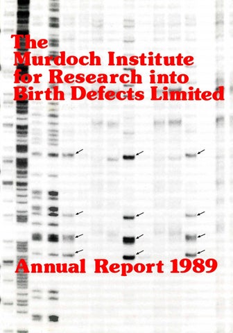
COVER Chemical cleavage of mismatch method of detecting mutations used to compare human and rat phenylalanine hydroxylase genes — the dark bands marked by arrows show differences between the genes of the two species.
The Murdoch Institute is remarkable interna tionally among Institutes working on genetic diseases for an even balance between fun damental research and clinical practice covering genetic disease in all age groups in the whole State of Victoria. This gives the Institute a an particular style of which we are very proud innovative approach to clinical work and a practical start to basic research. It also provides special opportunities for training free-thinking young clinicians and research workers. The Murdoch Institute is the major Australian centre for research on genetic diseases and for training clinical geneticists.
(
:•
k- ^
V:
n
!*•: Royal Children’s Hospital, Flemington Road, Parkvilie, Victoria. 3052.
I - tS. V ■
MAJOR DONORS TO THE MURDOCH INSTITUTE FOUNDERS — Donors of $1 million or more The Murdoch Family: Dame Elisabeth Murdoch Mr. Rupert Murdoch Mrs. Helen Handbury Mrs. Anne Kantor Mrs. Janet Calvert-Jones The late Sir Jack Brockhoff The Brockhoff Foundation The Scobie and Claire Mackinnon Trust BENEFACTORS — Donors of $250,000 or more The Miller Foundation The Helen M. Schutt Trust
MAJOR DONORS TO THE MURDOCH INSTITUTE TRUSTEES — Donors of $25,000 or more Mrs. M.L. Griffin J.B. Were & Son Charitable Foundation National Australia Bank Limited The Percy Baxter Charitable Trust The Ian Potter Foundation The late Mr. Clive Roxburgh Mrs. Joan Roxburgh H. & L. Hecht Trust The late Mrs. L.B. Quayle The News Corporation Limited Coles Myer Limited Qantas Repco Corporation Limited (Ariadne) CORPORATE SPONSOR GROUP — Corporations undertaking substantial future support Amcor ANZ Banking Group Coles Myer Limited National Australia Bank Limited Qantas
1 J
DONATIONS TO THE MURDOCH INSTITUTE 1st January — 31 December 1989
$
The Jack Brockhoff Foundation Cruden Investments Scobie & Claire Mackinnon Trust The Miller Foundation The Lynne Quayle Charitable Trust Coles Myer Ltd National Australia Bank ANZ Banking Group The Morris Family Trust The Ian Potter Foundation Rothschild Australia Percy Baxter Charitable Trust Mrs. Marie Louise Griffin Mr. S. Spadafora J.B. Were & Son Charitable Fund Amcor Clive & Jean Roxburgh Trust Arthur Andersen Foundation Western Mining Corporation Ltd The Shell Co. of Australia The Brash Charitable Foundation Mr. D. Slack
250,000 250,000 100,000 50,000 25,000 25,000 20,000 7,500 5,000 5,000 5,000 5,000 5,000 5,000 5,000 5,000 4,000 4,000 3,000 1,000 1,000 1,000
Mayne Nickless Ltd Dr. J.M. Gooch Annie Danks Trust In memory of Kathryn Anne Tolley Mr. F.D. Ryan Professor D.M. Danks Mr. S.F. Gooley Mr. K.C. Smart Mr. G.E. Heeley McMullin Unit Trust In memory of Laura Vanderwert Little People’s Association of Australia In memory of Emma Ritchie Mr. & Mrs. Hargreaves Mr. & Mrs. L. Barbieri Mrs. G. Grimwade Mrs. Ann Thompson Mr. R. Symons Mr. E. Oakes Mr. «&Mrs. S. King Dame Patricia Mackinnon Mr. R.E. Dowland Anonymous Mrs. J.M. Fox
1,000 1,000 1,000
902 750 640 500 500 500 500 455 350 220 200 150 100 100 100 100 100 100 50 40 30
CHAIRMAN’S REPORT
INDEX Chairman’s Report.................................................... Board of the Murdoch Institute................................. Finance Committee.................................................. Director’s Report...................................................... Research in Progress................................................. How Pairs of Chromosomes Separate and Why This Process Sometimes Fails........................... Senior Visiting Scientists......................................... Post Doctoral Fellows.............................................. Staff List of the Murdoch Institute............................ The Victorian Clinical Genetics Services................. The Board of The Victorian Clinical Genetics Services............................................................ Staff List of The Victorian Clinical Genetics Services............................................................ The Murdoch Institute Lecture Series — 1989......... Staff Involvement in Australian and International Scientific Community Activities...................... Editorial Boards....................................................... Postgraduate Degrees Awarded................................ Overseas and Australian Lectures and Seminars by Institute Staff................................ Collaborations.......................................................... Oliver Miller Protein Chemistry Laboratory............ Scobie and Claire Mackinnon Trace Element Group. Studies of Pyruvate Dehydrogenase......................... Molecular Genetics Studies..................................... Enzymology/Metabolism........................................ Cytogenetics............................................................ Tissue Culture Laboratory...................................... Epidemiology.......................................................... Clinical Projects ..................................................... List of Publications — 1989....................................
1 2 4 5 10 17 23 24 25 26
29 29 30 30 31 31
Mr Neil Walford
As is evident from a reading of the Director’s Report, the 1989 year was marked by further significant professional achievement. It was also a year when our funds were well managed and in which, due to the continuing support of our many benefactors, our financial capability was maintained. We are deeply grateful to all of our donors. May I express our very special thanks for the continuing substantial gifts which we have again received from both the Murdoch family and the Brockhoff Foundation. We are pleased that some major corporations have joined our Corporate Sponsors Group, undertaking support for at least 3 years, and hope that others will join them. I also wish to say how very appreciative we are of the decision by the Victorian Government to support us to the extent of $150,000 a year. Elsewhere in this Report, there are financial statements which show just how completely we depend on funds from various public and private sources. Fund raising is a matter which requires our unremitting attention if we are to continue and expand our research work over the long years ahead. And, of course, the proper management of our funds is of vital importance. Again, I place on record the thanks of the Board for the help and guidance we have received from our Finance Committee. Professor G.J. Fraenkel has recently announced his retirement as Chief Executive of the Royal Children’s Hospital Research Foundation and from the Board of the Murdoch Institute. I should like to record our appreciation of his contribution and wish him well in his retirement. His successor. Dr Ruth Bishop, will take his place on our Board. We extend to her a warm welcome and congratulate her on her appointment. I am delighted to report that one of our directors, Mrs Penny Lewisohn, has been elected President of the Royal Children’s Hospital and Chairman of its Research Founda tion. We all offer our very warm congratulations to Dame Elisabeth Murdoch, our very dear friend and our Patron, on her being awarded the AC in last year’s Queen’s Birthday honours. The standing and reputation of our Director, Professor David Danks, is firmly established in international medical circles. It is very fitting that he should now receive formal recognition in his own country by the award of an AO in the recent Australia Day honours.
31 34 36 38 ,42 ,44 .46 .48 49 .49 51 53 1
BOARD OF THE MURDOCH INSTITUTE
m
n
1 Mr. N. Walford, B.Com., F.C.A. Chairman
Mrs. J. Calvert-Jones
Mr. L. G. Cox, B.Com., A.A.S.A., F.S.I.A. I Vice Chairman
Dr. B. R. Catchlove, M.B., B.S., F.R.A.C.P., F.R.A.C.M.A., F.H.A.
Mr. J. A. Fitzgerald
Mr. J. S. Guest, A.M., O.B.E., V.R.D., B.Sc., M.B., B.S., F.R.C.S., F.R.A.C.S.
Professor P. D. Phelan, M.D., B.S., B.Sc., F.R.A.C.P.
i 4
2
Dr. G. L. Barnes, M.D., Ch.B., F.R.A.C.P.
Dr. R. G. H. Cotton, B.Ag.Sci., Ph.D., D.Sc.
Dr. R. Bishop, Ph.D., D.Sc.
Professor D. M. Danks, M.D., B.S., F.R.A.C.P.
1
''
Mrs. J. Lewisohn, B.A.
Professor G. B. Ryan, M.D., B.S., Ph.D., F.R.C.P.A., F.R.A.C.P.
Mrs. I. McFarling 3
DIRECTOR’S REPORT
FINANCE COMMITTEE
1
I
Mr. L.G. Cox, B.Com., A.A.S.A., F.S.I.A. lairman
Mr, G.E. Heeley, B.Ec., F.A.S.A.
Mr. C.P. Abbott, L.L.B., C.P.A.
Mr. D.E. Meiklejohn, B.Com., F.A.S.A., C.P.A. A.A.U.Q.
Professor David Danks
Mr. D.T. Craig, A.C.A.(N.Z.), A.A.I.B.
Mr. F.D. Ryan, F.C.S.
Mr. P.J. Griffin, B.Com., A.S.l.A. 4
V,-
■
‘•r :
■
This year, the fourth year of our establishment as the Murdoch Institute, has been a year of getting on with our standard business of doing research and helping patients. In other words, it has been a normal year for a research institute and generally a year of very satisfactory progress in a wide range of projects without any major new changes, after several years of considerable change. An outline of the progress on our major projects is provided in the section “Research in Progress” and the various research groups have provided more detailed project reports which are found in the latter part of this report. This year we have chosen to give a detailed account of studies of Dr K.H. Choo and his colleagues who are analysing the mechanisms which control the separation of the pairs of chromosomes which are found in human body cells in order to produce germ cells (ova and sperm) which contain just one of each chromosome pair, allowing sexual recombination to restore the paired system of chromosomes at the time of fertilization. This is clearly one of the most fundamental processes of human life, and indeed of the life of all higher organisms. An understanding of the mechanisms which control this process is critical in all biology and especially critical if we are to understand the birth defects which occur because of errors in this process. Choo’s group has made some encouraging progress towards this goal. I hope that the lay readers of our Report will feel this account interesting and comprehensible. There are always some projects which are at a slightly more exciting phase than others in a particular year. In 1989 the work of Garry and Ruth Brown and Henrik Dahl on the enzyme pyravate dehydrogenase (featured in the detailed lay report in 1988) continued on a “high” with a number of exciting new findings. The chemical cleavage of mismatch (CCM or HOT method) of recognizing gene mutations, discovered by Dick Cotton, was applied to a number of diagnostic problems during the year and lived up to our high expectations. It is gaining international recognition as the method of choice for the detection of mutations. Studies with copper transport moved ahead in a satisfying way in the hands of Jim Camakaris, Julian Mercer and Harry McArdle and our increased understanding of basic aspects of copper transport have started to bear frait in the development of new methods of treating Wilson’s disease and the disturbances of copper metabolism which occur in other forms of liver disease. During our first two years, the focus was upon establishing the Institute and its methods of operation. In the 1988 Report we emphasised the establishment of the Victorian Clinical Genetics Service. I had imagined that in this 1989 Report we would be giving our colleagues and supporters exciting news of the development of new laboratories and other new physical facilities. Last year I thanked the staff, scientists and clinicians, for the way in which they had coped with the overcrowding that had prevailed in 1987 and 1988 and suggested that they would be sustained through 1989 by the “prospects of additional space which are now visible to us at last”. It makes me very sad to report that the prospect of additional space is still about as far away as it was this time last year. Not only must I thank all staff members for the good humour with which they have coped with the severe overcrowding that persisted throughout 1989, but I must exhort them to be even more good humoured through 1990 5
when we will be even more overcrowded, having taken on several additional staff on the assumption that new space would be available. I know that we have a group of good natured people and that they will cope once again. I also know that the additional people who are joining us in 1990 are very bright, competent and enthusiastic and that we will enjoy getting on together even if we are working on top of one another. We will await the completion of the new tont building, the expanded second floor and the third floor laboratory with eager anticipation, knowing that their occupation will at last free up the 10th floor space we have been promised for so long. In an Institute like this, one of our major problems is to maintain a sufficient flow of funds for each year’s activities and to enable us to expand our activities a little each year. Last year we pointed out that the Institute needs to raise between $800,000 and $1 million each year to sustain its current level of activity. It gives me great pleasure to endorse the comments already made by the Chairman about the generous further support that has been provided by the Murdoch family, by the Brockhoff Foundation and by a number of major corporations. We are hoping that further major companies will join our Corporate Sponsors Group in the coming year. We are so grateful to the Victorian Government for the decision they took in 1989 to provide grants to each of the major medical research institutes in Melbourne to support their basic establishment costs. We were pleased to welcome the Treasurer, Mr Jolly, and some of his staff when they chose our seminar room to announce this new initiative which brought our particular Institute the sum of $150,000 per annum. The advent of the Victorian Health Promotion Foundation (VHPF) was another important milestone in medical research funding in Victoria. It was established by the Victorian Government with funds previously committed by tobacco companies for the promotion of sporting events in Victoria. Part of this money is made available as research grants and health development grants in fields of preventive medicine. Dick Cotton was successful in winning one of the three Program Grants awarded by the VHPF. This will support further development and evaluation of his chemical cleavage method of mutation detection, and brings approximately $150,000 per annum for three years to support three post doctoral scientists, one applying the method to mutations causing genetic diseases and the other two studying the epidemiology of dengue virus and rota virus. The latter two projects will be in collaboration with Dr Peter Wright in the Department of Microbiology at Monash University and with Dr Ruth Bishop in the Department of Gastroenterology of the Royal Children’s Hospital, respectively. The VHPF also awarded a project grant of $79,000 for the development of a DNA testing programme for presymptomatic diagnosis of Huntington’s disease. This work will be undertaken by Dr Les Sheffield in collaboration with Dr Ed Chiu of the Department of Psychiatry at the University of Melbourne. Miss Davina Hanson has continued to make a valuable contribution to our fundraising activities during 1989. It is very generous of her to spend so much time helping us in an honorary capacity and I am most grateful to her. A special effort has been made to bring the Institute to the attention of lawyers and the various trustee companies of Melbourne, 6
hoping that they may remember us when clients ask for ideas when making wills. As always, I have enjoyed wonderful support and advice about fundraising fix)m the Board, Finance Committee and Fundraising Committee and especially from Chairman, Neil Walford, and Deputy Chairman, Laurie Cox. In the latter part of the year Mrs Miriam Davidson also joined our fundraising activities. She is a Canadian who is in Melbourne for two years along with her husband. Professor Ron Davidson, and her particular talents in personal communication became obvious as soon as we met her. She is assisting in various matters of communication within the Institute, in the promotion of our public image and in fundraising. She has revitalised our internal newsletter under the title of “Murdocuments” and is finding other ways of encouraging staff to know one another even better and to enjoy even more their collaborative work together. Professor Ron Davidson himself is a very experienced and justly renowned medical geneticist whose arrival in June saved our clinical geneticists fi'om collapsing from exhaus tion. I have had the pleasure of knowing Ron since we trained together at John Hopkins Medical School in the early 1960’s and was delighted when I learned that he was taking a very early retirement fit>m his position as Chairman of the Department of Medical Genetics at McMaster University in Hamilton, Ontario, Canada and would be interested in coming to Melbourne to work as a clinical geneticist for 12 months from June 1989. When I made these arrangements with him in November 1988 I was aware that we were in desperate need of an extra clinical geneticist, but I did not know that Dr John Rogers would take ill in January 1989 and be out of action for over six months. By the time Ron Davidson arrived in June, the need of his input was quite desperate and he threw his full effort into the work from the first week. He proved to be just the person we needed and we were delighted when he agreed to stay for a second year and take on the direction of our clinic in the Royal CMdren’s Hospital when John Rogers decided to relinquish this role after his return to health. Not only has he shown a high level of skill in his personal clinical work and a quiet and efficient style of leadership, but his special interest in the education of medical geneticists and of genetic counsellors is proving very valuable. We were very sad when John Rogers developed a serious illness early in the year, but are very pleased by his good recovery. John’s decision to reduce his commitment to the Victorian Clinical Genetics Service to three days a week, and to spend time in private practice in grief counselling, ends his formal employment in the Institute. He has always had a good “sense of smell” for the research potential of clinical observations and we know that this will continue to work to the benefit of our research. We wish him well in his new career direction. In April, the death of Sue Tomkins shocked and saddened all staff of the Institute. Sue had been with us as a very efficient secretary and a greatly-loved person since 1977. Her wonderful courage and cheerfulness in the face of a very severe disability (spinal curvature causing chronic respiratory failure) inspired the admiration of all who knew her. On the one hand, she often looked so fi-ail that we wondered if she would survive the day at work; on the other hand, her condition had seemed stable for several years and we all
i
I
dreamed this might go on for a long time. Sue had always feared becoming too ill to work and her rapid death while “in harness” was as she would have chosen, even though it was a shock for her family and many fiiends. We have been fortunate to have several visitors in the Institute during 1989. It was a pleasant surprise when Dr Lawrie Austin, Reader in Biochemistry at Monash Univer sity, asked if he could spend six months sabbatical leave in our laboratories. We were sad that his wife’s illness made it impossible for him to spend his sabbatical overseas, but we were certainly the ones who gained. Lawrie has a wide international reputation as a neurochemist and has made special contributions to the understanding of muscular dystrophies. He has been a pioneer of techniques of cultivating muscle cells in the laboratory and has shown that the satellite cells of muscle fix)m which the actual muscle cells themselves differentiate, are more readily cultivated in the laboratory. With injection of normal cultivated muscle cells emerging as a possible treatment of Duchenne muscular dystrophy, the new methods of cultivating satellite cells which he developed during his leave are very significant. Another senior visiting scientist who has made a reputation for studies in mice joined us for a short sabbatical at the end of the year. Dr Harold Rauch has worked for the whole of his career in the Department of Zoology at the University of Massachusetts in Amherst. He is particularly known for the discovery of a strain of mice called “toxic milk” which have a defect in the transport of copper which has many resembl ances to that encountered in humans with Wilson’s disease. These mice have been of obvious interest to our research group for a number of years and we were delighted to be able to commence collaborative work with Hal in 1988. This collaboration has been greatly enhanced by working together with him and will continue after his return. He plans to retire shortly and is kindly allowing us to maintain the main stock of the toxic milk mice. Dr Irma Dianzani, a young paediatrician from Turin in Italy, has been working with Dick Cotton. Trained initially in paediatrics, she has developed a deep interest in metabolic diseases, and especially in phenylketonuria. She came to know Dick Cotton when her chief. Professor Bonzoni, began collaboration with Dick in the study of the rare pterin deficient forms of PKU. Irma came here to learn the chemical cleavage methods of identifying mutations and to apply it to patients with PKU from Italy. She learned it so well that she has developed some useful new modifications in its application. Her research has progressed very well. She is very industrious, determined, and skilfull and we have all come to like her very much. Lotte Hansen joined Henrik Dahl’s group in September 1988 coming from his own home city of Copenhagen, on sabbatical leave from the Department of Human Genetics, Arrhus University, Denmark where she is near completing her licentiat degree in molecular genetics. She has made rapid progress in identifying the mutations in a number of patients with pyruvate dehydrogenase deficiency, using the chemical cleavage method. Dr Scott Garrett came from South Dakota to join Julian Mercer and Jim Camakaris in a project analysing the expression of sheep metallothionein genes after transfer into Chinese hamster ovary cells. He had done his PhD studies with Dr. Brady, a well known expert on the chemistry of
metallothionein metal interactions and is supported by a grant from the Australian Research Council through the Depart ment of Genetics at Melbourne University. The annual meeting of the American Society of Human Genetics is one of the more important meetings in our discipline and we always ensure that at least two or three of our staff attend to hear the latest discoveries and to present some of the results of our research. It was particularly pleasing to find that at the 1989 meeting, held in Baltimore in November, Les Sheffield’s paper on the classification of chondrodysplasia punctata was chosen as one of the eight presented in the main plenary session of the congress, a considerable honour, and that Susan Forrest was chosen as one of the three finalists for the award of a prize for the best presentation by a post doctoral scientist. She spoke about the application of the chemical cleavage method to detection of mutations in PKU and her paper was very well received. It was certainly no insult to emerge as runner up to another Australian who was working in Boston. His project happened to produce some really remarkable and unexpected findings, making it one of the most novel presentations in the whole meeting and a very hard one to beat. Dick Cotton was in demand as a speaker at the Pteridine Symposium in Zurich and to tell about his chemical cleavage method in various laboratories in Britain and Europe. I was pleased that our work of many years on the role of metabolic diseases as causes of physical malfunctions was recognized by a request to organize a symposium on the subject at the International Congress of Paediatrics in Paris. The sympo sium was well attended and Garry Brown’s contribution was particularly well received. Agnes Bankier was invited to attend the rather select Dave Smith Workshop on malforma tions in Madrid. Jim Camakaris was an invited speaker at the Fourth International Conference on Bioinorganic Chemistry in Boston. These and other staff members presented many papers at Australian scientific conferences and lectured and/or taught in various programmes in Melbourne. Details are listed elsewhere in this Report. Once again, we can report exciting progress for POSSUM. The displays at the meeting of the American Society of Human Genetics and the International Congress of Paediat rics again attracted a great deal of attention. The laudatory comments of experts in the field of syndrome identification are particularly gratifying, for instance — “We continue to find that POSSUM is an excellent aid to us in clinical diagnosis. It is a particularly helpful way to orient the medical student, resident and postdoctoral fellow to a logical way to search for similar phenotypes. I am sure you are getting similar feedback from other centres, but I want to join in the chorus of praise for its value.” We are pleased with the steady progress of sales and particularly pleased that most of the major international journals of paediatrics and genetics have agreed to publish formal reviews of POSSUM. This should attract considerably more attention to its existence. With their usual energy, Agnes Bankier and her colleagues and John Marquet have managed to get Version 2.5 ready for distribution to POSSUM users right on time, despite great difficulties because of additional pressures of clinical work. This continued success in meeting deadlines is playing a very important part in building up the reputation of our product. In addition to all the work involved in producing Version 2.5, Agnes and John have found time to start planning for a sister 7
product dealing with hereditary bone disorders. We are really very fortunate to have people of such energy, enthusiasm and ability working on these projects — without them POSSUM would not exist. In October 1989 we said farewell to Garry and Ruth Brown and to Harry McArdle. Garry and Ruth had worked in the Institute since 1980 and had made important contributions in enzymology/metabolism and in cell biology, respectively. Garry has an encyclopaedic knowledge of metabolism and of enzymology and played an important role in the development of the Institute. He is also an outstanding teacher who will be missed by the medical students to whom he brought enlightenment about genetics and metabolism. Ruth had great skills in manipulation of cells in culture and worked successively with Jim Camakaris, Dick Cotton and with Gariy. This last collaboration was particularly successful in the development of new knowledge about pyruvate dehyd rogenase deficiency. Both Ruth and Garry have had a longterm love affair with Oxford, having worked there for two year before coming to Melbourne and returning for sabbatical leave in 1987. When the opportunity to join an exciting new research group in the Department of Biochemis try in Oxford arose, it was too good to refuse. In replacing Garry, we needed to maintain an international presence in research in metabolism/enzymology and the expertise required for diagnosis and treatment of local patients with inborn errors of metabolism. It is not essential for us to continue all his individual projects. Research on many important molecular aspects of pyruvate dehyd rogenase deficiency will continue in Henrik Dahl’s group. Geoff Thompson and David Howells, together with Denise Kirby and Effie Tsotsis and our colleagues in Clinical Biochemistry, will maintain the laboratory diagnostic skill required for patient care and will develop their own international standing for research in certain aspects of inborn errors of metabolism, different from those which interested Garry. Geoff has particular interests in, and experience with, assessment of enzyme function in the whole patient, using stable (non-radioactive) isotope tracers. David’s interest and background is with chemical disturbances of neurotransmit ters, especially those which are related to tyrosine and tryptophan and may be disturbed in phenylketonuria. We look forward to the development of their work with interest and confidence. Harry Mcardle was one of the most lively personalities to have come to the Institute. A Scotsman with a real drive and determination and a pleasant streak of aggression, he was always good for strong debate and full of bright ideas about his own work and about other people’s projects. His was the type of lively interaction which makes an institute tick. He was also a very competent experimental physiologist with a considerable expertise in membrane transport. Although he had not worked with copper before joining us in 1985, he did have extensive experience in the study of iron metabolism and he made a veiy rapid adaptation to the new, but related, element. From the time of his arrival here from Perth, he made it clear that he would return to Scotland after a few years in Melbourne, provided he could find an appropriate position. Although we were sorry to lose him, we shared his pleasure when an interesting position turned up in the Department of Paediatrics in Dundee. Harry will continue copper projects in his new position and 8
will collaborate with us in these. Some of the techniques he established will go on in our laboratory in the hands of Sharon Gross, Hayley Vogel and Leigh Ackland. Other projects have terminated and two young research assistants (Peter Kyriakou and Becky Erhlich) have moved on to other positions. Julian Mercer will now make copper metabolism the focus of his research, drawing upon his expertise in molecular genetics, rather than seeing himself as a molecular geneticist with an interest in copper. Some rearrangement of resources will allow us to start a new group in experimental embryology later in 1990, led by Dr Don Newgreen, an Australian who has built a big reputation for his work on the neural crest, a very important collection of cells of the early embryo, which play critical roles in development. He has worked in France, Germany and the UK over the last 10 years, and returned to Australia two years ago. We have always seen embryology as a very appropriate field of research in the Institute and have been keeping an eye out for an appropriate person for some years, ever since an earlier attempt at establishing a group terminated in 1987. An unplaimed benefit has come fi'om the various rearrangements of 1989. Our Molecular Genetics Unit has never had a scientific cohesion, but existed as a grouping of three independent established scientists (Drs Dahl, Mercer and Choo) held together by common techniques rather than linked projects. Now Henrik has taken charge of the PDH projects and of DNA diagnostic work, Julian has plunged fully and enthusiastically in to copper research and Choo’s studies of centromeric DNA and of DNA integration into chromosomes are flourishing and have led to extensive collaboration with our Cytogenetics Laboratory. Finally, molecular genetics has become an approach to research used by most of our scientists rather than a separate discipline. We now have Henrik’s group, Julian’s group and Choo’s group, and everyone uses molecular techniques when appropriate. Three of the young postdoctoral scientists who joined us early in 1988 moved on to permanent positions during 1989. Two of the three had achieved a substantial part of their objectives before departing. In these days of very uncertain career prospects in science, one cannot really blame a young scientist for grasping any reasonable career position that is offered, even if it comes at an awkward time for the current project. The alternative of offering careers in the Institute to all post docs is obviously ridiculous. Phil Kearney moved to the Garvan Institute (and has since moved out of science); Neil Fraser returned to a good career position at Oxford University; Connie Maragos is now at CSL. This has been a very busy year for our Business Manager, Aime Ellis, especially because of protracted negotiations over industrial relations and an increased load of detailed personnel matters. Many may not realize that Anne also manages the financial affairs of the Royal Children’s Hospital Research Foundation. The combining of these tasks is logical, but onerous. From its initiation in 1986, the Institute has employed its own staff, unlike the Research Foundation whose staff were employed by the Hospital". Some problems inherent in this arrangement became apparent to the Foundation at about the same time as the Institute decided to take over its own persoimel services (previously provided by the Hospital at a fee), so the Institute and Foundation combined to employ a part-time personnel
officer, Ms Lucy Griffiths. Even with this help, it is clear that Anne is over-loaded and will soon need some assistance with accounting. The industrial relations matter stemmed from an attempt by the Hospital Scientists’ Association to claim that scientists in hospital-based research institutes should come under their award After many sessions of debate and formalities, the Industrial Relations Commission found in our favour and agreed that affiliation with the Universities Staff Association was more appropriate. I am most grateful to Anne for her wise advice and actions in these and many other matters. She is respected and appreciated by all staff and we all share her happiness in her new marriage to Michael Cronin. She will still be known as Anne Ellis in her work in the Institute. Barry Holt has had an extremely busy year with great pressures in his role in designing and implementing the conversion of the west wing of the third floor from outpatient clinics to laboratory space for the Orthopaedic Research Unit, Surgical Research and the Department of Endocrinolo gy. Satisfying all the demands of three groups within limited space is never easy, but problems with Fire Department standards and the connection of fume hoods to existing ducting on upper floors added to the difficulty. Despite all this, he remained good natured and helpful to all Institute staff, always managing to find yet another desk, for yet one more new staff member and coping with his part-time studies for MBA. I am most grateful to him for all his efforts on our behalf and also to Andrew Grimes who has filled the breech most ably whenever Barry has been unavailable. We are indeed fortunate to have someone as capable as Andrew at the research bench and in laboratory management. There have been a number of changes in the leadership of our sister research organisations in Melbourne. Nearest to home. Professor Gus Fraenkel has retired from his position as part-time Chief Executive of the Royal Children’s Hospital Research Foundation and Dr Ruth Bishop has been appointed as his successor. We all respect Ruth as a scientist and admire her achievements in the discovery and study of rotavirus; we look forward to working with her as an administrator. Professor Derek Denton has retired after many years as the founding Director of the Howard Florey Institute and we congratulate him on his fine achievements and Professor John Coghlan on his appointment as Director. Professor Paul Komer led the Baker Institute through a long period of substantial growth in size and respect and will soon retire. We extend congratulations to Professor John Funder on his appointment as the new Director. Dr John Stocker, the dynamic and successful Chief Executive of AMRAD (Australian Medical Research & Development Corporation^ has been appointed Chief Execu tive of CSIRO. We will be very sorry to lose him from the AMRAD role and wish him well with the massive task he is tackling. Congratulations to John Grace on his promotion to Chief Executive from Business Manager, AMRAD. I lean heavily upon Neil Walford (Chairman) and Laurie Cox (Deputy Chairman & Chairman of the Finance Committee) for advice on many matters and they are always willing and helpful. Although my calls on other Board members occur less often, they are all equally willing to help. This support and guidance is greatly appreciated. Of course, the senior scientists and clinicians are
constantly advising me in many informal discussions and regular meetings. The Institute is ultimately what they make it through their professional achievements and it is to them that credit is due for anything the Institute has achieved, As in past years, Anne Ellis has played the major part in the production of this Report. Mr David Gibbs, previously of the Herald and Weekly Times, now of Australian Jewish News has been our advisor on its design and layout. We ^e most grateful to him for this assistance and for recruiting the generous assistance of those organisations which are acknowledged inside the back cover,
9
RESEARCH IN PROGRESS: Continuing the practice commenced in 1988, this section gives brief comments on senior staff members and their main activities during 1989. More detailed reports of their projects written in their own words can be found later in the Report. I apologise to those who read our Report carefully each year for certain elements of repetition which are inevitable in this section, both in regard to the background and interests of the staff members and in the work that they are doing, because most of the projects do spread over several years. Without this repetition, the Report would be unintelligible for first-time readers. Dr Dick Cotton, the Deputy Director, is best described as a biologist with a very broad background and range of interests. He leads the Olive Miller Protein Chemistry Research Group. The major goal of his research is a full understanding of the enzymes of phenylalanine metabolism defects which cause phenylketonuria (PKU), an important and preventable cause of mental retardation. More recently the continued development of the chemical cleavage method of mutation detection has become an important project. Ian Jennings, who has worked with Dick for many years, is particularly involved with the study of the structure of phenylalanine hydroxylase (PAH), the enzyme at fault in the majority of cases of PKU and Anthony Urban has been concentrating on the further development of the chemistry of the chemical cleavage method. Three post doctoral scientists — Susan Forrest, David Howells and Irma Dianzani — are using the chemical cleavage method and other DNA techniques to identify mutations present in individual patients with PKU. Susan and Irma are concentrating on PAH and David is smdying dihydropteridine reductase (DHPR), one of the enzymes at fault in a rare malignant form of PKU. The application of the chemical cleavage method to these genes has contributed to its refinement. The identification of mutations has expanded our understanding of the variation in severity of PKU and the determination of the particular changes that the mutations have caused in the enzyme feeds back into the basic work on the relationship between enzyme stmcture and function. Mutations which cause disease generally affect functionally important parts of the enzyme. Mapping the location of these mutations helps to identify the functionally critical regions of the enzyme. In the very long run, we believe that PKU will be treated by gene replacement or gene correction, a once-only treatment of a young baby with the disease which would give a lifelong cure without in anyway meddling with the general pool of human genes since the treatment would not influence the germ cells. To achieve this goal, it will be necessary to have a thorough and detailed understanding of the way in which PAH carries out its enzymic role and to develop safe and efficient methods of delivering the normal gene into the appropriate place in the chromosomes of the cells of the liver. It will take many years to develop these abilities. Dick Cotton’s work is likely to make a major contribution to the understanding of the enzyme and we hope that Dr Choo’s work will contribute to the development of a method of controlling gene insertion. In their basic studies of the PAH protein, Dick Cotton and Ian Jennings are collaborating with a number of groups in Melbourne, elsewhere in Australia and internationally as listed elsewhere in this Report. They are making steady 10
Dr Dick Cotton
Dr Julian Mercer
progress and have localised one of the critical functional regions of the enzyme via the new antibody approach. The antibody developed in these studies is starting to be useful in many overseas laboratories studying related proteins/en zymes. The chemical cleavage of mismatch (CCM) method has been used successfully to locate mutations in PAH, DHPR, collagen and pymvate dehydrogenase in our laboratories and in numerous other genes analysed overseas. Dick and his colleagues have been in great demand to give talks about the method. The Victorian Health Promotion Foundation Program Grant awarded to Dick during 1989 will expand his work with the CCM refining its application to human genetic diseases and determining its value in the analysis of the genetic changes which occur in the dengue virus, HTV and rotavirus, modifying the severity of disease and the effectiveness of immune responses to these important viruses. This work will be performed in collaboration with Dr Henrik Dahl, Murdoch Institute, Dr Peter Wright of the Department of Microbiology at Monash University, Dr Dale McPhee, Fairfield Hospital and with Dr Ruth Bishop of the Department of Gastroenterology, RCH. Three post doctoral scientists have been recruited and will start work early in 1990. In the Scobie and Claire MacKinnon Trace Element Research Group there are three senior scientists. Dr Julian Mercer, Dr Jim Camakaris and Dr Harry McArdle. Although their three groups function independently many projects involve collaboration between two or more of the senior people. Jim’s background in microbial genetics led to an extensive use of normal and mutant cultured cell lines of various types for the study of copper transport. Julian’s background in molecular genetics determined his concentra tion upon the genes encoding copper transport proteins and factors influencing their function. Harry’s background as a physiologist equipped him to probe some of the normal processes of copper transport in normal mouse and human cells. Julian Mercer’s work has maintained a focus upon the roles of two copper transport proteins, metallothionein and caeruloplasmin, in whole animals, especially mice and sheep. Previously many of his studies focussed upon the brindled and blotchy strains of mice which have abnormalities very similar to those in human Menkes’ disease. More
Dr Jim Camakaris recently, he has begun collaboration with Dr Harold Rauch of Amherst, Massachusetts, using the toxic milk mice which have a genetic defect which seems quite similar to that in humans with Wilson’s disease. In toxic milk mice, copper accumulates in the liver to very high levels and eventually causes liver damage in older mice, both male and female. However, the really striking effect that led to the recognition of the mutation was that seen in pups of pregnant females affected by the mutation. The supply of copper across the placenta is deficient and the supply in the milk is even more deficient so that pups are bom with some degree of copper deficiency and become deficient to a lethal degree during the first two weeks postnatally. Fostering to an unaffected mother can completely correct the problem. Initial studies conducted between the US and Melbourne focussed upon the possibility that mutations affecting the control of metallothionein or of caeruloplasmin might be present. The results eliminated this possibility. The only abnormalities observed were in the pups and could be attributed to eopper deficiency rather than to the gene defect. Further studies focussed upon binding proteins in the livers of the animals and these are looking more interesting. New techniques have made it possible to separate several proteins which bind copper in the liver and one of these is either absent or present in very low quantities in the toxic milk mice. Julian and Andrew Grimes are working hard to identify these proteins which are of interest as copper transport proteins and of special interest if one of them does turn out to be the site of the basic fault in the toxic milk mice, and perhaps in Wilson’s disease. Sheep handle copper rather differently from other species, tending to accumulate excessive amounts in the liver if even slightly greater than necessary levels are ingested. They develop liver damage if this overloading with copper is allowed to progress. Julian and Professor John Howell of Murdoch University, Perth, have previously shown that this is not a direct consequence of an abnormality of metallothio nein, but found some usual patterns of response of metallothionein production to dietary zinc which warrant exploration. In the process, Julian cloned the sheep metallothionein genes and there is considerable interest in determining whether any of these genes differs from the others in terms of responsiveness to zinc or expression in different tissues. Scott Garrett, an American post doc, is working with Julian and Jim Camakaris on an Australian Research Council Grant, expressing the various sheep
metallothionein genes in Chinese hamster ovary cells and studying the factors that control these. Chinese hamster ovary cells have a particular advantage for this work because they do not express their own inherent metallothionein genes and we therefore anticipate that metallothionein genes from other species that are introduced into these cells can be studied rather readily. The initial results are quite encouraging, the introduction of one of the sheep metallothionein genes having caused the cells to resist much higher than normal levels of copper in the medium without toxic effects. Julian also has other studies in progress looking at the response of the various MT genes to zinc, copper and liver toxic chemicals in whole sheep. This is a collaboration with Professor John Howell of Murdoch University in Perth and is funded by the Wool Research Corporation. Julian and Choo are planning a collaboration studying the effects of disraption of the metallothionein and caemloplasmin genes in transgenic mice. A necessary step towards this involved isolation of the mouse caeruloplasmin gene and a number of the properties of this gene have been studied in the process. Julian and Jennifer Paynter took the opportunity of examining caeruloplasmin gene expression in brindled mutants comparing it with copper deficient animals and showed that neither of these disturbances of copper status had any effect upon caeruloplasmin gene expression. Jim Camakaris trained originally in microbial genetics and then did a post doctoral fellowship at the Hall Institute with Dr Don Metcalfe in which he obtained extensive experience with cell culture. His work with us on copper metabolism has concentrated on the use of mutant cell lines in the analysis of copper transport. His position as Senior Lecturer in Human Genetics in the Department of Genetics, Melbourne Univer sity, brings a number of postgraduate students to work with him. During 1989 he and Angela Bruzanniti have made considerable progress is defining the methods of uptake oArdle has found in liver cells in culture — namely that the copper is brought into contact with the cell surface complexed to the amino acid histidine and is exchanged on the cell surface r to be involved. This mechanism is not altered by the mutation in Menkes’ disease, which must influence another process of copper transport within the cell. Jim and Rohan Farrell (initially as a BSc Honours student, then as a research assistant, and now as a PhD student) have been working hard to find satisfactory methods of analysing the copper-binding proteins within cells. It has become clear that the traditional methods of extracting and separating the proteins from cells allow the copper to shift from one protein to another so that the end result tells little about the original situation that existed in a living cell. They have speeded up the whole process of preparation and analysis, have used methods which separate copper proteins from cell mem branes and ways of protecting proteins which are easily changed by exposure to oxygen and have also developed methods of separating the proteins and then attaching copper isotope to those proteins which have a specific copper binding capacity. The ubiquitous metal-binding protein, metallothionein, is the problem in many of these processes for it seems to be able to pick up copper from other proteins once the cell is disrupted. Two cell lines (W7 lymphocytes 11
r
and Chinese hamster ovary cells) make very little metallothionein and they have proved useful in these studies. The logic behind working with cell lines from human and mouse mutants is that it may be easier to recognize a normal copper transport process by studying cells in which that component has been disrupted by mutation. So far, the spontaneously occurring mutations in man and mouse have resisted our efforts. Another approach is to deliberately generate mutations in copper transport proteins in cells in culture. For this purpose, the Chinese hamster ovary cell has many useful properties including a very low level of expression of metallothionein. Jasmine Georgiou has ex ploited this cell line, building upon work initiated by Janet Patton. By selecting cells that survive in media containing high levels of copper she has selected cells which have an abnormal resistance to the toxic effects of copper. These cells are likely to have a defect in the uptake of copper, an augmented ability to excrete copper or an ability to complex copper in a harmless form within the cells. Analysis of any of these variations is likely to lead to identification of copper transport proteins. One of the copper-resistant variants appears to overproduce a copper-binding protein which is not metallothionein. Jasmine is studying the mutations in two different copper resistant cell lines. Bacteria offer a readily manipulable form of cultured organism and techniques for analysing genetic faults in processes like copper transport are much more advanced in bacteria than in mammalian cells. Jim has therefore developed a collaboration with Dr Barry Lee from the Department of Genetics and Dr Suzanne Rogers, a post doctoral fellow, is conducting these studies. They have defined several genes involved in copper transport and are now isolating these genes, sequencing them and working out the structure of the proteins which they encode. It is rather unlikely that there will be any copper proteins in bacteria which are identicai to those in mammalian cells, but it is quite likely that there will be a similar set of processes in both organisms and that the regions of proteins to which copper actually binds may be retained through evolution. This work has been supported by a grant from the Australian Research Council. Harry McArdle brought a number of projects to comple tion before returning to his native Scotland in October. During his four years in Melbourne he studied many aspects of the uptake of copper into mouse liver cells in culture and into human fibroblasts in culture. He found very different systems in these two cell types. Copper appears to be carried to the liver attached to albumin and before it is taken up by the liver cells, a complex is formed between albumin, copper and the amino acid histidine from which a copper-histidine complex is released and attached to the surface of the liver cell. There appears to be a specific receptor which then transports the copper into a cell. This is not the mechanism utilised in fibroblasts, but it is still not clear exactly how the copper is taken up by these cells. Harry has been able to eliminate both albumin and histidine as the carriers which the fibroblast recognizes and has not succeeded in proving or excluding the possibility that caeruloplasmin plays this role. He finds uptake to be most efficient from copper salts in the cultures, but these are most unlikely to be available in the body as a source of copper for these cells. 12
of diagnostic work and it is essential that the service laboratories should be closely integrated with research in molecular genetics. Susan Forrest has also collaborated with Henrik in supervising this work, especially during Pam Dry’s absence on maternity leave.
Dr Harry McArdle
Dr Henrik Dahl
These studies have been run in parallel with Jim Camakaris’ studies of copper uptake into cultured lympho cytes making careful comparisons of the three cell systems all along the way. Harry has examined also the copper protein complexes that form after the copper is taken into cells, especially in liver cells. His approach has been to use powerful copper-binding chemicals (chelators) to recognise different pools of copper within the cell, some accessible to the chelators, and others inaccessible. This work has been assisted by the availability of several new copper chelators developed by Professor Alan Sargeson of the Australian National University. These are called sar and diamsar and appear to be the most potent chelators of copper yet used in biological systems. Copper which has recently been taken up by the cells is easily removed by these agents whereas copper that was taken up 24 hours earlier is much less accessible. There is clearly a change in the chemical form or the physical location of copper within the cell during this period. Harry and Peter Kyriakou have measured the effect of copper uptake upon the production of metallothionein and caerulo plasmin in liver cells and the amount of copper complexed to these proteins. Up till now it has not been possible to equate the pools of copper accessible or inaccessible to chelators with individual copper proteins. Harry has also examined some of the effects of the copper-binding agents that are used in the treatment of Wilson’s disease, penicillamine and tetrathiomolybdate. Penicillamine has very little effect on either the uptake of copper into liver cells or its removal from the cells, whereas tetrathiomolybdate is potent in preventing uptake and in removing copper that is already within the cells. The only effect of penicillamine demonstrated was a stimulation of the production of metallothionein. This may be the mode of action of this drug in Wilson’s disease since copper complexed to metallothionein is probably non-toxic. With Becky Ehrlich he studied transfer of copper from pregnant dam to fetus in the mouse, and to a lesser extent in the rat. Caeruloplasmin seems to be involved in the rat, but not so clearly in the mouse. Leigh Ackland has worked with Harry since his arrival in Melbourne, examining the uptake of zinc into fibroblastic cells and looking for abnormalities in the genetic disease, acrodermatitis enteropathica. This used to be a lethal disease before an English dermatologist showed that it could be cured by zinc supplements. In 1989 Leigh started studies towards a PhD and her more detailed studies of zinc uptake are progressing well.
Dr Garry Brown
Dr K.H. Choo
Henrik Dahl was trained in biochemistry in Denmark and had extensive experience in molecular genetics in one of the top European groups before joining the Institute in 1984. His work on genetic analysis of the Ela subunit of pyruvate dehydrogenase (PDH) has made particularly good progress during 1989. As described in detail in 1988’s Report, this enzyme is at the pivot point of the energy production in human cells. The project began in Garry Brown’s laboratory where the basic enzyme studies, enzyme purification and identification of the subunits of the enzyme was achieved. Then Henrik cloned the gene and the detailed analysis of the gene and of its relationship to the genetic disease of primary lactic acidosis began as a collaboration between Henrik and Garry and Ruth Brown. Hemik and Connie Maragos, a post doctoral scientist, analysed the mutation in the PDH gene in one severely affected girl and found a change in a region of the gene which may turn out to be a “hot spot” for mutations judging by subsequent results obtained by Lotte Hansen, a Danish scientist visiting on sabbatical leave. As more and more mutations are identified in different patients, we will get a clearer picture of the regions of the PDH molecule which are particularly important for its function, following the same logic described for phenylalanine hydroxylase. One of the very interesting findings about the PDH Ela gene has been its location on the X-chromosome and of even greater interest has been the discovery of a second gene encoding a similar, but not quite identical, subunit which is located on chromosome 4 and is functional only in sperm. It seems possible that there may be a number of sperm-specific genes which are encoded separately from the genes serving similar functions in other body cells. Henrik and his colleagues will be focussing on this possibility in 1990. The first application of the chemical cleavage method to mutations causing human genetic diseases was by Shireen Lamande in the Orthopaedic Research Unit guided by Henrik Dahl. Shireen is one of a number of people trained in the use of DNA technology by Henrik. Over several years she has become quite self-sufficient with these techniques and now Henrik acts as a consultant rather than as a supervisor. None the less this work continues to be a substantial interest for him. During 1989, Henrik has taken on formal supervision of the DNA diagnostic work in the Victorian Clinical Genetics Service within the Murdoch Institute and has played an important role in getting these DNA diagnostic techniques on to a sound scientific footing. This is a rapidly developing area
Dr Garry Brown developed a strong collaboration with Henrik Dahl during 1987 and 1988, as his major project on PDH reached the stage of gene cloning. Garry is a medical graduate with a very broad training in enzymology, organic chemistry and molecular biology. He has a particularly good understanding of human metabolism and the various factors which influence its balance. This provided an excellent background for investigation of hereditary metabolic diseases and for teaching about them. He has played an important role in the clinical work of the Institute on patients with metabolic disease and in the many discoveries that have been made in the Institute over the years relating to new inborn errors of metabolism. His knowledge, skills and teaching ability will be missed. During 1989 his efforts were almost totally absorbed by the exciting progress that was occurring in the studies of PDH and this work kept him busy until the last day before his departure to Oxford. These studies were performed in collaboration with his wife, Ruth, and Henrik Dahl and were presented in detail in the 1988 Report. PDH is a very complex enzyme made up of seven different subunits, each present in a number of copies and complete understanding of complex enzymes like this is one of the big challenges of modem biochemistry. It was this type of complexity which particularly interested Garry and he has made an important contribution towards its resolution. However, there is still a great deal more to be learned. The surprise finding that the gene encoding the Ela subunit of PDH was located on the X-chromosome opened up possible explanations of the very wide range of severity of the disease observed in male and female patients. It is now clear that the male patients who commonly present with severe overwhelming metabolic illness in the first week of life have a moderate degree of enzyme deficiency in all body cells. A complete deficiency of enzyme activity would be lethal during embryonic development in a male. On the other hand, females, having a mix of cells using one of their X-chromosomes and other cells using the other X-chromosome, can tolerate more severe mutations of PDH because this damages only half of their cells. The effects are then concentrated particularly in the brain, an organ with a speciai requirement for PDH and in which it is not good enough to have half the cells working well. It is unlikely that we will continue to study the whole PDH enzyme complex in the manner which Garry had originally intended, nor is it certain that he will personally go on to complete the dissection of this very complex molecule, a task which would take at least a decade of intensive work. His new appointment in Oxford will be taking him into rather different areas of biochemical and molecular genetics. Dr K.H. Choo trained in our own group as a PhD student and postdoctoral scientist working in cell biology, enzymolo gy and protein chemistry. He then trained in molecular genetics in two very good groups in Oxford and San 13
I
I
Francisco. This has been his field of work since he returned to Melbourne in 1984. One of Choo’s current major projects involves analysis of the repetitive DNA sequences around the centromere of chromosomes seeking to understand some of the factors which influence the segregation of chromosome pairs in cell division and the errors in this process. This work is explained in detail in this Report. Out of this work has come an interesting hypothesis about the evolution of the chromosomes 13, 14, 15, 21 and 22 of the human genome and about their peculiar susceptibility to translocation events. In the process, Choo has isolated some DNA sequences which are highly specific to chromosome 15 and can be of considerable use as reagents in detecting fragments of chromosome 15 in patients with chromosome rearrangement using a process called chromosome painting. Choo’s other major project involves studying the mechan isms which determine where DNA is inserted into the chromosomes and endeavouring to predetermine where DNA is inserted into the cells and the location at which injected DNA will be incorporated. In this work Dr Neil Fraser and Dr Anna Michalska have been his collaborators. There is good reason to believe that the site of insertion of added DNA is influenced by similarities between the sequence of bases in the added fragment and those present in the DNA of the cell. It should follow that insertion of relatively long fragments which match nearly perfectly the sequence of a “target” gene should be effective in directing the site of insertion. This has proved true, but the efficiency of targetting is only about 0.1 to 1.0 percent. This level of efficiency is nowhere near sufficient to be useful in human gene therapy, but it is useful in constructing genetic models of human genetic diseases in mice. A specific error introduced into the inserted DNA can disrupt the function of the normal mouse gene once inserted. Choo, Neil and Anna have managed to produce mouse embryonic stem cells with the metallothionein (MT) genes disrupted and will now insert these cells into mouse embryos to breed mice which will be unable to make MT. Study of copper and zinc transport and utilisation in these mice may at last tell us clearly the functions of MT. Disruption of the caeruloplasmin gene is the next objective and then other copper transport proteins as they are discovered. Dr Malgorzata Schmidt has focussed her attention on disturbances of the X-chromosome, especially those related to the fragile X syndrome. Fragile X syndrome is second only to Down’s syndrome in frequency among causes of mental retardation and is even more important in terms of preventive medicine because its X linked inheritance gives the opportunity to identify carrier women and offer them the means of avoiding the risk of having mentally retarded boys. Many of Malgorzata’s projects made use of a patient identified by Desiree Du Sart who is now working with Malgorzata, along with Paul Kalitsis. A mentally retarded girl had the Xq27 segment of one of her X chromosomes missing. This is the same segment which is fi-agile in the fragile X syndrome. It is not at all clear how this chromosome deletion is related to the girl’s mental retardation, but it has been possible to use this chromosome to identify with considerable certainty genes which are very close to the fragile site and distinguish them from genes 14
Dr Malgorzata Schmidt
Dr Les Sheffield
Dr Agnes Bankier
neural tube defects. He has also established a close collaboration, of mutual benefit, with the staff of the Victorian Birth Defects Register. His main study of the classification and aetiology of chondrodysplasia punctata (with Mrs Jane Halliday) has been slowed by the problems mentioned above. None the less, an exciting hypothesis based on a series of biochemical disturbances in recently discovered bone and cartilage proteins, known as osteocalcin and matrix gla-protein, has emerged and is being pursued in collaboration with groups in Melbourne and Sydney and with the world’s experts on these proteins in California. He has also found time to establish a new project of presymptomatic detection and counselling of patients with Huntington’s disease.
which are further away. There were also some interesting disturbances of the pattern of X chromosome inactivation in this girl’s cells in tissue culture which have led Malgorzata to examine carefully the published observations about X chromosome inactivation in patients who have a translocation between an X chromosome and an autosome. She has come up with a new hypothesis about the interaction between the autosomal fragment and the X-chromosomal fragment in these translocations. (Normal females have only one of their two X chromosomes active at any given cell; generally about half of these cells in any tissue of the body have one X chromosome active and the remainder have the other X chromosome active; this balance is altered when a fragment of the X chromosome is translocated on to an autosome or when part of the X chromosome is deleted). Malgorzata has developed special cell lines in which the X chromosome with the deletion has been transferred into a mouse cell as the only human chromosome. This cell line is particularly valuable for determining whether newly isolated genes or DNA fragments from the X chromosome do or do not lie within the Xq27 region. Since many groups around the world are working on the fragile X syndrome, Malgorzata’s work has brought her into collaboration with a large number of other research groups. These collaborators are listed elsewhere. Dr Les Sheffield came back to Melbourne from Adelaide because he wanted to apply his skills in epidemiology to research into the causes of birth defects, as well as continuing his interest in clinical genetics. The plan was for his clinical work to occupy no more than one-third of his time. Unfortunately, we have never had enough clinical geneticists to make this possible. Just when the recruitment of Professor Ron Davidson to start in June 1989 seemed to offer the opportunity to release Les for research, John Rogers’ illness thrust even more clinical and organisational load on him. Finally, in December 1989, we have rearranged schedules to give the intended time for his research. Despite these constraints, Les has managed to collaborate with Mr Ron Batagol, the Director of Pharmaceutical Services at the Royal Women’s Hospital, to establish a system for reviewing knowledge of teratological effects of drugs used during pregnancy and to update a book to guide obstetricians. He has assessed the reliability of the compute rised pharmacy record system as a record of drugs taken during pregnancy and is collaborating in a British based multicentre trial of vitamin therapy in the prevention of
Dr John Christodoulou
tl
For Dr Agnes Bankier, 1989 was a year of frantic effort. She was required to cope with the additional clinical load which fell upon her in John Rogers’ absence and still to produce on time at year’s end Version 2.5 of POSSUM. Before she knew about the extra clinical demand, she had committed herself irreversibly to upgrading the trait diction ary used to describe the features seen in undiagnosed patients. She, and other users, had become aware of a number of terms missing from the dictionary and of ambiguity of others. The trait dictionary was modified in content and in layout and made accessible on the computer screen as well as in the user’s manual. The program was modified to accommodate these changes (by John Marquet of Computer Power). Then came the long slog of recoding all 1600 syndromes in the data base (in addition to the addition of new syndromes and upgrading of commentaries needed in each update). Unfortunately, we were unable to find a suitable part-time medical person to help, so the whole task fell to Agnes, Meredith Wilson (Clinical Fellow) and two medical students (Yuri Kontrobarsky and Daniel Leong). Somehow it was completed in time for distribution to the 169 POSSUM users in 28 countries during January, 1990. Agnes even found time to add a valuable new feature — the chromosomal location of the gene concerned when this is known. She and John Marquet have also spent some time discussing with German colleagues a possible sister system dealing with the 300 hereditary bone disorders. The need to illustrate with X-rays of many bones at different ages makes videodisc an ideal medium. Dr John Christodoulou has concentrated on studying the functional properties of malonyl CoA decarboxylase during
1989 and has come up with some interesting findings about the localisation of this enzyme within the cells. His studies began because of two families with a deficiency of this enzyme who were identified in the Institute over the last 10 years. One of these has a mild deficiency and rather few symptoms, but the other has a severe deficiency and considerable delay in mental development plus intermittent episodes of metabolic illness during intercurrent infections. These effects were unexpected initially given the rather limited importance attributed at that time to this enzyme in mitochondria. However, Garry Brown was later able to show that the enzyme deficiency did lead to accumulation of some quite toxic metabolites within mitochondria which might explain some the symptoms. John’s findings have shown that the enzyme is present in peroxisomes as well as in mitochondria and to date he has not been able to show any differences in the properties of the enzyme present in the two organelles. Enzymes are directed into organelles by leader sequences which are clipped off as the enzyme is transported through the membrane of the organelle. Different leader sequences direct enzymes to mitochondria or to peroxisomes and it is rather surprising to find identical forms of an enzyme in these two organelles. John, Mrs Betty Lynch (recently retired Chief Dietitian), and Ms Jacky Wrennall (psychologist) also evaluated those PKU patients who have been taken off dietary treatment after completing an 8-10 year course of therapy. Reports from overseas have expressed concern about a small number of patients who have shown a distinct deterioration after ceasing the diet. Irritability, loss of concentration and actual measured loss of intellectual performance have been mentioned. The survey of 48 patients did not reveal symptoms of this type in any. None the less, the evidence from other clinics around the world is sufficient to be concerning and we have decided that in future our patients will be advised to change to a modified version of the diet at the age of 10 years rather than coming right off dietary control. A related study was conducted by Dr David Pitt who retired from the genetics group some years ago, but still has a lively interest in research related to PKU. More than 20 years ago he surveyed all known mentally retarded individuals in Victoria and found 51 adults and older children with PKU who had never been treated. He has followed all 46 surviving members of this group and it was pleasing to find that very few had shown any evidence of deterioration in their limited abilities over this long period. Dr Geoff Thompson joined the Institute at the end of 1988, coming from the Clinical Research Centre at Harlow and the Hospital for Sick Children, London, where he and Dr David Halliday had developed elegant methods of measuring enzyme activity in the whole body of patients with genetic enzyme defects. This method involves using chemical elements which have a slightly different atomic weight as constituents of metabolites of interest (e.g. phenylalanine in PKU) and measuring the distribution of these labelled compounds in the body using a technique called mass spectometry. These modified compounds are harmless. During 1989 Geoff has applied these techniques to the study of some of our patients with unusual metabolic diseases and to the assessment of various forms of treatment. 15
HOW PAIRS OF CHROMOSOMES SEPARATE AND WHY THIS PROCESS SOMETIMES FAILS Dr David Ravine has made good progress with his massive project on polycystic kidney disease. This dominantly inherited condition is passed from parent to child through many generations of the affected families causing renal failure in mid adult life or other complications like severe hypertension or cerebral haemorrhage. It is an important disease, being the cause in 15% of cases of renal failure requiring transplantation and we estimate that about 3000 individuals in Victoria have a 50% risk of developing the disease and of passing it on to some of their children. Although the gene that is causing the disease has not yet been isolated, several adjacent genes can be used to track the causative gene through affected families. It is also possible to diagnose patients by ultrasound scanning of the kidneys. David’s study is using the DNA method to measure the accuracy of diagnosis by ultrasound. He has obtained family histories and blood samples from 700 relevant family members and most of these have had ultrasound scans. The collaborators in the project are the Department of Nephrology and the Department of Radiology at the Royal Melbourne Hospital. He is now starting to analyse DNA samples.
Dr David Ravine
Dr Geoff Thompson
Dr Andrew Kornberg is training in paediatric neurology, and has chosen a genetic project on a neurological disease called neurofibromatosis. It causes large numbers of small benign tumours on nerves under the skin and in other parts of the body. Many of these are quite harmless, but some may cause pressure on important structures. This is another disease which can now be tracked through families using linked genes. In paediatrics, we see quite a number of children with neurofibromatosis bom to parents who have no evidence of the disease nor a family history of it. We think that these patients are generally the result of new mutations, but when we come to advise the parents about future pregnancies, there is always a lingering doubt that one or other parent might have the gene, but not show any manifestations that we can detect. Andrew is using DNA probes to check whether there are any individuals like this.
16
k
Sexual reproduction is one of the really fundamental characteristics of all higher organisms. The basic point of sexual reproduction is the production of offspring with approximately equal contributions of genetic information from each of the two parents. This ensures offspring which retain the essential characteristics of the species, but differ from parents in many details. Sexual reproduction contrasts with the system of asexual division used by bacteria in which progeny are genetically identical to parents. Fundamental to sexual reproduction is the existence of pairs of genes located in pairs of chromosomes. A special method of cell division called meiosis, is used to produce the germ cells (eggs and sperm). It achieves the separation of the pairs of chromosomes that exist in the body cells of the parent so that just one member of each pair is found in the germ cell. Fusion of two germ cells at fertilization restores the pairs of chromosomes and of genes. This whole system keeps constant the number of genes present in the cells of each individual, which ensures the maintenance of speciesspecific characteristics, but allows considerable reshuffling of the actual genes in each pair, thereby producing the diversity of normal individuals which is so typical of all animals, and especially of humans. Dr Choo and his colleagues are trying to unravel some of the mysteries of the process which actually controls the separation of pairs of chromosomes in the formation of germ cells. Errors in this process are particularly important in the production of genetic diseases. For instance, Down’s syndrome, the most common cause of mental retardation, is caused by failure of separation of the two chromosomes 21 in the formation of egg cells (most often) or sperm cells (less often). Fertilization of this type of egg cell by a sperm with one chromosome 21 produced a baby with three chromo somes 21 (trisomy 21). Although we regard Down’s syndrome as distressingly frequent among livebom babies (approximately 1 in 600) it is even more common in early pregnancies. Most embryos with trisomy 21 are miscarried. Errors in the separation of pairs of chromosomes can lead to trisomy (inclusion of an extra dose of a chromosome) or monosomy (just one of a pair of chromosomes). When errors of this type affect chromosomes other than 21, the consequences are even more serious. Babies with trisomy 13 and trisomy 18 occur about once in every 5000 births and have much more severe abnormalities than are seen in Down’s syndrome. Trisomies and monosomies for other chromosomes are found among embryos and fetuses which miscarry. Indeed, nearly half of all miscarriages are caused by errors in the separation of chromosomes during meiosis. For many years most of the research on chromosomal abnormalities in man was of an epidemiological type, looking at factors which influence the frequency of Down’s syndrome and other chromosomal errors. As early as the 1930’s, geneticists noticed an increased frequency of Down’s syndrome among babies bom to older mothers and correctly interpreted this as indicating chromosomal trisomy. This was 30 years before scientists learned to count human chromo somes. The interpretation was based on observations in laboratory animals showing that errors of chromosomal separation were more frequent in older animals. Studies of this type have contributed some useful ideas, but more recently there has been a swing to a more molecular approach, trying to understand exactly how the two
Fig. 1
FIRST MEIOTIC DIVI8IOH Pair of
Cwitromra (B)
(A)
r
r
'Ckri pairfiip Uyaapalal
DNA rapIleatkMi
Uahigap
(C) ■igratloa to polaa
tfhrfsioil
(D)
SECOND MEIOTIC DIVISION
♦
m Can dMalaa
The different steps in meiosis. chromosomes which constitute a pair can recognize one another during meiosis and how they are pulled (or pushed) apart into the different daughter cells at the end of the cell division. The diagrams demonstrate the steps in meiosis. For the sake of simplicity, just one pair of chromosomes is illustrated, but the same process occur in each of the 23 pairs of chromosomes in a human cell undergoing meiosis to form the egg or sperm cells. The process is essentially similar in both males and females, but there are two important differences. The first is that sperm production involves an equal division of the cell at each of two steps of meiosis so that four sperm are produced from one cell. In the female, the cell divisions result in a very unequal partitioning of the substance of the cell into one large cell and one tiny cell containing just the chromosomes. This is called a polar body. The two cell divisions produce just a single egg cell and three polar bodies, which are discarded. The first step in meiosis. Figure lA, is the doubling of the DNA content of the chromosomes with duplication of each 17
Fig. 2 I-
m
Fig. 4
e u
*
P
I
B
I
0
&
5
6
5^
m
W;‘,
)m
1
w
i 2
3
4
'w;
s1
m
g
A
2
1
B
3
5
4 ?|
y y
I
7
13
8
Ie 14
m M 19
20
9
10
11
R
I
i
15
16
17
i
« H
R
21
22
12
I
:8
6
• jr
9
12
11 —
10
IS
I
I X
« Y
X
Diagrammatic picture of all the human chromosomes. gene to give effectively two chromosomes held together at an important point called the centromere. The pairs of already doubled chromosomes find one another within the cell nucleus and line up in close apposition, one to the other. (IB) — This is called synapsis. After a short period lined up together the pairs of chromosomes then separate and are pulled apart to opposite poles of the cell (1C) before the substance of the cell divides to produced two daughter cells, each of which contains one doubled up chromosome (ID). The second meiotic division involves merely the separation of the two strands of the doubled chromosome with one going to each p^ole of the cell. When the cell divides, the two daughter cells have just one copy of each chromosome and therefore one copy of each gene. It is clear there are several important molecular mechan isms to be understood in the meiotic process. What signature on a chromosome enables it and its mate (the other member of the pair) to recognize one another so that the pair of chromosomes will align so exactly during the first meiotic division? What force then pushes or pulls these two chromosomes apart a short time later when the cell is ready to divide? The small region of the chromosome called the centromere plays a critical role in this process. It is the region which is always held in tightest apposition during the synapsis of chromosome pairs and is the site of attachment of the spindle fibres. These are contractile proteins and they pull the chromosomes apart during the later stages of cell division, but it is not clear whether they initiate the process of separation of chromosomes or whether there is some active unlocking and pushing apart of centromeres. All of this makes the structure of the centromere a matter 18
C
D
E_,6-
Sex _X 13----
17 -
i
F_„- -A -I
18 -
W
Microscopic picture of chromosomes from a patient carrying a of very considerable interest to geneticists. We are hoping that some special features of the arrangement of DNA in this region of the chromosome will unlock the secrets of its function. Yeasts are amongst the simplest of sexually reproducing organisms and scientists have concentrated upon them to gain some first knowledge of this type of mechanism. The region of the centromere has been separated from the rest of the chromosome and the structure of DNA there has been determined. A small region has been identified which is essential and sufficient to provide the functions of a centromere. Indeed, it has been possible to transfer this section of a yeast chromosome into other stretches of DNA and to create artificial chromosomes. The essential compo nent of a yeast centromere is a short sequence of nucleotides surrounded by repeated sequences.
20-
I
G
Zli at
22 -
translocation. The translocated chromosome is on the right side of the pair of chromosome 14.
I
I
It was to be expected that the structure and function of human centromeres would be much more complicated. A number of research groups around the world have made quite extensive studies of the arrangement of DNA in this region of the human chromosomes. Dr Huntington Willard and his colleagues in the Hospital for Sick Children in Toronto, which is often thought of as a sister institution to the Royal Children’s Hospital, Melbourne, has made a particularly good contribution. More recently Dr Choo, Bryce Vissel (a PhD student) and Elizabeth Earle (a research assistant with particular skills in cell and chromosome studies) have focussed their attention on this region of DNA and have made some very important contributions which are now being recognized internationally. Before describing their work, we must direct the reader’s attention to the chromosomes 13, 14, 15, 21 and 22 in a
Exchange between chromo some 14 and chromo some 21 leading to a Robertso nian trans location.
Fig. 3 21
:=
21
wmk
■/-■■if
"
14
14
19
j
Fig. 6
Fig. 5 (A)
13
13
21
■
21
— Short arm
[
*
I" .
14
14
14
14
iiisa
\^i\
— Centromere
(B)
■ .m
+
wa
iVi’iViV
13
21
13
21
(A). Pairing between chromosomes 13 and 14, and between 14 and 21 in an “inverted” fashion leads to 13-14 and 14-21 translocations respectively. (B). Pairing between chromosomes 13 and 21 in the same orientation does not lead to Robertsonian translocation.
normal human karyotype which is shown in Figure 2. It will be noted that the centromere is almost at the top of each of these chromosomes. Geneticists use the word acrocentric to describe this appearance. It is interesting that two of the most frequent trisomies (21 and 13) are from this group of five chromosome pairs and that some of these same five chromosome pairs are involved in another type of error in cell division called the formation of Robertsonian translocations. A translocation involves breakage of two chromosomes and the erroneous joining together of the broken fragments. Chromosomes are in fact breaking all the time and there is a very efficient method of repair which quickly joins the broken ends together again. Normally, the piece broken off a chromosome is correctly joined back to the same chromo some. However, if two chromosomes happen to lie close beside one another at the moment when they both break, it is possible for the repair mechanism to make the mistake of joining the pieces together in a new arrangement. Figure 3 shows the type of breakage and rejoining which causes a Robertsonian translocation between chromosomes 20
I;
14 and 21. This particular pair is chosen for discussion because this translocation is one of the more frequent encountered in humans. Because the centromere is so close to the top end of the acrocentric chromosomes the small piece that breaks both these chromosomes can be lost without damaging the function of the cell. A person who has a 14-21 translocation chromosome is perfectly normal. When a cytogeneticist examines the chromosomes they find what is seen in Figure 4. There are only 45 chromosomes (rather than 46) with just one normal chromosome 14 and one chromosome 21. However, the translocation chromosome contains all of the genes that would be found on a chromosome 14 plus those found on a chromosome 21 so that the individual has a normal complement of the genes expected on these two chromosomes. This is why this person develops quite normally. Problems arise only in reproduc tion. During meiosis the translocation chromosome may separate from the normal chromosomes 14 and 21 or may end up in the same germ cell as either of these, leading to babies with trisomy 14 or trisomy 21. In practise, only the latter
CHROMOSOME
13
21
■
— Long arm
14
Comparison of DNA sequences between chromosomes 13, 14 and 21. Each type of sequence is represented by a different geometric symbol. The common sequence (rectangular boxes), found between the three chromosomes (in an inverted orientation on chromosome 14), is proposed to be responsible for the 13-14 and 14-21 Robertsonian translocations. Virtual ■ identity of all known sequences on chromosomes 13 and 21 further suggest the likelihood of close interaction between these two chromosomes. Such interaction would lead to errors in meiotic separation and result in trysomy 13 or 21. seems to occur. This hereditary form of Down’s syndrome is uncommon, but important for the families concerned. Even more common than the 14 21 translocation among normal people is a 13 14 translocation. This is found in about 0.5% of normal people. In very few of these people this translocation seems to predispose to recurrence of trisomy 13. Most of these people have normal children, half of whom carry the translocation chromosome inherited from the parent, but a few babies are bom with trisomy 13. For many years cytogeneticists have noticed that the short arms (the part above the centromere) of acrocentric chromosomes are often found adjacent to one another when cell nuclei are examined under the microscope. It has been tempting to suggest that this tendency for acrocentric chromosomes to be associated physically with another may make translocations between them more frequent and may prevent proper separation of chromosome pairs, leading to trisomy. To evaluate this idea, we need to know more about the normal process of chromosome pairing and chromosome separation during meiosis.
It seems very likely that the force which holds chromo some pairs together along their whole length during the first meiotic division is the identity of DNA sequences along pairs of chromosomes. Pairs of genes have the same sequence of DNA and are found in identical positions on the paired chromosomes. In other words, the 10th gene from the bottom end of one chromosome 21 is the same as the 10th gene from the bottom end of any other chromosome 21 and the DNA sequence within these two genes is identical (or very close to identical). This identity of DNA sequence attracts the two DNA strands to one another just as the two strands of DNA that constitute a DNA molecule are themselves bonded together. This leads to the reasonable presumption that centromeres may be held together by an identity of sequence within this region. Several characteristics have been defined about the DNA surrounding the centromeres of human chromosomes. The most pronounced feature is the presence of a DNA sequence of about 170 nucleotides that is repeated thousands of times 21
1
SENIOR VISITING SCIENTISTS in a long tandem (end to end) array. Although all human centromeres carry such sequences, the detail of the sequence that is repeated is specific at most of the 23 pairs of chromosomes. The exceptions to this are the acrocentric chromosomes 13, 14, 15, 21 and 22 described in Figure 2. Instead of finding centromeric sequences specific to each of these chromosomes one finds families of sequences which are shared by two or more of these five pairs. This suggests that during meiosis when pairs of identical chromosomes (e.g. the two chromosomes 1 or two chromosomes 6 or two chromosomes 21) are normally pairing together, it may occasionally happen that two non-identical acrocentric chromosomes may undergo a similar pairing because they share the same repeated sequences within their centromeres. This might lead, for instance, to a pairing of chromosome 13 with 21 or chromosome 13 with 14. Such interaction is not a normal process and would happen only rarely, but might happen often enough to cause Roberstonian translocation and chromosomal trisomies involving these chromosomes. For more than 20 years it has been proposed that the very frequent involvement of chromosome 13 and 14 in 13-14 Robertsonian translocations and of chromosomes 14 and 21 in 14-21 Robertsonian translocations is due to a considerable amount of DNA sequence identity shared between these three otherwise different chromosomes. Since the Robertsonian translocation involves exchange of chromosome material at or near the centromere, it was proposed that these shared sequences might be found around this region. For a long time scientists were unsuccessful in identifying such sequences shared between these chromosomes. Recently, Dr Choo and his colleagues have isolated such a sequence from the centromeres of chromosomes 13, 14 and 21 and shown that there is no identical sequence in the centromeres of chromosomes 15 and 22. This finding does seem to explain specific interactions which do occur between these chromo somes. For their explanation to be accepted, they will have to explain why 13-14 and 14-21 translocations are frequent while 13-21 translocations are rare. They predict that the sequence that is shared between the centromeres of these three chromosomes is inverted in its orientation on chromosome 14 so that this chromosome can only pair with chromosome 13 or 21 in the “upside down” configuration needed for Robertsonian translocation to occur (Figure 4A). It is long established that paired chromosomes do exchange stretches of DNA by a process known as crossing over. Normally the stretches of DNA interchange are virtually identical, representing the comparable segments of two members of a chromosome pair, but in this situation crossing over would give rise to a 13-14 or 14-21 translocation. However, when chromosomes 13 and 21 come together they will have to pair in a “side by side” configuration (Figure 4B) and a crossing over event will only need exchange of a small amount of material at the top of the chromosomes and will not result in a Robertsonian translocation. The next step in the search will be to develop methods of determining the orientation of the shared repeat sequences on chromosome 13, 14 and 21. This is not easy to do, but Dr Choo has some ideas on how it may be achieved. Besides examining the structures of the centromeres, a considerable amount of effort has also gone into understanding the organisation of the DNA in the short arms of the 22
acrocentric chromosomes. A schematic comparison of the centromere and short arm regions of the five chromosomes is shown in Figure 5. It shows that these chromosomes share a number of common domains of DNA sequences in several different combinations. A close examination reveals a surprising similarity between chromosomes 13 and 21 throughout this region. This had led Dr Choo and his colleagues to propose that the presence of this relatively long stretch of sequence identity between these two chromosomes should cause occasional errors in pairing during meiosis so that chromosome 13 pairs with chromosome 21 instead of with its own mate. When this happens, there will be a risk that either an additional chromosome 21 or an additional chromosome 13 will find its way into the egg or sperm cell. Fertilization will then add a third dose of the particular chromosome to give trisomy 13 or 21. They are putting this mechanism forward as an explanation for the relatively common occurrence of these two trisomies and are suggesting that the higher frequency of trisomy 21 relative to trisomy 13 may be explained by a greater chance of a trisomy 21 embryo surviving to be bom alive, a reasonable suggestion given the much greater severity of the abnormali ties present in livebom babies with trisomy 13 relative to trisomy 21. Some support for the idea of pairing during meiosis of pairs of chromosomes which have only a relatively small region of identity can be found in the behaviour of the X and Y chromosomes in humans. During female meiosis the two X chromosomes behave just like any other pair of chromosomes and they are faithfully sorted so that each egg cell receives just one of the X chromosomes. In the male it is necessary for the sperm cells to end up containing either the X chromosome or the Y chromosome, not both and not neither. As we have already seen, the methods that have been developed to ensure such sorting of pairs of chromosomes involves pairing in the early part of meiosis. The X chromosome is about five times the size of the Y chromosome and the two chromosomes have very few functional genes in common. However, very careful chromosome studies have shown that during the early stages of meiosis, the X and Y chromosomes do always pair together by the very tops of their short arms. Recently DNA analysis has shown that there are identical DNA sequences in these regions of the two X chromosomes and that this is the explanation of the pairing. In this situation, the X and Y chromosomes are not offered any alternative other than to pair with one another by synapsis of these relatively short regions of chromosomal identity. In Choo’s proposal for chromosomes 13, 14 and 21 there is a competition between pairing of the two chromosomes 13, the two 14’s and the two 15’s driven by identity in the centromere plus identity of DNA throughout the whole length of the chromosome and a weaker tendency for chromosome 13 to synapse with a chromosome 21 or with an “upside down” chromosome 14, driven by a much shorter region of DNA identity. Naturally, the normal pairing of chromosome pairs will happen nearly all the time, but once in a while the erroneous pairing will occur and may lead to trisomy or to Robertsonian translocation. In these studies, we are seeing the start of a new era of molecular understanding of the behaviour of chromosomes and more exciting new information can be expected in the next few years.
i
Dr Lawrie Austin is very widely known as a neuroscientist with a very wide range of knowledge, who has made a particular contribution to research on muscular dystrophy. He is renowned for originality in development of techniques and we were therefore enthusiastic about the ideas which might rub off onto our young scientists during his time in the Institute on sabbatical leave from Monash University in the latter half of the year. The prediction proved well founded and we all benefitted from interactions with him. During his leave, he set himself the task of expanding some interesting findings, made first in Boston, in mice with X-linked muscular dystrophy (mdx mice). Following the discovery of the basic defect in Duchenne muscular dystrophy, a new muscle protein, dystrophin, was identified as one of the important structural components of muscle cells and as the site of the defect in this disease. Although it has long been known that muscle fibres are formed by the coalescence of a large number of muscle cells, it was still a surprise to find that if normal muscle cells are injected into the muscles of a mouse with muscular dystrophy, they will fuse with neighbouring muscle fibres and supply the whole fibre with the dystrophin which it lacks. This obviously raises the possibility of using this approach to the treatment of Duchenne muscular dystrophy in children although the thought of injecting muscle cells into thousands of different sites throughout the thigh of a child is rather daunting. Anyway, it is clear that this process needs to be understood much better and that efficient methods of cultivating large numbers of muscle cells for injection is required. Lawrie had previously developed special methods of cultivating muscle satellite cells, the precursors of muscle cells. Like most primitive cells, they multiply better than the differentiated muscle cells and seemed a more suitable cell to use in this type of treatment. He made great progress in this work during his time in the Institute by showing that a cellular growth factor (LIF) which was discovered recently at the Hall Institute, enhances greatly the multiplication of satellite cells.
Dr. Lawrie Austin
Dr. Hal Rauch
Dr Hal Rauch came to the Institute in November 1989, for 4 months, on sabbatical leave from the Department of Zoology, University of Massachusetts, Amherst, where he had worked as a mouse geneticist for many years. Some years ago he observed a litter of mice in which all the pups failed to thrive afterbirth, lost their pigmentation and died. A second litter suffered the same fate. Fostering the next litter of pups to another dam saved them. The condition was given the name toxic milk. Careful analysis of the situation allowed him to recognise that the pups were dying of copper deficiency due to inadequate copper in the dam’s milk. It was later found that such dams and their litter mates had a recessively inherited defect of copper transport which caused excessive accumulation of copper in the liver (with damage to this organ) in addition to defective secretion into the milk. We were naturally keen to study these mice which have a number of features in common with humans with Wilson’s disease and were very pleased when Hal arranged to come here on leave, to send mice on in advance and to allow us to work on with them, in collaboration, after he returns to the US. He plans to retire soon and to allow us to maintain the mice for research. During his time in Melbourne, Hal made interesting progress in using silver to identify copper-binding proteins. This metal is known to displace copper from metallothionein (a metal scavenging protein). Silver has radioactive isotopes which have a useable life much longer than the one or two days during which copper isotopes can be used as a label. His findings point towards a “new” copper-binding protein with properties similar to another protein detected by Rowan Farrell and Andrew Grimes.
Muscle culture no LIF
Muscle culture plus LIF 23
STAFF LIST — MURDOCH INSTITUTE
POST DOCTORAL FELLOWS
Administration
Postdoctoral Fellows
David Danks, A.O., M.D.,B.S., F.R.A.C.P. Scientific Director Dick Cotton, B.Ag.Sci., Ph.D., D.Sc. Deputy Scientific Director Anne Ellis, B.Sc., B.Bus.(Acc.), A.A.S.A. Business Manager Barry Holt, B.App.Sci.(M.T.), A.A.I.M.L.S. Laboratory Manager
Susan Forrest, B.Sc.(Hons.), D.Phil.(Oxon.) Neil Fraser, B.Sc.(Hons.), D.Phil.(Oxon.) (to 1.9.89) David Howells, B.Sc.(Hons.), Ph.D.(London) Phillip Kearney, B.Sc.(Hons.), Ph.D.(Monash) (to 23.3.89) Constantina Maragos, B.Sc.(Hons.), Ph.D.(Latrobe) (to 1.9.89) Anna Michalska, M.Sc., Ph.D.(Adelaide) Irma Dianzani, M.D.(Turin) Suzanne Rogers, B.Sc.(Hons.), Ph.D.
Scientists (Senior) Dr. Irma Dianzani
Dr. Anna Michalska
Dick Cotton, B.Ag.Sci., Ph.D., D.Sc. Jim Camakaris, B.Sc.(Hons.), Ph.D. Garry Brown, M.B., B.S., Ph.D. (to 31.8.89) K.H. Choo, B.Sc.(Hons.), Ph.D. Henrik Dahl, Ph.D. Harry McArdle, B.Sc.(Hons.), Ph.D. (to 20.10.89) Julian Mercer, B.Sc.(Hons.), Ph.D. Malgorzata Schmidt, M.D., Ph.D.
Clinical Scientists Agnes Bankier, M.B.,B.S., F.R.A.C.P. Ron Davidson, M.D., F.R.C.P.C., F.C.C.M.G., F.A.A.P. John Rogers, M.B.,B.S., D.C.H., F.R.A.C.P. Les Sheffield, B.Med.Sci., M.B.,B.S., M.Sc., D.C.H., F.R.A.C.P. Geoffrey Thompson, M.B.,B.S., F.R.A.C.P., M.D., Ph.D. Dr. Susan Forrest
Dr. David Howells
24
Dr. Neil Fraser.
Betty Lynch, D.LM.(Nutr.), Cert. Dietetics
Ph.D. Scholars Leigh Ackland, M.Sc. John Christodoulou, M.B.,B.S.(NH&MRC Medical Postgraduate Scholar) Rohan Farrell, B.Sc.(Hons.) Brendan Kirby, B.Sc.(Hons.) Bryce Vissell, B.Pharm.
M.D. Scholar David Ravine, M.B.,B.S. (NH&MRC Medical Postgraduate Scholar)
M.Sc. Scholar Jane Halliday, B.Sc.(Hons.)
Scientific Officers and Research Assistants
Dr. Constantina Maragos
Dietitian
Ruth Brown, M.Sc. (to 31.8.89) Daniel Chiu Tina Colgan, S.R.N. Maijorie Crawford, A.R.M.I.T. Judy Dodge, B.Sc.(Hons.), M.Sc. Pam Dry, B.Sc.(Hons.), Dip.Ed., Ph.D. Trevor Duke Elizabeth Earle, A.A.I.M.L.S. Becky Erlich, B.Sc.(Hons.) (to 23.6.89) Rosa de Faziom, Cert.App.Sci.(Med.Lab.) Gay Filby, B.Sc., B.A. Grant Flynn Andrew Grimes, B.App.Sci. Sharon Gross, B.Sc., Grad.Dip.Diet. Jane Halliday, B.Sc.(Hons.) Debbie Harris, B.Sc.(Hons.) (to 30.6.89) Ian Jennings, B.Sc. Paul Kalitsis, B.Sc. Denise Kirby, B.Sc.(Hons.) Yuri Kontrobarsky Peter Kyriakou, B.Sc.(Hons.) (to 10.10.89) Wendy McGarry, B.App.Sci.(App.Biol.) Helen McNeil, M.I. Biol. Sofia Mercer, S.R.N. Jenny Paynter, B.Sc.(Hons.) Effie Tsotsis, B.Sc. Jennifer Tursi, B.Sc. Anthony Urban, B.Sc.(Hons.) Hayley Vogel, B.App.Sci. Fiona Wakefield (to 3.2.89)
Technical Assistants Evelyn Boyer Mandy Boyer Sophie Gazeas Moira Graham Sharon Howlett Wendy Lindsay (to 3.11.89)
Administrative Assistant Lucy Griffiths, B.A.(Hons.), M.A.
Secretaries Debbie Davis Kristine Stephenson Susan Taaffe
Photography/Design Kati Bromley
Public Relations/Fund Raising Miriam Davidson Davina Hanson, B.A., M.B.A.
25
J
1
1
VICTORIAN CLINICAL GENETICS SERVICES During the last year, the Victorian Clinical Genetics Service (VCGS) has become more clearly identified in the minds of the public and the medical and allied professions, has established newborn screening for cystic fibrosis and a second DNA diagnostic laboratory, and has coped with a phase of severe understaffing in clinical genetics. It was necessary to defer the major objective of establishing further clinics in city hospitals and in rural areas. It has been a very difficult year for all the clinical staff and I am most appreciative of the cheerful way in which Les Sheffield, Agnes Bankier, the Clinical Fellows, Coordinators and all other staff have coped with the enormous load they had to carry. I hope we will never again need to ask them to carry such a workload. We were very sad when Dr John Rogers, Director of our clinics at the Royal Children’s Hospital (RCH) and Monash Medical Centre (MMC) developed a serious illness in January 1989. The treatment required was very debilitating, but fortunately was effective so that he was able to return to part-time work in August. We are pleased that he is now restored to near normal vigour. He has decided to reduce his work in genetics to three days a week and to establish a private practice in bereavement counselling in the remaining days, realising a plan that he had been developing for some years. John continues his role at MMC and his quarterly clinics in Tasmania, but has relinquished his position as Director of the RCH Clinic , remaining a Senior Geneticist in that Clinic. We are grateful to John for the part he has played in the development of our genetics clinics and look forward to working with him in this new relationship for many years. When we were given some extra funds for clinical geneticists in the 1988/89 budget, we hoped to recruit a young, but fully trained, new person and had one or two candidates in mind. However, these plans failed because of difficulties in finding an appropriate position for the spouse of one candidate and because of very unfavourable levels of Australian medical salaries relative to Europe, given the prevailing exchange rates. Coupled with the world-wide shortage of clinical geneticists, it became apparent that further long-term appointments would depend upon the young people currently in training in Australia (mostly in Melbourne). We therefore looked to see if any senior colleagues overseas might like a year’s sabbatical leave with salary and were fortunate enough to find that Professor Ron Davidson, Chairman of Medical Genetics, McMaster Uni versity, Hamilton, Ontario was taking very early retirement and was interested in joining us. Ron Davidson arrived in June, 1989, when all of our clinicians were exhausted by coping with the clinical load in John Rogers’ absence, and immediately threw himself into the clinical work with great enthusiasm. The backlog of work was caught up over three months and it became possible for Les Sheffield and Agnes Bankier to get back to epidemiology and POSSUM, respectively, and us to return to planning new raral and metropolitan services. Apart fi-om his experience and abilities in clinical genetics, Ron brings a special interest and ability in training of both clinical geneticists and genetic counsellors. He is assisting in the further development of our existing programme for training clinical geneticists and in setting up the training of genetic counsellors here. The Human Genetics Society of Australasia has established guidelines for training the genetic 26 i
counsellors who will be needed in the next few years and we need to implement our own system here. (Genetic counsellor is a term applied to non-medical professionals involved in genetic counselling — the doctors involved are called clinical geneticists.) In the USA there is formal training just as we have courses in physiotherapy and other health professional disciplines. However, the number of genetic counsellors required in Australia is too small for this approach. Instead, the plan is to offer “top-up” training to persons with initial qualifications in genetics, nursing, medical social work or psychology to make good the gaps in this prior training. This could be achieved by in-service experience, short courses in various aspects and by self-training with a tutorial system. We are hoping to place emphasis on self-training with tutorial assistance for our three Clinic Coordinators, all of whom have a nursing background. Dr Les Sheffield directed the RCH Clinic in John Rogers’ absence, but this activity and the extra clinical duties made it nearly impossible for him to find time for his epidemiological research, which is intended to be his principal activity. With Ron Davidson established and willing to stay until June 1991, it became clear that he should take over John Rogers’ administrative role and allow Les to get back to his research. Thus, from December 1989 Les is restricting his clinical work to that required at the Royal Women’s Hospital, his monthly clinic at the Royal Victorian Eye & Ear Hospital and involvement in the single “on-call” roster which covers all hospitals. Agnes Bankier took on John’s duties at MMC in addition to her RCH commitments and the Geelong Clinic and put her usual considerable energy into getting our new expanded services to MMC into full order. Previously, the genetics work had been incorporated within the Fetal Diagnostic Unit (FDU). With the appointment of Mary Ann Young as Coordinator and plans ahead for a DNA diagnostic group, it was important to establish an identity for the Genetics Clinic and to make it available for all genetic problems in patients of all ages. The value of having Genetic Coordinators at RWH and MMC has become fhlly apparent during 1989. Having someone available every day of the week has encouraged obstetricians at both hospitals to make much better use of the Genetics Clinic. The doctors of RWH have been pleased and surprised to find how much Anne Robertson can help them and their patients. At the MMC, where Anne was previously playing part of this role from within the FDU, the change in utilisation has been less dramatic, but still considerable. Mary Ann is becoming involved with many paediatric cases and we hope for greater future involvement in some of the genetic diseases in adult life. The establishment of a DNA diagnostic laboratory at MMC, staffed by Mrs Janice Brash and Ms Andrea Twomey has helped to make doctors more aware of the VCGS at MMC. The laboratory is supervised by Dr Don Bowden, a medical researcher from the Department of Anatomy at Monash University who has many years’ experience with DNA tests in his research on haemoglobin abnormalities in the Pacific Islands. Work in the laboratory is closely integrated with that in the DNA diagnostic group at the Murdoch Institute. Naturally, the first tests set up at MMC have been for thalassaemias. These are very severe blood disorders prevalent in Australia in Mediterranean people
Andrew Grimes working in the DNA diagnostic laboratory. (P-thalassaemia) and those from South-East Asia (athalassaemias). Patients with (3-thalassaemia require blood transfusions monthly throughout life and the severe form of a-thalassaemia is lethal before birth. Once the tests for thalassaemias were operating smoothly, those used for myotonic dystrophy and the haemophilias were transferred from the Murdoch Institute laboratory where the workload with cystic fibrosis, muscular dystrophy and Huntington’s disease was proving more than sufficient. Myotonic dystrophy is a progressive muscle disease of mid adult life, but sometimes occurs in a very severe form in newborn babies. As it is passed from parent to child (dominant inheritance) there is a demand for presymptomatic testing among young adults with an affected parent and for prenatal diagnosis of the severe neonatal form. Haemophilia A and B are two closely related severe bleeding disorders inherited as X linked recessives. This means the sisters of affected boys want to be tested for carrier status and carrier women often want to use prenatal diagnosis — both are possible. It will take some months for the transfer of these tests to be completed — in the meantime urgent tests are still handled at the Murdoch Institute laboratory. The demand for prenatal testing for cystic fibrosis (CF) has increased steadily. In 1989 35 couples had preliminary studies and 10 prenatal tests were performed. Fortunately, the announcement of the isolation of the CF gene in September 1989 and the description of a very common mutation causing the disease, allowed us to set up a rapid test which can be used in about 50 percent of families. Further
developments in knowledge of the mutations causing CF may soon make it possible to screen couples for carrier status before they have an affected child. Testing of possible carriers and prenatal diagnosis of Duchenne muscular dystrophy has continued at a rate comparable with 1988, but recognition of the high frequency of deletions has simplified the task. It is still difficult to give good advice to some mothers and sisters of a single affected boy when there is no previous family history of the condition. Newborn screening for CF began in January 1989 and by the year’s end 24 cases had been diagnosed. One case was missed giving a false negative result close to the 5-10 percent expected with presently available tests. To achieve these diagnoses, it was necessary to do second blood tests on 473 babies and sweat tests on 59 of these. The parents of babies who need second tests suffer considerable anxiety and it is difficult to ensure that all are counselled well. We can only contact them through the doctors who delivered the babies and, although we have gone to great lengths to assist, we know that the news has not always been passed on in a supportive way. An obvious deficiency in the information supplied to mothers at the time of the original blood test has been rectified and it seems that the level of anxiety is diminishing as doctors and the community become more accustomed to the procedure. Perhaps the long established newborn tests for phenylketonuria and hypothyroidism had established rather unrealistic expectations — each had an unusually low frequency of false positive initial test results. The 20 to 1 false positive rate of the CF test is more characteristic of screening procedures, for example, screen ing for cervical and breast cancers. Fortunately, it is likely that the new knowledge of the gene mutation can be used to avoid the need for the second blood sample. The DNA tests needed for presymptomatic testing for Huntington’s disease have been established in the laboratory at the Murdoch Institute with support from Institute funds. A grant from the Victorian Health Promotion Foundation will support the first 18 months of the extensive counselling process which is needed to help family members to decide whether they really want to use the type of test which is available and whether they could cope with a result which may show that they will definitely develop the disease. Many have an unrealistic expectation of a test which can show them to be unaffected without possibly revealing that they will develop the disease — no such test is likely. Unfortunately, some cannot be offered tests because key relatives are dead or uncooperative. This work involves Les Sheffield, Dr Ed Chiu from the Melbourne University Department of Psychiatry and Mrs Sue Mansie, a medical social worker previously attached to the Huntington’s Disease Association, but joining the Murdoch Institute in 1990. The next major task for the DNA diagnostic laboratories will be to take over presymptomatic testing for polycystic kidney disease when Dr David Ravine (Clinical Fellow) finishes his research study evaluating the roles of ultrasound and DNA diagnosis. This hereditary (dominant) disease causes 15 percent of cases of chronic renal failure needing transplantation and we estimate that over 3000 Victorians have a 50 percent chance of possessing the gene concerned. It is interesting to note the much lower level of anxiety about testing in these families compared with those with Hunting ton’s disease. There is also a huge potential workload in 27
THE VICTORIAN CLINICAL GENETICS SERVICES
I!'
presymptomatic testing for neurofibromatosis, the subject of a research study by Dr Andrew Komberg, a Fellow in Neurology who is working with us. Every few weeks some research group announces a DNA test for yet another disease. Some diseases are rare and will add little to the workload, but some are more frequent and must be dealt with soon. Fortunately, techniques are improving and becoming less labour intensive, but there will still be a need to expand the two DNA diagnostic laboratories very soon. The type of counselling about these tests is particularly demanding and further clinical geneticists and genetic counsellors will be essential. Two long established sections of our genetic service are our cytogenetics and metabolic/enzymology laboratories. The Cytogenetics laboratory (Scientist in Charge — Ms Margaret Leversha) performs about 1800 tests a year, on babies, children and adults with birth defects and obtains diagnostic findings in over 15 percent. Down’s syndrome is the most frequent diagnosis — chromosome analysis is used to confirm a clinical diagnosis and, particularly, to distinguish the rare hereditary cases from the non hereditary majority. Although uncommon, the hereditary cases are important because many relatives may be at risk of having children with Down’s syndrome. Much more frequent than hereditary Down’s syndrome is the fragile-X syndrome, an X-linked condition which is half as frequent as Down’s syndrome (all cases), making it the second most frequent cause of mental retardation and by far the most frequent hereditary cause. Modem cytogenetic techniques can recog nize a wide range of more subtle chromosome changes which are quite uncommon individually, but important collectively. Molecular genetics is starting to play an important role in helping cytogeneticists to confirm the precise details of these small changes. Dr Choo is taking a strong interest in this interaction and Dr Schmidt has a special involvement with molecular analysis of X chromosomal variants. For several years, the cytogeneticists have been struggling to cope with their workload. During 1989 they have finally got on top of it and now have nearly enough time to do the reading and extra testing which the unusual findings, which crop up every few weeks, deserve. This has been possible because of a combination of low staff turnover and a slowing in the rate of increase in work. The laboratory work concerned with diagnosis and management of patients with inborn errors of metabolism (genetic biochemical diseases) has been conducted as a collaborative effort between the Department of Clinical Biochemistry of the RCH and the Murdoch Institute since the
THE BOARD
early 1970’s. It is the only service of this type in Victoria and serves patients from all of Victoria and Tasmania plus the Riverina region of NSW. Samples of urine and/or blood are submitted from about 50 patients each week. Tests are performed in Clinical Biochemistry, our doctors provide clinical liaison, our laboratories perform some of the special back up tests required and both groups meet each week to review unusual results. The special back-up tests are rationalised between laboratories throughout Australia. This system continues to work very well. Dr Garry Brown has been the supervisor of this laboratory team and his departure posed a problem. Dr Geoff Thompson, who joined the Murdoch Institute/VCGS in December 1988 as a Clinical Fellow, has had extensive training in many of the clinical and laboratory aspects of this discipline and will assume principal clinical responsibility in 1990. He will be more able to contribute to laboratory supervision than his predecessors in this role and will combine with Dr David Howells to lead ouf metabolic research and service. David, who came to Melbourne as a post doc in Dick Cotton’s group, has had long experience with metabolic diseases which affect neurotransmitters (brain chemical messengers) before and during his PhD studies at the Institute of Child Health in London. Under Geoff and David, the special interests of the laboratory will change, but the standard of care and research will be maintained. Some tests offered at a national reference laboratory level will be phased out and others will be introduced, in consultation with colleagues interstate. The long-term care of patients with metabolic diseases makes up a substantial part of our clinical activities and provides most of the out of hours work. A number of metabolic diseases which used to be lethal can be controlled by complex special diets. Their care needs constant supervision by parents, whom we must educate, and by doctors and dietitians during the intercurrent illnesses which are so frequent in childhood. As Director of the VCGS, I must finish by repeating my special gratitude to my senior colleagues who carried so much pressure during 1989 and my confidence in the embellishments of our service which will now happen in 1990 rather than in 1989. I also wish to thank Mr Neil Walford (Chairman) and other Board members, Anne Ellis and Barry Holt for administrative support, and all staff members and collaborators.
Mr. N. Walford, B.Com., F.C.A. — Chairman Mr. L.G. Cox, B.Com., A.A.S.A., F.S.l.A. — Vice-Chairman Dr. G.L. Barnes, M.D., Ch.B., F.R.A.C.P.
Melissa Grey, B.Sc. Louise Hills, B.Sc. Rhonda Hutchinson, M.Sc. Margaret Leversha, B.Sc.(Hons.) Ralph Oertel, B.Sc. Vida Petrovic, B.Sc. Anne Robertson, B.Sc. Cathryn Vaux, B.Sc. Lucille Voullaire, M.Sc.
Neonatal Screening Laboratory
Dr. B.R. Catchlove, M.B., B.S., F.R.A.C.P., F.R.A.C.M.A., F.H.A.
Ivan Francis, B.Sc., Dip.Comp.Sci. Leonard Bonaquisto, B.Sc.(Hons.) Karina Forshaw Maureen Ryan Nick Tzanakos, B.App.Chem.
Professor D.M. Danks, M.D., B.S., F.R.A.C.P.
Co-ordinator — Royal Children’s Hospital Clinic
Mr. J.S. Guest, A.M., O.B.E., V.R.D., B.Sc., M.B., B.S., F.R.C.S., F.R.A.C.S. Dr. J.G. Rogers, M.B., B.S., D.C.H., F.R.A.C.P. Mr. G.E. Heeley, B.Ec., F.A.S.A.
STAFF LIST Clinical Geneticists David Danks, A.O., M.D.,B.S., F.R.A.C.P. Executive Director Ron Davidson, M.D., F.R.C.P.C., F.C.C.M.G., F.A.A.P. Director, RCH Clinic Agnes Bankier, M.B.,B.S., F.R.A.C.P. John Rogers, M.B.,B.S., D.C.H., F.R.A.C.P. Les Sheffield, B.Med.Sci., M.B.,B.S., M.Sc., D.C.H., F.R.A.C.P.
Clinical Fellows
i
Margaret Stebbing, S.R.N.
Co-ordinator — Royal Women’s Hospital Clinic Ann Robertson, S.R.N.
Co-ordinator — Monash Medical Centre Genetics Services Mary-Ann Young, S.R.N.
Social Worker Margaret Sahhar, B.A., Dip.Soc. Studies
Business Manager Anne Ellis, B.Sc., B.Bus.(Acc.), A.A.S.A.
Secretaries Sharon Grosvenor Michelle Halden Rahini Savananthan
Administrative Assistant Jo Wells
John Christodoulou, M.B.,B.S. David Ravine, M.B., B.S. Geoffrey Thompson, M.B.,B.S., F.R.A.C.P., M.D., Ph.D. Meredith Wilson, M.B., B.S., F.R.A.C.P.
DAVID M. DANKS Executive Director
Scientists — DNA Diagnosis
I
Michaela Balnaves, B.Sc.(Hons.) Janice Brasch, B.Sc.(Hons.), M.Sc. Steven Nasioulas, B.Sc.(Hons.) Andrea Twomey, B.Sc.(Hons.)
Cytogeneticists Sue Dale, B.Sc.(Hons.) Julie Davies, B.Sc. Desiree Dusart, B.App.Sci. Dean Foster, B.Sc. 28
29
MURDOCH INSTITUTE LECTURE SERIES — 1989 Dr. S. Tan, Department of Anatomy, University of Melbourne. The role of cytotactin and proteoglycan during neural crest migration. Dr. P. Barter, Baker Institute. The role of HDL in plasma cholesterol transport. Professor C. Masters, Department of Pathology, University of Melbourne. The role of chromosome 21 in Down’s Syndrome and Alzheimer’s disease. Dr. J. Martin, Department of Genetics, University of Melbourne. Studies of a dominant male sex determining system in insects. Dr. B. Jarrot, Department of Clinical Pharmacology, Austin Hospital. Antibody microprobes for measuring neuropeptide release in spinal chord. Professor R. Wettenhali, Department of Biochemistry, University of Melbourne. Applications of instrumentation in biomolecular research. Dr, A. Trounson, Centre for Early Human Development, Monash University. Micromanipulation of gametes and embryos. Professor Linnane, Department of Biochemistry, Monash University. The molecular biology of mitochondrial ATP synthetase: A multisubunit enzyme complex encapsulating the biosynthesis of the mitochondrial organelle.
!
Professor M. Hearn, Department of Biochemistry, Monash University. Epitope mapping of gonadotrophins. Dr. L. Wakelin, Cancer Institute, Melbourne. NMR studies of DNA — anticancer drug complexes. Dr. 1. Walker, Department of Veterinary Science, University of Melbourne. A molecular link between the complement pathway and the reproductive system.
li;,.
30
STAFF INVOLVEMENT IN AUSTRALIAN AND INTERNATIONAL SCIENTIFIC COMMUNITY ACTIVITIES
EDITORIAL BOARDS Professor D.M. Danks American Journal of Medical Genetics Birth Defects Encyclopedia Brain Dysfunction European Journal of Pediatrics Genomics Journal of Trace Elements in Medicine Journal of Trace Elements and Electrolytes in Health and Disease Molecular Biology and Medicine (Associate Editor) Prenatal Diagnosis
Dr. R.G.H. Cotton Member — NH & MRC Assigners Committee. Chairman — NH & MRC Regional Grants Committee. Professor D.M. Danks Deputy Chairman — Genetic Manipulation Advisory Committee, Australian Government. Member — Scientific Program Committee, 8th International Congress of Human Genetics. Member — International Organising Committee, Vth International Congress of Inborn Errors of Metabolism. Chairman — Expert Co-ordinating Committee on Genetic Services, Health Department Victoria. Member — Congenital Malformations Subcommittee, Consultative Council on Obstetric and Paediatric Mortality and Morbidity, Health Department Victoria. Chairman — Neonatal Metabolic Screening Joint-Committee, Human Genetics Society of Australasia and Australian College of Paediatrics. Member — Board of Censors in Clinical Genetics, Human Genetics Society of Australasia.
Dr. R.G.H. Cotton Pteridines
Society for Inherited Metabolic Disease, Florida, U.S.A. — Malformations associated with deficiency of pyruvate dehydrogenase. International Congress of Paediatrics, Paris, France — Malformation associated with deficiency of pyruvate dehydrogenase. Department of Biochemistry, Latrobe University, Melbourne — Pyruvate dehydrogenase deficiency. Department of Chemical Pathology, Adelaide Children’s Hospital — Pyruvate dehydrogenase deficiency. Human Genetics Society of Australasia (NSW Branch) — Genetic control of PDH Ela subunit. Oliver Latham Laboratory, Department of Health, NSW — Pyruvate dehydrogenase deficiency. Department of Chnical Endocrinology and Metabolism, University of Florida Medical School, USA — Genetic control of PDH Ela subunit.
Mrs. R.M. Brown
POSTGRADUATE DEGREES AWARDED Doctor of Philosophy Brendan Kirby — An application of the proton microprobe to the study of trace element distributions in tissues of the brindled mouse.
Dr. J.G. Rogers
OVERSEAS AND AUSTRALIAN LECTURES AND SEMINARS BY INSTITUTE STAFF
Member — Paediatric Examination Committee, Royal Australasian College of Physicians. Member — Congenital Malformations Subcommittee, Australian Drug Evaluation Committee. Member — Board of Censors in Clinical Genetics, Human Genetics Society of Australasia. Member — Expert Co-ordinating Committee on Genetics Services, Health Department Victoria. Member — Committee of National Association of Loss and Grief, Victoria.
Dr. A. Bankier
Dr. L.J. Sheffield Member — Australian Ionizing Radiation Advisory Council. Member — Non-ionizing Radiation Subcommittee, Radiation Advisory Committee, Health Department Victoria. Member — Expert Co-ordinating Committee on Genetic Services, Health Department Victoria. Member — Congenital Malformations Subcommittee, Consultative Council on Obstetric and Paediatric Mortality and Morbidity, Health Department Victoria. Chairperson — Prenatal Diagnosis Committee, Human Genetics Society of Australasia. Convenor — Working Party on Genetic Counselling, Human Genetics Society of Australasia. Member of Council — Human Genetics Society of Australasia. Secretary — Board of Censors in Genetic Counselling, Human Genetics Society of Australasia.
Dr. G.K. Brown
t
Tenth David W. Smith Workshop on Malformations and Morphogenesis, Madrid, Spain — CHARGE association: clinical features and developmental outcome. Human Genetics Society of Australasia, Alice Springs — Burden of genetic disease at the Royal Children’s Hospital. POSSUM Workshop, Madrid, Spain — Invited leader, rv Maratona di Dismorfologia, a workshop on dysmorphology, Milan, Italy — Invited speaker. UNESCO Regional Workshop on ’The use of Computers in the Teaching of Science and Mathematics in Asia and the Pacific”, RMIT, Melbourne — Invited participant. Diploma of Psychological Medicine Course, Prince Henry’s Hospital, Melbourne — Lectures on psychological aspects of genetic counselling (4 lectures). Nurses Postgraduate training, St. Georges Hospital, Melbourne — Lectures on genetic counselling. Nurses Midwifery training, Mercy Maternity Hospital, Melbourne — Lectures on genetic counselling/prenatal diagnosis (3 lectures). International Diabetes Institute, Caulfield — Counselling in diabetes.
Human Gene Mapping 10, Yale, USA — Regional localisation of the X-linked pyruvate dehydrogenase Ela subunit gene. Human Genetics Society of Australasia (NSW Branch) — Genetic control of PDH Ela subunit. Cytogenetics Department, Prince of Wales Hospital, Sydney — Chromosomal localisation of PDH genes.
Dr. J. Camakaris Fourth International Conference on Bioinorganic Chemistry, Boston, U.S.A. — Invited speaker on Copper transport and resistance. State University of New York, Buffalo, U.S.A. — Invited lecture on Copper transport and resistance. Department of Genetics, Latrobe University — Copper transport and' resistance.
Dr. K.H. Choo Genetics Department, University of Melbourne — Human centromere repetitive DNA and its relationship to Robertsonian translocations. Microbiology Department, Monash University — Genetic manipulation of animals. Microbiology Department, Monash University — Molecular genetics and gene replacement therapy.
Dr. J. Christodoulou Human Genetics Society of Australasia, Alice Springs — EEC syndrome in a father and daughter. Human Genetics Society of Australasia, Alice Springs — Use of the phenylpropionate load study in the diagnosis of medium chain acyl-CoA dehydrogenase deficiency. Human Genetics Society of Australasia, Alice Springs — Ring chromosome 22 karyotype in a patient with Opitz-Frias (G) syndrome. Australian Conference of Inborn Errors of Metabolism, Adelaide — Load studies in the diagnosis of inborn errors of metabolism. Royal Melbourne Institute of Technology, Melbourne — Lecture on genetic counselling to genetics students. Royal Children’s Hospital, Nursing Staff — Lectures on metabolic diseases.
il 'll
31
L
Dr. R.G.H. Cotton International Symposium on Pteridines and Folic Acid Derivatives, Zurich, Switzerland — Molecular defects in dihydropteridine reductase deficiency. Abbott Laboratories, Chicago, USA — Invited Lecturer — Chemical cleavage of mismatch for detection of mutations. Ludwig Institute, Melbourne — Chemical cleavage of mismatch method. Centre for Transplantation and Cancer Research, Melbourne — Chemical cleavage of mismatch method. Department of Medicine, Alfred Hospital, Melbourne — Chemical cleavage of mismatch method. Royal Melbourne Hospital — Chemical cleavage of mismatch method. Department of Biochemistry, Monash University, Melbourne — Chemical cleavage of mismatch method. Human Genetics Society of Australasia (NSW Branch) — Chemical cleavage of mismatch method. Centre for Immunology, Sydney — Chemical cleavage of mismatch method.
Dr. H.H.-M. Dahl Biotechnology and Genetic Disease, UCLA Symposium, Steamboat Springs, Colorado, USA — Analysis of collagen mutations by chemical cleavage of RNA mismatches. Organisation and Expression of the Genome, Lome — A new rapid method for the detection of base changes in RNA. Division of Genetics and Department of Pediatrics, University of Colarado, Denver, USA — Pyravate dehydrogenase: an enzyme with an interesting expression pattern and exciting genetics. Department of Molecular Biology and Plant Physiology, Arhus University, Arhus, Denmark — Pyravate dehydrogenase: exciting expression. Human Genetics Society of Australasia, Alice Springs — Pyravate dehydrogenase Ela: an autosomal, and not the X chromosome, locus is expressed in late spermatogenesis. Royal Australian College of Obstetrics & Gynaecology, Melbourne — Gene expression and inborn errors of metabolism. Royal Children’s Hospital and Royal Melbourne Hospital Joint Clinical Meeting — The clinical and developmental implications in an X-linked disorder: Why gene mapping matters. Department of Genetics, Queen Elizabeth Hospital, Adelaide — A chemical cleavage method for the detection and localisation of base changes in DNA: possible applications. Grand Round, Queen Elizabeth Hospital, Adelaide — Dissecting DNA to diagnose disease. Queen Elizabeth Hospital, Adelaide — The X-linked and autosomal forms of the human gene for pyravate dehydrogenase, subunit Ela. Adelaide Children’s Hospital, Adelaide — The pyravate dehydrogenase Ela subunit: expression and mutations. Department of Pathology, University of Melbourne, Melbourne — Human pyravate dehydrogenase: an enzyme 32
with an unusual expression profile and exciting genetics. Commonwealth Serum Laboratory, Melbourne — Human pyruvate dehydrogenase: not just another boring metabolic enzyme. Danish-Australian Cultural Society, Melbourne — Morality in genetic engineering.
Mr. A.B. Holt Australian Institute of Medical Laboratory Scientists, Adelaide — Laboratory Managers, Bom or Made: Professional Training for Managers — Invited speaker.
Dr. D.W. Howells
Professor D.M. Danks International Congress of Paediatrics, Paris, France — Invited Symposium Chairman/Organiser — Congenital malformations caused by inborn errors of metabolism. International Society for Trace Element Research in Humans, Tokyo, Japan — Invited speaker — Current understanding of Menkes’ disease. Australian Biotechnology Association, Melbourne — Invited speaker — Molecular genetics in medicine. Medical Research Week, Brisbane — Invited speaker — Impact of molecular genetics in medicine. Australian Perinatal Congress, Adelaide — Invited speaker — Prenatal diagnosis by DNA techniques. Australian Society of Microbiology, Adelaide — Invited speaker — Diagnosis of genetic diseases in 2000. Bioethics Conference, St. Vincent’s Hospital, Melbourne — Invited speaker — Gene therapy. Genetics for Medical Students, University of Melbourne — Course of 11 lectures. Biotechnology Course, Monash University — Diagnosis of genetic disease.
Ms. M. Leversha
Dr. L.J. Sheffield
Human Genetics Society of Australasia, Alice Springs — Chromosome instability associated with Seckel-like dwarfism.
Human Genetics Society of Australasia, Alice Springs — Comparison of LINKAGE, MENDEL and RISK DNA, in calculation of risk for DNA tests. Human Genetics Society of Australasia, Alice Springs — Importance of complete follow up of pregnancies monitored by new prenatal diagnostic technique (presented for J. Halliday). Human Genetics Society of Australasia, Alice Springs — Eventual outcome of ultrasound detection of bowed limbs in utero. Human Genetics Society of Australasia, Alice Springs — Two forms of ring 13; The child with rhabdomyosarcoma. Twin Register Conference, University of Melbourne — The difficulty of defining ascertainment probability using pre viously gathered information. University of Melbourne — Lectures on teratology to medical and genetics students. Lectures on clinical genetics to medical students. Royal Women’s Hospital, Melbourne — Lectures on genetics to midwives. Melbourne Epidemiology Group, Apollo Bay — The use of meta-analysis in cohort and case-control studies. Royal Children’s Hospital Research Foundation, Melbourne — Course on research methods (3 lectures).
Dr. A.E. Michalska ANZ Society for Cell Biology, Melbourne — Transfer of foreign DNA into embryonic stem (ES) cells by lipofection. ANZ Society for Cell Biology, Melbourne — Workshop on Gene transfection in animal cells.
Mrs. D. Du Sart
Dr. D. Ravine
Human Genetics Society of Australasia, Alice Springs — X chromosome inactivation in fibroblasts of mentally retarded female carriers of the fragile site Xq27.
Dr. S. Forrest American Society of Human Genetics, Baltimore, USA — Use of the chemical cleavage method to detect mutations causing phenylketonuria and dihydropteridine reductase deficiency. Department of Clinical Chemistry, University of North Carolina, USA — Use of the chemical cleavage of mismatch method. Institute of Molecular Medicine, Oxford, UK — Mutation detection in DNA and RNA. Department of Genetics, Oxford, UK — Methods of detection of single base substitutions.
Mr. A. Grimes Lome Genome Meeting — Structure of mouse caeruloplasmin gene.
Mr. I.G. Jennings International Symposium on Pteridines and Folic Acid Derivatives, Zurich, Switzerland — Pteridine mimicking antibodies.
Fourth International Workshop on the Fragile X Syndrome and X-linked Mental retardation. New York, U.S.A. — X chromosome inactivation in fibroblasts of mentally retarded fragile X carriers. Human Genetics Society of Australasia, Alice Springs — X chromosome inactivation in fibroblasts of mentally retarded female carriers of the fragile site Xq27. Genome Conference, Lome — Physical map of the human region X27-28.
Veterinary Biochemistry Course, University of Melbourne — Lectures on recombinant DNA technology (4 lectures, one practical). Science Teachers Association, Victoria College, Melbourne — Lecture and Workshop on DNA diagnosis.
Human Genetics Society of Australasia, Alice Springs — An interstitial deletion of the band 4pl5.3 defined by sequential replication banding.
I
Dr. M. Schmidt
International Symposium on Pteridines and Folic Acid Derivatives, Zurich, Switzerland — Characterisation of the mutation in dihydropteridine reductase deficiency. Society for Study of Inborn Errors of Metabolism, Munich, West Germany — Characterisation of the mutation in dihydropteridine reductase deficiency. Institute of Child Health, London, UK — Characterisation of the mutation in DHPR.
Dr. J. Mercer
Ms. J. Davies
Hospital, Melbourne — Diagnosis of genetic disorders at the DNA level. Victorian Dialysis and Transplant Association — DNA diagnosis of polycystic kidney disease.
Human Genetics Society of Australasia, Alice Springs — Difficulties encountered with DNA diagnosis of autosomal dominant polycystic kidney disease. Human Genetics Society of Australasia, Alice Springs — Polycystic kidney and liver disease. Human Genetics Society of Australasia, Alice Springs — Autosomal dominant polycystic kidney and liver disease. Melbourne Renal Group — Use of DNA probes for the diagnosis of autosomal dominant polycystic kidney disease. Grand Round, Royal Melbourne Hospital — The genetics of adult polycystic kidney disease. Radiology Meeting, Royal Melbourne Hospital — Diagnostic criteria for autosomal dominant polycystic kidney disease. Human Genetics Society of Australasia, Perth — The role of imaging and DNA-linkage studies for the diagnosis of autosomal dominant polycystic kidney disease. Nephrology Meeting, Prince Henry’s Hospital, Melbourne — The role of imaging and DNA-linkage studies for the diagnosis of autosomal dominant polycystic kidney disease. Medical Meeting, Hamilton Base Hospital, Hamilton — The New Genetics. Postgraduate Training Programme, Roval Children’s
Dr. G.N. Thompson Pediatric Research Society of Australia, Adelaide — In vivo propionate oxidation as a prognostic indicator in disorders of propionate metabolism. Pediatric Research Society of Australia, Adelaide — Elevated phenylalanine concentrations do not impair protein synthesis in phenylketonuria. Conference of Inborn Errors of Metabolism, Adelaide — In vivo kinetics studies of protein metabolism in inborn errors: Clinical applications, concentrating on therapy of disorders of propionate metabolism. Society for Study of Inborn Errors of Metabolism, Munich — A simple isotopic technique for assessing vitamin responsiveness in propionic acidaemia. Society for Study of Inborn Errors of Metabolism, Munich — Aggressive BCAA-free amino acid supplementation during illness may prevent acute 33
J
1
i!
|:
metabolic decompensation in maple syrup urine disease. Society for Study of Inborn Errors of Metabolism, Munich — Whole body protein synthesis is increased, rather than decreased, in patients with phenylketonuria. Grand Round, Monash Medical Centre, Melbourne — Recent advances in inborn errors of metabolism.
Dr. M. Wilson Audiology Course, University of Melbourne — Lectures on genetics. Cardiology Nursing Course, Royal Children’s Hospital — Genetic counselling in congenital heart disease. Midwifery Nursing Course, Mercy Maternity Hospital, Melbourne — Lectures on genetic counselling.
COLLABORATIONS Dr. A. Bankier Department of Surgery, Royal Children’s Hospital, Melbourne — The epidemiology of tracheo-esophageal fistula Computer Power Group (Mr. J. Marquet) — POSSUM Universitatkinderklinik, Mainz, Germany (Professor J.Spranger) — Bone dysplasias.
Dr. J. Camakaris Department of Genetics, University of Melbourne (Dr. B.T.O. Lee) — Molecular genetic analysis of copper transport in E.coli. Department of Biological Sciences, Birmingham University, U.K. (Professor N. Brown) — Molecular genetic analysis of copper transport in E.coli. Departments of Chemistry and Biochemistry, North-Western University, U.S.A. (Professor T.V. O’Halloran) — Molecular genetic analysis of copper transport in E.coli. Department of Physics, University of Melbourne (Dr. G. Legge) — Proton probe neuroanalysis of cultured cells. Department of Physics, University of Melbourne (Professor H. Bolot) — High resolution detection methods for radioactively labelled proteins on gels and columns.
Dr. K.H. Choo Department of Human Genetics, Medical College of Virginia, Virginia Commonwealth University, Richmond, Virginia, USA — Molecular analysis of Robertsonian translocations. Cytogenetics and Cell Biology Unit, Prince of Wales Hospital, Sydney (Dr. S. Purvis-Smith) — Molecular analysis of Robertsonian translocations.
Dr. R.G.H. Cotton Department of Biochemistry, Medical College of Ohio, Toledo, Ohio (Professor J. Freisheim) — Use of antiidiotypic antibodies in the study of dihydrofolate reductase and folate transport. MRC Unit of Immunochemistry, Oxford, U.K. (Dr. R.D. Campbell) — Analysis of chemical reactivity matched C 34
& T bases near mismatched bases. Department of Biochemistry, University of Liverpool, U.K. (Dr. M. Fisher) — Use of antibody PH7 to assay the phosphorylation state of rat phenylalanine hydroxylase. Department of Biochemistry, University of Bergen, Norway (Dr. A. Doskeland) — Ligand effects on phosphorylation of phenylalanine hydroxylase. Department of Microbiology, Monash University, Melbourne (Dr. P. Wright) — Variation in Dengue virus. Orthopaedic Research Unit, Royal Children’s Hospital, Melbourne (Dr. J. Bateman) — Detection of collagen mutations in osteogenesis imperfecta by the CCM method. Institute of Medical Genetics, Moscow (Dr. Chestkov) — Analysis of human phenylalanine hydroxylase using a panel of monoclonal antibodies. Institute of Clinical Pediatrics, Turin, Italy (Drs. A. Ponzone and O. Guardamagna) — Phenylalanine metabolism in various forms of hyperphenylalanaemia. University of Michigan, Ann Arbor, USA (Dr. R. Matthews) — Use of pterin antiidiotype antibodies in the study of 5,10 methylene tetrahydrofolate reductase. Department of Clinical Chemistry, Kinderspital, Zurich, Switzerland (Dr. G. Schoeden) — Use of pterin antiidiotype antibodies in the study of GTP cyclohydrolase. Department of Anatomy, University of NSW, Sydney (Dr. 1. Tork) — Antibody PH8 in the mapping of human neurones. Department of Medicine, University of Sydney (Dr. G. Halliday) — Antibody PH8 in the study of Parkinson’s disease. St. Vincent’s Hospital Research Institute, Melbourne (Dr. B. Kemp) — Structure function relationships of phenylalanine hydroxylase. John Curtin School of Medical Research, Canberra (Dr. W. Armarego) — Expression and structure studies of dihydropteridine reductase.
Dr.
Dahl
Orthopaedic Research Unit, Royal Children’s Hospital, Melbourne (Dr. J. Bateman and Professor W.G. Cole) — Detection of collagen mutations in osteogenesis imperfecta. Department of Biochemistry, Latrobe University, Melbourne (Dr. N. Hoogenraad) — Expression of the pyruvate dehydrogenase El a subunit.
Dr. J.F.B. Mercer Department of Zoology, University of Massachusetts, Amherst, U.S.A. (Dr. H. Rauch) — Toxic milk mouse studies. School of Veterinary Studies, Murdoch University, Perth (Professor J.McC. Howell) — Copper toxicosis in sheep. School of Veterinary Studies, University of Melbourne (Dr. I. Walker) — Sequencing of Cu-binding proteins. Department of Biochemistry, University of Adelaide (Dr. B.K. May) — Developmental profile of 5-aminolevulinate synthase in rats and sheep. CSIRO Division of Animal Production, Prospect (Dr. K. Ward) — Expression of metallothionein genes in normal and transgenic sheep.
Dr. D. Ravine Departments of Nephrology and Radiology, Royal Melbourne Hospital — Polycystic kidney disease.
Dr. M. Schmidt Consiglio Nazionale delle Ricerch, Istituto di Genetica, Biochimica ed Evoluzionistica, Pavia, Italy (Dr. Dr. Toniolo) — Cloning from Xq27. Department of Cell Biology, Erasmus University, Rotterdam, The Netherlands (Dr. B. Oostra) — Cloning from Xq27. Nuffield Department of Clinical Medicine, John Radcliffe Hospital, Oxford, U.K. (Dr. K. Davies) — Cloning from Xq27. Unite de Biologic Moleculaire et de Genie Genetique, Inserm, Strasbourg, France — Genetic map of Xq27-28. Department of Medical Genetics, Biomedical Center, University of Uppsala, Sweden — Pulse field map of Xq28. Cytogenetics Unit, Adelaide Children’s Hospital — Idunonate sulphatase deficiency; Genetic maps of Xq27.
Dr. L.J. Sheffield Pharmacy, Royal Women’s Hospital, Melbourne (Mr. R. Batagol) — Teratogenetic effects of drugs. Department of Chemical Pathology, Adelaide Children’s Hospital (Dr. A. Poulos) — Chondrodysplasia punctata. Department of Pediatrics, Emory University, Atlanta, Georgia, USA (Dr. G. Sherman) — Haemophilia genetics. Garvan Institute, Sydney (Dr. A. Morrison) — Chondrodysplasia punctata. Department of Medicine, Royal Melbourne Hospital (Dr. J. Wark) — Chondrodysplasia punctata. Department of Radiology, Monash Medical Centre, Melbourne (Dr. F. Jensen) — Chondrodysplasia punctata. Department of Psychology, Latrobe University, Melbourne (Dr. D. Loesch) — Fragile X syndrome.
Dr. G.N. Thompson Institute of Child Health, London, U.K. (Dr. J.V. Leonard) — Study of fat oxidation defects and of disorders of propionate metabolism. Hopital des Enfants Malades, Paris, France (Dr. J.L. Bresson, Professor J.M. Saudubray) — Study of disorders of propionate metabolism. Duke University, North Carolina, USA (Dr. D.S. Millington) — Study of fat metabolism and fat oxidation defects. Children’s Hospital of Philadelphia, U.S.A. (Dr. G. Berry) — Protein metabolism during acute illness in maple syrup urine disease. Clinical Research Centre, Harrow, U.K. (Dr. D. Halliday) — Development of stable isotope techniques.
35
OO O)
IIIIIISh^s
P. -dcr. 3-a-voo m^. 3 gogoa.5,3^
tl|i-l;4i§| g'S' - ffi a I’a
PO CD -j
so
p3 2 «
oI& sag 3-|a =« P p g- g £ ^ CJ. SS o S2.
C 5.
a i 5’ B i “
?
B 5" " B B KGO
5iofi
o
gOcL<’"^aS;0
0
Ipo-S'gog.,-,
’ «>a^‘5'o»=5a a a. o g; s- a s S 2fi>‘-hcy<ia‘°
M»
5
^ O ^
iP
,-l!^i3.a^C;N‘o3. bS"
g=^3-g8a^8
s^i-a' a-* s? E. g. =•'< 9 U.'< ^ 2: S’ si^’m pS'^S-S'SSSS fS”3'^af'>33 III g-p >^3 0 a.
-D-S-ogo-cnS'
g-^B. p. &
as
g*
m5
^ Ci* ^
O f=?*a 2.0ea0-‘^a!"
^3
B
^ £■
a.^1 -
VO
£
■
■Ta ? ^ a
'll s:^ §■ I- 5 ^ s -zi':
isl.^S’irl^S ^E«s-"i S'®
Cu fji
Vi
s a« ^ ^ 2 3
cn 'a £
g-^ S £. 2 <« sr
ll'^
I §,§ s. SB g
|§-i
s
111
^ ^ a.
a § 3 s 2^11
m.mn
^ ml: 2-3 5 il g2 11 B-i
2 gcro O g 2
litis ct<5g"rtp£g<»<i>^K i- O S
O
>
5:.^
Oi On § » ^ s
t*
a
®IFitFi8|i:g'?r
H
8 Si. '-’
ii
?s.“ i |5 li 15 25 g- f s f I “ah 5* a "l?.'t.i' a »..g>»”lil--f ^FP't s o ^ ^ ^1 - iuii a s <“ B <<Mc3r<po35h-< a £ I 3- g ^ 50 g 2'Eg^isiacg--^ar^ - fp?. s'fing af g«|-1181 •T3 S'! i?l|i-i?ls--'’ n « ft. g g o g f;- , .
^
O
O
li li
^ “
Fp^
"(aaD“'p^s^2"° W K *< „ O 5. s,»a,^ii§aE"“
li-sli * S' * i-'3'
p'^^’g. p p o K 2. s
^B ip ^«111 p "8 ^ B" S3 ?a 3 ia^3<s»§5-g.2-og
aai i-ai aaiFo'f? S g§E|.
2i ^ w
i
'§’§■
B
Oc
> 'is o <»
s3 $<0 Si li S> =s
K) H- •
I a. 3
*<
^1
3l
N
S- 2
2 S' a TO a
"II «< 3 K- £ g
1^ f= c:
8a. ag
(ft
3!
^3
E c Cft)
S
It i'-a'
Vi
C3
(ft
►1
n
sr
s*
"B B £ 8
» O)
o»
li li
o 9 o -<
5
I
3^
1.1.^ lf!«
111
0>
t
o
c r-
("id wf
8t
> o-»
o
O
P,|:
5i
a rs
ftj
i
m m¥
If
§2 ><
a 81
O
IgI K 8?
?= I i"" list 2|l||°|;il|| o S g. S§ 2»
O)
I
Q
"i g-BK-a £•
2
Ol
>
s§
3 aB «■ a. no a Sj o 5 S' S3 ^a £ X^a•
2
a
3 -S E
Qj 2
^ 5‘ B I S S-"S: B 03
I ri
§85 g
>
a-s aB Hiilll is 2.ios-g-
3-g-5;g:5>g^o>g
^
|.o2’ ||c:ao ||§ »| 3 S^ p .
” I a i i |. a- ^ S m»
|'|8BC
e
8ppi='“S|33 p
^ o g.p. "s'^ggais’ g' i p S a a-3' 3. Q ffi E ft S'S ® p -a 5) ■ ,g 3
3§£”a§>£ a (£• ^ g 8 i > i a 1 3 p
Vi
»I M.
r 0
I »
"'s ai as 11IIIgi"1 i y sr
gp
11?II11111 sS. a_ (j«
2
00
o
a -• B r S 2. g ^£I® « B
o o
U)
?0 n>
-1^
PI S S.. '3 »5 gO M. 0
I Ql 5-?
M
§3
oN»
^Ds>tij^e-a5 SsCS^'-Eft
g g <?
4:^
On
(ft
iit|i| |;g
i:g|
50
sr
^B3 OQ
s- ^ S: I
a
(ft
<1
<3-
£B>-
B
p^g'^_2-o3aE.|g. i e a 8 <5^ g 'S S'5’«5' 8 § >d f-
®
o
& S-o ^a ^ & a s » i
gni
g-
B ft (ft (6
11n> § |?|l|i S!.! i I o
£n
3*
§ C/5
5-^R-§S8^
S5P
3 ^
3- ^ N-. K
FI itfi^i:isS|| 5, “
2§
locgf.a-OS|.S|. a^S-Si^goq|8gg3„3§o^5-
:o|. II g:a|;s |ai
B
B
&p ^^5 3 Iw&N g-b s*
,^'^i"
(ft
K-.< g - " a. • ■ p SpEtg. o5* g as« o g.
§.1" 3-i g IS - if 11 ^2“ SgM gSg-p p I o 5:§ § |SS
:iS|a|fii§ts»sf
g
ft* a^'gla^ag-a '”|P'^?oSoOO
«
3 p p0.5.5 H-. c 2i£C^
|g|
^^ltf|l?^i|||l|l^^ §:S-SiaoO&a^S'i'lp'TsSgg^O ^*®
sr
^
jIPhIIiIII'i^iIsIi Is olS’flIfli lll-i||lg p 5* B
^ c/2 » 3
I j5 |. g ?? g
:r B § o a p.*” (M -S. 3 " a'” g ” § § .5 o SBC • M
13-3 n H,-. ^ aa® ^ a
g as§ 3 E “ § I & 5' ^
§gs2‘-“n^§§g3i3a3°iSh 3Q
liillFIsg FIF'iflil s
50
•"Is. 8.. s|- g*! S' ai% a• 3 Is o a D“
li f If?;-" ■s’l^ g£ng-|“'Er' ®
n IllfPilS. 3* *:;• a 5- 3®
m
«
a- s. (w B 50 5' S fu
I ^ s §■ g «> I
I '8 I a3.■ 3 90 si a ” |.,a 8 b gES'a.8^^phH a- 5' 2
a- a o ■
i a 3.8 S' S-.S p
5f 3b co
TO § 5 TO a a 3 a “ Si. a a TO = "* a o V
<S 5
• a. £
3 S' a O P
3 3 B£ £
la SLg
llFII ='i|IS O. 3 a. ^ 5 Ci
S ^ o gi g 3
Bs
g.g3£ap35
S 2
S 'E S B 2 § (ra
I% T
Vi
1 ^1 -TO
5:'i«l '“^iiii SS
2S
i
! ii
SCOBIE AND CLAIRE MACIONNON TRACE ELEMENT GROUP J.F.B. Mercer, J. Camakaris, H.J. McArdle
29
A. Grimes, K.H. Choo, P. Kearney, G. Filby, N. Fraser, J.F.B. Mercer Caeruloplasmin (CP) is a large glycoprotein found in the plasma. It contains 6 copper atoms per polypeptide chain of 1046 amino acids, and is divided into three domains which have similar amino acid sequence. The biological role for this protein has not been fully defined, there is some evidence that it is involved in copper transport and we are interested in determining the details of this role of CP. In previous reports we have described the isolation of CP cDNA clones using rat and human CP probes and these have been used to select mouse genomic clones. The sequence analysis of the clones has provided the first primary structure information for mouse caeruloplasmin. The amino acid conservation between the three domains of the molecular is 21%. There is a more highly conserved region from amino acids 134-282 in the first domain, which has 46% identity with the equivalent sequences in the other domain. Comparison with the human CP shows that essentially the same amino acids are conserved in the three domains of the human protein. Similar amino acids are also conserved in the blood clotting protein Factor VIII which also has a region with a triplicated structure. Although we have not yet completed the analysis of the genomic structure, it is clear that the gene structure is relatively complex, spanning at least 35 Kb, with at least 19 exons. We have not identified a promoter for the gene and it is possible that it is separated by an intron from the coding region.
49
104
jia
J36
'.43
soo
283
23C
248
30#
380
HCP KRsisj^nMutOMiryvHr* MCP coMvnnnriBcsr'rsM'a HOP sokUTMionritiftT: MCP 8»3aW«3.8S
Mouse caeruloplasmin cDNA and genomic clones
36
Sa
«3
88
HCP MCP
430
303
HOP MCP OMMM
34#
33#
fCQiiuiaot
2SC
3.80
no
*80
»78
as
38#
310
ittKttiftMliWMnUMttxwxa
'381
480
453
490
413
4^
tti
iu
»<»
#10
<8#
nvura
488
iOO
383
m
*80
HCP sv»»»M3 MCP MCP MCP HCP
32#
i«e
tii2TU»rcwictix*«sbZ<a»uis
m
»3S
S40
>33
«4#
»0
rasttnuKCi 340
7*0
739
780 «t9«’>S8e»Ma<it»nrnP»«>t34u
7«9
*90
>89
830
Its
m
*80
M9
*30
>40
HCP
8*0
39»«,
808#
8080
8939
8040
««6
870
8*0
IT#
MCP HCP «»i MCP
I
c»rHnrv»4 wmno MM* Wf
J.A. Paynter, J.F.B. Mercer Determination of the concentrations of metallothionein (MT) mRNA and MT in the liver of developing sheep has shown that the molecular ratio of MT to MT-Ia mRNA decreased from 1.3 to 3.2 x 10^ during gestation to between 0.28 and 0.64 X 10^ postnatally. The most likely reason for this change is a reduction in the stability of MT. There is some evidence that MTs are involved in the biliary excretion of copper. If the postnatal sheep has less MT than needed to complete the excretion of excess copper, the result could be a gradual accumulation of hepatic copper.
7*0
maT««»f»vW
MCP >39
Expression of metallothionein genes in the developing sheep
Above: Comparison of human and mouse caerulo plasmin.
1
DNA standards introduce errors in mRNA measurements using dot blots J.A. Paynter, J.F.B. Mercer Previously we had concluded that sheep produced higher hepatic levels of MT mRNA than rats in response to zinc. Subsequently we demonstrated that this difference was an artifact, that arose from the use of DNA standards to quantify the level of mRNA in the rat studies. This effect depends upon the conditions of hybridisation and are most pro nounced with an aqueous hybridisation system is used compared with 50% formamide. In the aqueous system the DNA standard gives about 5 times greater counts than in formamide and yet the counts from RNA are similar. This result emphasises the need to use RNA standards for RNA quantitation.
■M
f
Caeruloplasmin mRNA in the mottled mouse J.A. Paynter, J.F.B. Mercer The mottled mice (brindled and blotchy) have an X-linked disorder of copper metabolism which is analogous to Menkes syndrome. The pups suffer from a severe copper deficiency and the liver copper is very much lower than normal. We investigated the levels of CP mRNA in these mice to see if the mutation produced any alteration in mRNA either directly or as a secondary consequence of the copper deficiency. We found no difference in the CP mRNA levels between the mutant and normal, however, there was a difference in CP mRNA between some of the strains of mice used, for example the normals of the blotchy strain and about 30% of the level found in the C57 black mouse.
Studies on the toxic milk (tx) mouse A. Grimes, H. Rauch, S. Gross, H. Vogel, J. Mercer This mouse mutant provides a model of the human disorder of copper metabolism, Wilson’s disease. In both disorders copper accumulates to very high concentrations in the liver. 38
Effect of different dietary levels of copper and zinc on metallothionein mRNA in the sheep J.A. Paynter, J. McC. Howell (Murdoch University, W.A.)» J. Mercer We are continuing our collaboration into the role of metallothioneins in the copper toxicosis seen in sheep. This study is funded in part by the Australian Wool Corporation. We are determining the levels of MT mRNA in sheep which have been on diets containing various levels of copper and zinc. The interaction of copper and zinc is of particular interest since higher dietary levels of zinc may either reduce the accumulation of copper or protect the liver from accumulated copper by induction of MT. We have only just received the samples since the experiment went for a longer period than anticipated. Our preliminary analysis of the tissues suggests that zinc supplementation did reduce the copper accumulation in the liver and the levels of MT mRNA in the Cu Zn treated animals was lower than expected. We cannot make any conclusions about the possible significance of these observations until the analysis has been completed.
i'
Expression of sheep metallothioneins in Chinese hamster ovary cells S. Garrett, J. Camakaris, J.F.B. Mercer To continue our analysis of the role of MTs in the copper toxicosis of sheep we have set up a cell-culture system which will allow us to examine the effect of expression of the sheep MT genes in a cell line (CHO) which does not contain MTs. Comparison of the CHO clones containing the sheep genes with those expressing mouse MTs will allow us to determine if the sheep MTs have unusual metal binding or stability. We have transfected CHO cells with the sheep MT-Ia gene and established cell lines which are resistant to 40 p,M cadmium, whereas 1 p.M will kill the parental cells. We are now determining the level of expression of the MT gene in these cells and the pattern of metal resistance. This work is funded in part by a grant from the Australian Research Council.
Wi ^Andrew Grimes. This can result in severe liver damage and death. In previous work we have assessed the levels of mRNA for two known copper-binding proteins, metallothionein and caeruloplas min, and concluded that the mutation did not directly affect the regulation of these genes. Since then we have analysed the distribution of copper in the tx mouse by various chromatography techniques. Using gel filtration, the excess copper in the mutant liver appears in a peak that is a minor copper peak in the normal mouse. We are currently isolating this peak, since it is possibly a novel copper-binding protein. We have used the Cu-Westem blot procedure to demonstrate another interesting difference between the normal and mutant, which we are also investigating further. We are fortunate that the discoverer of the tx mouse. Dr. Harold Rauch, is spending a sabbatical with us and this should enable us to move more rapidly to establishing the molecular nature of the mutation.
Copper transport in lymphocytes
Toxic milk mouse.
J. Camakaris, A. Bruzanniti Further studies have been carried out on the effect of various ligands on copper (Cu) uptake by human continuous lymphoid cell lines. Histidine was found to stimulate uptake over a broad concentration range in both normal and Menkes lymphocytes, with maximal stimulation occurring at phy siological concentrations (lOOuM). Similar results were obtained when using the transformed mouse lymphocyte cell line, W7. As W7 cells do not express metallothionein genes, it appears that the stimulation is not dependent on metallothionein. In the presence of lOOuM Histidine uptake was biphasic — an initial rapid phase over the first two minutes followed by a slower linear phase over thirty minutes. Studies at 4°C indicated that histidine may be stimulating binding to putative Cu binding protein(s) on the cell membrane. Albumin and serum inhibit Cu uptake and 39 J
histidine alleviates this inhibition. The chelators EDTA and DIAMSAR markedly inhibit Cu uptake. The amino acids threonine cysteine and cystine also stimulate Cu uptake, but to a lesser degree than histidine. The possibility exists that Cu may be transported by amino acid membrane transport systems, in particular histidine. This would not be the case for fibroblasts where histidine has been shown to have a significant inhibitory effect on Cu uptake (McArdle and Gross).
Fractionation of copper-binding proteins by FPLC gel filtration R. Farrell and J. Camakaris Studies of intracellular Cu distribution have usually been performed by analysis of the distribution of ®^Cu amongst proteins separated by conventional gel filtration chroma tography. These gel filtration studies generally required large columns to obtain acceptable resolution of Cu-binding proteins. The large column size results in extended running times, during which Cu may exchange between proteins and counting efficiency of a short half-life isotope such as ^Cu (t% = 12.8 hours) diminishes. A rapid FPLC (Fast Performance Liquid Chromatography) gel filtration system has been developed to study the distribution of ^Cu amongst Cu-binding proteins in cultured cells. This method provides increased resolution and a decreased running time. The inclusion of a non-ionic detergent in the lysis and column buffers aids solubilisation of hydrophobic molecules, such as membrane associated proteins. This has resulted in a reduction in the size of the void volume peak normally associated with gel filtration analysis, and therefore a reduction of the “background” Cu in all column fractions (derived from void volume components). Furthermore the addition of the reducing agent, dithiothreitol to the lysis and column running buffer prevents oxidation and polymerization of metallothionein. In the absence of a reducing agent, metallothionein polymerizes to form high molecular weight complexes which will run in the void volume. However, it has been suggested that dithiothreitol aids the redistribution of Cu to metallothionein thus leading to artifactual Cu distribution results. Therefore we have developed an anaerobic buffer system to prevent oxidation of metallothionein with minimal effects on the observed Cu distribution.
Copper-resistant variants of cultured Chinese Hamster Ovary (CHO) cells J. Georgiou, C. Hanrahan, J. Camakaris Previous data have demonstrated in one experiment that the copper resistance phenotype in the variant SD-C13 is stable whilst in two other experiments growth of SD-C13 under non-selective conditions resulted in isolation of variants with reduced levels of copper-resistance. It was thought that this discrepancy may be related to use of different batches of fetal calf sera, but experiments using four different batches of sera have demonstrated that the high level Cu resistance phenotype in SD-C13 is unstable. Studies have also been carried out on an independently isolated copper-resistant variant, SC-C12. Cu accumulation is 40
reduced in this variant which may have an altered Cu uptake system. Kinetic studies measuring initial uptake of ®''Cu over very short times (5 seconds) have indicated the presence of a saturable Cu uptake/binding system in CHO cells with an apparent Km of 107uM Cu in the parental K1 cells and an apparent Km of 26uM in SC-C12 variants. Although Cu exchange phenomena cannot be excluded (over such short times), these data suggest that SC-C12 possesses a high affinity Cu binding component on the cell membrane. Following fractionation of ®^Cu labelled problems on FPLC gel filtration columns proportionately less ®^Cu was observed in the Cu-Zn superoxide dismutase peaks in SC-C12 compared to parental cells, and this is compatible with the findings of reduced Cu accumulation. The variants grow more slowly in media not supplemented with Cu which may indicate a Cu dependency.
Copper-transport and homeostasis in Escherichia coli S. Rogers, B.T.O. Lee (Genetics Department, University of Melbourne), J. Camakaris Copper transport is being studied in E.coli, chosen to provide a useful model system which is amenable to genetic and molecular analysis. Previous work was directed towards isolating and characterising several copper-sensitive/copperdependent mutants, and studies are in progress to clone genes involved in copper transport by complementation of these mutants with genomic DNA fragments. A 2.2kb cloned region partially complements a copper-uptake mutant, whilst a separate 2.4kb genomic insert completely complements a putative Cu storage/carrier mutant. The latter mutant has been designated cutE. Various experiments had suggested that over-expression of genes coding for Cu-binding proteins may be lethal to the cell. Recent experiments suggest that such over-expression may result from cloning of copper-transport genes with high copy number vectors. When a 2.4kb clone which comple ments cutE is sub-cloned into the high copy number vector pUC 19, plasmids are rescued, which although com plementing the mutant strain, carry two tandem pUC 19 repeats. It is thought that this rare cloning event, which occurs at a high frequency in this system, would effectively reduce expression of the cutE gene product whilst maintain ing the same number of plasmid replicators. Addition of Cu to selection media in the original transformation allows selection of transformants containing only one pUC 19 moiety. We assume this reflects the copper storage properties of the cutE gene product, as added Cu would prevent depletion of cellular Cu due to binding to the cutE product. These results should be applicable to the cloning of other copper transport genes, and elucidation of the phenomenon has allowed us to obtain the cutE clone in high copy number vectors.
nant designated pCo. pCo encodes at least three structural genes {pCo A, B, C) and one regulatory gene, pCoR. The mechanism of resistance involves enhanced efflux of copper. However as sufficient Cu must be retained by the cell to satisfy normal requirements, regulatory interactions must occur between the chromosomal encoded Cu transport system and the plasmid encoded Cu-resistance genes, Fragments of pCo have been ligated to the p-galactosidase gene to that Cu-inducible promotions can be identified by measuring fluctuations in activity of the enzyme Pgalactosidase. The plasmid encoded pCoR gene regulates expression from these promoters. However evidence has been obtained for regulation by two chromosomal encoded regulators, cutR and cutS, and these have been cloned. The cutR gene product probably regulates expression of both chromosomal and plasmid encoded genes. The cutS gene has signia gene involved in chemotaxis. Current evidence suggest that the cutS gene product, when over-expressed, may interact with’ the pCoR regulatory system due to a “cross-talk” phenomenon given the sequence homology.
Copper uptake by hepatocytes H.J. McArdle, S.M, Gross We have continued examining the mechanism whereby copper is taken up by hepatocytes in culture. Albumin is known to have a specific copper binding site situated at the N terminal end of the protein. Merely mixing copper and albumin together at pH 7.4 results in the copper binding to at least two and possibly more sites on the protein. However, it is possible to label the specific site selectively by incubating the copper and albumin together at pH 5.5, then raising the pH to 7.4. When we labelled the albumin with ^’’Cu, and then measured uptake of the radioactive copper, we found uptake was greater from the specific site than from the protein labelled at more than one site. We could increase copper uptake even more by adding histidine, which forms a very high affinity complex with copper and albumin. Thus, copper was taken up best from the complex which one would expect to bind the copper the tightest. The effect is specific to the binding site, because albumins which lack the specific binding site do not show this effect (eg. dog albumin). These data suggest strongly the existence of a specific copper carrier and further give some information on the nature of the recognition site. It has previously been shown that liver cells preferentially take copper up from a HiSjCu complex, and this, combined with our present data, argues that the copper is probably best recognised when it is in a flat ring surrounded by nitrogen atoms (in the histidine imidazole side groups).
Regulation of plasmid-mediated copper-resistance in Escherichia coli
The effect of intracellular copper on copper uptake, ceruloplasmin and MT mRNA levels
B.T.O, Lee, A. Bergemann (Genetics Department, University of Melbourne), J. Camakaris Copper-resistance in E.coli is mediated by a plasmid, PRJ1004, which carries a major copper-resistance determi-
H.J. McArdle, S.M. Gross, A. Sargeson (ANU) How copper inside the cell controls further copper uptake and copper protein synthesis is not well understood. Using diamsar, we are able to manipulate intracellular copper over a
wide range. We found that copper uptake and cemloplasmin expression were independent of copper concentration inside the cell. This data was in contrast to that found by other workers in the rat, and the discrepancy could be explained in ..several ways. Firstly, there could be a species difference • between mice and rats. This is quite possible, since mice seem to turn over their copper much more slowly than rats. Furthermore, mice have much lower ceruloplasmin levels than rats. They are also much more resistant to copper deficiency than rats. The second possibility relates to the rate of deprivation. Our experiments represent changes over a short term while those of others took place over a longer term. How this in turn relates to the different pools emptied by diamsar is of some interest.
The effect of chelators on metallothionein and ceruloplasmin mRNA levels H.J. McArdle, S.M. Gross, D.M. Danks Penicillamine is routinely used to treat patients with Wilson’s disease. It is thought that the chelator removes copper from the liver directly. We have studied this in hepatocytes in culture and found that, at least in short term experiments, this is not the case. Penicillamine did not change copper uptake, neither did it stimulate copper efflux from the cell. This was in contrast to the effect of tetrathiomolybdate and the chelators sar and diamsar. Penicillamine did, however, increase the levels of MT mRNA within the cells. It appeared to do this by mobilising copper from a carrier within the cell. Under normal circumstances, the copper would be passed through a series of unidentified carriers. It would seem that if there is too much copper for these carriers, then the excess induces metallothionein. Penicillamine would seem to act by removing copper from these carriers and putting it into the MT inducer pool. The effect is specific for copper, and zinc cannot substitute for copper, arguing that the carrier is specific for that metal. At this stage, we have not identified the carrier. Other experiments with the chelators have concentrated on the labile pool previously described. We showed that tetrathiomolybdate could act on this pool, although not as well as diamsar and also showed that it did not have this effect in fibroblasts.
Copper transport across the mouse placenta H.J. McArdle, R. Erlich During pregnancy, copper levels in the fetal liver increase. Why this should be so is not known. We investigated copper transport across the mouse placenta during pregnancy to see if we could start to answer this question. The data suggest strongly that during pregnancy, the mouse placental copper transport system develops enormous ly. From about day 14 to day 16 copper transport rates more than double. After day 16, the rates either decrease or increase somewhat more slowly than the placenta grows. Following a single bolus injection of copper, transport from mother to fetus is linear, for at least 48 h after injection. This suggested to us that the copper was being stored somewhere before being transferred to the fetus. The most likely candidate was either the placenta or the maternal liver. 41
with the copper being secreted as ceruloplasmin. The placenta was excluded as a possible store. Thus, we thought that we had definitive evidence for the role of ceraloplasmin in the transfer of copper — a subject of some controversy. Our theory was that radioactive copper was taken up by the liver, incorporated into ceruloplasmin, released into the circulation and transferred across the placenta. To demonstrate this, we studied the distribution of copper in pregnant mouse plasma 24 h after injection, and found, to our surprise, that there was still significant amounts of label on albumin. We verified that this was not a problem with our method, since repeating the experiments with rats gave the expected results. In the few rats that we did study, we found the same pattern of copper transfer, so we suspect that this system will show clear evidence in favour of ceruloplasmin having a role in copper transport across the placenta.
Distribution of copper in renal tubules in brindled mice B.J. Kirby, G.F.J. Legge (Physics Department, Univer sity of Melbourne), H.J. McArdle, D.M. Danks Accumulation of high levels of copper in the kidney is a hallmark of Menkes disease in humans and of brindled and blotchy mice. Although it had seemed probable that this copper would be accumulating in tubular cells, this had not been demonstrated clearly. The proton microprobe built in the Physics Department can measure quite low levels of copper with a spatial resolution of one micron and proved able to demonstrate that the copper was indeed in tubular cells. Correlation of serial sections analysed for copper content and for activity of alkaline phosphatase, an enzyme found in proximal, but not distal, tubular cells, showed that the proximal renal tubule was specifically affected. This information will be of considerable use in subsequent studies of the process of copper transport in the normal kidney.
Zinc transport in fibroblasts L.A. Ackland, H.J. McArdle Zinc is essential for a very diverse range of cellular functions. The transport of zinc across the cell membrane is a prerequisite for the cellular incorporation and function of this metal. We have been using human skin fibroblasts grown in tissue culture to investigate zinc accumulation. Although zinc is bound predominantly to proteins in the body, our results suggest that “ionic” zinc is taken up by the fibroblasts. The ionic zinc transport system identified in fibroblasts does not directly require cellular energy, that is, it is not primary active transport. Initial results however suggest that the Zn uptake process depends on K'*' ions. This result could be assisted with the presence of a Zn^'''/K''' counter transport system.
Detection of Zn-binding proteins L.A. Ackland, H.J. McArdle The identification of one or more specific cell membrane components involved in Zn^"'' transport would increase our understanding of the transport mechanism. To detect cellular Zn-binding proteins, we are using “Western” blot technique. Since there are numerous Zn-binding sites associated with the cell membrane, and probably many Zn-binding proteins, we 42
have investigated several methods of cell fractionation. Differential and density gradient have given centrifugation partial purification of the membrane. The limited success of this method appears to be caused by the extensive cytoskeleton of the tissue culture cells.
Zinc metabolism in acrodermatitis enteropathica L.A. Ackland, H.J. McArdle Acrodermatitis enteropathica (AE) is an autosomal recessively inherited disease, the main features of which are dermatitis, diarrhoea and alopecia. The nature of the primary defect is unknown although there is some evidence that gut Zn absorption is reduced. Oral Zn supplementation usually abolishes the symptoms of this disease. No difference between AE cells and controls could be shown in cellular Zn levels, number of membrane Zn-binding sites or the rate of Zn uptake using cultured skin fibroblasts. We plan to extend investigations of patient cells to determine the distribution of cellular Zn. Ref. 18.
Immunoassay for metallothioneins A. Ward, H.J. McArdle Alex Ward is a visiting scientist from the Lincoln School of Health Sciences, LaTrobe University. He is working in collaboration with Drs. Mercer and Camakaris on the measurement of minute amounts of the metal-binding protein metallothionein. Metallothionein levels vary considerably in certain inherited diseases and a rapid and sensitive means of measuring metallothionein levels would aid research into the nature of these genetic disorders. During 1989, Alex has worked with Dr. Harry McArdle to develop an enzyme-linked immunosorbent assay (ELISA) technique which provides extremely high sensitivity in detecting and measuring metallothionein levels. The techni que is now being used to measure the normal variation with age from neonate to young adult.
STUDIES OF PYRUVATE DEHYDROGENASE G.K. Brown, H-H.M. Dahl
Chromosome mapping of the human and mouse pyruvate dehydrogenase Ela subunit genes R.M. Brown, H-H.M. Dahl, G.K. Brown The pyruvate dehydrogenase (PDH) complex converts pymvate to acetyl CoA, an essential step in the aerobic glucose metabolism. Using in situ hybridisation and analysis of somatic cell hybrids with various human X-chromosome rearrangements we have shown that the functional gene for the Ela subunit (EC 1.2.4.1) of this complex in all somatic tissues analysed is located in the region p22.1 of the human X chromosome. This gene, PDHAl, contains 10 introns and
spans approximately 17 kb. An autosomal locus, PDHA2, showing significant cross-hybridisation with a PDH Ela cDNA probe, was detected on chromosome 4, band q22. This represents a testis specific PDH Ela gene. A similar gene arrangement was found in mouse where the X chromosome linked gene mapped to band F3, and the autosomal gene to chromosome 19, band B.
Genetic analysis of pyruvate dehydrogenase Ela subunit deficiency G.K. Brown, R.M. Brown, N.J. Fraser, H.-H.M. Dahl, D.M. Kirby, R.D. Scholem Following the mapping of genes for the Ela subunit of the pyruvate dehydrogenase (PDH) complex to two sites in the human genome, Xp22.1 and 4q22-23, we have been concentrating on aspects of PDH deficiency which only became apparent once the X-linkage of the disorder was established. These studies relate mainly to the clinical and pathological spectrum of PDH deficiency in heterozygous females and sampling problems which arise as a consequence of random X-inactivation. For these studies, we have combined measurements of PDH activity and Ela immunoreactive protein with direct analysis of the X-inactivation pattern using a highly polymorphic locus on the X chromosome, DXS255. In almost all females, a different number of copies of the VNTR segment of this locus are present on the two X chromosomes so they can be readily distinguished on the basis of different fragment sizes. In addition, the locus is differentially methylated on active and inactive X chromosomes. We have correlated expression of the normal and mutant X chromosomes with the pattern of X-inactivation in different tissues of female patients. Variations in the clinical course and pathological changes found at post-mortem can then be related directly to the underlying genetic mechanism. The same approach has been used to determine the origin and transmission of PDH Ela mutations. Most cases of PDH Ela deficiency are sporadic and it appears that almost all heterozygous females manifest the disease. However, it is often important to exclude the possibility that a particular female is a heterozygous carrier. There are two main clinical situations in which this arises. The first is the case of mothers of an affected child who may be carriers but do not manifest the disease as a result of a favourable pattern of X-inactivation. The second arises in the context of antenatal diagnosis in families with an affected child. A female foetus in a subsequent pregnancy may be misclassified as normal if the sample of chorionic villi has a preponderance of cells expressing the normal X chromosome. In both of these cases, direct analysis of the level of PDH Ela expression and the pattern of X-inactivation allows correct diagnosis. The results of these X-inactivation studies clearly have direct clinical application. However, they also provide information about the X-inactivation process itself. Wide variations in the pattern of X-inactivation in different tissues of human females have been found in these studies. These results suggest that, in contrast to the situation in other mammals such as the mouse, the cell pools for different tissues at the time of X-inactivation are very small in the human female.
lip:
Ruth Brown.
Evolution of pyruvate dehydrogenase Ela genes G.K. Brown, R.M. Brown As the autosomal gene is apparently only expressed in haploid germ cells of the testis, when the X-linked gene is absent or inactive, the evolution of this novel gene arrangement should be closely associated with the divergence of the sex chromosomes during mammalian evolution. Comparative mapping of PDH Ela genes in the mouse reveals an exactly comparable arrangement to that in man and these studies are currently being extended to include lower mammalian. forms such as marsupials and monotremes in which the sex chromosomes organisation is more primitive.
Structure and function analysis of the human pyruvate dehydrogenase Ela subunit genes H-H.M. Dahl, W. Hutchison, C. Maragos, R.M. Brown, D.M. Kirby, G.K. Brown We have previously shown that the gene for the Ela subunit of the pyruvate dehydrogenase complex, expressed in somatic tissues, is located on band p22.1 of the human X chromosome. This gene, PDHAl, contains 10 introns and spans approximately 17 kb. An autosomal locus, PDHA2, showing significant cross-hybridisation with a PDH Ela cDNA probe, was detected on chromosome 4, in the region q22-23. We have isolated human testis-specific PDH Ela cDNA clones. The homology with the X chromosome-linked cDNA coding sequence at the nucleotide level is 84%. Northern blot analysis confirmed the presence of a smaller testis-specific mRNA. Protein sequence analysis of human testis PDH Ela confirmed the existence of a testis-specific form of PDH Ela, and indicated that all postmeiotic spermatogenic cells express this subunit. The autosomal human gene was isolated from a chromosome 4 specific genomic library. The transcribed region of this gene is identical to the testis-specific cDNA sequence. It completely lacks introns and possesses characteristics of a functional processed gene. 43
Cloning and characterisation of the mouse pyruvate dehydrogenase Ela subunit genes H-H.M. Dahl, W. Hutchison Further study of the expression, function and regulation of the pyruvate dehydrogenase (PDH) Ela subunit would be easier if an animal model for pyruvate dehydrogenase deficiency existed and if they could be studied in animals. We have selected the mouse fo such studies, and have therefore started the characterisation of mouse PDH Ela cDNA and genomic clones. We have isolated cDNA clones from a mouse hver cDNA library and have started determining the DNA sequence. These clones represent the somatic form of the subunit. We have also isolated cDNA clones from an adult mouse testis cDNA library. The sequence of these differs slightly from the liver cDNA clones and they are the testis specific form of the enzyme. We have also isolated genomic mouse clones. Initial analysis indicates that a similar gene arrangement appear to exist in mouse as in man: the somatic form of the PDH Ela is located on the X chromosome by an intron-containing gene, whereas the testis specific gene is located on an autosome and coded for by an intronless gene.
Characterisation of naturally occurring mutations in the human X linked pyruvate dehydrogenase Ela subunit gene H-H.M. Dahl, L. Hansen, C. Maragos, G. Brown We are interested in studying the structure function relationship in pymvate dehydrogenase Ela and therefore wish to identify the mutations in a number of patients with defects in this subunit. Defects in PDH Ela usually result in lactic acidosis and/or abnormalities in brain structure and function. They are often lethal. We have used a modification of the chemical cleavage method developed by Dick Cotton in one heterozygous female and found that the mutation was a 7 base pair deletion. In the normal gene the 7 base pair deletion is part of a direct, tandem repeat in the coding region, and it is therefore tempting to speculate that this mutation is a result of unequal chromosome crossing over. The mutation should generate a truncated Ela protein. However, no mutant protein is detectable in immunoprecipitates from this patient. Mutations in males are less severe. Two mutations have been located in males.
Polymorphism analysis of the X-linked human pyruvate ^ dehydrogenase Ela subunit gene H-H.M. Dahl, S. Forrest, G. Brown, W. Hutchison Due to the severity of PDH deficiency most observed cases are likely to be the result of new mutations. However, the clinical presentation in heterozygous females is a result not only of the nature of the mutation, but also of the X chromosome inactivation pattern in the affected person. It is therefore possible that the clinical presentation in women with the same mutation could vary quite significantly. In order to follow a specific PDH Ela gene and to be able to offer prenatal diagnosis using DNA techniques, we have attempted to find polymorphisms in or near the X 44
chromosome linked gene. This work has revealed an extraordinarily conserved gene. Using the full length cDNA clone and also two single copy probes from the flanking region of the gene, we looked for restriction enzyme fragment length polymorphisms. More than 35 different restriction enzymes were tested, but no polymorphisms were detected. We have also applied the chemical cleavage method to this project. Two intron regions and two regions flanking the gene have been analysed and again no polymorphisms were noticed. We have recently identified and sequenced the DNA surrounding three CA repeat blocks located close to the 5’ end of the gene. Such CA repeats usually show variations in the number of repeat units, which can be detected using the polymerase chain reaction. Analysis of the individual variation of these three CA repeat blocks are under way.
MOLECULAR GENETIC STUDIES K.H. Choo, H.-H.M. Dahl
Isolation of alpha satellite DNA that are highly specific for human chromosome 15 K.H. Choo, E. Earle, B. Vissel, G. Filby We have isolated two distinct subfamilies of alpha satellite DNA (pTRA-20 and -25) fi'om human chromosome 15. In situ hybridisation experiments indicated that both subfamilies are highly specific for this chromosome. Southern analysis of a somatic hybrid cell line carrying only human chromosome 15 revealed a single higher-order genomic band of 1.8 kb for pTRA-20, and multiple higher-order bands of 2.5, 4.5 and 5 kb for pTRA-25. Analysis of total human genomic DNA confirmed the authenticity of these higher-order structures, and demonstrated polymorphic variations between different individuals. These sequences should be useful for the construction of centromere-based genetic linkage maps for human chromosome 15, and, in conjunction with the other alphoid sequences we and others have already reported for chromosomes 13, 14, 21 and 22, should allow a concerted analysis of the evolution and the possible aetiological role of these DNAs in aberrations commonly seen in these chromosomes.
Evolution of centromeric DNA and Robertsonian translocations K.H. Choo, B. Vissel, E. Earle Human alpha satellite DNA constitutes up to 10% of the genome. This DNA is present in all the centromeres, with chromosome-specific subfamilies being found for most of the chromosomes. However, the acrocentric chromosomes are exceptional in that several chromosomes may share a common alphoid subfamily, with each chromosome at the same time carrying more than one identifiable subfamily. Resolution of the complex structural organisation of these DNAs is important for the understanding the evolution and the aetiology of the common chromosomal aberration involving the acrocentric chromosomes. We have identified a number of different subfamihes of
alpha DNA which are shared between chromosomes 13, 14, 21 and 22 (but not 15; see earlier section). In particular, we have found a number of alphoid subfamiUes which are common to chromosomes 13, 14 and 21 and have provided for the first time a candidate sequence which may provide the necessary “molecular drive” to effect a high prevalence of Robertsonian translocations (or the joining of the long arms) involving these three chromosomes. We are currently employing pulsed field gel electrophoresis to examine the junction regions of these translocations to provide a more direct definition of the sequences involved. Parallel studies on the mouse centromeric satellite DNA has also been brought to a publication stage. These revealed extensive sequence homogenisation between all the chromo somes (which are acrocentric in nature) and suggested the possibility that the mechanism of Robertsonian translocations commonly seen in these chromosomes may be similar to that seen in the human acrocentric chromosomes. Further studies are aimed at long-range mapping by pulsed field gel electrophoresis.
Isolation of satellite 3 DNA for the short arm of human acrocentric chromosomes K.H. Choo, G. FUby, E. Earle, B. Vissel Besides the centromeric alpha satellite DNA, the short arm of the human acrocentric chromosomes carry a number of other families of repetitive DNA. Two of these are the ribosomal satellite DNA and satellite 3 DNA. The former has aheady been well studied by others but not the latter. In order to fully understand the mechanism of interaction between the different acrocentric chromosomes, it is important to clearly define the structure and the organisation of the satellite 3 DNA families on these chromosomes. We have therefore initiated a programme involving the isolation and the characterisation of a number of cloned satellite 3 sequences. In situ hybridisation is used to map the chromosomal location of the new clones. Despite the use of a chromosome 21 specific gene library as the starting material, we have isolated clones which map outside of the acrocentric chromosomes, to the heterochromatic regions of 9, 15 and Y. Detailed sequence analysis of a 1.7 kb satellite sequence on chromosome 9 provided evidence that this repetitive DNA may have originated from an ancestral viral gene. A number of satellite 3 clones which hybridises strongly to the short arm of the acrocentric chromosomes has also been isolated. These clones are currently being used for the long range mapping of the short arm of normal acrocentric chromosomes and the junction region of Robertsonian translocations. In addition, some of these probes have already proved useful for the study of unusual chromosomal rearrangements seen in the Cytogenetic laboratory (see project below).
Mapping of centromeric and acrocentric short arm satellite DNA by pulsed field gel electrophoresis B. Vissel, K.H. Choo The aim is to provide a long range genomic map of the centromere and the short arm of the human acrocentric chromosomes. The relative positions of the different
subfamilies of alpha satellite within the centromere, and their orientation in relation to the satellite 3 ribosomal DNA will be defined. This study also aims to investigate the presence of other non alpha DNA within the centromere. These studies should provide a more direct visualisation of how the different acrocentric chromosomes interact with each other, and how, through some of these interactions, abnormal rearrangements occur. Results obtained so far have indicated that alpha satellite DNA constitute about two megabases of DNA within the centromere. Where two different subfamilies exist on the same chromosome, each exists as a large discrete block, with no detectable infiltration by DNA of another subfamily. We have also successfully linked the alpha satellite DNA with the satellite 3 DNA on a single pulsed field fragment. However, the picture is not sufficiently complete to allow us to discuss the relationship between the map of the different chromo somes and how they might interact in giving risk to Robertsonian translocations at present.
Investigation of an unusual acrocentric P-arm variant by detailed cytogenetic techniques and in situ hybridisation L. Hill, E. Earle, V. Petrovic, L. Voullaire, D.M. Danks, K.H. Choo Routine cytogenetic analysis of a moderately retarded man has revealed an unbalanced chromosomal rearrangement which he has inherited from his phenotypically normal mother who carries a balanced rearrangement. The unba lanced rearrangement in the man on GTL banding was not unlike a normal variant of chromosome 14 that appears to have lost the satellite region (pl2-pl3). However, detailed analysis of this patient and his mother by silver staining and in situ hybridisation has clearly revealed the translocation of a portion of the short arm of chromosome 14 onto the q24.1 region of chromosome 8, with a reciprocal translocation of the terminal end of chromosome 8 onto the short arm of chromosome 14. The use of different acrocentric satellite DNA probes has allowed exact definition of the break point on chromosome 14 at the short arm satellite 3 region (between the ribosomal gene cluster and the centromeric alpha satellite DNA). The presentation of this case demonstrates the importance of the use of a variety of cytogenetic techniques in combination with in situ hybridisa tion methods in a routine laboratory for the clarification of unusual and sometimes obscure chromosomal variants.
Production of caeruloplasmin and metallothionein deficient mice by homologous recombination A.E. Michalska, N.J. Fraser, M. Crawford, G. Filby, K.H. Choo This project has progressed in three independent directions: (a) making of the targeting gene constructs; (b) establishing the embryonic stem (ES) cell and chimeric mouse production procedures; (c) testing out a new selection procedure for homologous recombination not involving ES cells. Two different constructs employing the mouse caeruloplasmin and metallothionein genes have already been made. 45
Both constructs were approximately 10 kilobases in size and contained the neomycin and HSV-TK selectable markers, and a mutation which disrapts the coding sequence. The procedure for the handling of the ES cells and for the production of chimeric animals have also been fully developed. In an attempt to by-pass the ES cells totally, we have tested out a method which involves direct injection of the gene constructs into fertilised mouse zygote followed by direct selection for homologous recombination events in a double selection system employing neomycin and gancyclovir. Unfortunately, whilst we have evidence (by PCR assay) that we were able to select for the correct event, the harsh selection regime has rendered the embryo incapable of further development in vivo. We will now concentrate our full effort on the ES cell approach to obtain the desired mutations.
Prenatal diagnosis of inherited genetic disorders H-H.M. Dahl, S. Forrest, S. Nasioulos, M. Marschall, P. Dry and clinical staff of the VCGS, MI. Restriction enzyme fragment length polymorphisms (RFLPs) can be used as convenient markers for genetic disorders and in many cases they have enabled or simplified prenatal diagnosis of inherited diseases. We have established the use of DNA techniques in the early diagnosis of Duchenne and Becker muscular dystrophy, cystic fibrosis, myotonic dystro phy, the thalassaemias, haemophilia A & B, phenylketonur ia, dihydropteridin reductase deficiency, ornithine transcarbamylase deficiency, Huntington’s chorea, neurofibromato sis and polycystic kidney disease. The RFLP patterns are usually determined by conventional Southern blotting methods. However, we are continuously trying to simplify the methods of diagnosis. We have tested a number of non-radioactive probe-labelling techniques, but have so far not found any of them sensitive enough to be of general practical use. We have also tested the use of the polymerase chain reaction (PCR) in prenatal diagnosis. This method requires that DNA sequence data near the RFLP site is available so suitable primers can be synthesised. When so, this approach promises to significantly reduce the time and effort in RFLP analysis. The PCR technique can also be successfully applied to the detection of common deletions such as those found in many patients with Duchenne or Becker muscular dystrophy. The simultaneous use of several sets of oligonucleotide primers allow us to scan for several deletion hot-spots at the same time.
Analysis of collagen mutations in patients with osteogenesis imperfecta H-H.M. Dahl in collaboration with J.F. Bateman, W.G. Cole and S.R. Lamande (Department of Paediatrics, University of Melbourne). Osteogenesis imperfecta is a group of biochemically and clinically heterogeneous connective tissue disorders in which 46
bone fragility is the main clinical feature. The lethal perinatal form (01 type II) is characterised by abnormalities in the type I collagen chains. We have adapted the chemical cleavage method (CCM) originally developed by Cotton et al in order to detect and locate base changes in RNA. Combined with the polymerase chain reaction and DNA sequencing it has enabled us to define and characterise a number of mutations in patients with 01 type H. Because the disorder is lethal and autosomal dominant all mutations are de novo mutations. In one patient (patient 0126), a single base insertion after base 4088 of preproal(I) mRNA of type I procollagen results in a truncated nonfunctional carboxyl-terminal proa 1(1) prop eptide. In addition, using a similar approach, the mutations in five other patients with 01 type II (patients 0130, 0131, 0135, 0159 and 0192) have been characterised. All were heterozygous single base mutations which led to the substitution of glycine residues in the helical region of the pro-a-chains. Four of these mutations were located in the pro-al(I) chain and the fifth in the pro-a2(I) chain.
a) catalase
b) dihydroxyacetone phosphate
c) Citrate synthase
ENZYMOLOGY/ METABOLISM G.K. Brown
Malonyl-CoA and its role in normal and abnormal human metabolism J. Christodoulou, G.K. Brown Over the last 12 months efforts have been directed at studying the effect of malonyl-CoA on a number of important metabolic pathways. These studies have given us tangible explanations for the biochemical abnormalities seen in one of our patients with a severe defect of the enzyme malonyl-CoA decarboxylase. Previously it was thought that malonyl-CoA decarboxylase was only found within the mitochondrion. Studies by us have shown that there also exists a peroxisomal form on this enzyme, and functional studies would suggest that the two enzymes represent the same gene product. In addition we have tentative evidence to suggest that our patient with the severe form of malonyl-CoA decarboxylase deficiency has a defect of both forms of enzyme. It is hoped that in the future the gene or genes for this enzyme will be isolated and characterised, thus allowing a more definitive analysis of the mutation in our patients and enabling us to gain further valuable insight into the regulatory role of malonyl-CoA.
e) dehydroepiandrosterone
g) lactate dehydrogenase
Detection, diagnosis and management of inborn errors of metabolism H. Croll, J. Pitt (Department of Clinical Biochemistry, Royal Children’s Hospital), D. Kirby, E. Tsotsis, G.N. Thompson, J. Christodoulou, G.K. Brown, D.M. Danks The Murdoch Institute Enzymology and Metabolism Labora tory plays an important role in collaboration with the Victorian Clinical Genetics Services and the Royal Chil dren’s Hospital in providing a service to the whole of Victoria for detection and definitive diagnosis of patients with inborn errors of metabolism as well as long term
Enzyme studies in rat liver demonstrating the existence of two forms of malonyl-CoA decarboxylase. 47
i
management. Our Laboratory provides quantitative mea surement of amino acids in blood, urine and cerebrospinal fluid, assessment of organic acids and cerebrospinal fluid and assays of a number of enzymes which are the ultimate cause of inborn errors of metabolism, especially those concerned with lactate metabolism and energy production in cells. For some of these assays our laboratory acts as a national reference centre. Others are important for monitoring the management of patients with life threatening inborn errors of metabolism. A particular reason for having a research laboratory involved in this type of work is the need for constant vigilance for new inborn errors of metabolism, not previously recognised. The yield of such new discoveries is diminishing, indicating that most of the conditions detectable by the existing techniques have now been identified. We are always on the lookout for new methods of screening. The consideration of the entire processes of metabolism makes it clear that there are still groups of metabolites for which we have no satisfactory screening test and it is probable that there are groups of diseases which have not yet been recognised in which these metabolites are pooling up in the body.
Use of the phenylpropionic acid load test in the diagnosis of medium chain acyl CoA dehydrogenase (MCAD) deficiency J. Christodoulou, J.J. Pitt, D.M. Danks As part of our metabolic service we have been offering the PPA load test to families who have lost a child with the Sudden Infant Deagested that up to 5% of cases of SIDS are due to the defect of fatty acid metabolism, MCAD deficiency. To date we have performed studies on 25 families and in none of them has a defect in fatty acid metabolism been found. Whether or not MCAD deficiency is as common a cause of SIDS as has been previously presumed, remains to be seen. It is hoped that this and other studies will enable us to answer this question with more certainty.
Stable isotope studies in inborn metabolic errors G.N. Thompson with J. Pitt (Department of Clinical Biochemistry) Research effort in inborn errors of metabolism has concen trated on in vitro investigation, largely because of the practical advantages of studying tissues in culture. In vivo methods would have clear advantages in their more direct relevance to patient care. Recent advances in stable isotope tracer technology have created new opportunities to investi gate metabolic pathways and metabolic interactions in patients with inborn metabolic errors. The methods employed in the Institute are concentrating on measurement of enzyme activity, on protein metabolism and on determining the effect of various treatments on production and disposal of the metabolites that accumulate abnormally in these disorders. Current projects are examining the effect of carnitine and vitamin B12 therapies in methylmalonic acidaemia (MMA). MMA is the most common disorder of organic acid metabolism; current treatment of the condition is far from satisfactory, and many children still die. Theoretical proposals and anecdotal descriptions of clinical benefit have 48
lead to the widespread use of carnitine in treatment of MMA and propionic acidaemia. However, a number of centres now have doubt about the value of the therapy. The benefit of therapy with vitamin B12 in selected “responsive” patients is more established, but in many cases it is difficult to assess whether an individual patient is vitamin-responsive. This project is using [‘^Cjpropionate infusion to examine more scientifically the proposed benefits of carnitine on propionate metabolism. Similar techniques have been employed to rapidly determine the benefits of vitamin B12 therapy; appraisal of children in our clinic with these refined techniques has resulted in cessation of weekly B12 injections in 2 children who had previously been thought to be vitamin responsive. It is expected that stable isotope in vivo techniques will be applied to a wide range of questions of clinical interest over the next few years.
Clinical trials in maple syrup urine disease and methylmalonic acidaemia G.N. Thompson with D. Francis (Department of Diete tics), G. Berry (Children’s Hospital of Philadelphia), J.V. Leonard (Institute of Child Health, London) and J.M. Saudubray (Hopital des Enfants Malades, Paris). Maple syrup urine disease (MSUD) is one of the more important inborn metabolic errors. Long term dietary therapy is now effective, but acute illnesses are still a major threat to survival. Understanding of protein metabolism in these children has lead to the development of a new regime for administration when these children become unwell. Applica tion of the regime in one child has resulted in marked improvements in metabolic control during minor infections. Further trials of the new “unwell” regime will be necessary before it can become widely accepted. By comparison with MSUD, chronic management of methylmalonic acidaemia (MMA) remains difficult. Stable isotope and other studies have demonstrated a possible therapeutic benefit of long term therapy with antibiotics. This strategy is based on the apparently significant gut bacterial production of propionate, which accumulates to toxic levels in this condition. Therapeutic trials of the antibiotic, metronidazole, are continuing in collaboration with overseas centres.
CYTOGENETICS M. Schmidt
Hunter Syndrome (Iduronate sulphatase deficiency) M. Schmidt, L. Sheffield, P. Kalitsis, D. Du Sart, P. Morris (Adelaide Children’s Hospital). The gene for Hunter syndrome has been recently cloned in Adelaide and subsequently mapped to Xq27.3 using a cell line from our patient with a deletion of this region. Even though this girl is lacking one of the iduronate sulphatase genes and the remaining copy appears to be inactive in 95% of her fibroblasts, she shows no symptoms of Hunter syndrome. Studies are underway to determine whether an extensive cell crossfeeding, or transcription of the IDS gene
from the late replicating X chromosome are involved. This is being done by Northern analysis and enzymatic analysis of hybrid clones containing the structurally normal, late replicating X chromosome from this patient (the latter test is carried out in Adelaide).
The subtracted DNA has been amplified by PCR reaction primed from previously ligated oligonucleotide linkers and cloned into to pUC vector. This library is to be screened for the sequences from Xq27 using previously characterised cell lines.
Functional disomies of the X chromosome are responsible for the cell selection and hence the X inactivation pattern in females with balanced X/autosome translocations
Physical map of the distal long arm of the human X chromosome
M. Schmidt, D. Du Sart, P. Kalitsis Our work on two balanced X autosome translocations and a review of 121 such published cases, revealed that the break points at Xp22 and Xq28 are frequently associated with failure of cell selection against the cells in which the translocated X chromosome is late replicating. This selec tion, otherwise very efficient when the X break point is positioned elsewhere, is commonly thought to be driven by spreading of X inactivation onto the adjacent autosomal segments. Our data indicates a different mechanism of this process. Firstly, spreading is merely independent of the position of the X break point. Secondly, the portion of the X chromosome translocated onto an autosome as a rule fails to be inactivated (by the criteria of DNA methylation and chromosome replication patterns). Thus, biological selection against the cells with the late replicating translocated X is in fact directed against the functional disomy X. The efficiency of this process appears to depend on the extent of the disomic region, which is in turn determined by the position of the X break point. Consequently in translocations with central break points, and hence with a large noninactivated region, the abnormal cells are eliminated from the body. When the break point is distal, however, the selection process is often not exerted. This leads to persistence of cells which are functionally disomic for a part of the X chromosome. Among patients reviewed, this situation was associated with mental retardation and/or other phenotypic abnormalites in 83% of the cases. We conclude that the outcome of cell selection against disomy X is the major determinant of the clinical status in most patients with balanced X/autosome transloca tions.
Cloning of sequences from the region Xq27 M. Schmidt, P. Kalitsis, D. Du Sart, H. Dahl The ultimate goal of this project is to clone multiple genomic fragments from the region of the fragile site Xq27, which is associated with heritable mental retardation. Sonicated genomic DNA from an X-only hybrid has been enriched in the sequences from Xq27 by a subtractive hybridisation against the DNA from another hybrid, containing del(X)(q27.1-q27.3). This deletion involves the locus for the fragile X syndrome and about 10Mb of sequences around it. It was found in a mentally retarded female patient in our laboratory, and the derived cell lines have been made available to our collaborators from the groups of J.L. Mandel, D. Toniolo, K. Davies, P. Pearson, M. Peterson and G. Sutherland who are also trying to clone from Xq27 region.
P. Kalitsis, M. Schmidt, D. Du Sart This is a long term project based on the use of pulse field electrophoresis and the cell lines specifically produced for this purpose. So far it has led to the establishment of two important facts. Firstly, that the gene for F8C is distal with respect to the loci DXS52, DXS33 and DXS15. Secondly, that the orientation of the distal loci, including color vision genes (RCP, GCP), G6PD and F8C with respect to the centromere is reverse to what is commonly assumed. This means that the locus for F8C, frequently used in linkage studies for the fragile X syndrome, is in fact at least 3Mb away from the locus FRAXA which accounts for the observed high recombination fractions.
TISSUE CULTURE LABORATORY Marjorie Crawford, Rosa De Fazio, Leigh Fauids (until June 1989), Grant Flynn (June-December 1989), Garry Brown, Dick Cotton. Tissue culture had a busy year again in 1989. We received almost 300 samples as skin biopsies and established cell lines and were kept busy transferring our records onto computer. Although a major part of this work has been aecomplished, there is still a lot of work to be done before the data is complete. So that tissue culture staff can feel they are more involved in the research of the Institute, it was decided that each person should spend a proportion of time collaborating on a research project. This idea seems to be working well and will be continued in the future. Marjorie is working with Anna Michalska in her work on homologous reeombination in mouse embryo skin eells, culturing the eells, performing the electroporation to introduce DNA and setting up the seleetion of cells with the desired DNA insertion. Rosa worked with Harry McArdle, endeavouring to produce monoclonal antibodies against copper binding proteins in liver cells. Grant worked with David Howells developing a strategy for amplifying coding sequences from genomic DNA without needing to work through the introns.
EPIDEMIOLOGY Les Sheffield
Study of the effects of drugs on the fetus during pregnancy L. Sheffield, J. Dodge, H. O’NeiU, R. Batagol (Royal Women’s Hospital). Four hundred women have now been interviewed as to thendrug taking behaviour during pregnancy and analysis is proceeding. Interesting results so far include the fact that 49
3
29% patients have medications prescribed by the general practitioner in addition to what was known to be prescribed in the Hospital, 76% of patients obtained nonprescription medication over a two month period in their pregnancies, and 36% of patients required special prompting to recall taking medication. Analysis of this data is proceeding with a view to deciding how to use this data in the study of birth defects and in particular to find out how reliable that data is that is recorded on the Hospital computer. The work so far shows that as expected it is only part of the total drug profile of any particular woman and we are looking at developing a diary system to try to get personal recording of all drugs taken during pregnancy.
Clinical radiological and biochemical features of chondrodysplasia punctata L. Sheffield, J. Halliday, D. Danks, J. Rogers, F. Jensen, (Radiology Department, Monash Medical Centre), N. Morrison (Garvan Institute, Sydney), A. Poulos (Adelaide Children’s Hospital), W. Farruggia and J. Wark (Department of Medicine, Royal Melbourne Hospital). Most of the patients in this study have now been recalled with very few outstanding ones due to inability to trace addresses. New XDRays were again reviewed with Dr F. Jensen and the critical and radiological features were summarised to present at the Baltimore American Society of Human Genetics meeting. In the search for a biochemical marker about twenty patients and sixty controls had serum osteocalcin levels measured in the Garvan Institute in Sydney. The results give reason to hope that further work will show that a disturbance of osteocalcin, or of the related cartilage protein, is responsible in some cases of this condition.
^ Genetics of Haemophilia A L. Sheffield, J. Halliday, J. Lloyd, B. Duncan and J. Braun (Adelaide Children’s Hospital), S. Sherman (Atlanta, Georgia, U.S.A.). Family data collected in Adelaide is being analysed in Atlanta to calculate the frequency of new mutations in ova. Biological fitness of female carriers has been assumed to be normal, but these women seem to have fewer children than other females. Two papers are being written, one on the theoretical aspects of mutation in haemophilia A, and the other one on the more practical aspects.
of application have commenced and are currently being evaluated. Firstly, there is presymptomatic diagnosis of Huntington’s disease linked to the services provided by Dr. Chiu (Psychiatrist) and Mrs. Sue Mansie (Social Worker of the Huntington’s Disease Association). A protocol involving multiple interviews and counselling support of the person requesting the test is required because the implications of knowledge about inheriting the gene are so large and affect the person having the test and his whole family. The second area involves prenatal diagnosis by “exclusion diagnosis”. This involves testing pregnancies at risk for Huntington’s disease in order to see if the disease can be excluded in the fetus and so provide reassurance for the parents. Both aspects of this new DNA test are being evaluated to see if they can become an established part of the clinical services of the Victorian Clinical Genetics Services.
Randomised control trial of vitamins and folic acid in the prevention of neural tube defects T. Colgan, L. Sheffield, A. Robertson We have continued on with this study as a participating centre for the MRC Trial co-ordinated in Britain. Patient enrollment has been slower than expected but this is a world wide finding. Three patients have delivered normal babies in the study and there are three patients who are pregnant at present. A further five patients are not yet pregnant but are enrolled in the study.
Ascertainment bias in families with Fragile X syndrome D. Loesch (Latrobe University), L. Sheffield A review has been undertaken of family data collected by Dr. D. Loesch of families with the Fragile X syndrome. (Many families were originally ascertained fi'om the Victorian Clinical Genetics Services Clinic and Cytogenetic Labora tory). Analysis of families by generation in multigeneration families shows that most gene carriers in the current generation are mentally retarded, but that fewer carriers in early generations were now affected. This suggested ascertainment bias was very important and may be the explanation for some of the aberrations made by others in Fragile X. Previous reports had postulated some form of progression in the severity of the mutation from generation to generation.
Molecular diagnosis of Huntington’s disease
Importance of complete follow up in assessment of fetal loss rates foUowing chorion villus sampling
P. Dry, L. Sheffield in coiyunction with E. Chiu (University of Melbourne) and S. Mansie (Huntington’s Disease Association) A programme was started in 1989 that aimed to apply new advances in the molecular understanding of Huntington’s disease together with a programme of counselling and support for patients having these new tests. A grant from the Victorian Health Promotion Foundation was used to support the commencement of this new work. Initially a detailed plan for counselling and evaluation of the programme was combined with development of the DNA probes. Two areas
J. Halliday, J. Lumley (Victorian Birth Defects Register), L. Sheffield Chorion villus sampling (CVS) was introduced into Victoria as a prenatal diagnostic procedure in 1986 and all outcomes of pregnancies monitored by CVS are being ascertained in order to determine risks associated with the procedure. The need for 100% ascertainment was exemplified by the 1986 and 1987 data collection. In 1986, 12% of pregnancy outcomes were put aside as “too hard” because preliminary attempts to follow them up had failed. The “fetal loss rate” doubled, from 4.7% to 9.4%, when outcome information on
50
these last pregnancies was obtained. In 1987, there were relatively fewer in the “too hard” category, 8%, but again it was in this group that a significant number of fetal losses were found. The total fetal loss rate was 2.3%, rather than 1.0% as first recorded. It is necessary to establish the risk rates associated with CVS, both transcervical and transabdominal, over a period of time. Complete ascertainment of the 1988 and 1989 outcomes, which is underway, will contribute further important information.
CLINICAL PROJECTS POSSUM A. Bankier, J. Marquet (Computer Power Group), M. Wilson, T. Duke, D. Chiu, Y. Kontrobarsky POSSUM, a computer-laser videodisc system designed to help geneticists and pediatricians to diagnose malformation patterns in their patients, has been developed by the Murdoch Institute in collaboration with the Computer Power Group. The project commenced in 1985 and was first available for commercial distribution in 1987. POSSUM remains the only system of its kind and has achieved an impressive record in a very short time. Not only has it proved effective in its designed aim of helping doctors in their diagnostic work but it has also proven to be an effective and much liked teaching tool for medical trainees. The POSSUM program is now installed in 165 Pediatric Centres in 28 countries. Sales of POSSUM have exceeded those of its competitor, the London Dysmorphology Database (soon to be redistributed as SYNDROME) which has been available for 5 years longer than POSSUM. POSSUM won the 1988 “Systems Innovation of the Year”, an Australian Information and Technology Award, granted by the Australian Computer Society and Pacific Computer Weekly. In 1989 POSSUM continued to grow and develop. A further 150 syndromes were added to the database, existing syndromes were updated and the entire database revised according to an improved coding system. A gene map has been added to the program. Some 700 letters were sent to colleagues worldwide, inviting them to submit pictures for inclusion in the next laserdisc, due for release in 1990. The response has been most encouraging with offers of substantial contributions and collaboration fixtm many centres.
POSSUM TEAM (from left) Trevor Duke, Meredith Wilson, Yuri Kontrobarsky, Daniel Chiu, Agnes Bankier.
Place of ultrasound and DNA analysis in early diagnosis of polycystic kidney disease D. Ravine, L. Sheffield, D. Danks, R. Gibson, R. Walker, J. Savage, P. KincaidDSmith (Royal Melbourne Hospital). 1989 involved an intensive effort to collect information and blood for DNA analysis from the families. About fifty families proved suitable for such analysis and blood as well as historical information was collected on about seven hundred individuals. A third of the fifty families were interviewed for information about polycystic kidney disease but did not prove suitable for DNA analysis. 1989 was the year of intensive family study and 1990 should see the DNA analysis proceeding so that the eventual aim of linking ultrasound with the gene presence should be realised. There is another study being carried out at the Royal Melbourne Hospital to determine the ftequency of cystic changes in normal kidneys. About three hundred patients have been enrolled in this and the study will continue through 1990.
The genetics of tracheoesophageal fistula
Phenylketonuria (PKU) follow-up study
N. Myers, Jocelyn Bradey, I. Francis, A. Bankier The surgeons at the Royal Children’s Hospital have had long experience in the management of tracheoesophageal fistula and are publishing their experience as a book on Ae subject. In order to clarify the genetics of this disorder, family studies were done by personal interview of 120 families who had a child bom with this defect in the period of 1975-1984. The results of these interviews are still being analysed.
J. Christodoulou, J. Wrenall and J. Astbury (Depart ment of Child Psychiatry, Royal Children’s Hospital), B. Lynch, D.M. Danks A clinical audit of older patients managed at the PKU Clinic of the Royal Children’s Hospital was carried out. The study group consisted of 48 children who had been on a phenylalanine-restricted diet for a period of time and whose diet had subsequently been stopped. No particular health 51
LIST OF PUBLICATIONS — 1989 problems were identified since coming off the diet. In a group of those individuals for whom serial IQ assessments were available, the IQ scores had remained stable since cessation of the phenylalanine-restricted diet. There was some evidence that subtle differences in cognitive function ing assessed by sensitive neuropsychological measures were associated with the levels of plasma phenylalanine and tyrosine. For the whole group, there was a statistically signficant correlation between delay in starting diet and/or higher initial plasma phenylalanine levels and a lower final IQ outcome.
In Press in previous reports, now published 1. CHOO K.H., FILBY G., EARLE E. and BROWN R. — Isolation of human chromosome 21 sequences and their application to in situ hybridisation. Hum Genet 81:49-53, 1989.
Natural history of untreated phenylketonuria (PKU) D.B. Pitt, J. O’Day (St. Vincent’s Hospital), D.M. Danks In 1968-69, David Pitt identified and assessed 51 older children and adults with PKU who had never been treated. In 1988-89 he succeeded in tracing all 46 survivors and found no evidence of progressive loss of intellectual function, although 5 severely retarded individuals have shown some neurological deterioration. Despite published claims that cataracts are a feature of untreated PKU, these were found in only 3 patients (6.5%) and in 10% of a control series of mentally retarded adults. These findings are encouraging when considering the likely outcome of treated PKU in later adult life.
Published and accepted for publication since 1988 report
3. CHRISTODOULOU J., HALL R.K., MENAHEM S., HOPKINS I.J. and ROGERS J. — Genetic and clinical features of the syndrome of progressive neurological de terioration and amelogenesis imperfecta. J Med Genet 25:827-830, 1988.
18. ACKLAND M.L., DANKS D.M. and McARDLE H.J. — Zinc transport by fibroblasts from patients with acrodermatitis enteropathica. Biol Trace Elem Res 22:257-263, 1989.
4. DAHL H-H.M. — Dissecting DNA to diagnose disease. Biology in Action 26:1, 1988.
19. AMAREGO W.L.F., DAHL H-H.M., COTTON R.G.H. and DIXON N.E. — High-level expression of human dihydropteridine reductase (EC 1.6.99.7), without N-terminal amino acid protection in E. coli. Biochem J 261:265-268, 1989.
5. DANKS D.M. and MERCER J.F.B. — Metallothionein and caeruloplasmin genes. Trace Element Metabolism in Man and Animals (TEMA 6) 287-291, 1988.
20. BANKIER A. — Hirschsprung’s disease, distinctive facies and microcephaly. J Med Genet 26:287-288, 1989.
7. GRIMES A., McARDLE H.J. and MERCER J.F.B. — A total extract dot blot hybridization procedure for mRNA quantitation in small samples of tissues or cultured cells. Anal Biochem 172:436-443, 1988. 8. HAAN E.A., MULLEY J.C., GIDEON A.K., SHEFFIELD L.J. and SUTHERLAND G.R. — Presymptomatic testing for myotonic dystrophy using linked DNA marker Apoc2. Med J Aust 149:326-329, 1988.
A. Kornberg (Department of Neurology, Royal Chil dren’s Hospital), H.-H.M. Dahl Parents of a child with neurofibromatosis, who themselves have no signs of the disease, are probably free of the mutant gene but most clinical geneticists remain concerned about the possibility that the mutant gene is present in one parent without causing any clinical effects. One clinical study has claimed that the NFl gene always causes some clinical effects. The availability of gene probes linked to the NFl gene allows identification of young adults within known affected families who have inherited the gene fi-om an affected parent. Careful clinical assessment of these persons can answer the question posed. A number of families have been seen and DNA analysis of blood samples is proceeding. These results will be combined with those of similar studies in other countries.
17. WEBB G.C., KRUMINS E.J.M., EICHENBAUM S.Z., VOULLAIRE L.E., EARLE E. and CHOO K.H. — Non C-banding variants in some normal families might be homogeneously staining regions. Hum Genet 82:59-62, 1989.
2. CHRISTODOULOU J., DEWAN P.A., TAN H.L. and ROGERS J.G. — Priapism: A rare complication of Fabry disease. Ped Surg 4:69-70, 1988.
6. de JONG A.P.J.M., HAAN E.A., MANSON J.I., WISE G.A., OUVRIER R.A., WADMAN S.K. — Kinetic study of catecholamine metabolism in hereditary progressive dystonia. Neuropaediatrics 20:3-11, 1989.
Is neurofibromatosis 1 (NFl) always clinically recognisable in older children and adults?
16. VOULLAIRE L.E. and WEBB G.C. — Complex chromo some rearrangements involving chromosomes 1:3 and 2:3 in two abnormal children. Clin Genet 34:313-320, 1988.
9. MERCER J.F.B., SMITH L, GRIMES A., McC HOWELL J., GILL P. and DANKS D.M. — Zinc, copper and metallothionein mRNA in sheep liver during development. Trace Elements in Man and Animals, TEMA 6 679-680, 1988.
21. BANKIER A. — Approach to the dysmorphic child. In Practical Paediatrics, second edition, M.J. Robinson ed, Churchill Livingstone (in press). 22. BANKIER A. — Genetic counselling. In Clinical Paediatric Surgery. Blackwells (in press). 23. BANKIER A. — Syndrome identification. In Atlas of Pediatric Oral Medicine and Oral Pathology. Chapman-Hall (in press). 24. BANKIER A., SHEFFIELD L. — Piebaldism-Naardenburg syndrome. Am J Med Genet (in press). 25. BATEMAN J.F., LAMANDE S.R., DAHL H-H.M., CHAN D., MASCARA T. and COLE W.G. — A frameshift mutation results in a truncated non-fimctional carboxyl terminal proal(l) propeptide of type I collagen in osteogenesis imperfecta. J Biol Chem 264:10960-10964, 1989.
10. MERCER J.F.B. — PKU under control. Biology in Action 27:3, 1988.
26. BROWN G.K. — Genes and chromosomes. In Practical Paediatrics, second edition. M.J. Robinson ed, Churchill Livingstone (in press).
11. PARNIAK M.A., JENNINGS LG. and COTTON R.G.H. — Interaction with a monoclonal antibody alters the expression of cooperativity by phenylalanine hydroxylase from rat liver. Biochem J 257:383-388, 1989.
27. BROWN G.K. — Molecular genetics. In Practical Paediat rics, second edition. M.J. Robinson ed, Churchill Livingstone (in press).
12. PETROVIC V. — A new variant of chromosome 3 with unusual staining properties. J Med Genet 25:781-782, 1988.
28. BROWN G.K. and DAHL H-H.M. — When mapping matters — the pyruvate dehydrogenase Ela loci. Today’s Life Science 1:48-53, 1989.
13. ROUGH D., CAMAKARIS J. and LEE B.T.O. — Copper transport in Escherichia coli. In Metal Ion Homeostasis: Molecular Biology and Chemistry. UCLA symposia on Molecular and Cellular Biology. D Winge and D Hamer eds. Alan R. Liss New York. 469-477, 1989.
29. BROWN G.K., BROWN R.M., SCHOLEM R.D., KIRBY D.M. and DAHL H-H.M. — The clinical and biochemical spectrum of pymvate dehydrogenase deficiency. Ann New York Acad Sci 573:360-368, 1989.
14. ROUGH D., LEE B.T.O. and CAMAKARIS J. — Genetic and molecular basis of copper resistance in Escherichia coli. In Metal Ion Homeostasis: Molecular Biology and Chemistry. UCLA Symposia on Molecular and Cellular Biology. D. Winge and D. Hamer eds. Alan R. Liss Inc, New York. 439-446, 1989. 15. SRIVASTAVA G., BORTHWICK I.A., MAGUIRE D.J., ELFERINK C.J., MERCER J.F.B., MAY B.K. and ELLIOTT W.H. — Heme regulation of 5-aminolevulinate synthase mRNA in different rat tissues and during development. J Biol Chem 263:13012-13016, 1988.
30. BROWN G.K., SCHOLEM R.D., CROLL H.B., WRAITH J.E. and McGILL J.J. — Sulphite oxidase deficiency: clinical, neuroradiological and biochemical features in two new patients. Neurology 39:252-256, 1989. 31. BROWN R.M., DAHL H-H.M. and BROWN G.K. — An homologous locus to the human X-linked pyruvate dehyd rogenase El subunit gene is located at the distal end of the mouse X-chromosome. Cytogenet Cell Genet 51:970, 1989. 32. BROWN R.M., DAHL H-H.M. and BROWN G.K. — Regional localisation of the X-linked pyruvate dehydrogenase Ela subunit gene. Cytogenet. Cell Genet 51:970, 1989. 53
52 * *
r
33. BROWN R.M., DAHL H-H.M. and BROWN G.K. — X-chromosome localisation of the functional gene for the El alpha subunit of the human pymvate dehydrogenase complex. Genomics 4:174-181, 1989. 34. CHEN J.D., HALLIDAY F., SHEFFIELD L., DICKINSON P., GRAY R., CONSTABLE I. and DENTON M. — Linkage heterogeneity between X-linked retinitis pigmentosa. Am J Hum Genet (in press). 35. CHOO K.H., EARLE E., VISSEL B. and FILBY R.G. — Identification of two distinct subfamilies of alpha satellite DNA that are highly specific for human chromosome 15. Genomics (in press). 36. CHOO K.J., VISSEL B. and EARLE E. — Evolution of alpha satellite DNA on human acrocentric chromosomes. Genomics 5:332-344, 1989. 37. CHOW C.W., FRERMAN F.E., GOODMAN S.I., BROWN G.K., PITT J. J. and DANKS D.M. — Striatal degeneration in glutaric acidaemia type n. Acta Neuropathol 77:554-556, 1989. 38. CHOW C.W., McKELVIE P.A., KLUG G.L. and ROGERS J.G. — Autosomal recessive hydrocephalus with third ventricle obstruction. Am J Med Genet (in press). 39. CHRISTODOULOU J., McDOUGALL P.M. and SHEF FIELD L. — Choanal atresia as a feature of ectrodactyly — ectodermal dysplasia — clefting (EEC) syndrome. J Med Genet 26:586-589, 1989. 40. COLE W.G., CHOW C.W., ROGERS J.G., BATEMAN J.F. — The clinical features of three babies with osteogenesis imperfecta resulting from the substitution of glycine by arginine in the proal(l) chain of type 1 procollagen. Genetics (in press). 41. COLLEY A.F., ROGERS J.G., LEVERSHA M., VOUL LAIRE L.E. — Five new cases demonstrating the behavourial manifestation of chromosomsal deletion 17pl2 (SmithMagenis syndrome). J Paediatr Child Health (in press). 42. COTTON R.G.H. — Detection of single base changes in nucleic acids. Biochem J 263:1-10, 1989. 43. COTTON R.G.H. and CAMPBELL R.D. — Chemical reactivity of matched cytosine and thymine bases near mismatched and unmatched bases in a heteroduplex between DNA strands with multiple differences. Nucl Acids Res 17:4223-4233, 1989. 44. COTTON R.G.H. and WRIGHT P.J. — Rapid chemical mapping of dengue virus variability using RNA isolated directly from cells. J Virol Methods 26:67-76, 1989. 45. COTTON R.G.H. — The molecular basis of phenylketonuria (review). J Inher Metab Dis (in press). 46. DAHL H-H.M., BROWN R.M., HUTCHISON W.M., MARAGOS C. and BROWN G.K. — A testis-specific form of the human pyruvate dehydrogenase El subunit is coded for by an intronless gene on chromosome 4. Genomics (in press). 47. DAHL H-H.M. and HAY D.L. — Molecular biology and its clinical applications. In Advanced Electrophoretic Techniques in Clinical Diagnosis, B. Biegler and S. Sykes eds, 47-53, 1989.
50. DAHL H-H.M., LAMANDE S.R., COTTON R.G.H., COLE W.G. and BATEMAN J.F. — A rapid chemical cleavage method for the detection and localization of base changes in RNA and DNA. Proceedings of the UCLA Symposia on Molecular and Cellular Biology; Biotechnology and Human Genetic Predisposition to Disease (Cantor, Caskey, Hood, Kamely and Omeim eds). Alan R. Liss New York (in press). 51. DAHL H-H.M., NASIOULAS S., MARSCHALL M., DRY P.J., DANKS D.M., SHEFFIELD L.J., DODGE J., FORREST S.M. — Diagnosis of genetic diseases using DNA methods. Today’s Life Science (in press). 52. DALE S., EARLE E., VOULLAIRE L., ROGERS J. and CHOO K.H. Centromeric alpha satellite DNA amplification and translocation in an unusually large chromosome 14p variant. Hum Genet 82:154-158, 1989. 53. DANKS D.M. and SAHHAR M. — Blame and compensation for birth defects. Ann Rev Terat (in press). 54. DANKS D.M. — Copper-induced dystonia secondary to cholestatic liver disease. Lancet (in press). 55. DANKS D.M. — Disorders of copper transport. In The Metabolic Basis of Inherited Disease. 6th ed. C.R. Scriver, A.L. Beaudfef,-W.L. Sly and D. Valle eds. McGraw-Hill Co. USA 1:141M431, 1989. 56. DANKS D.M. — Disorders of copper transport: the occipital hom syndrome and Menkes’ disease. In Extracellular Matrix and Inheritable Disorders of Connective Tissue. Alan R Liss (in press). 57. DANKS D.M. — Genetic disorders of copper transport — Menkes’ disease, occipital hom syndrome and Wilson’s disease. Proc of Second Meeting of International Society of Trace Element Research in Humans, Hiroshi Tomita, Springer-Verlag (in press). 58. DANKS D.M. — Disorders of copper metabolism. In The Principles and Practice of Medical Genetics. 2nd edition, A.E.H. Emery and D.L. Rimoin eds. Churchill Livingstone (in press). 59. DANKS D.M. — Prenatal and presymptomatic diagnosis for genetic diseases. In Prospects for Prevention in Medicine. Edward Arnold Pty Ltd (in press). 60. DANKS D.M. and BROWN G.K. — Inborn errors of metabolism in the neonate. In Textbook of Neonatology, 2nd edition. Dr N.R.C. Robinson ed, Springer-Verlag (in press). 61. DANKS D.M., METZ G., SEWELL R. and PREWETT E.J. — Wilson’s disease as a cause of cirrhosis in adults with no neurological abnormalities. BMJ (in press). 62. DANKS D.M., CAMAKARIS J., McARDLE H., MERCER J. — Menkes’ disease — a disorder of zinc metabolism? Lancet i:1399, 1989. 63. DANKS D.M. and ROGERS J.G. — Birth defects. In Practical Paediatrics, second edition. M.J. Robinson ed. Churchill Livingstone (in press).
48. DAHL H-H.M., HAY D.L. — Advanced electrophoretic techniques in clinical diagnosis. The Clinical Biochemist. 47-53, 1989.
64. DIANZANI I., FARINASSO L., FORTINA P., CAMASCHELLA C., PONZONE R., DAHL H-H.M., COTTON R.G.H, and PONZONE A. — RFLP’s of the phenylalanine hydroxylase gene in the Italian population. J Inher Metab dis 12:162-165, 1989.
49. DAHL H-H.M., LAMANDE S.R., COTTON R.G.H. and BATEMAN J.F. — Detection and localization of base changes in RNA using a chemical cleavage method. Anal Biochem 183:263-268, 1989.
65. DRY P.J., WAKE S., ROBINSON C.F., COLLEY P. and SHEFFIELD L.J. — Analysis of DNA probes for the prenatal diagnosis of cystic fibrosis. Med J Aust 151:131-136, 1989.
54
66. DZIADEK M. and MITRANGAS K. — Differences in the solubility and susceptibility to proteolytic degradation of basement-membrane components in adult and embryonic mouse tissues. Am J anatomy 184:293-310, 1989.
80. KITANO A., ENDO F., MATSUDA L, MIYABAYASHIS. and DAHL H-H.M. — Mutation of the El subunit of the pyruvate dehydrogenase complex in relation to heterogeneity. J Inher Metab Dis 12:97-107, 1989.
67. EARLE E., DALE S. and CHOO K.H. — Cytogenetic and molecular characterisation of a large 14p chromosomal variant involving amplification of satellite III DNA. Hum Genet 82:187-190, 1989.
81. LAMANDE S.R., DAHL H-H.M., COLE W.G. and BATEMAN J.F. — Characterization of point mutations in the collagen COLl A1 and COLl A2 genes causing lethal perinatal osteogenesis imperfecta. J Biol Chem 264:15809-25812, 1989.
68. ENDO F., TANOUE A., KITANO A., ARATA J., DANKS D.M., LAPIERE C.M., SEI U.Y., WEDMAN S.K. and MATSUDA I. — Biochemical basis of prolidase deficiency: polypeptide and RNA phenotypes and the relation to clinical phenotypes. J Clin Invest (in press). 69. FOWLER K.J. and HUTCHINSON R.G. — The effect of chlorpromazine on the mitotic index and chromosome morphology of various human diploid cell lines. Aust J of Med Lab Sci 10:42-44, 1989. 70. FRASER R.M., BROWN N.H. and BROWN G.K. — Differential methylation of the hypervariable locus DXS255 on active and inactive X-chromosomes correlates with the expression of a human X-linked gene. Genomics (in press). 71. GREEN A.K., COTTON R.G.H., JENNINGS I. — Experi mental determination of the phosphorylation state of phenyla lanine hydroxylase. Biochem J (in press). 72. HALLIDAY G.M. LI Y.W., BLUMBERGS P.C., JOH J.H., COTTON R.G.H., HOWE P.R.C., BLESSING W.W. and GEFFEN L.B. — Neuropathology of immunohistochemically-identified neurons in Parkinson’s disease. Ann Neurol (in press). 73. HALLIDAY G.M., BLUMBERGS P.C., COTTON R.G.H., BLESSING W.E. and GEFFEN L.B. — Loss of brainstem serotonin and substance P-containing neurons in Parkinson’s disease. Brain Res (in press). 74. HALLIDAY G.M., LI Y.W., JOH T.H., COTTON R.G.H., GEFFEN L.B. and BLESSING W.W. — Topography of monoamine and substance P-containing neurons in the human pons and midbrain. In Progress in Catecholamine Research. Part B: Central aspects. Neurolobiology, 42B:179-185, 1988. 75. HAY D.L. and DAHL H-H.M. — Recent advances in DNA hybridization methods. In Advanced Elecrophorectic Techni ques in Clinical Diagnosis. B. Biegler and S. Sykes eds. 54-59, 1989. 76. HAYASAKA K., BROWN G.K., DANKS D.M., DROSTE M. and KADENBACH B. — Cytochrome c oxidase deficiency in subacute necrotizing encephalopathy (Leigh’s syndrome). J Inher Metab Dis 12:247-256, 1989. 77. HERTZBERG M., JAHROMI K., FERGUSON V., DAHL H-H.M., MERCER J., MICKLESON K.N.P. and TRENT R.J. — Phenylalanine hydroxylase gene haplotypes in Polynesians: evolutionary origins and absence of alleles associated with severe phenylketonuria, am J Hum Genet 44:382-387, 1989. 78. HUGHES J.L., POULOS A., ROBERTSON E., CHOW C.W., SHEFFIELD L.J., CHRISTODOULOU J. and CAR TER R.F. — Pathology of hepatic peroxisomes and mitochondria in patients with peroxisomal disorders. Vir chows Archiv A Pathol Anat (in press). 79. JENNINGS LG. and COTTON R.G.H. — Strucmral similarities amongst enzyme pterin-binding sites as demons trated by amonoclonal anti-idiotypic antibody. J Biol Chem (in press).
82. LEE B.T.O., BROWN N.L., ROGERS S., BERGEMANN A., CAMAKARIS J., and ROUGH D.A. — Bacterial resistance to copper in the environment: copper resistance in E. coli as a model system. In Metal Speciation in the Environment. S. Gucer and J.A.C. Broekaert eds) SpringerVerlag (in press). 83. MARAGOS C., HUTCHISON W.M., HAYASAKA K., BROWN G.K. and DAHL H-H.M. — Structural organization of the gene for the El subunit of the human pyruvate dehydrogenase complex. J Biol Chem 264:12294-12298, 1989. 84. McARDLE H.H., GROSS S.M., CREASER I., SARGESON A.M. and DANKS D.M. — Effect of chelators on copper metabolism and copper pools in mouse hepatocytes. Am J Physiol 256:G667-G672, 1989. 85. McARDLE H.J., GROSS S.M., VOGEL H.M., ACKLAND M.L. and DANKS D.M. — The effect of tetrathiomolybdate on the metabolism of copper by hepatocytes and fibroblasts. Biol Trace Elem Res 22:179-187, 1989. 86. McARDLE H.J., GROSS S.M., DANKS D.M. and WEDD A.G. — The role of albumin’s copper-binding site in copper uptake by mouse hepatocytes. Am J Physiol (in press). 87. McARDLE H.J., KYRIAKOU P., GRIMES A., MERCER J.F.B., DANKS D.M. — The effect of D-penicillamine on metallothionein mRNA levels and copper distribution in mouse hepatocytes. Chemico-Biol Inter (in press). 88. McARDLE H.J., MERCER J.F.B., SARGESON A.M. and DANKS D.M. — The effect of cellular copper on copper uptake and metallothionein and caemloplasmin mRNA levels in mouse hepatocytes. J Nutr (in press). 89. MIHALIK S.J., MOSER H.W., WATKINS P.A., DANKS D.M., POULOS A. and RHEAD W.J. — Peroxisomal L-pipecolic acid oxidation is deficient in liver from Zellweger syndrome patients. Pediatric Research (in press). 90. OOSTRA B.A., HUPKES P.E., PERDON L.F., van BAKKER C.A., BENNEKOM E., HALLEY D.J.J., SCHMIDT M., DU SART D., SMITS A., WIERINGA B., van OOST B.A. — New polymorphic DNA marker close to the ftagile site FRAXA. Genomics (in press). 91. OOSTRA B.A., MAJOOR-KRAKAUER D.F., van BAK KER J.O., HELEM E., CALLEN D.F., SCHMIDT M., van OOST B.A. — Mapping of a new RFLP marker RNl (DXS 369) close to the fragile site FRAXA on Zq27-q28. Am J Med Genet (in press). 92. PAYNTER J.A., CAMAKARIS J. and MERCER J.F.B. — Analysis of hepatic copper, zinc, metallothionein and metallothionein-1A in developing sheep. Euro J Biochem (in press). 93. ROGERS J.G. — Genetic counselling. In Practical Paediat rics, second edition. M.J. Robinson ed, Churchill Livingstone (in press). 94. SHEFFIELD L.J. — Prenatal diagnosis. In Practical Paediat rics, second edition. M.J. Robinson ed, Churchill Livingstone (in press).
55 1,
THE MURDOCH INSTITUTE FOR RESEARCH INTO BIRTH DEFECTS LIMITED 95. SMALL G.M., SANTOS J.J., IMANAKAT., POULOS A., DANKS D.M., MOSER H.W., and LAZAROW P.B. — Peroxisomal integral membrane proteins in livers of patients with Zellweger syndrome, infantile Refsum’s disease and X-linked adrenoleukodystrophy. J Inher Metab Dis 11:358371, 1989. 96. SUTHERS G.K., HYLAND V.J., CALLEN D.F., BAKER E., SCHMIDT., SCHWARTZ C.E., ARCHIDIACANO N. — Physical mapping of new DNA markers at Xq26-28 with a panel of cell lines. Genomics (in press). 97. TAYLOR A.M. and ROGERS J.G. — Ethics and medical genetics in Australia. In Ethics and Human Genetics. J.C. Fletcher and D.C. Wertz eds. Springer-Verlag, Heidelberg 82-99, 1989. 98. TAYLOR D.S., DAHL H-H.M., MERCER J.F.B., GREEN A.K. and FISHER M.J. — The effect of streptozotocininduced diabetes on phenylalanine hydroxylase expression in rat liver. Biochem J 264:185-190, 1989. 99. THOMPSON G.N., CHRISTODOULOU J., DANKS D.M. — Metabolic stroke in methylmalonic acidemia: a reason for tighter metabolic control? J Pediatr 115:499-500, 1989. 100. SUTHERS G.K., HYLAND V.J., CALLEN D.F., BAKER E., SCHMIDT., SCHWARTZ C.E., ARCHIDIACANO N., — Physical mapping of new DNA markers at Xq26-28 with a panel of cell lines. Genomics (in press). 101. TREBLE N.J., JANSEN F.O., ROGERS J.G., COLE W.G., BANKEER A. — Development of the hip in multiple epiphyseal dysplasia; natural history and susceptibility to premature osteoarthritis. J Bone Joint Surg (in press). 102. VISSEL B and CHOO K.H. — Mouse major () satellite DNA is highly conserved and organised into extremely long tandem arrays: implications for recombination between non-homologous chromosomes. Genomics 5:407-414, 1989. 103. VOULLAIRE L.E., WEBB G.C. and LEVERSHA M. — Fragile X testing in a diagnostic cytogenetics laboratory. J Med Genet 26:439-442, 1989. 104. WALTER J.H., THOMPSON G.N., LEONARD J.V. and HALLIDAY D. — Propionate production in methylmalonic acidaemia. Lancet i:1050-1051, 1989. 105. WRAITH J.E., BANKER A., CHOW C.W., DANKS D.M. and SARDHARWALLA 1. — Geleophysic dysplasia. Am J Med Genet (in press). 106. ZAMOTRINSKY A.V., JENNINGS LG., COTTON R.G.H., CHESTKOV V.V. — Demonstration of phenylala nine hydroxylase antigen in human platelets. Immunochemi cal identification. Brikhimya (in press).
Directors’ Report The directors have pleasure in submitting their report for the year ended 31 December 1989. 1. DIRECTORS The names and relevant details of the directors of the company in office at the date of this report are: Dr. G.L. Barnes, M.D., Ch.B., F.R.A.C.P. Dr. Barnes is the Director of the Department of Gastroenterology, Royal Children’s Hospital. He repre sents the Hospital on the Institute’s Board. Dr. R.F. Bishop, Ph.D., D.Sc. Dr. Bishop is the Chief Executive of the Royal Children’s Hospital Research Foundation. Mrs. J. Calvert-Jones Mrs. Calvert-Jones is the Chairman of the Herald and Weekly Times Limited and represents Cruden Invest ments I*ty Ltd on the Institute’s Board. She is a member of the Council of the University of Melbourne. Dr. B.R. Catchlove, M.B., B.S., F.R.A.C.P., F.R.A.C.M.A., F.H.A. Dr. Catchlove is the Chief Executive of the Royal Children’s Hospital. He is also the Secretary of the Royal Children’s Hospital Research Foundation. Dr. R.G.H. Cotton, B.Ag.ScL, Ph.D., D.Sc. Dr. Cotton is Deputy Scientific Director of the Institute. Mr. L.G. Cox, B.Com., A.A.S.A., F.S.I.A. Mr. Cox is Vice-Chairman of the Board of the Institute and the Chairman of the Finance Committee. He is the Chairman of the Australian Stock Exchange Limited. Professor D.M. Danks, A.O., M.D., B.S., F.R.A.C.P. Professor Danks is the Scientific Director of the Institute and the Executive Director of the Victorian Clinical Genetics Services. He holds the Chair of Paediatric Research at the University of Melbourne. Mr. J.A. Fitzgerald Mr. Fitzgerald is the Managing Director of International Public Relations Ry. Ltd., Australia’s largest public relations company. Mr. J.S. Guest, A.M., O.B.E., V.R.D., B;Sc., M.B., B.S., F.R.C.S., F.R.A.C.S. Mr. Guest is a distinguished Melbourne surgeon and a Director of the Jack Brockhoff Foundation. Mr. W.H. Hodgson Mr. Hodgson was until recently Deputy Managing Director of the National Australia Bank Limited. He is a Director of various public and private companies. Mrs. P.M. Lewisohn, B.A. Mrs. Lewisohn is the President of the Board of Management of the Royal Children’s Hospital and represents the Hospital on the Board of the Institute. Mrs. I. McFarling Mrs. McFarling is a successful public relations advisor. Professor P.D. Phelan, B.Sc., M.D., B.S., F.R.A.C.P. Professor Phelan is the Stevenson Professor of Paediatrics at the University of Melbourne and a distinguished thoracic physician.
56
Professor G.B. Ryan, M.D., B.S • 9 Ph.D., F.R.C.P.A., F.R.A.C.P. Professor Ryan is Dean of the Faculty of Medicine, University of Melbourne. Mr. N. Walford, B.Com., F.C.A. Mr. Walford is the Chairman of the Institute’s Board. He is a Director of various companies. 2. At the date of this report, and since the date of the previous report, no director has declared any interest in any contract or proposed contract with the company. 3. The principal activities of the Institute during the course of the financial year were to promote and undertake medical research into the understanding, prevention and treatment of birth defects. 4. The net surplus of the Institute for the last financial year was $366,603. No provision is required for taxation as the company is exempt from Income Tax. 5. Review of operations The work of the Murdoch Institute continued satisfac torily during the year. Additional laboratory space and other new physical facilities were expected to become available during the year. However alterations to the Hospital building are behind schedule placing the likelihood of expanded facilities well into 1990. The Institute’s work was well supported from external sources. Further funding has been assured from the Murdoch family and the Brockhoff Foundation and a number of major corporations. In addition to the Centre Grant awarded by the NH & MRC to the Institute, new grants were received during the year from the Victorian Health Promotion Foundation and the Victorian Govern ment. Several eminent overseas and local scientists chose to spend time working at the Institute during the year. The Institute’s senior staff presented many papers at scientific conferences both overseas and within Austra lia, and lectured and/or taught in various programmes in Melbourne. 6. There were no significant changes in the state of the company’s affairs. 7. There were no significant post balance date events. 8. Since the end of the previous financial year, no director of the company has received or become entitled to receive a benefit other than: a) A benefit included in the aggregate amount of emoluments received or due and receivable by directors shown in the accounts; or b) the fixed salary of a full time employee of the company or of a related corporation, by reason of a contract made by the company or a related corporation with a director or with a firm of which he is a member, or with a company in which he has a substantial financial interest. By Order of the Board
R. NEIL WALFORD (Director)
LAURENCE G. COX (Director) Melbourne, 14th May, 1990.
THE MURDOCH INSTITUTE FOR RESEARCH INTO BIRTH DEFECTS LIMITED
THE MURDOCH INSTITUTE FOR RESEARCH INTO BIRTH DEFECTS LIMITED
Statement of Sources and Application of Funds
Notes to and forming part of the accounts
Year ended 31st December, 1989
Year ended December 31, 1989. Note
1989 $
1988 $
Inflow of funds Less Outflow from operations
5
2,971,039 2,572,042
2,629,185 2,320,727
FUNDS FROM OPERATIONS
6
398,997
308,458
7 7
1,109
38,825
1,109
38,825
80,413 1,000
29,337
81,413
29,337
481,519
376,620
SOURCES OF FUNDS
REDUCTION IN ASSETS Current assets Non-current assets
INCREASE IN LIABILITIES Current liabilities Non-current liabilities
7 1
TOTAL SOURCES OF FUNDS
APPLICATION OF FUNDS INCREASE IN ASSETS Current assets Non-current assets
8 8
402,980
402,980
REDUCTION IN LIABILITIES Current liabilities Non-current liabilities TOTAL APPLICATION OF FUNDS
8 8
362,374
362,374
f.
1. STATEMENT OF ACCOUNTING POLICIES 1.1 Basis of preparation of the financial statements (a) The Murdoch Institute for Research into Birth Defects Limited is classified by the Australian Taxation Office as a scientific institution and is exempt from income tax under Section 23(e) of the Income Tax Assessment Act. (b) The financial statements have been prepared in accordance with the historical cost accounting convention. Applicable approved accounting standards, Australian Accounting Standards and the disclosure requirements of Schedule 7 of the Companies (Victoria) Code have been adopted as the basis for preparing the financial statements. 1.2 Investments Investments are stated at cost. 1.3 Equipment The Murdoch Institute adopts the policy of writing off plant and equipment acquired for research and development activities in the year of purchase, against profit. This is a consistent policy with similar research institutes. As no future benefit will be derived from this plant and equipment, the prudent approach under AAS 13 is to write off the asset 100% in the year of purchase. This accounting treatment is consistent with that of the prior year. The amount of the write off was $150,938. 1.4 Employee entitlement The amounts expected to be paid to employees for their entitlements to long service and annual leave are accrued annually at current wage rates. Contributions to employee superannuation funds are charged against operating profit.
2. INVESTMENTS AT COST SHARES — Listed on a prescribed stock exchange — Unlisted GOVERNMENT BONDS — Listed on a prescribed stock exchange OTHER INVESTMENTS
78,539
1989
1988
$
$
2,053,033 16,000
2,245,500 16,000
2,069,033
2,261,508
6,325,242
5,729,787
8,394,275
7,991,295
2,347,900
2,374,627
6,551,321
5,683,714
8,899,221
8,058.341
248,228
326,767
45,235 44,032 8,687
37,600 54,942
97,954
92,542
14,246 481,519
376,620
TOTAL MARKET VALUE OF INVESTMENTS SHARES GOVERNMENT BONDS OTHER INVESTMENTS
Notes to and forming part of the accounts 3. CREDITORS & BORROWINGS Royal Children’s Hospital (This is a suspense account which is used for payments to creditors. The hospital pays the creditors on behalf of the Institute). 4. ACCRUED EXPENSES Salaries & Wages Annual Leave Superannuation
THE MURDOCH INSTITUTE FOR RESEARCH INTO BIRTH DEFECTS LIMITED
5, OPERATING SURPLUS BEFORE INCOME TAX HAS BEEN DETERMINED After crediting Grants — NH & MRC Grants — Other Donations Interest from Investments Dividends Net Gain Sale of Investments Income — Other
After charging Salaries & Wages Payroll oncosts Lab consumables Equipment & Furnishing Equipment maintenance Travel Central Services and Administration
6. FUNDS FROM OPERATIONS Operating Surplus Add non-fund items; — Provision for Long Service Leave
7. SOURCES OF FUNDS REDUCTION IN ASSETS 1) Current Assets — Cash — Accrued Income
2) Non-current Assets INCREASE IN LIABILITIES 3) Current liabilities — Creditors & Borrowings — Accrued Expenses — Grants in advance
1989
1988
$
$
840,346 105,454 790,037 468,426 226,393 353,802 186,581
807,699 209,547 877,319 326,497 209,651 191,580 6,892
2,971,039
2,629,185
1,510,298 157,370 425,838 150,938 32,178 82,592 245,323
1,423,138 102,832 403,948 178,814 18,207 59,693 153,465
2,604,436
2,340,097
366,603
8.
5,124
398,998
294,212
967 142
38,514 311
1,109
38,825
1,000 1,000
1988 $
402,980
362,374
402,980
362,374
REDUCTION IN LIABILITIES 1) Current Liabilities — Creditors & borrowings
78,539 78,539
2) Non-Current Liabilities — Payments from Long Service Leave Provision 9.
REMUNERATION OF DIRECTORS REMUNERATION Amounts received or due and receivable from the company by directors of the company. Number of directors whose remuneration was within the following bands 0 — 9,999 $85,000 — $90,000 $100,000 — $105,000
14,246
102,689
86,251
No
No
14
14 1
1
289,088
32,394
5,413 75,000
APPLICATION OF FUNDS INCREASE IN ASSETS 1) Current Assets — Investments
1989 $
2) Non-Current Assets
10.
80,413 4) Non-current liabilities — Special Purpose Fund
THE MURDOCH INSTITUTE FOR RESEARCH INTO BIRTH DEFECTS LIMITED
21,444 7,893 29,337
SUPERANNUATION BENEFITS Superannuation contributions paid in respect of directors. The directors believe that the provision of full particulars would be unreasonable.
7,114
91,658
THE MURDOCH INSTITUTE FOR RESEARCH INTO BIRTH DEFECTS LIMITED Statement by Directors
In the opinion of the directors of the Murdoch Institute for Research into Birth Defects Limited; (a) The accompanying Statement of Income and Expenditure is drawn up so as to give a true and fair view of the surplus of the company for the period ended 31 December, 1989. (b) The accompanying Balance Sheet is drawn up so as to give a true and fair view of the state of affairs of the company as at 31 December, 1989. ... (c) As at the date of this statement, there are reasonable grounds to believe that the company will be able to pay its debts as and when they fall due. 2. The company’s accounts have been made out in accordance with applicable approved accounting standards and Australian
1.
Accounting Standards. By Order of the Board
R. NEIL WALFORD (Director)
Acknowledgments The Vlurdoch Institute for Research into Birth Defects Limited acknowledges the following donations: Typesetting by The Herald and Weekly Times Limited.
HWT Color reproduction by Wilke Color.
LAURENCE G. COX (Director)
PRINTERS
Melbourne, 14th May, 1990. Paper by Dalton Fine Paper,
FINE PAPER Printing plates by A.C.P. Polychrome Ltd.
©
A.C.P. POLYCHROME LIMITED
Cover lamination by All-Kotes (Aust) Pty. Ltd.
^\I/^
^nflMfiKdlES Printing by Brownhall Printing Pty Ltd.
/•
f
i
*
\ k
k„i
a a a
k; if
if I €
(D
IfSi;
i *1 iSli