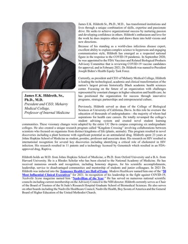
James E.K. Hildreth, Sr., Ph.D., M.D. President and CEO, Meharry Medical College, Professor of Internal Medicine
James E.K. Hildreth Sr., Ph.D., M.D., has transformed institutions and lives through a unique combination of skills, expertise and passionate drive. He seeks to achieve organizational success by nurturing passion and developing confidence inothers. Hildreth’s enthusiasmand love for the work he does inspires others and draws them into bold visions and new directions.
Because of his standing as a world-class infectious disease expert, excellent ability to explain complex science to laypersons and engaging communication style, Hildreth has emerged as a respected national figure in the response to the COVID-19 pandemic. In September 2020, he was appointed to the FDA Vaccines and Related Biological Products Advisory Committee that is reviewing COVID-19 vaccine candidates forapproval,and inFebruary 2021, Dr. Hildrethwas named toPresident Joseph Biden’s Health Equity Task Force
Currently, as president and CEO of Meharry Medical College, Hildreth is leading the technological, academic and clinical transformation of the nation’s largest private historically Black academic health sciences center. Focusing on the future of an organization with challenges represented by constant changes in higher education and health care, he has positioned the organization for success through innovative programs, strategic partnerships and entrepreneurial culture.
Previously, Hildreth served as dean of the College of Biological Sciences at University of California, Davis. In this role he oversaw the education of thousands of undergraduates the majority of whom had aspirations for health care careers. He totally revamped the college’s student advising system and created novel student learning communities. These visionary changes were adapted by the entire UC Davis campus comprising six undergraduate colleges. He also created a unique research program called “Kingdom Crossing” involving collaborations between scientists who focused on organisms from distinct kingdoms of life (plants, animals). This program resulted in novel discoveries including a plant hormone with significant potential as an antimalarial drug. Hildreth spent 23 years at Johns Hopkins School of Medicine as student, postdoc, professor and associate dean. His research on HIV resulted in international recognition for several key discoveries including identifying a critical role of cholesterol in HIV infection. His research resulted in 11 patents and a technology licensed by Genentech which resulted in an FDAapproved drug, Raptiva.
Hildreth holds an M.D. from Johns Hopkins School of Medicine, a Ph.D. from Oxford University and a B.A. from Harvard University. He is a Rhodes Scholar who has been elected to the National Academy of Medicine. He has received numerous awards and recognition, including honorary degrees, for his scientific accomplishments, leadership, service to disadvantaged communities and mentorship of students and junior colleagues. In 2021 Dr. Hildreth was inducted into the Tennessee Health Care Hall of Fame Modern Healthcare named him one of the “50 Most Influential Clinical Executives” for 2021. In recognition of his leadership in the fight against COVID-19, Nashville Scene magazine named him “Nashvillian of the Year.” He has served on numerous national scientific councils including currentmembership on theAdvisory Counciltothe NIH director. Hildrethcurrentlyserves as Chair of the Board of Trustees of the St Jude’s Research Hospital Graduate School of Biomedical Sciences. He also serves on otherboards including the Nashville Healthcare Council, Nashville Health, Boy Scouts ofAmerica and the General Board of Higher Education of the United Methodist Church.
Anil Shanker, M.S.,
Ph.D.
Senior Vice President for Research and Innovation, Meharry Medical College, Professor of Biochemistry, Cancer Biology, Neuroscience & Pharmacology
Focused on mission, excellence, community trust, and innovation, Dr. Anil Shanker builds strategic partnerships and operating systems that translate biomedical innovation into health impact especially for communities historically left out of discovery and care. As Senior Vice President for Research and Innovation at Meharry Medical College and a key member of the President's Executive Leadership Council, Dr. Shanker leads a $125M+ annual R&D enterprise, scaling tri-sector sponsored programs and collaborative networks 4.5-fold aligning strategy, governance, talent, and resource allocation to drive institutional transformation. Across complex external landscapes, Dr. Shanker forges mission-aligned alliances with NIH/NSF, major foundations, and biopharma to move programs from concept to execution. This includes NIH’s $165M+ AIM-AHEAD national consortium building AI talents, technology, and capacity, and multi-partner initiatives that expand diverse clinical research participation (e.g., Novartis/Sanofi Beacon of Hope and Chan Zuckerberg Initiative Accelerate Precision Health).
In moments that tested institutional resilience, Dr. Shanker drove crosscampus alignment and stakeholder diplomacy to secure reinstatement of $30M+ in NIH grants through compliance-forward risk remediation Dr. Shanker steers globally scaled, long-horizon science partnerships through the Diaspora Human Genomics Institute (a Meharry-incorporated nonprofit) to advance community-anchored data governance and global collaboration. He co-developed Together for CHANGETM a 10-year, $80M public–private consortium with Regeneron, AstraZeneca, Novo Nordisk, and Roche to build a genomics resource of ≥500,000 African-ancestry participants, among the largest global efforts, while enabling under-resourced institutions to lead discovery.
He published on practical roadmaps for institutional growth (Academic Medicine, Nature World View). Dr. Shanker serves on the U.S. National Academies of Science, Engineering and Medicine Board on Health Sciences Policy and the AAMC Research Advancement and Development leadership group. Distinctions include Overseas Fellow of the Royal Society of Medicine (UK), Fellow of the International Union Against Cancer, Champion of the Society for Immunotherapy of Cancer, and recognition by the United Nations Academic Impact. With more than three decades across three continents as a scientist-educator,Dr. Shankercompleted Harvard’s Institute forEducationalManagement executive program. Central to his work in cancer immunology is mentoring 200+ emerging leaders and building cultures of excellence, integrity, and execution so AI-enabled translational engines can advance health across all populations.
Dr. Shanker earned his PhD in biotechnology with a focus on tumor immunology from the School of Biotechnology atBanarasHindu University and his MSandBSdegrees inzoology (specialization incellbiology)fromthe University of Delhi. He conducted postdoctoral studies at the Centre d’Immunologie de Marseille-Luminy, France and worked as aScientistattheNationalCancerInstitute, Frederick,Maryland before joining Meharry MedicalCollege, Nashville, TN in 2010. He is also an adjunct faculty member at the Vanderbilt-Ingram Cancer Center; the Vanderbilt Institute for Infection, Immunology and Inflammation, and the Vanderbilt Memory and Alzheimer’s Center.
Tshilidzi Marwala, M.E.,
Ph.D.
Professor and Rector, United Nations University | UnderSecretary-General of the United Nations
Prof. Tshilidzi Marwala is the Rector of the United Nations University (UNU) and an Under-Secretary-General of the United Nations. Since assuming this role in March 2023, he has led the UN’s global academic think tank dedicated to advancing research, policy engagement, and capacity development to address pressing global challenges.
Prior to joining UNU, Prof. Marwala served as Vice-Chancellor and Principal of the University of Johannesburg, South Africa (2018–2023), where he strengthened the institution’s global research profile and international partnerships. He previously served as Deputy ViceChancellor for Research and Internationalization (2013–2017) and Executive Dean of the Faculty of Engineering and the Built Environment (2009–2013). Earlier in his career, he was Associate Professor and later Full Professor at the University of the Witwatersrand (2003–2008), and Executive Assistant to the Technical Director at South African Breweries (2001–2003). He was also a Postdoctoral Research Associate at Imperial College London.
Prof. Marwala holds a PhD in Artificial Intelligence and Engineering from the University of Cambridge (United Kingdom), a Master of Mechanical Engineering from the University of Pretoria (SouthAfrica), and a Bachelor of Science degree (magna cum laude) from Case Western Reserve University (USA). He has also completed executive leadership and management programmes at Columbia Business School and Harvard Business School.
An internationally recognized scholar, Prof. Marwala’s multidisciplinary research focuses on the theory and applications of artificial intelligence across engineering, social sciences, economics, finance, politics, and medicine. He is a co-holder of five patents and has served as a visiting scholar and professor at universities in the United States, the United Kingdom, China, and South Africa. Prof. Marwala has served on numerous global and national policy bodies and has worked closely with United Nations entities including UNESCO, UNICEF, WHO, and WIPO. He is a member of the UN Secretary-General’s Scientific Advisory Board and a Fellow of the American Academy ofArts and Sciences, The WorldAcademy of Sciences (TWAS), the Academy of Science of South Africa, and the African Academy of Sciences.
A prolific author, Prof. Marwala has written more than 25 books, including Leadership Lessons from Books I Have Read (2021) and Leading in the 21st Century: The Call for a New Type of African Leader (2021), along with numerous book chapters, journal and conference papers, and more than 250 magazine articles and newspaper opeds. His many honors include the Order of Mapungubwe, one of South Africa’s highest national awards, and the Science-for-Society Gold Medal from the Academy of Science of South Africa. In 2021 he was named IT Personality of the Year by the Institute of IT Professionals South Africa and was recognized among the 100 Most InfluentialAfricans of 2024 by NewAfrican Magazine
Richard J. Reddick, Ed.D.
Distinguished Service Professor and Senior Vice Provost for Undergraduate Education, The University of Texas at Austin
Dr. Richard J. Reddick is the inaugural Senior Vice Provost for Undergraduate Education at The University of Texas atAustin, where he serves as the university’s chief student success officer. In this role, he provides strategic leadership for undergraduate curriculum, enrollment, and academic success initiatives. He previously served as Dean and Senior Vice Provost of the Undergraduate College and as the inauguralAssociate Dean for Equity, Community Engagement, and Outreach in the College of Education.
A Distinguished Leadership Service Professor in the Program in Higher Education Leadership within the Department of Educational Leadership and Policy, Dr. Reddick has been a faculty member at UT Austin since 2007. He previously served as Assistant Director of the Plan II Honors Program in the College of LiberalArts and holds courtesy faculty appointments in African and African Diaspora Studies and the John L. Warfield Center forAfrican and African American Studies. He is also a Fellow of the Institute for Urban Policy Research andAnalysis and the Center for the Study of Race and Democracy. His university service includes co-chairing the Council for Racial and Ethnic Equity and Diversity (CREED), chairing The Eyes of Texas History Committee, and serving on the Signature Course Advisory Committee, the Faculty Working Group on Academic Integrity, and the Core Curriculum Task Force. In 2020, he was selected to the inaugural cohort of the Provost’s Distinguished Leadership Service Academy. Dr. Reddick’s influence extends nationally and internationally. In 2018, he was appointed Visiting Associate Professor at the Harvard Graduate School of Education, where he continues to serve as Faculty Co-Chair of the Institute for Educational Management and teaches in the Institute for Management Leadership in Education. His scholarship centers on cross-cultural and cross-identity mentoring, cultural taxation, faculty of color at historically White institutions, Black families in American society, and work–family balance among faculty, using ethnographic approaches to examine mentoring relationships and faculty experiences in higher education.
An award-winning teacher and scholar, Dr. Reddick has authored dozens of peer-reviewed journal articles and public essays and is the author or co-editor of four scholarly books. His most recent book, Restorative Resistance in Higher Education: Leading in an Era of Racial Awakening and Reckoning (Harvard Education Press, 2023), examines leadership, mentoring, and institutional responsibility during periods of social transformation. His research and commentary have been featured on NPR, the Associated Press, PBS, the BBC, CNN, and in publications including Fortune, Nature, and The Chronicle of Higher Education. His TED Talk, “Nurturing Relationships Across Difference,” was named a 2025 Editor’s Pick.
Dr. Reddick has received numerous honors recognizing his contributions to scholarship, teaching, and service, including UT Austin’s Eyes of Texas Excellence Award, the Outstanding Young Texas Ex Award, the John L. Warfield Center for African and African American Studies Teaching Award, the Black Faculty Staff Association Faculty Member of the Year Award, the Austin L.E.A.D.S. Award, the Outstanding Community Based Learning Professor Award at the Tower Awards, the President’s Award for Global Learning, the university’s Presidential Citation, and the Distinguished Alumni Award from The University of Texas at Austin.
A first-generation college graduate, Dr. Reddick earned his B.A. in Plan II Honors from UT Austin before teaching in Houston’s Fifth Ward and working in student affairs at MIT, Cal Poly San Luis Obispo, and Emory University. He later received his master’s and doctoral degrees from the Harvard Graduate School of Education, where he served as Class Marshal, edited the Harvard Educational Review, worked with the School Leadership Program, served as a School Director with Teach For America, and co-founded the Harvard Alumni of Color Conference.Beyond the academy, he is co-founder and board chair of Montessori For All, serves on the Austin Regional Board for IDEA Public Schools, co-hosts the NPR podcast Black Austin Matters, and has appeared on nationally televised game shows including Wheel of Fortune, Jeopardy!, Who Wants to Be a Millionaire?, and Trivial Pursuit
JasonA. Deakings, Ph.D., M.S.P.H. Assistant Professor and Associate Director, Center for Health Equity
University of Pittsburgh School of Public Health
Dr. Jason A. Deakings is a non-traditional researcher and educator whose work bridges academia, community leadership, and structural change. He serves as Assistant Professor and Associate Director for the Center for Health Equity at the University of Pittsburgh School of Public Health, where he advances innovative, community-centered approaches to addressing health inequities.
With more than 15 years of experience navigating the nonprofit and community development sectors, Dr. Deakings has centered his career on strengthening community capacity,expanding socialmobility, and improving supportservices for historically marginalized populations. His work focuses on developing actionable solutions to structural and systemic barriers that have disproportionately impacted Black communities. By leveraging cross-sector partnerships and community expertise, he has contributed tothe development of researchprotocols and procedures, secured and supported grant funding totaling over $5 million, and strengthened organizational capacity across multiple institutions.
Dr. Deakings’ scholarship primarily examines the interaction of Black communities within clinical and public health settings, particularly surrounding service uptake and persistent health disparities. He integrates mixed-methods and non-traditional research approaches with community engagement and healing-centered practices to improve health outcomes. His work has supported Black men, women, and young adults in addressing STI/STD stigma, reproductive health access,justice systeminvolvement, substance-use disorder, the built environment, and health literacy for communities most affected by policy-driven inequities.
Committed to expanding the boundaries of public health research, Dr. Deakings advances models that prioritize lived experience, trust-building, and culturally responsive methodologies. His work has been presented nationally and internationally, including at the United Nations, and continues toinform conversationson equity, systemschange, and community-informed research practices.
Beyond scholarship, Dr. Deakings is deeply committed to improving quality of life and wellbeing acrosscommunities by filling gaps where they exist and fostering environments rooted in dignity and empowerment. His guiding philosophy is to lead with love and to be “the salt of the Earth,” striving to positively transform the life course of all those with whom he shares space.
Vence L. Bonham, Jr., J.D. President and CEO,
The Diaspora Human Genomics Institute
Professor and Founding Director, Meharry Center for Bioethics, Social and Behavioral Research
Meharry Medical College
Vence L. Bonham, Jr., J.D., is President and Chief Executive Officer of the Diaspora Human Genomics Institute and Professor and Founding Director of the Meharry Center for Bioethics, Social and Behavioral Research at Meharry Medical College. An internationally recognized leader at the intersection of law, genomics, and bioethics, Professor Bonham’s work focuses on advancing health equity and examining the ethical, legal, and social implications of genomic science.
Prior to joining Meharry Medical College, Prof. Bonham held several senior leadership roles at the National Institutes of Health (NIH), including serving as Acting Deputy Director of the National Human Genome Research Institute (NHGRI). Before that appointment, he served as Senior Advisor on Genomics and Health Disparities, where he played a critical role in guiding national conversations on equity in genomic research and the responsible integration of genomic discoveries into clinical and public health practice. He was also a faculty member in the NHGRI Intramural Research Program, where his scholarship examined the societal implications of genomics, issues of race and identity in biomedical research, and strategies to address persistent health disparities.
Prof. Bonham’s academic career reflects a commitment to interdisciplinary scholarship and education. He previously held a tenured faculty appointment in the College of Human Medicine at Michigan State University and later served as an adjunct faculty member at the Saint Louis University School of Law while at the NIH. His teaching and research have influenced a generation of scholars and practitioners working at the intersection of biomedical science, public policy, and social justice. He earned his Juris Doctor from The Ohio State University Moritz College of Law and received a Bachelor of Arts with honors from Michigan State University. Throughout his career, Prof. Bonham has emphasized the importance of integrating legal and ethical perspectives into biomedical innovation, particularly as genomic technologies continue to transform health care.
Prof. Bonham has authored more than 100 peer-reviewed publications and is widely recognized for his leadership in advancing ethical and equitable approaches to genomic research. His contributions to science, mentorship, and public engagement have been honored with numerous awards, including the Advocacy Award from the American Society of Human Genetics and the Outstanding Faculty Mentorship Award from NHGRI. A dedicated mentor, he has guided many trainees into successful careers in medicine, law, public health, and bioethics.
Through his scholarship, leadership, and mentorship, Prof. Bonham continues to shape national and global conversations on the ethical integration of genomics into medicine and the pursuit of equitable health outcomes for diverse populations.
Eric L. Moore, Ph.D. Director, Army Research Laboratory, U.S. Army Combat Capabilities
Development Command (DEVCOM); Deputy to
the Commanding General, DEVCOM
Aberdeen Proving Ground, Maryland
Dr. Eric L. Moore currently serves as Director of the U.S. Army Combat Capabilities Development Command (DEVCOM) Army Research Laboratory in Adelphi, Maryland, and as the Deputy to the Commanding General at DEVCOM headquarters at Aberdeen Proving Ground. In this dual leadership role, he provides strategic direction for the Army’s foundational research enterprise and helps guide research, development, and engineering initiatives that advance the technological superiority of the United States Army.
Dr. Moore assumed leadership of the Army Research Laboratory on October 1, where he leads the Army’s only foundational research laboratory. The Army Research Laboratory bridges academia, industry, and Army expertise to ensure that basic scientific discovery drives innovation and the development of transformative technologies that give warfighters a decisive advantage now and in the future. Under his leadership, the laboratory oversees highly skilled teams of technical experts working across a broad spectrum of scientific disciplines, including quantum computing, advanced materials, energy sciences, biotechnology, and human performance.
Based in Adelphi, Maryland, the Army Research Laboratory operates seven locations across the United States, housing world-class research facilities and collaborative partnerships designed to accelerate innovation and technological advancement. Dr. Moore brings nearly four decades of combined military and federal service to his current role and was appointed to the Senior Executive Service on August 21, 2016. Within the Army, senior executives serve as the civilian counterparts of general officers. Prior to his appointment as director, Dr. Moore served for three years as Deputy to the Commanding General at DEVCOM headquarters, where he provided strategic leadership and served as a catalyst for research, development, and engineering initiatives across the command.
Earlier in his career, Dr. Moore served as Director of the DEVCOM Chemical Biological Center, formerly known as the Edgewood Chemical Biological Center, and his first assignment in the Senior Executive Service was as the Center’s Director for Research and Technology.
An expert in chemical and biological defense programs and medical countermeasures, he previously held several leadership positions at the Defense Threat Reduction Agency, including Chief of the Advanced and Emerging Threat Division and Chief of the Basic and Supporting Science Division. Prior to entering civilian service, Dr. Moore served as a U.S. Army officer. His military career culminated at the Armed Forces Medical Intelligence Center, where he served as the Defense Intelligence Agency’s Senior Scientific and Technical Intelligence Officer responsible for chemical, biological, radiological, and nuclear medical countermeasures worldwide.
Dr. Moore earned his doctorate in neurophysiology from Meharry Medical College and a bachelor’s degree in biology from Fisk University. He was commissioned as a U.S. Army officer through the U.S. Army ROTC program at Vanderbilt University.
His distinguished military and civilian service has been recognized with numerous honors, including the Federal Laboratory Consortium Laboratory Director of the Year Award, the Department of Defense Distinguished Civilian Service Award, the Defense Meritorious Service Medal (with one Oak Leaf Cluster), the Joint Service Commendation Medal, the Army Commendation Medal, and the National Defense Service Medal with one bronze star.
Tameka A. Clemons, Ph.D.
Clinical Associate Professor of Biochemistry, Tilman J. Fertitta
Family College of Medicine, University of Houston
Dr. Tameka A. Clemons is a Clinical Associate Professor of Biochemistry at the Tilman J. Fertitta Family College ofMedicine atthe University ofHouston, where she is dedicated toadvancing medicaleducation and biomedicalresearch while mentoring the next generation of scientists and physicians. A native of Detroit, Michigan, Dr. Clemons has built a career grounded in academic excellence, scientific discovery, and a strong commitment to expanding opportunities in biomedical science.
Dr. Clemons earned her Bachelor of Science degree in Chemistry from Xavier University of Louisiana, a historically Black university known for producing a large number of African American graduates who go on to careers in medicine and science. She later received her Ph.D. in Biochemistry from Meharry Medical College, where she was named an Alfred P. Sloan Fellow in recognition of her academic promise and research potential. Her doctoral training provided a strong foundation in biochemical research and prepared her for a career that bridges scientific discovery with education and mentorship.
Throughout her career, Dr. Clemons has been actively engaged in research focused on understanding complex biological processes and advancing knowledge in neurodegenerative disease. Her current research interests include the molecular and biochemical mechanisms underlying Alzheimer’s disease, with a particular focus on identifying pathways that may contribute to disease progression and potential therapeutic targets. Her work contributes to broader scientific efforts aimed at addressing the growing public health burden of neurodegenerative disorders.
Dr. Clemons has successfully secured research support through several competitive grants, including the prestigious Research Initiation Award from the National Science Foundation. Her contributions to science and education have also been nationally recognized. In 2020, she was featured by Cell Press as one of the “100 Inspiring Black Scientists in America,” an honor that highlights scientists whose work and leadership are helping to shape the future of research and innovation.
In addition to her research accomplishments, Dr. Clemons is deeply committed to teaching and mentorship. As a faculty member at the Tilman J. Fertitta Family College of Medicine, she teaches biochemistry to medical students, helping to prepare future physicians with a strong foundation in biomedical science. She considers mentorship to be one of the most rewarding aspects of her career and has guided more than twenty students through research projects, many of whom have gone on to pursue careers in science, medicine, and related fields.
Through her work as a researcher, educator, and mentor, Dr. Clemons remains committed to fostering scientific curiosity, supporting emerging scholars, and contributing to advancements that improve human health. She continues to inspire students and colleagues alike through her dedication to science, education, and the cultivation of future leaders in biomedical research.
Leah Alexander, Ph.D., M.P.H.
Associate
Professor, Department
of Public Health School of Global Health,
Meharry
Medical College
Dr. Leah Alexander is an Associate Professor in the Department of Public Health within the School of Global Health at Meharry Medical College in Nashville, Tennessee. With more than 20 years of experience in public health education and community-engaged research, Dr. Alexander has dedicated her career to advancing health equity through scholarship, leadership, and meaningful community partnership.
A proud graduate of Spelman College, Dr. Alexander earned both her Master of Public Health and Doctor of Philosophy degrees from the University of Alabama at Birmingham. Her academic and professional journey reflects a longstanding commitment to addressing structural inequities that shape health outcomes, particularly within historically marginalized communities.
Dr. Alexander’s research centers on building authentic partnerships that honor community expertise as essential to effective public health practice. Her funded research portfolio includes initiatives focused on HIV prevention and increasing PrEP awareness among African American women and students attending Historically Black Colleges and Universities (HBCUs). She has also led projects examining health promotion within faith-based organizations, leveraging trusted community institutions to advance preventive health strategies. Her work in public interest technology explores how digital tools and innovation can be used ethically and effectively to reduce health disparities and amplify community voices. A significant component of Dr. Alexander’s scholarship is dedicated to maternal health equity. She is the founding Director of a Health Resources and Services Administration (HRSA)-funded Maternal Health Excellence Research Center (MHERC), where she leads interdisciplinary efforts to strengthen research, education, workforce development, and advocacy aimed at improving maternal health outcomes. Under her leadership, the center advances evidence-based solutions while prioritizing culturally responsive care and systems-level change.
In 2024, Dr. Alexander co-founded the Public Health Arts and Technology (PHAT) Lab, an innovative initiative designed to integrate art, storytelling, and technology into public health research and practice. The PHAT Lab seeks to elevate community narratives, foster creative engagement, and expand the ways public health communicates and collaborates with the populations it serves.
In addition to her research and leadership roles, Dr. Alexander is a dedicated educator and mentor who prepares future public health professionals to approach their work with cultural humility, scientific rigor, and a justicecentered lens. Her teaching emphasizes the importance of ethical engagement, interdisciplinary collaboration, and sustainable community impact.
Dr. Alexander lives in Nashville with her husband, Dr. Olayinka Otukpe and two children, Iyanu and Imisi, who continually inspire her commitment to justice-driven public health.
Karen M. Winkfield, M.D., Ph.D.
Ingram Professor of Cancer Research
Executive Director, Meharry–Vanderbilt Alliance Professor of Radiation Oncology, Vanderbilt University Medical Center
Dr. Karen M. Winkfield is a nationally recognized radiation oncologist, physician-scientist, and implementation scientist whose work focuses on advancing cancer care and eliminating health disparities. She is the Ingram Professor of Cancer Research at Vanderbilt University School of Medicine, Executive Director of the Meharry–VanderbiltAlliance, and Professor of Radiation Oncology at Vanderbilt University Medical Center. Through her clinical leadership, research, and community-engaged scholarship, Dr. Winkfield works to improve equitable access to highquality cancer care and to expand participation of underrepresented communities in clinical research.
Before joining Vanderbilt University in 2020, Dr. Winkfield served at Atrium Health Wake Forest Baptist, where she was Associate Director for Community Outreach and Engagement and Director of the Office of Cancer Health Equity. In these roles, she led initiatives focused on addressing cancer disparities and strengthening partnerships between academic medical centers and the communities they serve. Dr. Winkfield’s academic journey reflects extraordinary determination and achievement. Born in 1970 into a family of Jehovah’s Witnesses who were opposed to formal education, she pursued higher education and scientific training with distinction. She earned a Bachelor of Science in Biochemistry from Binghamton University before completing both a Ph.D. in Pathology (2004) and an M.D. (2005) at the Duke University School of Medicine. While at Duke, she became the second Black woman to complete the prestigious Medical Scientist Training Program (MSTP), which prepares physician-scientists to integrate biomedical research with clinical practice. She subsequently completed her residency in radiation oncology at Harvard University.
An internationally respected voice in oncology and health equity, Dr. Winkfield is an implementation scientist whose research focuses on translating scientific advances into practical strategies that improve cancer outcomes for underserved populations. Her work emphasizes community engagement, culturally responsive care, and innovative approaches to increasing participation in cancer research and treatment. She is the co-founder and director of the Association of Black Radiation Oncologists, an organization dedicated to advancing diversity, mentorship, and equity within the field of radiation oncology.
Dr. Winkfield also plays a significant leadership role in national public health initiatives. She co-leads the Inclusive Participation Workgroup of the National Institutes of Health Community Engagement Alliance (CEAL) teams working to address COVID-19 disparities, helping to promote trust, community partnership, and equitable access to health information and services.In recognition of her leadership and impact, Dr. Winkfield was appointed by U.S. President Joe Biden in September 2021 to serve a six-year term on the National Cancer Advisory Board, which advises the National Cancer Institute on national cancer research priorities and policy. She has also been recognized as one of the 100 Influential Women in Oncology by OncoDaily.
Through her clinical practice, research, mentorship, and national leadership, Dr. Winkfield continues to advance cancer research and champion equitable care for patients and communities across the United States.
The Honorable Marsha Blackburn, United States Senator from Tennessee
In 2018, the people of Tennessee elected Marsha Blackburn as the first woman to represent the Volunteer State in the United States Senate. As Tennessee’s senior senator, she serves on the Deputy Whip Team and on the Finance; Commerce, Science & Transportation; Veterans’ Affairs; and Judiciary Committees. She also serves as Ranking Member on the Commerce Subcommittee on Consumer Protection, Product Safety, and Data Security and the Judiciary Subcommittee on Human Rights and the Law.
Blackburn has dedicated her public service to promoting opportunity for Tennesseans, breaking barriers for women, and strengthening America’s prosperity. She began her public service career in 1995 as Executive Director of the Tennessee Film, Entertainment, and Music Commission. While serving in the Tennessee State Senate, she led a successful statewide grassroots campaign to defeat a proposed state income tax, earning a reputation as an anti-tax champion. Before her election to the Senate, she represented Tennessee’s 7th Congressional District in the U.S. House of Representatives, where she became a leading advocate for a smaller, more accountable federal government.
In the Senate, Blackburn has focused on fiscal responsibility, economic growth, and strategic trade policies to strengthen U.S. competitiveness. She introduced legislation calling for 1%, 2%, and 5% across-the-board reductions in nondefense, non-veterans, and non-homeland security spending to curb excessive federal expenditures. A strong supporter of the military, she champions policies supporting active duty servicemembers, National Guard members, and their families, and in 2022 successfully led the repeal of the military’s COVID-19 vaccine mandate. Blackburn has also been a leading advocate for veterans, expanding access to health care through the community care program and introducing the bipartisan Transparency and Effective Accountability Measures (TEAM) Veteran Caregivers Act and the Strengthening VA Cybersecurity Act, both signed into law. Her Rural Health Agenda proposes legislation to expand care access, support providers, and help communities integrate health care into local economic development. Throughout her time in Congress, Blackburn has worked to hold the Chinese Communist Party accountable, advancing legislation addressing Beijing’s influence over global supply chains, technology infrastructure, and international organizations. Following China’s crackdown in Hong Kong in 2019, she led bipartisan legislation signed into law by President Trump prohibiting the export of crowd-control equipment to the Hong Kong Police Force. Her bipartisan Open Technology Fund Authorization Act, supporting global internet freedom against authoritarian censorship, was also enacted into law.
Blackburn has also worked to strengthen the U.S.–Taiwan relationship, visiting Taiwan in August 2022 to meet with President Tsai Ing-wen and reaffirm support for stronger economic and diplomatic ties. A longtime advocate for the creative community, she has championed initiatives including the AM/FM Act, the HITS Act, and tax classification reforms for self-employed workers included in the CARES Act. In the House, she founded the bipartisan Songwriters Caucus and helped pass the Music Modernization Act, which modernized music licensing, and the BOTS Act, empowering the Federal Trade Commission to combat digital ticket scalping.
On border policy, Blackburn has advocated fully funding the U.S. Border Patrol, resuming construction of a physical barrier, imposing tougher penalties for drug smuggling, and combating cross-border human trafficking. Throughout her public service, Blackburn has also worked to protect women and children. She introduced the Kids Online Safety Act, and in 2024 her bipartisan REPORT Act, requiring technology companies to report child sex trafficking to the National Center for Missing and Exploited Children, was signed into law. She also introduced the Speak Out Act, enacted in 2022 to prohibit nondisclosure agreements in cases of sexual harassment and assault. In 2019, her bipartisan Women’s Suffrage Centennial Commemorative Coin Act, honoring the 100th anniversary of women gaining the right to vote, was signed into law by President Trump.
Eric Floyd, M.S., Ph.D., M.B.A Chief Regulatory and Quality Officer, Silence Therapeutics; Principal and CEO, Floyd Regulatory Strategic Consulting; Chairman, Meharry Board of Trustees Research and Innovation Committee
Dr. Eric Floyd is the Chief Regulatory and Quality Officer at Silence Therapeutics and a seasoned pharmaceutical executive with more than 28 years of experience in regulatory affairs and global drug development. Throughout his career, he has held senior regulatory leadership roles of increasing responsibility at leading pharmaceutical and biotechnology companies, including Merck, Aventis, Novartis, Lundbeck, Axovant Sciences, and Neurogene Inc. His work has focused on advancing innovative therapies and addressing the unmet medical needs of patients who have limited or no treatment options.
Prior to joining Silence Therapeutics, Dr. Floyd served as Chief Regulatory Officer at Neurogene Inc., a biotechnology company dedicated to developing life-changing genetic medicines for patients and families affected by rare and devastating neurological diseases. Earlier in his career, he served as Global Head of Regulatory Affairs at Axovant Sciences and as U.S. Head of Regulatory Affairs at Lundbeck. In these roles, he provided strategic regulatory leadership and guided clinical development programs that resulted in multiple successful drug approvals, including orphan drug approvals for Sabril, Onfi, and Northera, as well as approvals for treatments addressing major depressive disorder (Trintellix) and schizophrenia (Rexulti).
Dr. Floyd is also the Principal and Chief Executive Officer of the Floyd Regulatory Strategic Consulting Group, LLC, where he provides strategic regulatory guidance to biotechnology and pharmaceutical organizations navigating complex regulatory pathways.
In addition to his industry leadership, Dr. Floyd is actively engaged in academic and advisory roles. He serves as an adjunct faculty member in the Department of Neuroscience at both Harvard Medical School and Wake Forest University School of Medicine, where he contributes to education and mentorship in neuroscience and translational medicine. He has also held several governance and advisory positions in the biotechnology and investment sectors, including serving as a board member of Scilex Pharmaceuticals, former Chairman of the Board of Virpax Pharmaceuticals, and an advisor to 2Flo Ventures.
Dr. Floyd is deeply committed to academic and institutional leadership and currently serves as an alumni member of the Board of Trustees of Meharry Medical College as well as the University of Illinois Alumni Association. His dedication to education and mentorship extends beyond his professional roles, as he actively supports programs that encourage students to pursue careers in science, medicine, and biotechnology.
Dr. Floyd earned his undergraduate degree in Biology from the University of Illinois, a Master’s degree in Neuroscience from Tennessee State University, and a Doctorate in Neurophysiology from Meharry Medical College. He also holds an Executive MBA in Pharmaceutical Marketing from Saint Joseph’s University and an MBA in International Business from INSEAD Business School in Fontainebleau, France, reflecting his strong foundation in both science and global business leadership.
Outside of his professional responsibilities, Dr. Floyd is passionate about mentorship and community engagement. He dedicates time to mentoring high school and college students and actively participates in community service initiatives through Alpha Phi Alpha Fraternity, Inc.
MD, DHL(Hon), FAAFP
Executive Vice President and Provost, Meharry Medical College
Dr. Jeannette E. South-Paul joined Meharry Medical College as the Executive Vice President and Provost in December 2021. Prior to this appointment, she was the Andrew W. Mathieson UPMC Professor and Chair of the Department of Family Medicine at the University of Pittsburgh School of Medicine from 2001 – 2020 retiring from Pitt in 2020. Prior to joining the faculty at the University of Pittsburgh School of Medicine and UPMC, she served as a Medical Corps officer in the U.S. Army, retiring in 2001 while serving as Chair of Family Medicine at the Uniformed Services University of the Health Sciences and previously as Vice President for Minority Affairs at the same institution.
Dr. South-Paul has served in leadership positions in the Society of Teachers of Family Medicine (STFM), the American Academy of Family Physicians (AAFP), the Association ofAmerican Medical Colleges (AAMC), and the Association of Departments of Family Medicine (ADFM) to include serving as President of the Uniformed Services Academy of Family Physicians (USAFP) and the STFM.
After more than 10 years of service as a member of the Meharry Medical College Board of Trustees, Dr. South-Paul stepped off the Board to begin her new leadership role. She is excited to collaborate with the academic leaders of the five schools (Medicine, Dentistry, Graduate Studies, Applied Computational Sciences and Global Health) of this historic institution as she continues to serve in academe. She is a member of the NationalAcademy of Medicine, the Gold Humanism Society, and the Alpha Omega Alpha Medical Honorary Society
Dr. South-Paul is a sought-after speaker by academic, clinical and community organizations as well as health industry groups who are seeking perspectives on health disparities, workforce diversity, leadership and racial and social justice.
Jeannette E. South-Paul,
Jasbir Dhaliwal, Ph.D. Distinguished University Professor and Executive Vice President for Research & Innovation, University of Memphis
Dr. Jasbir Dhaliwal serves as Distinguished University Professor and Executive Vice President for Research & Innovation at the University of Memphis, where he provides strategic leadership for the university’s research enterprise and innovation initiatives. In this capacity, he advances interdisciplinary collaboration, strengthens partnerships with industry and government, and fosters an environment that supports technology commercialization and entrepreneurial engagement. As part of these responsibilities, Dr. Dhaliwal also leads the FedEx Institute of Technology, an advanced technology research organization that serves as the front door to the university’s research capabilities and innovation infrastructure. The institute promotes interdisciplinary research clusters, corporate engagement, entrepreneurship, and technology commercialization. In addition, he serves as Executive Director of the University of Memphis Research Foundation and is the founding President of its innovation subsidiary, UMRF Ventures Inc.
Prior to his current role, Dr. Dhaliwal served as Vice Provost for Academic Affairs and Dean of the University of Memphis Graduate School, where he provided strategic oversight for the university’s portfolio of 129 graduate programs enrolling approximately 4,800 master’s and doctoral students. He holds master’s and doctoral degrees from the University of British Columbia in Canada and has held international leadership roles including Deputy Director of the Center for Management of Technology at the National University of Singapore. Earlier in his career, he founded the first Canadian university-based internet incubator at what is now the School of Interactive Arts and Technology at Simon Fraser University. An internationally recognized scholar in information systems, Dr. Dhaliwal’s research focuses on the testing of complex, large-scale, real-time global information systems. He has published extensively in leading scholarly journals including Information Systems Research, IEEE Transactions on Engineering Management, IEEE Software, European Journal of Information Systems, International Journal of Electronic Commerce, Journal of Strategic Information Systems, and Communications of the Association for Information Systems. He is also co-author of the book E-Business Innovation, published by Prentice Hall/Pearson Education, and has served as Program Chair of the Pacific Asian Conference on Information Systems.
In recognition of his academic leadership, Dr. Dhaliwal was awarded the Papasan Family Professorship for Exemplary Leadership in 2013 for founding the Systems Testing Excellence Program, now the largest software testing research center in academia. His research has attracted more than $23 million in external funding and includes projects with organizations such as FedEx, the U.S. Department of Defense, the Defense Information Systems Agency, the Department of Homeland Security, IBM, Microsoft, Johnson & Johnson, Ericsson Telecommunications, Nomura Japan, and the Port of Singapore Corporation. Dr. Dhaliwal has also contributed to national research and policy leadership. He is a Past President of the Tennessee Conference of Graduate Schools, was appointed by Governor Bill Haslam to the Tennessee State Energy Policy Council, and has served on the Advancement Committee of the Council of Graduate Schools. He remains active with the Council for Competitiveness and, in 2022, was elected to the Executive Committee of the Council on Research of the Association of Public and Land-Grant Universities. Under his leadership, the University of Memphis expanded its research enterprise, with research awards increasing by more than 250 percent, National Science Foundation funding rising 147 percent, and research expenditures growing to nearly $100 million while doubling the university’s patent portfolio. He also led the launch of the university’s first research park for technology companies. These efforts helped the university achieve Carnegie R1 designation in 2021 and earn the AASCU Excellence and Innovation Award for Regional and Economic Development (2018), as well as recognition as a national finalist for the APLU Innovation and Economic Prosperity Award (2020).
Aramandla Ramesh, Ph.D. Associate Vice President for Research and Innovation, Professor, Department of Biochemistry, Cancer Biology, Neuroscience & Pharmacology, Meharry Medical College
Dr. Aramandla Ramesh serves as Associate Vice President for Research & Innovation. An internationally recognized toxicologist and environmental health scientist, Dr. Ramesh’s research focuses on the mechanisms by which environmental pollutants contribute to toxicity and cancer, with particular emphasis on polycyclic aromatic hydrocarbons (PAHs) and perand polyfluoroalkyl substances (PFAS).
Dr. Rameshearnedhis firstPh.D. inMarineMicrobiology from Annamalai University in India in 1986 and a second Ph.D. in Environmental Toxicology from Ehime University inJapan in1992. His areas of expertise include the bioavailability, toxicokinetics, biotransformation, and acute and subchronic toxicity of PAHs and PFAS, chemicals widely distributed in the environment and known for their potential impacts on human health.
Current research in Dr. Ramesh’s laboratory investigates the role of environmental toxicants in colon cancer, particularly the effects of benzo(a)pyrene (BaP), a fat-soluble compound belonging to the PAH family. His studies have demonstrated that exposure to BaP and related PAHs through diets high in saturated fats can induce cytochrome P450 (CYP) family enzymes, leading to the formation of reactive metabolites that persist in target tissues and contribute to increased DNA damage. Ongoing research in his laboratory seeks to better understand how environmental exposures combined with dietary practices such as excessive consumption of meat and fat products contaminated with pollutants contribute to toxicity and cancer development.
In addition to his research, Dr. Ramesh has extensive experience mentoring and training undergraduate and graduate students in environmental health and toxicology. His work has been supported by major federal agencies including the U.S. Department of Energy, the National Institutes of Health, the National Science Foundation, and the U.S. Environmental Protection Agency.
Dr. Ramesh is widely recognized for his contributions to the field of toxicology and serves on the editorial boards of several leading scientific journals, including Toxicology Mechanisms & Methods, Archives of Environmental Contamination & Toxicology, Journal of Toxicology & Environmental Health, Environmental Chemistry & Ecotoxicology, Journal of Applied Toxicology, Toxicology & Applied Pharmacology, and Polycyclic Aromatic Compounds. Through his research, mentorship, and leadership, Dr. Ramesh continues to advance scientific understanding of environmental toxicants and their role in human disease.
Nicole C. Kleinsteuer, Ph.D.
National
Institutes of Health (NIH)
Deputy Director for Program
Coordination, Planning, and Strategic Initiatives
Nicole C. Kleinstreuer, Ph.D., is the NIH Deputy Director for Program Coordination, Planning, and Strategic Initiatives. In this role, she leads the Division of Program Coordination, Planning, and Strategic Initiatives (DPCPSI) within the NIH Office of the Director, which oversees trans-NIH programmatic research and strategic policy initiatives, including the NIH Common Fund and offices focused on women’s health, data science, AIDS research, disease prevention, behavioral and social sciences, dietary supplements, and tribal health, among others.
Dr. Kleinstreuer is internationally recognized for her leadership in developing innovative, human-relevant research strategies that advance public health protection. Prior to her current position, she served as Director of the National Toxicology Program Interagency Center for the Evaluation of Alternative Toxicological Methods (NICEATM), within the National Institute of Environmental Health Sciences (NIEHS). She also served as Executive Director of the congressionally mandated Interagency Coordinating Committee on the Validation of Alternative Methods (ICCVAM) and as the US National Co-Coordinator for the Organization for Economic Cooperation and Development (OECD) Test Guidelines Programme. In these roles, she led interagency and international efforts to promote new approach methodologies (NAMs), reduce animal testing, and integrate computational modeling, artificial intelligence, and systems toxicology into regulatory science. Her work spans translational bioinformatics, predictive modeling, and quantitative risk assessment. She has authored over 150 peer-reviewed publications and received numerous honors, including the 2019 Society of Toxicology Achievement Award and the 2025 Enhancement of Animal Welfare Award, as well as multiple NIH Director’s and NIEHS Merit Awards.
Dr. Kleinstreuer holds B.S. degrees in biomedical engineering and applied mathematics from the University of North Carolina at Chapel Hill and a Ph.D. in bioengineering from the University of Canterbury. She completed postdoctoral training in computational toxicology at the U.S. Environmental Protection Agency and holds adjunct faculty appointments at Yale University and UNC Chapel Hill. She is deeply committed to mentorship, public health protection, and scientific innovation that enhances the translation of biomedical research to real-world impact.
Jayanta Bhattacharya, M.D., Ph.D.
Director of the National Institutes of Health (NIH)
Jayanta "Jay" Bhattacharya, M.D., Ph.D., took the helm as 18th director of the National Institutes of Health, the nation’s medical research agency, on April 1, 2025. President Trump nominated Dr. Bhattacharya for the position on Nov. 26, 2024, and the U.S. Senate confirmed him on March 25, 2025.
Dr. Bhattacharya, a renowned doctor, researcher, health economist, previously held a tenured professorship in the medical school at Stanford University in California. His research focused on population aging and chronic disease, particularly on the health and well-being of vulnerable populations. He has published over 170 research papers in peer-reviewed journals in medicine, epidemiology, health policy, economics, statistics, science policy, and public health, as well as a leading textbook on health economics.
During the pandemic, Dr. Bhattacharya coauthored the Great Barrington Declaration, which called for opening schools and lifting lockdowns while better protecting older populations who were most vulnerable to the disease.
Dr. Bhattacharya held numerous additional appointments at Stanford University, including courtesy appointments at the Stanford Institute for Economic Policy Research, the Stanford Freeman Spogli Institute and Stanford’s Hoover Institution, and Economics department. Previously, he conducted research at the National Bureau of Economic Research and the SPHERE Institute, a policy research firm. Before joining Stanford, he was an economist at the RAND Corporation and worked as a visiting economics professor at the University of California, Los Angeles.
Dr. Bhattacharya is a longtime NIH grantee and has served as a standing member of multiple NIH review committees. He earned his bachelor’s and master’s degrees in economics from Stanford University. He then completed medical school and earned a Ph.D. in economics also from Stanford University.
Dr. NeslonAdams
Rajbir Singh, M.D., Ph.D.
Associate Professor of Internal Medicine at Meharry Medical College; Executive Director of the Center of Excellence for Clinical and Translational Research; Executive Director of Precision Medicine Health Trials Design; Principal Investigator of the GREAT Health Study
Dr. Rajbir Singh is an Associate Professor of Internal Medicine at Meharry Medical College, where he serves as Executive Director of the Center of Excellence for Clinical & Translational Research, Executive Director for Precision Medicine Health Trials Design, and Co-Director of the Center for Women’s Health Research; he also chairs the Institutional Review Board. His work advances health for all and inclusion focused clinical trials and precision medicine through community-engaged genomics, biorepositories, and data infrastructure. Dr. Singh leads or collaborates on major initiatives including ECSTATIC (CTSAconsortium), the NHGRI-funded Center for Genome Research at Meharry, AIM-AHEAD, PCORnet/MSCDRN, and industry partnerships such as Sanofi’s Beacon of Hope and Genentech’s Lighthouse trial.
Trained at the All India Institute of Medical Sciences (AIIMS) in New Delhi for his MBBS (MD) he has co-authored publications in Clinical and Translational Science, Genetics in Medicine, and the Journal of the American Heart Association, and is a frequent national speaker on inclusive trial design.
He is the lead Principal Investigator of the GREAT (Genomics REsearch Advancing Transformational) Health Study.
SACS-V-1-26
DETECTING EARLY DISEASE-ASSOCIATED INSTABILITY BY MODELING GENOME ARCHITECTUREASADYNAMIC, HIGH-DIMENSIONAL SYSTEM
1
Pamela Djan1,Aize Cao1
School ofApplied Computational Sciences, Meharry Medical College, Nashville, TN
Purpose & Rationale: Many genomic studies rely on low-dimensional and static analyses that limit the ability to detect early regulatory and structural changes associated with disease. These approaches often fail to integrate three-dimensional genome organization with regulatory activity and nuclear positioning, reducing their ability to capture coordinated genome remodeling. This study aims to develop a mechanistic, high-dimensional framework that models genome architecture as a dynamic system governing disease initiation, progression, and therapy response. The primary goal is to identify coordinated genome instability before major molecular or clinical changes occur. Methods: A genomic coordinate system was constructed by integrating structural, regulatory, and nuclear organization features derived from experimentally grounded epigenomic and three-dimensional genome datasets. Genomic regions were represented as trajectories across multiple biological conditions. The framework was implemented and evaluated on human chromosomes 17 and 8 to assess numerical stability, biological relevance, and internal consistency. Results: The coordinate system was successfully established on chromosomes 8 and 17 and demonstrated stable and biologically coherent behavior across dimensions and conditions. Major features exhibited appropriate dynamic range and internal consistency. Regions with coordinated regulatory andstructural disruption wereconsistently detectedand formed localizedpatternsrather than diffuse background signals. Diagnostic analyses indicated that these patterns were not driven by technical artifacts. Conclusion: This work establishes a validated, high-dimensional representation of genome architecture as a dynamic system. The framework provides a foundation for systematic investigation of genome remodeling and supports future studies across chromosomes and disease contexts. This platform enables mechanistic analysis of early genome instability with potential relevance to precision medicine and therapeutic response.
Project support: [This research is supported by HRSAgrant UR650342]
SACS-V-2-26
PREDICTING CANCER SURVIVAL RATES USING MACHINE LEARNING
School ofApplied Computational Science, Meharry Medical College, Nashville, TN
Purpose & Rationale: Early cancer detection significantly improves survival outcomes, yet prognostic assessment remains challenging for oncologists. Machine learning offers promise in identifying complex patterns within clinical and genomic data that traditional statistical methods may miss. This study develops a predictive model to support oncologists in prognostic decisionmaking for cancer survival outcomes. Methods: This study utilized datasets from SEER, cBio Portal, CibersortX, and AllofUs, a comprehensive dataset over 150,000 patient samples . Data preprocessing included handling missing values, feature engineering for tumor sites, one -hot encoding of categorical variables, and standardization of numerical features, resulting in 538 final features. The target variable was binary overall survival status (LIVING=0, DECEASED=1). Three machine learning models : Logistic Regression, XGBoost, and XGB -Lite were trained and evaluated. Critical to this analysis was identifying and correcting data leakage, where the "Overall Survival (Months)" feature was inappropriately predictive. Results: The initial XGBoost model achieved 88% accuracy with 95% ROCAUC, but after removing the leakage variable, the final XGB –Lite model achieved more realistic metrics: 80% accuracy, 0.7628 precision, 0.8092 recall, and 0.8796 ROC AUC. The most influential features in the corrected model were Current Age, Fraction Genome Altered, Sample Coverage, and MSI Score; all clinically relevant factors in cancer prognosis. Conclusion: This research demonstrates successful development of a machine learning model for cancer survival prediction using rigorous methodology. The final model represents a promising step toward a clinical decision-support tool for oncologists. Future work should focus on external validation, exploration of additional biomarkers, and refinement using advanced deep learning techniques. This research was supported by Meharry Medical College.
SACS-P-1-26
PROT-B-GAN:ADVERSARIAL GENERATIVE MODELING FOR HYPOTHESIS DISCOVERY IN BIOLOGICAL KNOWLEDGE GRAPHS
Jaylin Dyson1,Aize Cao1
1School ofApplied Computational Sciences, Meharry Medical College Nashville TN USA
Knowledge graph completion (KGC) has emerged as a powerful approach for protein function prediction and drug discovery by integrating heterogeneous biological data. However, existing KGC pipelines often overproduce high-degree hub entities, recycle trivial tail predictions, and provide limited interpretability, reducing their practical utility in protein informatics. We introduce Prot-B-GAN, a framework to overcome these limitations by integrating semantic grounding, structural reasoning, and query aware context modeling. Prot-B-GAN’s framework builds on the established effectiveness of Generative Adversarial Networks (GANs), consisting of two networks: a generator and a discriminator. The generator generates candidate tails given (head, relation) as a KGC problem. The generator includes a composite loss function that promotes realism and helps fight hub bias through a frequency-aware penalty and a lightweight diversity or rare-entity reward. The discriminator serves as a classifier labeling realism. In an adversarial manner, the two components train each other to improve performance on the KGC and labeling tasks, while also supporting interpretability through a query-aware evidence subgraph or contextaware explanation for predictions. We evaluate Prot-B-GAN on the PRO-HGT, OGB-BioKG, and YAGO3-10 benchmarks, demonstrating improved ranking performance, reduced hub dominance, and greater diversity in predicted protein functions and repurposing-relevant drug associations (for example drug target and drug disease links) compared with existing KGC models. By explicitly rewarding rare yet plausible entities and producing interpretable, context aware predictions, ProtB-GAN provides a principled framework for protein function prediction and drug repurposing in biomedical knowledge graphs.
Funding: The National Science Foundation HBCU Excellence Research grant no. 2302637
1Department of Biomedical Data Science, School of Applied Computational Sciences, Meharry Medical College, Nashville, TN
2School ofApplied Computational Sciences, Meharry Medical College, Nashville, TN
Background: Pharyngitis, commonly referred to as sore throat, has multiple etiologies that impact both treatment and transmission of the disease. Pharyngitis caused by bacterial infections require antibiotics however, nonbacterial and viral infections are treated with rest. Misdiagnosing etiology can potentially lead to antibiotic resistance and disease transmission. Rationale & Goal: Pharyngitis can be diagnosed by a clinician however, rapid antigen detection tests (RADTS) are necessary to diagnose etiology. Due to the cost and resources necessary to correctly diagnose pharyngitis etiology, there is a need for better diagnostic tools. This study aimed to investigate the relationship between clinical symptoms and pharyngitis etiology and predict pharyngitis etiology using clinical symptoms. Methods: We performed a retrospective study using an open-source dataset from a 2024 study published by a group of clinicians in Iran. The dataset contains 742 patients visiting general practitioners and emergency medicine physicians and contains 20 clinical symptoms, such as vertigo, myalgia, rhinorrhea and etc., and pharyngitis etiology diagnosis. We performed chi-square tests to investigate the statistical significance of relationships between clinical symptoms and pharyngitis etiology. For prediction, we employed a logistic regression model. Results: There was a statistically significant relationship between pharyngitis etiology and cough and vomit (p < 0.05). The associated p-value for rhinorrhea and other clinical symptoms were greater than the significance value of 0.05, therefore all other clinical symptoms were found tobe independentof pharyngitis etiology.Forpharyngitisetiology classification,our modelfurther indicated that in addition to cough and vomit, myalgia are the most important symptoms to accurately differentiate between bacterial and nonbacterial etiology of pharyngitis, with an accuracy of 73% and AUC of 0.65. Conclusion: Our preliminary results show a statistically significant relationship between pharyngitis etiology and clinical symptoms of cough and vomit demonstrating the potential use of AI/ML tools in diagnosing pharyngitis etiology.
SACS-P-3-26
ASSESSING ENVIRONMENTAL RISK FACTORS IN SMART GRIDS:AFRAMEWORK FOR REDUCING GREENHOUSE GAS EMISSIONSANDANALYZING PLANT EFFICIENCY
Michael Paul1, Sajid Hussain &Asmah Muallem1
1Department of Computer Science and Data Science, School ofApplied Computational Sciences, Meharry Medical College, Nashville, TN
2School ofApplied Computational Sciences, Meharry Medical College, Nashville, TN
Background: The advancement of smart grid technologies is transforming the power sector by improving efficiency and sustainability while introducing new environmental and operational challenges that require systematic evaluation. As electric grids in the United States become increasingly digital and decentralized, assessing and managing environmental risks associated with power generation is essential for guiding decarbonization efforts and supporting policy decisions. Rationale & Goal: This study aims to address the growing need for comprehensive tools thatevaluateenvironmental riskandperformancewithincurrentsmartgrids.Thegoal istodevelop a data-driven framework capable of quantifying greenhouse gas emissions, identifying efficiency determinants, and generating insights that support targeted emission reduction strategies for plants that use smart grid technology. Methods: The proposed framework integrates facility-level operational data with machine learning regression models and geospatial analysis. Environmental Protection Agency’s Emissions & Generation Resource Integrated Database was used to analyze performance metrics. With this analysis, we were able to model key emission drivers, measure efficiency variations, and develop spatial mapping tools that reveal regional emission patterns and facility disparities. Results: Model outcomes indicate that heat input (51.4%), net generation (22.6%), nameplate capacity (12.4%), and capacity factor (11.7%) collectively explain over 85% of the variance in greenhouse gas emissions among U.S. power generating facilities. Geospatial analysis identifies distinct regional clusters of high emission intensity. Scenario modeling demonstrates that coal-to-gas substitution could reduce emissions by up to 60%, while incremental efficiency improvements contribute an additional 3% reduction. Conclusions: These findings emphasize that effective emissions mitigation requires both technological modernization and datainformed operational planning. By integrating statistical modeling with geospatial intelligence, thisframeworkprovidesapractical approach foradvancingdecarbonization andsupporting amore resilient, sustainable power infrastructure.
Project Funding: Research funded under the Department of Energy Office of Environmental Management Technology Operations Office Program.
SACS-P-4-26 (Selected for Oral Presentation)
INVESTIGATING THE ENVIRONMENTAL RISKS FOR CARDIORESPIRATORY DISEASES
Sade Graves1, Eugene Levin¹
1Department of Biomedical Data Science, School of Applied Computational Sciences, Meharry Medical College, Nashville, TN
2School ofApplied Computational Sciences, Meharry Medical College, Nashville, TN
Background: Coronary heart disease (CHD), chronic obstructive pulmonary disease (COPD), and stroke are included among the types of health conditions that impact U.S. mortality rates.Although individual risk factors are known, the population-level spatial associations between air pollution (PM₂.₅, ozone) and the occurrence of these diseases require an integrated methodological investigation.Rationale &Goal: First,thegoalof this studyistomeasurecounty-levelassociations between pollutants and disease frequency. The second objective is to develop predictive spatial models using an integrated geospatial and machine learning framework. Methods: We conducted a cross-sectional analysis of 3,145 U.S. counties, merging 2024 CDC places disease prevalence data with EPA air quality and Toxic Release Inventory data. Analytical methods included choropleth mapping, hotspot analysis (Getis-Ord Gi*), correlation tests, and machine learning (Random Forest, XGBoost), monitoring demographic, and behavioral confounders. Results: The final analytic sample included 1,775 counties. Spatial analysis revealed distinct disease clusters, including stroke in the Southeast, COPD in Appalachia/Midwest, and CHD in the Ohio River Valley. Also, significant urban-rural disproportions existed, with rural occurrence 21-25% higher (allp<0.001). Strokeprevalence correlated moderatelywithozone(r= -0.317, p<0.001). Machine learning modelseffectively predicted prevalence(R² upto0.855), with higher performanceinrural counties. Feature importance analysis showed pollution variables, particularly ozone, contributed considerably more predictive power in rural models (51-71%) than in urban models (40-51%). Conclusion: This study revealed strong geographic patterning and urban-rural disproportions in cardiopulmonary diseases linked to air quality. The predictive importance of pollution in rural models suggests greater environmental vulnerability. Findings support targeted public health education and interventions in high-risk clusters. This work was completed as a capstone project in partial fulfillment of the MS in Biomedical Data Science degree.
SACS-P-5-26
EXPLORING CARDIOMETABOLIC HEALTHAMONG WOMEN USING HORMONAL CONTRACEPTIVES
Judith Dike1,Aize Cao1
1Department of Biomedical Data Science, School of Applied Computational Sciences Meharry Medical College, Nashville, TN
Background: Hormonal contraceptive drug (HCD) use is widespread among reproductive aged women, but its long-term cardiometabolic impact remains uncertain. Studies suggest oral contraceptive users have increased hypertension risk compared to non-users. Rationale & Goal: This study investigates whether long-term HCD use is associated with increased risks of hypertension, high cholesterol, and type 2 diabetes among reproductive-age women. Methods: We analyzed electronic health records from women aged 18–50 years reporting hormonal contraceptive use between January 2017 and December 2024 in the NIH All of Us Research Program. Predictors included demographic variables (age at HCD, income, race/ethnicity, education). Outcomes were hypertension, hyperlipidemia, and type 2 diabetes diagnoses (ICD-10 defined) after contraceptive initiation. Associations were assessed using logistic regression and Cox proportional hazards models. Results: Among 14,871 women (mean age=32.3 years), racial/ethnic compositionwas51.3%White,24.7%Hispanic/Latino,15.5%Black,and8.5%other. Following contraceptive use, 9.3% developed hypertension, 8.6% hyperlipidemia, and 3.9% type 2 diabetes. Median time-to-event was approximately 5.3 years for all outcomes. Each additional year of age at contraceptive start increased odds of hypertension (OR=1.08, 95% CI: 1.07–1.09), hyperlipidemia (OR=1.08, 95% CI: 1.08–1.09), and type 2 diabetes (OR=1.06, 95% CI: 1.05–1.07). Higher BMI significantly increased odds of all outcomes, particularly type 2 diabetes (OR=2.79, 95% CI: 2.40–3.25). Cox models confirmed these patterns. Conclusion: HCD users demonstrated elevated risks of hypertension, lipid disorders, and type 2 diabetes. Each additional year in age at HCD initiation was linked to an 8% increase in hypertension odds. With nearly 1 in 10 users developing hypertension, findings underscore the importance of regular cardiometabolic monitoring in HCD users.
This work was supported by NIH NLM 5R25LM01421 and HRSAUR650342.
SGH-V-1-26
AN EVALUATION OF POVERTYAND RELATED LAWSAND POLICIES ON DIABETES DEVELOPMENT INASAMPLE OF REGARDS PARTICIPANTS
Holly R. Tomlin1, 11, 21, 3 Monika Safford1, Tyson Brown1, 4 Hugo Quezado-Pinedo1, 5 Ene
1Meharry Medical College, School of Global Health, Nashville, TN
2Weill Cornell Medicine, Department of General Internal Medicine, New York, NY
3Yale University School of Public Health, Department of Biostatistics, Division of Consumer Health Informatics, New Haven, CT
4Duke University, Durham, NC
5 Population Health Sciences, Duke University, Durham, NC
6Department of Epidemiology, University ofAlabama, Birmingham,AL
7Larner College of Medicine, University of Vermont, Colchester, VT
8Department of Population Health Sciences, Division of Biostatistics, Weill Cornell Medicine, New York, NY
9Population and Community Health Consultant, Brooklyn, NY
Purpose & Rationale: Legal epidemiologic analyses can account for correlates of structural racism, including poverty–a key antecedent to disease development. Few studies have operationalized legal epidemiology models to study the effects of poverty and related laws on development of diabetes mellitus (DM) since some jurisdictions declared racism a public health crisis. Herein, we quantified the impact of poverty and related state laws on DM development. Methods: First, the WHOCSDHframeworkwasusedtostudycorrelates ofstructuralracism, includingpoverty among 2,079 REGARDS participants who developed DM after 10 years. A proxy for poverty, Black:White % living below the median Federal Poverty Line (FPL) were analyzed and stratified by race. Second, legislation for six structural-racism related state laws that promulgate poverty were evaluated for REGARDS jurisdictions. Results: Black participants living in states with above-median Black:White % living below FPL had greater progression to diabetes than those living above FPL (22.0 vs 19.5; p=0.05) compared to White participants (13.0 vs 12.3; p=0.34), respectively. Poverty (evaluated by FPL quartiles) showed a protective effect for White, but not Black participants. The association between state-level poverty-related laws and DM development was significant in the unadjusted model for Black (OR, 1.04 [95%CI 1.00, 1.08); p = 0.04) but not White (OR, 1.03 [0.99, 1.06]; p = 0.10) participants. The odds of developing diabetes were higher for White (OR, 1.13 [1.00, 1.29]) vs Black (OR, 1.02 [0.88, 1.17]; p = 0.82) participants who lived in states with more discriminatory than protective laws. Conclusions: Development of DM is a downstream consequence of upstream exposure to social conditions (poverty) that are shaped by state laws. Although poverty is a fundamental cause of DM, a state-legal index as a longitudinal exposure variable may better explain the cumulative impact of DM development for more robust measures of poverty.
Funding Source: The REasons for GeographicAnd Racial Differences in Stroke (REGARDS-MI) is funded by R01HL165452.
SGH-V-2-26
MIND THE GAP: COLONIALATTITUDESAMONG GLOBAL DEVELOPMENT PROFESSIONALS
Yeabsira T. Mehari1 , Eugene T. Richardson1
1
Harvard University, Boston, MA
1Department of Global Health and Social Medicine, Harvard Medical School, Boston, MA
Background: Global development emerged from colonial administrative and medical systems, yet limited empirical research has examined whether colonial attitudes persist among contemporary development professionals. Rationale & goal: This study investigates whether colonial attitudes are present among global development workers and whether differences exist by race and organizationallocation,with the goal ofgeneratingempirical evidence toinformdecolonialreform efforts within the sector. Methods: A cross-sectional anonymous online survey was administered to global development professionals worldwide using a 28-item instrument assessing attitudes, beliefs, and perceptions related to authority, hierarchy, saviorism, and equity. A total of 284 respondents participated, with 248 usable responses. Paired t-tests were conducted to examine differences by race, comparing Black and Brown respondents with White respondents, and by work location, comparing country office staff with headquarters-based staff. Results: Significant differenceswere observed acrossmultipledomains.Black andBrownrespondentsreportedgreater perceived inequities in compensation, decision-making authority, and intellectual recognition than White respondents. Country office staff were more likely than headquarters staff to report unequal power dynamics and limited programmatic autonomy.Although most respondents rejected overtly colonial beliefs, many acknowledged the presence of paternalism and structural inequity within the sector while disassociating these dynamics from their own behavior. Conclusion: The findings indicate that colonial attitudes persist in global development in measurable ways, particularly through perceived hierarchies and unequal authority. These results identify a gap between individual self-perception and systemic practice and underscore the need for institutional accountability and structural reform.
CROSS-CULTURAL
1
Nadiyah Bunzy
SGH-V-3-26
PERSPECTIVES ONAUTISM DIAGNOSIS: INSIGHTS FROM CAREGIVERSAND PROVIDERS
2Center forAutism Services, Science and Innovation (CASSI), Kennedy Krieger Institute, Baltimore, MD
Background: Early identification of Autism Spectrum Disorder (ASD) depends on timely developmental screening, culturally responsive communication, and access to diagnostic services. Globally, disparities in health system capacity, sociocultural beliefs, and environmental instability influencehowcaregiversrecognizedevelopmentalconcernsandpursueevaluation.Understanding caregiver perspectives across diverse contexts is critical to advancing equitable and culturally informed autism screening and diagnostic practices. Rationale & Goal: This study compares caregiver experiences, perceptions, and needs related to autism screening and diagnosis in the United States and Turkey to identify universal and context-specific factors influencing timely recognition and service engagement. Methods: A qualitative, cross-cultural design was employed using semi-structured interviews with parents and caregivers of children with autism or developmental delays. U.S. participants were recruited through providers located in the Washington, DC–Maryland–Virginia (DMV) region, while Turkish participants were recruited through providers serving families displaced following the 2023earthquake. Interviews were audio-recorded, transcribed verbatim, and analyzed thematically using an inductive coding approach focused on communication, diagnostic experiences, service access, cultural beliefs, and family coping. Results: Five thematic domains emerged across both contexts: daily life challenges, diagnostic process experiences, healthcare and community system needs, perceptions of autism, and family dynamics. U.S. caregivers emphasized systemic barriers including long waitlists, limited childcare accommodations, and inadequate developmental education. Turkish caregivers described inconsistent screening practices, scarce therapeutic services, and significant economic and environmental strain related to post-earthquake displacement. Cultural beliefs shaped developmental interpretations, with Turkish caregivers often attributing autism to milestone delays, consanguinity, pregnancy stress, or environmental exposures. Across both settings, caregivers expressed a strong need for accessible information, reduced stigma, and coordinated, culturally tailored support systems. Conclusion: Findings underscore shared and context-specific barriers affecting autism recognition and care. Strengthening culturally informed screening, expanding caregiver education, and enhancing coordinated community-based services are essential for promoting equitable developmental health outcomes globally. This project was supported by funding from RITA-T revenue.
SGH-P-1-26 (Selected for Oral Presentation)
VOICES IN RECOVERY: UNDERSTANDING WORKFORCE CHALLENGESAND SUPPORT FORADDICTION TREATMENT STAFF INACTIVE RECOVERY
Frock1, R.¹²,Daniel E. Dawes1
1School of Global Health, Meharry Medical College, Nashville, TN
2The George Washington University, Washington, DC
Peer Support Workers (Peers) are increasingly integrated into addiction treatment settings as recovery-oriented models expand nationally, yet limited empirical research examines the organizational factors that influence their wellbeing and job satisfaction. Background: Peers bring lived experience that enhances engagement and outcomes, but they often encounter stigma, burnout risk, and inconsistent supervisory models. Rationale & Goal: This study sought to elevate the voices of Peer SupportWorkers in active recovery and identify workplace supports and barriers affectingtheirsatisfaction,integration, andretention. Methods:Amixed-methodsdesignwasused, employing a voluntary, anonymous Qualtrics survey distributed to 41 employees at a New Mexico for-profit addiction treatment center. Nineteen respondents participated (46.3% response rate). Quantitative data included Likert-scale items analyzed using Spearman’s Rank Correlation, while qualitative data underwent thematic analysis to identify patterns related to supervision, stigma, workplace culture, and organizational policies. Results: Supervisor support demonstrated a very strong positive correlation with job satisfaction (ρ = 0.92), while workplace stress showed a strong negative correlation with job satisfaction (ρ = –0.79). Thematic analysis revealed key supports recovery-informed supervision, reduced stigma, workplace flexibility, mental health resources, and strong peer cohesion. Barriers included organizational stigma, burnout, insufficient supervision, role conflict, rigid policies misaligned with recovery principles, and difficulty separating personal recovery from professional responsibilities. Conclusion: Findings indicate that Peer Support Workers require supervisory and organizational structures distinct from traditional clinical models. Recovery-informed leadership, stigma reduction, reflective supervision, and policy realignment are essential for retention and wellbeing.This study provides empirical support for emerging frameworks such asAltarum’s Five Steps for Peer Integration and highlights the need for systems-level reforms that honor lived experience.
SGH-P-2-26
BLACKK.A.R.E.INITIATIVE:ADDRESSINGCHRONICKIDNEYDISEASE DISPARITIES IN NASHVILLE'S AFRICAN AMERICAN COMMUNITY
Franklin H. Thurman II1 , Allysceaeioun Britt1
1School of Global Health, Meharry Medical College, Nashville, TN
The Black K.A.R.E. (Kidney Awareness, Resources, and Education) Initiative is a communitybased health program addressing chronic kidney disease (CKD) disparities in African American communities. CKD is a progressive condition characterized by gradual loss of kidney function over time, disproportionately affecting African Americans. Implemented by The Links, Incorporated, a civic organization focused on enriching, sustaining, and ensuring the cultural and economic survival of African Americans and those of African ancestry, Black K.A.R.E. emphasizesculturallyresponsive education,screening,andcommunityempowerment.Thisreview examines the Music City Chapter's implementation of the Black K.A.R.E. Initiative in Nashville, highlighting a partnership-driven model that integrates education, screening, and referral to address CKD disparities. Since 2021, the chapter has collaborated with Meharry Medical College, engaging medicalstudentsfromtheSchoolof Medicineandpublichealth students from theSchool of Global Health to deliver community-based health education, conduct screenings, and connect participants to care. Individuals identified without insurance were referred to the Meharry Medical College Salt Wagon Clinic, a medical student-run clinic providing accessible healthcare services. Key programmatic outcomes demonstrate increases in participant knowledge of CKD risk factors and screening indicators based on pre/post assessment data collected during screening events. These findings illustrate the effectiveness of culturally informed community engagement in improving health literacy and promoting preventive behaviors. The Music City Chapter's implementation of Black K.A.R.E. illustrates a scalable, community-centered model that bridges civic leadership and academic health resources to promote early awareness, screening, and prevention of CKD among African Americans. This work was supported by Meharry Medical College and Dr. Britt's Community Research Lab (The BRIGHT Lab).
SGH-P-4-26
PARTICIPATORY MAPPING1 OF ACCESSIBILITY BARRIERS AS SOCIAL DETERMINANTS OF HEAL TH IN A DISABILITY -INCLUSIVE CAMPUS COMMUNITY
Keona Gwinn1, Wansoo Im1 , 1Department of Graduate Studies, School of Global Health, Meharry Medical College, Nashville, TN
Background: People with disabilities experience disproportionate health disparities, including limited access to usual care, unmet medical needs, mental distress, and social isolation, which are compounded by inaccessible built environments and emergency preparedness gaps as well as broader social determinants of health such as transportation, housing, and neighborhood safety. Rationale & Goal: To address these inequities, we aimed to assess local accessibility and mobility infrastructure around a health sciences campus using a disability inclusive participatory mapping approach that centers lived experience. Methods: Graduate public health students at Meharry Medical College, including a student with a disability as co-investigator , conducted walkability audits of nearby neighborhoods, documenting sidewalks, crosswalks, crosswalk signals, curb ramps, public transit stops, building entrances, and other mobility-related infrastructure. Teams used field surveys, direct observation, and geotagging to map areas of concern and engaged community members during site walks to capture perceived barriers. Results: Preliminary observations revealed missing or broken curb ramps, uneven sidewalks, poorly timed pedestrian signals, obstructed or discontinuous pathways, and gaps in emergency accessibility features, such as limited tactile or audio cues on evacuation routes and lack of adaptive equipment in community emergency kits. These micro-level barriers, often invisible in traditional planning, function as social determinants of health by hindering mobility, limiting access to healthcare facilities and healthy food outlets, reducing opportunities for safe physical activity, and increasing injury risk, particularly for wheelchair users and others with mobility impairments. Conclusion: Participatory accessibility mapping functioned as both a data-generation and educational tool, deepening students’ understanding of disability inclusion and health equity, elevating disabled leadership, and prompting early dialogue with campus and city stakeholders about infrastructure improvements. Ongoing analysis and development of a replicable toolkit of survey instruments, training materials, and mapping templates aim to support wider adoption of disability-inclusive community health assessments and inform public health planning, campus improvements, and advocacy efforts.
SOGS-V-1-26
TITLE: EVALUATING INEQUITIES IN RESTRICTED TREATMENTACCESS FOR BLACK POSTPARTUM INDIVIDUALS WITH OPIOID USE DISORDER
Rudy White1, Taneisha Gillyard-Cheairs and Smita Misra1
1Department of Biomedical Sciences, School of Graduate Studies, Meharry Medical College, Nashville, TN
Background: Opioid use disorder (OUD) remains a leading contributor to maternal mortality in the United States, with the greatest risk occurring during the postpartum period. Medications for opioid use disorder (MOUD) including buprenorphine, methadone, and naloxone are effective in reducing opioid-related morbidity and mortality; however, access to these treatments remains inequitable. Structural barriers within the perinatal healthcare system disproportionately limit treatment initiation, continuity, and sustained engagement among Black postpartum individuals. Rationale & Goal: Persistent racial disparities in postpartum OUD outcomes indicate that systemic inequities continue to shape access to evidence-based care. This review aims to examine how structural and policy-level barriers influence disparities in diagnosis, treatment initiation, and continuity of MOUD among Black postpartum individuals, and to identify opportunities for equitable interventions during the high-risk postpartum period. Methods:Aliterature review was conducted using PubMed to identify relevant peer-reviewed studies. Search terms included “medications for opioid use disorder (MOUD),” “medication-assisted treatment,” “maternal health,” “mortality,” “postpartum,” “opioid use disorder,” “Black maternal” “comorbidities,” “mental health,” and “racial disparities.”Articles were evaluated for evidence related to treatment access, healthcare system barriers, prescribing practices, and postpartum outcomes across racially diverse populations. Results: Evidence suggests that Black postpartum individuals are less likely to receive timely OUD diagnoses, initiate MOUD during pregnancy, or maintain treatment during the postpartum period. Disparities are compounded by reduced access to buprenorphine, greater reliance on restrictive methadone-only treatment settings, underutilization of mental health services, limited psychiatric support, and provider shortages. Structural contributors including Medicaid coverage limitations, restrictive prescribing practices, and punitive substance-use and child welfare policies further exacerbate inequities in care engagement and treatment continuity. Conclusion: The postpartum period represents a critical yet under-addressed window for intervention. Reducing opioid-related maternal mortality requires dismantling structural barriers through equitable Medicaid policy implementation, expansion of culturally responsive and integrated care models, non-punitive public health approaches, and improved access to diverse MOUD options. Centering Black postpartum individuals in research, policy, and clinical practice is essential to reducing racial disparities and improving maternal health outcomes.
SOGS-P-1-26
ANNEXINA6–ENRICHMENT IN EXTRACELLULAR VESICLES DRIVES THEIR UPTAKE IN TRIPLE-NEGATIVE BREAST CANCER CELLS
Alayjha D. Edwards1, Destiny D. Ball1, Perrin J. Black1, Noni Diarra1, Antonisha R. McIntosh1 , andAmos M. Sakwe1
1Department of Biomedical Sciences, Meharry Medical College, Nashville, Tennessee
Introduction: Triple negative breast cancer (TNBC) is an aggressive and clinically challenging subtype characterized by the absence of estrogen receptor, progesterone receptor, and HER2, limiting the effectiveness of therapies targeting these receptors. Its molecular heterogeneity contributes to inconsistent treatment responses and an elevated risk of recurrence. Increasing evidence implicates extracellular vesicles (EVs), particularlyAnnexinA6 (AnxA6) enriched EVs from cancer or stromal cells in cancer progression, metastasis, and drug resistance but the mechanisms remain poorly understood. In this study, we investigated how AnxA6 influences EV uptake in phenotypically distinct TNBC cells. Methods: RNA interference was used to downregulateAnxA6 inTNBC cells. EVs were isolated by differential velocity ultracentrifugation and validated by ZetaView Nanoparticle TrackingAnalysis and transmission electron microscopy (TEM).WesternBlottingvalidated EVassociatedproteins.EVassociatedcholesterolwasassessed by the Amplex red assay, while EV uptake was assessed using PKH26 fluorescently labeled EVs and confocal microscopy. Statistical analyses were performed using Student’s T-test or ANOVA, with p<0.05 considered statistically significant. Results: Compared to AnxA6 depleted cells, AnxA6expressingTNBCcellssecretedsubstantially moreEVs.EVsfromAnxA6expressingcells were enriched in cholesterol and Rab7 consistent with the role of AnxA6 in EV trafficking. Live cell EVuptake assaysshowedthatAnxA6enrichedEVsweretakenupmore efficientlybyrecipient TNBC cells than EVs from AnxA6depleted cells. The internalized EVs accumulated in the perinuclear late endosomal compartment specifically in AnxA6 expressing mesenchymal-like TNBC cells. We also show that the protein cargo of EVs from AnxA6-low epithelial TNBC cells is distinct from that of EVs fromAnxA6-high mesenchymal-like TNBC cells. Conclusion: These findings suggest at least in part that AnxA6 influences TNBC progression, drug resistance and metastasis via AnxA6 dependent enrichment of cholesterol and cell type specific oncoproteins in TNBC cell derived EVs.
Funding: This project was supported, in part, by NIH/NIGMS SC1GM139814, 5T32GM144927, NIH U54MD007586 and an Education Gift from Dr.Bernard Crowe
SOGS-P-2-26
WHOLE-BLOOD TRANSCRIPTOMIC EFFECTS OF UTERINE FIBROID GWAS VARIANTS IN WOMEN OFAFRICANANCESTRY
Mileati Nelese
1, Amadou Gaye1
1Department of Integrative Genomics and Epidemiology, School of Graduate Studies, Meharry Medical College, Nashville, Tennessee
Background: Uterine fibroids are highly prevalent benign tumors of the uterus and represent a major source of gynecologic morbidity, with a disproportionate burden among women of African ancestry. Although genome-wide association studies (GWAS) have identified numerous loci associated with uterine fibroid risk, most signals lie in non-coding regions and are derived largely from European ancestry populations, limiting functional interpretation and relevance for populations most affected by disease. Integrative genomics approaches are needed to elucidate ancestry-enriched molecular mechanisms underlying uterine fibroid susceptibility. Methods: We integrated ancestry-stratified GWAS signals for uterine fibroids with cis-expression quantitative trait locus (eQTL) mapping using matched whole-genome sequencing and whole-blood transcriptomic data from 180AfricanAmerican women enrolled in the GENE-FORECAST study. GWAS variants associated at p ≤ 1×10⁻⁴ were tested for regulatory effects on the expression of nearby genes (±1 Mb). African-ancestry-specific and African-enriched variants were identified using population allele frequencies, and replication across hormonally relevant tissues was assessed using GTEx. Results: We identified 2,840 significant eQTL-gene associations involving 2,159 unique variants and 226 protein-coding genes. Many regulated genes mapped to pathways central to uterine fibroid biology, including estrogen signaling (ESR1, ESR2, SFRP4), growth factor signaling (PDGFRB), and epigenetic regulation (DNMT3B). In addition, several highly regulated genes not previously implicated in uterine fibroids, XKR9, TSGA10, SELENOP, and CCDC152, exhibited coherent gene expression regulatory architectures and were enriched for African-prevalent variants. We identified 20 African-ancestry-specific eQTL-gene associations involving ASPM, GPR135, and SLC2A14, as well as 68 variants with allele frequencies at least twofold higher in African populations. Replication in GTEx was limited to highly common variants, and none of theAfrican-ancestry-specific or African-enriched variants were represented, highlighting structural limitations of existing reference resources. Conclusions: By integrating ancestry-stratified GWAS with functional regulatory genomics in African American women, this study reveals gene-level regulatory architectures and population-enriched mechanisms that may contribute to uterine fibroid susceptibility. Our findings demonstrate that ancestry-aware functional analyses can uncover biologically meaningful regulatory signals obscured in combined meta-analyses and emphasize the need for more inclusive functional genomics resources to improve mechanistic understanding of diseases marked by persistent population disparities.
SOGS-P-3-26
TRANSCRIPTOMIC PROFILING OF ENZALUTAMIDEAND CANAGLIFLOZINTREATED CASTRATION-RESISTANT PROSTATE CANCER CELLS
Lincoln LiburdII1, Siddharth Pratap1, LaMonica V. Stewart1 1School of Graduate Studies, Meharry Medical College, Nashville, Tennessee
Background/Rationale:Androgen deprivation therapy (ADT) is standard for metastatic prostate cancer, yet most cases progress to castration-resistant prostate cancer (CRPC). Enzalutamide (ENZ) inhibits androgen receptor (AR) signaling but loses efficacy with development of CRPC Canagliflozin (CAN), an FDA-approved sodium-glucose cotransporter 2 (SGLT2) inhibitor for diabetes, reduces AR expression and function, and suppresses CRPC cell proliferation. The mechanisms underlying CAN’s antiproliferative and AR-suppressive effects remain unclear, warranting transcriptomic investigation in metastatic CRPC (mCRPC). Goal: To define transcriptomic alterations induced by ENZ and CAN in C4-2B cells by identifying overlapping and distinct biological pathways associated with treatment. Methods: C4-2B cells were treated with ENZ, CAN, or DMSO (vehicle control) in 10% FBS for 24 hours. Total RNAwas extracted, quantified, and analyzed by RNA sequencing. Sequencing data underwent quality control and principal component analysis (PCA) to confirm reproducibility and treatment-specific clustering. Differentially expressed genes were mapped using the DESeq2 package in R and defined as having a fold change > |1.5|, and FDR < 0.05 (low stringency), or Bonferroni corrected p < 0.05 (high stringency). Gene set enrichment analysis (GSEA) was performed on the ranked gene lists to identify enriched KEGG pathways. Results: ENZ was associated with 464 DEGs and CAN with 4,883 (FDR < 0.05); 193 ENZ and 2,853 CAN genes remained significant after Bonferroni correction. Both treatments downregulated canonical AR targets (KLK3, NKX3.1, FKBP5), confirming AR inhibition. GSEA identified CAN-specific suppression of DNA replication, cell cycle, and Fanconi anemia repair pathways. Guided by these pathways, CAN significantly repressed CCNA1, PRMT1, and BLM, indicating impaired replication and repair. Preliminary validation further showed CAN-mediated downregulation of HNRNPM, implicating posttranscriptional regulation. Conclusion: RNA-seq demonstrated that ENZ and CAN suppress AR signaling. However, CAN uniquely represses replication and DNArepair pathways, distinguishing its mechanism. Future integration of coding and noncoding RNA-seq data will refine regulatory networks and reveal therapeutic vulnerabilities in mCRPC.
SOGS-P-4-26
GENETICALLY-PREDICTED PLACENTAL GENE EXPRESSION LINKS TO UTERINE FIBROIDSAND ENDOMETRIOSIS
Alexis T.Akerele1, Gabrielle Hampton2, Jeewoo Kim2, 41, 51, 61, James Jaworski3, Toni J. Lewis5,Atlas Khan7, Laura J. Rasmussen-Torvik1 , 8, Yuan Luo8, 91, Elizabeth A. Jasper1 , 4, 101 , Jacklyn N. Hellwege2, 51, Todd L. Edwards1 , 3, Digna R. Velez Edwards1 , 4, 101
1School of Graduate Studies, Meharry Medical College, Nashville, TN
2Vanderbilt Genetics Institute, Vanderbilt University Medical Center, Nashville, TN
3Division of Epidemiology, Department of Medicine, Vanderbilt University Medical Center, Nashville, TN
4Division of Quantitative and Clinical Sciences, Department of Obstetrics and Gynecology, Vanderbilt University Medical Center, Nashville, TN
5Division of Genetic Medicine, Department of Medicine, Vanderbilt University Medical Center, Nashville, TN
6Medical Scientist Training Program, Vanderbilt University School of Medicine, Nashville, TN
7Division of Nephrology, Department of Medicine, Vagelos College of Physicians & Surgeons, Columbia University, New York, NY
8Department of Preventative Medicine, Northwestern University, Chicago, IL
9Institute for Augmented Intelligence in Medicine, Northwestern University, Chicago, IL
10Department of Biomedical Informatics, Vanderbilt University Medical Center, Nashville, TN
Background, Rationale & Goal: Mother-to-child disease transmission begins, with the placenta playing a critical role in pregnancy and offspring health. Uterine leiomyomata (fibroids, UFs) and endometriosis (ENDO) are common gynecologic diseases that have substantial overlap in symptomology and risk factors However these conditions are historically investigated independently of one another, and their etiologies and drivers of risk remain unclear. The goal of this study was to detect shared associations between placental gene expression and ENDO and UFs. Methods: Genome-wide association study (GWAS) summary statistics were utilized from a published study of UFs (PMID: 40050615) and a meta-analysis for ENDO (24,092 cases and 548,255 controls) from a BioVU GWAS and published data. We estimated genetically predicted gene expression (GPGE) using S-PrediXcan across 48 tissue gene expression models in the GTEx v7 database and a placental gene expression model developed from a placental expression quantitative trait loci study. Placenta samples were excised at birth from the fetal layer of placentas of healthy pregnancies. Results: We identified 54 and 27 genes where predicted expression in the placenta was significantly associated with UFs and ENDO (PFDR<0.01), respectively. Twentyone of these genes were shared between UFs and ENDO. The significant GPGE associations in placenta tissue were compared to the associations of the other 48 GTEx v7 tissue types to identify placenta-specific associations. There were 40 and 13 significant gene-tissue associations specific to the placenta across UFs and ENDO, respectively. Eight of the placenta-specific genes were shared across UFs and ENDO. The strongest shared placenta-specific associations included PRKCI (UF beta=-0.27, SE=0.3, PFDR=5.87x10⁻²³, ENDO beta=-0.04, SE=0.004, PFDR=9.01x10⁻¹²) and HRH1 (UF beta=-0.02, SE=0.005, PFDR=1.11x10⁻³, ENDO beta=0.004, SE=0.001, PFDR=2.20x10⁻³). Conclusions: Our results provides evidence further supporting a shared developmental origin between UFs and ENDO.
Project support: National Institutes of Health R01HD093671, R01HD074711, T32GM080178, K12HD043483, and T32HL007737.
PLASMAHISTONE H1 STRATIFIES SALT-SENSITIVE VERSUS SALT-RESISTANT IMMUNEAND BLOOD PRESSURE RESPONSES IN HUMANS
Kensley Horner1, Lale Ertuglu2,Annet Kirabo3, and Ashley Mutchler4 1School of Graduate Studies, Meharry Medical College, Nashville, TN
2Department of Medicine, Vanderbilt University Medical Center, Nashville, TN
3Department of Medicine, Division of Clinical Pharmacology and Genetic Medicine, Vanderbilt University Medical Center, Nashville, TN
4Department of Biomedical Sciences, School of Graduate Studies, Meharry Medical College, Nashville, TN
Purpose & Rationale: Salt-sensitivity of blood pressure (SSBP) is characterized by exaggerated blood pressure responses to sodium intake. SSBPis associated with oxidative stress and driven by immune activation. Circulating histones, including histone H1, are associated with inflammation and may reflect immune priming. We hypothesized that baseline plasma histone H1 relates to saltinduced hemodynamic and oxidative responses in salt-sensitive (SS) individuals and may indicate underlying inflammatory priming. Methods: Participants completed a controlled inpatient dietary salt-loading and -depletion protocol. Salt sensitivity was defined using the Salt-Sensitivity Index (SSI). Plasma collected at baseline was analyzed for histone H1 and IL-1β. PBMCs were used to quantifyisolevuglandins(IsoLGs)inCD14⁺monocytesatbaseline,saltloading,andsaltdepletion. Linear regression tested associations within SS and SR groups and interaction effects. Results: Baseline IL-1β trended higher in SS versus SR (p = 0.093). Baseline H1 did not differ; however, H1 showed strong phenotype-specific relationships with salt-evoked responses. In SS individuals, baseline H1 was positively associated with SSI (β = 2.38, p = 0.049), whereas no association was observed in SR. Similarly, in SS participants, H1 was associated with the salt-induced change in %IsoLG⁺ CD14⁺ monocytes (β = 18.55, p = 0.022). No association occurred in SR, but the interaction was significant (Δβ = –20.02, p = 0.036). Baseline H1 was not associated with baseline SBP or baseline monocyte IsoLGs. Conclusion: SS participants exhibited higher baseline plasma IL-1β levels than SR individuals, consistent with a pro-inflammatory circulating milieu. Baseline plasma histone H1 showed phenotype-specific associations with salt-evoked responses in SBPand IsoLG⁺ classical monocytes. This aligns with prior work linking salt-induced IsoLG⁺ monocyte responses to SSI, supporting a role for inflammatory priming in salt-sensitive hypertension. These findings suggest that, in addition to IsoLGs, circulating histone H1 may reflect heightened inflammatory and hemodynamic responsiveness in SSBP.
This project is supported by internal departmental research funds from the Department of Biomedical Sciences at Meharry Medical College and the National Institutes of Health T32 grant under award number GM144927
SOGS-P-6-26
INTERPRETABLE MACHINE LEARNING DECODES MULTISCALE FRACTALSPECTRAL SIGNATURES OF PATHOLOGICALANGIOGENESIS IN ISCHEMIC
RETINOPATHY
FapianeyAlexandre1, Harshana Rajakaruna2,Aleah Brokemond1, Ravirajsinh N. Jadeja1 , Pamela M. Martin1,Anil Shanker2, Menaka C. Thounaojam1
1Department of Biomedical Sciences, School of Graduate Studies, Meharry Medical College, Nashville, TN
2The Office for Research and Innovations, Meharry Medical College, Nashville, TN
Purpose & Rationale: Ischemic retinopathies, including retinopathy of prematurity (ROP), disrupt the hierarchical organization of the retinal microvasculature, yet comprehensive quantitative biomarkers that capture these multiscale abnormalities remain limited. Existing approaches often fail to link vascular morphology to underlying biological mechanisms. There is a need for interpretable, physiologically grounded metrics that quantify the breakdown of vascular organization associated with hypoxia-driven neovascularization. Methods: We developed an interpretable computational framework that integrates fractal geometry, spectral scaling analysis, stochastic texture metrics, and graph based vascular topology. From retinal flat mount images, the method extracts biologically significant descriptors, including fractal dimension, lacunarity, Hurst exponent, vessel tortuosity, and branching angle entropy. These features quantify vascular stochasticity, self-organization, and structural instability across spatial scales. Model inference is performed using ensemble decision trees trained on these physiologically meaningful metrics to preserve interpretability and mechanistic relevance. Results: Using the oxygen induced retinopathy (OIR) mouse model of ROP, we identified a consistent pathological signature characterized by reduced fractal complexity and weakened long range spatial correlations, reflected by a lower Hurst exponent, alongside increased lacunarity, tortuosity, and branching angle disorder. Decreased fractal dimension and Hurst exponent, together with elevated lacunarity and tortuosity, emerged as the most discriminative features, reflecting capillary dropout and aberrant neovascular growth. At the global scale, OIR retinas exhibited pronounced vascular fragmentation, including reduced fractal dimension, loss of long-range spatial dependencies, and reduced network connectivity. Conclusion: This work establishes a quantitative, biologically interpretable framework for automated microvascular phenotyping. Ischemic remodeling in the retina isshownto involve acoordinatedbreakdownofdeterministicfractalstructureand stochastic scaling behavior. In contrast to opaque deep learning approaches, the proposed pipeline enables direct mechanistic linkage between model predictions and vascular biology. This framework provides a robust foundation for translational angiogenesis research and supports objective, mechanism informed diagnostics for retinal vascular disease. This work was supported by R01EY034568-01 and CZIAccelerated Precision Health Program.
SOGS-P-7-26
SEQUENTIAL INVASIONASSAYS IDENTIFY CARBONICANHYDRASEASADRIVER OF INVASIVENESS IN EPITHELIAL TRIPLE-NEGATIVE BREAST CANCER
Perrin Black II1, Destiny Ball1, Antonisha McIntosh1,Alayjha Edwards1, SewedoAjisegiri1 , Olga Korolkova1, Vineeta Sharma1, Qingguo Wang1, Amos M. Sakwe1 1Department of Biomedical Sciences, School of Graduate Studies, Meharry Medical College, Nashville, TN
Background: Triple-negative breast cancer (TNBC) is an aggressive subtype characterized by lack of hormone receptors, rapid tumor growth, high rates of relapse and metastasis, and limited treatment options. Its tumor microenvironment is highly heterogeneous, containing mixtures of rapidly-growing epithelial (BSL) cells and more invasive mesenchymal-like (MSL) tumor cells, making treatment options and overall prognosis considerably worse compared to other breast cancer subtypes. Rationale & Goals: Annexin A6 (AnxA6), a calcium-dependent scaffolding protein, is expressed at high levels in MSL TNBC but at low levels in BSL TNBC, and has been linked to proliferation, motility, and drug resistance. However, it remains unclear how invasive TNBC subpopulations and altered AnxA6 expression contribute to the invasive behavior of these distinct TNBC subgroups. Methods: AnxA6 was downregulated using short hairpin RNAs in parental (PAR) BSL (MDA-468) and MSL (BT-549) TNBC cell lines. Invasive (INV) subpopulations were then isolated from both control (NSC) and AnxA6-deficient (A6sh) cells using sequential Boyden chamber invasion assays. Parental and invasive populations were analyzed for invasive markers, cancer stem cell (CSC) expression, drug sensitivity, oncoprotein expression, and differential gene expression. Results: In BSL TNBC, invasiveness increased in an AnxA6-dependent manner in INV subpopulations compared to parental cells. BSL INV subpopulations showed reduced sensitivity to doxorubicin, while MSL INV subpopulations showed reduced sensitivity to paclitaxel. Flow cytometry revealed elevated CD49fhigh/EpCAMhigh CSC expression in BSL INV cells. Conclusion: Sequential invasion assays successfully identified Carbonic anhydrase IX (CA9) as a differential driver of invasiveness, was upregulated in an AnxA6-dependent manner specifically in BSL INV subpopulations, and CA9-knockdown reduced their invasiveness and viability, and increased sensitivity to carboplatin. RNA sequencing and RT-qPCR validation identified EDN2 and PDXN upregulation in BSL INV cells, while BIRC3 and PARP9 were elevated in MSL INV cells.
This project was supported, in part, by NIH/NIGMS SC1GM139814, NIH U54MD007586 and an Education Gift from Dr. Bernard Crowe.
SOGS-P-8-26
IMPACT OFARTERIAL STIFFENING ON CEREBROVASCULARAND COGNITIVE FUNCTION
C. Jeff1 , 2, S Reasonover2, M. Sobanko3, L. Zhong2, F. Miller2, Harrison2, Santisteban1
1School of Graduate Studies, Meharry Medical College, Nashville, TN
2Department of Genetic Medicine and Clinical Pharmacology, Vanderbilt University Medical Center, Nashville, TN
3Vanderbilt University, Nashville, TN
Purpose and Rationale: Hypertension and aortic stiffening expose the cerebral microcirculation to damaging pulsatile forces leading to impaired cerebral blood flow and neuroinflammation. One mechanistic link between vascular injury and neuroimmune activation is Growth arrest specific 6 (Gas6)-Axl signaling pathway. Hypertension-induced mechanical stress induces endothelial Gas6 release, promoting cytokine production through Axl activation. In the brain, Axl regulates microglial activation. We’ve previously found that AngII-induced arterial stiffening increased microglial-vascular interactions. We hypothesize that arterial stiffening impairs cognitive function through activation of inflammatory Gas6-Axl signaling promoting microglial-vascular interactions. Methods: We induced a brief episode of hypertension in 3-month-old male C57BL/6 mice via a two-week s.c. infusion of angiotensin II (AngII; 600ng/kg/min) using osmotic minipumps.Bloodpressurewas measured twiceweeklybytail-cuffplethysmography.Following pump removal, mice were randomized to vehicle or Axl inhibitor R428 treatment (25mg/kg/day in drinking water) for two months. We assessed cognitive function at 1- and 2- months post-infusion using a neurobehavioral battery including Y-maze, Novel Object Recognition, Barnes maze, and Nest Building (n=8-10 mice/group). Results: Systolic blood pressure increased in AngII infused groups and returned to baseline after pump removal on day 14. R428 treatment did not affect systolic blood pressure in saline or AngII groups. We’ve previously found that AngII exposure reduced the number of spontaneous alternations during the Y maze, indicative of impaired spatial working memory. However, in this study, we did not observe any difference with either AngII or R428. Conclusion:Together, these findings suggest that blockade ofAxl with R428 does not affect memory function in saline or AngII treated mice. However, the degree to which R428 can cross the blood-brain barrier is not know. Therefore, future studies will investigate endothelial deletion of Gas6 or central blockade of Axl to determine its role in cognitive decline during arterial stiffening.
DEFININGADRENERGIC RECEPTOR EXPRESSION IN IMMUNE CELLS TO INFORM NEUROIMMUNE SIGNALING
LaCara R. Bell1, Salvador González-Ochoa1,Alla Ivanova1,Anil Shanker1 1Dept. of Biochemistry, Cancer Biology, Neurosciences and Pharmacology, School of Graduate Studies, School of Medicine, Meharry Medical College, Nashville, TN
Background: Adrenergic signaling is a major pathway in both the neural and immune systems; however, the mechanisms by which receptor-specific signaling modulates neuroimmune crosstalk remain poorly understood. To begin bridging this knowledge gap, we investigated which immune cells express adrenergic receptors and how receptor expression differs across immune cell types. Rationale and Goal: Defining basal adrenergic receptor expression on immune cells is crucial for understanding how changes in adrenergic and calcium signaling impact brain-resident and infiltrating immune cell function. For this study, we focused on analyzing adrenergic receptor expression in natural killer (NK) and cytotoxic T cells, as these cells are known to respond to neuroinflammation by infiltrating the brains of patients with neurodegenerative diseases (ND) via the blood-brain barrier. Methods: We used datasets from CELLxGENE, a public RNA-seq database, to analyze levels of adrenergic receptors in immune cells based on gene expression and gene frequency. This analysis identified three adrenergic receptors consistently expressed in both humans and mice: Adrenergic Receptor Alpha 1a (ADRA1a), Beta 1 (ADRB1), and Beta 2 (ADRB2). To validate protein-level expression, we designed a flow cytometry antibody panel and analyzed splenocytes from wild-type 129sv mice using a 96-well format. The cells were stained with an antibody cocktail that enabled targeted detection of lineage markers for cytotoxic T cells and natural killer cells (CD3, DX5, NKp46, CD8, and CD11b), and of adrenergic receptors (ADRA1a, ADRB1, and ADRB2). Results: This data revealed that all three adrenergic receptors are present at the protein level in cytotoxic T and NK cell populations, with ADRB2 abundantly expressed in both populations. Conclusion: Flow cytometry validation of adrenergic receptor RNA-seq data in immune cell populations establishes a foundation for our functional studies investigating how adrenergic signaling influences calcium signaling and neuroimmune crosstalk in neurodegenerative diseases.
Funding Source: This project is funded by the GRISE Training Grant at Meharry Medical College 5T32GM144927-04
SOGS-P-10-26
OSTEOSTATIN (PTHRP 107-111) INHIBITS OSTEOBLAST MARKERS, BUT RESCUES
BONE LOSS CAUSED BY BREAST CANCER CELLS EXPRESSING PTHRP-36-67
Déja Grant1, 21, 41, Maddie S. Das1, Julia H.Ahn1, 51, Gwenyth J. Joseph1, 31, 41, Jeremy F. Kane1, 31, 41, JailynA. Smith1, 31, 41, Lawrence T. Vecchi III1, Rachelle W. Johnson1, 41, 51
1Department of Biomedical Sciences, Meharry Medical College, Nashville, TN 2Program in Cancer Biology, Vanderbilt University, Nashville, TN
3Vanderbilt University, Nashville, TN
4Vanderbilt Center for Bone Biology, Vanderbilt University Medical Center, Nashville, TN
5Department of Medicine, Vanderbilt University Medical Center, Nashville, TN
Parathyroid hormone-related protein (PTHrP-36-139 amino acids, aa) secreted by breast cancer cells promotes their outgrowth in bone, but we have shown the PTHrP C-terminus inhibits bone metastatic outgrowth. Osteostatin (PTHrP107-111 aa), a peptide in the PTHrP C-terminus, is highly conserved with no known receptor or clear understanding of how it regulates bone resorption. To investigate the function of osteostatin in breast cancer bone colonization, we stably expressed an empty vector control or a truncated form of PTHrP-36-67 with deletion of the entire C-terminus (including osteostatin) in human MCF7 breast cancer cells, which do not express PTHrP. MCF7 cells or MCF7 PTHrP-36-67 cells were inoculated into athymic nude mice by intracardiac injection, and mice were treated daily with either vehicle (PBS) or 80g/kg of recombinant osteostatin by subcutaneous injection. Osteostatin treatment increased tumor burden in the bone marrow, quantified by flow cytometry, in MCF7 inoculated mice (p<0.0017), but reduced tumor burden in MCF7 PTHrP-36-67 inoculated mice (p<0.0001), suggesting osteostatin stimulates tumor growth in bone, but is inhibited by tumor cell secretion of PTHrP-36-67. MCF7 PTHrP-36-67 inoculated mice had reduced bone volume (p<0.0029) by microCT, but this was partially rescued by osteostatin treatment (p<0.0273). Osteostatin increased the Tnfsf11/Tnfrsf11b ratio (p<0.0061) in homogenized bone by qPCR analysis, but surprisingly reduced Ctsk expression (p<0.0432) in mice inoculated with MCF7 cells, with no change in CTX-1 serum levels. Osteostatin also reduced Runx2 (p<0.0119), Sp7 (p<0.0098), and Alpl (p<0.0038) in mice inoculated with MCF7 cells. However, mice inoculated with MCF7 PTHrP-36-67 cells had increased serum P1NP levels (p<0.0463), suggesting truncated PTHrP inhibits osteoblast differentiation but increases osteoblast activity and bone formation. Our data suggest that osteostatin alters both tumor progression and physiologic bone remodeling, but the effects are influenced by expression of distinct PTHrP isoforms in the microenvironment.
SOGS-P-11-26
EXPLORING MATERNAL HEALTH COMPLICATIONSAND SUPPORT GAPS IN THE FOURTH TRIMESTER:AMIXED METHODS STUDY
Chiamaka S. Onyekwere1, Briar Tomeau1, Cassie Philogene1, and Taneisha Gillyard Cheairs1,2
1Department of Biomedical Sciences, School of Graduate Studies, Meharry Medical College, Nashville, TN
2Department of Political Determinants of Health, School of Global Health, Meharry Medical College, Nashville, TN
Background: The postpartum period, also known as the “fourth trimester,” is a critical window in which many women experience significant health complications. However, postpartum care is often fragmented and underutilized, particularly among Black women, contributing to persistent racial disparities in maternal morbidity. Rationale & Goal: This project aimed to assess the types and frequency of postpartum complications experienced by patients, as well as their perceptions of care during the fourth trimester. By integrating patient voices and survey data, we sought to identify key gaps and opportunities for improving support. Methods: Our ongoing research employs a mixed methods approach. A virtual focus group was conducted with postpartum individuals (N=4) to gather qualitative insights. Thematic analysis was performed to extract core themes. Concurrently, an anonymous survey captured self-reported health complications, followup care, and demographic data. Descriptive statistics and age-stratified comparisons were conducted. Results: Participants reported a wide range of complications, with hypertension (29%), breastfeeding difficulties (26%), and mental health challenges (20%) among the most common. Additional complications such as infections, C-section healing issues, and blood clots were also reported. Qualitative themes included the need for more consistent follow-up, culturally respectful care, and inclusion of partners/families. Participants emphasized the emotional toll of unmet needs and a desire for holistic postpartum support. Conclusion: Findings highlight the complex and often overlooked health challenges that arise after childbirth. Integrating patient voices with clinical data underscores the urgent need for more responsive, accessible, and culturally aligned postpartum care models, particularly for Black mothers navigating systemic inequities.
FundingAcknowledgment: This research was supported by a pilot grant under NIH P50MD017347.
SOGS-P-12-26
IMPLICATION OF
WNT5A‑Β‑CATENIN
PATHWAY IN THEADAPTATION OF TNBC CELLS TO HYPERCALCEMIA
Noni T Diarra1, Destiny Ball1, Perrin Black1, Alayjha Edwards1,Antonisha McIntosh1,Amos M Sakwe1
1Department of Biomedical Sciences, School of Graduate Studies, Meharry Medical College Introduction
Triple Negative Breast Cancer (TNBC) is an aggressive subtype of breast cancer frequently associated with bone metastasis, and progressive development of cancer induced hypercalcemia. Although hypercalcemia has been shown to be associated with more aggressive tumors in premenopausal women and larger tumors in postmenopausal women, the mechanisms by which cancer induced hypercalcemia influences TNBC progression remain unknown. Rationale & Goal: Extracellular calcium is sensed by the calcium-sensing receptor (CaSR), and expression of the A986S CaSR variant is associated with high circulating calcium and secondary malignancy to bone and respiratory organs. Prolonged exposure of TNBC cells to high Ca²⁺ reduces CaSR sensitivity to Ca²⁺ and enhances cell growth and motility. This study sought to identify genes involved in the adaptation of TNBC cells to high-calcium microenvironments. Methods: Total RNAwas isolated from two phenotypically distinct high Ca²⁺-adapted TNBC cell lines and control breast epithelial cells and analyzed by bulk RNA sequencing. Differentially expressed genes (DEGs) were filtered based on fold change (-2<FC>2), expression in the control breast epithelial cells, and clinical relevance assessed by Kaplan–Meier survival analysis. Western blotting, immunofluorescence microscopy, and qRT-PCR were used to validate protein and transcript expression of selected genes. Results: Bulk RNA sequencing revealed 1663 unique DEGs in mesenchymal-like and 410 genes in epithelial TNBC cells, with 120 TNBC-specific genes relative to control cells after filtering. Kaplan–Meier analysis showed 29 genes associated with relapsefree survival and 13 with distant metastasis-free survival, validated by qRT-PCR. WNT5A was prioritized for further study. High extracellular Ca²⁺ increased WNT5A and promoted β-catenin nuclear redistribution in MDA-MB-468 cells, and elevated both proteins in BT-549 cells, with attenuated responses in A986S CaSR-expressing cells. Conclusion: Our data suggests the significance of the WNT5A/β-catenin pathway in the CaSR mediated adaptation of TNBC cells to high calcium environments and identifies potential molecular drivers of hypercalcemia mediated TNBC progression.
Funding: This project was supported, in part, by NIH/NIGMS SC1GM139814, 5T32GM144927, NIH U54MD007586 and an Education Gift from Dr. Bernard Crowe
SOGS-P-13-26
TUSC2 LOSS WAKES THE IMMUNE SYSTEM UPWHEN NOTHING IS WRONGAND HITS SNOOZE WHEN CANCER SHOWS UP
Muna Mohammed1, Salvador Gonzalez Ochoa1, Jane Tonello1, Thanigaivelan Kanagasabai1 , Mark Berger1,Alla Ivanova2*1, Anil Shanker1, 2*1
1School of Graduate Studies, Meharry Medical College, Nashville, TN
2Department of Biochemistry, Cancer Biology, Neuroscience and Pharmacology, Meharry Medical College, Nashville, TN
3GenePrex,Austin, TX
Background: Mitochondrial calcium (mitoCa²⁺) integrates Ca²⁺-dependent signaling with metabolic output in cytotoxic immune cells, sustaining activation while coupling these signals to ATP production. Tight regulation of mitoCa²⁺ is essential for balancing immune activation, restraint, and long-term cellular fitness in NK cells and CD8⁺ T lymphocytes. Rationale & Goal: Tumor suppressor candidate 2 (TUSC2), frequently deleted in lung and other cancers, functions as a mitochondrial Ca²⁺ sensor that regulates Ca²⁺ exchange between the cytosol and mitochondria. Mice lacking TUSC2 develop spontaneous tumors, chronic inflammation, and autoimmune phenotypes, suggesting dysregulated immune activation. We hypothesized that TUSC2 loss disrupts contextual control of cytotoxic immune cells promoting inappropriate activation at baseline while driving functional exhaustion within the tumor microenvironment (TME) and that restoration of TUSC2 would alleviate cytotoxic lymphocyte exhaustion in cancer. Methods: Naïve Tusc2wild-type(WT) andknockout(KO)micewere immunophenotypedby multiparametric flow cytometry.Parallel cohortswerechallengedsubcutaneouslywith344SQtumorcell line andtreated with TUSC2-expressing lipoparticles (Quar Oze) or empty-vector controls. Immune populations from tumors, were analyzed, with emphasis on cytotoxic lymphocyte differentiation and inhibitory receptor expression. MitoCa2+ was tracked in vitro after treatment with Quar Oze or EV. Results: Naïve Tusc2KO mice exhibited reduced expression of inhibitory receptors, including CD96, TIGIT, CTLA-4, on NK cells and CD8⁺T cells, consistent with loss of basal immune restraint. Following tumor challenge, Quar Oze markedly reduced tumor burden. Within TME, Quar Oze significantly reduced exhaustion-associated receptor expression on NK and CD8⁺T cells. Granzyme-B significantly increased compared to EV indicating recovery of cytotoxic immune competence. TUSC2 loss slows the mitoCa2+ buildup, meaning higher concentrations of cytoCa2+ that results in failure maintaining cytotoxic cell activation. Conclusion: TUSC2 calibrates cytotoxic immune activation by coupling mitochondrial Ca²⁺ homeostasis to immune inhibitory pathways, preventing inappropriate activation at baseline while preserving effective antitumor immunity under pathological stress.
Funding: T32 training Grant. U54 MD007593, U54 CA163069, SC1 CA182843 (AS). NIH AIM-AHEAD grant,Agreement NO. 1OT2OD032581 (AS), the NHGRI Meharry Center for Genome Research grant UG3HG013248 (AS), and the Chan Zuckerberg Initiative grant under award number CZIF2022-007043 (AS).
SOGS-P-15-26
ENRICHMENT OF FETUIN-A-HISTONE COMPLEXES IN EXTRACELLULAR
ENHANCES THEIR UPTAKE IN PROSTATE CANCER CELLS
VESICLES
Keyona S. Williams1,Alayjha D. Edwards1, Shanilah Frierson1, Destiny D. Ball1,Antonisha R. McIntosh1,Amos M. Sakwe1, and Josiah Ochieng1
1Department of Biomedical Sciences, School of Graduate Studies, Meharry Medical College, Nashville, Tennessee
Exosomes, a subtype of extracellular vesicles, are secreted by the fusion of multivesicular bodies (MVBs) and the plasma membrane. Exosomes are key mediators of intercellular communication and play critical roles in tumor growth and metastasis. The internalization of Fetuin-A (FA), a multifunctional glycoprotein, has been found to modulate cancer cell growth and invasiveness. Previous data suggest that FAserves as a ligand for Toll-like receptor 4 (TLR4); however, its role in exosome uptake remains unknown. This study aimed to evaluate how interactions of FA and histones influence TLR4-mediated exosome uptake in prostate cancer cells. Exosomes were isolated from conditioned media of prostatic LNCaP cells by differential velocity centrifugation and validated by ZetaView Nanoparticle Tracking Analysis and Western blotting. Interaction and bindingaffinityofFAandhistone H2AwereperformedbyusingtheNicoyaOpenSPR.FA–histone complexes were generated by suspending purified FAand Histone H2Ain PBS and incubated with rhodamine isothiocyanate labeled exosomes. Uptake was quantified by confocal microscopy. We demonstrate that FA interacts with histone H2A with an association constant of value of 2.73e-9. We next showed that compared to FA and Histones controls, FA–H2A complexes enhanced exosome uptake confirmed by intracellular localization of internalized exosomes. The FA-H2A mediated exosome uptake was dependent on TLR4 as down regulation of TLR4 in LNCaP cells significantly attenuated the uptake of FA-H2A loaded exosomes. These findings identify FA–histone complexes as critical modulators of extracellular vesicle dynamics and uptake via aTLR4mediated mechanism and suggest TLR4 as a potential therapeutic target in prostate cancer.
EXPLORING THE PROTEOMIC SIGNATURE OF WEIGHT LOSSANDASSOCIATIONS WITH COLORECTAL
CANCER
Zebenay W. Bitew1, 31, Rebecca C. Richmond1, 21, Laura J. Corbin1, 21, Nicholas J Timpson1 , 21, Jane M. Blazeby1, ChrisA. Rogers1, Sophie Fitzgibbon1, 21,Alix Groom1, 21, Susan Ring1 , 21, Emma E. Vincent1, 21, 31, Lucy J. Goudswaard1, 21, MatthewA. Lee1,Amadou Gaye1
1Population Health Sciences, Bristol Medical School, Faculty of Health Sciences, University of Bristol, Bristol, United Kingdom
2Medical Research Council Integrative Epidemiology Unit, University of Bristol, Bristol, United Kingdom
3Translational Health Sciences, Bristol Medical School, University of Bristol, Bristol, United Kingdom
4International Agency for Research on Cancer, World Health Organisation, Lyon, France
5Department of Integrative Genomics and Epidemiology, School of Graduate Studies, Meharry Medical College, Nashville, TN
Background: Weight loss interventions, such as bariatric surgery, effectively reduce adiposity. There is evidence that these interventions also reduce adiposity-associated disease burden, including cancer. How changes in disease burden occur is unclear. We used rich proteomic data from a unique weight-loss intervention study, By-Band-Sleeve, where individuals were randomizedto threetypesofbariatricsurgery, to investigatewhether proteomicchangesassociated with bariatric surgery could impact colorectal cancer (CRC) risk. Methods: Paired plasma samples from 125 participants collected at baseline and 3 years post-randomization underwent proteomic analysis using the Olink Explore 3072 platform. Protein levels were inverse rank-transformed, and the effects of bariatric surgery on protein levels were assessed using a linear mixed model. Intervention-associatedproteinswere investigatedfor associationwith CRCrisk usingtwo-sample Mendelian randomization (MR) and genetic colocalization with data from the UK Biobank and the Genetics and Epidemiology of Colorectal Cancer Consortium. Results: Of 2,919 proteins, 10% changed after bariatric surgery (Bonferroni p < 2.7 × 10⁻⁵), with 170 increased and 121 decreased. Of the intervention-associated proteins, there was evidence from MR that decreases in circulating AFM increased risk of rectal cancer (OR=1.60; 95% CI: 1.26, 2.00) whereas increases in circulatingAGER and TSPAN8 decreased risk of rectal (OR=0.62; 95% CI: 0.53, 0.73) and colon (OR=0.88; 95% CI: 0.81, 0.95) cancer, respectively. Genetic colocalization evidence supported the AFM-rectal cancer and TSPAN8-colon cancer results. Conclusion: This study used complementary study designs to investigate proteomic changes induced by bariatric surgery and the anticipated downstream effects on CRC risk.We complemented trial-based proteomic analyses with MR. The findings show that bariatric surgery–induced weight loss alters circulating proteins, some of which are causally linked to colorectal cancer and adiposity-related oncogenic pathways, including Wnt/β-catenin signaling.
Funding information: By-Band-Sleeve was funded by the National Institute of Health and Care Research (NIHR) Health Technology Assessment Programme (HTA 09/127/53). We also acknowledge funding from the MRC ConDuCT-II Hub for Trials Methodology Research and the NIHR Biomedical Research Centre (BRC) at the University of Bristol (IS-BRC-1215-20011).This trial was designed and delivered in collaboration with the Bristol Trials Centre, a UKCRC-
registered clinical trials unit (CTU), which receives NIHR CTU support funding. The views expressed are those of the author(s) and not necessarily those of the NIHR or the Department of Health and Social Care. Proteomics data generation in BBS was funded by a Wellcome Trust Investigator Award (202802/Z/16/Z).
SOGS-P-17-26
THE ROLE OF DJ-1 IN METHAMPHETAMINE-INDUCED DOPAMINE TRANSPORTER
1Department of Biomedical Sciences, Meharry Medical College, Nashville, Tennessee
2Department of Professional Medical Education, Meharry Medical College, Nashville, Tennessee
Parkinson’s Disease (PD) is the world’s second leading neurodegenerative disorder, characterized by the progressive loss of dopaminergic neurons in the nigral striatum. The loss of dopaminergic neurons affects the mesocorticolimbic pathway for reward and cognition, and the dorsal striatal pathway for motor senses. While age is the main contributor to PD, mutations in the PARK7 gene that encodes the DJ-1 protein can cause hereditary early-onset Parkinsonism. Similarly, methamphetamine (METH) can also cause METH-induced Parkinsonism, increasing the risk of parkinsonism nearly 3 times for individuals abusing methamphetamine. METH disrupts dopamine transporter (DAT) regulatory properties by competitively inhibiting DAuptake. METH also causes reverse transport of dopamine (DA), depletion of vesicular stores, and oxidative stress mimicking aspects of PD. DJ-1 is a redox-sensitive, neuroprotective protein that modulates dopamine transporter (DAT) activity to maintain dopamine homeostasis. Current studies show that loss of DJ-1 disrupts DAuptake and storage, increasing vulnerability to neurotoxicity.Although DJ-1 and DAT are known to form complexes on the plasma membrane, the direct effect of METH on DJ-1 remains unknown. PD etiology highlights the importance of defining molecular mechanisms, which this study aims to address. We hypothesize that METH-induced changes in DAT and DJ-1 interaction are due to a state of change in DJ-1 activity and/or conformation. This study uses immunocytochemistry, co-immunoprecipitation, and immunoblot analysis to investigate DAT and DJ-1 colocalization and functional activity to uncover mechanisms influencing PD pathology. Humanembryonickidney cellsstablyexpressingYFP-DATandneuroblastoma cellsdifferentiated into dopaminergic neurons were treated with 10µM of METH at various time points, and DJ-1 and DAT localization were assessed using immunofluorescence. Findings from this study show that DAT and DJ-1 colocalization are time-dependent upon METH exposure, suggesting changes in interaction. This preliminary data gives precedence for future mechanistic studies regarding DJ-1 function in METH-induced DAT dysfunction.
Ami Patel1, 2Gunaraj Dhungana1, 2Chethan Sampath1, 2Pandu Gangula1
1School of Graduate Studies, Meharry Medical College, Nashville, TN
2Oral Diagnostic Sciences and Research, School of Dentistry; Department of Research, School of Dentistry, Meharry School of Dentistry, Nashville, TN
Purpose & Rationale: The relationship between periodontal disease and systemic health has become an expanding area of research, particularly regarding its role in cardiovascular disease. Growing evidence suggests that periodontal pathogens, especially red complex bacteria, may contribute to vascular endothelial dysfunction through inflammatory and oxidative stress pathways. This has motivated efforts to better understand the biological mechanisms linking oral infection to cardiovascular pathology, with a focus on disruptions in nitric oxide signaling and increased reactive oxygen species production. Methods: The present systematic review examined the association between periodontal pathogens (red complex) and cardiovascular endothelial dysfunction. We searched the literature for the past 10 years’ time on PubMed for experimental studies investigating periodontal infection, endothelial function, oxidative stress, and NO-related pathways. From these studies, we synthesized three major categories of findings: systemic inflammatory and oxidative stress biomarkers, measures of endothelial function, and molecular signaling pathways related to NO bioavailability and redox regulation. Results: Across studies, periodontal disease was consistently associated with elevated ROS, reduced NO signaling, and impaired endothelial-dependent vascular responses. Several mechanistic pathways were repeatedly implicated, including suppression of Nrf2-mediated antioxidant defenses and activation of proinflammatory signaling cascades that exacerbate endothelial injury. To complement these findings, we conducted a preliminary analysis using a murine model of periodontal infection for 24 weeks to evaluate systemic NO and ROS levels. Consistent with trends observed in the literature, infected mice demonstrated increased ROS levels and reduced NO bioavailability compared to healthy controls, supporting a redox-mediated mechanism of endothelial dysfunction. Conclusion: Our findings identify oxidative stress–driven NO dysregulation as a key pathway linking periodontal disease to cardiovascular endothelial dysfunction. Evidence supports a mechanistic pathway in which red complex periodontal pathogens promote cardiovascular endothelial dysfunction through oxidative stress–driven NO dysregulation. These findings strengthen the biological link between PD and cardiovascular disease and highlight redox pathways as potential therapeutic targets.
Funding: Research reported in this presentation was supported by the National Institute of Dental & Craniofacial Research under award number U01DE033241 (Pandu Gangula) and National Institute of General Medical Sciences under award number R16GM149440 (Pandu Gangula), and National Institute on Minority Health and Health Disparities (NIMHD), USA, Grant (UC2MD019626, PG, Drs. Lindsey and Chearis).
SOGS-P-19-26
FETAL HEMOGLOBIN REGULATES OXYGEN HANDLINGAND VASCULAR INJURY DURING PHASE I RETINOPATHY OF PREMATURITY
Marcus Jackson1, Gayatri Seth1, Menaka C. Thounaojam1, Pamela M. Martin1, and Ravirajsinh N. Jadeja*1
1Department of Biomedical Sciences, School of Graduate Studies, Meharry Medical College, Nashville, TN
Background: Retinopathy of Prematurity (ROP) is a leading cause of preventable childhood blindness that affects preterm infants exposed to postnatal oxygen fluctuations. Current therapies, including laser ablation and intravitreal anti-VEGF injections, are directed at advanced disease and may interfere with normal neurovascular development. These limitations underscore the need for physiologic, preventive strategies targeting early disease mechanisms. Rationale and Goal: Fetal hemoglobin (HbF) has higher oxygen affinity than adult hemoglobin (Hb) and is essential for fetal oxygen transport. In preterm infants, red blood cell transfusions accelerate the transition from HbF to adult Hb, increasing susceptibility to oxygen‑mediated complications such as ROP. Clinical studies and our odds‑ratio analysis indicate that higher HbF levels are associated with reduced ROP severity. We hypothesize that persistence of HbF mitigates oxygen‑driven retinal vascular injury by limiting oxidative stress and pathological angiogenesis. Methods: To test this hypothesis, we used the oxygen‑induced retinopathy (OIR) model in neonatal Townes (human globins) and wild‑type C57BL/6J (human globins) mice. Pups were exposed to hyperoxia (75% oxygen) from postnatal day (P) 7 to P12, and retinas were collected at P12. Hb isoform expression was assessed by Western blotting. Retinal vaso‑obliteration, vascular architecture, and spatial hypoxia were analyzed using isolectin B4 staining,AngioTool software, and Hydroxyprobe. In parallel, primary retinal pigment epithelial (RPE) cells were cultured under hyperoxic conditions to assess hemoglobin isoform expression in vitro. Results: OIR disrupted HbF expression and altered Hb chain composition.Townes mice exhibited distinct retinal vascular pathology and improved retinal hypoxia tolerance compared with wild-type mice, indicating that hemoglobin composition modulates retinal oxygen homeostasis. These findings were corroborated in RPE cells exposed to hyperoxia. Conclusion: Together with clinical evidence, our data provide the first in vivo demonstration that HbF dynamics protect retinal vascular development. Preserving HbF may represent a novel, physiologic strategy to prevent ROP.
Funding: - EY34568, EY035336, EY033264, HL007737, and MMC-CZIAccelerated Precision Health program
SOGS-P-20-26
PROTECTIVE
MECHANISMSAGAINST
UTERINE FIBROIDS IN WOMEN WITH SICKLE CELL DISEASE: INSIGHTS FROMALL OF US DATA
Madisyn Elam ¹, Taneisha Gillyard Cheairs ¹
1Department of Biomedical Sciences, School of Graduate Studies, Meharry Medical College, Nashville, TN
Purpose and Rationale: Uterine Fibroids (UFs) or leiomyomas are benign tumors arising from abnormal growth of uterine smooth muscle cells. Their pathophysiology is multifactorial, involving aberrant angiogenesis, altered growth factor signaling, inflammation, and maladaptive responses to hypoxia. Sickle Cell Disease (SCD) is a hereditary hemoglobinopathy caused by a point mutation in the β-globin gene, resulting in hemoglobin S, which polymerizes under hypoxia, causing dehydration, rigidity, and the classic sickled morphology. Although UFs and SCD are distinct conditions, both involve perturbations in hypoxia and vascular biology. Notably, two prior studies found UF prevalence to be significantly lower in women with SCD compared to those with sickle cell trait or no SCD, suggesting that aspects of the SCD systemic environment may confer protection against UF development. Methods: Using the All of Us Research Workbench, we are conducting a retrospective, cross-sectional analysis to investigate UF prevalence among women with and without SCD. This study aims include: (1) comparing UF prevalence between SCD and non-SCD cohorts, and (2) characterizing demographic and clinical factors that distinguish women with SCD who develop fibroids from those who do not. Cohorts were defined using structures electronic health record data. Descriptive statistics, bivariate analyses, and multivariable logistic regression will be conducted to assess associations and significance. Results: Early results confirm the prevalence previously found between UFs and SCD persists in theAll of Us dataset.Analyses of other clinical biomarkers is currently underway. Conclusions: This study aims to provide population-level evidence supporting a potential association between SCD and UF development. Findings from this work will inform continued investigation focused on identifying biological mechanisms that may serve as protection against UF development and occurrence.
Project Funding Statement: National Institutes of Health National Institute of General Medical Sciences G-RISE at Meharry Medical College (T32GM144927)
1
SHARED GENETICARCHITECTURE OF PREECLAMPSIAAND ESSENTIAL HYPERTENSION
MohammedA. Farahat1,Amadou Gaye1
Department of Integrative Genomics and Epidemiology, School of Graduate Studies, Meharry Medical College, Nashville, TN
Background: Genetic overlap between preeclampsia and blood pressure–related traits may reflect shared mechanisms involving vascular function, endothelial signaling, renal regulation, and inflammatory pathways. However, pregnancy introduces unique physiological demands that may engage biological processes not fully captured by studies of hypertension outside of pregnancy. Distinguishing shared versus trait-specific genetic contributors is therefore critical for clarifying disease etiology and guiding functional follow-up. Rationale & Goal: The goal of this project is to identify and characterize genetic variants shared across preeclampsia, essential hypertension, systolic blood pressure (SBP), and diastolic blood pressure (DBP), while distinguishing variants that appear to be specific to hypertensive disorders of pregnancy. Methods: Publicly available genome-wide association study (GWAS) summary statistics for preeclampsia, essential hypertension, SBP, and DBP were obtained from the GWAS Catalog. Variants reaching an association p-value ≤ 1 × 10⁻⁴ were extracted. Variants observed in only one phenotype were classified as trait-specific. To assign putative target genes, trait-associated variants were intersected with cis-expression quantitative trait loci (eQTL) data from the GTEx database. Genes linked to shared and trait-specific variants were evaluated using pathway enrichment analyses to identify overrepresented biological processes across relevant tissues, including whole blood and vascular-related tissues. Results: Cross-trait analyses identified 51,750 variants associated with preeclampsia, 663,197 with essential hypertension, 1,065,509 with SBP, and 841,752 with DBP, of which 4,791 variants were shared across preeclampsia and blood pressure–related traits.Among shared variants, 33 were supported by whole-blood eQTLs and mapped to 27 genes. Pathway enrichment analyses highlighted processes related to alcohol and catecholamine metabolism and vascularorendothelial responsestoshearstress,supportedbygenes includingALDH2,SULT1A1, PECAM1, and ADCY6. Conclusion: These findings provide preliminary evidence of shared genetic signals and biologically relevant pathways linking preeclampsia with blood pressure traits. Variants uniquely associated with preeclampsia further suggest pregnancy-specific genetic mechanisms that warrant separate investigation in future studies.
SOGS-P-22-26
FARNESOID X RECEPTOR SIGNALING PROTECTS RETINAL FUNCTIONAND LIMITS
ANGIOGENESIS IN CHOROIDAL NEOVASCULARIZATION
Chanel Harris1,Amamoo, Ronny1, Ravirajsinh N. Jadeja1, Pamela M. Martin1, Menaka C. Thounaojam1
1Department of Biomedical Sciences, Meharry Medical College, Nashville, Tennessee
Background: Neovascular age-related macular degeneration causes severe vision loss and is driven by excessive angiogenesis, inflammation, and oxidative stress. Early progression is characterized by choroidal neovascularization (CNV), in which abnormal, fragile vessels grow from the choroid through Bruch’s membrane of the retina. Rationale & Goal: Emerging evidence indicates that the Farnesoid X Receptor (FXR), a bile acid-activated nuclear receptor known for regulating vascular integrity, inflammation, barrier function, angiogenesis, and oxidative stress, may be a potential regulator of CNV. Methods: To investigate the role of FXR in angiogenic signaling, WT and FXR⁻/⁻ choroidal explants were cultured on Matrigel and assessed for endothelial sprouting. Primary choroidal endothelial cells from WT and FXR⁻/⁻ mice were then exposed to hypoxia to induce angiogenesis, and a tube formation assay was conducted. In vivo, a laser-induced CNV model was employed in WT and FXR⁻/⁻ mice to disrupt the Bruch’s membrane, promoting CNV. Fourteen days following laser injury, lesions were evaluated by fundus imaging, fluorescein angiography (FA), optical coherence tomography (OCT), and H&E staining. Additionally, choroidal flat mounts were stained for isolectin-B4 (angiogenesis), and Western blotting was performed to evaluate FXR-related angiogenic signaling changes. Results: Ex vivo FXR⁻/⁻ choroidal explants showed disorganized endothelial sprouting and a higher number of vascular tubes but significantly reduced tube length and total vascular area, indicating increased initiation of angiogenic sprouting with impaired vessel extension. In vivo, FXR⁻/⁻ mice developed significantly larger CNV lesions, confirmed by fundus, FA, OCT, and H&E-stained sections. Choroidal flat mounts showed larger lesions in FXR⁻/⁻ mice with increased isolectin-B4 staining, and Western blots confirmed elevated angiogenic markers. Conclusions: Our data reveal that FXR deficiency promotes CNV and elevates the expression of angiogenic markers within CNV lesions, underscoring a pivotal role for endothelial FXR signaling in suppressing fibrovascular remodeling and limiting neovascularization in the choroidal vasculature.
Funding: The T32GM144927, NIH-NEI R01EY034568-01, and the Chan Zuckerberg Initiative (CZI) grant under award number CZIF2022-007043, provided funding for this project.
SOGS-P-23-26
ASNSD1ASAMULTI-TISSUE MEDIATOR OF MATERNAL DIET–DRIVEN
1Department of Integrative Genomics and Epidemiology, Meharry Medical College, Nashville, TN
2Department of Computer Science and Information Technology, California State University Channel Islands, CA
3Department of Biological, Geological and Environmental Sciences, Cleveland State University, Cleveland, OH
4National Center for Quantitative Biology of Complex Systems, Madison, WI
5Morgridge Institute for Research, Madison, WI
6Department of Biomolecular Chemistry, University of Wisconsin-Madison, Madison, WI
7Department of Chemistry, University of Wisconsin-Madison, Madison, WI
Introduction:Previousworkfromour laboratorydemonstratedthat maternaldiabetesisadominant determinant of offspring type 2 diabetes susceptibility and that a high-fiber maternal diet provides strong protection against type 2 diabetes in Nile rat offspring. Despite this clear protective phenotype, the molecular mechanisms through which maternal fiber intake confers long-lasting metabolic resilience in offspring remain incompletely understood. Rationale and Goal: The goal of this study was to isolate the effects of maternal high-fiber diet and determine how early-life dietary exposure shapes offspring transcriptomic and metabolic profiles prior to the onset of type 2 diabetes. Methods and Results: To assess maternal diet effects independently of offspring diet, we compared offspring born to regular chow fed dams and high-fiber fed dams, with all offspring weaned to a high-fiber diet. In a second, independent experiment, dams and offspring were maintained on regular chow, and offspring were stratified by early-onset diabetes status at 12 weeks of age. Across both experiments, 13 metabolically relevant tissues and plasma were collected. Multi-tissue transcriptomic profiling and plasma untargeted metabolomics were performed to identify molecular signatures associated with maternal diet and early diabetes susceptibility. Across tissues, most transcriptomic changes were tissue-specific; however, Asnsd1 emerged as the only gene consistently altered in the aorta, brown adipose tissue, and skeletal muscle. Exposure to a high-fiber maternal diet increased Asnsd1 expression across these tissues, whereas early-onset diabetes was associated with reduced expression. Among circulating metabolites, succinate was the only metabolite significantly correlated with Asnsd1 expression, specifically in the aorta. Conclusion: These findings provide a systems-level, multi-tissue transcriptomic–metabolomic framework linking maternal high-fiber diet to offspring diabetes susceptibility. The results identifyASNSD1 as a maternal diet–sensitive, tissue-specific candidate gene and highlight succinate signaling as a potential mediator of early-life nutritional programming in type 2 diabetes risk.
Jolene Mach1, Ayo P. Doumatey2,AdebowaleA.Adeyemo2, Charles N Rotimi2,Antwi-Boasiako Oteng1, 21
1Department of Integrative Genomics & Epidemiology, School of Graduate Studies, Meharry Medical College, Nashville, TN
2Center for Research on Genomics and Global Health, National Human Genome Research Institute, National Institutes of Health, Bethesda, MD
Circulating triglycerides is an independent risk factor for obesity-related cardiometabolic disorders, including dyslipidemia, insulin resistance, type 2 diabetes, and atherosclerosis. These conditions disproportionately affect individuals of African ancestry, yet their molecular and genetic drivers remain incompletely understood. In a genome-wide association study conducted in an East African population, we identified genetic variants near the Vacuolar Protein Sorting 13 Homolog C (VPS13C) gene that are associated with lower circulating triglycerides levels. Correlational analyses further showed thatVPS13C expression aligns with canonical adipogenesis and lipogenesis genes, including PPARG, ADIPOQ, FABP4, and CIDEA. Differential gene expression analyses revealed that higher VPS13C expression was associated with lower fasting triglyceride levels after adjusting for age, sex, percent fat mass, and type-2 diabetes status.To elucidate the cellular and mechanistic role of VPS13C in triglyceride metabolism, we conducted in vitro studies using differentiating murine 3T3-L1 adipocytes with siRNA-mediated knockdown of VPS13C. Functional assays demonstrated that Vps13c knockdown significantly reduced lipid droplet formation and decreased intracellular glycerol, fatty acid, and triglyceride levels. Moreover, key markers of adipocyte differentiation such as Pparg,Adipoq, Fabp4 and lipogenesis such as Srebp1c, Fasn, Dgat2 were markedly downregulated following Vps13c suppression. Together, these findings identify VPS13C as a novel regulator of adipocyte function and triglyceride metabolism, with higher adipose expression linked to favorable circulating triglyceride profiles. This work provides functional insight into genetic determinants of triglyceride regulation in understudied populations and highlights VPS13C as a potential therapeutic target for cardiometabolic disease.
This study and future follow-up studies are supported by the NIH Intramural Research Program and the Chan Zuckerberg Initiative’s Accelerate Precision Health Program at Meharry Medical College.
SOGS-P-25-26
TRYPANOSOMACRUZI PROTEINS INDUCE EXPRESSION OF PIRNAS
COMPUTATIONALLYPREDICTED TO TARGETAND REGULATEACSL4 EXPRESSION IN TNBC CELL LINES
Sewedo B.Ajisegiri1, Said B. Gonzalez1, Pushkar Shivam1, Destiny Ball1, Inmar Osi1,Ayorinde Cooley1, Kayla J. Rayford1, Amos M. Sakwe and Pius N. Nde1
1Department of Biomedical Sciences, School of Graduate Studies, Meharry Medical College, Nashville, TN
Breast cancer is a heterogeneous disease composed of multiple molecular subtypes with distinct therapeutic vulnerabilities. Among these, triple‑negative breast cancer (TNBC) is an aggressive subtype defined by the absence of estrogen, progesterone receptor, and HER2 expression resulting in limited therapeutic options and poor clinical outcomes. Beyond genetic and transcriptional alterations, literature increasingly documents that cancer cells behavior is significantly shaped by post‑transcriptional regulatory mechanisms. Piwi‑interacting RNAs (piRNAs), a class of small non‑coding RNAs known for gene regulation are known to be expressed in somatic cells, although their contribution to cancer‑associated metabolic regulation remains underexplored. Parasite‑derived proteins, including Trypanosoma cruzi (T. cruzi) proteins, can alter host metabolism and induce experimental perturbations of cellular metabolism. Metabolic reprograming especially lipid metabolic pathways regulating ferroptosis play a central role in TNBC adaptive plasticity.Acyl-CoAsynthetase long-chain family member 4 (ACSL4) is essential for polyunsaturated fatty acid metabolism and ferroptosis sensitivity in TNBC. In this study, we challenged TNBC cells (MDA-MB-468 and BT-549) with T. cruzi cytoplasmic (CYT) and membrane (MEM) protein extracts, purified RNAs for sequencing and qPCR, and used cell lysates for immunoblotting assays. We show a discordance between the relative ACSL4 mRNA and protein expression levels quantified by qPCR and immunoblotting, respectively. Small RNA profiling combined with miRANDA-based alignment was used to identify piRNAs predicted to target ACSL4. Several piRNAs targeting ACSL4, including hsa-piR-1421, hsa-piR-26832, hsapiR-5746, hsa-piR-28276, and hsa-piR-26626, were enriched following treatment. These findings suggest that T. cruzi proteins induce the expression of piRNA that can regulate ACSL4 protein expression despite increased relative transcript levels. Suggesting that the expressed piRNAs may serve as a regulatory factor for modulating lipid metabolism and ferroptosis in TNBC cells that can be exploited for the development of novel therapeutics.
Supported by NIH Training Grants: GM144927, HL007737, AI007281, GM139814, GM151274 and FD110 CC10138 PG10.
ROLE OF HCAR2 RECEPTOR IN REGULATING RETINAL VASCULAR PATHOLOGY IN ROP
Amira-Nuriya McKinney1,Aleah J. Brokemond Gayatri Seth1, Menaka C. Thounaojam1 , Pamela M. Martin1, and Ravirajsinh N. Jadeja
1Department of Biomedical Sciences, School of Graduate Studies, Meharry Medical College, Nashville, TN
Hydroxycarboxylic acid receptor 2 (HCAR2) is a ketone body receptor expressed in retinal epithelial cells and microglia and has been implicated in maintaining blood–retinal barrier integrity, vascular stability, and inflammatory regulation. Prior studies suggest HCAR2 may protect against retinal vascular dysfunction, highlighting its potential as a therapeutic target. Retinopathy of Prematurity (ROP), a leading cause of childhood blindness, lacks effective noninvasive preventive therapies. This study investigates the role of HCAR2 in retinal vascular injury and recovery during ROP. HCAR2 knockout (HCAR2⁻/⁻) mice on a C57BL/6 background andage‑matchedwild‑type(WT)controlsweresubjectedto theoxygen‑inducedretinopathy(OIR) model. Pups were exposed to hyperoxia beginning at postnatal day (P)7, followed by hypoxia at P12. Retinas were harvested at P17 (peak retinopathy) and P60 to assess vascular recovery. Retinal flat mounts were stained with isolectin B4 and imaged using fluorescence microscopy. Vaso‑obliteration and neovascularization were quantified using OIRseg image analysis software. At P17, HCAR2⁻/⁻ mice exhibited significantly increased vaso‑obliteration and pathological neovascularization compared with WT controls. By P60, WT mice demonstrated near‑complete vascular recovery, whereas HCAR2⁻/⁻ mice showed persistent retinal pathology, including dilated, tortuous, and restricted vessels, as well as areas of incomplete vascularization. Loss of HCAR2 resulted in prolonged and exacerbated retinal vascular damage following OIR exposure. These findings demonstrate that HCAR2 plays a critical role in protecting retinal vasculature from oxygen‑induced injury and promoting vascular recovery. Absence of HCAR2 worsens and prolongs ROP‑like pathology, supporting its role in maintaining vascular integrity under hypoxic stress. Targeting HCAR2 signaling may represent a promising, noninvasive therapeutic strategy for preventing or mitigating ROP, warranting further investigation into its downstream substrates and mechanisms.
SOGS-P-27-26
FXR DELETIONAMPLIFIES STRESS-DRIVEN ENDOTHELIAL-GLIAL DYSREGULATIONAND VASCULAR PATTERNING IN THE OIR MODEL OF ROP.
Briah Bailey1, Fapianey J.Alexandre1, Gayatri Seth1, Ravirajsinh N. Jadeja1, Pamela M. Martin1, Menaka C. Thounaojam1
1Department of Biomedical Sciences, School of Graduate Studies, Meharry Medical College, Nashville, TN
Background: Retinal vascular development relies on coordinated interactions between migrating endothelial cells and the underlying astrocyte scaffold, a process disrupted in retinopathy of prematurity (ROP).The bile-acid-activated nuclear receptor Farnesoid X receptor (FXR) regulates vascular and inflammatory pathways, yet its role in retinal angiogenesis is undefined. Using the oxygen-induced retinopathy (OIR) model, we examined how loss of FXR signaling alters vascular patterning during hyperoxia-induced vaso-obliteration (P7-P10) and hypoxia-driven neovascularization (P12-P17).-acid-activated nuclear receptor -induced retinopathy (OIR) model, we examined how loss of FXR signaling alters vascular patterning during hyperoxia-inducedobliteration (P7-driven neovascularization) (P12) Rationale and Goal: Although FXR contributes to systemic vascular homeostasis, its function in retinal neurovascular development is unknown. Because astrocyte-directed endothelial migration is essential for angiogenesis and impaired in ROP, we sought to determine how FXR deficiency affects vascular survival, glial integrity, and pathological angiogenesis during OIR.-directed endothelial migration is essential for angiogenesis and impaired in ROP, we sought to determine how FXR deficiency affects vascular survival, glial integrity, and pathological angiogenesis during OIR. Methods: Wildtype (WT) and FXR-null (FXR-/-) mice underwent standard OIR. Pups were exposed to hyperoxia from P7, and retinas were collected at P10 and P17; age-matched controls remained in room air. Retinal flat mounts were stained for astrocytes (GFAP) and vasculature (isolectin), and cryosections at both timepoints wereimmunostained forGFAPto assessglialreactivity.Vascular andglialfeatureswerequantified using ImageJ and Python-based analyses in relation to vaso-obliterated and neovascular regions.type (WT) and FXR-null (FXR-matched controls remained in room air. Retinal flat mounts were stained for astrocytes (GFAP) and vasculature (-based analyses in relation to -obliterated and neovascular regions. Results: FXR deletion impaired early angiogenesis by disrupting endothelial migration along the astrocytic template.At P10, FXR-/- retinas exhibited enlarged vaso-obliterated zones, reduced vascular density, diminished astrocyte coverage, and enhanced gliosis, indicating compromised endothelial-glial coordination. By P17, FXR deficiency further exacerbated disease severity, with expanded central avascular areas, increased neovascular tuft formation, and persistent reactive gliosis, reflecting heightened sensitivity to hypoxia-induced pathologica angiogenesis.-obliterated zones, reduced vascular density, diminished astrocyte coverage, and enhanced gliosis, indicating compromised endothelial-induced pathological angiogenesis. Conclusion: FXR is a key regulator of retinal vascular stability, endothelial-glial interactions, and angiogenic responses. FXR deficiency intensifies hyperoxia-induced vascular loss and augments hypoxia-driven neovascularization. FXR signaling represents a promising therapeutic target for promoting vascular repair and mitigating neurovascular dysfunction in ROP.-induced vascular loss and augments hypoxia-driven neovascularization. FXR signaling represents a promising therapeutic target for promoting vascular repair and mitigating neurovascular dysfunction in ROP.
Project Support: This work was supported by the National Eye Institute (R01EY034568-01), the CZIAccelerated Precision Health Program, and the National Institute of General Medical Sciences (T32GM144927).
SOGS-P-28-26
INVESTIGATION ON THE ROLE OFA-SYNUCLEIN IN DOPAMINE TRANSPORTER MULTIMERIZATIONAND REGULATION
1Departments of Biomedical Science, School of Graduate Studies, Meharry Medical College, Nashville, TN
2Biochemistry, Cancer Biology, Neuroscience, and Pharmacology, School of Medicine, Meharry Medical College, Nashville, TN
3Professional and Medical Education Departments, School of Medicine, Meharry Medical College, Nashville TN
4Department of Biology, Tennessee State University, Nashville, TN
Dopamine transporter (DAT) function is essential for the neuronal health of dopamine (DA) neurons, and its dysregulation is implicated in neuropsychiatric conditions, including addiction, Parkinson’s disease, depression, and schizophrenia. DAT mediates DA reuptake, a process critical for maintaining DAhomeostasis and limiting oxidative stress. Psychostimulant methamphetamine (METH) is a substrate for DAT, competing with DA reuptake and stabilizing an outward-facing conformation that promotes DA efflux, further disrupting dopaminergic signaling. Therefore, understanding the mechanisms that regulate DAT is key to addressing both disease‑related and drug‑induced alterations. α‑Synuclein (aSyn) represents a compelling regulatory candidate due to its involvement in multiple pathologies, most notably Parkinson’s disease. aSyn contributes to pathology by forming aggregate inclusions that impair cellular homeostasis through membrane permeabilization and protein misfolding. Although the physiological role of aSyn remains incompletely defined, prior work from our laboratory demonstrates that aSyn forms complexes with DAT at the plasma membrane, correlating with increased plasma membrane DAT expression and enhanced METH‑induced DA efflux. Whether these complexes persist when DAT is multimerized, however, remains unknown. Additionally, the influence of aSyn’s own protein–protein interactions on aSyn/DAT complex formation and downstream DAT regulation has not been characterized. We utilized fluorescence recovery after photobleaching (FRAP) to assess the impact that oligomerized DAT has on aSyn. Furthermore, we assessed aSyn‑dependent protein interactions with co‑immunoprecipitation and immunohistochemistry. These studies aim to clarify how aSyn modulates DAT organization and function, providing insight into mechanisms underlying both neuropsychiatric disease and psychostimulant‑induced dysregulation.
SOGS-P-29-26
INSTITUTIONAL RESEARCH CAPACITYASSESSMENTAT MEHARRY MEDICAL COLLEGE
Brandon Harris1, Taneisha Gillyard-Cheairs1, Pandu Gangula2, Melanie Ragin1, and Merry L. Lindsey1
1School of Graduate Studies, Meharry Medical College, Nashville, TN
2Department of Oral Diagnostic Sciences & Research, School of Dentistry, Meharry Medical College, Nashville, TN
Purpose and Rationale: Meharry Medical College, established in 1876, is the largest private historically black college exclusively dedicated to training health care professionals and biomedical scientists committed to reducing health disparities. While we are the nation’s largest private historically black academic health sciences center, we are also currently a resource-limited institution. The purpose of the Strengthening Research Opportunities for Growth at Meharry (Meharry‑STRONG) initiative is to expand and enhance our research capacity by identifying current strengths, barriers, and opportunities across all schools and research disciplines. Methods: To inform this effort, a comprehensive 52‑item institutional survey was administered from March to September 2025, yielding 711 analyzable responses representing faculty, staff, students, alumni, andtraineesacross37departments.Results:Findings revealedthat46%ofrespondentsaredirectly involved in research, spanning biomedical sciences, clinical research, public health, data science, and applied computational sciences. Respondents expressed moderate confidence in existing pre‑ and post‑award infrastructure, though notable gaps remain in administrative efficiency, incentive structures, and availability of mentorship and training opportunities. Key barriers to research engagement included workload demands, limited administrative support, insufficient resources, and complex institutional processes. Motivating factors centered on improving patient care, advancing scientific knowledge, and contributing to community impact. Importantly, 305 (43%) respondents expressed interest in participating in working groups to address institutional policies, research infrastructure, and faculty‑specific barriers. Conclusions: Through collaborative action planning, the Meharry‑STRONG initiative will develop action plans to implement targeted and feasible strategies that enhance research productivity, increase external funding competitiveness, and advance Meharry’s mission to improve health outcomes through rigorous scientific inquiry. Finally, Meharry-STRONG will provide Meharry with an actionable and realistic plan to increase our competitiveness in the biomedical research enterprise and will foster an environment conducive to developing research careers for our trainees and faculty.
Funding: 5UC2MD019626
SCARS BENEATH THE SKIN: HOW STRESSAND TRAUMACAN MANIFESTAS FIBROIDS IN BLACK WOMEN
Kerstyn Jones1, Taneisha Gillyard-Cheairs1
1Department of Biomedical Sciences, School of Graduate Studies, Meharry Medical College, Nashville, TN
Background: Stress and trauma… two factors that are mentally and emotionally damaging. They can cause heart disease, diabetes, and high blood pressure and chronically cause mental disabilities and nervous breakdowns. Stress and trauma can additionally affect women creating hormonal imbalances which eventually affect the uterus. Rationale & Goal: My family has a history of fibroids, and I’ve always wanted to study on the linkage to their formation psychologically and reproductively. The goal is to connect the neurological pathways of stress and trauma to the formation of fibroids and give detail on why and how black women are at the highest rate of diagnosis compared to other races. Methods: I conducted charts and graphs from external sources, including one from the University of California’s Ovarian Aging Study. Conclusion: Dr. Monica Ploetzke with McLeod Health found that 25% of African American women will suffer from fibroids by the age of 25 and 80% of them will suffer from fibroids by the age of 50.Also stating that black women will suffer at an earlier age, creating a 2-3 times higher chance of undergoing surgery. Researchers have found that black women sit at the highest rate of stress compared to other races, causing higher rates in fibroid diagnosis.
SOGS-P-31-26
INNOVATIVEAPPROACHES TO DENTAL TISSUE ENGINEERING: BIOASSEMBLYBOT®400 & BIOINKS
1Department of Bioengineering, School of Graduate Studies, Meharry Medical College, Nashville, TN
Dental tissue engineering is a promising field in regenerative dentistry that has the capability to transform the discipline of oral health using bio-fabrication technology. This study explores the potentialof theBioAssemblyBot(BAB)and bio-inks inbio-printing dental tissue.TissueStructure
Information Modeling (TSIM) uses 3D virtual models to guide the precise layer-by-layer deposition of biomaterials to create dental scaffolds. Other bio-inks like collagen, gelatin methacryloyl, alginate, and fibrin are tested to enhance cell adhesion, structural stability, and biocompatibility. Dental pulp stem cells, keratinocytes, and fibroblasts are also introduced to enable tissue regeneration. Priorities of the study for use in future applications in regenerative dentistry include optimizing print accuracy, scaffold durability, and cellular viability. Some of the issues that are likely to be faced include viscosity of the bioink, crosslinking efficiency, and longterm stability. This research advances the potential of 3D bioprinting in producing functional, personalized dental tissue, offering an eco-friendly, customized answer to tooth replacement and restoration.
SOGS-P-32-26
PARASITE‑DERIVED MOLECULES INDUCE PRODUCTION OF PRO-INFLAMMATORY CYTOKINES IN TRIPLE‑NEGATIVE BREAST CANCER CELLS
Said B. Gonzalez1, Pushkar Shivam1, Destiny Ball1, Sewedo B.Ajisegiri1, Inmar Osi1,Ayorinde Cooley1, Kayla J. Rayford1, Amos M. Sakwe and Pius N. Nde1
1Department of Biomedical Sciences, School of Graduate Studies, Meharry Medical College, Nashville, TN
Triple‑negative breast cancer (TNBC) is an aggressive subtype characterized by poor prognosis, early metastatic spread, therapeutic resistance, and high rates of relapse. Although chemotherapy, surgery, and radiation remain the standard of care, their benefits are limited and often accompanied by substantial health risks. Monoclonal antibodies, show restricted efficacy in TNBC due to the tumor’s pronounced heterogeneity, underscoring the need for safer and more effective therapeutic strategies. Growing evidence suggests that parasite‑derived biomolecules may serve as unconventional yet promising anti‑tumor agents. However, the molecular pathways involved remain largely undefined, particularly in heterogeneous cancer cell populations such as those often found in TNBC. Prior studies demonstrate that Trypanosoma cruzi–derived proteins can inhibit thegrowthof severalcancers,including breast cancer, butthemechanismsdrivingT.cruzi extract–induced cytotoxicity in TNBC cells have yet to be elucidated. Preliminary studies revealed that pro-inflammatory proteins are upregulated after treatment of TNBC cells with T. cruzi extracts. In this study, we aim to understand the signaling mechanisms modulated in phenotypically distinct TNBC cells, MDA-MB-468 (epithelial-like) and BT-549 (mesenchymal-like), after treatment with cytotoxic T. cruzi cytosolic and membrane extracts. We treated TNBC cells, MDA-MB-468 and BT-549, with T. cruzi cytosolic and membrane extracts. Purified RNA was used for RT-qPCR, while the conditioned media was used for ELISAs and for cytokine array assays. We evaluated the levels of cytokines, including IL-6, IFN-γ, IL-10, and IL-1β secretion in condition media. RT‑qPCR analysis revealed significant induction of TNF and IL‑6 transcripts, with ~1‑fold and ~2‑fold increases, respectively, in TNBC cells exposed to T. cruzi membrane extract. We observed increased secretion of pro-inflammatory cytokines (IL-6, TNF-α, IL-1β, etc.) after treatment. The findings highlight NF-κB/NLRP3 pro-inflammatory signaling as a potential mediator of parasite extract-induced TNBC cell death, offering new mechanistic insight into parasite-derived molecules as candidate anti-cancer therapeutics or additives.
Supported by NIH Grants: GM144927, HL007737,AI007281, GM139814, and GM151274
NSF grant: 2229521 and DoE grant: G-SOW-A-02557 (PNN).
SOGS-P-33-26
PROTECTIVE EFFECTS OF BUTYRATE IN MOUSE MODELS OF PHASE I RETINOPATHY OF PREMATURITY
Aleah J. Brokemond1, Gayatri Seth1, Fapianey J.Alexandre1, Menaka C. Thounaojam1, Pamela M. Martin1, and Ravirajsinh N. Jadeja1
1Department of Biomedical Sciences, School of Graduate Studies, Meharry Medical College, Nashville, TN
Background: Retinopathy of prematurity (ROP) is a progressive retinal disorder affecting preterm infants and remains a leading cause of childhood blindness. Current treatment options including laser photocoagulation, cryotherapy, vitrectomy, and intravitreal anti‑VEGF therapy are invasive, target advanced disease stages (phase II and beyond), and can impair normal retinal maturation. Therefore, early, safe, non‑invasive therapeutic strategies are critically needed to prevent or reduce early vascular injury. Rationale and Goal: Preterm birth is associated with reduced production of short‑chain fatty acids (SCFAs) due to immature gut microbiota. Consistent with this, our preliminary studies in premature infants and oxygen‑induced retinopathy (OIR) mice demonstrated significantly decreased butyrate levels. Given the anti‑inflammatory and vasoprotective properties of butyrate, this study evaluates its protective effects in two mouse models of Phase I ROP. Methods: In the OIR model, newborn mice (P7) and their nursing dams were exposed to 75% oxygen until P9. Oral butyrate (500 mg/kg) or vehicle (PBS) was administered from P7–P9. Room‑air mice served as controls. In the hyperglycemia‑associated retinopathy (HAR) model, hyperglycemia was induced using daily streptozotocin injections (50 mg/kg) from P1–P9, and butyrate or vehicle was administered orally from P1–P9. Retinas were collected at P9, whole‑mounted, and stained with isolectin‑B4. Vaso‑obliteration was quantified using OIRseg, and vascular density was analyzed with ImageJ. Results: Vehicle‑treated OIR mice exhibited prominent central vaso‑obliteration and reduced vascular density compared to room‑air controls,confirmingrobustretinalvascular injury.Butyrate‑treatedOIRmiceshowedsignificantly reduced vaso‑obliteration, improved vascular branching, and enhanced revascularization. Similar effects were observed in the HAR model, with butyrate reducing capillary dropout and increasing vascular density relative to hyperglycemic controls. Conclusion: Butyrate significantly protects against early vascular injury in both OIR and HAR models, supporting its potential as a safe, non‑invasive early‑stage therapeutic candidate for ROP.
Funding: R56EY035336 and CZIAccelerated Precision Health Program (RNJ).
SOGS-P-34-26
INTEGRATIVE OMICSANALYSIS OF GWAS DATAREVEALSAFRICANANCESTRYSPECIFIC FUNCTIONAL VARIANTSASSOCIATED WITH TRIPLE-NEGATIVE BREAST CANCER
Han Le1,Amos Sakwe2,Amadou Gaye1
1Department of Integrative Genomics and Epidemiology, School of Graduate Studies, Meharry Medical College, Nashville, TN
2Department of Biomedical Sciences, School of Graduate Studies, Meharry Medical College, Nashville, TN
Purpose & Rationale: Triple-negative breast cancer (TNBC) disproportionately affects women of African ancestry, yet the biological mechanisms underlying this disparity remain poorly understood. While recent genome-wide association studies (GWAS) have identified numerous TNBC risk variants, their functional consequences have not been systematically evaluated in African-ancestry populations. We hypothesized that ancestry-specific regulatory variants drive distinct transcriptomic and proteomic landscapes that contribute to TNBC aggressiveness. Methods:Weextracted2,849uniqueTNBC-associatedvariantsfromtheGWASCatalog. Ofthese, 206 were excluded due to absence in our whole-genome sequencing (WGS) dataset or low minor allele frequency (<0.01). We prioritized the remaining 2,643 bi-allelic variants for analysis in the GENE-FORECAST cohort. We integrated WGS with whole-blood transcriptomic (n=242) and proteomic data (n=362) to perform cis-eQTL and cis-pQTL analyses in discovery and validation sets. Results: We identified 38 cis-eQTL associations and 5 cis-pQTL associations that were statistically replicated across independent discovery and validation datasets within our cohort. Both omics layers converged on a single gene, Hemoglobin Zeta (HBZ). We identified five functional variants (four SNPs and one INDEL) in strong linkage disequilibrium that consistently upregulate HBZ mRNA and the corresponding circulating embryonic zeta-globin protein. These variantswerefound almost exclusively inAfrican andAfrican-admixed populationsacrossGENEFORECAST, 1000 Genomes, BioVU, andAll of Us datasets. Conclusion: Our integrative analysis reveals variants of the embryonic HBZ gene as a novel, ancestry-specific TNBC-associated risk factor detectable at both transcriptomic and proteomic levels. This suggests that aberrant reactivation of this embryonic globin may provide a selective advantage in the hypoxic TNBC microenvironment. These results highlight a unique mechanism of tumorigenesis in women of African ancestry, offering a potential distinct biomarker and therapeutic target for this high-risk population.
This work was supported by the Chan Zuckerberg Initiative through the Foundation for Accelerating Precision Health Program to Advance Genomics Research at Meharry Medical College.
SOGS-P-35-26
MICROBIALAND INFLAMMATORY SIGNALING IN WOUND HEALING: IMPLAICATIONS FOR ORAL
TISSUE REPAIR
Johndylon Jeffrey1, Sarika Saraswati2
1School of Graduate Studies, Meharry Medical College, Nashville, TN
2Department of Biological Sciences, Tennessee State University, Nashville, TN
Oral tissues are constantly exposed to resident microbiota, creating a unique and highly dynamic environment for wound healing. Following injury, microbial signals and inflammatory pathways coordinate the body’s immune response and tissue repair processes. Dysregulation of these signaling mechanisms may lead to prolonged inflammation, delayed healing, or fibrotic tissue outcomes. This project aims to examine how microbial and inflammatory signaling pathways regulate the balance between inflammation and tissue remodeling.Afocused synthesis of relevant literature was conducted to evaluate key pathways, including Toll-like receptor (TLR), nuclear factor kappa B (NF-KB), and transforming growth factor beta (TGF-β), and their roles in regulating inflammatory responses and fibroblast mediated tissue repair during oral wound healing. Findings indicate that prolonged NF-KB mediated inflammation may impair healing, whereas TGF-β driven fibroblast activation supports tissue remodeling and regeneration. Understanding these interactions may inform therapeutic strategies to improve recovery following dental extractions, periodontal procedures, and other oral surgeries.
SOGS-P-36-26
EXPLORING THE ROLE OF GLUCOCORTICOID -HMGB1 SIGNALING IN ENDOTHELIALANGIOGENIC DYSFUNCTION:AMODEL FOR VASCULAR STRESS
IN PREECLAMPSIA
Briar Tomeau¹, Taneisha G. Cheairs¹, Sanika Chirwa2
¹Department of Biomedical Sciences, School of Graduate Studies, Meharry Medical College, Nashville, TN,
2Department of Biochemistry, Cancer Biology, Neuroscience, and Pharmacology, School of Medicine, Meharry Medical College, Nashville, TN
Preeclampsia is a hypertensive disorder of pregnancy marked by systemic endothelial dysfunction and an increased risk of long -term maternal cardiovascular disease. Chronic maternal stress and dysregulated hypothalamic -pituitary -adrenal (HPA) axis activity in preeclampsia result in sustained glucocorticoid (GC) exposure , yet the mechanisms linking GC sig naling to endothelial dysfunction remain incompletely understood. Emerging evidence suggests that prolonged GC exposure may alter glucocorticoid receptor GR) dynamics, impairing anti -inflammatory pathways. One such mediator is high mobility group box 1 (HMGB1), a damage -associated molecular pattern (DAMP) implicated in vascular inflammation and angiogenic imbalance in preeclampsia. We hypothesize that chronic glucocorticoid exposure induces HMGB1 signaling through dysregulated glucocorticoid receptor (GR) pathways, resulting in impaired angiogenic capacity in endothelial cells. This study aims to explore how chronic GC exposure affects GR signaling, HMGB1 expression and release, and angiogenic function in human endothelial cells. P rimary human umbilical vein endothelial cells (HUVECs) will be treated with dexamethasone to model chronic glucocorticoid exposure. GR signaling activity will be evaluated via NR3C1 and FKBP5 expression using an NCounter Pathway Panel. HMGB1 levels will be assessed in cell lysates and conditioned media, and angiogenic function will be measured through tu be formation assays and VEGF expression analysis. These studies will provide preliminary mechanistic insight into GC -induced endothelial dysfunction and the role of HMGB1 in modulating angiogenic capacity. Findings may inform future in vivo investigations and therapeutic strategies targeting stress -responsive inflammatory signaling in preeclampsia.
FundingAcknowledgement: G-RISE at Meharry Medical College
SOGS-P-37-26
NON-ERYTHROID HEMOGLOBIN SUBUNITSAS MODULATORS OF RETINAL OXIDATIVE
STRESS
Railyn S. Webster1,Ankit Seth1, Ravirajsinh N. Jadeja1, Menaka C. Thounaojam1, Pamela M. Martin1
1Department of Biomedical Sciences, School of Graduate Studies, Meharry Medical College, Nashville, TN
The retinal pigment epithelium (RPE) is essential for retinal homeostasis and is highly vulnerable to oxidative injury in ischemic retinopathies. Reduced retinal perfusion drives hypoxia-induced oxidative stress, ultimately contributing to pathological neovascularization and vision loss. Although hemoglobin (Hb) is traditionally considered erythrocyte‑restricted, recent evidence shows that Hb subunits are expressed in several non‑erythroid tissues. Our laboratory has identified strong α-, β-, and γ-globin expression in the RPE, suggesting a previously unrecognized role for non‑erythroid Hb in retinal redox balance. Under normal conditions, Hb may act as a local oxygen buffer and antioxidant; however, under sustained oxidative stress, oxidized forms of Hb (metHb, ferrylHb) can become cytotoxic by generating reactive oxygen species and releasing free heme. To investigate the functional significance of RPE-expressed Hb, we exposedARPE‑19 cells to oxidative stress using cigarette smoke extract (CSE), sodium iodate (SI), and hypoxic conditions. MTT assays at 24 and 48 hours showed significant reductions in viability across all treatments.To determine whether hemoglobin genotype influences RPE susceptibility to oxidative damage, we used Townes mouse models expressing HbAA, HbAS, or HbSS. Primary RPE cells isolated from these mice were treated with graded doses of CSE and SI, and viability decreased in all genotypes. We next assessed oxidative stress–induced changes in Hb subunit expression in primary RPE from HbAA and HbAS mice after SI treatment. qPCR revealed distinct responses: α‑globin increased in AA‑RPE but decreased in AS‑RPE, while β‑globin was reduced in both. These findings indicate that hemoglobin genotype alters globin regulation under oxidative stress, potentially modifying RPE redox homeostasis. This work addresses an important health disparity: hemoglobin variants such as sickle cell trait, highly prevalent in individuals of African descent, may reduce physiologic resilience to chronic oxidative stress. Identifying this mechanism provides a foundation for precision‑based strategies to mitigate disproportionate vision loss in high‑risk populations.
This work was supported by National Institutes of Health Grants #T32AI007281, #T32HL007737, #T32GM144927, #R44EY033264-02A1 and CZIAccelerated Precision Health Program.
SOGS-P-38-26
FOREVER CHEMICALS IN FLOW: PFASACROSS MAJOR TENNESSEE RIVERS
1Department of Biomedical Science, School of Graduate Studies, Meharry Medical College, Nashville, TN
2Department of Biochemistry, Cancer Biology, Neuroscience & Pharmacology, School of Medicine, Meharry Medical College, Nashville, TN
3Department of Public Health, School of Global Health, Meharry Medical College, Nashville, TN
4Department of Chemistry, Middle Tennessee State University, Murfreesboro, TN
5Department of Energy, Savannah River National Laboratory,Aiken, SC
Per- and polyfluorinated substances (PFAS), often called, forever chemicals, are persistent synthetic compounds known for their strong carbon-fluorine bonds. PFAS have become a growing concern because of their widespread use in industrial and consumer products. Due to their resistance to natural breakdown, PFAS impact water, sediment, and soil quality, creating a threat to human health through dietary, drinking water, and dermal exposure. In Tennessee, major river systems supply water for drinking, agriculture, and daily use.Therefore, monitoring PFAS in water and sediment is essential to understand contamination patterns throughout Tennessee's rivers. This study analyzed PFAS contamination in water and sediment from theTennessee-, Elk-, and Harpeth Rivers. The collected samples were processed and extracted using solid-phase extraction method and analyzed using a triple quadrupole liquid chromatography-tandem mass spectrometer (LCMS/MS). PFAS were detected in all water and sediment samples. Elk River water samples showed elevated concentrations of PFBS (3533 ± 25.82 ng/L) and PFHpA (1400 ± 5 ng/L) compared to Tennessee River (48.85 ± 5 ng/L for PFBS and 362 ± 4.75 ng/L for PFHpA), and Harpeth River (83 ± 4.3 ng/L for PFBS and 14 ± 2.6 ng/L for PFHpA). Similarly, Elk River sediments contained higher concentrations of PFBS (38 ± 13.91 ng/g) and PFHpA(44 ± 6.6 ng/g) than sediment from Tennessee River (0.013 ± 0.001 ng/g for PFBS and 0.35 ± 0.001 ng/g PFHpA) and Harpeth River (0.11 ± 0.01 ng/g for PFBS and 0.04 ± 0.009 ng/g for PFHpA). The presence of PFAS in both water and sediments highlights their environmental persistence and mobility. In summary, our findings demonstrate widespread PFAS contamination in Tennessee rivers and underscore the importance of monitoring water and sediment. Exploring effective remediation strategies, such as microbial and thermal degradation strategies deserves consideration to mitigate PFAS contamination.
This work was supported by DOE grants DE-EM0005266 and DOE-BSRAG-SOW-A-02557 and NIH grants LUH3HG013248-02, R25GM160671, and GM151274.
SOGS-P-39-26
RAC2ASADRIVER OF DRUG RESISTANCE IN TRIPLE-NEGATIVE BREAST CANCER
1Department of Biomedical Sciences, School of Graduate Studies, Meharry Medical College, Nashville, TN
2Department of Biochemistry and Cancer Biology, School of Medicine, Meharry Medical College, Nashville, TN
Triple-negative breast cancer (TNBC) is an aggressive and heterogeneous subtype of breast cancer characterized by the absence of estrogen receptor (ER), progesterone receptor (PR), and low or absenthumanepidermalgrowth factorreceptor2(HER2)expression.Thelackof these therapeutic targets limits treatment options and contributes to poor clinical outcomes. Tyrosine kinase inhibitors (TKIs) provide a targeted strategy by disrupting receptor tyrosine kinase (RTK) signaling pathways that support tumor growth and survival. However, drug resistance remains a major obstacle to durable therapeutic responses. In this study, we proposed that chronic TKI exposure induces transcriptional reprogramming in TNBC cells, thereby promoting drug resistance. RNA sequencing was used to profile differentially expressed genes (DEGs) in control and lapatinib-resistant MDA-MB-468 cells, an EGFR-amplified TNBC model. We identified 129 DEGs that were associated with relapse-free survival of basal-like breast cancer patients and validated five of the most significantly altered genes using qPCR andWestern blotting. Ras-related C3 botulinum toxin substrate 2 (RAC2), a small Rho GTPase, emerged as a leading candidate, exhibiting strong upregulation in resistant cells. This induction was specific to RAC2 when compared with related Rho GTPases. RAC2 expression varied acrossTNBC cell lines and patientderived xenograft (PDX) models, suggesting cell type–specific regulation. Ectopic overexpression of RAC2 in MDA-MB-468 cells increased viability following lapatinib and neratinib treatment and significantly enhanced resistance to neratinib. Additionally, RAC2 upregulation promoted greater migratory and invasive capacity, as shown by transwell migration and Matrigel invasion assays. Collectively, these findings identify RAC2 as a potential novel mediator of TKI resistance in TNBC and highlight its potential as a therapeutic target to restore drug sensitivity in resistant TNBC cells.
This research was funded by the National Institutes of Health under award numbers GM139814, GM144927, HL007737,AI007281, MD007586 and CA163069
SOGS-P-40-26
PROTEIN INTERACTION PATHWAYS LINKINGANNEXINA6 TOADAPTIVE DRUG RESISTANCE IN TNBC.
Esly Mesadieu1 ,Amos M. Sakwe1
1Department of Cancer Biology, School of Graduate Studies, Meharry Medical School, Nashville, TN
Triple-negative breast cancer (TNBC) is an aggressive form of breast cancer that lacks estrogen receptor, progesterone receptor, and HER2 expression, which limits targeted treatment options and contributes to high rates of therapeutic resistance. Increasing evidence suggests that treatment failure in TNBC is driven in part by adaptive, non-genetic mechanisms. One protein that has emerged as an important regulator in this process is Annexin A6 (ANXA6), a calcium-dependent phospholipid-binding protein involved in membrane organization and cellular signaling. This research examines the role ofANXA6 in mediating adaptive drug resistance in TNBC. This study comprehensively reviewed literature focusing on molecular and cellular studies that investigated ANXA6 expression, cholesterol trafficking, membrane organization, and growth factor receptor signaling in TNBC models. Particular attention was given to studies evaluating therapeutic stress, hypoxic tumor microenvironments, and exosome-mediated signaling mechanisms. The reviewed studies demonstrate that ANXA6 is consistently upregulated in drug-resistant TNBC cells and contributes to altered cholesterol homeostasis and membrane dynamics, resulting in sustained epidermal growth factor receptor (EGFR) signaling. Furthermore, hypoxia-induced ANXA6 expression and exosome-mediated transfer of ANXA6 were shown to promote resistance across cancer cell populations by enhancing survival signaling and metabolic adaptation. Overall, these reports identify ANXA6 as a key regulator of adaptive drug resistance in TNBC as a scaffolding protein that influences drug resistance via interaction with numerous protein families.
SOGS-P-41-26
GPR109A-DEPENDENTABCA1/ABCG1 SIGNALING GOVERNS REVERSE CHOLESTEROL TRANSPORT IN RETINAL PIGMENT EPITHELIUM IN VIVO
1Department of Biomedical Sciences, School of Graduate Studies, Meharry Medical College Nashville, TN
Background: Altered cholesterol homeostasis within the retinal pigment epithelium (RPE) may be a key contributor to outer retinal pathology and age-related macular degeneration (AMD). However, the upstream mechanisms controlling reverse cholesterol transport (RCT) remain unclear. Rationale & Goal: Based on prior in vitro identification of GPR109A as an ABCA1 regulator, this study explored the in vivo role of GPR109Ain retinal RCT. Methods: Retinas and primary RPE were isolated from wild-type (WT) and GPR109A knockout (GPR109A−/−) mice. ABCA1 and ABCG1 expressions were evaluated using qPCR, Western blotting, and immunofluorescence. Cholesterol content in serum, neural retina, and RPE was determined by Amplex Red assays. Oil Red O staining was used to detect lipid content. RPE cultures were treated with β-hydroxybutyrate, nicotinic acid, and L-2-oxothiazolidine-4-carboxylic acid to examine GPR109A-dependent transporter induction. Results: ABCA1 and ABCG1 localized primarily to the basolateral RPE membrane. GPR109A−/− retinas displayed decreased ABCA1 and ABCG1 expression and increased levels of serum and retinal cholesterol, as well as more lipid deposition. GPR109AagonistsinducedABCA1inWTRPEwithin24hoursandABCG1by72hours,whereas GPR109A−/− cells showed no response. Conclusion: GPR109A thus appears to regulate RCT, and its absence disruptsABCA1/ABCG1 signaling and promotes systemic and retinal cholesterol accumulation. Additionally, its pharmacologic activation enhances transporter expression. The availability of natural and synthetic GPR109A ligands suggests translational potential, indicating that GPR109Amay represent a therapeutic target for lipid-related retinal disorders such asAMD.
This study was supported by grants EY035336, EY033264, and EY034568
SOGS-P-42-26
DEVELOPMENT AND CHARACTERIZATION OF A STREPTOZOTOCIN-INDUCED DIABETIC TOWNES HUMANIZED HEMOGLOBIN MOUSE MODEL TO INVESTIGATE HEMOGLOBIN-RELATED RETINAL PATHWAYS IN DIABETES
Ronny Amamoo1,2, Railyn Webster1, Menaka Thounaojam1, Ravirajsinh Jadeja*1,2 and Pamela M. Martin*1,2
1Department of Biomedical Sciences, School of Graduate Studies, Meharry Medical College, Nashville, TN
2James and Jean Culver Vision Discovery Institute, Medical College of Georgia at Augusta University, Augusta GA
Background: Diabetic retinopathy (DR) remains a leading cause of vision loss. Although murine models have yielded substantial mechanistic insight, they do not fully model human retinal pathophysiology, particularly with respect to hemoglobin-dependent processes that may influence diabetic retinal injury. Rationale & Goal: The absence of human hemoglobin biology in conventional strains highlights the need to evaluate humanized systems for modeling diabetic retinal injury. To address this gap, we developed and characterized a streptozotocin (STZ)-induced diabetic retinopathy model in HbAA Townes mice expressing human hemoglobin and compared outcomes with the conventional C57BL/6J strain. Method: Diabetes was induced through serial intraperitonealSTZinjections,followedby longitudinalassessmentofsystemicglycemicmarkers, visual function, and retinal structure and pathology. Results: Both strains developed sustained hyperglycemia, elevated HbA1c, and weight loss, confirming successful diabetes induction. Diabetic HbAA mice exhibited progressive visual acuity impairment and retinal dysfunction, reflected by reduced electroretinographic b-wave amplitudes. Optical coherence tomography imaging revealed thinning of the retinal nerve fiber layer, outer nuclear layer, and total retina, and RBPMS immunostaining confirmed ganglion cell loss. Fluorescein angiography identified early vascular leakage andcapillary dropoutbeginning at 3 monthsand progressingthrough the8-month duration of study. Diabetic HbAA retinas additionally exhibited reactive gliosis and microglial activation and increased oxidative stress. Moreover, HbAA mice showed reduced lipid peroxidation and marked ZO-1 upregulation, indicating a potential compensatory mechanism supporting blood-retina barrier integrity that was absent in C58BL/6J mice. Conclusion: These findings demonstrate that HBAA mice reproduce hallmark features of non-proliferative DR and offer a humanized model for investigating early neurovascular pathology and therapeutic interventions in diabetic retinopathy.
This work was supported by National Institutes of Health (NIH) grant R01EY033264 and conducted under Institutional Animal Care and Use Committee (IACUC) protocol #24061486.
SOGS-P-43-26
TUSC2 LOSSASADRIVER OFAGGRESOME FORMATION INAMODEL OF SPORADIC ALZHEIMER’S DISEASE
George Babineaux III1,3, Jane Tonello1, Thanigaivelan Kanagasabai1, Vineeta Sharma1, Olga Koralkolva1,Alla Ivanova1, * , Anil Shanker1,2,3, *
1Department of Biochemistry, Cancer Biology, Neuroscience and Pharmacology, School of Medicine, Meharry Medical College Nashville, TN
2Office for Research and Innovation, Meharry Medical College, Nashville, TN
3Department of Biomedical Sciences, School of Graduate Studies, Meharry Medical College, Nashville, TN
Background:Alzheimer’s disease (AD) is the most common form of dementia, yet for 30+ years, mostpreclinicalresearch hasfocused onfamilialAD(fAD)mousemodelsoverexpressingmutated APP or PSEN proteins found in fAD (5% of all cases). These models highlight amyloid plaques as a cause of disease and fail to reflect various mechanisms underlying sporadic AD (sAD), characterized by late onset (>95% of cases). Here, we employ a Tumor Suppressor Candidate 2 (TUSC2) knockout (KO) mouse model of premature aging that displaysAD-like phenotypes such as inflammaging, memory and hearing impairment, and sleep dysregulation. We hypothesized that Tusc2 loss promotes the formation of aggresomes, considered precursors to amyloid plaques. Rationale & Goal: Bone marrow–derived macrophages (BMDMs) were differentiated from young (3–4 months) and aged Tusc2 wild-type (WT) and KO mice, then exposed to proteostatic stressors: inflammation (LPS, L), mitochondrial impairment (Rotenone, R), and autophagy inhibition (Chloroquine, C). Dual-stressor conditions (L+R, L+C, R+C) were also applied. Cells were stained for aggregates (0.05–0.5 µm²) and aggresomes (1–25 µm²), imaged using Nikon microscopy, and quantified using Nikon Elements. Results: Young female Tusc2 KO BMDMs exhibited significantly elevated basal aggresome levels relative to WT. Despite WT BMDMs displaying increased aggresome formation following proteostatic stress, autophagy inhibition (C) re-established genotype-specific accumulation differences. Findings indicate that Tusc2 loss induces chronic proteostatic stress and restricts autophagic capacity. The absence of further induction after inflammation or mitochondrial dysfunction suggests that baseline proteostatic stress is near-saturated, likely driven by chronic mitochondrial impairment, ROS elevation, Ca²⁺ dysregulation, and sustained proteasome burden. Conclusion: These findings establish Tusc2 KO mice as a biologically relevant model of sporadic AD, recapitulating key features of pathological aging, including mitochondrial dysfunction, elevated ROS, disrupted Ca²⁺ signaling, and proteostatic stress and identify autophagy impairment as a potential upstream driver of protein aggregation in sAD pathogenesis.
Funding acknowledgment: This study was supported by the following National Institutes of Health (NIH) grants: U54 MD007593, U54 CA163069, SC1 CA182843 (AS), U54 MD00758638 (NIMHD), the NIHAIM-AHEAD grant 1OT2OD032581, the NHGRI Meharry Center for Genome Research grant 1UG3HG013248-01, the RCMI grant 3U54MD007586-37S1, and the Geneprex, Inc grant.
SOGS-P-44-26
PROTEOMICALTERATIONSASSOCIATED WITH ORAL CONTRACEPTIVE USE IN HYPERTENSIVE
PREMENOPAUSALAFRICANAMERICAN WOMEN
Ashton Oliver1 , 3Lisa Deroo, 4MalakAbbas, 5Gabriel Goodney, 2Merry L. Lindsey, 1Han Le, 1Antwi-Boasiako Oteng, 1Amadou Gaye
1Department of Integrative Genomics and Epidemiology, Meharry Medical College, Nashville, TN
2Department of Biomedical Sciences, Meharry Medical College, Nashville, TN
3National Human Genome Research Institute, National Institutes of Health, Bethesda, MD
4National Institute of Diabetes and Digestive and Kidney Diseases, National Institutes of Health, Bethesda, MD
5National Heart Lung and Blood Institute, National Institutes of Health, Bethesda, MD
Background: Hypertension is a leading contributor to cardiovascular disease in reproductive-age women, with African American women experiencing disproportionate risk and severity. Oral contraceptive (OC) use has been implicated in elevating blood pressure, but the underlying molecular mechanisms remain unclear. Understanding the proteomic alterations associated with OC use may provide novel insights into the biological pathways contributing to OC-related hypertensive risk. Rationale & Goal: Identify proteomic signatures associated with oral contraceptive use among premenopausal African American women with stage 2 hypertension and elucidate pathways that may contribute to blood pressure regulation in this population. Methods: We evaluated 2,941 serum proteins to assess associations of oral contraception use in 51 premenopausal women with untreated stage 2 hypertension (13 OC users and 38 non-users). A generalized linear model was fitted for each protein, adjusting for age, body mass index, alcohol consumption, level of education, and physical activity. Pathway enrichment analysis was carried out to identify common pathways among the proteins associated with OC use. Gene Ontology enrichment analysis wasconducted togain insight intothefunctionalcharacteristics oftheproteins and the underlying biology. Results: 44 proteins were significantly associated with OC use (FDR ≤ 0.05),Among these, 13 proteins were elevated in OC users, including SERPINA6, SERPINA7, and TNFSF13, markers implicated in hormone transport, RAAS activation, and chronic inflammation. PLAU and PLAUR were enriched in sympathetic nervous system signaling and coagulation pathways, while SCG3 suggested neuroendocrine involvement. Enrichment analysis revealed overrepresentation of pathways related to complement and coagulation cascades, cholesterol metabolism, and the RAAS system. Conclusion: OC use among premenopausal African American women, with stage 2 untreated hypertension, is associated with alterations in circulating proteins involved in neurohormonal signaling, immune modulation, lipid regulation, and vascular remodeling. The findings provide novel insights into the proteomic landscape associated with oral contraceptive in these women.
Funding: This project is supported by the 5T32HL007737-29
SOGS-P-45-26
DJ-1 (PARK7)AND DOPAMINE TRANSPORTER (DAT) INTERACTIONS IN THE PRESENCE OF METHAMPHETAMINE
Nickolas Cotter1,Asriel Walker1, Kelley A. Pryor2, J. Shawn Goodwin2, and Shalonda Ingram1,2
1Department of Professional Medical Education, School of Medicine, Meharry Medical College, Nashville, TN
2Department of Biomedical Sciences, School of Graduate Studies, Meharry Medical College, Nashville, TN
The dopamine transporter (DAT) is essential for the reuptake of released neurotransmitter dopamine (DA), making it a target of interest for psychostimulants. DAT has been shown to interact with multiple different types of proteins at the presynaptic plasma membrane, such as polymerizing actin proteins (profilin and cofilin-1 (CFL1)), redox proteins (peroxireductases and PARK7), and calcium proteins such as annexin A6. These proteins were studied using HEK 293 cells stably expressing YFP-DAT, treated with 10µM of methamphetamine (METH) to assess its effects on their function. In this follow-up research, we have chosen to focus on DJ-1 (PARK7) and its interactions with DAT. DJ-1is a chaperone protein that interacts with DAT and has been implicated in the pathogenesis of diseases such as Parkinson’s and Huntington’s. DJ-1 has been shown to increase surface DAT levels without increasing DAT expression, by stabilizing DAT at the cell membrane. In the presence of METH, DAT and DJ-1 were shown to come into close molecular proximity. Co-IP also showed that the DJ-1 and DAT interaction changed over time following METH exposure, suggesting a regulated and dynamic interaction. Continuing this research from a DJ-1-centered perspective, western blot analyses (WBA) were condutced after treating SH-SY-5Y cells differentiated into human dopaminergic neurons treated with METH for various times. Our results showed upregulation of DJ-1 expression and interaction at different timepoints in the presence of METH. These results allow us to start to unravel how changes in DJ1 regulation facilitate its interaction with DAT and its further involvement in Parkinson’s and Hunting’s disease.
SOD-P-1-26
COMPARATIVEANALYSIS OF CYTOKINE PROFILES IN PERIODONTAL
DISEASEAND HIV INFECTION
ChikezieAnige1, Jaden Knight1, DanielAnige1, Gunaraj Dhungana1, Dollada Srisai1, Chethan Sampath1, India Menefield1, Ethel Harris2, Leela SubhashiniAlluri3, Vladimir Berthaud2, David Mott3, James Cade1, Alexys Ferguson1, Derek Wilus4, Mohammad Tabatabai4, Zaid Khoury1 , Cherae Farmer-Dixon1, Pandu R Gangula1
1Department of Oral Diagnostic Sciences & Research, School of Dentistry, Meharry Medical College, Nashville, TN
2Department of Internal Medicine, Meharry Community Wellness Center, School of Medicine, Meharry Medical College, Nashville, TN
3Department of Periodontology, School of Dentistry, Meharry Medical College, Nashville, TN
4Office for Research & Innovation, Meharry Medical College, Nashville, TN
Purpose & Rationale: Cytokine and chemokine signaling functions to maintain immune balance while promoting chronic inflammatory conditions. This study will examine cytokine levels during standard body function, periodontal disease, and human immunodeficiency virus (HIV) infection to reveal how immune systems function in these different conditions. Healthy people use these mediators to sustain balanced immune responses which protect against pathogens and enable normal tissue development. Methods: Informed consent was obtained from all participants upon Institutional Review Board approval from Meharry Medical College (IRB #22-12-1265). Following inclusion and exclusion criteria, participants were divided into four groups: Group 1 (HIV-PD-, n=19), Group 2 (HIV−PD+), n=15), Group 3 (HIV+PD-, n=12), and Group 4 (HIV+PD+, n=13). Unstimulated saliva was analyzed for 96 biomarkers, including cytokines, chemokines, and growth factors, using Luminex xMAP technology. Statistical analysis was done using the Kruskal-Wallis test (p<0.05). Data are presented as Mean ± SD. The study selected a specific set of biomarkers which included IL-21, IL-22, IL-23, TNF-α, IFN-α2, IP-10 (CXCL10), RANTES (CCL5), MIP-1β(CCL4), IL-10, andVEGF-Abecause they participate in inflammatory signaling and chemokine driven immune cell movement, immune system control along with tissue reconstruction. Utilizing the Kegg pathway database, we were able to analyze and identify diseasespecific immune signaling pathways. Results: In periodontal disease, dysregulation of this cytokine network leads to chronic localized inflammation, increased recruitment of immune cells, and progressive tissue breakdown. In individuals diagnosed with HIV these mediators show two main features which include ongoing immune activation throughout the body and changes in chemokine communication together with immune system burnout and control problems. This phenomenon exists in people who undergo antiretroviral treatment. Conclusion: This research project discovers the role of above biomarkers and its function in inflammatory conditions such as periodontal disease, and HIV.
Funding: Dental student research 1s supported by the HRSA-COE funding (D34HP00002) to the School of Dentistry, Meharry Medical College, and Nashville, Tennessee. NIH- Research reported in this presentation was supported by the National Institute of Dental & Craniofacial Research under award number U01DE033241 (Pandu Gangula) and National Institute of General
Medical Sciences under award number R16GM149440 (Pandu GanguL), and National Institute on Minority Health and Health Disparities (NIMHD), USA, Grant (UC2MD019626, PG, Drs. Lindsey and Chearis)
SOD-P-2-26
MAPPING DRY MOUTH: SOCIAL RISK FACTORSAND SALIVARY FLOW RATE IN PERSONS LIVING WITH HIVAND PERIODONTAL DISEASE: PHASE I STUDY
1Department of Oral Diagnostic Sciences & Research, School of Dentistry, Meharry Medical College, Nashville, TN
2Department of Internal Medicine, Meharry Community Wellness Center, School of Medicine, Meharry Medical College, Nashville, TN
Purpose and Rationale: Research examining persons living with HIV and decreased salivary flow highlightsa linkbetweenoralhealth andsystemicconditionsin immunocompromisedpopulations. Xerostomia, or reduced salivary flow, is a complication influenced by HIV and antiretroviral therapy. This condition increases the risk of oral infections and dental caries and can impair eating and speech. This study suggests that social determinants of health, including geographic location as indicated by zip codes, may play a role in the prevalence and severity of xerostomia in HIVpositive individuals. Zip codes reflect differences in socioeconomic status, access to healthcare, community resources, and transportation. Individuals in underserved areas experience difficulty managing HIV, increasing the risk of salivary gland dysfunction due to limited dental care, poor nutrition, or systemic stress. Methods: Twenty-three HIV+, virally suppressed patients from Meharry Community Wellness Center were grouped into those with hyposalivation without periodontal disease (PD-, HIV+; n=30) and those with periodontal disease (PD+, HIV+; n=44). Medical and dental records from the CareWare electronic health record system included salivary flow rates, medications, comorbidities, zip codes, and HIV- health markers. Results: In the PD-, HIV+ group, unstimulated salivary flow rates ranged from 0.00 to 0.2 mL/min, indicating hyposalivation. Two patients lived in the same zip code; however, neither shared the same medications nor comorbidities. In the PD+, HIV+ group, unstimulated salivary flow rates ranged from 0.01 to 0.24 mL/min. Three of seventeen patients lived in the same zip code, but none shared the same medications or comorbidities. Conclusion: HIV+ patients with and without periodontal disease commonly show decreased salivary flow. Although medications contribute to hyposalivation, no correlation was found related to zip codes. Other determinants of health may affect salivary flow and quality of life, supporting early-periodontal disease detection and improved treatment strategies.
Funding: Dental student research is supported by the HRSA-COE funding (D34HP00002) to the School of Dentistry, Meharry Medical College, and Nashville, Tennessee. NIH- Research reported in this presentation was supported by the National Institute of Dental & Craniofacial Research
under award number U01DE033241 (Pandu Gangula) and National Institute of General Medical Sciences under award number R16GM149440 (Pandu Gangula). Biostatistics Core was partly funded by RCMI (MD007586). HRSA grant award H76UA01706; NIH/NIAID grant award P30 AI110527, TN-CFAR.
SOD-P-3-26
THE EFFECTIVENESS OF NANO-HYDROXYAPATITE IN CARIES REMINERALIZATION –ALITERATURE REVIEW*
Hassenboehler, Cameron1, Schwecke1, Megan1, Berta1, Lemlem1, Butler1, Mcayla1, Oluyode1 , Oluwatosin1, Price1, Mirissa1 1Department of Pediatric Dentistry, Meharry Medical College School of Dentistry, Nashville, TN
Purpose & Rationale: Topical fluoride has been used as an enamel remineralizing agent since the 1940s. While researchers have begun to investigate alternative materials for the remineralization and prevention of initial caries, there remains less awareness about these alternative materials.This literature review investigates the effectiveness of nano-hydroxyapatite as a remineralizing agent in comparison to topical fluoride and MI Paste. Methods:Aliterature search was performed using the keywords Fluoride, Nanohydroxyapatite, Enamel, Caries, In Vivo, Remineralization. Articles from 2009 to 2021 that evaluated materials using Knoop Microhardness testing, DIAGNOdent scores, and pH cycling were included in the study. Results: Six articles met inclusion criteria. Evidence demonstrates that nano-hydroxyapatite has a comparable or an even greater effect on remineralizing enamel caries than topical fluoride-based agents. Knoop Microhardness, DIAGNOdent, and pH cycling scores exhibited increased deposits of nano-hydroxyapatite on demineralized surfaces of enamel. Conclusion: Nano-hydroxyapatite serves as an effective alternative or addition to topical fluoride in remineralization of enamel caries. Additional clinical research is needed to establish the optimal frequency of use of nano-hydroxyapatite, as well as best means of introducing this new material into dental practice.
SOD-P-4-26
COMPARINGALVEOLAR BONE RESORPTION OF NATURAL ROOTATTACHMENT SYSTEMS TO IMPLANT RETAINED OVERDENTURES
Elizabeth Knight1,Angela Graves1, Bryan Baker1
1Department of Restorative Dentistry, School of Dentistry, Meharry Medical, College
Purpose & Rationale: Alveolar bone resorption presents an ongoing clinical challenge in edentulous patients receiving removable prosthetic rehabilitation. Although implant-retained
overdentures are considered the gold standard for improving prosthesis stability and preserving residual ridge bone, retained natural root attachment systems offer a conservative alternative. This review evaluates whether root-supported attachment systems can reduce alveolar bone resorption compared with implant-retained overdentures. Methods: A literature review was conducted, focusing on biomechanical principles, alveolar bone preservation, and patient-centered outcomes associated with implant-retained and root-supported overdentures in conjunction with a clinical case study. The clinical case was analyzed alongside the literature to illustrate treatment planning considerations, attachment selection, and clinical application of root-supported overdentures. Results: The reviewed evidence suggests that preservation of natural tooth roots maintains periodontal ligament proprioception and physiologic load transmission, which may contribute to reduced vertical and horizontal alveolar bone loss. Root-supported attachment systems demonstrated favorable stress distribution and potential to slow residual ridge resorption, particularly in medically compromised or financially limited patients. The clinical case demonstrated satisfactory short-term outcomes in prosthesis stability, patient satisfaction, and radiographic bone preservation. Implant-retained overdentures consistently showed superior retention and long-term stability; however, root-supported systems exhibited meaningful biological and functional benefits under appropriate clinical conditions. Conclusion: While implant-retained overdentures remain the preferred option for many edentulous patients, natural root-supported attachment systems represent a viable, minimally invasive alternative with potential advantages in alveolar bone preservation. These findings highlight the importance of individualized treatment planning and support the selective use of root-supported overdentures. Further long-term comparative studies are needed to strengthen clinical guidelines and predictability of outcomes. We thank the Meharry Medical College School of Dentistry for supporting our student and faculty research endeavors and providing an educational grant to help fund the clinical case presented.
SOD-P-5-26
INCIDENTAL DETECTION OF CENTRAL GIANT CELL GRANULOMAINAN ADOLESCENT FEMALE: COMBINED CASE REPORTAND LITERATURE REVIEW
Winford, Jaila1, Price, Marissa1
1Department of Pediatric Dentistry, Meharry Medical College School of Dentistry, Nashville, TN
Purpose & Rationale: Central Giant Cell Granuloma (CGCG) is a benign intrabony lesion that is characterized by proliferation of fibroblasts. Although slow growing and asymptomatic, CGCG can show aggressive behaviors and is often confused with other jaw lesions like aneurysmal bone cyst and other fibro-osseous lesions. This case report presents a female, teenage patient who
presented with incidental detection of a mandibular radiolucency. Methods: A case report was conducted on a 13-year-old female who presented for a new patient exam at Meharry Medical College School of Dentistry in custody of a state facility. The patient was followed from initial presentation to diagnosis. A literature review was conducted on radiolucent jaw lesions in adolescents. Key words (CGCG, Dentigerous Cyst, Aneurysmal Bone Cyst, KOT [OKT], Ameloblastoma, Radiolucencies in jaw of adolescents, and incisional biopsy in adolescent) were searched in PubMed and Google Scholar with publication date 2005 to 2025. Articles were reviewed for lesion type, size, location, shape, patient demographics and signs and symptoms to develop a differential diagnosis. Results: Excisional biopsy indicated that the patient presented with CGCG. While typical presentation is a multilocular radiolucency in the body of the mandible affecting females under age 30, this case presented atypically with a rapidly progressive lesion in the ramus. The case further highlights the complexity of timely care delivery to pediatric patients in state custody. Conclusion: CGCG is initially an asymptomatic lesion. Early identification through routine radiographs is imperative. Diagnosis can be complicated by the similar radiographic presentation to other lesions and atypical presentations. While follow up on this patient couldn’t be performed due to complex social history, this patient’s case highlights the importance of routine dental imaging. Further publications are encouraged to highlight atypical presentations of jaw lesions in pediatric patients.
SOD-P-6-26
DEXMEDETOMIDINE (PRECEDEX)ASASEDATIONADJUNCT IN ORALAND
MAXILLOFACIAL SURGERY PATIENTS WITH AHISTORY OF CANNABIS USE
Maurice Thompson II1, Raymond Tunoa-Scanlan1, Robin Daniel1,Shirin Shahnaseri1
1School of Dentistry, Meharry Medical College, Nashville, TN
2Department of Oral and Maxillofacial Surgery, School of Dentistry, Meharry Medical College, Nashville, TN
Purpose and Rationale: Cannabis use continues to rise, and an increasing number of patients presenting for oral and maxillofacial surgery (OMFS) report regular use. Chronic cannabis exposure alters CB1 (Cannabinoid Receptor 1) signaling in the central nervous system, which affects responses to commonly used anesthetic agents. Clinically, this is associated with higher
sedative requirements, variable cardiovascular responses, and airway reactivity. These issues pose challenges in office-based sedation, where consistent anesthetic depth and physiologic stability are essential. Dexmedetomidine is a useful non-opioid drug used to manage pain and sedation in the ICU and operating room. ThisAlpha2-adrenergic agonist drug will produce sedation independent of Gamma-aminobutyric acid (GABA) pathways while preserving respiratory function and dampening sympathetic activity. This review explores current evidence supporting its use as an adjunct for conscious sedation in cannabis-using OMFS patients.Methods:Aliterature review was conducted using PubMed, PubMed Central, Scopus, and Google Scholar, along with practice guidelines from American Association of Oral and Maxillofacial Surgeons (AAOMS). Search terms included dexmedetomidine, cannabis, marijuana, oral surgery, sedation, and anesthesia interactions. Relevant case reports, clinical studies, systematic reviews, and professional recommendations published through 2025 were evaluated. Articles addressing anesthetic considerations in cannabis users and the pharmacologic effects of dexmedetomidine in procedural or outpatient sedation were analyzed. Results:Available evidence indicates chronic cannabis users frequently require increased doses of GABA based anesthetics and exhibit greater hemodynamic variability and airway hyperresponsiveness. Across multiple clinical settings, dexmedetomidine provides reliable conscious sedation and analgesia, improves cardiovascular stability, maintains respiratory drive, and reduces anesthetic and opioid requirements. Conclusion: Cannabis use presents growing challenges for conscious sedation in OMFS practice. Dexmedetomidine offers a physiologically sound adjunct by stabilizing sympathetic responses, preserving respiration, and reducing anesthetic burden. Further research is needed to establish OMFS-specific dosing strategies and sedation protocols for this patient population.
SOM-P-7-26
PLASMAIMMUNE BIOMARKERS IN PERIODONTITIS PATIENTS WITHAND WITHOUT HIV INFECTION
Margaret Tunoa-Scanlan1 , Kayla Moncrief1, Gunaraj Dhungana1, Dollada Srisai1, Chethan Sampath1, Ethel Harris2, Leela Subhashini Alluri3, Vladimir Berthaud2, David Mott3, James Cade1,Alexys Ferguson1, India Menefield1,Anaite Montes Bu4, Derek Wilus5, Mohammad Tabatabai5, Zaid Khoury1, Cherae Farmer1, Pandu R Gangula1
1Department of Oral Diagnostic Sciences & Research, School of Dentistry, Meharry Medical College, Nashville, TN
2Department of Internal Medicine, School of Medicine, Meharry Medical College, Nashville, TN
3Department of Periodontology, School of Dentistry, Meharry Medical College, Nashville, TN, 4Center for Women’s Health Research, School of Medicine, Meharry Medical College, Nashville, TN
5Office for Research and Innovation, Meharry Medical College, Nashville, TN
Purpose & Rationale: Periodontal disease (PD), largely driven by red complex bacteria, induces destructive inflammation of the periodontium and is more severe in immunocompromised individuals, including those with HIV. While biomarkers of PD and HIV have gained interest, plasma biomarkers remain underexplored. This study aims to evaluate immune responses in individuals with periodontitis, with or without HIV infection. Methods: Informed consent was obtained from all participants upon Institutional Review Board approval from Meharry Medical College. Following inclusion and exclusion criteria, participants were divided into four groups: Group 1 (HIV-PD-, n=19), Group 2 (HIV−PD+, n=15), Group 3 (HIV+PD-, n=12), and Group 4 (HIV+PD+, n=13). Whole blood was collected and processed for plasma. Apanel of 96 immunerelated biomarkers from plasma was measured using Luminex xMAP multiplex technology. Statistical analysis was done using the Kruskal-Wallis test (p<0.05). Data are presented as Mean ± SD. Results: Plasma cytokine immune profiling revealed distinct immune responses across the four groups. Several biomarkers showed a significant difference, indicating differential immune activation in PLWH with or without PD. Immune regulatory and apoptosis cytokines were reduced in HIV+PD+ in comparison to HIV−PD+ which exhibited a greater number of significant cytokines and stronger correlations, inducing a state of profound immune exhaustion and dysregulation. Furthermore, plasma biomarkers were correlated with salivary biomarkers to evaluate stronger association with disease stage/severity. Conclusion: Assessing the levels of cytokines in plasma may potentially be a resource for gum disease and immune dysregulation management.
Funding: Dental student research is supported by the HRSA-COE funding (D34HP00002) to the School of Dentistry, Meharry Medical College, and Nashville, Tennessee. NIH- Research reported in this presentation was supported by the National Institute of Dental & Craniofacial Research under award number U01DE033241 (Pandu Gangula) and National Institute of General Medical Sciences under award number R16GM149440 (Pandu Gangula), and National Institute on Minority Health and Health Disparities (NIMHD), USA, Grant (UC2MD019626, PG, Drs. Lindsey and Chearis).
SOD-P-8-26
SYSTEMIC LUPUS ERYTHEMATOSUSAND IMPLANT THERAPY: CHALLENGES IN IMPLANT-DRIVEN CARE
Avona Rios1 , Marcello Baptiste1, LeelaAlluri1 1Department of Periodontics, School of Dentistry, Meharry Medical College, Nashville, TN
Systemic lupus erythematosus (SLE) is a chronic condition that affects the immune system and can involve the oral cavity, bone health, and wound healing. These effects have raised concerns about the predictability of dental implant therapy in patients with SLE. Historically, clinicians have approached implant treatment with caution due to disease activity and the use of immunosuppressivemedications,whichoften limited implanttherapyin thispopulation. However,
more recent research suggests that favorable outcomes are possible when patients with SLE are carefully selected. Clinical observations have shown that patients with well-managed SLE can receive dental implant therapy and demonstrate successful healing. The purpose of this review was to identify the factors that influence implant eligibility in patients with SLE, to outline absolute and relative contraindications for dental implant therapy, and to summarize reported implant outcomes in this patient population.Areview of the peer-reviewed literature, including systematic reviews, case reports, and observational studies, was conducted using PubMed, PubMed Central, and other open-access databases. The literature was evaluated for disease control in SLE, medication use, oral health status, bone quality, and dental implant survival. This report did not include any patient data. The findings indicate that implant eligibility in patients with SLE depends ondiseasestability, medical management,and localoralconditions.Activediseaseflares andhighdose immunosuppressive therapy are considered contraindications to implant placement. Lowdose immunosuppression, controlled xerostomia, and mild bone changes are considered relative contraindications and require careful risk assessment. Evidence from the literature, supported by clinical experience, suggests that implant survival rates in patients with well-controlled SLE are comparable to those in otherwise healthy individuals. Overall, SLE alone should not be considered a reason to avoid dental implant therapy. When guided by evidence-based medical and oral evaluations, clinicians can safely and effectively provide implant rehabilitation in carefully selected patients, with reliable outcomes.
SOD-P-9-26
IMPROVING ORAL HEALTH OUTCOMES:ASTUDY OF CARIES PREVALENCEAND PARENTAL RESPONSEAMONG HEAD START PARTICIPANTS
Meagan Schwecke1, Zanobia Bakari1, Alex Delva1, Gaylyn Farmer1, McKelvie Maxwell1, Rex Okonkwo1, Zoe Williams1, Mirissa Price1, Shana-Warren Byers1 1Department of Pediatric Dentistry, Meharry Medical College, School of Dentistry, Nashville, TN
Purpose&Rationale:Thepurposeof thisstudy isto evaluatetheprevalenceofdentalcariesamong Head Start attendees in Nashville, Tennessee, between the ages of 3 and 6 years old, and to determine whether the child receives follow-up care within one academic year. Methods: A retrospective chart review was conducted using electronic dental health records of children enrolled in Head Start programs who received dental screenings or examinations betweenAugust 24, 2022 and November 6, 2025 in Nashville, Tennessee. Patient data was de-identified and recorded using unique numerical identifiers. Variables collected included patient date of birth, datesof initial andfollow-upscreeningsorexaminations,andclinicalfindingssuchas thepresence
or absence of dental caries and restorations at each visit. Descriptive statistics were applied, and the Friedman test was used to assess follow-up care patterns. Results: Of 1135 patients, 222 received follow-up exam inYear 2 and 36 received follow-up exam in Year 3. FromYear 1 toYear 2, patients had a statistically significant increase in dmft (χ²(1) = 59.52, p < 0.001). 71.0% (49/69) of children showed no evidence of receiving treatment after one year and 62.5% of children showed no evidence of receiving treatment after two years. Conclusion: At follow-up screenings and exams, children with identified dental caries at Head Start screenings do not receive followup dental treatment and have increase in caries prevalence over time, suggesting gaps in care delivery and the need for improved follow-up in establishing dental homes.
SYNTEHSIS IN HUVECS INFECTED WITH POLYBACTERIAL PATHOGENS
Orlando K. Walker II1, Gunaraj Dhungana1, Chethan Sampath1, and Pandu R. Gangula¹
1Department of Oral Diagnostic Sciences & Research, School of Dentistry, Meharry Medical College, Nashville, TN
Purpose & Rationale: Periodontal disease is a chronic inflammatory condition that contributes to systemic vascular dysfunction through immune dysregulation and endothelial injury. Polymicrobial periodontal pathogens can disseminate beyond oral tissues, disrupt endothelial nitric oxide (NO) signaling, and promote oxidative stress. Impaired endothelial nitric oxide synthase (eNOS) coupling, reduced tetrahydrobiopterin (BH₄) biosynthesis, and suppression of Nrf2 weaken antioxidant defenses, while activation of TLR4 and NF-κB amplifies inflammation. This study examined how polybacterial infection alters endothelial redox balance and whether Lsepiapterin or CDDO-Me can restore nitric oxide synthesis which is key for vascular function. Methods: Human umbilical vein endothelial cells (HUVECs) were cocultured with a polybacterial consortium (P. gingivalis, T. forsythia, T. denticola, F. nucleatum) using the HoxBan system for 12–72 hours, with or without L-sepiapterin (100 µM) or CDDO-Me (100 nM). NO and BH₄ levels were quantified using Griess and ELISA assays, while oxidative stress was assessed by ROSGlo™. Gene expression of redox, and nitric oxide synthase was measured by qRT-PCR. Data were
analyzed using Student’s t-test or two-way ANOVA (p < 0.05). Results: Polybacterial infection significantly increased ROS, iNOS, while reducing NO, BH₄, eNOS, DHFR, GCH-1, Nrf2, and antioxidant genes. L-sepiapterin restored NO bioavailability, improved BH₄-dependent eNOS coupling, and reduced oxidative stress. CDDO-Me enhanced Nrf2 activation. Conclusion: Polybacterial periodontal infection induces endothelial dysfunction through oxidative stress, and impaired nitric oxide syntehsis. L-sepiapterin and CDDO-Me mitigate these effects by restoring NO signaling and enhancing antioxidant defenses, supporting dual redox and metabolic modulation as a therapeutic strategy.
Funding: Dental student research is supported by the HRSA-COE funding (D34HP00002) to the School of Dentistry, Meharry Medical College, and Nashville, Tennessee. NIH- Research reported in this presentation was supported by the National Institute of Dental & Craniofacial Research under award number U01DE033241 (Pandu Gangula) and National Institute of General Medical Sciences under award number R16GM149440 (Pandu Gangula), and National Institute on Minority Health and Health Disparities (NIMHD), USA, Grant (UC2MD019626, PG, Drs. Lindsey and Chearis).
SOD-P-11-26
DRYTRUTH: SALIVARY FLOW RATE, COMORBIDITIES & MEDICINESAMONG PERSONS LIVING WITH HIVAND PERIODONTAL DISEASE
1Department of Oral Diagnostic Sciences & Research, School of Dentistry, Meharry Medical College, Nashville, TN
2Department of Internal Medicine, School of Medicine, Meharry Medical College, Nashville, TN
Purpose and Rationale: Saliva plays a vital role in oral and systemic health by cleansing, delivering immune factors, and buffering acids. Reduced salivary flow, or xerostomia, can increase oral infection risk and signal systemic diseases. Previous studies link reduced salivary flow to severe periodontal disease and HIV infection, due to the virus, opportunistic infections, or antiretroviral therapy. Beyond HIV, other systemic conditions affect salivary flow. This study analyzes salivary flow rates in HIV+ patients, with and without periodontal disease, and their relationships with systemic conditions and medications. Methods: A sample size of 52 HIV+, virally suppressed patients from Meharry Community Wellness Center were grouped into those without periodontal disease (PD-, HIV+; n=20) and those with periodontal disease (PD+, HIV+; n=32). Data from medical and dental records, stored by electronic health recording system CareWare, included salivary flow rates, medications, comorbidities, and HIV-related health markers. Results: PD-, HIV+ group: Unstimulated salivary flow rates ranged from 0.00 to 0.33 mL/min; five of six values
were below the normal 0.3–0.4 mL/min range, indicating xerostomia. Antiretroviral therapy and vitamin D likely did not reduce flow, but medications such as albuterol, cetirizine, and atorvastatin may have contributed. The marked flow reduction suggests other factors besides medication. PD+, HIV+ group: Unstimulated salivary flow rates ranged from 0.01 to 6.0 mL/min, with most measurements below normal. Severe hyposalivation (≤0.10 mL/min) occurred in seven samples; moderate hyposalivation in five. Several medications known to cause dry mouth were common, but antiretroviral therapies were unlikely contributors. Medication effects likely combined with other patient factors to reduce flow. Conclusion: HIV-positive patients, especially those with periodontal disease, frequently show reduced salivary flow. While medications contribute, other factors also impact salivary flow rate. Understanding these factors may improve treatment and early disease detection.
Funding: Dental student research is supported by the HRSA-COE funding (D34HP00002) to the School of Dentistry, Meharry Medical College, and Nashville, Tennessee. NIH- Research reported in this presentation was supported by the National Institute of Dental & Craniofacial Research under award number U01DE033241 (Pandu Gangula) and National Institute of General Medical Sciences under award number R16GM149440 (Pandu Gangula). Biostatistics Core was partly funded by RCMI (MD007586). HRSA grant award H76UA01706; NIH/NIAID grant award P30 AI110527, TN-CFAR.
SOD-P-12-26
PRIMARY BARRIERS OF HPV VACCINATIONSAMONGADOLESCENTSAND THEIR PARENTS/GUARDIANS IN THE DENTAL SETTING
1Department of Oral Diagnostic Sciences & Research, School of Dentistry, Meharry Medical College, Nashville, TN
2Department of Pediatric Dentistry, School of Dentistry, Meharry Medical College, Nashville, TN
Purpose and Rationale: The Human Papillomavirus (HPV) is transmitted through intimate contact and is a known cause of oropharyngeal and cervical cancers. Despite HPV vaccination being highly effective in preventing HPV-related cancers when administered as early as 9-15 years of age, vaccination rates remain unsatisfactory in the United States. Oftentimes, the dental setting is overlooked as a venue for patient education regarding HPVprevention and vaccination. This study aimed to integrate HPV education for adolescents and their parents/guardians at the Meharry Pediatric Dental Clinic, while also identifying key barriers to HPV vaccine uptake. Methods: Following the viewing of an educational video on HPV, adolescents and their parents/guardians completed surveys assessing HPV knowledge, vaccine attitudes, perceived relevance, communication with providers, and barriers to vaccination. Results: Survey responses revealed 31.4% (11/35) of parents/guardians and 44.7% (17/38) of adolescent patients were not interested in receiving the HPV vaccine in the dental clinic setting. The primary reasons cited were lack of trust in dental providers (4/11 parents/guardians) and fear of needles (7/17 adolescents). Conclusion: Preliminary results from this study show many parents/guardians are reluctant to
vaccinate their children due to the lack of knowledge on the role of HPV vaccine in preventing HPV-related cancers and a lack of confidence in dental providers administering vaccines. By providing families with clear, age-appropriate education, and improving dental professionals' support and training, a rise in HPVvaccination rates may take place. Dental professionals have the potential to significantly contribute to the prevention of HPV and the enhancement of adolescent health with the correct education being promoted.
Funding:
American Cancer Society (Diversity Cancer Research and Development Grant- Award Number: DICRIDG-21-071-01-DICRIDG; PI Dr. SamuelAdunyah)
SOD-P-13-26
THE PROTECTIVE EFFECT OF CINNAMALDEHYDEAGAINST P. GINGIVALIS
ENDOTHELIAL DYSFUNCTION VIAPPARΓ/NRF2 PATHWAY IN MICE.
Aaliyah Gray1, Chethan Sampath2, Sasanka S. Chukkapalli3, and Pandu R. Gangula2
1General Practice Residency Clinic, Meharry Medical College, Nashville TN
2Department of ODS & Research, School of Dentistry, Meharry Medical College, Nashville, TN
3Department of Biomedical Engineering, TexasA&M University, College Station, TX
Purpose & Rationale: Periodontal disease, particularly infection with Porphyromonas gingivalis (Pg),hasbeen implicatedin endothelialdysfunction andcardiovasculardisease;however, itsdirect vascular effects and interaction with metabolic stress remain incompletely understood. This study aimed to establish the role of Pg infection in endothelial dysfunction under normal and high-fat diet (HFD) conditions and to evaluate the protective potential of cinnamaldehyde (CNM). The impact of Pg infection was assessed by changes in metabolic parameters, vascular relaxation, and organ pathology. The study sought to delineate the molecular mechanisms underlying CNM’s vasoprotective effects, focusing on activation of PPARγ/Akt/eNOS and Nrf2/ARE signaling pathways. Methods: Mice were orally infected with Pg and were fed on standard chow (ND) or on a high-fat diet (HFD) for a period of 24 weeks. Groups of HFD or ND mice were supplemented with CNM three times a week for 24 weeks. Serum and (mesenteric & aortic) specimens were collected at the end of the experimental period, and biochemical parameters were analyzed. Results: It was observed that Pg infection on ND mice induces obesity, insulin resistance, and upregulated the mRNA levels of inflammatory mediators in turn impaired vascular relaxation. Whereas Pg infection on HFD exacerbates inflammatory responses and vascular relaxation. CNM supplementation significantly regulated weight gain, plasma glucose, insulin levels, liver, and kidney weights, and attenuated vascular relaxation in both ND and HFD groups. Our results showedsupplementation ofCNMactivates endothelialPPARγ andNrf2/eNOSsignaling cascades. Conclusion: Thus, it can be construed by observations of this study that increasing NO bioavailability and antioxidant status in vascular and myocardial tissue can a potential strategy to prevent the onset of vascular dysfunction and CVDs and could be an effective therapeutic mode of choice for countering cardiac and vascular disorders.
Funding:The authors acknowledge RCMI Infrastructure Core (CRISALIS) grant U54MD007586. The research was supported by the National Institute of Dental & Craniofacial Research under award number U01DE033241 (Pandu Gangula) and National Institute of General Medical Sciences under award number R16GM149440 (Pandu Gangula).
SOD-P-14-26
PARAFUNCTIONAL JAWACTIVITY DURING CLEARALIGNER THERAPYAND ITS POTENTIAL ROLE IN TMJ PAIN:ALITERATURE REVIEW
Zoe Williams1, Sabrina Jonassaint1, Pandu Gangula1 1Oral Diagnostic Sciences and Research, Meharry Medical College, School of Dentistry, Nashville, TN
Purpose and Goals: Clear Aligner Therapy (CAT) are removable appliances that guide tooth movement and are normally well tolerated. Changes in occlusion and jaw positioning may trigger daytime cleaning and bruxism, increasing load on the masticatory muscles and temporomandi bular joint. These behaviors may contribute to muscle tension, fatigue, and temporomandibular disorder (TMD). This review examines whether CAT promotes daytime parafunctional activity and how it may be linked to TMD, aiming to identify at -risk patients and develop strategies to reduce TMJ symptoms during treatment. Methods: A literature search of PubMed and Google Scholar reported studies published from 2018 to present, analyzing clear aligner therapy, parafunctional jaw activity, and TMJ pain. Keywords included Clear Aligners, Parafunctional Habits, Bruxism, Clenching, Jaw Pain, Anxiety, TMD, Invisalign, and Orthodontic Appliance. Seven articles met the inclusion criteria and were considered directly relevant to the research question.Applicable articles were reviewed and integrated to identify patterns, outcomes, and gaps in current research. Results: Tran et al. reported CAT caused tooth pain and triggered mild jaw muscle tenderness (p<0.001). The Otago study reported occlusal discomfort levels rose in all participants after passive aligner insertion (p<0.001), with greater increases in the high oral parafunction group (p=0.048), who also had higher somatization scores (p=0.006) and more TMD discomfort (p≤0.004). Heleeiwa-Feriolo found that in 100 subjects, muscle contraction, jaw muscle pain, pain in the masticatory muscles, TMJ pain, and bruxing and clenching with aligners were statistically significant (p=0.0001). Another study of dental students developed a scale to assess cognitive and emotional aspects of psychological distress linked to parafunctional habits, reporting mean scores of anxiety 8.94 ± 4.96, depression 9.92 ± 6.02, and stress 10.40 ± 5.31 (p<0.001). Conclusion: Clear aligner therapy may increase parafunctional behaviors su ch as bruxism and clenching, which can contribute to TMJ discomfort. Psychological stress may further exacerbate these effects.
ANTIRETROVIRAL
SOD-P-15-26
THERAPY (ART)AFFECTS SALIVARY BIOMARKERS IN PERSONS LIVING WITH HIV (PLWH)AND PRERIODONTAL DISEASE:ALITERATURE REVIEW
Himali Shah1, Bhavyasri Gaddam1, Chethan Sampath1, Pandu R Gangula1
1Department of Oral Diagnostic Sciences & Research, School of Dentistry, Meharry Medical College, Nashville, TN
Purpose & Rationale: Antiretroviral therapy (ART) and HIV have a substantial impact on salivary biomarkers, including the microbiome, protein levels, electrolytes, and oxidative stress markers. Saliva provides a straightforward Non-Invasive Monitoring method of tracking the course of an illness and the effectiveness of treatment. The purpose of this review is to investigate whether specific biomarkers are differentially expressed in persons living with HIV (PLWH) upon ART and with periodontal disease (PD). Method:A comprehensive literature search was conducted for studies published between 2015 and 2025 using relevant keywords related to HIV, ART, saliva, and periodontal disease. Eligible studies were identified from the following databases: PubMed, Google Scholar, Semantic Scholar, National Library of Medicine (NIH), Europe PMC, and F1000Research. Results: Approximately 15 Articles were reviewed of various cross-sectional studies and literature reviews. 11 articles were found to be most relevant and used in the current study. In addition to key biomarkers, changes in the microbiome are correlated with viral load and immunological state (CD4 levels), and certain salivary metabolites (such as citric acid and amino acids) can reflect disease stage and progression. The most commonly used ART regimen is a combination of drugs like Tenofovir (TDF/TAF): A foundational NRTI, often with Emtricitabine (FTC): Very common, paired with Tenofovir (TDF/FTC or TAF/FTC), Lamivudine (3TC): Interchangeable with FTC, also used with Tenofovir, Bictegravir (BIC): An integrase inhibitor, part of the popular Biktarvy (BIC/TAF/FTC) pill, and Dolutegravir (DTG): another key integrase inhibitor, used with TAF/FTC or TDF/FTC. ART differentially regulates the biomarkers and reduces oral lesions by restoring salivary equilibrium and thus improving oral health. Conclusion: This review will enable the identification and assessment of salivary biomarkers, which can be used for disease progression & intervention in patients with HIV infection with and without PD.
Funding: Research reported in this presentation was supported by the National Institute of Dental & Craniofacial Research under award number U01DE033241 (Pandu Gangula) and National Institute of General Medical Sciences under award number R16GM149440 (Pandu Gangula), and
National Institute on Minority Health and Health Disparities (NIMHD), USA, Grant (UC2MD019626, PG, Drs. Lindsey and Chearis).
SOD-P-16-26
DYSREGULATION OF SALIVARY PRO-INFLAMMATORY CYTOKINES IN PATIENTS WITH PERIODONTAL DISEASEAND HIV INFECTION
Kenya Rivers1, Joshua Williams1, Gunaraj Dhungana1, Dollada Srisai1, Chethan Sampath1, India
Menefield1 Ethel Harris2, Leela Subhashini Alluri3, Vladimir Berthaud2, David Mott3, James Cade1,Alexys Ferguson1, Derek Wilus4, Mohammad Tabatabai4, Zaid Khoury1, Cherae FarmerDixon1, Pandu R Gangula1
1Department of Oral Diagnostic Sciences & Research, School of Dentistry, Meharry Medical College, Nashville, TN
2Department of Internal Medicine, School of Medicine, Meharry Medical College, Nashville, TN
3Department of Periodontology, School of Dentistry, Meharry Medical College, Nashville, TN
4Office for Research & Innovation, Meharry Medical College, Nashville, TN
Purpose & Rationale: Periodontal disease (PD) and human immunodeficiency virus (HIV) can both be characterized by chronic immune activation and cytokine dysregulation, leading to connective tissue destruction and impaired host defense. The study's aim was to examine these biomarkers and characterize their functions, signaling pathways, and role in normal versus periodontal disease and HIV. Methods: Informed consent was obtained from all participants upon Institutional Review Board approval from Meharry Medical College (IRB #22-12-1265). Following inclusion and exclusion criteria, participants were divided into four groups: Group 1 (HIV-PD-, n=19), Group 2 (HIV−PD+), n=15), Group 3 (HIV+PD-, n=12), and Group 4 (HIV+PD+, n=13). Unstimulated saliva was analyzed for 96 biomarkers, including cytokines, chemokines, and growth factors, using Luminex xMAP technology. Statistical analysis was done using the Kruskal-Wallis test (p<0.05). Data are presented as Mean ± SD. Bioinformatic pathway analysis was performed using the KEGGs database. Salivary cytokines were assigned to major signaling pathways such as NF-κB, MAPK, JAK-STAT, and Th1 and Th17 differentiation pathways. Functional evaluation was conducted by combining KEGG pathway data with known immunologic mechanisms to assess cytokine behavior across periodontal disease and HIV related immune dysfunction. Results: PD showed a significant increase in IL-1α, IL-1β, IL-6, IL-17A, IL17F, and IL-18, with NF-κB and MAPK pathway activation causing connective tissue damage and alveolar bone resorption. Elevated IL-12(p40) suggests an increase in Th17 activity. IL-12(p70) levels varied, consistent with a compromised Th1 response in an advanced PD. In HIV, IL-1β, IL6, and IL-18 remained \ elevated, reflecting chronic immune activation, while IL-17Aand IL-17F were functionally suppressed due to Th17 cell loss. Regulatory cytokines IL-17E (IL-25) and IL15 also showed altered signaling in both conditions, contributing to immune dysregulation.
Conclusion: This bioinformatic cytokine network analysis identifies a shared pattern of cytokine signaling dysregulation in periodontal disease and HIV.
Funding: NIH- Research reported in this presentation was supported by the National Institute of Dental & Craniofacial Research under award number U01DE033241 (Pandu Gangula) and National Institute of General Medical Sciences under award number R16GM149440 (Pandu Gangula),and NationalInstituteon MinorityHealth andHealth Disparities(NIMHD),USA,Grant (UC2MD019626, PG, Drs. Lindsey and Chearis).
DISTINCT SALIVARY MICROBIOME PROFILESACROSS PERIODONTAL DISEASE STAGESAND HIV STATUS
Chethan Sampath1, Dollada Srisai1, Gunaraj Dhungana1, India Menefield1, Ethel Harris2, Leela SubhashiniAlluri3, Vladimir Berthaud2, David Mott3, James Cade1,Alexys Ferguson1, Derek Wilus4, Mohammad Tabatabai4, Zaid Khoury1, Cherae Farmer1, Pandu Gangula1
1Department of Oral Diagnostic Sciences & Research, School of Dentistry, Meharry Medical College, Nashville, TN
2Department of Internal Medicine, School of Medicine, Meharry Medical College, Nashville, TN
3Department of Periodontology, School of Dentistry, Meharry Medical College, Nashville, TN
4Office for Research & Innovation, Meharry Medical College, Nashville, TN
Purpose & Rationale: Periodontal disease (PD) is a chronic inflammatory condition driven by microbial dysbiosis and host immune responses. While Red Complex bacteria Porphyromonas gingivalis (Pg), Treponema denticola (Td), and Tannerella forsythia (Tf) are well established periodontal pathogens, the contribution of phyla level microbial shifts to PD severity, particularly in persons living with HIV (PLWH), remains incompletely understood. Methods: In this IRB approved cross-sectional study (IRB #22-12-1265), 163 participants were stratified into four groups based on HIV and PD status: HIV−/PD− (n = 62), HIV−/PD+ (n = 52), HIV+/PD− (n = 20), and HIV+/PD+ (n = 29). Unstimulated saliva samples were analyzed using quantitative PCR to quantify Red Complex species and phyla-specific 16S rDNA targets, including total bacteria, Bacteroidetes, Firmicutes, and Bifidobacteria. Periodontal disease staging was determined using standardized clinical criteria. Associations between microbial burden and PD stage were assessed using Spearman correlation and multivariate regression analyses adjusting for age, sex, smoking status, and HIV status. Results: Salivary Red Complex bacterial burden increased significantly with advancing PD stage (p < 0.05). PD positive participants exhibited higher total Red Complex
SOD-P-17-26
load than periodontally healthy controls, with P. gingivalis showing the strongest association with disease severity. At the phyla level, Firmicutes abundance was significantly reduced in HIVpositive individuals (p < 0.05), while total bacterial and Bacteroidetes loads were lower in HIVpositive groups. Data for Bifidobacteria demonstrated a trend toward increased abundance in PDpositive groups, irrespective of HIV status. Total Red Complex burden emerged as an independent predictor of PD stage. Conclusion: Increased salivary Red Complex and Spirochaetes burden is strongly associated with periodontal disease severity, while broader phyla level changes reflect HIV related oral microbial imbalance. Combined species and phyla level profiling may improve periodontal disease assessment and risk stratification, particularly in immunocompromised populations.
Funding: Dental student research is supported by the HRSA-COE funding (D34HP00002) to the School of Dentistry, Meharry Medical College, and Nashville, Tennessee. NIH- Research reported in this presentation was supported by the National Institute of Dental & Craniofacial Research under award number U01DE033241 (Pandu Gangula) and National Institute of General Medical Sciences under award number R16GM149440 (Pandu Gangula), and National Institute on Minority Health and Health Disparities (NIMHD), USA, Grant (UC2MD019626, PG, Drs. Lindsey and Chearis).
SOD-P-18-26
THE ROLE OF DENTISTS IN HPV EDUCATION FORADOLESCENTSAND PARENTS/GUARDIANS:APILOT STUDY
1Department of Oral Diagnostic Sciences and Research, Meharry Medical College, Nashville, TN, USA
2Department of Pediatric Dentistry, School of Dentistry, Meharry Medical College, Nashville, TN, USA
Purpose & Rationale: Human Papillomavirus (HPV) is a group of viruses, transmitted sexually through skin-to-skin contact. Some high-risk strains of HPV, such as 16 and 18, can lead to different types of cancer, including cervical and oropharyngeal. Currently, there is no cure for HPV but there is a preventative vaccine (Gardasil 9) that protects against most of the HPV strains. The vaccine is recommended by the CDC starting at ages 9-12 years old. The American Dental Association urges dentists to support HPV vaccination as a preventative measure against oropharyngeal cancer. The purpose of this pilot study is to understand the role of dental professionals in HPV education and vaccination uptake. Methods: Adolescents and their parents/guardians viewed an educational HPV vaccine video and completed a survey assessing knowledge, attitudes, and vaccination intent using self-administered questionnaires. This is an ongoing investigation done in the Meharry Pediatric Dental Clinic with consent obtained prior. Results: From the surveys, 85.7% (30/35) of parent/guardian respondents reported having prior
knowledge of HPV, compared to only 47.7% (17/38) of adolescent participants. After the educational video and discussion with the dentist, roughly half of the adolescents (55.3%) were interested in receiving the vaccine and 57.1% of parents/guardians expressed interest in their child receiving the vaccine. Conclusion: This pilot study demonstrates that while dentist-provided education may increase awareness of HPV, vaccine hesitancy remains prevalent among both adolescentsand their parents/guardians.Thefindings highlight asignificant knowledge gap among adolescents and suggest that one-time education may be insufficient to increase vaccination uptake. Dental professionals are well positioned to initiate conversations about HPV prevention, but additional strategies, including educational counseling and campaigns in the school system, may be necessary to improve HPV vaccination uptake. Further research with larger sample sizes is needed to better understand how dental settings can support long-term HPV prevention efforts.
Funding: American Cancer Society (Diversity Cancer Research and Development Grant- Award Number: DICRIDG-21-071-01-DICRIDG; PI Dr. SamuelAdunyah)
SOD-P-19-26
HEALTH-RELATED SOCIAL NEEDS, ORAL HEALTH BEHAVIORS,AND PERIODONTAL RISK IN PERSONS LIVING WITH HIV
Purushottam Panday1, Eliza Mall1, Bhavyasri Gaddam1, Ethel Harris2, Leela Subhashini Alluri3, Vladimir Berthaud2, David Mott3, James Cade1, Dollada Srisai1, Gunaraj Dhungana1 , Alexys Ferguson1, Cherae Farmer1, Derek Wilus4, Mohammad Tabatabai1, Zaid H. Khoury1 , India Menefield1, Chethan Sampath1, Pandu R Gangula1
1Department of Oral Diagnostic Sciences & Research, School of Dentistry, Meharry Medical College, Nashville, TN
2Department of Internal Medicine, School of Medicine, Meharry Medical College, Nashville, TN
3Department of Periodontology, School of Dentistry, Meharry Medical College, Nashville, TN
4Office for Research & Innovation, Meharry Medical College, Nashville, TN
Purpose and Rationale: Oral care habits and systemic health are strongly associated, but only a few studies simultaneously explored their interaction in populations with complex medical needs. This larger investigation of 163 subjects focused on associations among oral health behaviors, social barriers, and clinical dental assessments in jointly predicting periodontal and systemic health risk, especially in persons living with HIV (PLWH). Methods: This IRB approved study enrolled 163 participants 18 years and above, divided into four groups: Group 1 [HIV- and (no periodontal disease) PD-], Group 2 (HIV- and PD+), Group 3 (HIV+ and PD-), and Group 4 (HIV+ and PD+).
Frequency analysis was performed on all categorical variables. Depending on the frequency of each cell, a Chi-square or Fisher’s exact test was used. If Fisher’s exact test failed, the p-value was computed based on Monte-Carlo simulation. For continuous variables, Kruskal-Wallis was used to determine group differences. Results:Among 163 participants, 52% were male and 48% female, with HIV+ individuals representing nearly one-third of the cohort. Oral health education was significantly loweramong HIV+andPDgroups(p<0.001).Dailyflossingremained more common among PD-negative groups (69%). Notably, tooth loss was higher among HIV+ participants
(57%), and periodontal indicators (bleeding on probing, deep pockets >5 mm) clustered in HIV+/PD+ participants, suggesting synergistic vulnerability. Importantly, when oral and dental variables were combined, poor oral hygiene habits and unmet social barriers (limited access to oral health education and preventive care) emerged as a powerful predictor of clinical dental pathology (caries, restoration and tooth mobility), thereby highlighting the importance of prevention. Conclusions: This larger cohort study reinforces evidence that oral health behaviors significantly influence dental disease burden and periodontal health, particularly among persons living with HIV. These findings highlight social barriers, such as limited oral health education and dental coverage, in exacerbating PD risk.
Funding: Dental student research is supported by the HRSA-COE funding (D34HP00002) to the School of Dentistry, Meharry Medical College, and Nashville, Tennessee. NIH- Research reported in this presentation was supported by the National Institute of Dental & Craniofacial Research under award number U01DE033241 (Pandu Gangula) and National Institute of General Medical Sciences under award number R16GM149440 (Pandu Gangula). Biostatistics Core was partly funded by RCMI (MD007586). HRSA grant award H76UA01706; NIH/NIAID grant award P30 AI110527, TN-CFAR.
SOD-P-20-26
UNSTIMULATED SALIVARY FLOW RATE IN REALTION TO HIV STATUS, PERIDONTAL HEALTHAND SYSTEMIC CONDITIONS
Shakol Hamadamin¹, Bhavyasri Gaddam¹, Himali Shah¹, Ethel Harris², Leela Subhashini Alluri³, Vladimir Berthaud², David Mott³, James Cade¹, Dollada Srisai¹, Gunaraj Dhungana¹, Alexys Ferguson¹, Cherae Farmer¹, Derek Wilus⁴, Mohammad Tabatabai⁴, Zaid H. Khoury¹, India Menefield¹, Chethan Sampath¹, Pandu R. Gangula¹
1Department of Oral Diagnostic Sciences & Research, School of Dentistry, Meharry Medical College, Nashville, TN
2Department of Internal Medicine, School of Medicine, Meharry Medical College, Nashville, TN
3Department of Periodontology, School of Dentistry, Meharry Medical College, Nashville, TN
4Office for Research & Innovation, Meharry Medical College, Nashville, TN
Background: Salivary flow rate is a key indicator of salivary gland function, and reductions in flow may result in xerostomia, increasing susceptibility to dental caries, mucosal infections, and periodontal disease. Individuals living with HIV are particularly vulnerable to hyposalivation due to immune dysregulation, viral effects on salivary tissues, and prolonged exposure to antiretroviral therapy. Purpose and Rationale: Although both HIV infection and periodontal disease have been implicated in salivary gland dysfunction, their relative contributions along with systemic and demographic factors to changes in unstimulated salivary flow are not well defined. This study aimed to clinically evaluate the independent and combined effects of HIV status, periodontal disease, and selected health-related variables on unstimulated salivary flow rate. Method:
Following Institutional Review Board approval, 163 participants were enrolled and categorized into four groups: HIV-negative without periodontal disease (PD), HIV-negative with PD, HIVpositive without PD, and HIV-positive with PD. Unstimulated whole saliva flow rate was measured, and demographic, medical, and oral health data were obtained. Data normality was assessed using the Shapiro–Wilk test. Group comparisons were conducted using the Kruskal–Wallis test with Dunn’s post hoc analysis, while associations between variables were examined using the Mann–Whitney U test and Spearman correlation analysis. Results: Unstimulated salivary flow was significantly lower among HIV-positive participants compared with HIV-negative individuals. Increasing age demonstrated a significant inverse relationship with salivary flow rate. Reduced flow was also associated with a history of sexually transmitted diseases and lower educational attainment. In contrast, periodontal disease presence or severity, body mass index, sex, diabetes mellitus, hypertension, hepatitis B status, and smoking history were not significantly associated with salivary flow rate. Conclusion: HIV infection and advancing age emerged as primary clinical predictors of reduced unstimulated salivary flow, whereas periodontal disease alone did not independently affect salivary secretion. These findings support the need for routine assessment of salivary gland function in individuals living with HIV and highlight the influence of systemic and demographic factors on salivary health.
Funding: Dental student research is supported by the HRSA-COE funding (D34HP00002) to the School of Dentistry, Meharry Medical College, and Nashville, Tennessee. NIH- Research reported in this presentation was supported by the National Institute of Dental & Craniofacial Research under award number U01DE033241 (Pandu Gangula) and National Institute of General Medical Sciences under award number R16GM149440 (Pandu Gangula), National Institute on Minority Health and Health Disparities (NIMHD), USA, Grant (UC2MD019626, PG, Drs. Lindsey and Chearis) and HRSAgrant H76HA01706 (Vladimir Berthaud).
SOM-P-21-26
ALTERED SALIVARY BIOMARKERS IN PEOPLE LIVING WITH HIV INFECTIONAND PERIODONTAL DISEASE
Cleveland Chandler1, Garrick Jada1, Gunaraj Dhungana1, Dollada Srisai1, Chethan Sampath1 , Ethel Harris2, Leela Subhashini Alluri3, Vladimir Berthaud2, David Mott3, James Cade1, Alexys Ferguson1,Anaite Montes Bu5, Derek Wilus4, Mohammad Tabatabai4, Zaid Khoury1, Cherae Farmer-Dixon1, Pandu R Gangula1
1Department of Oral Diagnostic Sciences & Research, School of Dentistry, Meharry Medical College, Nashville, TN
2Department of Internal Medicine, School of Medicine, Meharry Medical College, Nashville, TN
3Department of Periodontology, School of Dentistry, Meharry Medical College, Nashville, TN,
4Office for Research & Innovation, Meharry Medical College, Nashville, TN
5Center for Women’s Health Research, School of Medicine, Meharry Medical College, Nashville, TN
Purpose & Rationale: Periodontitis is a chronic inflammatory disease associated with immune dysregulation; however, the extent to which these immune changes occur independently of HIV infection remains unclear. Understanding immune alterations attributable specifically to periodontitis may improve disease characterization and inform targeted therapeutic strategies. This studyaims toevaluatealterationsin immunefunction inindividualswithperiodontitis,irrespective of HIV status. Methods: Informed consent was obtained from all participants upon Institutional Review Board approval from Meharry Medical College. Following inclusion and exclusion criteria, 59 participants were enrolled in Meharry Dental Clinics and Community Wellness Center. Participants were divided into four groups: Group 1 (HIV-PD-, n=19), Group 2 (HIV PD+), n=15), Group 3 (HIV+PD-, n=12), and Group 4 (HIV+PD+, n=13). Unstimulated saliva was analyzed for 96 biomarkers, including cytokines, chemokines, and growth factors, using Luminex xMAP technology. Statistical analysis was done using the Kruskal-Wallis test (p<0.05). Data are presented as median value with interquartile range. Results: Salivary immune profiling identified significant alterations in multiple cytokines, chemokines, and cytotoxic mediators across study groups. Pro-inflammatory and immune-regulatory cytokines, including IL-5, IL-18, TNF-α, APRIL, differed significantly between groups (p < 0.05). Several chemokines and cytotoxic/apoptotic markers also demonstrated significant group-level differences (p < 0.05). Overall, GRO-α, G-CSF, MDC, RANTES, MIP-3α, and Granzyme B were elevated in periodontitis-positive groups irrespective of HIV status, whereas sFasL levels were reduced in individuals with HIV and periodontitis, indicating distinct patterns of inflammatory activation and immune suppression. Conclusion: The elevation of inflammatory and chemokine markers in periodontitis-positive groups, coupled with suppression of immune-regulatory and cytotoxic mediators in HIV-associated disease, reflects distinct immune modulation at the oral mucosal interface. Collectively, this study supports the utility of salivary biomarkers as noninvasive tools for assessing immune dysregulation and informing future diagnostic and therapeutic strategies.
Funding: Dental student research is supported by the HRSA-COE funding (D34HP00002) to the School of Dentistry, Meharry Medical College, and Nashville, Tennessee. NIH- Research reported in this presentation was supported by the National Institute of Dental & Craniofacial Research under award number U01DE033241 (Pandu Gangula) and National Institute of General Medical Sciences under award number R16GM149440 (Pandu Gangula), and National Institute on Minority Health and Health Disparities (NIMHD), USA, Grant (UC2MD019626, PG, Drs. Lindsey and Chearis).
SOD-P-22-26 (Selected for Oral Presentation)
IDENTIFICATION OF SALIVARY IMMUNE BIOMARKERSAND THEIR FUNCTIONAL PATHWAYSASSOCIATED WITH PERIODONTAL DISEASE SEVERITYWITHAND WITHOUT HIV INFECTION
Alluri3, Vladimir Berthaud2, David Mott3, James Cade1, Dollada Srisai1, Gunaraj Dhungana1 , Alexys Ferguson1, Cherae Farmer1, Derek Wilus4, Mohammad Tabatabai4, Zaid H. Khoury1 , India Menefield1, Chethan Sampath1, Pandu R Gangula1
1Department of Oral Diagnostic Sciences & Research, School of Dentistry, Meharry Medical College, Nashville, TN
2Department of Internal Medicine, School of Medicine, Meharry Medical College, Nashville, TN
3Department of Periodontology, School of Dentistry, Meharry Medical College, Nashville, TN
4Office for Research & Innovation, Meharry Medical College, Nashville, TN
Purpose & Rationale: Periodontal disease (PD) is more prevalent and severe in individuals living with HIV due to persistent immune dysregulation. Saliva offers a non-invasive medium to assess local immune responses linked to periodontal destruction. This study examined associations between salivary immune biomarkers and PD severity in HIV-positive individuals and explored related biological pathways using the KEGG database. Methods: A cross-sectional study was conducted among HIV-positive and HIV-negative participants with PD following established clinical criteria (IRB #22-12-1265): HIV with PD (n=48), and HIV+ with PD (n=29). Periodontal status was determined through clinical examination and radiographic assessment, and HIV status was confirmed via medical records or self-report. Unstimulated, whole saliva and plasma from the blood were collected under standardized conditions, centrifuged, and stored at −80°C. A total of 96 salivary biomarkers were quantified using Luminex® xMAP® technology. Associations between biomarker levels and PD stages were analyzed using Spearman rank correlation (α=0.05). Biomarkers showing significant correlations were mapped to immune pathways using KEGG pathway database. Results: Of the 96 biomarkers analyzed, PD severity showed significant positive correlations with salivary concentrations of GM-CSF (r=0.331),IL-1RA(r=0.346), BAFF (r=0.401), IL-34 (r=0.378), and sFas (r=0.363) (all p<0.05), indicating enhanced immune activation and apoptotic signaling. Conversely, CCL28 (r=−0.417) and IL-4 (r=−0.441) showed significant inverse correlations with PD stage, suggesting diminished mucosal immune regulation in advanced disease. KEGG pathway analysis identified enrichment of cytokine–cytokine receptor interaction, NF-κB, JAK–STAT, B-cell activation, and apoptosis pathways. Conclusion: Salivary immune biomarker profiles are significantly associated with PD severity with and without HIVand align with key immune and inflammatory pathways. These findings support saliva-based biomarker and pathway integration as a non-invasive approach for monitoring periodontal disease progression in HIV-infected populations.
Funding: Dental student research is supported by the HRSA-COE funding (D34HP00002) to the School of Dentistry, Meharry Medical College, and Nashville, Tennessee. NIH- Research reported
in this presentation was supported by the National Institute of Dental & Craniofacial Research under award number U01DE033241 (Pandu Gangula) and National Institute of General Medical Sciences under award number R16GM149440 (Pandu Gangula), National Institute on Minority Health and Health Disparities (NIMHD), USA, Grant (UC2MD019626, PG, Drs. Lindsey and Chearis) andHRSAgrant H76HA01706 (Vladimir Berthaud).
SOD-P-23-26
INVESTIGATING SALIVARY & PLASMABIOMARKERS IN INDIVIDUALS WITHAND WITHOUT HIVAND PERIODONTAL DISEASE:ACASE SERIES STUDY
Noor Chaudhry1,Aaliyah Gray1, Gunaraj Dhungana1, Dollada Srisai1, Chethan Sampath1, Ethel Harris2, Leela SubhashiniAlluri3, Vladimir Berthaud2, David Mott3, James Cade1,Alexys Ferguson1, Derek Wilus4, Mohammad Tabatabai4, Zaid Khoury1, Cherae Farmer-Dixon1, Pandu R Gangula1
1Department of Oral Diagnostic Sciences & Research, School of Dentistry, Meharry Medical College, Nashville, TN
2Department of Internal Medicine, School of Medicine, Meharry Medical College, Nashville, TN
3Department of Periodontology, School of Dentistry, Meharry Medical College, Nashville, TN,
4Office for Research & Innovation, Meharry Medical College, Nashville, TN
5Center for Women’s Health Research, School of Medicine, Meharry Medical College, Nashville, TN
Purpose & Rationale: Periodontal disease (PD) is a chronic inflammatory condition affecting the supporting structures of the teeth and has well-established links to systemic diseases, including HIV. HIV infection is characterized by chronic immune activation and inflammatory dysregulation, which may exacerbate periodontal inflammation and tissue destruction. While previous research has independently linked periodontal disease and HIV to elevated inflammatory markers, limited data exist on their combined effect on salivary biomarkers.This case series aimed to explore the relationship between periodontal status, HIV infection, and selected salivary and plasma biomarkers to provide preliminary insights into overlapping inflammatory pathways.
Methods: The study was approved by the Meharry Medical College Institutional Review Board. Four middle-aged African American male participants were categorized into four groups: PD−/HIV−, PD+/HIV−, PD−/HIV+, and PD+/HIV+. Periodontal status was determined through clinical examination and radiographic assessment, and HIV status was confirmed via medical records or self-report. Unstimulated, whole saliva and plasma from the blood were collected under standardized conditions, centrifuged, and stored at −80°C. Biomarker concentrations were quantified using Luminex® xMAP® technology. Results: Biomarkers examined were differentially expressed between saliva and plasma across the four groups. In saliva: 1) IL-8 levels increased progressively across groups, with the highest concentration observed in the PD+/HIV+ participant, suggesting an additive inflammatory effect; 2). G-CSF levels were markedly elevated in the PD+/HIV+ group compared to all others, indicating heightened immune stress: 3) SDF-1 was minimally detected in HIV-negative participants but substantially elevated in HIV-positive individuals, with the highest levels again observed in the PD+/HIV+ group and finally, 4) GMCSF showed variable expression across groups. Molecular signatures pertaining to these markers and associated functional pathways have been analyzed using KEGG pathway database. Conclusions: This case series suggests that periodontal disease and HIV infection may exert synergistic effects on salivary and plasma inflammatory biomarkers.
Funding: Dental student research is supported by the HRSA-COE funding (D34HP00002) to the School of Dentistry, Meharry Medical College, and Nashville, Tennessee. NIH- Research reported in this presentation was supported by the National Institute of Dental & Craniofacial Research under award number U01DE033241 (Pandu Gangula) and National Institute of General Medical Sciences under award number R16GM149440 (Pandu Gangula), National Institute on Minority Health and Health Disparities (NIMHD), USA, Grant (UC2MD019626, PG, Drs. Lindsey and Chearis) and HRSAgrant H76HA01706 (Vladimir Berthaud).
SOD-P-24-26
ASSOCIATION BETWEEN SALIVARY FLOW RATEAND SALIVARY BIOMARKERS IN INDIVIDUALS WITH OR WITHOUT HIVAND PERIODONTAL DISEASE
Vishakha Malhotra1, Bhavyasri Gaddam1, Himali Shah1, B, Ethel Harris2, Leela Subhashini Alluri3, Vladimir Berthaud2, David Mott3, James Cade1, Dollada Srisai1, Gunaraj Dhungana1 , Alexys Ferguson1, Cherae Farmer1, Derek Wilus4, Mohammad Tabatabai4, Zaid H. Khoury1 , India Menefield1, Chethan Sampath1, Pandu R Gangula1
1Department of Oral Diagnostic Sciences & Research, School of Dentistry, Meharry Medical College, Nashville, TN
2Department of Internal Medicine, School of Medicine, Meharry Medical College, Nashville, TN
3Department of Periodontology, School of Dentistry, Meharry Medical College, Nashville, TN
4Office for Research & Innovation, Meharry Medical College, Nashville, TN
Purpose & Rationale : Periodontal disease (PD) is a chronic inflammatory disease that is most common in people living with HIV (PLWH). The objective of this study was to evaluate the
relationship between salivary flow rate, PD stage, age, and HIV infection, and to identify salivary biomarkers associated with these variations. Methods: Following inclusion and exclusion criteria, this study (IRB #22 -12-1265) enrolled 59 participants stratified into four groups based on HIV status and periodontal disease (PD): HIV-negative without PD (n=20), HIV -negative with PD (n=14), HIV -positive without PD (n=12), and HIV -positive with PD (n=13). Salivary samples were collected to assess flow rate, profile 96 biomarkers, including cytokines, chemokines, growth factors, and a ge. Normality of data distribution was evaluated using the Shapiro –Wilk test, and group differences in salivary flow rate were analyzed using the Kruskal –Wallis test with Dunn’s post hoc test and Bonferroni correction. Spearman correlation analysis was done to identify significant biomarker associations across the four groups. Results: Significant differences in salivary flow rate across groups, were observed, revealing lower flow rates in HIV+, PD – vs. HIV –, PD – (p=0.025), HIV+, PD+ vs. HIV –, PD – (p=0.0005), and HIV+, PD+ vs. HIV –, PD+ (p=0.003). Correlation analysis identified IL -6 as positively correlated, while CCL28 was negatively correlated with age. In the group with healthy individuals, VEGF -A (p=0.003) was positively correlated, while IL -1α (p=0.018) was negatively correlated with salivary flow rate. In the group with HIV – and PD+ individuals, the IL -17α, Eotaxin, MCP -3 (r = -0.66 to –0.65) and, in the group with HIV + and PD + individuals, IL -11, Lymphotactin, MCP -4, and TSLP (r =0.69 to –0.66) had moderately strong to weak negative correlations. Conclusion: Salivary biomarker discovery along with saliva flow rate can be used as a diagnostic, prognostic and intervention marker to treat PLWH with PD.
Funding: Dental student research is supported by the HRSA -COE funding (D34HP00002) to the School of Dentistry, Meharry Medical College, and Nashville,Tennessee. NIH - Research reported in this presentation was supported by the National Institute of Dental & Craniofacial Research under award number U01DE033241 (Pandu Gangula) and National Institute of General Medical Sciences under award number R16GM149440 (Pandu Gangula ), and National Institute on Minority Health and Health Disparities (NIMHD), USA, Grant ( UC2MD019626, PG, Drs. Lindsey and Chearis).
SOD-P-25-26
EFFECT OF HYPERTENSIONANDANTIHYPERTENSIVE MEDICATIONS ON IMPLANTSUPPORTED OVERDENTURE OUTCOMES
Chaunice Johnson1, Taylor Shaw1, William Davis1 1General Practice Residency Department (GPR), Meharry School of Dentistry, Nashville, Tennessee
Purpose & Rationale: Hypertension is a common systemic condition among dental implant patients and may influence osseointegration and peri-implant tissue health. Antihypertensive medications, particularly calcium channel blockers, have been associated with oral soft tissue changes that could affect implant outcomes. This study aimed to evaluate the impact of hypertension and antihypertensive medication on the success and clinical management of implantsupported overdentures. Methods: A retrospective analysis was conducted comparing implantsupported overdenture outcomes among patients with diagnosed hypertension receiving antihypertensive therapy, including calcium channel blockers, and patients without hypertension or not taking antihypertensive medications. Implant survival, prosthetic complications, and periimplant clinical parameters were assessed and compared between groups. Two articles that were
published in 2024 were found by searching the key phrase “correlation between hypertension and dental implants”. Results: Implant survival rates and overdenture performance were comparable between hypertensive patients, regardless of antihypertensive medication use, and nonhypertensive controls.Nostatisticallysignificantdifferenceswereobservedin implantfailure rates or prosthetic complications among the study groups. Conclusion: Within the limitations of this study, hypertension and its pharmacologic management, including calcium channel blocker therapy, did not negatively affect the success of implant-supported overdentures. These findings suggest that well-controlled hypertensive patients may be considered appropriate candidates for implant-supported overdenture therapy without increased risk of adverse outcomes.
SOD-P-26-26
THROMBOCYTOPENIAASSOCIATED WITH HIV: CONTRIBUTE GINGIVAL BLEEDING INDEPENDENT OF PERIDOTNAL DISEASE
Tahirah Bennett1, Ethel Harris1
1Department of Internal Medicine, School of Dentistry, Meharry Medical College, Nashville, TN
Purpose & Rationale: Thrombocytopenia is a hematologic condition defined by abnormally low plateletcounts1, which can impair hemostasis and wound healing (Jinna et al.1, 2025). Platelets play a critical role in1 controlling bleeding, and reduced platelet levels may result in mucosal bleeding, including bleeding within the oral cavity. While gingival bleeding is most commonly associated with periodontal disease, systemic conditions such as thrombocytopenia may produce similar oral manifestations. Human immunodeficiency virus (HIV) infection is frequently associated with hematologic abnormalities, including HIV -related thrombocytopenia, which may occur independently of periodontal pathology. Methods: This literature -based review examines the relationship between thrombocytopenia and oral bleeding in HIV -positive individuals, with an emphasis on distinguishing systemic causes of gingival bleeding from periodontal disease. Results: Review of seven existing PubMed literature indicates that HIVassociated thrombocytopenia results from both immune -mediated peripheral platelet destruction and impaired platelet production due to megakaryocyte involvement, mechanisms that may contribute to mucosal bleeding even in the absence of active periodontal disease (Scaradavou, 2002). While oral -speci fic bleeding outcomes are infrequently evaluated in HIV -focused studies, case -based evidence from viral -associated thrombocytopenia supports a systemic contribution to gingival bleeding. In a reported case of hepatitis B –associated thrombocytopenia, active gingival bleeding occurred in the setting of severe peripheral thrombocytopenia despite only mild bone marrow megakaryocyte suppression, suggesting a peripheral, likely autoimmune, mechanism of platelet destruction (Hafeez et al., 2016). Improvement in platelet counts following thrombopoietin receptor agonist therapy further supports an immune -mediated pathogenesis (Hafeez et al., 2016). Conclusion: HIV-associated thrombocytopenia represents a potential systemic contributor to gingival bleeding that may be misattributed to periodontal disease in clinical settings. Increased awareness of hematologic influences on oral bleeding may support appropriate referral and interdisciplinary management. Further clinical studies are needed to directly assess gingival bleeding outcomes in HIV -positive patients with thrombocytopenia.
SOM-V-1-26
LEUKEMOID REACTIONASAPARANEOPLASTIC INDICATOR OFADVANCED CERVICAL CANCER
NafisaAlamgir1, Nnenna E. Achebe1, Cristen Flewellen1, Imani Gaines1, Michelle Kaimenyi1 , Leslie C. Appiah1
1School of Medicine, Meharry Medical College, Nashville, TN
2Department of OB/GYN, School of Medicine, Medical College, Nashville, TN
Leukemoid reaction (LR), defined as non-malignant leukocytosis exceeding 50 × 10⁹/L, is a rare paraneoplastic manifestation associated with aggressive solid tumors and has been reported in fewer than ten cases of cervical cancer worldwide, with only one previously described in the United States. This study aims to describe a rare case of advanced, untreated cervical squamous cell carcinoma (SCC) complicated by LR and to contextualize its diagnostic and prognostic significance. A retrospective review of the patient’s clinical presentation, laboratory results, imaging, andhospitalcoursewasperformedand comparedwithpublishedreportsof LRincervical cancer. A 54-year-old woman with a prior diagnosis of cervical cancer, untreated due to mistrust of medical institutions, presented with severe vaginal bleeding, pelvic pain, urinary and fecal incontinence, and hypotension. Laboratory studies revealed extreme leukocytosis of 55.7 × 10³/µL with neutrophilic predominance, without evidence of infection or hematologic malignancy. Imaging demonstrated findings consistent with advanced cervical SCC, including bladder involvement and severe bilateral hydronephrosis. Despite antimicrobial therapy and supportive care, leukocytosis remained markedly elevated, raising suspicion for a cytokine-mediated LR. The patient left the hospital against medical advice, precluding definitive oncologic management. Comparison to reported cases suggests that tumor-driven secretion of granulocyte colonystimulating factor (G-CSF) and interleukin-6 may underlie this presentation and is associated with poor outcomes. This case reinforces that LR may serve as a biomarker of tumor aggressiveness and advanced disease in cervical cancer, emphasizing the importance of early recognition to guide diagnostic evaluation and urgent oncologic intervention.
SOM-V-2-26
EXPLORING RESIDENT PHYSICIAN BURNOUT THROUGHAQUALITATIVE LENS:AN OCCUPATIONAL HEALTH PERSPECTIVE
JoséAlmonte1, Bolanle Ogunde1 1Department of Family and Community Medicine, School of Medicine, Meharry Medical College, Nashville, TN
Background: Burnout is a prevalent syndrome among resident physicians, with reported rates ranging from 40% to 70% across specialties. It is characterized by emotional exhaustion1 , depersonalization1, and reduced personal accomplishment1, and is driven by factors such as long work hours1, sleep deprivation1, high workload1, and limited autonomy. Beyond individual wellbeing1, burnout has implications for patient care quality1, workforce retention1, and healthcare system sustainability. From an occupational and environmental medicine perspective1, resident burnout represents a workplace health concern that demands closer study.Objectives: This project aims to explore how resident physicians experience and perceive burnout, identify contributing factors across demographic and programmatic contexts, and compare findings with existing literature to inform institutional and policy-level interventions. Methods:Aqualitative design will be used, employing semi-structured interviews with resident physicians across training programs. An interview guide was developed to capture manifestations of burnout, contributing factors, coping strategies, and suggested solutions. Transcripts will be analyzed using thematic coding, with attention to differences by postgraduate year, sex, and specialty. Anonymity will be maintained to promote open discussion. Results: Data collection and analysis are currently in progress.Anticipated results include identification of key themes in resident burnout and insights into subgroup differences. Findings will be compared with published studies to assess consistencies and highlight novel perspectives. Conclusions: This study will contribute to a better understanding of resident physician burnout from an occupational health perspective. By amplifying resident voices and situating findings within the broader literature, the project aims to inform targeted interventions that can improve physician well-being, training environments, and ultimately patient care outcomes.
SOM-V-3-26 (Selected for Oral Presentation)
IMPACT OFAGE, RACE, SOCIOECONOMIC STATUS,AND RURAL LOCATION ON OUTCOMES FOLLOWING OSTEOSARCOMATREATMENT:ANATIONAL CANCER DATABASEANALYSIS
DavidAStewart1, Nia Boles1,Avi NAlbert1, Jaylin Billie1, Natesh Saini1, Karim Masrouha1 , 1Department of Orthopedic Surgery, School of Dentistry, Meharry Medical College, Nashville, TN
2NYU Langone Health, New York, NY
Background: Osteosarcoma is a rare, aggressive primary bone malignancy that often requires complex surgical care, and disparities in sociodemographic and geographic factors may influence postoperative outcomes and survival. Rationale & Goal: This study used the National Cancer Database (NCDB) to evaluate whether older age, racial minority status, lower socioeconomic status,andruralresidenceareassociatedwithpatient outcomes.Methods:Datawasretrospectively obtained from the National Cancer Database for osteosarcoma patients diagnosed from 2004 to 2022.Analyzed patient outcomes included length of hospital stay, unplanned 30 day readmission, 30 and 90 day mortality, and overall survival. Results:Atotal of 7,912 patients were included with mean age 44.5 ± 20.2 years; 4,329 (54.7%) were men. Race was White in 6,113 (77.3%) and Black in 1,237 (15.6%). Residence was metropolitan in 6,243 (78.9%) and rural in 127 (1.6%), and 1,292 (16.3%) were in the lowest income quartile. Gross total resection was recorded in 4,534 (57.3%) and subtotal resection in 801 (10.1%); chemotherapy was administered in 5,299 (67.0%) and radiation in 959 (12.1%). Mean time from diagnosis to surgery was 77.8 [66.6] days and mean length of stay was 6.8 [9.3] days. Conclusion: In this national cohort of surgically treated osteosarcoma, factors that reflect vulnerability and access to care co-occurred, including differences in age distribution, racial composition, income quartile, insurance status, and residence classification.
SOM-V-4-26
PROGNOSTIC INFLUENCE OF SURGICAL MARGINS, CHEMOTHERAPY RESPONSE, METASTATIC STATUS,AND RADIATION THERAPY IN OSTEOSARCOMAPATIENTS:A NATIONAL CANCER DATABASEANALYSIS
1School of Medicine, Meharry Medical College, Nashville, TN
2Department of Orthopedic Surgery, NYU Langone Health, New York, NY
Background: Osteosarcoma is an aggressive primary bone malignancy with heterogeneous outcomes despite multimodal therapy. Prognosis may be influenced by factors such as surgical
margin status, chemotherapy response, metastatic disease at diagnosis, and radiation utilization. Rationale & Goal: The current study employed the National Cancer Database (NCDB) to evaluate whether positive surgical margins, inadequate chemotherapy response (less than 90 percent tumor necrosis), metastatic disease at diagnosis, and receipt of radiation are associated with outcomes following osteosarcoma resection. Methods: Data was retrospectively obtained from the National Cancer Database (NCDB) for patients diagnosed from 2004 to 2022.Analyzed outcomes included length of hospital stay, unplanned 30-day readmission, 30-day mortality, 90-day mortality, and overall survival. Survival was assessed using Kaplan Meier tests. Results: The cohort included 7,912 patients with mean age 44.5±20.2 years, of which 4,329 (54.7%) were men. Gross total resection occurred in 4,534 (57.3%) and subtotal resection in 801 (10.1%), while 1,898 (24.0%) had no surgical procedure of the primary site recorded. Chemotherapy was administered in 5,299 (67.0%) and radiation in 959 (12.1%). Mean time from diagnosis to surgery was 77.8±66.6 days and mean length of hospital stay was 6.8±9.3 days. In unadjusted survival analyses, overall survival differed by chemotherapy status (p<0.001) and did not differ by metastatic status (p = 0.271) or radiation status (p = 0.330;). Conclusion: In the current national osteosarcoma surgical cohort, treatment utilization varied, with chemotherapy more common than radiation, and overall survival differed by chemotherapy exposure but not by radiation exposure in available comparisons.
SOM-P-1-26
PREDICTING TRIPLE NEGATIVE BREAST CANCER SURVIVAL USING MULTIMODAL DATAANALYSIS
1School of Medicine, Meharry Medical College, Nashville, TN
2Meharry Medical College School ofApplied Computational Science, Nashville, TN
Breast cancer is the second most common cancer among women in the United States, but for nonHispanic Black women and Hispanic women, breast cancer is the leading cause of cancer death. ( U.S. Department of Health and Human Services, 2025) Triple-negative breast cancer (TNBC) is a highly aggressive subtype of breast cancer distinguished by the absence of estrogen, progesterone and HER2 receptor, and comprises 10-15% of all breast cancer cases.Triple negative breast cancer (TNBC) is an aggressive subtype of breast cancer, which accounts for 10-15% of all cases of breast cancer. TNBC also tends to be more common in Black women and in women who are younger than 40 years old. (American Cancer Society, 2025) Though there are a variety of factors that can help determine incidence and progression of cancer, including genetic mutations, stage and grade, research is not yet conclusive on why younger women and Black women are disproportionately affected. These traditional prognostic markers have provided limited predictive value, and environmental factors are still poorly understood. This study was designed to examine demographic data via machine learning to reveal predictive patterns for TNBC survival,
particularly in underrepresented populations. This was accomplished using data from the SEER database, through case listings. Exploratory analysis was done on the case listings to identify potential correlations, and these cases were then used to train a XGBoost model to predict survival months. This model returned an average error of 5 months when predicting survival months for each case, however when the vital status and death classifications were removed as variables, the average error returned as over twice as much. Though this indicates that predictive ability based on demographic trends alone is not yet very accurate, it is still possible to use to identify trends in demographics and to begin more targeted research on those demographics for the future.
This project was funded by the Meharry Vanderbilt Tennessee State University Cancer Partnership Summer Cancer Research Training Program, which is sponsored by grant number 5 U54 CA 163069 14, Dr. SamuelAdunyah PI, Meharry Medical College.
SOM-P-2-26
INHIBITORY EFFECTS OF INDUCTION OF MASSIVEAPOPTOSIS (PRIMA-1MET) ON TUMORIGENIC CHEMOKINES IN OVARIAN CANCER CELLS.
Audene Wilkinson1 , Deok Soo-Son1 1School of Medicine, Meharry Medical College, Nashville, TN
Purpose & Rationale: Ovarian cancer is a highly lethal gynecologic malignancy with a high prevalence of TP53 mutations, which are associated with poor prognosis and limited therapeutic options. In addition to dysregulated p53 signaling, elevated chemokine expression contributes to tumor progression, cellular proliferation, and metastasis. This study investigates the therapeutic potential of PRIMA-1MET, a small molecule that restores p53 function, to suppress tumorigenic chemokine production in ovarian cancer cells. Procedures: OVCAR3 ovarian cancer cells, characterized by inactive p53 promoter activity, were cultured under standard conditions. Cell viability was assessed using MTTcytotoxicity assays to determine the metabolic activity of treated cells. Protein expression and relative abundance were analyzed via Western blotting to identify potential therapeutic targets within the cell line. To evaluate p53 transcriptional activity, cells were transiently transfected with luciferase reporter plasmids containing the p53 promoter, and luminescence was measured to quantify promoter activation. A Human XL Cytokine Array was performed to profile cytokine expression and assess changes in signaling protein concentrations following attempts to restore p53-mediated tumor suppressor activity. Unpublished Data: Preliminary findings indicate that treatment of OVCAR3 ovarian cancer cells with PRIMA-1MET resulted in a significant downregulation of the chemokines CXCL1, CXCL8, and CXCL20, suggesting reduced pro-inflammatory and pro-metastatic signaling. PRIMA-1MET treatment led to a marked decrease in phosphorylated NF-κB activity, indicating suppression of this transcriptional regulator of cytokine expression. Consistent with its proposed mechanism of reactivating mutant p53, PRIMA-1MET induced p53-driven luciferase reporter activity, while
concurrentlyreducingCXCL8 promoter activitythat contains anNF-κBbindingsite.Conclusions: These findings suggest that PRIMA-1MET may restore p53 function and suppress NF-κB–mediated chemokine signaling, thereby potentially limiting ovarian tumor progression and metastasis while highlighting its overall therapeutic potential.
Funding Acknowledgement: This project was supported by grant funding from NCI (R25CA281834 and U54CA163069).
SOM-P-3-26
MAPPING THE POPULATION OF HAIR TYPESAND ETHNICITIES IN CURRENT ALGORITHMIC DATASETS REGARDING SCALPAND HAIR REPRESENTATIONAND DISEASE TYPES:ASCOPING REVIEW
1Meharry School of Medicine, Meharry Medical College, Nashville, TN
2Stanford Medicine Department of Dermatology, Stanford University, Palo Alto, CA
3Stanford Department of Biomedical Science, Stanford University, Palo Alto, CA
As artificial intelligence (AI) becomes increasingly integrated in dermatology, hair and scalp disorders remain underrepresented. Training datasets should be generalized to patients across diverse hair types and skin tones. This study maps the existing literature onAI applications in hair and scalp disease1, identifying gaps in hair and ethnic types while evaluating algorithmic bias. Following PRISMA-ScR1, PubMed and IEEE Xplore (January 2015–July 2025) were utilized to search for peer-reviewed studies that applied AI to hair/scalp imaging. Two reviewers screened titles1, abstracts1, and full-texts. Extracted data included dataset characteristics1, performance metrics1,andreportingof hair andethnoracial/Fitzpatricktype.Wilcoxon rank-sum testscompared accuracies between studies that did and did not report these variables. Forty-six studies met inclusion. Only 7 of 46 (15.2%) reported hair type; 5 of 46 (10.9%) were mono-ethnic; 3 of 46 (6.5%) described “diverse” samples; and 36 of 46 (78.3%) provided no ethnoracial data. Only 2 of 46 studies (4.35%) reported Fitzpatrick types1, with fewer than 4% reporting ofTypeV-VI. Studies reporting race/ethnicity/Fitzpatrick had lower average accuracies (87.7%) than those that did not (93.0%)1, though statistically insignificant (U=1, p=0.201). Reporting hair type revealed lower average accuracy (86.3% vs 93.1%)1, lacking significance (U=1, p=0.055) but interpretation was limited by the sheer scarcity of studies with robust1, diverse hair type representation. Current AI studies rarely report on hair or skin type. Emerging trends towards lower performance in the limited diverse cohorts indicate the risk of algorithmic bias. Standardized hair typing1, enriched
inclusion of underrepresented groups1, and stratified reporting are essential for equitable dermatologicAI and real-world practice.1
The project was supported and funded through the efforts of the Stanford-HBMC program.
SOM-P-4-26
EXAMINING THE SENSITIVITY OF THE 2023AMERICAN CONGRESS OF REHABILITATION MEDICINE’S DIAGNOSTIC CRITERIAFOR MILD TRAUMATIC BRAIN INJURY USINGASPORT-RELATED CONCUSSION SAMPLE
Avi N.Albert1, 2;Anthony E. Bishay1, 31, 4; Olivia Shaffer1, 3; Kristen L. Williams1, 3; Samuel Fitch1, 3; Scott L. Zuckerman1, Douglas P. Terry1, 31
1School of Medicine, Meharry Medical College, Nashville, TN
2Department of Neurological Surgery, Vanderbilt Sports Concussion Center, Vanderbilt University Medical Center, Nashville, Tennessee
3Department of Neurological Surgery, Vanderbilt University Medical Center, Nashville, TN
4Vanderbilt University School of Medicine, Nashville, TN
Background: While sport-related concussion (SRC) is a common form of mild traumatic brain injury (mTBI), diagnosis is based on athlete-reported symptoms, and clear diagnostic criteria remain elusive. The American Congress of Rehabilitation Medicine (ACRM) updated its mTBI definition to improve diagnostic consistency, creating a framework for full and suspected mTBIs. Rationale & Goal: The current study aimed to 1) retrospectively apply the updated ACRM mTBI criteria to high school athletes previously diagnosed with SRC using the Concussion in Sport Group Definition (CISG), and 2) compare demographics, medical history, and recovery outcomes between full versus suspected mTBI. Methods: A retrospective cohort of concussed athletes ages 14-18 years who presented within 72 hours of injury. All athletes were diagnosed with SRC by a certified athletic trainer (ATC) or physician using CISG. Using the electronic medical record, participantswere classifiedasfullorsuspected mTBI perACRM2023criteria:FullmTBIrequired a plausible mechanism plus a clinical sign or at least two symptoms plus an exam finding; Suspected mTBI required a plausible mechanism plus at least two symptoms or at least two exam findings. Groups were compared across demographics, medical history, and recovery metrics (return-to-learn, symptom resolution, return-to-play). Results: Of 181 patients (mean age 16.3±1.3 years;35.9%female),114(63.0%) metfull mTBI and 67(37.0%)hadsuspectedmTBI.Significant differences included higher rates of family migraine history (24.8% vs 7.7%, p=.019) and on-field evaluations (50.4% vs 38.5%, p<.001) in the full group. No significant differences in return-to-
learn (Mdn: 4.0 vs 3.0), symptom resolution (Mdn: 11.0 vs 12.5), or return-to-play (Mdn: 15.0 vs 14.5) were noted. Conclusion: Among high school athletes with SRC, most met the updated full ACRM mTBI criteria, with the rest meeting suspected mTBI criteria. Results suggest high sensitivity for theACRM definition across diverse concussion presentations.
SOM-P-5-26
DEVELOPINGACRISPR-BASED GENE THERAPY FOR GENETIC DILATED CARDIOMYOPATHY
Ryla Traylor1, Giovanni Torres1, Maryam Kay1, Ioannis Karakikes1
1Cardiovascular Institute, Stanford University School of Medicine, Stanford, California
Genetic Dilated Cardiomyopathy (DCM) is a common cause of heart failure and death, linked to mutations in over 50 genes. This genetic diversity presents major challenges for developing effective treatments. Emerging gene-editing technologies, like CRISPR-Cas9 a molecular tool that can target and repair faulty DNA offer promising strategies to correct mutations at the source. Our study targets the TNNT2 gene, which encodes cardiac troponin T, a protein essential for heart muscle contraction. We aim to genetically correct TNNT2 mutations and evaluate this approach in two models: cardiomyocytes (CMs) derived from stem cells of DCM patients and genetically modified transgenic mice. In vitro, we will assess CM contractility using engineered tissue systems. In vivo, we will use echocardiography to measure cardiac function and structure before and after treatment. If successful, this research could contribute to the development of a new targeted approach for treating genetic forms of DCM.
This project was funded by the Stanford Cardiovascular Institute, as well as the Stanford HBMC program.
SOM-P-6-26
CHARACTERISTICS OFADULTS PARTICIPATING IN COMMUNITY BLOOD PRESSURE SCREENING EVENTS:AN EXPLORATORY CROSS-SECTIONALANALYSIS
EmmanuelAdediran1; Jatheender Kumar1; Dontal Johnson1; Lisa Watson1; Unjanae Johnson1; Lisa Sherden1; Chike Nzerue1;Adrian Samuels1; Flora Ukoli1
1Department of Surgery, School of Medicine, Meharry Medical College, Nashville, TN
Background: Community health events can be useful venues in hypertension control interventions, especially in low-income populations. This project performed blood pressure (BP) screenings at community health fairs and other social events in Nashville, Tennessee. We aimed to 1) identify the demographic and clinical characteristics of individuals who attend BP screening events in Nashville, 2) examine whether participant characteristics differed by screening locations, and 3)
identify attendees with possible hypertension or new cases of high BP (130/80). Methods: The project collected demographic (e.g., gender, age, and alcohol use), clinical characteristics (e.g., history of stroke and heart attack, diabetes status, and medication status), and BP measures (systolic and diastolic) data from May 2025-September 2025. The second and third BP measures of three readings taken 3 minutes apart were averaged for this project. Chi-squared and multivariable logistic regression analysis estimated the differences across participant characteristics. Statistical significance was set at p<.05. Results: Of 272 participants screened, 46.3%(n=126) occurred atpublic housing communities.Participant’s BPwas135/80, on average. Overall, participants were often 65 years old (43.4%), female (51.8%), and obese (48.9%). Men were significantly more likely to be screened at health workshops (OR: 3.58, 95% CI: 1.92-6.68) and female attendees at churches (OR: 2.10, 95% CI: 1.14-3.86). Possible hypertension was identified in 13.9% (n = 38) health fair participants. Conclusions: The study revealed that people already diagnosed with hypertension, or those who deem themselves at high risk, attend community BP screening events. Women more often participated at public housing events and churches, while men participated at health workshops. Young adults did not seem attracted to BP screening. These factors should be considered when developing, implementing and evaluating lifestylemodificationinterventionsforhypertensioncontrol.Furtherstudieswithalargerandmore diverse cohort will be needed to validate these findings.
FundingAcknowledgement: National Institute of Health (NIH) P50 Grant: #5P50MD017347-04
EVALUATION
OF GPT-5.2
SOM-P-8-26
FOR MELANOMADETECTIONACROSS DIVERSE SKIN TONES
Katie Frederickson1 , Samuel E. Adunyah1 , Quingguo Wang1
1Department of Biochemistry, Cancer Biology, Neurosciences and Pharmacology, School of Medicine, Meharry Medical College, Nashville, TN
Malignant melanoma (MM) is the most aggressive form of skin cancer, for which early detection is critical and strongly associated with improved survival outcomes. Recent advances in large language models (LLMs), such as ChatGPT and Gemini, present promising opportunities to support melanoma screening and clinical decision-making. However, despite increasing interest in LLM-based dermatologic applications, their diagnostic reliability across diverse skin tones remains insufficiently characterized. In this study, we systematically evaluated the performance of GPT-5.2 across skin pigmentation groups using Milk10K, a clinically curated, publicly available dermatology dataset comprising paired dermoscopic and clinical close-up images with histopathology-confirmed diagnoses and standardized skin tone annotations. GPT-5.2 was assessed on two clinically relevant tasks: binary malignancy discrimination and top3 differential diagnosis. A balanced subset of 460 lesions (92 per skin tone class) was randomly selected for evaluation. Across both tasks and imaging conditions, GPT-
5.2 demonstrated moderate diagnostic performance with broadly consistent accuracy, F1 score, and agreement across skin tone groups, without evidence of systematic performance decline in darker skin tones. The incorporation of clinical close-up images provided modest improvements in overall performance while maintaining similar behavior across pigmentation classes. These findings suggest that GPT-5.2 exhibits stable melanoma-related diagnostic performance across diverse skin tones on this dataset. The study limitations and implications for future development of LLM-based dermatologic tools are also discussed.
SOM-P-9-26
CAREGIVER SELF-EFFICACYAND QUALITY OF LIFEAFTER RESPIRATORYRELATED HOSPITALIZATIONS IN CHILDREN WITH MEDICAL COMPLEXITY
1School of Medicine, Meharry Medical College, Nashville, TN
2Department of Pediatrics, Division of Hospital Medicine, Stanford University, Palo Alto, CA
Children with medical complexity (CMC) are characterized by chronic medical conditions, functional limitations, medical fragility, and high healthcare utilization. Caregivers of CMC often experience significant demands, particularly surrounding hospitalizations for acute respiratory infections, which may impact caregiver self-efficacy and overall quality of life. This study aimed to describe self-efficacy scores among caregivers of children with medical complexity. Selfefficacy data was collected using a validated PROMIS-based parental self-efficacy survey administered during hospitalization, 30 days post-discharge, and 90 days post-discharge. Raw summed and prorated scores (range 30–150) were used to assess changes in self-efficacy over time, as clinical cutoff values have not yet been established for this PROMIS-based measure. Survey data was extracted from REDCap and analyzed using descriptive statistics, including measures of central tendency and dispersion. Changes in self-efficacy scores over time were also evaluated. Participants were recruited when the tracheostomized child was hospitalized for an acuterespiratory infection.Among the31 caregivers whocompletedallthreesurveys,self-efficacy scores were generally high across time points, with median (IQR) scores of 134 (116–145) at baseline, 138 (131–146) at 30 days post-discharge, and 136.5 (125.5–145) at 90 days post-
discharge, demonstrating variability between participants. Measures of central tendency suggested overall stability or improvement in self-efficacy, while individual trajectories showed differences across race, insurance type, and care coordination program participation. Conclusion: Descriptive analysis of caregiver self-efficacy reveals overall high scores with notable individual variation over time. These findings highlight the importance of examining caregiver-level factors when evaluating quality of life outcomes in families of children with medical complexity.
Dr. Russell and this project was supported through the Laurie Kraus Lacob Faculty Scholar in Pediatric Translational Medicine Endowed Faculty Scholar of the Stanford Maternal and Child Health Research Institute.
SOM-P-10-26
LOW UPTAKE OF VOLUNTARY PATIENT-REPORTED OUTCOMES FOR TOTAL HIP AND KNEEARTHROPLASTY:ANATIONWIDE CMS DATASETANALYSIS
HanaAbbas1, Robert H.Ablove1, Michael Hopson1 , 1Meharry Medical College, School of Medicine, Nashville, TN
2Department of Orthopedics and Sports Medicine, Jacobs School of Medicine and Biomedical Sciences, State University of New York at Buffalo, Buffalo, NY 3Houston Methodist Orthopedics & Sports Medicine, Houston, TX
Background: Patient-reported outcomes (PROs) are increasingly recognized as essential measures of functional recovery and quality of life following orthopedic procedures, particularly total hip and knee arthroplasty (THA/TKA). Despite their clinical relevance, participation in the Centers for Medicare & Medicaid Services (CMS) THA/TKA Patient-Reported Outcomes Performance Measure (PRO-PM) remains voluntary and is not directly tied to reimbursement incentives.
Rationale & Goal: This study sought to quantify national and state-level hospital participation in voluntary PRO reporting for THA/TKA and to characterize geographic disparities in reporting patternsacrosstheUnitedStates.Methods:Aretrospective, cross-sectionalanalysiswasconducted using CMS’s publicly available “Patient Reported Outcomes Facility” dataset, covering January 1 through June 30, 2023. Hospitals were classified by whether they submitted PRO data for THA/TKAduring the study period. National and state-level reporting rates were calculated, and a post hoc chi-square analysis compared the ten highest- and ten lowest-reporting non-zero states. Datavisualization wasperformedusingPython-based analytictools.Results:Revealed that among 4,593 U.S. hospitals, only 88 (1.92%) voluntarily reported PRO data for THA/TKAduring the sixmonth period. The majority of states demonstrated reporting rates below 2%, with many showing no participation at all. Connecticut and Pennsylvania emerged as notable outliers, each exceeding a 10% hospital-level reporting rate. Comparison of the ten highest- versus ten lowest-reporting non-zero states demonstrated a statistically significant difference (χ² = 30.21, p < 0.05), indicating
that observed geographic disparities were unlikely due to chance. Conclusion: Despite national emphasis on value-based care, voluntary PRO reporting for THA/TKAremains exceptionally low among U.S. hospitals. Structural barriers, including lack of mandates, financial disincentives, and workflow constraints, likely contribute to poor adoption, while higher-performing states may offer insightsintofacilitatorsofsuccessfulimplementation. ProjectSupport:Thisstudyutilizedpublicly available, de-identified data provided by the Centers for Medicare & Medicaid Services (CMS).
SOM-P-11-26
MUSIC GENREAND ELECTROENCEPHALOGRAPHY CORRELATES UNDER VARYING ATTENTIONAL LOADS -IMPLICATIONS FOR PATIENT CARE IN INTENSIVE CARE SETTINGS
M. Michel1, M. Dolan5; C. Sicard1; S. Essani1; T. Lee1; V. Vundavilli2 D. Mcsherry1; O.Adada1; R. Kleinpell3; S. Williams Roberson4; JJ. Schlesinger1
1Anesthesiology, Vanderbilt University Medical Center, Nashville, TN
2Departments of Neurology and Biomedical Engineering, Vanderbilt University Medical Center, Nashville, TN
3Center for Research Development and Scholarship, Vanderbilt University School of Nursing, Nashville, TN
4Neurology, Vanderbilt University Medical Center, Nashville, TN
5School of Medicine, Meharry Medical College, Nashville, TN
Music is a non-invasive therapeutic tool shown to modulate stress responses, enhance mood, and aid pain management, particularly in patients with limited communication. Electroencephalography providesbedsideassessmentofneuralactivityin critically ill populations. This study investigates the dose-response relationship between music and brain activity by examining how rhythmic structure and genre influence electroencephalography measures in healthy adults.The goal is to determine how music genre and beat salience interact with attentional load to modulate EEG markers of neural engagement. Electroencephalography data were recorded from 40 healthy subjects using a 32-channel BrainCap MR system. Subjects listened to four music genres, Western “classical” instrumental, ambient, English popular, and foreign popular, each with high and low beat salience across three attentional conditions: eyes closed-EC, eyes open-EO, Purdue peg board test-PG. Electroencephalography data were processed and analyzed using MATLAB®. Distributions of 26 electroencephalography metrics were examined using medians, interquartile ranges, and Kruskal-Wallis tests. Approximate Entropy (ApEn), a measure of signal complexity, increased as attentional load intensified (p = 5.6 × 10⁻⁵), indicating that cognitively
demanding tasks elicit greater signal complexity in electroencephalography activity. Attentional load, a surrogate for patient acuity, was statistically significant across conditions, underscoring ApEn as a potential marker of neural engagement. Supporting findings showed higher relative alpha power in low-attention conditions like ambient music, low salience (median=1.13), compared to high-attention states like English pop, high salience (median=3.32), suggesting that certaingenresmay promote acalmerneuralstate.In contrast,elevatedrelativedelta powerin highload conditions and low signal variability may reflect pathological or sleep-like brain states, indicating that certain music under high cognitive demand could disrupt neural regulation in vulnerable populations. The results underscore the importance of tailoring ICU music interventions by genre, beat salience, and environmental demand, supporting the use of electroencephalography-informed music therapy in clinical settings.
SOM-P-12-26
MRI-BASED VOLUMETRICASSESSMENT OF DESMOID TUMORS FOLLOWING MRIGUIDED CRYOABLATION
Vidhi Dave1, Vipul Sheth1
1Department of Radiology, School of Medicine, Meharry Medical College, Nashville, TN
2Body MRI, Stanford University School of Medicine, Stanford, CA
Desmoid tumors (DT) are rare, locally aggressive, soft-tissue neoplasms characterized by uncontrolled collagen deposition. Traditional treatments, including surgery, radiation, and systemic therapy are limited by recurrence, risks of disfigurement, malignancy, and patient intolerance. Cryoablation is a minimally invasive, repeatable option for local control. MRI is a promising yet underutilized modality for tumor surveillance across the treatment continuum, including guidance of cryoablation.Accurate post-treatment assessment of DTs is challenging due to unpredictable growth, anatomic complexity, and variable treatment response. Although MRI offers high soft tissue contrast and reproducible tumor visualization, its role longitudinal volumetric monitoring of DTs remains underexplored. This study evaluates the utility of MRIbased volumetric tracking of DTs following cryoablation. This ongoing, single-institution study (Stanford University; IRB #32682) tracks DTvolume changes in patients treated with MRI-guided cryoablation. Patients underwent multiparametric MRI including T1-weighted (pre- and postcontrast with and without fat suppression) and T2-weighted imaging. Enhancing tumor components were manually segmented across serial MRIs using HOROS (Figure 1, yellow outline). Non-enhancing, collagen-rich regions corresponding to scarring were excluded. Tumor volumes were measured longitudinally. Six patients have been enrolled to date, with follow-up imaging spanning 3-15 months. Tumor volumes were normalized to baseline imaging obtained one day prior to treatment (month 0).All patients demonstrated dynamic volumetric changes over time with three demonstrating overall reduction and three exhibiting biphasic changes. MRI enabled clear delineation of tumor margins, ablation zones, and nearby vascular structures, with post-contrast enhancement aiding in distinction between viable tumor and peri- and postcryoablation changes. Heterogenous response patterns highlight the value of MRI-based
volumetrics for patient-specific, longitudinal follow-up. Larger studies with extended follow-up are needed to validate MRI volumetrics as a reference standard for DT surveillance and treatment. Figure 1
This study was funded by the REACH-HBMC Program at Stanford University.
SOM-P-13-26
CRYSTAL STRUCTURE OF ENOYL-COAHYDRATASE FROM MYCOBACTERIUM SMEGMATIS
1School of Medicine, Meharry Medical College, Nashville, TN
2Department of Biochemistry, Cancer Biology, Neuroscience & Pharmacology, School of Medicine Meharry Medical College, Nashville, TN 37208
3Department of Life and Physical Sciences, Fisk University, Nashville, TN
4Department of Medicine, Vanderbilt University Medical Center, Nashville, TN
65Protein Structure and X-ray Crystallography Laboratory, University of Kansas, Lawrence, KS Center for Global Infectious Disease Research, Seattle Children Research Institute, Seattle, WA
7Seattle Structural Genomics Center for Infectious Disease (SSGCID), Seattle, WA
Keywords: Mycobacterium Smegmatis; Mycobacterium tuberculosis; Acetyl-CoA Hydratase, mycobacterial β-oxidation; X-ray crystallography PDB references: 7MCM, 6WYI Synopsis Mycobacterium Smegmatis protein Enoyl-CoA reductase (MysmA. 00358.n) was purified and crystallized and its structure was determined at 2.05 Angstrom resolution Abstract: Enoyl-CoA hydratase from Mycobacterium smegmatis catalyzes the second step in fatty acid β-oxidation by mediating the syn-addition of a water molecule across the double bond of a trans-2-enoyl-CoA thioester, yielding a β-hydroxyacyl-CoAthioester an essential reaction in mycobacterial fatty acid metabolism. Here, we report the 2.05 Å resolution X-ray crystal structure of enoyl-CoAhydratase determined using molecular replacement with multiple homologous models. Protein crystals were obtained in 200 mM magnesium chloride, 100 mM Tris/HCl pH 7.0, and 10% (w/v) PEG 8000, and the diffraction data were collected at the Advanced Photon Source Beamline 21-ID-F (Argonne, IL). The structure has two molecules per asymmetric unit and reveals a crotonase superfamily α/β fold that provides detailed insights into the enzyme’s active site architecture, substrate recognition, and catalytic mechanism. These structural data lay the groundwork for understanding the molecular basis of fatty acid metabolism in mycobacteria tuberculosis and may inform future drug discovery efforts targeting β-oxidation pathways in pathogenic species
SOM-P-14-26
NATURALAND INDUCED EPITHELIAL-MESENCHYMAL TRANSITION RESULTS IN EPIGENETIC SILENCING OF HER2 OVEREXPRESSION
1
Monica
Bailey1 , Jordan Dillard1, Yin Ye1 , Sanford H. Barsky1
School of Medicine and the Department of Pathology,Anatomy and Cell Biology, School of Medicine, Meharry Medical College,
Nashville, TN
Background: Epithelial-mesenchymal transition (EMT) is a well-known phenomenon that has been implicated in diverse biological processes ranging from embryonal development to cancer invasion and metastasis. In epithelial-derived cancers which both invade and metastasize as epithelial clumps or clusters, EMT would have to be followed by MET (mesenchymal-epithelial transition) since both the initial cancer and the metastasis appear epithelial in nature. Rationale and Goal: Since there is a rare subset of breast carcinomas, however, that exhibit biphasic epithelial and mesenchymal differentiation, so-called metaplastic carcinomas and since select breast carcinoma cell lines that can be induced to undergo EMT, we exploited both situations to study the effects of EMTon Her2 expression. Methods: Once EMTwas confirmed, we wanted to investigate the effects of EMT beyond the immediate gene expression pattern that traditionally defined it. Although approximately 90% of metaplastic breast carcinomas are triple negative, 5-10% amplify and overexpress HER2. We then conducted both observational studies in these biphasic HER2 overexpressing metaplastic breast carcinomas and experimental studies with a HER2 overexpressing cell line, the HTB20, where TGFβ1 induced EMT. Results: Based on RT-PCR and immunocytochemical studies, EMT was naturally occurring in metaplastic breast carcinomas. HER2 gene amplification was equally present in both the epithelial and mesenchymal phases but both HER2 mRNA and protein levels were essentially silenced in the areas having undergone EMT. Similarly in the experimental studies whereTGFβ1 induced EMT, HER2 gene amplification persisted but HER2 mRNA and protein levels were similarly silenced. Conclusion: These studies provide direct evidence that both naturally occurring and experimentally induced EMT results in epigenetically silencing of HER2 overexpression. This raises the distinct possibility that many triple negative breast cancers may in fact exhibit HER2 amplification but through the process of EMT, exhibit epigenetic silencing of Her2.
Support:This work was supported by the Department of Defense Breast Cancer Research Program Grants BC990959, BC024258, BC053405, Meharry Medical College start-up funds and its Translational Pathology Shared Resource Core, supported by NIH U54CA163069.
SOM-P-15-26
HYPOXIA-INDUCED TUMOR EMBOLIC BUDDING IN INFLAMMATORY BREAST
CANCER LINKS HYPOXIASIGNALING WITH E-CADHERIN PROTEOLYSIS
Taylor L. Davis1, Lincoln Liburd1, Siddharth Pratap1, Qingguo Wang1 , Sanford H. Barsky1
1Departments of Pathology,Anatomy and Cell Biology, School of Medicine, Meharry Medical College, Nashville, TN
2Microbiology, Immunology and Physiology; School of Medicine, Meharry Medical College, Nashville, TN
3Department of Biochemistry and Cancer Biology, Meharry Medical College, Nashville, TN
Background: Inflammatory breast cancer (IBC) is characterized by numerous tumor emboli especially within dermal lymphatics. Our recent studies using a patient-derived xenograft (PDX) model of IBC suggested that the phenomenon to account for this observation is geometric budding manifested both in vivo as budding emboli and in vitro, as budding spheroids. Rationale and Goal: The mechanism of budding is not completely understood but involves calpain-mediated proteolysis of E-cadherin. To gain a fuller understanding of the mechanism of budding, the study investigated the effects of hypoxia. Methods: The study investigated whether hypoxia might stimulate budding in vitro and if so, whether this manipulation might link known hypoxia-related pathways to E-cadherin proteolysis and subsequent budding.To this end, we carried out timed next generation bulk RNA sequencing over a period of 24-96 hr of hypoxia v normoxia. Results: Our present study observed that budding increased 100-fold under in vitro conditions of hypoxia and peaked at 72 hr. Bioinformatic transcriptome analysis of the hypoxia v normoxia data revealed upregulation of well-known hypoxia-mediated pathways which stimulated downstream upregulation of calpain 2 transcription leading to E-cadherin proteolysis. Conclusion: This study both confirmed the role of hypoxia-induced calpain-mediated proteolysis of E-cadherin in vitro and also raised the hypothesis that hypoxia itself in vivo may promote metastasis by causing increased geometric budding of lymphovascular tumor emboli and resulting micrometastasis via this same mechanism.
Support:This work was supported by the Department of Defense Breast Cancer Research Program Grants BC990959, BC024258, BC053405, Meharry Medical College start-up funds and its Translational Pathology Shared Resource Core, supported by NIH U54CA163069.
SOM-P-16-26
ADENOCARCINOMASAND THEIR DERIVED LYMPHOVASCULAR EMBOLI CONTAIN DUAL COMPARTMENTAL POPULATIONS OF TUMOR MICROVESICLES WHICH DIFFER IN SIZE, CONTENTAND FUNCTION
KelechiA.Ajoku1, Jordan Dillard1, Yin Ye1, Sanford H. Barsky1
1Department of Pathology,Anatomy and Cell Biology, School of Medicine, Meharry Medical College, Nashville, TN
Background:Adenocarcinomas metastasize as tight aggregates of circulating tumor cells. Using a PDX model of inflammatory breast cancer (Mary-X), which exhibited florid lymphovascular emboli in mice and high-density tumor aggregates (spheroids) in vitro, we previously demonstrated that these are mediated by an overexpressed E-cadherin axis. In addition we observed that Mary-X spheroids secrete 10-fold higher levels of microvesicles (MVs) than other carcinoma cell lines. Rationale and Goal: We wanted to further characterize the nature of these microvesicles, specifically their distribution and content. Methods: MVs were isolated by ultracentrifugation, their size determined by electron microscopy as well as nanoparticle tracking and their content by global mRNA, miRNA, and protein profiling. Results:Within the high density spheroids, intercellular spaces existed which contained numerous MVs. These intraspheroidal (IS) MVs entrapped between the E-cadherin adherens junctions could be isolated by dissociating the spheroids in Ca+2-free media with EDTA. These IS MVs compared to the extraspheroidal (ES) MVs found in the conditioned media (CM) comprised a significant (approximately 25%) subpopulation of MVs which persisted for several days (day 3: 0.48 v 2.0; day 7: 1.1 v 3.9; day 14: 0.64 v 3.2 (# x 10E10)). The IS MVs differed in size compared to the ES MVs. By nanoparticle tracking analysis, IS MV mean volume was 79 nm compared with ES MV of 152 nm. These size differences were confirmed by transmission electron microscopy (TEM). The proteome profile of the IS MVs differed from that of the ES MVs, the former characterized by autocrine signaling molecules and the latter by paracrine signaling molecules. Conclusion: It would be attractive to hypothesize that IS MVs are responsible for autocrine phenomenon such as budding whereas ES MVs are responsible for paracrine phenomenon such as lymphovasculogeneis and micrometastasis.
Support:This work was supported by the Department of Defense Breast Cancer Research Program Grants BC990959, BC024258, BC053405, Meharry Medical College start-up funds and its Translational Pathology Shared Resource Core, supported by NIH U54CA163069.
SOM-P-17-26
EXPLOITING EXOSOME PROFILING TO IDENTIFY AUSEFUL SCREENING MARKER FOR INFLAMMATORY BREAST CANCER
1
Korey O. Adams1, Jordan Dillard1 , Sanford H Barsky1
Department of Pathology,Anatomy and Cell Biology, School of Medicine, Meharry Medical College, Nashville, TN
Background: Inflammatory breast cancer (IBC) is a deadly form of human breast cancer in need of an effective screen. When the disease turns the breast red and tender, it is usually too late as the numerous lymphovascular emboli which the disease produces has already metastasized. We have developed a PDX model of IBC called Mary-X that exhibits florid lymphovascular emboli in vivo and high-density spheroids in vitro. Rationale and Goal: Because Mary-X spheroids also secrete 10-fold higher levels of macrovesicles (MVs) than other carcinoma cell lines. MVs are membrane bound structures that contain both internal proteins, mRNAs and microRNAs. Both the proteome and RNA profiles of secreted tumor exosomes qualitatively and quantitatively differ from the soluble proteome and RNA profiles in serum. Therefore, the search for a useful marker that can lead to an effective cancer screen might prove fruitful if the circulating exosome fraction and not serum is studied for this purpose. Methods: MVs were isolated by ultracentrifugation of conditioned media, their size determined by nanoparticle tracking and their content by global mRNA, miRNA, and protein profiling. Results: Using Mary-X spheroids, we have obtained an in vitro proteome profile of secreted microvesicles (MVs), predominantly exosomes, which differed significantly from the proteome profile of secreted soluble markers in conditioned media (CM). We are in the process of obtaining similar comparative microRNAprofiles. In the in vitro exosome proteome screen, five of the most abundant proteins were Annexin A2, CD133, CD13, Ezrin and heat shock protein 70 (HSP-70). Each of these proteins has been reportedly associated with the malignant phenotype and metastasis. Neither of these proteins appeared in the soluble conditioned media fraction. Conclusion: From these in vitro studies, exploiting exosome profiling in vivo in the Mary-X PDX model may identify a useful screening tumor marker for IBC.
Support:This work was supported by the Department of Defense Breast Cancer Research Program Grants BC990959, BC024258, BC053405, Meharry Medical College start-up funds and its Translational Pathology Shared Resource Core, supported by NIH U54CA163069.
SOM-P-18-26
WHY
StanleyAkujor1, Yin Ye1, Sanford H. Barsky1
1Department of Pathology,Anatomy and Cell Biology, School of Medicine, Meharry Medical College, Nashville, TN
Background: Human breast cancer which affects 1/8 women is rare at a cellular level. Even in the setting of germline BRCA1/BRCA2, which is present in all breast cells, only solitary cancers or cancers arising at 2-3 separate foci occur. The overwhelming majority of breast cells (109-1012 cells) resist transformation. Rationale and Goal: Our hypothesis to explain this rareness of transformation is that mammary oncogenesis is regulated by the cell of origin’s critical window of differentiation so that target cells outside of this window cannot transform. Methods: Our novel hypothesis differs from both the multi-hit theory of carcinogenesis and the stem/progenitor cell compartmental theory oftumorigenesisand utilizestwowell establishedmurinetransgenic models of breast oncogenesis, the FVB/N-Tg(MMTV-PyVT)634Mul/J and the FVB-Tg(MMTVerbB2)NK1Mul/J. Tail vein fibroblasts from each of these transgenics were used to generate iPSCs. Results:When select clones were injected into cleared mammary fat pads, but not into nonorthotopic sites of background mice, they exhibited mammary ontogenesis and oncogenesis with the expression of their respective transgenes. iPSC clones, when differentiated along different nonmammary lineages in vitro, were also not able to express their oncogenic transgene nor exhibit either mammary ontogenesis or oncogenesis in vivo. Conclusion: Therefore, in vitro and in vivo regulation of differentiation is an important determinant of breast cancer oncogenesis.
Support:This work was supported by the Department of Defense Breast Cancer Research Program Grants BC990959, BC024258, BC053405, Meharry Medical College start-up funds and its Translational Pathology Shared Resource Core, supported by NIH U54CA163069.
SOM-P-19-26
AGING MAY REGULATE CANCER INCIDENCE BYALTERING THE CRITICAL WINDOW OF STEM CELL DIFFERENTIATION THAT DETERMINES SUSCEPTIBILITYTO TRANSFORMATION
B. Steven,Assaad1 , Yin Ye1, Sanford H. Barsky1
1School of Medicine, Department of Pathology,Anatomy and Cell Biology,
Meharry Medical College, Nashville, TN
Background: Cancers are thought to be diseases of aging and the multi-hit theory of carcinogenesis has been invoked to explain this relationship where an accumulation of “hits” over time are necessary for cancer emergence. But cancers do not increase exponentially over one’s entire lifetime but rather given cancers show a peak incidence at certain decades of life followed by a decline. Rationale and Goal: We advance a different hypothesis to account for the peak age incidence of different cancers: that transformation depends on a critical window of stem cell differentiation. Before and after this cindow has been reached, target cells cannot transform even when they receive the necessary ‘hits’ or have the requisite transforming suppressor genes or oncogenes. Methods: To test our critical window of differentiation hypothesis, we utilized two established murine transgenic models of breast oncogenesis, the FVB/N-Tg(MMTVPyVT)634Mul/J and the FVB- Tg(MMTV-ErbB2)NK1Mul/J. Tail vein fibroblasts of these mice were used to generate oncogene-iPSCs via retroviral transfection of pMXs- mSox2, pMXsmOct3/4, pMXs-mKlf4, pMXs-mc-Myc and pMX-GFP. Results: Emerging iPSC clones were positive for alkaline phosphatase, a known iPSC marker. Select iPSC clones exhibited other markers of pluripotent stem cells including SSEA-1, sox-2 and nestin. Most importantly these oncogene containing-iPSCs did not express their respective oncogene in vitro. However when injected into the mammary fat pad, they expressed their respective oncogene and gave rise to both mammary ontogenesis and oncogenesis. When select clones were injected into non-orthotopic sites of background mice, they failed to exhibit mammary ontogenesis and oncogenesis. iPSC clones, when differentiated along different non-mammary lineages in vitro, were also not able to exhibit mammary ontogenesis or oncogenesis in vivo. Conclusion: Our findings suggest that a critical window of differentiation is required for transformation and that both in vitro and in vivo regulation of differentiation is an important determinant of oncogenesis.
Support:This work was supported by the Department of Defense Breast Cancer Research Program Grants BC990959, BC024258, BC053405, Meharry Medical College start-up funds and its Translational Pathology Shared Resource Core, supported by NIH U54CA163069.
SOM-P-20-26
THE ROLE OF DAIRY PRODUCTS IN PROSTATE CANCER RISKAMONG MEN OF AFRICAN DESCENT IN THE UNITED STATES
SunyaAghili1 , FloraA. M. Ukoli1
1Department of Surgery, School of Medicine, Meharry Medical College, Nashville, TN
Prostate cancer (PCa) is the most commonly diagnosed cancer and the second leading cause of cancer-related death among men in the US. Dairy products have shown mixed associations with PCa risk. While some meta-analyses report increased PCa risk with high dairy intake, others show
no association. Given that dietary habits are modifiable, the aim of this secondary analysis is to assess the role of dairy intake in PCa risk and determine how dietary changes may help reduce cancer risk. Data were collected from age-eligible men ofAfrican descent residing in Washington, D.C.,andNashville,TN,usinga demographic survey,a3-monthdietary assessment, andan annual food frequency questionnaire (FFQ). Descriptive statistics and Pearson’s chi-square tests were used to examine categorical differences, while independent samples t-tests were used to assess differences in quantitative dairy intake. Statistical analysis was completed using SPSS version 30. Atotal of 231 men participated, including 50 cases and 181 controls. Compared to controls, cases were significantly older, more likely to be married and either retired or disabled, and less likely to report an annual income below $25,000. In total, 228 participants reported consuming at least one dairy product in the past year, with no significant difference between groups in overall dairy consumption. However, cases consumed significantly less whole milk (22.0% vs 37.6%) and pizza (64.0% vs 74.6%) and were significantly more likely to consume skim milk (18.0% vs 6.6%), compared to controls. Consumption of other dairy products was similar between groups. Overall, cases consumed less dairy compared to controls, which may reflect dietary changes made postdiagnosis based on lifestyle modification advice received during cancer treatment. Future studies should include newly diagnosed cases who have not received dietary counseling to better evaluate the role of dairy intake in PCa risk.
This project was supported by the Meharry Cancer Summer Research Program through grant funding from NCI/NIH (R25CA281834).
SOM-P-21-26
TRANSCRIPTIONAL CONSEQUENCES OF IN VIVOALVEOLAR TYPE I CELL DEPLETION DURINGALVEOLOGENESIS
Taiana J. James1, Shriya Garg1, Nicholas M. Negretti1,Alex Sharkey1, Devan Wilkins1 , Shawyon Shirazi1, Christopher S. Jetter1,Arline Pierre-Louis1, Jonathan Kropski1 , Jennifer M. S. Sucre1
1School of Medicine, Meharry Medical College, Nashville, TN
2Vanderbilt University Medical Center, Department of Pediatrics, Nashville, TN
This project was supported by the Vanderbilt–Meharry James Puckette Carter Summer Scholars Program and the Burroughs Welcome Fund (G-1-21707). During normal lung development,
coordinated cellular and molecular interactions generate the massive alveolar surface area required for gas exchange.Alveolar type I (AT1) cells line the distal lung epithelium and play a critical role in cell–cell signaling and expression of basement membrane components, forming the alveolar gas-exchange interface with the capillary endothelium.AT1 cell loss and dysfunction are observed in the setting of preterm birth and are associated with chronic respiratory deficits; however, the role of AT1 cells in regulating alveolar development and repair remains incompletely understood. Our research seeks to define the role ofAT1 cells in alveologenesis and identify molecular targets toinformtherapeuticstrategiesthatpromote lunggrowthandregenerationfollowingneonatal lung injury. Preliminary data support the hypothesis that AT1 cells recruit alveolar capillaries (aCaps) to form a shared basement membrane and functional aCap–AT1 interface. We therefore employed an inducible AT1-cell depletion model using AgerCreERT2;DTA+ transgenic mice to assess the impact of AT1 loss during postnatal lung development. Single-cell RNA sequencing was performed across six FLEX-seq runs to generate transcriptomic profiles of developing lungs with and without AT1 depletion. Consistent with our hypothesis, AT1 cell depletion produced a histopathologic phenotype characterized by dilated distal airspaces, similar to neonatal lung injury. Lung morphometry demonstrated decreased alveolarization at postnatal day 10, with fewer and larger alveoli as measured by increased mean linear intercept and air space volume density. RNA in situ hybridization revealed clusteredAT1 cells and abnormal spatial relationships between AT1 cells and aCaps. Preliminary single-cell analyses indicate shifts in epithelial cell-state composition and transcriptional profiles following AT1 loss. Together, these findings support a central role for AT1cellsin coordinating alveolardevelopmentduringacriticalsaccular-stagewindow,withdirect implications for neonatal lung disease and strategies to promote lung growth and resilience following injury.
SOM-P-22-26
DOSE-RESPONSE LETHALITYAND IPOC EFFICACY INARAT MODEL OF VENTRICULAR FIBRILLATION
Mark R. Sourial1, 21, Zhu Li1, Matthew B. Barajas1, 31, Matthias L. Riess1, 31, 41
1Anesthesiology, Vanderbilt University Medical Center, Nashville, TN
2School of Medicine, Meharry Medical College, Nashville, TN
3Anesthesiology, TVHS VAMedical Center, Nashville, TN
Background: Out-of-hospital cardiac arrest (OHCA) results in high mortality due to ischemiareperfusion injury, yet pre-clinical testing of interventions often fails due to inconsistent injury severity. Rationale & Goal: Validating a translatable animal model requires establishing a "therapeutic window" where injury is lethal but treatable. We aimed to define the dose-response
relationship of arrest "down-time" on lethality and evaluate the efficacy of Ischemic PostConditioning (IPOC) using a matched-severity analysis. Methods: Adult rats underwent electrically induced ventricular fibrillation (VF) via esophageal pacing followed by mechanical CPR.Thedurationof the ischemic"down-time"andsubsequentresuscitation effort wasmodulated to titrate injury severity. Subjects were randomized to Standard CPR or an IPOC protocol consisting of controlled pauses during resuscitation. Outcomes included survival time, telemetry (EEG/temperature), and arterial blood gas analysis. Results: We established a clear lethality threshold based on ischemic dose.Ashortened "down-time" and 1.5-minute resuscitation resulted in 100% long-term survival, whereas prolonged "down-time" requiring >11-minute resuscitation was uniformly lethal (0% survival). Within the intermediate "therapeutic window" (~7-minute resuscitation), a matched comparison revealed that the IPOC-treated subject survived 18 hours compared to only 8 hours for the resuscitation-time-matched control. Notably, the IPOC subject achieved this extended survival despite exhibiting the most severe metabolic acidosis (pH 7.08) andhyperglycemia(550 mg/dL)of the cohort.Conclusion:Thisstudyvalidatesatunablerat model of VF cardiac arrest where survival outcomes strictly correlate with "down-time" and resuscitation effort. Preliminary matched data suggests IPOC may significantly extend survival specifically within the intermediate severity window, potentially by decoupling metabolic injury from immediate mortality.
This project was supported by the Department ofAnesthesiology at Vanderbilt University Medical Center and the TVHS VAMedical Center.
SOM-P-23-26
OPTIC NERVE HEAD STRAINASAGLAUCOMAPROGNOSTIC FACTOR: INITIAL RESULTS FROM THE STRAIN STUDY
Jithin Yohannan¹, Kelly Clingo², Thomas Johnson¹, Bryce Chiang¹, Thao Nguyen1,2, Harry Quigley1
1Department of Ophthalmology, Wilmer Ophthalmological Institute, Johns Hopkins School of Medicine, Baltimore, MD
2Department of Mechanical Engineering, Johns Hopkins University, Baltimore, MD
3School of Medicine, Meharry Medical College, Nashville, TN
Background: Open angle glaucoma (OAG) is characterized by progressive optic nerve damage, yet reliable prognostic biomarkers reflecting disease susceptibility and progression remain limited. Biomechanical deformation of the lamina cribrosa (LC) has been implicated in glaucomatous injuryand may representa prognostic factor.Rationale&Goal:TheSTRAIN(STRainAssessment
In glaucomatous Neuropathy) study evaluates LC strain responses to intraocular pressure (IOP) change as a potential prognostic biomarker for OAG progression. This analysis aimed to characterize LC strain behavior and identify demographic and ocular factors associated with strain compliance.Methods:Fifty-ninepatients(100eyes) withopenangleglaucomaunderwent changes in intraocular pressure by temporarily stopping or starting glaucoma eye drops or by undergoing selective laser trabeculoplasty. Optical coherencetomographyoftheopticnerveheadwasobtained at baseline and following IOP change. Digital volume correlation was applied to estimate threedimensional LC strains. Strain compliances (strain per mm Hg IOP change) were analyzed in relation to age, axial length, optic disc area, retinal nerve fiber layer (RNFL) thickness, and visual field mean deviation (MD). Results: Axial LC strain (Ezz) was compressive with IOP elevation andtensilewithIOPreduction. Mean effectivestrain was4.60%,maximum principalstrain1.08%, and maximum shear strain 1.13%. Older age was associated with lower strain compliance, while longer axial length and larger disc area were associated with greater compliance (p < 0.05). Eyes with mild-moderate visual field loss and thinner RNFL demonstrated higher strain compliance, whereas eyes with advanced damage showed reduced compliance. Sex, race, central corneal thickness, corneal hysteresis, and baseline IOP were not significantly associated with strain compliance. Conclusion: LC strain compliance varies in association with ocular anatomy and disease severity, demonstrating a biphasic relationship with glaucomatous damage. These findings suggest LC strain as a biomechanical biomarker worthy of further investigation in glaucoma prognosis.
Funding Acknowledgment: This study was supported in part by National Eye Institute–funded research grants, Research to Prevent Blindness, endowed professorships, and unrestricted research support.
SOM-P-24-26
INTRACELLULARATP ISADRIVER OF CATHETER BIOFILM FORMATION IN CHRONICALLY CATHETERIZED PATIENTS
YasminAbdellatiff1,Alexandra LaPat Polasko2,Alexander Jeon2,Avanti Ramraj2, Fernando J. Garcia-Marques3,Abel Bermudez3, Kathryn Kapp3, Dwayne Seeram4, Heekuk Park4, Sharon Pitteri3, Anne-Catrin Uhlemann4, James D. Brooks2
1School of Medicine, Meharry Medical College, Nashville, TN
2Department of Urology, Stanford University, Standford, CA
3Department of Radiology, Stanford University, Standford, CA
4Division of Infectious Diseases, Columbia University, Standford, CA
BACKGROUND: Catheter-associated urinary tract infections (CAUTIs) are among the most common healthcare-associated infections and are complicated by biofilm formation and encrustation, affecting up to 50% of long-term catheterized individuals. Despite their clinical impact, the biological drivers of biofilm formation in catheter-associated uropathogens remain
poorly understood. RATIONALE & GOAL: This project aimed to characterize the catheter biome and identify key microbial drivers associated with catheter biofilm formation. METHODS:Atotal of 38 urinary catheters were collected from a VeteransAffairs catheter change clinic. Biofilm was removed using sequential vortexing and sonication and suspended in sterile PBS. Biofilm biomass was quantified using the crystal violet assay. IntracellularATP levels, CFUs, turbidity, and protein concentrations were measured from biofilm eluates. Shotgun proteomics was performed to compare protein expression between high, moderate, and low biofilm-forming catheters, and STRING analysis was performed to identify enriched functional pathways. An additional experiment was conducted using Enterococcus faecalis ATCC 29212 cultured under varying pH (7.5, 6.5, 5.5) to assess changes in intracellular ATP (due to increased proton environments) and biofilm formation. RESULTS: Biofilm quantification revealed three distinct groups: high, moderate, and low biofilm formers. High biofilm-forming catheters exhibited significantly elevated intracellularATPlevels, which correlated with biofilm biomass (R² = 0.76, p < 0.001). In contrast, CFUs, protein, and turbidity did not distinguish groups. Proteomic analysis demonstrated increased expression of ATP-generating proteins and reduced expression of proteins associated with proton leakage in high biofilm formers. Consistent with these findings, increased proton environments(viapH)resultedin increased intracellularATPand biofilmproduction in E.faecalis.
CONCLUSION: These findings support a model in which microbial attachment to catheter surfaces modulates bacterial proton motive force, elevating intracellular ATP and driving biofilm. Ongoingworkischaracterizing the genomicprofiles ofuropathogens toidentify organism-specific mechanisms underlying these community-level proteomic signatures.
This project was supported by the Stanford REACH-HBMC Summer Research Program, the Stanford K12 Grant (1K12DK137162), and the O’Brien Urology Research Center (U54 DK130065).
SOM-P-25-26
TRACING THE DEVELOPMENTALAND MECHANISTIC ORIGIN OF BAF INDUCED CONGENITAL HYDROCEPHALUS
Ran Chen1,Arjun Rajan2, 31, Ryan Humphires2, Luke Liu2, Ryann Fame2
1School of Medicine, Meharry Medical College, Nashville, TN
2Stanford University, 2Graduate program in Developmental Biology, Stanford, CA
3Department of Neurosurgery, Stanford University, Stanford, CA
Background: Congenital hydrocephalus is a pediatric disease wherein inborn genetic mutations lead to excess cerebrospinal fluid in the brain ventricles. Recent whole-exome sequencing studies of 5 large independent pediatric cohorts have identified a handful of congenital hydrocephalus risk genes. Among them, BAF155 is a high-confidence congenital hydrocephalus risk gene that is associated with the BAF chromatin remodeling complex. Studying BAF155 associated congenital hydrocephalus has proven challenging in mouse models due to embryonic lethality of BAF155 mutants. Rationale & Goal: It is critical to develop a mammalian model of BAF155 associated
congenital hydrocephalus to perform a developmental study of disease formation. In doing so, we can find when in development hydrocephalus is first detectable and begin to understand which cell-types are responsible for the formation of hydrocephalus. Methods: We developed a mouse model that conditionally knocks down BAF155 in the neural progenitors starting before neurulation using the CRE-LOX system. We characterized this model using MRI, patency tests, histology, and genomic readouts. Results: Our model successfully recapitulates the human BAF155 phenotype, as conditional heterozygous knockout of BAF155 in neural progenitors results in a 1/3 penetrance of hydrocephalus by post-natal day (P)10 in mice. We can detect hydrocephalus as early as P1, along with aqueductal stenosis also arising at this time. Conclusion: This model provides an excellent platform to investigate the mechanism through which hydrocephalus arises, and future work will continue to characterize the molecular changes that arise in neural progenitors upon partial loss of BAF155 that lead to stenosis causing events in neurodevelopment.
SOM-P-26-26
MALE BREAST CANCER INABRONX URBAN POPULATION:ASINGLE-INSTITUTION
RETROSPECTIVE OBSERVATIONAL STUDY
Christian L. Sellers1, NatalieAdam1, Takouhie Maldjian2
1School of Medicine, Meharry Medical College, Nashville, TN
2Deartment of Radiology, Montefiore Medical Center, Einstein College of Medicine, New York City, NY
Purpose: To describe the clinical characteristics of newly diagnosed male breast cancer in the Bronx, an underserved population with elevated risk for adverse health outcomes. Methods: We retrospectively identified male patients who underwent mammography between January 1, 2016, and October 1, 2024. Primary outcomes included the prevalence of biopsy-confirmed male breast cancer and associations with gynecomastia andTNM stage at diagnosis. Clinical data (TNM stage, receptor status, risk factors, and demographics) were documented for patients with confirmed malignancy. Mammograms were reviewed by two breast imagers who determined the presence of gynecomastia by consensus. Analyses were descriptive. Results: A total of 423 screening and 1,775diagnosticmammogramswereperformed inmen.Twenty-sixpatientshad biopsy-confirmed breast cancer, including two bilateral and four multifocal cases. Gynecomastia was present in 69% (18/26) of patients and was similar across demographic groups. Of three patients with Stage IV disease, two were Black and one was White. Stage III or higher disease occurred in 29% of Black patients, 12% of White patients, and 0% of Hispanic patients. Conclusion: Male breast cancer in this Bronx cohort was frequently associated with gynecomastia and demonstrated significant
demographic disparities, with Black patients more likely to present with advanced-stage disease. These findings underscore the need for further investigation in larger, multi-institutional cohorts and may inform targeted outreach and early detection efforts in underserved populations. Results are limited by small sample size, single-institution design, and descriptive analysis.
SOM-P-27-26
FROM SEVERE DISEASE TO MINIMALLY SYMPTOMATIC: TREATMENT OUTCOMES INAERD
1Division ofAllergy and Immunology, Scripps Clinic, San Diego, CA
2School of Medicine, Meharry Medical College, Nashville, TN
Aspirin-Exacerbated Respiratory Disease (AERD) is a chronic condition characterized by asthma, chronic rhinosinusitis with nasal polyps (CRSwNP), and sensitivity to aspirin and other cyclooxygenase-1 inhibiting NSAIDs. AERD is difficult to manage, with sinonasal symptoms representing the most debilitating aspect of the disease. Recent advancements in biologics and aspirin therapy after desensitization (ATAD) have enabled patients to be virtually asymptomatic. This study aimed to identify characteristics associated with minimally symptomatic sinonasal diseaseinpatientswithAERD.Thisambispectivestudyincluded80 patientswithAERD evaluated at Scripps Clinics. Patients completed questionnaires on medical history and Sinonasal Outcome Test (SNOT-22) scores were collected. Minimally symptomatic burden (MSB) was defined as SNOT-22 ≤ 7. Patients were categorized by treatment regimen: advanced therapies including (biologics and/or ATAD) intranasal corticosteroids (INCS) only, combination therapy (advanced therapy + INCS), or no treatment. Of the 80 patients, 27 achieved MSB. Among the 44 patients receiving advanced therapy alone, 18 (40.9%) met MSB criteria. Over half of the patients who achieved MSB were treated with advanced therapy (66.7%). This study highlights that patients with AERD can achieve minimal or near-complete absence of sinonasal symptoms. Advanced therapies were more associated with achieving MSB compared to other regimens. 77.8% of patients who were minimally symptomatic did not require rescue systemic steroids, highlighting the potential for adequate disease control. Despite these findings, a significant number of patients continued to experience high symptom burden. Further study is necessary to identify factors predicting treatment response and goals of therapy in AERD.
Funding for this project was provided by Scripps Health & Dr. Eric Topol.
SOM-P-28-26
TARGETING HEALTH – RELATED SOCIAL NEEDS INARTHROPLASTY: FOOD & NUTRITIONACCESS
Oluwafemi Gbayisomore1, Haley M. Cox1,Alejandro Rivera-Vega1, Ryan Mullan1, Emily Parker1, David H. Gibson1, Lee Rubin1, Ilda Molloy1, Daniel Wiznia1
1School of Medicine, Meharry Medical College, Nashville Tennessee
2Department of Orthopedics & Rehabilitation, Yale School of Medicine, New Haven, CT
3Universidad Central del Caribe, Bayamón, Puerto Rico
Food Insecurity is an underrecognized risk factor that may worsen outcomes for patients recovering from total joint arthroplasty (TJA). Malnutrition is an outcome of food insecurity and a known surgical risk, yet perioperative care rarely addresses underlying social needs such as food access or financial strain. Recovery after TJA demands adequate nutrition and adherence to postoperative care, which food insecure patients may struggle to achieve. Food insecure patients may experience inadequate outcomes post operatively after TJA of the hip and knee due to decreased nutritional status when compared to food insecure patients. The goals of the study were to characterize the prevalence and lived experiences of food and nutrition insecurity among pre and post TJA patients. We quantified the association between food and nutrition insecurity using patient reported outcomes (PROMIS Pain Interference and Mobility). This study was a prospective, mixed-methods study atYale New Haven Health. TJApatients were recruited at their scheduled pre – or post operation visits. Food and nutrition insecurity were validated with screeners+PROMIS.Resultsshowed22.3%of patientsreported usingtheSupplementalNutrition Assistance Program (SNAP) in arthroplasty clinic. Food insecurity affects more than one in eight arthroplasty patients (12.5%). More than one in four arthroplasty patients report concerns that the food they have access to is harming their health (28.4%). Overall, food insecure patients experienced decreased mobility pre-operation, increased pain pre-operation, decreased mobility post-operation and increased pain post-operation. Food insecurity affects a significant proportion of TJApatients and amplifies musculoskeletal disparities, including pre - and post – TJAmobility ad pain interference. Routine screening for food insecurity in arthroplasty clinics could identify patients at risk for delayed recovery or poorer functional outcomes. Future work will focus on the
correlation between food insecurity and post-operative complications, reoperations, emergency department visits and readmissions.
Research reported in this study was supported by the National Institute on Aging of the National Institutes of Health under Award Number T35AG049685.
SOM-P-29-26
INFLUENZAVACCINATION UPTAKE INAMULTICENTER CLINIC-BASED COHORT DURING THE 2024-2025 SEASON
Azmi Marouf1, MayAung1,Alina Seletska1, Hashim Salameh1, Pawel Lysikowsk1,Anaite Montes2, Richard D. Fremont1
1Internal Medicine, Meharry Medical College, Nashville, TN
2Meharry Medical College, Nashville, TN
Purpose and Rationale: Influenza vaccination remains a critical strategy to reduce morbidity and mortality from seasonal influenza. Contemporary, real-world data on uptake in diverse clinical populations are essential to guide targeted vaccination strategies. We examined influenza vaccination status and associated demographic factors in a multicenter sample of clinic patients during the 2024-2025 season. Methods:Between September 1, 2024, and April 30, 2025, adult patients across multiple outpatient clinics were invited to share their influenza vaccination status. Institutional Review Board approval was obtained. Of 967 patients approached, 316 consented (response rate: 32.7%). Vaccination status was determined by documentation of vaccine receipt during the clinic visit or by patient self-report. Demographic, language, and insurance data were collected. Chi-Square and Fisher’s exact test was used for categorical analyses. Results: Of the 316 participants, 245 (77.5%) were vaccinated and 71 (22.5%) were not. The median age category was 40-59 years (33.2%), with additional representation from 60-64 years (24.7%), 65-69 years (16.8%), and 70-74 years (14.6%). Females comprised 57.6% of participants. The cohort was racially diverse, with 51.1%AfricanAmerican, 36.2% White, and 12.7% identifying as other race; 9% identified as Hispanic. Insurance coverage was primarily commercial (39.1%), followed by Medicare (16.0%), none (20.8%), and other plans. Vaccination uptake increased with age, ranging from 61.5% in patients aged 30-39 to 84.8% in those aged 70-74. Prior influenza vaccination in 2023-2024 strongly predicted uptake in 2024-2025 (100% vs 14.3%; p<0.0001). Uptake was significantly higher among Hispanic compared with non-Hispanic participants (92.9% vs 76.4%; p=0.03). Conclusion: In this multicenter, clinic-based cohort, influenza vaccination uptake during the 2024-2025 season was 77.5%. Prior vaccination was associated with influenza vaccination, while disparities were observed by ethnicity
SOM-P-30-26
TUSC2 DEFICIENCY IMPACTS NK CELL MATURATIONANDAGING-ASSOCIATED FUNCTIONAL DECLINE
Salvador Gonzalez Ochoa1, †1, Metin L.Aksu1, 2†1, Karan Lagisetty1, Edward J. Hughes1, 21 , MunaA. Mohammed1, 21, Jane Tonello1, Thanigaivelan Kanagasabai1,Alla Ivanova1, *1, Anil Shanker1, 21, *1
† Equal contributors; * co-senior authors
1Department of Biochemistry, Cancer Biology, Neuroscience and Pharmacology, School of Medicine, Meharry Medical College Nashville, TN, USA.
2School of Medicine, Meharry Medical College, Nashville, TN, USA.
Purpose & Rationale: Aging is a biological process that drives metabolic and functional decline across physiological systems, including immune system. Natural killer (NK) cells, key mediators of tumor and viral surveillance, have emerged as a promising immunotherapeutic tool; however, the molecular and metabolic pathways that shape NK-cell function during aging remain unclear. We show that expression ofTumor Suppressor Candidate 2 (TUSC2), a mitochondrial protein with structuralfeaturesconsistentwith acalciumsensor,steadilydeclinesacrossmultiplehumantissues with age. We further investigated how the loss of TUSC2 impacts NK-cell development and differentiation of functional NK-cell subsets. Methods: Splenocytes were collected from young (1–3 months) and aged (18–21 months) Tusc2 wild-type (WT) and knockout (KO) mice. Single cells in suspensions were stained with NK lineage and maturation markers: CD122, CD49b, CD11b, and NKp46 to define developmental stages I–IV. Following surface staining, cells were labeled intracellularly to identify transcription factors and functional mediators. Results: NK cells at developmental stages (I–III) showed comparable frequencies between WT and KO mice, indicating preserved early progenitor commitment. In contrast, loss of Tusc2 in aged-mice caused downregulation ofTcf7 (Tcf1) and Gata3, indicating loss ofstem-like transcriptional programs and impaired maturation. Aged Tusc2-KO NK cells also exhibited elevated expression of IL15Rα, FasL, and CX3CR1 at early stages, reflecting heightened activation potential and migratory responsiveness, yet failed to acquire effector markers such as CD16, indicating uncoupling between activation, licensing, and effector differentiation. Consistent with this impaired maturation progression, the frequency of Stage IV (mature NK) populations markedly declined with age in Tusc2-KO mice, whereas WT mice preserved Stage IV NK-cell populations. Conclusion: These findings indicate that TUSC2 is required for proper coordination of
developmental transcriptional programs and for the acquisition of effector and metabolic competence of NK-cell during aging, particularly within terminal effector subsets. Funding acknowledgment: This study was supported by the following National Institutes of Health (NIH) grants: U54 MD007593, U54 CA163069, SC1 CA182843 (AS), U54 MD007586-38 (NIMHD), the NIHAIM-AHEAD grant 1OT2OD032581, the NHGRI Meharry Center for Genome Research grant 1UG3HG013248-01, the RCMI grant 3U54MD007586-37S1, and the Geneprex, Inc grant.
SOM-P-31-26
NOVEL
ELEMENTS FOR NUCLEAR EXPRESSION FOR STABILIZATION OF POLYADENYLATED RNAVIATRIPLE HELIX FORMATION
John Harrington¹, Kazimierz Tycowski², Joan Steitz²
1School of Medicine, Meharry Medical College, Nashville, TN
2Department of Molecular Biophysics and Biochemistry, Yale University School of Medicine, New Haven, CT
Elements for Nuclear Expression (ENEs) stabilize RNA by forming triple helices with poly(A) tails. We found that the Xenopus CR1-2 ENE stabilizes b-globin reporter mRNA2.5-fold and that the stabilization can be improved by introducing two GAAA tetraloops to enhance ENE folding. We established that a conserved guanosine nucleotide (G26) is essential for RNAstabilization.The distance from the 4-way junction to G26 is also important for the ENE stabilization activity. Taken together, this suggests that both sequence identity and spatial geometry are required for optimal RNA protection. Together, our findings highlight the structural logic behind the CR1-2 ENEmediated stabilization and its potential utility in engineering durable therapeutic mRNAs. Thank you to the Steitz Lab for providing funding and resources.
SOM-P-32-26
AQUAGMIRE: MIRS IN COCAINE TREATED CELLS
Shuwei
Cai1,Antoinette Watson1, Binah Baht Ysrayl1 1School of Medicine, Meharry Medical College, Nashville, TN
Background: Prolidase is the rate limiting enzyme of collagen turnover and synthesis, being the onlyenzymecapableof cleavingtheC-terminusofProline andHydroxyprolinedipeptideresidues. Previous research has indicated that cocaine increases Prolidase expression in a dose dependent manner, which is implicated in the breakdown of the blood brain barrier and possible HIVassociated neurocognitive disorder in cocaine users. Prolidase mRNA between cells treated with varying cocaine levels was not significantly different, suggesting a post-transcriptional modification to Prolidase mRNA. Rational & Goal: In this study, micro RNAs (miRs) were analyzedtodetermineif thesesequencesare associatedwithincreasedcocaine concentration,since small 8-22 RNA sequences can bind to the 3’-UTR region of mRNA, downregulating protein expression while still retaining mRNA. It is expected that increased concentration of cocaine exposure will have increased expression of miRs. Methods: In-silico analysis of miRs was conducted using Targetscan 8.0 to determine binding to Prolidase mRNA. Conservation of miRs across species was also considered. Specific binding of miR-mRNA structures and mean free energies was then determined using RNA Hybrid 2.2 and RNA Bifold. After in- silico analysis, HEK-293T cells were cultured and treated with 0, 5, 10, 25, and 50 uM of cocaine, respectively. RT-qPCR analyzing levels of conserved hsa-miR-325-3p was conducted using ABM Onescript and Qiagen MiRCury kits with SYBR Green. Results: Five miRs: hsa-miR-6827-5p, hsa-miR6776-5p, hsa-miR-1343-3p, hsa-miR-4708-3p, hsa-miR-504-3p, and hsa-miR-325-3p had the highest binding affinity to Prolidase mRNA.Asixth miR, hsa-miR-325-3p, was also included due to its conservation. Treated cells yielded a drastic reduction in MiR-325-3p relative to untreated cells from RT-qPCR results. Conclusion: Despite decreased hsa-miR-325-3p detection with cocainetreatment, a proper “housekeeping”PCR targetthatremainsconstant throughouttreatment was not determined. Further research should focus on refining RT-qPCR for reliability/reproducibility, analyzing other miRs of interest, determining Prolidase expression/activity/phosphorylation, and miRs in other cell lines.
SOM-P-33-26
TRANEXAMICACID USEAND OUTCOMES IN ELDERLYTRAUMAPATIENTS
Rohan Pethkar1, Bailey Segura1, Jordan Rahm1, Mercedes Huens1, Darrell L. Hunt1
1School of Medicine, Meharry Medical College, Nashville, TN
2Tristar Skyline Medical Center-HCAHealthcare, Nashville, TN
Background Elderly trauma patients, especially those on anticoagulants, face increased risk of hemorrhage and poor outcomes. While tranexamic acid (TXA) has demonstrated mortality benefits in trauma care, its effectiveness in geriatric populations remains unclear. This study assessed the impact of prehospital TXA administration on blood transfusion requirements, ICU length of stay (LOS), and discharge disposition among elderly trauma patients, stratified by injury severityand anticoagulantuse.Methods:We conductedaretrospective,propensity-matchedcohort study of 142 trauma patients aged >60 admitted to a Level I trauma center between January 2023 and December 2024. Patients were grouped by Injury Severity Score (ISS <16 vs ≥16) and blood thinner use. Those with hemoglobin <8 g/dL were excluded. Primary outcomes included packed red blood cell (PRBC) transfusions within 48 hours, ICU LOS (for all patients and survivors), and discharge disposition (home, SNF/LTC/IPR, or hospice/morgue). Results: Among patients with ISS <16, TXAuse was associated with fewer PRBC transfusions (1.48 vs 2.36 units, p=0.03), and amonganticoagulatedpatientsin thisgroup,1.42 vs2.44units(p=0.03).ICU LOSwasalsoshorter with TXAin this subgroup (3.48 vs 5.27 days, p=0.03); for survivors, 3.25 vs 4.98 days (p=0.03). Anticoagulated patients with ISS <16 who received TXA had similarly reduced ICU LOS. In contrast, patients with ISS ≥16 showed no significant differences in transfusions or ICU LOS with TXA, regardless of anticoagulant use. Discharge disposition did not differ significantly across any group (p > 0.05). Conclusion: In summary, prehospital TXA administration was associated with reduced transfusion needs and ICU stays in elderly trauma patients with lower injury severity, including those on blood thinners. No benefit was observed in higher severity cases, suggesting TXA’s utility may be limited to select lower-acuity geriatric trauma populations. Further research is needed to optimize patient selection and TXAprotocols
EFFECT OFANTIBIOTIC TREATMENT ON CHIMERICANTIGEN RECEPTOR T CELL PHENOTYPEANDANTI-TUMOR FUNCTION
Ericson
Onyewuenyi1, Jiayi Xie2, Sarah Elkordy2, Melody Smith2
1School of Medicine, Meharry Medical College, Nashville, TN
2Department of Medicine, Division of Blood and Marrow Transplantation and Cellular Therapy, Stanford University School of Medicine, Stanford, CA
ChimericAntigen Receptor (CAR)T cell therapy has enhanced cancer treatment by harnessing the body's immune cells to target and destroy malignant cells, particularly in hematologic malignancies traditionally treated with chemotherapy. However, CAR T cell therapy has shown to have challenges such as antigen-negative tumor escape and the development of therapeutic resistance. While its efficacy is well documented, new research suggests that the microbiome of cancer patients may influence therapeutic outcomes. Antibiotics have been shown to significantly alter the diversity of the bacteria in the gut by its destructive effect on nonpathogenic bacteria causing dysbiosis. The microbiome plays a critical role in immune system regulation, potentially affecting how patients respond to CAR T cell therapy. This is a major concern due to antibiotics being readily prescribed to cancer patients receiving treatment to prevent further infection in this immunocompromised population. Several studies have reported correlations between antibiotic use and reduced effectiveness of cancer treatment, suggesting that antibiotic-induced change in microbiome may compromise anti-tumor immune responses. Understanding how different classes of antibiotics impact the success of CAR T cell therapy could provide critical insights into optimizing pretreatment protocols and improving patient outcomes. This study aims to examine the impact of gut anaerobic antibiotic (piperacillin-tazobactam) induced dysbiosis on CAR T cell phenotype and functions. By evaluating the transduction efficiency, CD4/CD8 ratio, memory and exhaustion markers, andcytotoxicfunction, thisresearchaimsto clarifytheroleof the microbiome in modulating CAR T cell therapy. Our findings indicate that piperacillin-tazobactam treatment disrupts intestinal anaerobes, reduce CD8 effector memory T cells within the spleen, and upregulate the exhaustion marker CD39 on CAR T cells. However, the cytotoxicity function of the CAR T cells was unaffected by antibiotics. Future studies on antibiotics use with CAR T cell therapy can help understand how they affect clinical practice.
ThisprojectwassupportedbytheREACH-HBMCStanfordMedicineSummer Research Program.
FOUR-FACTOR FRAILTY
SOM-P-35-26
INDEX (4FFI) CORRELATES WITH CLINICAL OUTCOMES OF ISOLATED HIP FRACTURES
C. Boothe1, B. Segura1, J. Rahm1, J. Evans1, D.L. Hunt1
1School of Medicine, Meharry Medical College, Nashville, TN
2Trauma Center, TriStar Skyline Medical Center, Nashville, TN
Frailty is a syndrome of impaired physiologic resilience that greatly increases susceptibility to adverse outcomes. Numerous frailty indices exist; however, the 4-factor frailty index (4FFI) combines functional and phenotypic patient characteristics easily obtained in a standard history and physical. Simplistic frailty indices are necessary for quick assessments, especially in emergent situations. The goal of this study was to assess the relation between 4FFI and clinical outcomes in geriatric isolated hip fracture patients. A single-center, retrospective cohort study was conducted comparing geriatric traumapatients(age75+)withisolated hipfractureswhopresented to anACSverified level II trauma center between January 1, 2022, and December 31, 2024. The 4FFI was calculated for each patient by tallying 1 point for each positive variable: dementia, poor nutritional status (BMI < 18.5 kg/m2), history of falls, and non-independent functional status. Patients were stratified into cohorts of "not frail (NF)” with 4FFI=0-1, and “frail (F)” with 4FFI=2+. The measured outcome was patient’s discharge disposition including inpatient rehab (IPR), skilled nursing facility (SNF), home with services, hospice, or morgue. Proportions of NF versus F patients for each disposition were compared using “N-1” Chi-square statistics with significance set at p<0.05. Sample included 305 patients; 162(53.1%) were classified as “frail”, and 143 (46.9%) as “not frail."Average cohort age was 85.25 years with a greater proportion being female (70.2% vs 29.8%). When compared to NF patients, frail patients were less likely to discharge to IPR [33% NF vs 8.6% F, p<0.001], and more likely to discharge to SNF [36% NF vs 55% F, p=0.013], or to die (morgue + hospice), [5% NF vs 16% F, p=0.0035]. The 4FFI allows for a holisticsimplistic assessmentoffrailty.In thisstudy,frailpatientsweremorelikely tohave adverse clinical dispositions, including death.
SOM-P-36-26
LOCAL FIELD POTENTIAL BIOMARKERS OF NON-MOTOR SYMPTOMS IN PARKINSON’S DISEASE
1School of Medicine, Meharry Medical College, Nashville, TN
2Vanderbilt University School of Medicine, Nashville, TN
3Department of Neurological Surgery, Vanderbilt University Medical Center, Nashville, TN
Parkinson’s Disease (PD) is a progressive neurodegenerative disorder characterized not only by motor dysfunction but also by a wide range of non-motor symptoms (NMS), including cognitive impairment, mood and sleep disturbances, autonomic dysfunction, and sensory complaints. These non-motor symptoms emerge early in the disease progression and contribute significantly to reduced quality of life. Despite advances in deep brain stimulation (DBS) for motor symptom control, therapeutic innervation for NMS remain limited and incompletely understood. Identification of reliable neurophysiological markers may improve mechanistic understanding and may guide development of more precise neuromodulation therapies.The goal is to evaluate current evidence on local field potential signatures associated with NMS domains in PD and examine their potentialclinical relevance.Afocused systematicreviewofpeerreviewed literaturewasconducted emphasizing studies investigating oscillatory activity within basal ganglia-cortical networks during cognitive, emotional sleep, and autonomic processing in PD. Existing evidence suggests that abnormal modulation of ow frequency and beta band activity is linked to dysfunction in limbic and associative circuits, which may underline several non-motor manifestations. Additionally, emerging studies indicate that electrophysiological markers may help explain variable non motor outcomes observed with DBS and support the feasibility of adaptive neuromodulation approaches targeting non-motor networks. Overall, these findings highlight the importance of circuit-specific and patient tailored neuromodulation strategies and the potential role of LFP based biomarkers in improving treatment of NMS symptoms in PD.
Funding:This work wassupported byVanderbilt University Medical Center andVanderbilt School of medicine.
SOM-P-37-26
DIAGNOSTIC DELAYSAND NONSTANDARD TREATMENT CHARACTERIZEADULT WILMS TUMOR IN THE SEER DATABASE
Edward J. Hammond-DeBrady1 , Tatiana E. Zabaleta1 , Shilin Zhao1 , Jennifer H. Aldrink1 , Daniel Benedetti1, Harold N. Lovvorn III1
1
Monroe Carell Jr. Children's Hospital at Vanderbilt, Nashville, TN
Background: Wilms tumor (WT) is the most common pediatric kidney cancer but comprises only 0.5%ofadultrenal malignancies.AdultswithWTexperiencepoorer outcomesrelativeto children, possibly due to different tumor biology, delays in diagnosis and therapy initiation, or less treatment tolerance. The purpose of this study was to compare patient and disease characteristics among patients with WT to optimize adult care. Methods: The NCI Surveillance, Epidemiology, and End Results (SEER)-17 database was queried for allWTcases (8960/3) registered between 2000-2022. Patients were grouped into adult (>18 years) or pediatric (£18) ages. Categorical and continuous variables were analyzed using Pearson Chi-square and Wilcoxon tests, respectively. Survival was estimated using Kaplan-Meier curves and Cox regression. Results: SEER-17 included 2,949 patients with WT: 108 (3.7%) adults and 2,841 children. Median age at diagnosis was 34.5 [IQR:24.75-50.75] years for adults and 3.0 [IQR:1-5] for children. Female sex predominated: 66% adults, 54% children (p=0.015). Race did not differ significantly between age groups, although Hispanic-Latino ethnicity was more common among children (27%) than adults (14%, p=0.002). Histology confirmed definitive diagnosis more consistently among children than adults: 99% to 86% (p< 0.001). Interval from diagnosis to treatment was shorter for children (4.2±14.5) than adults (22.9±36.5; p< 0.001) All primary WT occurred in the kidney among children relative to 93% of adults (p< 0.001). Bilateral WT occurred in 8% of children compared to 3% of adults (p< 0.001). Adults received fewer radical and more partial nephrectomies (p< 0.001). Lymph node sampling was more common for children than adults, of whom 53% had no nodes sampled (p< 0.001). Chemotherapy was administered to 91% of children but only 52% of adults (p< 0.001). Similarly, radiation was administered to 46% of children but only 19% of adults (p< 0.001). WTspecific death occurred in 27% of adults but only 7% of children (p< 0.001). Kaplan-Meier and Cox analysis showed inferior survival for adults (Figure; p<0.001). Conclusion: SEER-17 underscores the poorer cancer-specific survival of adults with WT compared to children. Diagnostic delays and variability in multimodal treatment may indicate lack of disease familiarity, in which case earlier consultation with experts in pediatric WT may improve outcomes for adults.
AMACHINE LEARNING
APPROACH TO PREDICTING PROSTATE RADIOTHERAPY PROS
Oren D. Edrich1, K. Ruwani M. Fernando1, Evangelia Katsoulakis1,Aasha I. Hoogland1
Heather S.L. Jim1 , and Issam El Naqa1
1School of Medicine, Meharry Medical College, Nashville, TN
2Department of Machine Learning, Moffitt Cancer Center, Tampa, FL
Background: Prostate radiotherapy utilizes high-energy radiation to eradicate cancer cells in prostatic adenocarcinoma. External beam radiation therapy shoots beams of photons from outside the body, passing through surrounding tissue, to reach its target. Patient reported outcomes (PROs) assess treatment-related toxicity and provide insight into the quality of life of a patient posttreatment. Rationale & Goal: Machine learning (ML) models in radiotherapy toxicity is an emerging field with limited research on prostate PRO prediction. The objective of this study is to evaluate whether ML models can predict PROs after prostate radiotherapy. Methods: In this study, we analyzed the data of patients treated with radiotherapy for prostate cancer at Moffitt Cancer Center between 2001 and 2022. The clinical input variables included 47 variables consisting of dosimetrics, pre-treatment lab values, and demographics. PROs were measured using two questionnaires (American Urologic Association and Expanded Prostate Index Composite), measuring urinary frequency, urgency, and stream. The PROs were mapped to none/mild and moderate/severe categories for questionnaires taken one to three years post-treatment. Models were trained using 5-fold cross-validation and evaluated usingAUC and F1 scores. Results:Atotal of 57 patients, with a mean age of 64 years (SD = ±9), were included in this retrospective study. For urinary frequency prediction, logistic regression achieved the highest unweighted F1 score of 0.63 (SD = ±0.07) with anAUC-ROC of 0.67. For urinary urgency prediction, XGBoost achieved the highest unweighted F1 score of 0.66 (SD = ±0.10) with an AUC-ROC of 0.66. For urinary stream prediction, XGBoost achieved the highest unweighted F1 score of 0.68 (SD = ±0.12) with an AUC-ROC of 0.69. Conclusion: These findings indicate that ML models have the potential to predict clinically relevant PROs in prostate radiotherapy.
Funding: This work is funded by DOD grant W81XH-22-1-0277 and P30 CA076292.
SOM-P-39-26
THEASSOCIATION OF FOOD DESERT POPULATIONSAND PARASPINAL MUSCULAR FAT INFILTRATION
Malia Colden1, Calvin Chapman III1, Ryan Snowden1 1School of Medicine, Meharry Medical College, Nashville, TN
Tennessee OrthopedicAlliance, Murfreesboro, TN
Background:Access to nutritious food is increasingly recognized as a social determinant of health with potential downstream effects on metabolic and musculoskeletal outcomes. Food deserts are geographic areas characterized by limited availability of affordable, nutrient-dense foods. Residence within these areas has been associated with poorer diet quality and higher prevalence of obesity and cardiometabolic disease, although causal relationships remain complex and variable across populations. Obesity and metabolic dysfunction are, in turn, linked to adverse changes in musculoskeletal tissue composition, including fatty infiltration of paraspinal muscles, which is associated with impaired spinal stability, reduced bone mineral density, and degenerative spine pathology. Emerging evidence suggests that muscle quality, rather than muscle mass alone, may serve as a sensitive marker of cumulative metabolic stress and nutritional imbalance. Despite growing interest in the interaction between environmental food access and metabolic health, little is known about how residence in food deserts relates specifically to paraspinal muscle composition. Understanding this association may help elucidate a potential pathway by which structural inequities in food access contribute to spine-related morbidity. Rationale & Objectives: The objective of this study is to examine the association between residence in a food desert and fat infiltration within the paraspinal musculature. Methods: This project is a retrospective cohort study of Tennessee Orthopedic Alliance(TOA) patients who received spinal lumbar MRI imaging within the last 5 years. We will be using the TOAInteleviewer electronic health record for patient selection and paraspinal musculature imaging. Spinal imaging will be graded based on the Goutallier Grading for Lumbar Paraspinal Muscles. Patients’home addresses will also be obtained from the electronic health record and used to determine residence within food deserts, as determined by the United States Department ofAgriculture. Results:At this stage of our research, data collection has been completed and the analysis of that data is ongoing. Results will be available at the time of presentation. Conclusion: Residence in a food desert is anticipated to be associated with increased fat infiltration of the paraspinal musculature compared with residence in food-secure areas. These findings would support a potential pathway linking structural food access inequities to adverse musculoskeletal health and underscore the need for longitudinal studies to establish causality and intervention targets.
SOM-P-40-26
TUSC2 LOSS DISRUPTS BONE MARROW NK CELL MATURATIONAND PROMOTES
AGE-DEPENDENT RETENTION OF DYSFUNCTIONAL NK CELLS
Edward J. Hughes1, Salvador Gonzalez Ochoa2, Metin L. Aksu1,Alla Ivanova2,Anil Shanker2 1School of Medicine, Meharry Medical College, Nashville, TN
2Department of Biochemistry, Cancer Biology, Neuroscience, and Pharmacology, Meharry Medical College, Nashville, TN
Background: Tumor Suppressor Candidate 2 (Tusc2) is a Ca²⁺ and metabolism-associated gene whose expression declines with aging. Recent evidence indicates that Tusc2 deficiency impairs Natural Killer (NK) cell immunity and homeostasis; however, the stage-specific mechanisms by which Tusc2 regulates NK cell development, maturation, and retention within the bone marrow (BM) remain undefined. This study aimed to delineate Tusc2’s role in NK cell development and functional programming in the BM across aging. Methods: BM from wild-type (WT) and Tusc2knockout (KO) mice was analyzed at young (1–3 months) and aged (18–21 months) time points. A 22-parameter high-dimensional flow cytometry panel was implemented on the Cytek Amnis CellStream platform to assess surface receptors, cytotoxic mediators, transcription factors, and migratory markers. NK developmental stages (I–IV) were resolved using CD122, CD49b, CD11b, and NKp46-based gating strategies. Results: Stage I NK progenitor commitment was preserved in Tusc2-KO mice, indicating intact lineage specification in BM. However, beginning at Stage II, Tusc2 deficiency induced premature and dysregulated effector-associated markers, including increased expression of perforin, CD16, CX3CR1, and inflammatory mediators, particularly in aged mice. Stage III NK cells in Tusc2-KO BM failed to extinguish early effector programs and exhibited impaired IL-15Rα expression, suggesting defective homeostatic signaling. At Stage IV, Tusc2-KO NK cells accumulated within the BM had reduced T-bet expression despite preserved Eomes expression, alongside aberrant cytotoxic marker expression. This was exacerbated with aging, consistent with age-dependent retention of terminally differentiated but functionally unstable NK cells within the BM compartment. Conclusion: Tusc2 is dispensable for early NK lineage commitment but is essential for enforcing late-stage developmental checkpoints coordinating effector programming, homeostatic responsiveness, and BM egress. Loss of Tusc2 promotesage-dependentretentionof phenotypically matureyetfunctionallydysregulated NKcells in the BM, providing a mechanistic link between aging, impaired NK cell immunity, and defective immune surveillance.
This study was supported by the following National Institutes of Health (NIH) grants: U54 MD007593, U54 CA163069, SC1 CA182843 (AS),U54 MD007586-38 (NIMHD), the NIHAIMAHEAD grant, Agreement NO. 1OT2OD032581, the NHGRI Meharry Center for Genome Research grant 1UG3HG013248-01, the RCMI grant 3U54MD007586-37S1, the Geneprex, Inc grant and Chan Zuckerberg Initiative grant under award number CZIF2022-007043.
SOM-P-41-26
TUSC2 EXPRESSION MARKS FUNCTIONALLYENHANCED NATURAL KILLER CELLS WITH ELEVATED CYTOTOXICAND METABOLIC CAPACITY IN-SILICO
Metin L.Aksu1, Salvador Gonzalez Ochoa2, Edward J. Hughes1,Alla Ivanova2,Anil Shanker2
1School of Medicine, Meharry Medical College, Nashville, TN
2Department of Biochemistry, Cancer Biology, Neuroscience, and Pharmacology, Meharry Medical College, Nashville, TN
Background: For decades, NK cells have been considered key effectors in immune surveillance and cancer control. However, the development of NKcell–based therapies has revealed major gaps in our understanding of the intrinsic metabolic mechanisms regulating NK cell function and how these pathways decline with age. Tumor suppressor candidate 2 (TUSC2/FUS1), linked to aging and IL-15 signaling, has been associated with defects in NK cell homeostasis when systemically silenced. Nevertheless, the functional role of intrinsic TUSC2 expression in individual NK cells remains unexplored. We hypothesized that intrinsic TUSC2 expression marks NK cell subpopulations with enhanced metabolic fitness and cytotoxic capacity. Methods: We performed single-cell RNA sequencing analyses of publicly available datasets comprising peripheral blood NK cells (Rebuffet et al., 2024; n=32,124 cells) and healthy and tumor tissue–derived NK cells (Tang et al., 2023; n=57,692 cells). NK cells were annotated into developmental and functional subtypes (NK2, NKint, NK1A, NK1B, NK1C, NK3) usingAUCell-based gene signature scoring. Cells were stratified by TUSC2 expression, and cytotoxic, receptor, exhaustion, and cytokine signatures were quantified alongside metabolic pathways including glycolysis, oxidative phosphorylation, and fatty acid metabolism. Statistical comparisons were performed using Mann–Whitney U tests with FDR correction. Results: Across blood, normal tissue, and tumor microenvironments, TUSC2⁺ NK cells exhibited significantly increased expression of cytotoxic effector machinery (p<0.0001) and were preferentially enriched within mature cytotoxic NK1B andNK1Csubsets(p<0.0001).Importantly,TUSC2+ cells exhibitedelevatedglycolysis,oxidative phosphorylation, and fatty acid metabolism (p<0.0001). Cytokine production remained unchanged (p>0.05), suggesting TUSC2 specifically associates with direct cytolytic function rather than cytokine-mediated immunity. Conclusion: Intrinsic TUSC2 expression identifies a metabolically robust, cytotoxic NK cell state conserved across tissue contexts. These findings position TUSC2 as a candidate biomarker of NK cell competence and supports targeting TUSC2-associated pathways to improve the efficacy of CAR-NK and other NK-based immunotherapies.
This study was supported by the following National Institutes of Health (NIH) grants: U54 MD007593, U54 CA163069, SC1 CA182843 (AS),U54 MD007586-38 (NIMHD), the NIHAIMAHEAD grant, Agreement NO. 1OT2OD032581, the NHGRI Meharry Center for Genome Research grant 1UG3HG013248-01, the RCMI grant 3U54MD007586-37S1, the Geneprex, Inc grant and Chan Zuckerberg Initiative grant under award number CZIF2022-007043.
SOM-P-42-26
THE ROLE OF RSK IN OLEICACID-INDUCED DE NOVO LIPOGENESISAND LIPID METABOLISM IN HEPATOCELLULAR CARCINOMACELLS
NanaArhin1,Aviv Preminger1, Deborah Lannigan1, 31
1School of Medicine, Meharry Medical College, Nashville, TN 2Vanderbilt University, Nashville, 3Vanderbilt University Medical Center, Nashville, TN
Hepatocellular carcinoma (HCC) progression is fueled by metabolic adaptations, including upregulatedfattyacid(FA)uptakeanddenovolipogenesis(DNL).p90ribosomalS6kinase(RSK) has been implicated in HCC progression, although studies report mixed effects on oncogenic signaling. RSK is activated by the extracellular signal-regulated kinase (ERK) 1/2, a key effector in the mitogen-activated protein kinase (MAPK) pathway. Mouse models indicate that RSK 2 upregulates hepatic DNL enzymes; however, its role in driving HCC through DNL regulation remains unclear. We hypothesized that RSK influences DNL through the liver X receptor (LXR), a transcription factor that induces the expression of lipogenic enzymes. Thus, we treated HepG2 cells with the FA, oleic acid (OA), to assess lipid-driven metabolic changes in HCC, and lipid accumulation was quantified using Oil Red O staining. The LXR agonist T0901317 was added to these OA-treated HepG2s to determine whether RSK modulates LXR-mediated lipogenic gene expression. OAtreatment of HepG2s induced a 2-fold increase in lipid accumulation compared to the control. Notably, the RSK 1/2 inhibitor, C5”, failed to reduce lipid levels in OA-treated cells. Furthermore, T0901317 did not increase lipid accumulation in OA-treated cells. Cell proliferation, measuredby CellTiter-Glo assays,did not changewithincreasingOAconcentrations(0-1000μM) and C5 at its IC50 of 15 μM. Thus, although OA administration promotes lipid accumulation in HCC cells, these findings suggest that RSK may not be the primary driver of lipid accumulation and DNL modulation with exogenous FA sources. Moreover, exogenous FA sources may have caused HCC cells to bypass DNL, explaining why LXR activation did not alter the lipid profile of these cells. Overall, these findings suggest that HCC cells are highly adaptable and do not rely solely on internal DNL when FAs can be scavenged from their environment.
This project is supported by Grant Number 5 U54 CA163069-14, with Dr. SamuelAdunyah as PI, Meharry Medical College.
SOM-P-43-26
AGE-DEPENDENT LONGITUDINAL MICROSTRUCTURAL VULNERABILITYAND NETWORK REORGANIZATION WITHIN THE FRONTOPARIETAL NETWORK
Marlon Gonzales1 , Panpan Zhang2,3,4, Dandan Liu2 ,3,4, Kimberly R. Pechman2,3 Niranjana Shashikumar2,3 Yukti Vyas2,3, BennettA. Landman2,3,5,6,7, Derek B.Archer2,3,8, Timothy J. Hohman2,3,8,Angela L. Jefferson2,3,8,9, Sarah E Goodale2,3
1School of Medicine, Meharry Medical College, Nashville, TN
2Vanderbilt Memory andAlzheimer’s Center, Vanderbilt University School of Medicine, Nashville, TN
3VanderbiltAlzheimer’s Disease Research Center, Nashville, TN
4Department of Biostatistics, Vanderbilt University Medical Center, Nashville, TN, USA
5Department of Electrical and Computer Engineering, Vanderbilt University, Nashville, TN
6Department of Radiology and Radiological Sciences, Vanderbilt University Medical Center, Nashville, TN
7Department of Biomedical Engineering, Vanderbilt University, Nashville, TN
8Department of Neurology, Vanderbilt University Medical Center, Nashville, TN
9Department of Medicine, Vanderbilt University Medical Center, Nashville, TN
Background: Aging is associated with functional and microstructural changes within the frontoparietal network (FPN), a key system supporting executive control and working memory. Functional magnetic resonance imaging (MRI) studies have demonstrated age-related changes in functional connectivity (FC), yet how these changes relate to underlying white-matter structure vulnerability remains unclear. Rationale & Goal: We investigated diffusion and functional biomarkers of the FPN longitudinally in a cognitively unimpaired aging cohort to determine early drivers of age-related change. Methods: A subset of Vanderbilt Memory and Aging Project participants (n=96, 71.6±6.3 years) underwent serial 3T MRI sessions. Twenty-one free-water (FW) FPN tracts were extracted from diffusion MRI. FMRI data were parcellated (Schaefer atlas) to compute within and across- network FC using Pearson correlation. Linear-mixed effects (LME) models evaluated 1) the age- and time-related changes of FW and FC and 2) whether changes in FW and FC are associated with rates of change in executive function and memory. The models included sex, race, apolipoprotein E ε4 status, diagnosis, and Framingham Stroke Risk Profile. Results: LME models revealed that age significantly influenced the rate of longitudinal change in 24 FPN-related white matter tracts across 366 diffusion scans (Table 1), and 12 within-network FPN FC regions across 381 functional scans after false-discovery rate correction (Table 2). A significant FW x Time interaction (Table 3) indicated that increasing FW over time was associated with faster executive function decline, while Time x FC was not significant (p>0.05). Conclusion: IncreasedFWsuggestsearlymicrostructuralvulnerability intheFPNprecedingwidespreadaxonal degeneration. Older baseline age was associated with greater within-network FC change, consistentwith age-dependentnetworkreorganization,whiletheabsenceofacross-networkeffects suggests that early functional alterations are spatially specific. Lastly, longitudinal microstructural degeneration within the FPN, rather than FC changes, is more closely linked to executive function decline in this early-stage cohort. Table 1: Rate of Change in Frontoparietal Diffusion-Weighted Imaging Metrics Note. TCATT = Transcallosal Connections Table 2: Rate of Change in WithinFrontoparietal Network Connectivity Table 3: Longitudinal effects of Within-Frontoparietal Network Connectivity and Frontoparietal Free Water Tracts on Cognition.
FundingAcknowledgment: This work was supported by the National Institutes of Health (NIH) under grants K00-AG079810, R01-AG034962, R01-AG056534, K24-AG046373, UL1TR000445, UL1-TR002243, P20-AG068082, P30-AG086403, S10-OD023680, and S10OD021771; theAlzheimer’sAssociation (IIRG-08-88733); the Richard Eugene Hickman Alzheimer’s Disease Research Endowment; and the Vanderbilt Memory &Alzheimer’s Center.
SOM-P-44-26
OUTPATIENT DIURETIC INTENSIFICATIONASAPRACTICAL MARKER OF DISEASE PROGRESSION IN TRANSTHYRETINAMYLOID CARDIOMYOPATHY
Vanessa Okooboh1, John Isaiah Jimenez1, Nixuan Cai1, Hiroki Kitakata1, Kevin M.Alexander1
1Meharry Medical College, School of Medicine, Nashville, TN
2Department of Medicine, Stanford, University, School of Medicine, Stanford, CA
Transthyretin amyloidosis (ATTR) is a progressive, infiltrative cardiomyopathy caused by misfolded transthyretin protein deposition in the myocardium, leading to heart failure and increased mortality. ATTR affects an estimated 100,000-150,000 individuals worldwide and is underdiagnosed, with wild-type ATTR predominantly impacting older adults and hereditary variants disproportionally affecting Black populations. Disease monitoring remains challenging due to heterogeneous progression, limited access to specialized amyloid centers, and reliance on biomarkers such as NT-proBNP that may not fully capture functional decline. Given barriers to frequent specialty follow-up and the need for practical, progression metrics, we evaluated outpatient diuretic intensification (ODI) as a surrogate marker of worsening status and disease severity. We aimed to assess the relationship between ODI and all-cause mortality, and to identify clinical predictors of ODI within anATTR population. We conducted a retrospective cohort study of 111 patients with ATTR cardiomyopathy followed at the StanfordAmyloid Center. Crude odds ratio calculations were conducted to examine predictors of ODI and all-cause mortality, adjusting for age, sex, race, and ATTR genotype. ODI was associated with increased all-cause mortality (odds ratio [OR] 2.6, 95% CI 1.6-4.4). Older age (75 years) demonstrated the strongest association with mortality (OR 3.8, 95% CI 2.2-6.4). In ODI predicting models, older age (OR 2.0, 95% CI 1.3-3.0) revealed a strong association. Black race (OR 1.4, 95% CI 0.9-2.4) and variantATTR (OR 1.1, 95% CI 0.7-1.8) were associated with higher odds of diuretic intensification but did not reach clinical significance, while sex was not associated and may instead demonstrate a protective effect (OR 0.8, 95% CI 0.3-1.5). These descriptive findings support future incorporation of ODI into multivariable predictive models to enhance individualized risk stratification and longitudinal monitoring. ODI may enable earlier identification of disease progression in patients with limited access to amyloid centers, complementing established biomarkers such as NT-proBNP. This project was supported, in part, by the REACH-HBMC Stanford Medicine Summer Research Program.
SOM-P-45-26
VITAMIN DAND BONE HEALTH IN SPINE SURGERY: PREVALENCE, CLINICAL OUTCOMES,AND SUPPLEMENTATION STRATEGIES
Joseph Johnson1, HanaAbbas1,Autumn Key1, Ryan D. Snowden1, 31, 1School of Medicine, Meharry Medical College, Nashville, TN
2Tennessee OrthopedicAlliance, Murfreesboro, TN
3Thomas F. Frist Jr. College of Medicine, Belmont University, Nashville, TN
Vitamin D plays a critical role in bone metabolism and calcium homeostasis. Despite its importance for skeletal health, hypovitaminosis D is highly prevalent among patients undergoing spinal fusion surgery. This review examines the relationship between vitamin D status and spine surgery outcomes, including fusion rates, pseudarthrosis risk, and postoperative functional recovery. A literature search was conducted using PubMed/MEDLINE and Google Scholar through January 2025. Studies measuring serum 25-hydroxyvitamin D [25(OH)D] in adult spinal fusion patients and reporting clinical or radiographic outcomes were included. Vitamin D deficiency (<20 ng/mL) prevalence ranges from 27-30% in spine surgery populations, while combined deficiency/insufficiency (<30 ng/mL) ranges from 57-84%, well above general population rates.A large database analysis of 108,137 patients demonstrates that hypovitaminosis D independently predicts hardware failure (OR 1.14, 95% CI 1.04-1.26) and revision surgery (OR 1.17, 95% CI 1.10-1.25) after controlling for multiple confounders. Meta-analytic evidence confirms elevated fusion failure risk (OR 2.46, 95% CI 1.24-4.90). Supplementation protocols significantly raise serum levels (p<0.001 for all regimens), though only 42.6% of patients achieved sufficiency (≥30 ng/mL) at 6 months, with poor follow-up compliance (31%) highlighting implementation challenges. High-quality observational evidence and mechanistic data support vitamin D optimization before spine surgery. Given the high prevalence, consistent associations with complications, favorable safety profile, and low cost, preoperative screening warrants consideration in this population. Supplementation appears safe and may accelerate fusion based on limited RCT evidence
1School of Medicine, Meharry Medical College, Nashville, TN
2Department of Radiology and Imaging Sciences, Indiana University School of Medicine, Indianapolis, IN
Alzheimer’s Disease (AD) is a neurodegenerative disease that is characterized by progressive memory and cognitive decline. It is marked by Beta-Amyloid (Aβ) protein buildup in the brain, especially in default mode network (DMN) regions. Even though the causes ofAD are not widely understood,it ishypothesized thatdepression mayhavearole inADdevelopmentandprogression, since it has been observed that depression is common in AD patients. In order to investigate that, this study analyzes the longitudinal changes in depressive symptoms, as measured by the Geriatric Depression Scale (GDS) and Aβ accumulation in key DMN brain regions. Alzheimer’s Disease Neuroimaging Initiative (ADNI) data was utilized for the purposes of this study. Patients were divided into three groups based on cognitive function (Normal Cognition, Mild Cognitive Impairment, andAlzheimer’s Disease).After data was sorted, longitudinal changes in GDS scores
and Aβ values were assessed using Linear Mixed Models (LMM), with significance level at p<0.05. The analysis of GDS values across time showed that score increased over time, especially 2-3 years vs baseline. Both the AD group and MCI group had higher scores than the NC group. The analysis of Aβ values over time yielded results showing that values increase over time for all groupsandhavesignificantrisesin2-3 yearrange.Onaverage,ADgroupmembershadthe highest Aβ values, with MCI group members having fewer, and NC group members having the lowest. We found that Aβ accumulation in DMN regions and depression accelerate during the transition from MCI to AD. This implies that early identification of rapidly increasing Aβ levels in individuals with MCI may help identify those at high risk for progression to AD, guiding early intervention strategies.
This project was funded by the IU IMPRS program and Sarah Roush Fellowship Program.
SOM-P-47-26
SYSTEMATIC REVIEW OF EXPERIMENTALAND CLINICAL STUDIES OF FETAL BOVINE COLLAGEN-BASED DERMAL SCAFFOLDS IN WOUND HEALINGAND RECONSTRUCTIVE SURGERY
Mehul Satish Prabha1, Jordan Johnson1, Daniella King1, Claudy Sarpong1, Barite Gutama1 , Franklin Gergoudis1, Emmanuel Giannas1 , William C. Lineaweaver1 1School of Medicine, Meharry Medical College, Nashville, TN 2Department of Plastic Surgery, Vanderbilt University Medical Center, Nashville, TN
Purpose & Rationale: Soft tissue defects from trauma, oncologic resection, and chronic wounds affect millions annually, with treatment costs exceeding $25 billion. Fetal bovine collagen (FBC)based scaffolds possess distinct structural advantages including elevated type III collagen and enhanced mechanical strength. However, clinical evidence remains fragmented. This systematic review evaluates available evidence on FBC scaffold efficacy in wound healing and reconstructive surgery. Methods: Following PRISMAguidelines, we searched PubMed, Embase,Web of Science, and Cochrane Library through November 2025. Studies evaluating FBC in human subjects, animal models, and in vitro contexts were included. Two reviewers independently screened records using Covidence software. Risk of bias was assessed using Cochrane RoB 1, ROBINS-I, and SYRCLE tools. Due to heterogeneity in designs and outcome measures, narrative synthesis was conducted. Results: Twenty-two studies (15 clinical, 7 preclinical) published between 2008-2024 met inclusion criteria. Clinical applications included chronic wound healing (9/15), with diabetic foot ulcers most common (6/15), followed by venous leg ulcers (4/15) and burns (2/15). Complete closure rates ranged from 75.9-100%, with healing times of 4-26 weeks. Preclinical studies demonstrated cellular infiltration, neovascularization, and tissue remodeling with minimal inflammatory response. However, only one randomized controlled trial was identified, with 21/22
studies being non-comparative observational designs. Conclusion: FBC scaffolds demonstrate biological plausibility and clinical feasibility across diverse wound healing applications. Preclinical data support favorable integration, while clinical studies report encouraging outcomes. However, predominance of non-comparative studies prevents definitive conclusions regarding superiority over established alternatives. High-quality randomized controlled trials with standardized outcome measures are needed to establish evidence-based guidelines. This research was supported by Vanderbilt University Medical Center.
SOM-P-48-26
GENOMICS INFRASTRUCTUREAND BIOREPOSITORY RESOURCESACROSS SIX NHGRI-FUNDED CENTERS FOR GENOME RESEARCH:ACROSS-SECTIONAL SURVEY
Maria Johnson Irudayam1,7+,, Huizhen Zhang2+,, Jingwei Gu3+, , Bikhyat Adhikari3+,,Auwal Bala4+,, FranciscoA. Fernandez-Lima3, Zoran Bursac3, Stephen Black3, Marianna Baum3 , Jeremy W. Pettit3, Deepa Bedi5 , Honghe Wang5, Balasubramanyam Karanam5, Melissa B. Davis6, Martini Rachel6, Erica L. Johnson6, Brian Rivers6,Anderson Winkler4, John Blangero4 , Sarah Williams-Blangero4, Maarit Tiirikainen2 , Youping Deng2 , Yonglu Pan2, Olga Korolkova7,8 , Alla Ivanova1,8, Harshana Rajakaruna1,7 , Thanigaivelan Kanagasabai1,8, Ying Dang7,8 , Qiujia Shao7,8 , Vineeta Sharma7,8, Victor Paromov7,8, Jane Tonello7,8, Sergey V. Ivanov7,8, Dorin B. Borza1,9, Rajbir Singh1,10, Sanika Chirwa1,8 , Aramandla Ramesh1,7,8,Ananda Mohan Mondal3, Jacob Galan4,* , Xuexia Wang3,*, Lang Wu2*,Anil Shanker1,7,8,*, Qingguo Wang1,8,*
1Meharry Center for Genome Research, Meharry Medical College, Nashville, TN
2Pacific Center for Genome Research, University of Hawaiʻi at Mānoa, Honolulu, HI
3FIU Center for Genome Research, Florida International University, Miami, FL
4Center for Genome Research, University of Texas Rio Grande Valley School of Medicine, Brownsville, TX
5Carver Genomic Research Center, Tuskegee University, Tuskegee,AL
6Institute of Genomic Medicine Center for Genomic Diversity, Morehouse School of Medicine, Atlanta, GA
7The Office for Research and Innovation, Meharry Medical College, Nashville, TN
8Department of Biochemistry, Cancer Biology, Neuroscience and Pharmacology, Meharry Medical College, Nashville, TN
9Department of Microbiology, Immunology, and Physiology, Meharry Medical College, Nashville, TN
10Department of Internal Medicine, Meharry Medical College, Nashville, TN
+ Maria Johnson Irudayam, Huizhen Zhang, Jingwei Gu, Bikhyat Adhikari, andAuwal Bala are joint first authors.
Purpose and Rationale: Rapid advances in human genetics and genomics underscore the urgent need to strengthen genomics infrastructure, resources and capacity at institutions that serve diverse and historically underrepresented populations, often with limited resources. These institutions are critical for addressing chronic diseases, including cancer, that disproportionately affect the communities they serve, and for developing a strong workforce in genomics and precision medicine. This study aimed to assess the current genomics infrastructure and resources of institutions within the Centers for Genome Research (CGR) Consortium to understand their genomic research capacity. Methods: A cross-sectional survey was conducted between August and November 2024 among six CGR Consortium institutions established by the National Human Genome Research Institute between 2023 and 2024 and curated with resource updates in March 2026: University of Hawai‘i at Mānoa, University of Texas Rio Grande Valley, Meharry Medical College, Morehouse School of Medicine, Tuskegee University and Florida International University. The survey consisted of 32 questions addressing key domains, including sequencing technologies, proteomics instrumentation, and human biorepository sample collection and management. Results: All six institutions reported operating or planning to establish human tissue biorepositories. The survey data provide detailed information on available biospecimens, sample types, and donor racial and ethnic backgrounds across institutions. In addition, the study identified the sequencing platforms and other essential genomics and proteomics equipment currently in use at each site. Conclusion: The findings offer a comprehensive overview of existing genomics infrastructure and research capacity across CGR Consortium institutions. These insights highlight shared strengths and gaps, inform future collaboration and strategic planning, and support efforts to build and sustain robust genomics programs that advance precision medicine and human health research for the communities they serve. SOM-P-49-26
EXPLORING POST-CAR T-CELL IMMUNOTHERAPYTOXICITY MANAGED BY TOCILIZUMAB IN PEDIATRICACUTE LYMPHOBLASTIC LEUKEMIAPATIENTS WITH THE HLA-DRB*15ALLELE
Mudiare Ikoba1, Dr. Christina Baggott1, Dr. Liora Schultz1, and Dr. Vivian Saper1
1Meharry Medical College School of Medicine, Nashville
2Division of Pediatric Hematology, Oncology, Stem Cell Transplantation & Regenerative Medicine, Stanford University, Stanford, CA
3Department of Pediatrics, Division of Allergy, Immunology, and Rheumatology, Stanford University, Stanford, CA
The utility of CAR T-cell immunotherapy for hematologic malignancies is well established, but research examining the complications and adverse effects after CAR T-cell immunotherapy is not yet as robust. Targeting malignant cells triggers cell-mediated response mechanisms that kill cancerous cells that may induce systemic inflammation. This can be managed with immunomodulatory treatments like tocilizumab, an IL-6 inhibitor. However, immunomodulators mayprecipitateseveresystemic inflammatory conditionsincludingdrugreactionwith eosinophilia and systemic symptoms (DReSS), cytokine release syndrome (CRS), and immune effector cellassociated hemophagocytic lymphohistiocytosis-like syndrome (IEC-HS). Previous rheumatologic work has revealed an increased risk for DReSS-like reactions following treatment with IL-6 and IL-1 inhibitors in individuals with HLA-DRB*15 alleles. While CRS and IEC-HS have similar diagnostic criteria, they remain clinically distinct from DReSS. This study seeks to explore whether CRS and IEC-HS occurrence implicating tocilizumab complicates treatment of post-CAR T-cell toxicities in patients with the HLA-DRB1*15 allele. A multisite retrospective chart review of data from 102 patients aged 11 months to 24 years old with acute lymphoblastic leukemia was conducted. Demographic information, disease burden pre-CAR T-cell infusion, and adverse effects associated with systemic inflammation were analyzed. Patients with and without the HLA-DRB1*15 allele were identified for comparison. Preliminary research revealed a trend in patients with the HLA-DRB1*15 who experienced IEC-HS. Further statistical analyses of the data are needed to control for confounding variables and establish a causal relationship between HLA allele and post-CAR T-cell immunotherapy complications. Understanding this relationship permits prognostic strategies to identify patients at highest risk of severe drug toxicity post-CAR T-cell therapy. If this relationship between HLA allele and tocilizumab toxicity is further supported, patients may benefit from shorter-acting or alternative immunomodulatory treatments to minimize the risk of life-threatening systemic inflammation. Thisworkwasmadepossiblebyfunding throughthe Stanford-HBMCSummerResearchProgram. SOM-P-50-26
LAYILIN EXPRESSION MEDIATES MACROPHAGE PHENOTYPEAND FUNCTION IN MURINE MODELS OF COLITIS
Philenchy Oliver Monfiston Séjour1, Lydia Smith1, 21, Kim Quiesser1, NansyAlbtoush1 , Gabriel Leung1,Aaron Petrey1, 21
1Molecular Medicine Program, University of Utah, Salt Lake City, UT
2Department of Pathology, Division of Microbiology and Immunology, University of Utah, Salt Lake City, UT
3School of Medicine, Meharry Medical College, Nashville, TN
Inflammatory bowel disease (IBD) is an incurable, chronic, inflammatory condition that leads to the degradation of the intestinal barrier and intense infiltration of immune cells2 such as macrophages (Mφ), which are key drivers of colitis. This is due to their ability to effectively sense their surrounding environment and clear foreign pathogens from sites of inflammation in gastrointestinal tissue. Hyaluronan (HA) serves as a regulatory molecule for immune responses in IBD3 and is often found at elevated levels compared to normal tissue. Transmembrane receptor Layilin (LAYN) binds to HAand is expressed on immune cells, including Mφ, but its role in IBD remains elusive. Our work aims to elucidate the novel effects of LAYN on the phenotype and function of Mφ during murine models of colitis. We hypothesize that LAYN expression on intestinal Mφ plays a protective role in murine colitis, and that loss of LAYN leads to exacerbated inflammation.To investigate how LAYN expression alters gastrointestinal inflammation response, we examined colonic tissue differences in wild-type vs LAYN-deficient (LAYN-KO) murine lines exposed to 2% DSS for 5 days. To investigate the effect of LAYN expression in Mφ, we examined the level of phagocytosis, via pHrodo BioParticles and mRNAexpression via qPCR at fixed time points on immortalized RAW 264.7 Macrophage-like cells after LPS stimulation. We also investigated the effects of Clodronate-Mφ-depletion on murine DSS-induced colitis.We found that LAYN-KO mice showed increased susceptibility to DSS-induced colitis and higher Mφ frequency in colonic tissue.When clodronate-depleted, the LAYN-KOmice were protected from exacerbated colitis. When stimulated with LPS, RAW 264.7 cells express LAYN at a higher rate at 24hrs coinciding with reduced proinflammatory function and supporting our hypothesis. The next steps we would like to take are qPCR analysis of stimulated LAYN-KO Mφ as well as IHC staining of human IBD tissue.
This project was supported and funded by NIH RO1HL167919 and the University of Utah, Department of Pathology Education Funds (PISCES).
SOM-P-51-26
HIGH-SALT ENVIRONMENTS ENHANCE HIV-1 INFECTIVITY
Juan Galindo1, Prem Prakash1, 21, Rajasree Chakraborty1, 21, Muthian Gladson1, 21 Gehad Elkady1,2, Han Le4, Faben Zeleke1,2, Soumya Mahapatra1,2, Shilpak Bele1,2, Jianqiang Shao6, Olga Korolkova3, Vineeta Sharma3, Victor Paromov7, Wanjala Celestine5,Annet Kirabo4, Antentor O. Hinton4, Muthukumar Balasubramaniam1,2, Chandravanu Dash1,2
1Center forAIDS Health Disparity Research, School of Medicine, Meharry Medical College, Nashville, TN
2Department of Microbiology, Immunology, and Physiology, School of Medicine, Meharry Medical College, Nashville, TN
3Department of Biochemistry, Cancer Biology, Neuroscience and Pharmacology, School of Medicine, Meharry Medical College, Nashville, TN
4Vanderbilt University, Division of Molecular Physiology & Biophysics, Nashville, TN
5Vanderbilt University, Department of Medicine, Division of Infectious Diseases, Nashville, TN
6University of Iowa, Central Microscopy Research Facility, Iowa City, IA
7Meharry Medical College, MRRCC Proteomics sub-core, Nashville, TN
HIV-1 primarily targets and replicates within CD4+ T-cells, with emerging evidence indicating enhanced viral infectivity under hypertonic conditions induced by elevated sodium chloride (NaCl) levels. Such environments mimic the physiological stress associated with hypertension (HTN), prompting investigation into potential links between HIV-1 pathogenesis and HTN. While the interplay between HIV-1 and HTN is still being explored, prior research has highlighted high salt alters protein profiles in HIV-infected Jurkat cells, a model for CD4+ T-cells. This investigation focused on the expression patterns of Complex I (a key component of oxidative phosphorylation, OxPhos) and RRM2 (ribonucleotide reductase regulatory subunit M2) in HIV-1-infected Jurkat cells exposed to varying NaCl concentrations. Cells were infected with HIV-1, maintained in media supplemented with different NaCl levels, and subsequently lysed to obtain protein extracts. Protein quantification was performed using the bicinchoninic acid (BCA) assay, followed by separation via SDS-PAGE and detection through Western blotting. Expression levels were assessed quantitatively by spectrophotometry. Initial findings reveal a downregulation of Complex I (OxPhos) and an upregulation of RRM2 in HIV-1-infected cells under high NaCl conditions. These observations imply that osmotic stress from elevated salt may alter key metabolic and replicative protein dynamics in T-cells during HIV infection.
SOM-P-52-26
METABOLIC SYNDROME1 AND SCHISTOSOMA: MECHANISMS OF INNATE IMMUNE TRAINING
Mason N. Petty¹, Elyssa Winterton², Keke C. Fairfax²
1School of Medicine, Meharry Medical College, Nashville, TN
2Dept. of Pathology, University of Utah, Salt Lake City, UT
Metabolic Syndrome (Mets) is categorized by a cluster of conditions (obesity, insulin resistance, dyslipidemia, and hypertension) that increase the risk of diseases such as ASCVD, T2D, and MASLD[1]. In 2012, 34% of Americans were reported to have Mets, with a global prevalence of 25% (adults)[2]. It has been observed that when mice are infected with S. mansoni, they express improved insulin sensitivity, lower body weight, fat percentage, and rates of atherosclerosis and T2D[3-4]. Additionally, Helminth infections have been associated with metabolic reprogramming
and M2 macrophage induction. We have found that when Soluble egg antigens (SEAs) are injected into mice, they express results consistent with the infection, which has been seen and males, however, less in females. Using bone marrow-derived macrophages (BMDM) from male and female high-fat diet (HDF) ApoE -/- knockout mice, we investigated metabolic respiratory capacity (MRC) and gene expression concerning SEAs and Progesterone (P4) concentrations in vitro. We believe that administration of SEA protects against Mets through improved cellular respiration and fatty acid oxidation, and that Progesterone may have a suppressive effect.We found that SEA improves MRC, which may be associated with increased Free Fatty Acid Receptor 2 (FFAR2) expression using a Seahorse assay and qPCR. Additionally, groups with SEA and Progesterone show suppressed MRC and increased Mitochondrial Transcription Factor A (Tfam). These results begin to establish some of the potential mechanisms of adaptations seen in BMDM under SEA stimulation and/or female hormone introduction.
Funding:
University of Utah Dept of Pathology Education Funds; 5R01Ai58710 to K.Fairfax
SOM-P-53-26
TRACKING HIV RESERVOIRACTIVITY IN THE BRAIN USING MICROGLIA -DERIVED EXTRACELLULAR VESICLES
Nina Goodson1, Ramesh Gogulothu1, Waldemar Popik1
1Center forAIDS Health Disparities Research, Internal Medicine, School of Medicine, Meharry Medical College, Nashville, TN
AfricanAmericans living with HIV face an increased risk of accelerated neurocognitive aging and HIV-associated neurocognitive disorders (HAND) due to persistent viral reservoirs in the brain and associated neuronal mitochondrial dysfunction. Microglia, th e brain's resident immune cells, are a major reservoir for latent HIV and contribute to chronic neuroinflammation and neuronal damage despite effective antiretroviral therapy (ART). Understanding the mechanisms that sustain neuroinflammation and viral pers istence is essential for improving clinical outcomes and identifying novel therapeutic targets for HAND. We hypothesized that HIV transcript levels in microglia -derived extracellular vesicles (MEVs) reflect HIV reservoir activity in brain microglia. To test this, we quantified HIV transcripts in MEVs released by latently infected HC69 microglial cells under basal and TNFα -stimulated conditions, as well as in blood -derived MEVs from uninfected and HIV -infected individuals on ART. MEVs from TNFα –treated micr oglial HC69 cells and from HIV -infected individuals exhibited significantly elevated levels of HIV transcripts, specifically Long LTR and Tat -Rev, indicating increased viral transcription in microglia. These findings support the use of MEVs as sensitive, n oninvasive biomarkers of brain HIV reservoir activity and underscore the role of inflammation in maintaining viral persistence in the brain, contributing to neurocognitive decline in people living with HIV.
The ENVIT Program is funded by the NIAID/NIH grant number R25AI64610.
SOM-P-54-26
AN EMPIRICALASSESSMENT OFARTS-BASED STORYTELLINGASACATALYST FOR MATERNAL HEALTHADVOCACY
GabrielleAlston1, Cynthia Harris2, LeahAlexander2
1School of Medicine, Meharry Medical College, Nashville, TN
2Department of Public Health, School of Global Health, Meharry Medical College, Nashville, TN
Black women in the United States experience maternal mortality at rates three to four times higher than White women due to structural factors such as systemic racism, inequitable access to care, and bias within clinical systems. Despite increased national attention, awareness alone has not translated into sustained advocacy or systemic change. Historically, social movements have relied on storytelling, particularly through the arts, to humanize injustice and mobilize collective action. Centering Black women’s maternal near-miss narratives through arts-based storytelling may deepen understanding of structural inequities and strengthen self-efficacy for maternal health equity action. Mama Stories (MS) is a choreopoem developed from qualitative inter views with Black women who experienced life-threatening complications before, during, or after pregnancy. The production emerged from a community-engaged partnership among the Meharry Maternal Health Excellence Research Center, the Morehouse Center for Women’s Health Equity, and Actor’s Bridge Ensemble. Following Institutional Review Boar d appr oval, deidentified inter views were shar ed with the Meharry research team.Amultidisciplinary group of artists conducted a secondary qualitative analysis to identify recurring themes and illustrative quotes incorporated into the MS script.Audience members were recruited using convenience sampling and completed pre- and post-performance surveys assessing knowledge, attitudes, and self-efficacy for Black maternal health equity. Participants were predominantly female (72%). The sample was 60% African American and 37% White. Preliminary analyses indicate increased knowledge and selfefficacy following the performance, including fewer “don’t know” responses and greater confidence to advocate. Arts-based storytelling shows promise as a complementary public health strategy to support maternal health equity action nationally.
SOM-P-55-26
AI QCT DETECTION OF SEX SPECIFIC PATTERNS IN PLAQUE BURDENAND CLINICAL RISK
Arshiya Dhiman1, N.A. Khan2, E. Hu, K. Kozakowski, T. Lee, S. Quintero, A. Karmach, G. Cobb, J. Gonzalez, S. Newlander, L. Silva, M. Hermel, S. Bhavnani, A.A. Robinson, G. Wesbey
1School of Medicine, Meharry Medical College, Nashville, TN
2Scripps Prebys Cardiovascular Institute, La Jolla, CA Scripps Clinic, La Jolla, CA
3United Medical Doctors Cardiology, La Jolla, CA
Background Cardiovascular disease risk is associated with plaque volume. However, women generally have lower plaque volume, more non-calcified plaque, and smaller coronary arteries compared to men, and unclear how this impacts clinical outcomes. Artificial intelligence quantitative coronary CT angiography (AI QCT) provides automated plaque characteristics which can be evaluated for its relationship with clinical outcomes in women and men. Purpose: To evaluate sex specific differences in AI QCT (Cleerly) plaque characteristics and clinical outcomes. Methods: Adults clinically referred for coronary CT angiography from 2017-2018 were using cardiac-gated CT. Patients with prior revascularization were excluded. 461 adults met inclusion criteria. AI QCT quantified total plaque volume, calcified plaque, non-calcified plaque, low density plaque, vessel volume, lumen volume, lumen area, vessel length, plaque diffuseness, diameter/area stenosis. Percent atheroma volume was calculated as plaque volume divided by vessel volume multiplied by 100. Coronary calcium scores were extracted from anonymized reports using natural language processing with manual audit. The primary endpoint was first hospitalization for non-fatal acute coronary syndrome, non-fatal stroke, new atrial fibrillation, new heart failure, delayed revascularization (greater than 90 days), or cardiovascular death. Cox proportional hazard ratios were calculated for the full cohort and by sex. Results: A total of 461 adults were included, 179 women and 282 men. Mean age for women was 67 years and for men was 64 years. Men had higher body mass index and incidence of smoking exposure. Hypertension, diabetes mellitus, and LDL cholesterol were similar across sex. Median follow up was 7.3 years in women and 7.6 years in men. All plaque characteristics differed between men and women (P <
0.01) (Figure 1). There were 119 primary endpoint events: 41 in women and 78 in men. 15 were lost to follow up or had no events. Women had fewer total events but both men (N = 33) and women (N = 16) had the highest non-fatal acute coronary syndrome events. Event free survival was higher in women. Plaque volume1, percent atheroma volume1, plaque diffuseness1, and stenosis were associated with the primary endpoint. For both men and women1, stenosis area and diameter were significant predictors. However1, in men1, low density plaque was the most significant predictor while in women plaque diffuseness was more associated with events compared to men (HR 1.74 vs 1.1, p < 0.01) (Figure 2). Conclusions: Sex differences in plaque characteristics were present across the cohort. While women had lower plaque volume1, there was a stronger association between plaque diffusiveness and clinical outcomes compared to men. Men with low density plaque were more likely to have clinical outcomes. These observations show that different plaque characteristics drive clinical outcomes in women and men.
SOM-P-56-26
E-CADHERIN REGULATES HIF-ΑIN VITRO IN INDUCED 3D SPHEROID MODELS OF HUMAN BREAST CANCER THROUGH BOTH MTORAND NOTCH1 SIGNALING
Dollada Srisai1, Yin Ye1, and Sanford H. Barsky1
1Department of Oral Diagnostic Sciences, School of Dentistry, Meharry Medical College, Nashville, TN
2Department of Pathology,Anatomy and Cell Biology, School of Medicine, Meharry Medical College, Nashville, TN
Background: Both spontaneous and induced 3D spheroid models are one of many in vitro models that recapitulate aspects of in vivo cancers. Rationale and Goal: Although numerous studies have described the spatiotemporal relevance of these 3D models, there have been a paucity of studies investigating the signaling pathways which are activated during spheroidgenesis. Methods: Since in vitro 3D spheroidgenesis is thought to reflect at least some of the in vivo aspects of cancer biology which undoubtedly involve cell adhesion, metabolism and hypoxia-related pathways and since we previously investigated these pathways in a model of spontaneous spheroidgenesis, we presently investigate these pathways in a model of induced spheroidgenesis with comparative studies involving a series of well-known E-cadherin positive (MCF-7, HTB-126, HTB-27) and Ecadherin negative (MDA-MB-468, MDA-MB-231, BT-549) human breast carcinoma cell lines. Results: Our findings demonstrate that during early induced spheroidgenesis, E-cadherin regulates hypoxia-inducible factor 1-alpha (HIF-1α) predominantly through PI3K /AKT / mTOR signaling and to a lesser extent through Notch1 signaling. Both the knockout of E-cadherin and calpainmediated E-cadherin proteolysis result in a remarkable reduction in HIF-1α. Conclusion: Because 3D spheroid models recapitulate, in part, some of the in vivo stages of cancer progression which include primary tumor clusters, lymphovascular emboli and micrometastases, the signaling pathways present in these 3D spheroid models likely have relevance in vivo.
Support: This work was supported by the Department of Defense Breast Cancer Research Program Grants BC990959, BC024258, BC053405, Meharry Medical College start-up funds and its Translational Pathology Shared Resource Core, supported by NIH U54CA163069.
1Center forAIDS Health Disparities Research, School of Medicine, Nashville, TN
2Department of Microbiology, Immunology and Physiology, School of Medicine, Nashville, TN
3School of Dentistry, Meharry Medical College, Nashville, TN
BACKGROUND: The HIV/AIDS pandemic remains a global health crisis, with no cure or preventive vaccine currently available. Antiretroviral therapy suppresses HIV-1 replication by targeting viral proteins, but emerging drug resistance warrants the urgent development of alternative therapeutic strategies. Targeting host factors essential for HIV-1 replication is an attractive therapeutic strategy because it imposes a higher genetic barrier to viral escape. PIN1 is a human peptidyl-prolyl isomerase that regulates the structure, stability, localization, and function of its target proteins. PIN1 has been implicated in early stages of HIV-1 replication, specifically viral capsid disassembly and viral DNAintegration, and is also incorporated into progeny virions. However, the underlying molecular mechanisms remain unclear, and whether virion-incorporated PIN1 and target cell-associated PIN1 serve distinct or complementary functions has not been explored. RATIONALE & GOAL: While PIN1 has been linked to early HIV-1 replication events, its incorporation into progeny virions suggests additional, uncharacterized functional roles in subsequent infection cycles. We hypothesize that PIN1 modulates both early and late stages of HIV-1 replication. This study investigates novel functional relationships between PIN1 in its nuclear-localized and virion-incorporated forms and HIV-1 replication. METHODS: PIN1 was overexpressed in HEK293T cells co-transfected with the HIV-1 proviral plasmid pNL4-3, and effects on viral protein expression and infectivity were assessed by western blot andTZM-bl assay, respectively. PIN1 knockout Jurkat T-cells were generated using CRISPR-Cas9, and the impact of PIN1 depletion on HIV-1 infection was evaluated by flow cytometry. RESULTS: PIN1 overexpression in producer cells enhanced both HIV-1 production and virion infectivity. Conversely, PIN1 knockout in target T-cells significantly reduced HIV-1 infection. CONCLUSION: These findings reveal a novel role for PIN1 in promoting HIV-1 replication in producer cells and validate its functional importance in natural target T-cells. Together, these results position PIN1 as a promising host-directed antiviral target warranting further investigation.
This project was supported in part by NIH Grants: R25AI1647610; R25AI164610; P30AI117970; P30AI110527; U54MD007586-36.
SOM-P-58-26
EMOTIONAL STOP SIGNAL TASK: IDENTIFYING CONNECTIONS BETWEEN MESOLIMBICAND MOTOR NEURAL NETWORKS
Rachel Starnes12, Helen Qian2, Leah Mann2, Vamsi Nandamuri2, Isabel Long2, Sarah Bick2
1School of Medicine, Meharry Medical College, Nashville, TN
2Department of Neurological Surgery, Vanderbilt University Medical Center, Nashville, TN
Background: Emotions have the critical ability to modulate movements. For example, fear can elicit a response of fleeing or freezing. This correlation between emotional processing and movement begs the question: do limbic structures directly contribute to motor output? This study investigates motor responses and inhibitions to positive, negative, and neutral emotional stimuli to understand the connection between limbic and motor neural networks. Rationale and Goal: Our understanding of limbic contribution to movement is limited by the spatial and temporal resolutions of functional imaging methods. fMRI offers high spatial but low temporal resolution, while EEG offers the inverse. Our goal is to use the excellent spatial and temporal resolutions of intracranial Local Field Potential (LFP) recordings to identify limbic contributions to motor initiation and inhibition. Methods: 18 epilepsy patients implanted with stereo-EEG electrodes to localize seizures participated in an emotional stop signal task while LFPs were recorded from the electrodes. Subjects made speeded motor responses to happy, angry, or neutral go signals and occasionally inhibited motor responses when presented with a stop signal. Each subject performed four 75 trial blocks.ANOVAcompared go and stop reaction times across emotional valence, while LFPs were compared across motor response and emotional condition. Results: On individual subject level, eleven patients had significantly shorter reaction times to happy compared to angry or neutral go stimuli (p<0.05). The remaining subjects showed no significant differences in reaction times to different emotional go stimuli. We found decreased beta power in the motor cortex, amygdala, and anterior cingulate during movement, and beta power increases in the amygdala after failed motor inhibition. Conclusion: Our emotional stop signal task elicited motor variation across emotional valence in the majority of patients.The amygdala and anterior cingulate showed beta power decreases analogous to the motor cortex during movement in response to emotional stimuli.
SOM-P-59-26
THE IMPACT OF GLP-1 RECEPTORAGONISTS IN OCCUPATIONAL HEALTH SETTINGS:ACOMPREHENSIVE LITERATURE REVIEW
Divine-FavourAnene1, Bolanle Ogunde1
1Department of Occupational and Environmental Medicine, School of Medicine, Meharry Medical College, Nashville, TN
Background: Obesity and type 2 diabetes mellitus (T2DM) affect over 40% and 10–15% of working-age adults in the United States, respectively, contributing to increased absenteeism, presenteeism, workplace disability, and employer healthcare costs. Glucagon-like peptide-1 receptor agonists (GLP-1 RAs) such as semaglutide and tirzepatide have demonstrated transformative efficacy in weight reduction and glycemic control, yet their implications for occupational health remain insufficiently characterized. Rationale & Goal: As GLP-1 RA prescribing expands and medication costs decline substantially with oral formulations now available from $149/month employers increasingly face decisions regarding formulary coverage, workplace accommodation, and program integration. This review aims to evaluate the evidence on GLP-1 RA effects on workplace productivity, absenteeism, safety, and economic outcomes for employers. Methods: A comprehensive literature review was conducted using peerreviewed medical journals, occupational health publications, employer benefit analyses, and clinical guidelines. Evidence was synthesized across GLP-1 RA pharmacology, clinical trial outcomes, workplace productivity data, safety profiles, and cost-effectiveness analyses. Results: Clinical trials demonstrate 15–22.5% body weight reductions and HbA1c decreases of 1.0–2.0% with current GLP-1 RAs, alongside significant cardiovascular and renal protective effects. Extrapolation from these outcomes suggests potential reductions in absenteeism, presenteeism, and disability claims. However, gastrointestinal side effects affect 20–40% of users, discontinuation rates reach 37–81% within one year, and direct occupational health outcome data remain limited. The 2025–2026 pricing reforms have substantially improved cost-effectiveness, with short-term employer ROI now potentially achievable within 12–24 months for high-risk populations. Conclusion: GLP-1 RAs represent a promising but complex intervention for occupational health. Their integration into workplace health programs requires comprehensive implementation strategies addressing clinical oversight, side effect management, equity of access, and sustained medication adherence. Rigorous prospective research on direct workplace outcomes is critically needed.
SOM-P-60-26
POLO-LIKE KINASE 1 IN ORAL SQUAMOUS CELL CARCINOMA:AN UNDEREXPLORED TARGET WITH TRANSLATIONAL POTENTIAL
1School of Medicine, Meharry Medical College, Nashville, TN
2Department of Oral Diagnostic Sciences & Research, School of Dentistry, Meharry Medical College, Nashville, TN
3Department of Biochemistry, Cancer Biology, Neuroscience & Pharmacology, School of Medicine, Meharry Medical College, Nashville, TN
4School of Graduate Studies, Meharry Medical College, Nashville, TN
Purpose & Rationale: Oral squamous cell carcinoma (OSCC) represents a major global health burden with high morbidity and mortality, particularly in patients who are not candidates for surgical intervention due to advanced disease, comorbidities, or limited access to specialized care. Current nonsurgical treatments, including chemoradiation, are associated with significant toxicity and therapeutic resistance, highlighting the need for selective molecular targets. Polo-like kinase 1 (PLK1), a serine/threonine kinase essential for mitotic progression, is frequently overexpressed in OSCC and correlates with aggressive tumor behavior and poor clinical outcomes. Methods: A structured literature search was conducted using PubMed, PubMed Central, and Google Scholar with Boolean combinations of “oral squamous cell carcinoma,” “PLK1,” “targeted therapy,” “mitosis,” “apoptosis,” and “radiosensitization.” Preclinical and translational studies evaluating PLK1expression,functional dependency, andpharmacologicinhibition in OSCCorheadand neck squamous cell carcinoma were reviewed. Preliminary in vitro experiments were performed using the human OSCC cell line OECM-1 treated with the selective PLK1 inhibitor BI2536 (0.001–1.0 µM). Results: PLK1 was consistently identified as a critical proliferative dependency in OSCC. Preclinical studies demonstrate that PLK1 inhibition induces mitotic arrest, apoptosis, tumor growthsuppression,andradiosensitization.BI2536treatmentresultedin dose-andtime-dependent reductions in OECM-1 cell viability at low nanomolar concentrations, by morphological changes consistent with mitotic disruption. No clinical trials to date specifically target PLK1 in OSCC, revealing a significant translational gap. Conclusion: PLK1 represents a compelling and underexplored therapeutic target in OSCC. Targeted PLK1 inhibition may enhance therapeutic selectivity while reducing systemic toxicity, supporting further mechanistic, translational, and oral cancer–specific clinical investigations.
Funding: This research was partly supported by the American Cancer Society (ACS), Diversity in Cancer Research Institutional Development Grant (DICRIDG2107101) to Dr.Adunyah. SOM-P-61-26
EARLY PRESENTATION OF SUICIDAL THOUGHTSAND BEHAVIORS IN BLACK SCHOOL-AGED CHILDREN WITHADHD: PRELIMINARY DESCRIPTIVE FINDINGS
AlissaAltidor1, Taylor Harris1, Ndeah Terry1, Kryztal Pena1, Maya Hareli1, Andrea Spencer1
1School of Medicine, Meharry Medical College, Nashville, TN
2Ann & Robert H. Lurie Children’s Hospital of Chicago, Chicago, IL
3Department of Psychiatry and Behavioral Sciences, Feinberg School of Medicine, Northwestern University, Chicago, IL
Attention-deficit/hyperactivity disorder(ADHD)isthemostcommonpsychiatricdiagnosisamong Black school-aged children who die by suicide. Despite the rise in suicidal thoughts and behaviors (STB) and suicides among Black school-aged children with ADHD, this population remains underrepresented in suicide research. ADHD-related emotional dysregulation, combined with social and structural stressors experienced by Black children, may contribute to early vulnerability to suicidality. This study aims to examine patterns of suicidal thoughts and behaviors reported by Black school-aged children withADHD.We conducted a preliminary descriptive analysis of Black children aged 6-11 with ADHD enrolled in an ongoing longitudinal cohort study examining the impact of racism on the development of STB with planned follow-up over two years. Child interviews utilized standard assessments of STB (C-SSRS). Caregivers completed standardized questionnaires assessing child psychiatric symptoms (K-SADS) as well as the C-SSRS. Analyses focused on the prevalence of STB, patterns of caregiver-child reporting discordance, and associated clinical characteristics of affected children. At baseline, 48% of dyads endorsed a lifetime history of STB and 36% reported STB within the past 6 months. Concordance between parent and child reports was higher for absence of STB (86%) than for presence of STB (61%). Reporting discordance was notable: 39% of children denied STB despite parent endorsement, while 14% of children endorsed STB despite a negative parent report. Children with past 6-month STB were slightly older and more likely to have comorbid anxiety, emotional regulation/disruptive behavior disorders, and post-traumatic stress disorder compared to those without recent STB. The preliminary data suggest that STB is common in this cohort and characterized by heterogenous caregiver-child reporting patterns. The presence of psychiatric co-morbidities highlights the importance of comprehensive approaches to suicidality assessment in Black School-age populations. These findings support further investigation into relational and cultural factors influencing suicide risk detection, reporting, and prevention.
NUCLEAR ELONGATION OF ESOPHAGEAL BASAL CELLS IN EOSINOPHILLIC ESOPHAGITIS OCCURS DUE TO IL-13 MEDIATEDACTIVATION OF STAT6
Andrew Michael1, 11, 2 MatthewA. Buendia1, 3Aaron Kwag1, 2 Jennifer M. Pilat1, 2Anna Brooke Harris1, 2Amanda Muir1, 9 Justin Jacobse1, 21,4 Xi Steven Chen1, 51, 6 Girish Hiremath1, 3 YashA. Choksi2, 71, 81
1School of Medicine, Meharry Medical College, Nashville, TN
2Department of Medicine, Division of Gastroenterology, Hepatology and Nutrition, Vanderbilt University Medical Center, Nashville, TN
3Division of Pediatric Gastroenterology, Hepatology and Nutrition, Vanderbilt Children’s Hospital, Vanderbilt University Medical Center, Nashville, TN
4Willem-Alexander Children's Hospital, Department of Pediatrics, Leiden University Medical Center, Leiden, The Netherlands
5Department of Public Health Sciences, University of Miami Miller School of Medicine, Miami, FL
6Sylvester Comprehensive Cancer Center, University of Miami Miller School of Medicine, Miami, FL
7Tennesee Valley Health System, Veteran’sAffairs, Nashville, TN
8Center for Mucosal Inflammation and Cancer, Vanderbilt University Medical Center, Nashville, TN
9Children’s Hospital of Philadelphia, Philadelphia, PA
Background: Eosinophilic esophagitis (EoE) is a chronic inflammatory esophageal disorder. EoE and gastroesophageal reflux disease (GERD) can present with similar symptoms, including dysphagia or heartburn. Similarly, esophageal biopsies from EoE and GERD patients can exhibit eosinophilia and basal cell hyperplasia (BCH). This overlap of clinical and histological findings can make distinguishing EoE and GERD challenging. Our lab has previously observed that the nuclear shape of esophageal epithelial basal cells is elongated in EoE versus GERD and control patients, offering a potential diagnostic tool. Rationale: IL-13 is a th2 cytokine prominent in EoE that drives BCH, epithelial remodeling, and eosinophilia. We hypothesized that IL-13 causes altered basal cell nuclear shape through STAT6 activation. Methods: To test this hypothesis, we measured basal cell nuclear shape in human and murine esophageal organoids treated with IL-13 versus control. Additionally, nuclear shape in esophageal epithelial basal cells was measured in mice treated with IL-33, an IL-13 dependent murine model of experimental EoE, versus control. Next, nuclear shape was measured as the maximum/minimum axis ratio using H&E sections of the esophagus. To determine whether this change in nuclear shape was due to IL-13 activation of STAT6, EPC2-hTERT cells (immortalized human esophageal epithelial cells) were stained with NucBlue Live Ready Probe and treated with IL-13 with or without a STAT6 inhibitor for 48hrs. Due to potential off-target STAT6 inhibitor effects, we generated control and STAT6 knockdown EPC2-hTERT cell lines using a lentiviral approach. Results: Mice treated with IL-33, and both human and murine organoids treated with IL-13, displayed increased basal cell nuclear max/min ratios compared to controls. In EPC2-hTERT cells, IL-13 treatment significantly increased the nuclear max/min axis ratio but was rescued by pharmacologic and genetic STAT6 inhibition. Conclusion: These findings suggest that esophageal basal cell nuclear elongation in EoE is mediated by the IL-13/STAT6 pathway.
Funding and support provided by the Vanderbilt Student Research Training Program through the NIDDK (NIH 5T35DK007382-46).
SOM-P-63-26
AGE MATTERS: HOW AGE EFFECTS BRAIN METASTASIS PROGNOSIS BETWEEN GENOTYPES
Cameron Brown1 , Monique Awanyai1, Victor Damptey1, Maxwell de la Paz1, Asma Ibrahim1 , Ruby Jewell1 , Leon Bernal-Mizrachi1
1School of Medicine, Meharry Medical College, Nashville, TN
2Department of Cancer Medicine, Wake Forest University School of Medicine, Winston-Salem, NC
3University of California-Davis School of Medicine, Sacramento, CA
4The Warren Alpert Medical School of Brown University, Providence, RI
5University of Missouri-Columbia School of Medicine, Colombia, Missouri
6Virginia Commonwealth University School of Medicine, Richmond, VA
7Alabama College of Osteopathic Medicine, Dothan, AL
Non-Small Cell Lung Adenocarcinoma (NSCLC) is the most common type of lung cancer, accounting for approximately 80% of all lung cancer cases. It is also the cancer type most likely to metastasize to the brain, with central nervous system involvement occurring in up to 60% of patients2 . Many NSCLC patients who develop brain metastases experience recurrence following initial treatment. This recurrence can be quantified using Brain Metastasis Velocity (BMV), a metric that captures the rate of new brain metastases over time. Using BMV and patient genomic data, we conducted a retrospective cohort study to assess trends in brain metastasis recurrence based on age and specific genetic mutations. Data were collected from institutional radiation and genomic databases. Patients were grouped by the presence of targetable mutations Wildtype (WT), KRAS, and EGFR and, in a secondary analysis, by non-targetable mutations PTEN, MMP13, and TP53. Our primary analysis revealed that among older patients with WT and EGFR mutations, BMV was higher in younger patients, whereas KRAS mutations were associated with higher BMV in older patients. In our secondary analysis, PTEN and MMP13 mutations were too infrequent for meaningful analysis. However, TP53 mutations were consistently observed across all mutation and age groups, except in the KRAS-mutant cohort under 65, where only 30% had a TP53 mutation compared to 80% in the patients above 65. The results of the study presented some evidence that could be clinically relevant. In future studies we would like to expand the cohort of patients and have access to larger data sets to investigate to possibly provide support for the results found in this study. Keywords: [NSCLC, BMV, KRAS, EGFR].
SOM-P-64-26
ACLAIMS-BASED RETROSPECTIVE STUDY ON SITE SPECIFIC TREATMENT PATTERNSAND TIME TO CARE IN MELANOMAPATIENTS USING MARKETSCAN
Department of Biomedical Informatics and Data Science, School of Medicine, Yale University, New Haven, CT
2School of Medicine, Meharry Medical College, Nashville, TN
Introduction: Melanoma, the most aggressive skin cancer, remains a major public health concern in the United States, with incidence rising among older adults. The anatomical site of melanoma is an important prognostic factor, yet its influence on treatment patterns and time to care is understudied in real-world datasets. In this study, we compared upper versus lower extremity melanoma using the Merative MarketScan Commercial and Medicare databases (2015–2024) to examine how site affects treatment outcomes. Methods:We conducted a retrospective cohort study using MarketScan data, including adults (≥18 years) with upper or lower extremity melanoma identified by ICD-10 codes (C43.6x, C43.7x). We identified treatments (excision, chemotherapy, immunotherapy, radiation, surgery) using CPT, ICD-10-PCS, and HCPCS codes. We evaluated treatment patterns with propensity score matching and time to treatment using Kaplan–Meier analysis. Results: Most melanoma cases occurred in adults aged 50–80. More men with melanoma were covered by Medicare (54.9%), while more women were covered by Commercial insurance (58.1%). Across the four geographic regions (North, North Central, West, South), the North Central region showed the highest incidence in both Commercial (20.6%) and Medicare (36.7%) cohorts, and most patients identified as white. Upper extremity melanoma was the most common site in both Commercial (18.2%) and Medicare (19.3%). Excision was the most frequent treatment (Commercial 24.4%; Medicare 20.1%), followed by excision combined with other surgical procedures (10.9%; 12.5%). Time-to-treatment was significantly shorter for upper extremity melanoma than lower extremity in both insurance groups (p<0.05; Kaplan–Meier p<0.001). Propensity score matching showed higher surgical rates for upper extremity cases, while lower extremitycasesmoreoftenreceivedchemotherapyandcomplex regimens. Conclusion:Theresults show consistent disparities in treatment patterns and timeliness of care by anatomical site. Understanding site-specific variation may guide efforts to improve equity and optimize melanoma management across populations.
SOM-P-65-26
CEACAM1–TIM-3 MEDIATED CD8⁺ T CELL EXHAUSTIONAND IMPAIRSANTITUMOR IMMUNITY
1Department of Biochemistry, Cancer Biology, Neuroscience and Pharmacology, School of Medicine, Meharry Medical College, Nashville, TN
2The Office for Research and Innovation, Meharry Medical College, Nashville, TN,
3Department of Biomedical Sciences, School of Graduate Studies, Meharry Medical College, Nashville, TN
Background: T cell exhaustion is a hallmark of impaired anti-tumor immunity caused by chronic antigen exposure in the tumor microenvironment (TME). Exhausted CD8⁺ T cells show reduced cytotoxicity and sustained inhibitory receptor expression. TIM-3, an immune checkpoint receptor on T, NK, and myeloid cells, contributes to T cell dysfunction. CEACAM1 (CD66a), expressed on epithelial, vascular, immune, and cancer cells, forms heterodimers withTIM-3 that are required for its inhibitory signaling. Although this pathway suppresses T cell responses, its role in CD8⁺ T cell exhaustion in cancer remains unclear. We hypothesize that CEACAM1–TIM-3 signaling drives the development and maintenance of T cell exhaustion in the TME, limiting effective CTLmediated tumor clearance. Methods: LL/2 (Lewis lung carcinoma) syngeneic tumor models were established by subcutaneous implantation of tumor cells into mice. Tumors were harvested at defined time points, and tumor-infiltrating lymphocytes (TILs) and tumor cell suspension were analyzed by multiparameter flow cytometry. CD8⁺ T cell subsets were characterized based on CEACAM1 expression and co-expression of exhaustion-associated markers, including PD-1, TIM-3, and CTLA-4. Results: We identified a tumor cell expressing high levels of CEACAM1 together with multiple inhibitory receptors, including PD-1, TIM-3, and CTLA4 on CD8⁺ T cells. CEACAM1⁺ CD8⁺ T cells showed enriched expression of gene signatures characteristic of terminal T cell exhaustion. These cells were present at higher frequencies within tumor samples, indicating enrichment of dysfunctional CD8⁺ T cell states in the TME. The co-expression of CEACAM1 with TIM-3 and other checkpoints supports a functional link between CEACAM1 signaling and the exhausted phenotype. Conclusion: Our findings identify the CEACAM1–TIM3 axis as a key regulator of CD8⁺ T cell exhaustion in lung cancer. Targeting this pathway may help reinvigorate exhausted T cells, strengthen anti-tumor immunity, and improve responsiveness to immunotherapy.
Funding: This study was supported by the following National Institutes of Health (NIH) grants: U54 MD007593, U54 CA163069, SC1 CA182843 (AS), and U54 MD007586-38 (TK). This research was partly supported or funded by the NIH AIM-AHEAD grant, Agreement NO. 1OT2OD032581 (AS), the NHGRI Meharry Center for Genome Research grant UG3HG013248 (AS), and the Chan Zuckerberg Initiative grant under award number CZIF2022-007043 (AS).
SOM-P-66-26
NEURON-DERIVED MTDNAAND MICROGLIAL HIV RNAIN BLOOD PLASMA EXTRACELLULAR VESICLESAS BIOMARKERS OF NEUROCOGNITIVE RISK IN VIRALLY SUPPRESSEDAFRICANAMERICANS.
Ramesh Gogulothu1, 21, Vladimir Berthaud1, Tarik Smith1, 21, Derek Wilus1, Franklin Nouvet1 , Venkateswara RaoAmara1, 21, Waldemar Popik1, 21
1Department of Internal Medicine, School of Medicine, Meharry Medical College, Nashville, TN
2Center forAIDS Health Disparities Research, Meharry Medical College, Nashville, TN
3School of Global Health, Meharry Medical College, Nashville, TN
4Meharry RCMI Research Capacity Core, Meharry Medical College, Nashville, TN
Background: African Americans living with HIV experience disproportionate neurocognitive decline that may be driven by neuronal mitochondrial dysfunction and persistent HIVtranscription in brain microglia. Despite long-term viral suppression, chronic oxidative stress, inflammation, and ART-related mitochondrial toxicity can accelerate neurocognitive aging. Nicotine exposure may further amplify mitochondrial injury and neuroinflammatory signaling. Rationale and Goal: Brain microglia are long-lived HIV reservoirs capable of sustaining low-level viral transcription that can promote neuronal injury even under effective ART. Because cerebrospinal fluid (CSF) sampling is invasive and impractical for routine CNS assessment, we sought minimally invasive blood-based biomarkers neuron-derived extracellular vesicle (NEV) mitochondrial DNA (mtDNA) and microglia-derived extracellular vesicle (MEV) HIV RNA that reflect neuronal mitochondrial stress and microglial reservoir activity in virally suppressed African Americans. Methodology: NEVs and MEVs were immuno-isolated from plasma using cell-type–specific antibodies. Participants were stratified by HIVstatus, smoking, and sex. NEVmtDNAcontent was quantified by qPCR, while MEV-associated HIV RNA transcripts were detected by RT-qPCR. Total plasma EV mtDNA was also measured to compare systemic versus neuron-enriched mitochondrial stress. Results: HIV-positive men exhibited the highest NEV mtDNA content, indicating pronounced neuronal mitochondrial injury despite viral suppression. Smoking produced a modest additional increase, suggesting HIV status is a primary driver of mitochondrial stress. Women showed minimal mtDNA elevation across groups, suggesting sex-specific resilience. Plasma EVmtDNAlevels paralleled NEVpatterns, supporting concordance between systemic and neuron-enriched mitochondrial stress. MEVs from HIV-positive men contained detectable HIV RNA transcripts, consistent with ongoing microglial HIV transcription under ART. Conclusion: These data indicate heightened neuronal mitochondrial stress and persistent microglial HIV transcription in virally suppressed African American men, whereas women appear comparatively protected. NEV-associated mtDNA and MEV-associated HIV RNA are promising, minimally invasive biomarkers for monitoring neurocognitive risk and CNS reservoir activity and may support precision-medicine approaches in neuro-HIV.
Funding: This research is partly funded by NIH/NIDA R21DA052832 (W.P.). The authors acknowledge Meharry’s institutional support through the Chan Zuckerberg Initiative’s Foundation for Accelerate Precision Health Program to Advance Genomics Research at Meharry Medical College (CZIF2022-007043).
SOM-P-67-26
BILEACID RECEPTORASAREGULATOR OF RETINAL BARRIER INTEGRITYAND FIBROTIC REMODELING
Hadassah Som-Pimpong1, MohammadA. Nisar2, Brandon Battle3, Ravirajsinh N. Jadeja3 , Pamela M. Martin3, 4, Menaka C. Thounaojam3.4
1School of Medicine, Meharry Medical College, Nashville, TN
2Medical College of Georgia atAugusta University, Augusta, GA
3Department of Biomedical Sciences, School of Graduate Studies, Meharry Medical College, Nashville, TN
4Vanderbilt Vision Research Center, Vanderbilt University Medical Center, Nashville, TN
Purpose: Subretinal fibrosis irreversible vision loss in neovascular age-related macular degeneration (nAMD). Retinal pigment epithelium (RPE) integrity the outer blood-retinal barrier dysfunction, epithelial-mesenchymal transition (EMT), drives. Farnesoid X receptor (FXR) antifibrotic in non-ocular tissues its role in RPE homeostasis or fibrosis is unknown and the focus of this study. Methods: Transmission electron microscopy (TEM) assessed ultrastructural changes in WT and FXR⁻/⁻ mouse eyes at 12 and 24 months.At 12 months RPE barrier integrity in FXR⁻/⁻ mice was assessed byWestern blot of ZO-1 and RPE flat-mount immunostaining.To assess FXR’s role in EMT, WT and FXR⁻/⁻ primary RPE cells mice were treatedTGFβ (10ng/ml) for 72 hours. EMT progression was assessed by αSMA, fibronectin, and MCT-1 immunostaining. Western blot quantifiedfibronectin andcollagenIexpression inWT,FXR⁻/⁻TGFβgroups,Results:At6 months, TEM early ultrastructural in FXR⁻/⁻ mice, shortened RPE apical processes, mitochondrial dysmorphology, and RPE nuclei. 24-month-oldFXR⁻/⁻ miceve changes, lipid droplet, RPE dysmorphology, sub-RPE electron-dense deposits, thickening and delamination of Bruch’s membrane immature choroidal vessels with irregular lumens and persistent fibrillar material, consistent early. 12-month-old FXR⁻/⁻mice showed reduced ZO-1 expression andRPE hypertrophy, hyperplasia, lipofuscin accumulation, and peripheral nuclear condensation. In vitro, FXR⁻/⁻ RPE displayed heightened sensitivity to TGFβ-induced EMT, with increases in αSMAand fibronectin and MCT-1 expression. FXR⁻/⁻+TGFβ cells showed higher fibronectin and collagenI levels than WT+TGFβ cells, despite similar baseline levels. Results demonstrate FXR deficiency promotes EMT and fibrotic remodeling in the RPE. Conclusion:These findings reveal a novel role forFXRinmaintaining RPEbarrier integrity andEMT-drivenfibrosis,highlightingFXRsignaling as a promising therapeutic target for slowing in nAMD.
FundingAcknowledgement: NIH Grant R01EY034568-01 and CZIAccelerated Precision Health Program
SOM-P-68-26
EVALUATING RISK FACTORS FOR MALNUTRITION IN CHILDREN WITHAND WITHOUT SICKLE CELL DISEASE IN NORTHERN NIGERIA
Jennifer Salako1,UmmaA. Ibrahim2,Aisha B. Musa3, OizaAliu-Isah2, Khadija Bulama4, Aisha Mukaddas4, Khadija Kamal4, Rifkatu NasiruAuta4, MuhammadAdam5, Nafiu Hussaini5, Bilya S. Musa6, Yvonne Carroll7, Ibrahim M Idris8, Michael R. DeBaun9, 10 Lauren J. Klein9 , 1
1School of Medicine, Meharry Medical College, Nashville, TN
2Department of Pediatrics, Bayero University/Aminu Kano Teaching Hospital, Kano, Nigeria
3Department of Pediatrics,Aminu Kano Teaching Hospital, Kano, Nigeria
4Department of Pediatrics,ASPIRE Team,Aminu Kano Teaching Hospital, Kano, Nigeria
5Department of Mathematical Sciences, Bayero University Kano, Kano, Nigeria
7Department of Hematology, St Jude Children’s Research Hospital, Memphis, TN
8Department of Hematology, Bayero University/Aminu Kano Teaching Hospital, Kano, Nigeria
9Vanderbilt Institute for Global Health, Nashville, TN
10Department of Pediatrics Vanderbilt-Meharry Center of Excellence in Sickle Cell Disease, Vanderbilt University Medical Center, Nashville, TN
11Department of Pediatrics, D. Brent Polk Division of Pediatric Gastroenterology, Hepatology, and Nutrition at Monroe Carell Jr. Children's Hospital at Vanderbilt, Nashville, TN
Background: Children with sickle cell disease (SCD) are at high risk of malnutrition, which is associated with increased morbidity and mortality. Risk may be amplified in northern Nigeria, where both SCD prevalence and childhood malnutrition are high. Longitudinal data describing the development of underweight in young children with SCD compared with peers without SCD remain limited.Wehypothesizedthattheprevalenceofunderweightatenrollmentwouldnotdiffer, whereas the incidence rate of underweight would be higher among children with SCD during follow-up. Methods: We conducted a longitudinal cohort study atAminu Kano Teaching Hospital in Kano, Nigeria, between December 2022 and June 2025, enrolling children identified through newborn screening. Weight-for-age z-scores (WAZ) were calculated using WHO Child Growth Standards. Underweight was defined as WAZ < −2. Baseline prevalence of underweight was estimated by study group. Incident underweight during follow-up was defined as the first occurrence of WAZ < −2 among children not underweight at baseline. Incidence rates were calculated as new cases per 1,000 child-months at risk. Results: 277 children were enrolled, including 33 with SCD (32 HbSS, 1 HbSC) and 244 without SCD. Mean age at enrollment was 4.11 months (SD 1.68). At baseline, underweight prevalence was 27.3% (9/33) among children with SCD and 16.8% (41/244) among children without SCD (p=0.2). Among children not underweight at baseline (n=227; 24 with SCD and 203 without SCD), follow-up accrued 4,484 child-months at risk (SCD: 432.7; non-SCD: 4,051.3) with 46 incident underweight cases (SCD: 10; non-SCD: 36).The incidence rate of underweight during follow-up was higher in children with SCDthaninpeerswithoutSCD(23.11vs8.88per1,000child-months).Conclusion:Ahighburden of underweight was observed in children with SCD. Next steps include evaluating how biological markers, particularly hemoglobin levels, contribute to becoming underweight.
Jennifer Salako was supported by theAmerican Society of Hematology (ASH) Inclusion Pathway Medical StudentAward.
SOM-P-69-26
Michael Delfino1,Azhar Ali1,Alla Ivanova3, Minu Chaudhuri2
1School of Medicine, Medicine, Meharry Medical College, Nashville, TN
2Department of Microbiology, Immunology, and Physiology, School of Medicine, Meharry Medical College, Nashville, TN
3School of Graduate Studies, Meharry Medical College, Nashville, TN
Non-small cell lung cancer (NSCLC) accounts for 85% of all lung cancers, the second most common cancer worldwide and the deadliest. FUS1/TUSC2 (FUSion1/Tumor Suppressor Candidate 2) is a tumor suppressor frequently deleted in lung cancer. Previous studies have shown that FUS1/TUSC2 plays a role in maintaining mitochondrial Ca2+ dynamics. The mammalian inner mitochondrial membrane subunit 50 (Timm50) translocase is highly expressed in the blood of NSCLC patients and is associated with a poor disease outcome. Besides acting as a protein transporter into the mitochondria, Timm50 also functions in maintaining mitochondrial membrane potential, regulating the MAPK/ERK growth pathway, and downregulating E -cadherin, thereby promoting tumor progression. It has not been investigated whether Timm50 and FUS1/TUSC2 crosstalk to maintain mitochondrial functions.We used the NSCLC (A549andH1299) and healthy bronchial epithelial (BEAS -2B) cell lines to investigate the effect of Timm50 knockdown in the absence and presence of FUS1/TUSC2 expression on mitochondrial activities. Our results show that Tim m50 expression levels are 2-3-fold higher in A549 and 5-6-fold higher in H1299 compared to the BEAS -2B. Seahorse analysis showed that basal respiration rate and ATP production capacity are higher inA549 than in BEAS -2B, indicating that the cancer cell depends more on mitochondrial activity. SiRNA -mediated knockdown of Timm50 in A549 cells reduced basal respiration and ATP production. Interestingly, transfection of A549 cells wit h the FUS1/TUSC2 gene -containing lipoparticle (REQORSA) in combination with Timm50 siRNA significantly increased basal respiration and ATP production compared with the control; however, this effect was not observed in H1299 cells. FUS1/TUSC2 alone showed a minimum effect in both A549 and H1299 cell lines. Together, these results indicate that Timm50 and Fus1/Tusc2 crosstalk to maintain mitochondrial activities in a cell -specific manner, possibly via Ca2+ dynamics.
Funding for this project was provided via grants from Meharry Medical College.
SOM-P-70-26
BEYOND MOTHERS: MALE CARE GIVERSAS ESSNTIAL PARTNERS IN PEDIATRIC BLACK HAIR HEALTHAND HAIR LOSS PREVENTION
Ogheneruona Eruvwetere1, Dontal Johnson1
1School of Medicine, Meharry Medical College, Nashville, TN
2Department of Pediatrics, School of Medicine, Meharry Medical College, Nashville, TN
Background: Male caregivers play an increasingly active role in Black children’s hair care yet remain largely excluded from pediatric Black hair health education. Limited data exist on how men engage with and respond to culturally tailored interventions addressing hair loss prevention, including traction alopecia (TA) and central centrifugal cicatricial alopecia (CCCA). Objective:To characterize knowledge acquisition, behavioral responsiveness, and educational receptivity among
male caregivers following pediatric Black hair health education. Methods: Male participants enrolled in a community-based pediatric Black hair health educational program completed preand post-intervention surveys assessing hair health knowledge, awareness of high-risk styling practices, and self-reported hair care behaviors. Educational content emphasized early recognition and prevention of TA and CCCA through culturally informed guidance. Results: Male caregivers demonstrated substantial post-intervention behavioral changes, including reduced use of tight hairstyles, heat, hair extensions, and chemical relaxers. Participants reported increased willingness to modify hair care practices and a greater likelihood of discussing hair and scalp concerns with healthcare providers. Despite lower baseline exposure to hair health education, men showed high educational uptake and actionable behavior change following the intervention. Conclusion: Male caregivers represent an under-recognized yet highly responsive target population for pediatric Black hair health interventions. Pronounced behavioral shifts following education suggest that targeted inclusion of men may enhance early prevention efforts for TA and CCCA in Black children. Future interventions should intentionally engage male caregivers and evaluate sustained behavioral outcomes.
SOM-P-71-26
DYNAMIC REGULATION OF IMMUNE CHECKPOINT MOLECULES IN RETINAL CELLS BY CANCER THERAPIES
Zane Wilson1, 31,Ankit Seth1, Salvador Gonzalez Ochoa1, Kanagasabai Thanigaivelan1 , Antonisha McIntosh1,Amos Sakwe1, Ravirajsinh N. Jadeja1,Anil Shanker1, Menaka C. Thounaojam1, Pamela M. Martin1
1Department of Biomedical Sciences, School of Graduate Studies, Meharry Medical College, Nashville, TN
2Department of Biochemistry, School of Medicine, Meharry Medical College, Nashville, TN 3School of Medicine, Meharry Medical College, Nashville, TN
Immune checkpoint inhibitors (ICIs) targeting CTLA-4 and PD-1/PD-L1 have transformed cancer therapy but are associated with immune-related adverse events, including ocular toxicities in 3% of treated patients. The retinal pigment epithelium (RPE) and retinal vascular endothelial cells are essential regulators of retinal immune homeostasis, yet it remains unclear whether these barrierforming cells express immune checkpoint molecules relevant to ICI-mediated inflammation. This study examined modulation of CTLA-4 and PD-L1 in ARPE-19 and human retinal endothelial cells (HRECs) under inflammatory, chemotherapeutic, and tumor-derived stress. To model inflammation, ARPE-19 and HRECs were exposed to LPS for 24–72 hours and analyzed by Western blotting. Both cell types exhibited basal CTLA-4 and PD-L1 expressions. LPS increased
PD-L1 and reduced CTLA-4, showing dynamic immune modulation in retinal cells. Sublethal carboplatin produced similar PD-L1 increases, especially in HRECs, with decreased CTLA-4, indicating conserved checkpoint regulation across cellular stressors. Tumor-retina crosstalk was evaluated using tumor-conditioned media from chemotherapy-treated triple-negative breast cancer cell lines (MDA-MB-468 and MDA-MB-231). As with LPS and carboplatin, tumor-conditioned media increased PD-L1 and reduced CTLA-4 in both ARPE-19 and HRECs, suggesting that tumor-derived signals can directly modulate retinal immune checkpoint dynamics. In vivo relevance was assessed using C57BL/6 mice implanted with LL/2 Lewis lung carcinoma. After four weeks, retinal cells were isolated and analyzed by flow cytometry with retinal cell-type markers. Retinal endothelial populations from tumor-bearing mice displayed altered checkpointassociated markers consistent with in vitro findings, supporting that systemic tumor burden modifies retinal immune regulation. Collectively, these results show that RPE and HREC express immune checkpoint molecules and adapt to inflammatory, chemotherapeutic, and tumor-derived cues. Non-immune retinal cells may drive ocular immune dysregulation and contribute to vasculoophthalmic and RPE-related irAEs during cancer therapy. Understanding checkpoint regulation at the blood–retina barriers may enable prediction, monitoring, and mitigation of retinal toxicity from ICIs and chemotherapy.
This project was supported by grant funding from NCI/NIH (R25CA281834) and CZI
Accelerated Precision Health Program (R01EY034568-01)
SOM-P-72-26
COST-EFFECTIVENESSAND POTENTIAL VALUE OF T-REGULATORY CELL THERAPY IN THE TREATMENT OF MODERATE-SEVERE CROHN’S DISEASE
BethelAbraham¹ , Dula Jadambaa¹, Eric Stone¹ 1School of Medicine, Meharry Medical College, Nashville, TN 2Hopkins Bloomberg School of Public Health, Baltimore, MD
Objectives: To evaluate the cost-effectiveness of a hypothetical allogeneic T-regulatory cell therapy compared with infliximab and ustekinumab for moderate to severe Crohn’s disease, and to estimate its potential value as reflected in its economically justifiable price at a standard willingness-to-pay threshold. Methods: A hybrid decision tree-Markov model with 8-week cycles and a 6-year time horizon was constructed to estimate the costs and health outcomes of ustekinumab and infliximab compared to T regulatory (Treg) cells from the US healthcare sector perspective. Transition probabilities for infliximab (IFX) and ustekinumab (UST) were sourced from Aliyev et al (Pharmacotherapy 2019). As no clinical data exist for allogeneic Treg therapy in treating Crohn’s disease, we modeled a plausible incremental benefit scenario in which Treg therapy reduced the probability of surgery and moderate to severe disease by 20 percent relative to UST. Costs included drug acquisition, administration, monitoring, surgery and disease management. Outcomes reflected quality-adjusted life years (QALYs), total costs, incremental cost-effectiveness ratios (ICERs), net monetary benefit (NMB), and the economically justifiable price (EJP) at a willingness-to-pay (WTP) threshold of $150,000/QALY. Results: Treg therapy
results in an ICER of $65,917/QALY versus IFX. IFX dominated UST. At a WTP of $150,000/QALY, Treg yielded the highest net monetary benefit of $529,388, demonstrating it was the most cost-effective option evaluated. The economically justifiable price for Treg therapy was $95,382, indicating the maximum cost at which it would remain cost effective under base case assumptions. Conclusions: Based on the assumptions used in our model, allogeneic T regulatory cell therapy could be a cost-effective treatment for Crohn’s disease. Further analysis is needed to determine the real-world manufacturing costs and clinical outcomes as Treg therapies advance through development.
SOM-P-73-26
COMBINED NUTRITION EDUCATIONAND VOCATIONAL TRAINING FOR CAREGIVERS OF CHILDREN WITH SICKLE CELLANEMIAAND SEVERE MALNUTRITION:AFEASIBILITY STUDY
Eliza Brown1, ShehuAbdullahi1, Safiya Gambo1, 4,1, Hassan Murtala1, Halima Kabir1, Khadija Shamsu1,Awwal Gambo1, SaifuddeenAdamu1, RumanatuAbdumalik1, Bilya Musa1, SariAcra1 , Virginia Stallings1, Leshana Saint Jean1, Mark Rodeghier1, Michael DeBaun1, 8,1, Lauren Klein1, 8,1, 9,1
1School of Medicine, Meharry Medical College, Nashville, TN
5Vanderbilt University Medical Center, Pediatrics, Nashville, TN
6Children's Hospital of Philadelphia/University of Pennsylvania, Philadelphia, PA
7Rodeghier Consultants, Chicago, IL
8Vanderbilt Institute for Global Health, Nashville, TN
9University of North Carolina Gillings School of Global Public Health, Nutrition, Chapel Hill, NC
Malnutrition is a major cause of morbidity and mortality for children living in low- and middleincome countries. Among older children (5-12 years old) with sickle cell anemia (SCA), underweight (weight-for-age z-score <-1.0) is associated with premature death. Caregivers largely determine children’s diets, but in resource-limited settings, constrained household income can prevent families from acting on nutrition guidance. Despite this, evidence is lacking on interventions that combine caregiver nutrition education with vocational training to address knowledge and economic barriers. We evaluated the feasibility of delivering a combined nutrition education and vocational training program for caregivers of older children with SCA and severe
malnutrition (BMI z-score <-3.0). This evaluation was conducted as an ancillary feasibility cohort study nested within our trial for the management of severe acute malnutrition in children aged 512 years old with SCA (SAMS trial, NCT03634488) in Kano, Nigeria. Caregivers of children enrolled in the SAMS trial were invited to participate in three nutrition education and three vocational training sessions, accompanied by a designated partner of their choosing. Pre- and postintervention questionnaires assessed caregiver knowledge and practices and evaluated the perceived implementation and impact. Feasibility outcomes were recruitment, session attendance, and retention through the post-intervention assessment. All invited caregivers enrolled, attended the sessions, and completed follow-up (100%; n=27). After the intervention, 100% of caregivers (n=27/27) reported incorporating new foods into their child’s diet, and 82% (n=22/27) reported implementing the nutrition education daily over the preceding seven days.Among caregivers with available vocational follow-up data, 95% (n=20/21) reported that vocational training positively influenced their household income. Of those reporting positive income influence, 80% (n=16/20) usedincomefromvocationalactivitiesto purchasefood.Delivering acombinedcaregivernutrition education and vocational training program within an ongoing SCAmalnutrition trial was feasible. Future larger trials should assess the impact on health outcomes.
Project Support: Eliza Brown was supported by the Vanderbilt-Meharry James Puckette Carter Summer Scholars' Program. This research was supported by the Eunice Kennedy Shriver National Institute of Child Health & Human Development of the National Institutes of Health (NIH): R21HD097992; Fogarty International Center and the National Heart, Lung, and Blood Institute of the NIH: D43TW009337; and the National Institute of Diabetes & Digestive & Kidney Diseases of the NIH: T32DK007673
SOM-P-74-26
INVESTIGATING THE ROLE OF FXR IN MODULATING EPITHELIAL‑TO‑MESENCHYMAL TRANSITION DURING THE DEVELOPMENT OF SUB‑RETINAL FIBROSIS
Hadassah Som-Pimpong1, MohammadA. Nisar2, Brandon Battle3, Ravirajsinh N. Jadeja3 , Pamela M. Martin3, 4, Menaka C. Thounaojam3,4
1School of Medicine, Meharry Medical College, Nashville, TN
2Medical College of Georgia atAugusta University, Augusta, GA
3Department of Biomedical Sciences, School of Graduate Studies, Meharry Medical College, Nashville, TN
4Vanderbilt Vision Research Center, Vanderbilt University Medical Center, Nashville, TN
Purpose & Rationale: Subretinal fibrosis (SF) causes irreversible vision loss in neovascular agerelated macular degeneration (nAMD). Retinal pigment epithelium (RPE) integrity preserves the outer blood-retinal barrier. RPE dysfunction, via epithelial-mesenchymal transition (EMT), drives SF. Farnesoid X receptor (FXR) is antifibrotic in non-ocular tissues yet its role in RPE homeostasis or fibrosis is unknown and the focus of this study. Methods: Transmission electron microscopy (TEM) assessed ultrastructural changes in WT and FXR⁻/⁻ mouse eyes at 12 and 24 months.At 12 months RPE barrier integrity in FXR⁻/⁻ mice was assessed by Western blot of ZO-1 and RPE flat-
mount immunostaining. To assess FXR’s role in EMT, WT and FXR⁻/⁻ primary RPE cells mice were treated +/-TGFβ2 (10ng/ml) for 72 hours. EMT progression was assessed by αSMA, fibronectin, and MCT-1 immunostaining. Western blot quantified fibronectin and collagen I expression in WT, FXR⁻/⁻ and +TGFβ2 groups, normalized to actin. Results: At 6 months, TEM showed early ultrastructural changes in FXR⁻/⁻ mice, including shortened RPE apical processes, mitochondrial dysmorphology, and fewer RPE nuclei. 24-month-old FXR⁻/⁻ mice showed degenerative changes including, lipid droplet buildup, RPE dysmorphology, sub-RPE electrondense deposits, thickening and delamination of Bruch’s membrane, immature choroidal vessels with irregular lumens and persistent fibrillar material, consistent with early SF. 12-month-old FXR⁻/⁻ mice showed reduced ZO-1 expression and RPE hypertrophy, hyperplasia, lipofuscin accumulation, and peripheral nuclear condensation. In vitro, FXR⁻/⁻ RPE displayed heightened sensitivity to TGFβ2-induced EMT, with increases in αSMA and fibronectin and reduced MCT-1 expression. FXR⁻/⁻ +TGFβ2 cells showed higher fibronectin and collagen I levels than WT+TGFβ2 cells, despite similar baseline levels. Results demonstrate FXR deficiency promotes EMT and fibrotic remodeling in the RPE. Conclusion: These findings reveal a novel role for FXR in maintaining RPE barrier integrity and limiting EMT-driven fibrosis, highlighting FXR signaling as a promising therapeutic target for slowing SF in nAMD.
FundingAcknowledgement: NIH Grant R01EY034568-01 and CZIAccelerated Precision Health Program
SOM-P-75-26
SINGLE-NULEUS RNA-SEQUENCING OF MERKEL CELL CARCINOMAREVEALS TRANSCRITPTIONAL SIGNATURESASSOCIATED WITH IMMUNOTHERAPY RESPONSE
Faith Jean1, Magdalena Matusiak1
1School of Medicine, Meharry Medical College, Nashville, TN
2Department of Pathology, Stanford University, Standford, CA
Merkel Cell Carcinoma (MCC) is a rare, aggressive skin cancer that can be driven by Merkel Cell Polyomavirus or UV light exposure. The introduction of Immune Checkpoint Blockade (ICB) has transformed MCC treatment with response rates of 50–70% in advanced disease. However, significant variability exists in patient responses, with some experiencing durable remissions while others exhibit primary resistance or early progression. Our objective was to identify cellular and molecular correlates of ICB-response. To this end we collected pre-treatment FFPE tissue samples from ten patients with advanced MCC that were treated at the University Medical Center Mainz. Single-nucleus RNA sequencing data (snRNA-seq) were processed with standard normalization and scaling. Expression-based clustering was performed utilizing the Louvain algorithm on principal components to identify transcriptionally similar cell populations. We aimed to identify distinct T cell populations (CD4+, CD8+, regulatory T cells, etc.) using canonical T cell markers. We identified 5 distinct T cell populations: naive CD4+ T cells, memory CD8+ T cells, exhausted CD8+ T cells, regulatory T cells (Tregs), and effector CD8+ T cells. Analysis revealed distinct T cell population patterns between immunotherapy responders and non-responders. Naive CD4+ T cells were enriched in responders, while exhausted CD8+ T cells and Tregs were enriched in nonresponders. However, the small sample size (N=5 per group) limited the statistical power for definitive conclusions. This preliminary analysis identifies specific T cell subpopulations driving
immunotherapy response in MCC patients. Validation of this experiment with larger cohorts is needed to confirm these findings and establish clinical significance.
This project was funded through the Stanford Reach-HBMC program.
SOM-P-76-26
GLIOBLASTOMAMULTIFORME TUMOR VOLUMEAND PERSISTENCE OF CHIMERIC ANTIGEN
RECEPTOR T CELLS FOLLOWING TREATMENT
Devi Veerrappan1, Reena Thomas1
1School of Medicine, Meharry Medical College, Nashville, TN
2Department of Neurology and Neurological Sciences, Standford University, Stanford, CA
Background: Glioblastoma Multiforme (GBM) is one of the most common and aggressive highgrade primary brain tumors that arises from the glial cells of the central nervous system and occurs most often in the frontal and temporal lobes. Despite current treatment options, long-term prognosis of GBM remains low with no significant improvement rates. Chimeric antigen receptor (CAR)-expressing T cells target the B cell marker CD19 and have shown increased efficacy in B cell lymphomas, acute lymphoblastic leukemia, and chronic lymphocytic leukemia suggesting benefits in GBM. B7-Homologue3 (B7-H3) is a type 1 transmembrane glycoprotein that shares homologous domains with PD-L1 and recently gained increased attention as an important immune checkpoint molecule. B7-H3 is highly expressed in GBM, contributing to tumor invasiveness, metastatic potential, and poor outcomes, making it an ideal target for immune therapies. This study aims to quantify the persistence of CAR-T cells in the context of GBM tumor volume following neurosurgicaldebulkingprocedure.Methods:For imaging,aradiologysoftwareknownasthemint Lesion was utilized to provide Tumor Response Assessment by Criteria (TRAC) reports that consist of volumetric measurements of the tumor lesions (target enhancing lesions, non-target enhancing lesions, and non-target non-enhancing lesions). The Response Assessment in NeuroOncology (RANO) criteria is used as a radiologic assessment of treatment response in high-grade gliomas. Fluorescence-activated cell sorting (FACS) is a type of flow cytometry technique that uses highly specific antibodies labeled with fluorescence conjugates. FACS analysis takes a sample mixture of cells and sorts them into different populations based on their specific biomarkers and fluorescence characteristics and with the help of Cytek Aurora, separates the cells into various groups to provide a graphical representation. Results: B7-H3 CAR-T cell persistence was increased in patients with a larger post-surgical tumor volume. CAR-T cells remained detectable till the end of the cycle (Day 28) for all subjects. This was reflected through the ratio of the percentage of CD8 CAR-T cells and the follow-up tumor volume. Increased B7-H3 CAR-T cell persistence was demonstrated with a larger ratio and reflected a larger post-surgical tumor volume.
Conclusion: To conclude, B7-H3 CAR-T cells are highly persistent in patients with a large postsurgical tumor volume making CAR-T cell therapy a more promising regimen in patients with the first recurrence of glioblastoma.
SOM-P-77-26
ANNEXINA6:ANOVEL DOPAMINE TRANSPORTER INTERACTING PROTEIN THAT MAY ELUCIDATE THE MECHANISMS OF THE METHAMPHETAMINEAND DOPAMINE TRANSPORTER INTERACTOME
Benae Clardy1,Asriel Walker2, Nicholas Cotter2, Shalonda Ingram1,2, J. Shawn Goodwin1
1Department of Biomedical Sciences, School of Graduate Studies, School of Medicine, Meharry Medical College, Nashville, TN
2Department of Professional Medical Education, School of Medicine, Meharry Medical College, Nashville, TN
Methamphetamine (METH) is a potent psychostimulant that is known to be widely abused. Recent reports have revealed that as of 2024 there are approximately 2.5 millionAmericans that have used METH in the past 12 months, ultimately increasing the rate of overdoses dramatically since the mid-2010's. This drug of abuse disrupts dopamine (DA) homeostasis. This can have adverse short and long-term side effects. Short-term effects include euphoria and heightened reward sensitivity, increased locomotor activity, and reduced appetite. While long-term effects include dopaminergic neurotoxicity, cognitive impairments, and motor impairments. METH exerts addictive characteristics by targeting the dopamine transporter (DAT), a protein responsible for the regulation of dopamine in the brain, located on the plasma membrane of presynaptic DAneurons. Dysregulation of DA neurotransmission, and protein-protein interactions have been shown to be caused by the presence of METH. Furthermore, previous studies have shown that METH activates the sigma 1 receptor; this activation releases intracellular calcium, activating CAMKII, which in turn binds to the C-terminus of DAT, phosphorylating the N-terminus. However, the mechanisms by which METH causes disruption of DA homeostasis remain poorly understood. Interestingly, AnnexinA6 is a calcium-dependent membrane binding protein that we have shown to co-localize with DAT at the plasma membrane. Previous proteomics studies in our lab identifiedAnnexinA6 as a key interacting protein of DAT in response to METH exposure. Annexin A6 has yet to be studied in the context of DAT dysregulation by METH. This study aims to leverage Annexin A6 to further understand and study the mechanisms involved in DATdysregulation, specifically in the presence of METH. We hypothesize that Annexin A6 is a key protein in regulating the calcium response induced by METH compared to DAneurotransmission.
This work is funded by the Department of Biomedical Sciences, School of Graduate Studies at MeharryMedicalCollege:NationalInstitutesofHealthNationalInstituteofAllergy andInfectious Diseases Molecular Microbial Pathogenesis Training Program 5T32AI007281-35
1Department of Preventive Medicine and Public Health, Occupational and Environmental Medicine, School of Medicine, Meharry Medical College, Nashville, TN
Background: Needlestick and sharps injuries (NSIs) pose significant occupational hazards for dental healthcare workers (DHCWs), with transmission risks for hepatitis B (30%), hepatitis C (1.8%), and HIV(0.3%).At our institution, 72.4% of occupational injuries are sharps-related, with dental personnel accounting for 88.1% of incidents. Rationale & Goal: Knowledge gaps regarding post-exposure protocols and underreporting of injuries necessitated a targeted educational intervention. This study evaluated the effectiveness of a COM-B (Capability, Opportunity, Motivation–Behavior) model-based intervention in improving NSI prevention knowledge among dental students, residents, and faculty. Methods: Aprospective pre-post quality improvement study was conducted. Participants received a 40-minute structured educational presentation addressing bloodborne pathogen transmission, CDC-recommended prevention strategies, and post-exposure protocols. Knowledge was assessed using an adaptedAlsabaani KAP instrument (13 items). Statistical analysis included Welch’s t-test, Mann-Whitney U test, and Cohen’s d effect size. Results: 158 participants completed pre-test and 126 completed post-test assessments. Mean knowledge scores improved significantly from 59.5% to 72.5% (p<0.001), with Cohen’s d=0.77 indicating medium-to-large effect size. Eight of thirteen questions showed significant improvement. Greatest gains occurred in post-exposure protocol knowledge: tetanus misconception correction (+41.3%), HCV prophylaxis understanding (+29.1%), and HCV testing timeline (+24.3%). Awareness of reporting procedures increased from 62.7% to 99.2%. Conclusion: A COM-B model-based educational intervention significantly improved NSI prevention knowledge among DHCWs. Five-year longitudinal surveillance will assess translation of knowledge gains into reduced injury rates.
SOM-P-79-26
DJ-1 (PARK7)AND DOPAMINE TRANSPORTER (DAT) INTERACTIONS IN THE PRESENCE OF METHAMPHETAMINE
Nickolas Cotter1,Asriel Walker1, Kelley A. Pryor2, J. Shawn Goodwin2, and Shalonda Ingram1,2
1Department of Professional Medical Education, School of Medicine, Meharry Medical of College, Nashville, TN
2Department of Biomedical Sciences, School of Graduate Studies, Meharry Medical of College, Nashville, Tn
The dopamine transporter (DAT) is essential for the reuptake of released neurotransmitter dopamine (DA), making it a target of interest for psychostimulants. DAT has been shown to interact with multiple different types of proteins at the presynaptic plasma membrane, such as polymerizing actin proteins (profilin and cofilin-1 (CFL1)), redox proteins (peroxireductases and PARK7), and calcium proteins such as annexin A6. These proteins were studied using HEK 293 cells stably expressing YFP-DAT, treated with 10µM of methamphetamine (METH) to assess its effects on their function. In this follow-up research, we have chosen to focus on DJ-1 (PARK7) and its interactions with DAT. DJ-1is a chaperone protein that interacts with DAT and has been implicated in the pathogenesis of diseases such as Parkinson’s and Huntington’s. DJ-1 has been shown to increase surface DAT levels without increasing DAT expression, by stabilizing DAT at the cell membrane. In the presence of METH, DAT and DJ-1 were shown to come into close molecular proximity. Co-IP also showed that the DJ-1 and DAT interaction changed over time following METH exposure, suggesting a regulated and dynamic interaction. Continuing this research from a DJ-1-centered perspective, western blot analyses (WBA) were condutced after treating SH-SY-5Y cells differentiated into human dopaminergic neurons treated with METH for various times. Our results showed upregulation of DJ-1 expression and interaction at different timepoints in the presence of METH. These results allow us to start to unravel how changes in DJ1 regulation facilitate its interaction with DAT and its further involvement in Parkinson’s and Hunting’s disease.
SOM-P-81-26
EFFECTIVENESS OFASTRUCTURED EDUCATIONAL INTERVENTION ON WEIGHT LOSS MEDICATION KNOWLEDGEAMONG RESIDENT PHYSICIANS
Varsha Das1, Bolanle Ogunde1
1Department of Preventive Medicine and Public Health, Occupational and Environmental Medicine, School of Medicine, Meharry Medical College, Nashville, TN
Background: Obesity affects approximately 42% of U.S. adults and contributes to substantial workplace productivity losses estimated at $8.65 billion annually through absenteeism and presenteeism. Despite FDA-approved weight loss medications demonstrating efficacy (5-15% body weight reduction), resident physician knowledge and prescribing confidence remain limited, particularly regarding occupational health implications. Rationale & Goal: Insufficient training, low prescribing confidence, and knowledge gaps regarding weight management pharmacotherapy and its workplace implications necessitated a targeted educational intervention. This study evaluated the effectiveness of a structured educational intervention in improving weight loss medication knowledge, prescribing attitudes, and occupational health perspectives among resident physicians. Methods: A pre-post quality improvement study was conducted at Meharry Medical College. Twenty-five resident physicians across family medicine (76%), occupational medicine (16%), and preventive medicine (8%) participated. Participants received a 60-minute structured educational session covering weight loss medication pharmacology, clinical guidelines, workplace productivity impacts, and fitness-for-duty considerations. Knowledge, attitudes, and prescribing confidence were assessed using a comprehensive survey instrument (18 items across 6 domains). Statistical analysis included paired t-tests, McNemar’s tests, and Cohen’s d effect size. Results: All 25 participants completed pre- and post-intervention surveys (100% response rate). Mean composite knowledge scores improved significantly from 54.3% to 82.7% (p<0.001), with Cohen’s d=2.54 indicating a large effect size. Greatest knowledge gains occurred in weight loss medicationdomains:FDA-approvedmedication identification(+56%), expectedweightlossrange (+60%), and long-term therapy understanding (+76%). Comfort discussing weight management increased from 32% to 76% (p<0.001). Prescribing confidence improved from 20% to 72% (p<0.001). Knowledge-related prescribing barriers decreased dramatically from 88% to 24% (p<0.001). Recognition of obesity’s workplace impact and appropriate fitness-for-duty evaluation factors also improved substantially across all measured domains. Conclusion: A structured educational intervention significantly improved resident physicians’ knowledge, attitudes, and confidence regarding weight loss medications and their occupational implications. Integration of weight management pharmacotherapy into residency curricula is supported, particularly for specialties addressing workplace health. Future longitudinal study will assess translation of knowledge gains into prescribing behavior change and patient outcomes.
1Department of Preventive Medicine and Public Health, School of Medicine, Meharry Medical College, Nashville, TN
2Vanderbilt University Medical Center, Nashville, TN
Health Disparities for Black and Hispanic women exist partly due to discrimination and racism in medical care settings. This has contributed to medical mistrust and may lead to disengagement in pregnancy care. Therefore, dismantling mistrust requires a multi-strategy approach and is essential for reducing health disparities and improving the well-being of women and their families. It is also vital to determine if the implementation of key strategies improves patient satisfaction on the PREM survey. As part of the Tennessee Initiative for Perinatal Quality Care, Vanderbilt Medical Center participated in the “Best forAll” project. Vanderbilt implemented key strategies, including respectful care training for staff and disaggregated data by race, ethnicity, and payor status. A baseline PREM survey score was obtained prior to the implementation of all the key strategies and was continuously obtained throughout the QI project (one year). Average PREM survey scores were averaged by quarters and compared by race, ethnicity, and payor status. Two-sided t-test and a p-value of < 0.05 was significant. Hispanic women had a lower PREM satisfaction score, from a total of 1703 responses and an average score of 87.73%, compared to Non-Hispanic women, 4670 responses with an average score of 93.82% (p=0.03). Black, White, and Asian women had comparable PREM satisfaction scores. Moreover, publicly insured patients with 4670 responses had an average PREM satisfaction of 91% compared with 2905 responses 95%of privately insured patients (p=0.46). Hispanic and publicly insured patients had lower patient satisfaction scores compared with other Ethnic groups. Further study needs to be done on how to improve patient satisfaction amongst these groups. Implementing a comprehensive approach for all patients that incorporates respectful care, disaggregating data based on personal characteristics, and involving patient/family partners has promise in improving patient satisfaction scores over time.
SOM-P-83-26
POOP-BASED FERTILIZERS,AN ENVIRONMENTAL PERIL: THE SAGAOF PFASAND BIOSOLIDS
1Department of Biochemistry, Cancer Biology, Neuroscience & Pharmacology, School of Medicine, Meharry Medical College, Nashville, TN
2Department of Biomedical Science, School of Graduate Studies, Meharry Medical College, Nashville, TN
Per- and polyfluoroalkyl substances (PFAS) are persistent environmental contaminants known to accumulate in municipal biosolids, which are increasingly used in commercial fertilizers. Land application of such products represents a potential pathway for PFAS introduction into agricultural soil. However, limited data are available on PFAS occurrence and composition in commercial biosolid-containing fertilizers. In this study, twenty-five anonymized commercial fertilizers containing biosolids were analyzed for PFAS using LC–MS/MS following an EPAMethod 1633–based protocol. Each fertilizer was analyzed across five independent analytical datasets (n = 5). PFAS were detected in most fertilizers with total PFAS (ΣPFAS) concentrations ranging from approximately 9 to 45 ng/g dry weight. Fertilizers named as LSO-1 (24 ng/g±2.2), LSO-2 (35 ng/g±5), LSO-7 (24±6 ng/g), MOE (25 ng/g±5), GEGE (33ng/g±6), ESRP(42 ng/g±7), SAN, and (27 ng/g±5) exhibited the highest ΣPFAS concentrations, indicating elevated PFAS loading associated with specific biosolid sources. Across most fertilizers, perfluoroalkyl carboxylates (PFCAs) and perfluoroalkyl sulfonates (PFSAs) were both prevalent, with PFOA, PFOS, PFHxS, PFDA, and NEtFOSAAconsistently detected at higher levels relative to other PFAS. In fertilizers, including MOE, ESRP, GEGE, and MCYGF, elevated concentrations of the short-chain PFAS PFHxA were also observed, contributing substantially to ΣPFAS. PFAS composition profiles differed among fertilizers, suggesting heterogeneity in biosolid origin and treatment processes. Commercialfertilizerscontainingbiosolidsare anunder-recognizedsourceofPFASinagricultural soils. These findings provide baseline data for assessing exposure risks and guiding strategies to limit PFAS transfer to soil and water. Product variability highlights the need for routine monitoring and further study of PFAS fate and exposure pathways from land-applied biosolid amendments.
This work was supported by DOE grants DE-EM0005266, DOE-BSRAG-SOW-A-02557; NIH grants LUH3HGO13248-02, R25GM160671, GM151274; TTAC grantAW-100106-2
SOD-P-27-26 ???
UNUSUAL PRESENTATION OF BILATERAL SUBMANDIBULAR EPIDERMOID CYSTS
EMBEDDED IN PLATYSMAFASCIA
Micheal J. Curry1, Yahnae Jones¹; Marquita Wilkerson¹; Shirin Shahnaseri1, Zaid H. Khoury1 , Pandu R. Gangula1
1School of Dentistry, Meharry Medical College, Nashville, TN
2Department of Oral and Maxillofacial Surgery, School of Dentistry, Meharry Medical College, Nashville, TN
3Department of Oral Diagnostic Sciences & Research, School of Dentistry, Meharry Medical College, Nashville, TN
Purpose & Rationale: Epidermoid cysts are common benign cutaneous lesions but are rarely encountered in the submandibular region, particularly as bilateral lesions embedded within the platysma fascia. Their atypical location and inflammatory presentation frequently result in misdiagnosis and repeated courses of antibiotic therapy. This report describes a rare bilateral presentation in a patient with significant systemic comorbidities, emphasizing diagnostic pitfalls, surgical anatomy, and histopathologic correlation. Methods:A56-year-oldAfricanAmerican male with a three-year history of progressively enlarging bilateral submandibular masses underwent comprehensive clinical examination and contrast-enhanced computed tomography to assess lesion extent and exclude salivary gland or deep cervical space involvement. Based on imaging findings, excisional biopsy of the left-sided lesion was performed using a layer-preserving surgical approach with careful identification and protection of regional neurovascular structures. Results: Imaging demonstrated well-circumscribed superficial soft tissue lesions located superficial to the platysma muscle without invasion of adjacent structures. Surgical excision yielded an intact, encapsulated cyst with minimal blood loss and no perioperative complications. Histopathologic analysis revealed a cyst lined by keratinizing stratified squamous epithelium containing laminated keratin debris, confirming the diagnosis of an epidermoid cyst with no evidence of dysplasia or malignancy. Conclusion: This case highlights the importance of maintaining a broad differential diagnosis for submandibular masses and utilizing appropriate imaging to guide management. Complete surgical excision with preservation of the intact cyst capsule is essential to minimize recurrence. Awareness of regional anatomy and patient-specific comorbidities is critical for safe surgical planning and optimal outcomes in rare presentations of submandibular epidermoid cysts.