Implant Practice US Winter 2025/Spring 2026 Vol 19 No 1
Winter 2025/Spring 2026 n Vol 19 No 1
Editorial Advisors
Jeffrey Ganeles, DMD, FACD
Dan Holtzclaw, DDS, MS
Gregori M. Kurtzman, DDS
Jonathan Lack, DDS, CertPerio, FCDS
Samuel Lee, DDS, DMSc
David Little, DDS
Brian McGue, DDS
Ara Nazarian, DDS
Jay B. Reznick, DMD, MD
Steven Vorholt, DDS, FAAID, DABOI
Brian T. Young, DDS, MS
CE Quality Assurance Board
Bradford N. Edgren, DDS, MS, FACD
Fred Stewart Feld, DMD
Gregori M. Kurtzman, DDS, MAGD, FPFA, FACD, FADI, DICOI, DADIA
Justin D. Moody, DDS, DABOI, DICOI
Lisa Moler (Publisher)
Mali Schantz-Feld, MA, CDE (Managing Editor)
“Change or rot in the past.”
The above expression is applicable and especially true when looking through the lens of how implant treatment has evolved over 50 years. We could never have envisioned that the stainless steel screws that were created for denture retention in severely atrophic mandibles by an orthopedic surgeon would have completely altered the way we practice. Implants are now the standard of care for replacing teeth, stabilizing prostheses, and anchorage for facial prosthetics. We never would have thought a 3-unit bridge would not be the ideal treatment.
Most of the profound changes have been the modifications in implant design. Connection interfaces evolved from external to internal, and platform switching became an accidental finding that seemed to improve crestal bone retention. Probably the most profound improvement was the change to a roughened surface that produced a significant decrease in healing time and more predictable integration. Now nano surfaces have improved predictable integration and long-term success.
My office has maintained an implant study club for over 25 years, with guest speakers in oral surgery, prosthodontics, periodontics, and restorative dentistry. The current shift has been away from treatment planning and prosthetic design and is now focused on repairs and saving cases with screw and abutment fractures. Peri-implant disease has also been a popular topic.
Circulation Disclosure: Total Circulation May Vary.
The next paradigm shift in how we practice will be the incorporation of AI learning into implant treatment planning and prosthetic design. The RealGUIDE® planning software (ZimVie) has already empowered AI in its latest iteration. There is robotic guidance to facilitate precise placement of implants. Surgical guides, restorations, and abutments can be milled in-office. All of these changes were not available 15 years ago. It is a true testament to our commitment as a profession to continually try to improve treatment outcomes.
The next important area that needs improvement is in implant record-keeping. Too many patients now require treatment of old implants, and the records and information are not available. Fractured screws, broken abutments, and restorations have become a large part of dental practice. There is a continual stream of posts on Facebook dental groups of doctors trying to identify implants that need treatment. The common factor is the dentists need to know the type and size of the implant. Patients frequently don’t remember who or where the implant was placed, making identification and ordering the correct parts almost impossible.
I have solved this pervasive problem for my patients by registering their implants on a dentist-only dental implant database. The patients understand the value and appreciate us anticipating their future needs. There is a $10 fee per implant to cover data storage costs. This becomes a registration fee of $20/implant for the patient. This solves a huge problem in the dental implant industry and provides passive income for the practice. It is one of the simplest and best solutions to this issue and will soon transform how we all store and share our patient implant information. The site is Lumifi Dental.
I challenge you to continually strive to improve how you diagnose, treatment plan, and execute implant procedures. Be honest about how you define success. Learn from cases that weren’t perfect. Don’t be afraid of change. We will all benefit from a willingness to analyze our successes and failures. How we practice in 10 years will be different. Let’s all enjoy the present but maintain a hopeful anticipation of the future of implant treatment.
Kenneth Goldberg, DMD, is a Board-certified oral/maxillofacial surgeon in private practice in Central California. He is an advisor to Lumifi Dental which is a not-forprofit entity. Dr. Goldberg and his partner have a full scope practice with a focus on implants and implant reconstruction.
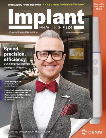
Speed, precision, efficiency
DEXIS Imprevo delivers the digital trifecta
Cover image of Dr. Joe Mehranfar courtesy of DEXIS.
IMPLANT CONCEPTS
Implant-specific evidence for non-abrasive peri-implant maintenance and treatment
Drs. Paras Ahmad, Jake Zellner, Michael A Pikos, Richard Miron, and Georgios A. Kotsakis discuss the importance of preserving titanium surface integrity
CONTINUING EDUCATION A life-saving exam
Dr. Brett Gilbert and Jonathan Gegerson provide a call to action for head and neck screening in dental practice 16
Subscription Rate 1 year (4 issues) $149 https://implantpracticeus.com/subscribe/ CONNECT
How to submit an article to Implant Practice US
Implant Practice US is a peer-reviewed, quarterly publication containing articles by leading authors from around the world. Implant Practice US is designed to be read by specialists in Periodontics, Oral Surgery, and Prosthodontics.
Submitting articles
Implant Practice US requires original, unpublished article submissions on implant topics, multidisciplinary dentistry, clinical cases, practice management, technology, clinical updates, literature reviews, and continuing education.
Typically, clinical articles and case studies range between 1,500 and 2,400 words. Authors can include up to 15 illustrations. Manuscripts should be double-spaced, and all pages should be numbered. Implant Practice US reserves the right to edit articles for clarity and style as well as for the limitations of space available.
Articles are classified as either clinical, continuing education, technology, or research reports. Clinical articles and continuing education articles typically include case presentations, technique reports, or literature reviews on a clinical topic. Research reports state the problem and the objective, describe the materials and methods (so they can be duplicated and their validity judged), report the results accurately and concisely, provide discussion of the findings, and offer conclusions that can be drawn from the research. Under a separate heading, research reports provide a statement of the research’s clinical implications and relevance to implant dentistry. Continuing education articles also include three to four educational aims and objectives, a short “expected outcomes” paragraph, and a 10-question, multiple-choice quiz with the correct answers indicated. Questions and answers should be in the order of appearance in the text, and verbatim. Product trade names cited in the text must be accompanied by a generic term and include the manufacturer, city, and country in parentheses.
Additional items to include:
• Include full name, academic degrees, and institutional affiliations and locations
• If presented as part of a meeting, please state the name, date, and location of the meeting
• Sources of support in the form of grants, equipment, products, or drugs must be disclosed
• Full contact details for the corresponding author must be included
• Short author bio
• Author headshot
Pictures/images/tables
Illustrations should be clearly identified, numbered in sequential order, and accompanied by a caption. Digital images must be high resolution, 300 dpi minimum, and at least 90 mm wide. We can accept digital images in all image formats (preferring .tif or jpeg).
Ensure that each table is cited in the text. Number tables consecutively, and provide a brief title and caption (if appropriate) for each.
Disclosure of AI use
Authors must disclose any AI used in researching, writing, or creating their articles. This includes a language model, machine learning, or similar technologies to create or assist with this submission. If AI was used, please provide a description of the AI-generated content and the name, model or tool used, and manufacturer. If AI was used, please confirm that you take responsibility for the integrity of the content that you are submitting.
References
References must appear in the text as numbered superscripts (not footnotes) and should be listed at the end of the article in their order of appearance in the text. The majority of references should be less than 10 years old. Provide inclusive page numbers, volume and issue numbers, date of publication, and all authors’ names. References should be submitted in American Medical Association style. For example:
Journals: (Print)
White LW. Pearls from Dr. Larry White. Int J Orthod Milwaukee. 2016;27(1):7-8. (Online)
Or in the case of a book: Pedetta F. New Straight Wire. Quintessence Publishing; 2017.
Website:
Author or name of organization if no author is listed. Title or name of the organization if no title is provided. Name of website. URL. Accessed Month Day, Year. Example of Date: Accessed June 12, 2011.
Author’s name: (Single) (Multiple) Doe JF Doe JF, Roe JP
Permissions
Written permission must be obtained by the author for material that has been published in copyrighted material; this includes tables, figures, pictures, and quoted text that exceeds 150 words. Signed release forms are required for photographs of identifiable persons.
Disclosure of financial interest
Authors must disclose any financial interest they (or family members) have in products mentioned in their articles. They must also disclose any developmental or research relationships with companies that manufacture products by signing a “Conflict of Interest Declaration” form after their article is accepted. Any commercial or financial interest will be acknowledged in the article.
Manuscript review and extra issues
All clinical and continuing education manuscripts are peer-reviewed and accepted, accepted with modification, or rejected at the discretion of the editorial review board. Authors are responsible for meeting review board requirements for final approval and publication of manuscripts. If additional issues are desired, they must be ordered from the publisher when the page proofs are reviewed by the authors. The publisher does not stock back issues. Reprints can be purchased after the issue is published.
Proofing
Page proofs will be supplied to authors for corrections and/or final sign off. Changes should be limited to those that are essential for correctness and clarity.
Articles should be submitted to:
Mali Schantz-Feld, editor in chief, at mali@medmarkmedia.com
Speed, precision, efficiency
DEXIS Imprevo delivers the digital trifecta
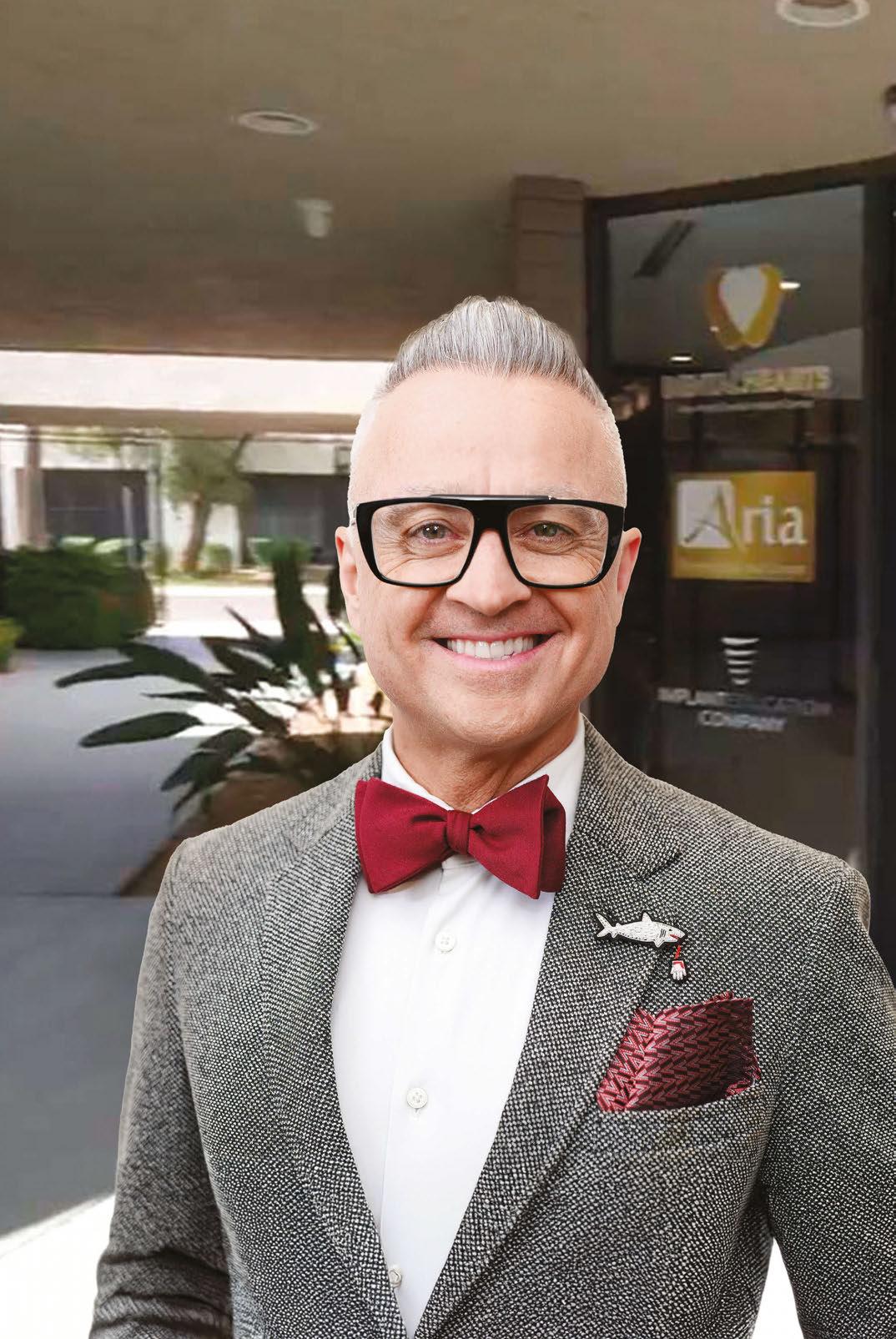
Across the globe, doctors share a common mission: to create a seamless, stress-free patient experience that builds trust and drives case acceptance. In pursuit of this mission, clinicians like Dr. Joe Mehranfar, owner of Aria Dental Implant Center, are embracing digital transformation. As practices become more digitally advanced, and patients increasingly expect the latest technologies from their dental care, doctors seek solutions that adapt to their evolving needs.
For Dr. Mehranfar, intraoral scanners have become an essential tool in his practice’s digital workflow, empowering him and his team to treat more patients with complex conditions, with clinical confidence and efficiency. Intraoral scanners play a central role in Dr. Mehranfar’s treatment planning, surgical procedures, collaboration, and design for a wide range of crowns, bridges, implants, and other restorations.
A long-time user of DEXIS imaging technology, Dr. Mehranfar recently added the company’s next-generation wireless Imprevo intraoral scanner to his digital toolbox. Featuring a host of technological innovations, Imprevo delivers the digital efficiencies, precision, and speed Dr. Mehranfar’s practice needs to expand and compete, while delivering the exceptional experience his patients have come to expect.
“Imprevo is an incredibly useful and versatile tool in my practice, from initial consultation to final restoration,” says Dr. Mehranfar. “There’s a lot of innovation packed into this slender, elegant scanner.”
Imprevo innovation in practice
Since acquiring his first Imprevo 6 months ago, Dr. Mehranfar has used the intraoral scanner primarily for his practice’s All-on-4 surgical implant surgeries — although he emphasizes that Imprevo is ideal for a wide range of procedures. With applications for image acquisition, treatment planning, all the way to the fabrication of the prosthetic and final implant, Imprevo serves as a versatile tool that translates to clinical efficiency and positive patient outcomes.
Sleek scanner design elevates the experience
Right out of the box, Dr. Mehranfar and his team could see that the Imprevo was different from other intraoral scanners on the market. Featuring a sleek ergonomic wireless design that’s
light and thin, Dr. Mehranfar and his team have found the Imprevo incredibly easy for both patient and user.
Around 20% slimmer than its DEXIS 3800W predecessor, Imprevo fits nicely in the hand as well as in the patient’s mouth. Along with a trimmer body, Imprevo features a 12% smaller tip compared to the 3800 series for a less intrusive presence, with no shrinkage to the field of view.
“Imprevo takes the freedom of wireless scanning to a new level,” adds Dr. Mehranfar. “Our patients as well as our team appreciate the sleeker, slimmer, lightweight design that makes scanning a smooth, quick, and comfortable experience.”
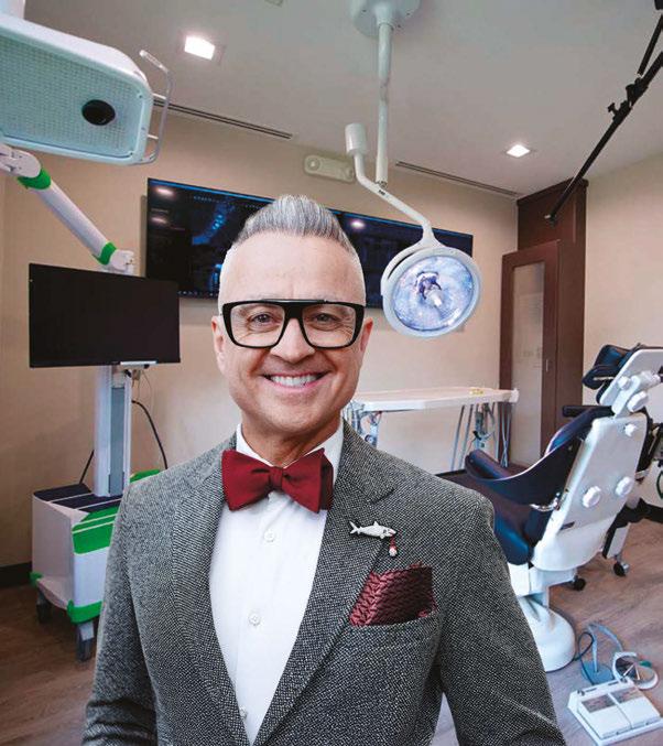
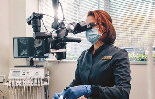
Dr. Joe Mehranfar
Imprevo may be slimmer, but there’s no shortage of thoughtful, intuitive features designed to make it easier for Dr. Mehranfar’s team to do their jobs. To reduce user fatigue, DEXIS product designers put Imprevo’s button within easy reach. The built-in GeniusControl functionality gives his team remote control of an on-screen scanning menu that eliminates the need to touch the computer or mouse during the scanning process, reducing the risk of cross-contamination. A simple wave of the wand is all it takes to rotate the on-screen digital impression to the right position.
While you can conveniently charge the Imprevo without having to remove the battery, the magnetic battery attachment makes it easy to install and change the device’s battery when needed. Imprevo designers thought of the smallest details, including the wrist strap that helps prevent droppage. Together, these attentive design features add up to a scanner that makes for a better experience — for both patient and clinician.
Precision meets speed in every scan
Inside Dr. Mehranfar’s practice, the Imprevo now plays an integral part in the patient’s journey from day one. During his patient’s initial consultation visit, Dr. Mehranfar uses Imprevo to capture intraoral scans, along with the ORTHOPANTOMOGRAPH™ OP 3D™ LX imaging platform to capture CBCT images, for a complete picture of the patient’s oral anatomy.
Imprevo integrates into your digital workflow
“Imprevo scans provide valuable data as I complete the smile design and present the case to the patient,” says Dr. Mehranfar. “The scans help illustrate to the patient what their final smile will look like, encouraging case acceptance.”
Imprevo innovation extends beyond esthetics and ergonomics. This next-generation scanner leverages cutting-edge laser technology and a dual sensor to capture ultra-clear scans in even the most challenging oral environments. With one of the
With its host of doctor-inspired advancements, Imprevo fits seamlessly into your practice’s digital workflow, integrating with existing DEXIS systems, including the i-CAT™ FLX V-Series as well as the OP 3D LX and EX, enabling the merging of 3D images and intraoral scans. Imprevo also plays well with other technology investments you’ve made, including common practice management software systems.
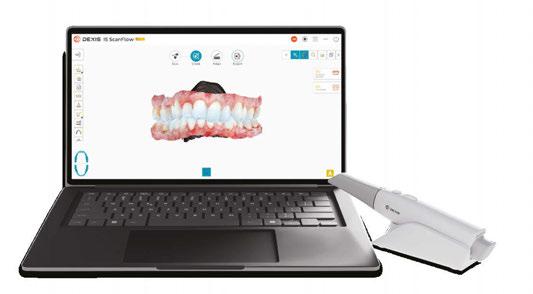
The latest generation of DEXIS intraoral scanners, Imprevo, is powered by the newest version of intuitive IS ScanFlow software featuring a simplified image tool bar, a streamlined scanning and exporting experience, and the ability to easily switch between various clinical workflows and treatment plans. Coupled with the IS Voyager and All-In-One mobile computer solution, Imprevo brings detailed accurate scans right to your patient’s chairside to support increased case acceptance.
Imprevo additionally integrates with the award-winning DTX Studio™ Clinic, so you can leverage the software’s wide range of powerful, AI-enabled tools to import, export, organize, position, merge, and interpret your intraoral scans with automated efficiency and accuracy.
Practices seeking to offer more services in-house will appreciate Imprevo’s integration with popular exocad 3D modeling software and Circle One™ smart design tools for 1-minute crown customization. Imprevo also works with SprintRay 3D Printing Solutions for same-visit prosthetics.
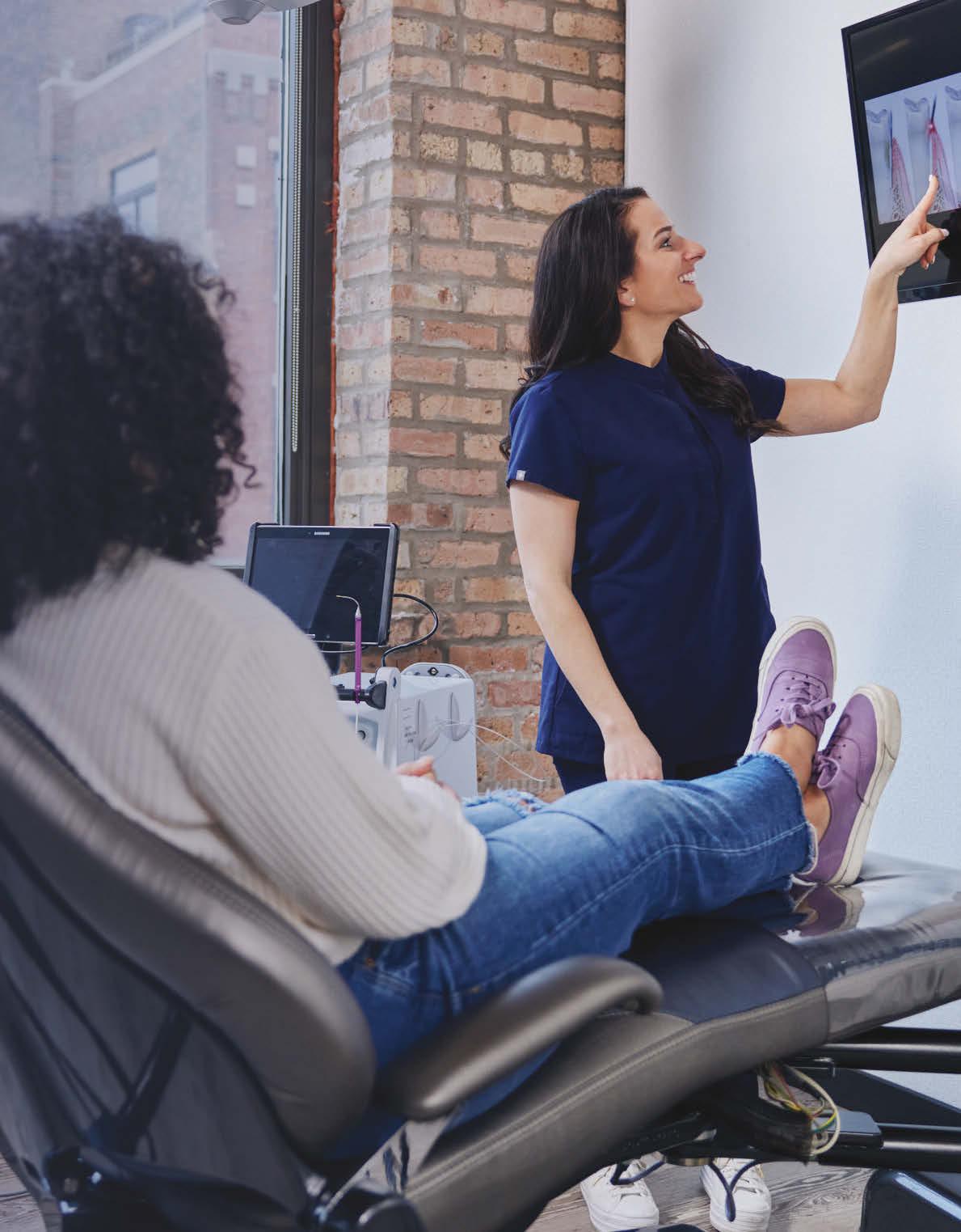
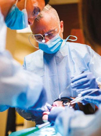
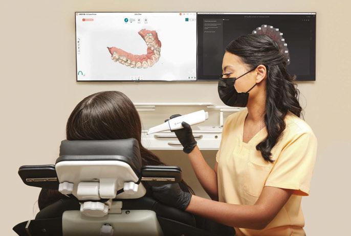
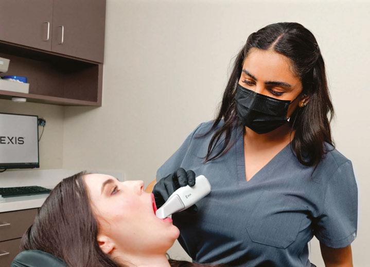
Assistant scanning a patient using DEXIS Imprevo
industry’s best 25 mm depth of field — 40% greater than the prior 3800 model — the Imprevo allows Dr. Mehranfar’s team to effortlessly capture images in those tight, hard-to-reach spots.
This depth of field doesn’t come at the cost of field of view, either, bringing confidence and clarity to every scan. Imprevo’s laser-powered precision delivers increased accuracy and data density, resulting in fewer discrepancies that require time-consuming rescans.
With a higher frame rate than its predecessor, Imprevo employs patent-pending parallel processing and advanced algorithms that allow more scan data to be processed with less compression required. The result is a refined mesh that allows for greater detail, particularly in the margin and scanbodies. Adding to the image quality is the unique laser light, designed to enhance color rendering for more photorealistic images of the scan area even on reflective surfaces.
“With Imprevo, we see high-resolution, full-arch scans rendered in real time and in stunning clarity,” explains Dr. Mehranfar. “This greatly increases our confidence in scanning results and supports better outcomes for our patients.”
He adds, “The high quality of the scan image also helps me communicate better with my patients so they can feel confident in my treatment plan.”
Imprevo processing technology allows Dr. Mehranfar and his team to work quickly as well as with confidence. Featuring twice the frame processing speed (FPS) as the 3800W — 40 FPS compared to 20 FPS — combined with its impressive depth of field, the Imprevo captures faster scans. This translates into enhanced clinical efficiency and reduced chair time for his patients.
As digital dentistry continues to evolve, solutions like the DEXIS Imprevo will play a pivotal role in helping practices stay agile, efficient, and patient-focused.”
Support for improved surgical outcomes
When it comes time for surgery, Dr. Mehranfar again turns to the Imprevo to scan the upper jaw and teeth with palatal fiducial markers that promote accurate and predictable restorations. Once surgery is completed, and the scanbodies and multi-unit abutment caps are placed, Dr. Mehranfar reapproximates the tissue and then uses the Imprevo again to scan the soft tissue, abutment caps, and palatal markers.
The pre-surgery and post-surgery markers are merged with the initial smile design so that the lab can fabricate the interim prosthesis. Over the next 2 hours, Dr. Mehranfar’s design team designs the interim prosthetic for the patient, which is then printed in his office on a SprintRay 3D printer.
“Without ever seeing the patient, our smile design team can virtually upload and merge the pre-surgery data and the post-surgery data to create the prosthetic,” Dr. Mehranfar explains. “That way, the patient can walk out of my office on the same day, already fitted with a prosthetic.”
Dr. Mehranfar then takes an Imprevo scan of the bite and redesigns the prosthetic as needed for a prototype, which is 3D printed and delivered the next day. Using the Imprevo one final time, Dr. Mehranfar takes another scan of the upper and lower teeth and bite, then sends this data to the designer for the final bridge to be fabricated.
“Imprevo has proven essential in capturing the proper data,” says Dr. Mehranfar. “With Imprevo, I can produce lab-ready files that ensure the right fit for my patients. It’s been an instrumental addition to my practice for smoother collaboration, fewer remakes, and consistently excellent results.”
Imprevo inspires a new standard in intraoral scanning
As digital dentistry continues to evolve, solutions like the DEXIS Imprevo will play a pivotal role in helping practices stay agile, efficient, and patient-focused. Putting a host of advancements in the hands of clinical teams, the Imprevo intraoral scanner is poised to redefine digital dentistry, empowering practices to advance their capabilities from initial consultation through final implant delivery. IP
Assistant scanning a patient using DEXIS Imprevo
Implant-specific evidence for non-abrasive peri-implant maintenance and treatment
Drs. Paras Ahmad, Jake Zellner, Michael A Pikos, Richard Miron, and Georgios A. Kotsakis discuss
the importance of preserving titanium surface integrity
Mechanical implant surface cleaning remains a constituent of peri-implantitis therapy, but its standalone effectiveness is increasingly called into question in light of titanium particle-induced inflammation. The evolving comprehension of peri-implantitis as a condition shaped by both material and microbial-driven determinants highlights the requirement for biomaterial-aware and integrative therapeutic approaches that address both the physicochemical and biological intricacies of the condition.1
1. Preserving titanium integrity in peri-implant decontamination
Mechanical debridement remains a cornerstone of peri-implantitis therapy due to its ability to disrupt microbial biofilms.2 However, conventional approaches employing ultrasonic scalers with metal tips or titanium curettes have raised concerns regarding iatrogenic surface damage.3 In vitro and preclinical studies demonstrate that abrasive instrumentation can disrupt the titanium-oxide passivation layer, generating titanium microparticles and nanoparticles that provoke inflammatory tissue responses
Paras Ahmad, DDS, MSC, PhD, is a post-doctoral associate at Rutgers School of Dental Medicine in New Jersey and lead researcher at Advanced PRF Education, Florida. Dr. Ahmad has no conflicts of interest.
Jake Zellner, DMD, MS, is a practicing Periodontist at Periodontal Associates of North Florida and a Diplomate of the American Board of Periodontology. Dr. Zellner has no conflicts of interest.
Michael A. Pikos, DDS, is the CEO and founder of Coastal Jaw Surgery and the Pikos Institute. He is a Diplomate of the American Board of Oral and Maxillofacial Surgery and, Diplomate of the American Board of Oral Implantology/Implant Dentistry. Dr. Pikos has no conflicts of interest related to this work.
Richard Miron, DDS, PhD, is a lead educator and researcher at Advanced PRF Education and an Adjunct Visiting Faculty in the department of Periodontology at University of Bern, Switzerland. Dr. Miron has no conflicts of interest related to this work.
Georgios A. Kotsakis, DDS, MS, PhD, is professor of Oral Biology and assistant dean for Clinical Research, and Director of Research at Rutgers School of Dental Medicine. He is a Diplomate of the American Board of Periodontology.
Disclosure: Dr. Kotsakis has received honoraria from Salvin Dental related to the training on the use of the Impla-Safe protocol. Dr. Kotsakis is co-inventor in a patent related to waterjet irrigation.
and osteolytic activity, even in the absence of bacteria.4,5 These findings emphasize that surface preservation is not only desirable but essential to maintain peri-implant tissue health. Thus, the latest evidence-based guidelines, including the AAP/AO consensus,6 shifted the paradigm towards non-abrasive decontamination methods that prioritize both preservation of surface integrity and microbial control. In-vitro reports have shown that surface-friendly decontamination procedures not only minimize titanium particle release but also cause significantly enhanced re-osseointegration potential compared to their abrasive counterparts.7 Particularly, approaches using non-metallic instruments or water-jet irrigation have exhibited superior results regarding the preservation of the implant’s biocompatible surface characteristics and facilitating cell attachment, proliferation, and differentiation on previously contaminated implant surfaces.7
A pivotal advancement in this area was reported by Daubert, et al.,8 who provided the first human data substantiating the deleterious effects of abrasive mechanical instrumentation on titanium release and clinical outcomes. The study showed that using abrasive tools, i.e., titanium curettes, to clean implants can lead to nearly 10 times more titanium particle release over time in humans than when using gentler, nonabrasive methods. This wasn’t just seen in lab tests; in real patients, it was linked to deeper peri-implant probing depths remaining unresolved after treatment. These findings suggest that titanium particles released during aggressive cleaning may play a role in damaging the tissues around implants and worsening peri-implant disease.8
These outcomes highlight a critical evolution in the comprehension of the peri-implantitis treatment landscape: therapeutic efficacy must now be assessed not merely by biofilm removal but also by the ability to maintain the chemical and structural stability of the implant surface. In summary, the implant-host-microbiome interface is a delicate balance affected by both iatrogenic and biological factors. Chemical or mechanical trauma to the titanium surface can propagate or exacerbate inflammatory pathways, independent of microbial presence, hence decreasing the overall efficacy of conventional antimicrobial strategies.
2. Tailoring peri-implantitis therapy to implant-specific factors
Beyond their direct pro-inflammatory effects, titanium particles also seem to indirectly mediate disease severity by impairing host immune surveillance and promoting dysbiosis within the peri-implant microbiome.9 This shift in microbial ecology
improves biofilm pathogenicity and may contribute to the well-documented resistance of peri-implant biofilms to conventional antimicrobial strategies.10
Biofilms, structured microbial communities encased in an extracellular polymeric matrix, demonstrate increased resistance to both antibiotic therapy and host immune mechanisms.11 The affinity of bacterial biofilms for titanium surfaces, combined with the anatomical limitations of accessing the subgingival peri-implant space, render effective decontamination particularly challenging. These mechanical and biological barriers mandate treatment strategies that go beyond conventional periodontal approaches, which were originally developed for natural dentition.11
In addition to biofilm-associated factors, other implant-specific, non-plaque-related contributors have been identified. One important example is excess dental cement, especially resin-based materials, which have been shown to induce peri-implant inflammation and bone loss when inadvertently retained in the peri-implant sulcus. Wilson has reported a causal association between peri-implantitis and excess submucosal cement, highlighting the necessity for rigorous cementation procedures in implant-supported restorations.12
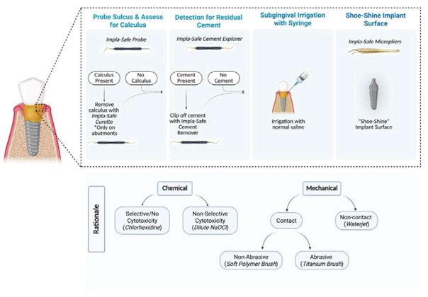
Collectively, these outcomes reinforce the need for implant-specific treatment workflows that diverge from conventional periodontal procedures applied to teeth. The biological differences between implant surfaces and natural teeth, such as the absence of periodontal ligament as well as submucosal calculus in implants and varying connective tissue attachment, demand a tailored clinical strategy. One such workflow, devised by the authors and grounded in current evidence on implant surface preservation, emphasizes non-abrasive decontamination approaches and the identification of modifiable implant-specific predisposing factors.13 This stepwise protocol, referred to as the “Impla-Safe” protocol, is designed to maintain peri-implant tissue health while minimizing iatrogenic damage to the titanium surface, hence preserving the potential for re-osseointegration and long-term implant success (Figure 1).
Step 1: Peri-implant probing — approach and instrumentation
Accurate assessment of peri-implant tissue starts with adequate probing, which remains a critical step in the diagnosis of peri-implant diseases. Contrary to misconceptions, dental implants can and should be probed routinely to assess peri-implant mucosal health, such as bleeding on probing, PPD, and suppuration. Nonetheless, probing around implants needs modifications in technique owing to distinct histological and anatomical differences compared to natural teeth.
Unlike the robust connective tissue attachment around natural dentition, the peri-implant mucosa demonstrates a weaker epithelial attachment and lacks true connective tissue fiber insertion into the implant surface. This anatomical variation makes the peri-implant interface more prone to mechanical trauma during probing, requiring the use of decreased probing forces, generally not exceeding 0.25 N, to avoid disruption of the fragile soft tissue seal.
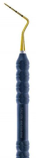
To facilitate accurate and gentle probing, the use of titanium implant-specific periodontal probes that have greater elasticity is
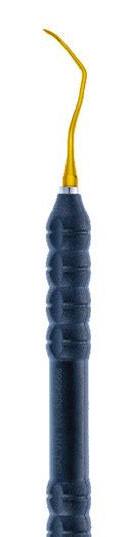
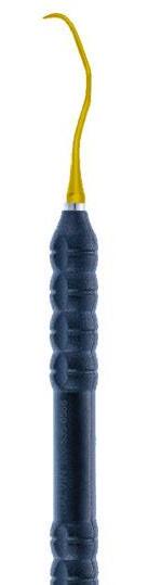
recommended. Implant probes should be manufactured in a soft, and preferably titanium alloy, with a narrower and more elastic structure than conventional plastic or metal probes. Examples include the including the titanium Impla-Safe Probe (Salvin Dental, Charlotte, North Carolina) and the steel ACE probe (PDT, Missoula, Montana) that have flexible shafts with a very high elasticity modulus that improves tactile feedback by gently flexing upon contact with the implant surface, decreasing the risk of surface abrasion or iatrogenic trauma to the epithelium. This flexibility is important as a compensatory mechanism for the lack of elasticity of the peri-implant as compared to periodontal tissues. In essence, the lack of elasticity of the peri-implant tissues due to differences in collagen organization and attachment as compared to teeth is compensated for by the elasticity of the probe to prevent a tear in the peri-implant junctional seal that could be caused by the pressure exceeding the modulus of elasticity of the tissues. Moreover, unlike bulky plastic probes that may undermine accuracy and access, flexible probes permit a more precise evaluation of submucosal conditions while maintaining minimal invasiveness.
Step 1B: Management of calculus on dental implants — minimizing risk to titanium integrity
When calculus is detected during clinical evaluation of an implant site, careful attention must be paid to both its location and the biological implications of mechanical debridement. Published literature suggests that supramucosal calculus formation is relatively frequent on transmucosal abutments but rarely occurs on the submucosal “threaded” portions of the implant body. This discrepancy is mainly due to the structural and electrochemical variations between titanium oxide and natural tooth root cementum. Cementum, with its surface porosity and organic composition, enhances the retention and nucleation of mineralized deposits, while the more inert and smoother titanium oxide surface lacks these features, deeming submucosal calculus formation rare.
When a visible supramucosal calculus is found on prosthetic or abutment components, eradication should be conducted utilizing implant-safe nonabrasive instruments. Specifically, curettes made of soft titanium alloys, engineered to have a decreased Rock-
Figure 1: The Impla-Safe Stepwise protocol for non-surgical peri-implantitis management
well hardness (e.g., Wingrove Titanium curette, PDT, Missoula, Montana) than the implant fixture itself, are recommended. Such instruments are designed to effectively disrupt and eradicate mineralized deposits without undermining the integrity of the titanium surface or inducing surface microdamage that may enhance corrosion or bacterial colonization, and caution is needed as not all titanium curettes have less hardness that titanium implant alloys.
Submucosal instrumentation on the implant body should be approached with extreme care. Routine curettage in this area is not recommended unless there is clear, clinically justified evidence of hard deposit retention. Aggressive mechanical debridement of the submucosal titanium surface can disrupt the thin and critical titanium oxide passivation layer, resulting in the release of titanium microparticles. These particles have been demonstrated to provoke local pro-inflammatory responses, exacerbate tissue deterioration, and contribute to peri-implantitis progression. Hence, when submucosal decontamination is indicated, it should be conducted utilizing non-abrasive, evidence-based techniques that prioritize preservation of the implant’s surface integrity.
A vital part of the prevention and treatment of peri-implantitis is identifying implant-specific risk factors, which differ from those associated with natural dentition. Among these, residual excess cement has been consistently identified as one of the most deleterious contributors to peri-implant tissue inflammation and bone loss. Particularly in cement-retained implant restorations, retained submucosal cement, specifically resin-based materials, can induce a robust inflammatory response and act as a nidus for pathogenic biofilm formation.
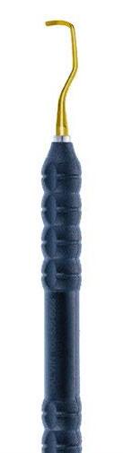
Despite its clinical relevance, detection of residual cement presents a significant diagnostic limitation, especially when thin films are embedded submucosally or integrated within the mucosal seal. Visual inspection alone is frequently inadequate, and definitive identification usually mandates invasive methods, including endoscopic visualization and flap reflection. In this regard, the utilization of specialized diagnostic instruments becomes indispensable. This workflow incorporates the Impla-Safe Explorer (Salvin Dental, Charlotte, North Carolina), a purpose-designed instrument with a narrow, flexible tip tailored to navigate the unique anatomical contours of the implant-abutment-crown complex. Its design accommodates the “mushroom-like” emergence profile usually evident in implant prosthetics, permitting precise exploration at the critical junction where residual cement is most probable to accumulate. The approach relies heavily on improved tactile perception. During probing, the clinician should note subtle changes in resistance or elasticity. A typical “bounce” or deflection response felt upon contact may suggest the presence of residual cement. This non-invasive cue allows targeted cement retrieval efforts without requiring flap elevation, hence preserving soft tissue integrity.
Step 2B: Treatment protocol after identifying residual cement
The eradication of excess cement alone can lead to the resolution of peri-implant mucositis or early peri-implantitis in nearly two-thirds of affected cases, underscoring its significance as a modifiable predisposing factor. Nonetheless, the method of removing excess cement must balance efficacy with preservation of the implant’s biological and structural integrity. Therefore, a single-stroke, conservative removal approach is emphasized. The objective is to disengage and extract the cement in one controlled motion, instead of repetitive scraping or scaling that could damage the peri-implant surface.
For this purpose, the Impla-Safe Cement Remover (Salvin Dental, Charlotte, North Carolina) is used. This instrument features an implant-specific sickle design with a micro-serrated working end, engineered to aid precise engagement of residual cement deposits, especially within the narrow submucosal interface at the crown-abutment junction. The micro-serrations offer mechanical interlocking with resin cement while minimizing friction against the titanium surface. The removal approach involves: (1) apical engagement of the cement margin with the serrated tip; (b) application of controlled coronal force in a single directional stroke; and (c) immediate re-evaluation employing tactile exploration to verify complete retrieval. Avoidance of multiple scaling strokes is crucial, as even seemingly minor mechanical trauma to the implant surface can lead to iatrogenic corrosion, particle shedding, and loss of surface biocompatibility.
Step 3: Sulcular irrigation with isotonic saline solution
After removing the residual cement, the peri-implant sulcus should be thoroughly irrigated with sterile isotonic saline (0.9% NaCl). This step acts as an essential adjunct to the preceding mechanical intervention by facilitating the eradication of disrupted biofilm fragments, inflammatory exudate, free-floating bacterial colonies, and titanium microparticles that may have been released during instrumentation. The irrigation should be performed using a side-vented, blunt-tipped irrigation syringe to avoid undue trauma or pressure to the sulcular epithelium. Gentle flushing ensures that any loosely attached debris is physically displaced, especially from the complex micro-topographies of the implant collar and abutment interface, which are prone to har-
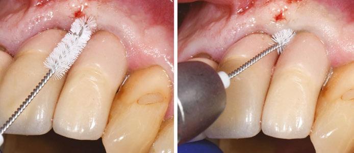
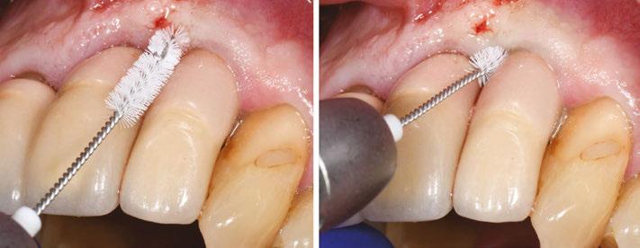
Figure 2: Clinical depiction of the use of the rotating nylon microbrush to reach intrasulcularly in an area where a curette cannot due to crown proximity. Note how the brush bristles conform as they enter the sulcus providing intimate adaptation between the implant threads for improved cleaning
North America’s most respected, doctor-led, multi-specialty partnership organization for growth and long-term success.
boring residual contaminants. In case the inflammation at the sites persists, such as in a case of refractory or recurrent peri-implant mucositis, the clinician can consider a diluted NaOCl irrigation with concentrations of no more than 1%, as this concentration ensures selective cytotoxicity. This means that it has antibacterial effects but does not compromise bone progenitor cells or affect regeneration. Importantly, chemotherapeutics with non-selective cytotoxicity should not be used on the implant threads as they get adsorbed and kill tissues cells such as fibroblasts and osteoblasts compromising peri-implant tissue attachment.
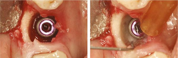
Figure 3: Clinical image of a peri-implant defect treated with an air-free waterjet (Bacterator; ImplantSci Ltd., Nicosia, Cyprus) that can clean the implant without surface damage and can be directly employed in the intrabony defect without risk for emphysema. Note the fan shapes targeted waterstream that can achieve high surface coverage. (Clinical case courtesy of Dr. Ioannis Vergoulis, Rhodes, Greece)
Step 4: Mechanical biofilm control with implant-specific, non-abrasive approaches
After initial debridement and sulcular irrigation, a critical next step in implant maintenance and peri-implant treatment is mechanical biofilm disruption using implant-safe, non-abrasive approaches. Given the delicate nature of the peri-implant interface and the risk of undermining the titanium surface integrity, biofilm control must be performed with precision tools that eradicate microbial deposits while preserving the implant’s passivated oxide layer.
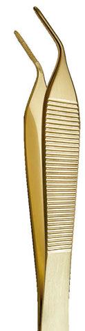
The recommended approach involves utilizing titanium micropliers to hold a small piece of sterile gauze pre-soaked in 0.9% isotonic saline, followed by gentle “shoe-shine” strokes along the exposed implant collar and threads. This approach offers: (a) tactile control and visibility; (b) effective mechanical disruption of loosely attached biofilm; (c) minimal risk of particle shedding or surface modification; and (d) compatibility with titanium surfaces without causing iatrogenic microdamage. This gauze-based method enables clinicians to maintain mechanical precision and biocompatibility, especially in sites where complex thread geometries may harbor persistent microbial colonies. In instances where additional antimicrobial support is needed, agents used must be rigorously chosen for their selective or low cytotoxicity to host tissue, as mentioned above. Notably, chlorhexidine gluconate gets adsorbed on titanium when used locally and demonstrates cytotoxicity, which compromises tissue regeneration and should not be used in implant treatment when a pocket extending to the threads exists.
Alternatively, the recent consensus data suggest the use of mechanical non-abrasive means of cleaning to remove biofilm while maintaining implant surface integrity, such as: (a) polymer-based micro-brushes, engineered for implant use that provide contact-based mechanical debridement that is gentle enough to avoid titanium surface damage, while being structurally capable of disrupting biofilm matrices. These are specifically advantageous for complex or narrow prosthetic interfaces where gauze cannot sufficiently conform; and (b) waterjet irrigation systems, such as continuous-flow or pulsatile devices that help efficient eradication of detached biofilm and inflammatory byproducts from submucosal areas without direct mechanical friction.
Examples of mechanical contact maintenance instruments are the Impla-Safe® brush (Salvin Dental, Charlotte, North Carolina) and the Labrida BioClean™ Brush (Straumann AG, Andover,
Massachusetts)14 both of which are mounted on reduction handpieces as rotating microbrushes. In contrast to rigid or metallic debridement instruments, soft polymer microbrushes comprise medical-grade polymer bristles, in the case of the BioClean brush enhanced with chitosan, particularly engineered to selectively interfere with biofilm while protecting the integrity of the implant surface. Clinical studies have indicated that the brush can substantially attenuate peri-implant mucosal inflammation and enhance clinical indices when utilized in combination with nonsurgical therapy, without causing noticeable alterations to the surface of titanium implants.14 Its non-metallic and flexible design renders it especially effective for cleaning exposed implant threads and complex prosthetic contours where traditional polymer tips or gauze may prove ineffective. Importantly, its utility has been reported as compatible, patient-friendly, and safe for multiple maintenance visits, in accordance with the principle of minimally invasive, surface-conserving peri-implant care.15
Both in vitro and human clinical evidence supports the use of non-metallic polymer microbrushes as an effective implant-safe method for biofilm disruption. A clinical trial showed that these brushes significantly reduced peri-implant mucosal inflammation while preserving the integrity of the titanium surface, with significantly better performance compared to titanium curettes.8 Their flexible, synthetic bristles are engineered to conform to complex implant geometries, allowing efficient access to narrow prosthetic interfaces that are difficult to clean with gauze alone. These findings suggest that polymer microbrushes may offer a gentle yet effective alternative to abrasive instrumentation in non-surgical peri-implantitis management.8
Beyond mechanical contact cleaning, new data suggests that mechanical non-contact cleaning with the use of air-free waterjet irrigation is a very promising and effective adjunct for peri-implant decontamination.6,16 An air-free waterjet system was employed in a preclinical study to deliver continuous saline flow without air infusion, minimizing the risk of subcutaneous emphysema while efficiently flushing biofilm and debris from the implant surface.16 Early results demonstrated significantly improved cleaning efficacy both in terms of bacterial and biofilm removal as compared to titanium brushes and curettes suggesting that air-free waterjet irrigation may optimize non-surgical peri-implantitis therapy beyond mechanical contact abrasive cleaning.16
In conclusion, according to the AO/AAP consensus on peri-implantitis, effective peri-implantitis management requires therapeutic strategies that preserve titanium surface integrity while
addressing both microbial and material-driven inflammation.6 Emerging evidence underscores that abrasive instrumentation may exacerbate disease by releasing titanium particles, reinforcing the need for implant-specific, non-abrasive decontamination workflows. Clinicians should prioritize gentle mechanical protocols, targeted risk-factor control such as cement removal, and the use of surface-friendly biofilm disruption tools to support tissue stability and re-osseointegration. Integrating these principles into routine practice offers an evidence-driven approach for improving peri-implant health and long-term implant success.
REFERENCES
1. Kotsakis GA, Black R, Kum J, Berbel L, Sadr A, Karoussis I, Simopoulou M, Daubert D. Effect of implant cleaning on titanium particle dissolution and cytocompatibility. J Periodontol. 2021 Apr;92(4):580-591. doi: 10.1002/JPER.20-0186. Epub 2020 Sep 11.
2. Ahmed P, Bukhari IA, Albaijan R, Sheikh SA, Vohra F. The effectiveness of photodynamic and antibiotic gel therapy as an adjunct to mechanical debridement in the treatment of peri-implantitis among diabetic patients. Photodiagnosis Photodyn Ther. 2020 Dec;32:102077. doi: 10.1016/j.pdpdt.2020.102077. Epub 2020 Nov 4.
3. Harrel SK, Wilson TG Jr, Pandya M, Diekwisch TGH. Titanium particles generated during ultrasonic scaling of implants. J Periodontol. 2019 Mar;90(3):241-246. doi: 10.1002/ JPER.18-0230. Epub 2018 Nov 14.
4. Fox SC, Moriarty JD, Kusy RP. The effects of scaling a titanium implant surface with metal and plastic instruments: an in vitro study. J Periodontol. 1990 Aug;61(8):485-90. doi: 10.1902/jop.1990.61.8.485.
5. Mann M, Parmar D, Walmsley AD, Lea SC. Effect of plastic-covered ultrasonic scalers on titanium implant surfaces. Clin Oral Implants Res. 2012 Jan;23(1):76-82. doi: 10.1111/j.1600-0501.2011.02186.x. Epub 2011 Apr 13.
6. Wang HL, Avila-Ortiz G, Monje A, Kumar P, Calatrava J, Aghaloo T, Barootchi S, Fiorellini JP, Galarraga-Vinueza ME, Kan J, Lin GH, Ravida A, Saleh MHA, Tavelli L; AO/AAP
Consensus Participants; Rosen PS. AO/AAP consensus on prevention and management of peri-implant diseases and conditions: Summary report. J Periodontol. 2025 Jun;96(6):519541. doi: 10.1002/JPER.25-0270. Epub 2025 Jun 12.
7. Yen Nee W, Raja Awang RA, Hassan A. Effects on the Titanium Implant Surface by Different Hygiene Instrumentations: A Narrative Review. Cureus. 2022 Oct 30;14(10):e30884. doi: 10.7759/cureus.30884.
8. Daubert D, Lee E, Botto A, Eftekhar M, Palaiologou A, Kotsakis GA. Assessment of titanium release following non-surgical peri-implantitis treatment: A randomized clinical trial. J Periodontol. 2023 Sep;94(9):1122-1132. doi: 10.1002/JPER.22-0716. Epub 2023 May 2.
9. Safioti LM, Kotsakis GA, Pozhitkov AE, Chung WO, Daubert DM. Increased Levels of Dissolved Titanium Are Associated With Peri-Implantitis - A Cross-Sectional Study. J Periodontol. 2017 May;88(5):436-442. doi: 10.1902/jop.2016.160524. Epub 2016 Nov 18.
10. Wilson TG Jr. Bone loss around implants-is it metallosis? J Periodontol. 2021 Feb;92(2):181185. doi: 10.1002/JPER.20-0208. Epub 2020 Aug 13.
11. Rosen PS. What Is an Effective Method for Treatment of Peri-implantitis? Compend Contin Educ Dent. 2020 Jul/Aug;41(7):378-383.
12. Wilson TG Jr. The positive relationship between excess cement and peri-implant disease: a prospective clinical endoscopic study. J Periodontol. 2009 Sep;80(9):1388-92. doi: 10.1902/jop.2009.090115.
13. Monje A, Amerio E, Cha JK, Kotsakis G, Pons R, Renvert S, Sanz-Martin I, Schwarz F, Sculean A, Stavropoulos A, Tarnow D, Wang HL. Strategies for implant surface decontamination in peri-implantitis therapy. Int J Oral Implantol (Berl). 2022 Sep 9;15(3):213-248.
14. Wohlfahrt JC, Aass AM, Koldsland OC. Treatment of peri-implant mucositis with a chitosan brush-A pilot randomized clinical trial. Int J Dent Hyg. 2019 May;17(2):170-176. doi: 10.1111/idh.12381. Epub 2019 Feb 7.
15. Khan SN, Koldsland OC, Roos-Jansåker AM, Wohlfahrt JC, Verket A, Mdala I, Magnusson A, Salvesen E, Hjortsjö C. Non-surgical treatment of mild to moderate peri-implantitis with an oscillating chitosan brush or a titanium curette-12-month follow-up of a multicenter randomized clinical trial. Clin Oral Implants Res. 2023 Jul;34(7):684-697. doi: 10.1111/ clr.14078. Epub 2023 May 3.
16. Jazaeri MS, Siddiqui DA, Tsai YC, Gabel K, Lorenzana Z, Kotsakis GA. Biofilm removal capacity and titanium surface integrity in non-abrasive versus abrasive peri-implantitis cleaning interventions. J Periodontol. 2025 Dec 10. doi: 10.1002/jper.11371. Epub ahead of print.
Precision Meets Protection in Implant Care
Impla-Safe™ is the first implant-specific instrument set for peri-implant maintenance and non-surgical management of peri-implant inflammation.
The flexible Titanium Probe enables safe implant probing without disrupting the delicate epithelial seal, while the NEW Impla-Safe Rotary Brush minimizes iatrogenic damage during implant cleaning.
Evidence-based and easy to use, Impla-Safe™ helps clinicians deliver safe, effective care and is essential for every implant maintenance protocol.
A life-saving exam
Dr. Brett Gilbert and Jonathan Gegerson provide a call to action for head and neck screening in dental practice
Abstract
This article underscores the critical role of the dental professional in the early detection of head and neck cancers through a systematic Conventional Visual and Tactile Examination (CVTE). Inspired by the personal story of my close friend, Jonathan, whose cancer diagnosis may have been expedited by routine dental screening, this article explores the components of the head and neck exam, its clinical rationale, and the current evidence supporting its implementation as a standard of care in dental settings.
Jonathan’s story in his own words: a missed opportunity at the dental chair
Educational aims and objectives
This self-instructional course for dentists looks at the critical role of the dental professional in the early detection of head and neck cancers.
Expected outcomes
Implant Practice US subscribers can answer the CE questions by taking the quiz online at implantpracticeus.com to earn 2 hours of CE from reading this article. Correctly answering the questions will demonstrate the reader can:
• Recognize the importance of the Conventional Visual and Tactile Examination (CVTE) in routine dental visits for early detection of head and neck cancers.
• Identify and describe each component of a systematic extraoral and intraoral head and neck exam. Understand the current evidence-based guidelines that support the implementation of CVTE in dental practice.
• Apply practical strategies for incorporating the CVTE into clinical workflows to enhance patient outcomes. 2
There I sat waiting for the doctor to examine my concern of a lump on the right side of my neck. I assumed it was nothing to be concerned about. The doctor entered the room, and I showed her the area. She felt around my neck on both sides, felt under my arms, and then asked a couple questions. “Have you had any dental work recently?” I responded, “No.” She then asked, “Have you had any infections in the mouth, or a root canal or anything?” Once again, my response was, “No.” She then looked at me and said, “It may be cancer.” I was stunned, and my body and mind froze in that moment, as that was the last thing I thought I was going to hear. Other than the lump on my neck, I was in perfect health. I worked out constantly, ate a Keto diet, never smoked, and limited all fatty foods and alcohol. There was no reason I should have cancer — at least that is what I thought.
Brett E. Gilbert, DDS, FICD, graduated from the University of Maryland Dental School (DDS, 2001, Endo, 2003). He is a professor in the Department of Endodontics at the University of Illinois at Chicago and a Diplomate of the American Board of Endodontics. He is the founder of the Access Endo Impact Academy, a global direct mentorship continuing education platform. He is a fellow in the International College of Dentists and a contributing consultant for Oral Health Journal. Dr. Gilbert lectures nationally and internationally on clinical endodontics and personal wellness and is the host of the On The Cusp podcast. Dr. Gilbert is a global key opinion leader in the area of endodontics and new technology. He is a partner in Specialized Dental Partners and has a private practice, King Endodontics PLLC, limited to Endodontics in Niles, Illinois.
Jonathan Gegerson, cancer survivor, has many dentists in his family tree — his great uncle, father and brother were all dentists. He worked for Envista with Kerr as the Global Trainer and then with Orascoptic as a Regional Manager. His 14-year career in the dental industry ended in 2021 when he could no longer perform his duties because of cancer. Since then, he has written a book Perspective C, (available on Amazon), to inspire and help people going through an illness. Gegerson has personally endured seven surgeries, 67 sessions of radiation, and over 200 sessions of chemotherapy. He believes that the biggest lesson is to approach everything with a desired outcome predetermined and to keep smiling. jonathangegerson@gmail.com
Since my initial diagnosis in May 2019, I have received over 200 rounds of chemotherapy, 67 sessions of radiation, and six surgeries. The side effects from all these treatments have been overwhelming at times. I lost my ability to chew and must be on a soft, liquid diet. My mouth opens just wide enough for a spoon. I was on a feeding tube for 6 months. I have constant lymphedema of my face, tongue, and throat which creates speaking problems, swallowing concerns, and at times, vision issues. I have radiation scars on my face, and one of my vocal cords was paralyzed during my recent 17-hour surgery, which was to help correct lymphedema as my airway was being constricted. I ended up in the ICU twice due to airway constriction. My body was literally suffocating itself.
I am the second patient to receive the type of surgery I had for head and neck lymphedema. My recovery took 7 months, and for two of those months, I was in the hospital. I share this because there were opportunities to catch the cancer sooner. Those opportunities were in my own power as well as the power of my dentist and hygienist. I mention my hygienist and dentist as I would see them regularly — at least every 6 months. If the cancer was caught sooner, it may have prevented such an
extensive treatment plan, limited all my side effects, and resulted in a better lifestyle after treatment.
I would rather have been told there was a concern 3 or 6 months before by my dentist or hygienist and addressed the concern with my primary doctor immediately. Early detection results in better patient outcomes. I was informed by my oncologist that out of 4 million people, 10 to 15 people get the type of cancer I was diagnosed with: Salivary Duct Carcinoma HER2 Positive. I was also told that I have been beating the odds, and I am one of a kind. I would rather be many of a kind when it comes to living through and after cancer. This is one of my motivations to encourage all hygienists and dentists to perform a cancer screening exam that includes the neck. Most people with my diagnosis are not here to tell their story. I intend to purposefully speak for them and myself as I encourage all dental professionals to perform a head and neck exam on every adult patient, every time they see them!
The clinical imperative
The dental setting provides a unique, often untapped opportunity to detect early signs of head and neck malignancies. The Conventional Visual and Tactile Examination (CVTE) is a simple, low-cost, evidence-based procedure that can uncover abnormalities before they become life-threatening.
The American Dental Association (ADA) recommends that clinicians perform a systematic CVTE for all adult dental patients, including inspection and palpation of the face, neck, and regional lymph nodes to detect tissue changes, masses, or asymmetry.1 This recommendation is echoed by the American Academy of Otolaryngology–Head and Neck Surgery, which emphasizes evaluation of the skin, salivary glands, thyroid, and lymph nodes using bimanual palpation techniques.2
Despite this guidance, implementation in general practice remains inconsistent.3
Step-by-step: performing the Conventional
Visual and Tactile Examination (CVTE)
General observation
Observe facial symmetry, skin color/lesions, swelling, and visible masses. Note signs of asymmetry, facial droop, or skin changes.
Lymph node palpation
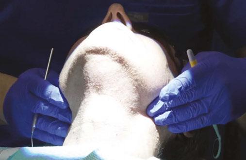
Preauricular and postauricular, submental and submandibular, cervical chain (anterior and posterior), supraclavicular nodes. Use gentle, circular pressure with the pads of your fingers, bilaterally when appropriate.
Salivary glands
Palpate parotid, submandibular, and sublingual glands for enlargement, tenderness, or firmness (Figure 1).
Thyroid gland
Visually inspect for enlargement while the patient swallows. Palpate the thyroid gently from behind the patient using both hands (Figure 2).
Figure 2: Thyroid exam
Figure 1: Salivary gland extraoral exam
Figure 3: Intraoral vestibule exam
Figure 4: Lateral tongue exam
Floor of mouth (Bimanual palpation)
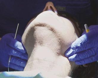
Place one gloved hand inside the mouth and one under the chin. Gently compress to assess for firm masses or nodularity.
Intraoral exam
Thorough inspection of the lips, buccal mucosa, gingiva, hard/soft palate, tongue (dorsal, lateral, and ventral), and oropharynx. Use gauze to pull and manipulate the tongue for complete visualization (Figures 3-4).
A case from my chair: CVTE in action
While Jonathan’s story reflects the devastating consequences of a missed opportunity for early detection, I also carry with me the opposite experience — one in which a thorough Conventional Visual and Tactile Examination (CVTE) led to the early diagnosis of a life-threatening malignancy.
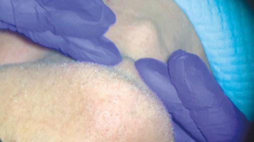
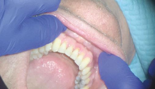
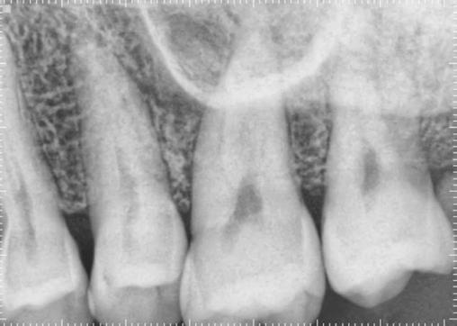
In 2016, a patient presented to my endodontic clinic for evaluation of a sore area on the palatal tissue adjacent to tooth No. 14 (Figure 5). As with every patient encounter, I performed a standard diagnostic endodontic evaluation alongside a systematic CVTE.
Clinical Findings
• Percussion: Negative
• Palpation: Positive on the palatal surface of tooth No. 14
• Periodontal Probing: Within normal limits
• Mobility: Within normal limits
• Pulpal Sensitivity (Cold): Normal response
These results pointed toward an endodontic diagnosis for tooth No. 14 of normal pulp with symptomatic apical periodontitis (due to a positive finding of palpation on the palate). But what caught my attention was not the tooth — it was the tissue.
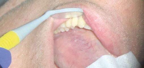
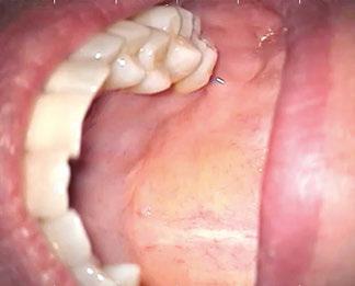
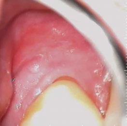
During the CVTE, I noted a small raised swelling on the palatal surface (Figure 6) and a separate white nodular lesion on the buccal gingiva adjacent to tooth No. 14 (Figure 7). Importantly, these findings did not align with any odontogenic pathology typically associated with tooth No. 14. That clinical inconsistency provided the moment to pause and widen the diagnostic lens.
Referral and diagnosis
Given the suspicious nature of these findings, I referred the patient to an oral and maxillofacial surgeon for biopsy. Oral and Maxillofacial (OMS) surgeons are often the first line of defense for dentists who may be unsure about a diagnosis that does not appear to be odontogenic in origin. The term “non-leo” refers to a lesion of non-endodontic origin. As a dental specialist, determination that findings are not adding up to a diagnosis of dental origin must be referred immediately.
OMS is the first line of referral for dentists as these specialists often have had extensive medical and hospital training. A dentist must understand that when they detect an irregular finding, no matter how small, it is critical to make this referral. Immediate referral will shorten the time between detection and diagnosis.
In many cases, the referral may seem like it was excessive if the OMS does not diagnose a problem. As clinicians, we should never allow doubt to creep into our minds by thinking that the finding is likely not significant. In fact, a non-significant diagnosis is the hope of the referral! The conversation we have with a patient in this moment should be calm in tone and decisive. I often will have my clinical team call the OMS office to set up a consultation appointment while the patient is still in our office. We must consider that a patient may listen to your concern but not act by making an appointment which could decrease the chances of a good outcome if a serious medical diagnosis is confirmed.
This patient was compliant and presented to the OMS the next day. The OMS performed their own examination and determined that a biopsy was necessary. The biopsy results revealed:
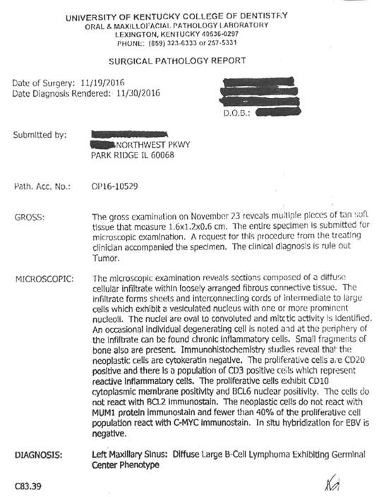
“Diffuse large B-cell lymphoma of the left maxillary sinus, germinal center phenotype” (Figure 8).
The patient was subsequently referred to oncology and underwent successful treatment for which they were extremely grateful that the detection of such a devastating systemic illness was detected early in the dental chair.
Clinical takeaway
This case illustrates the critical importance of integrating CVTE with routine diagnostic protocols. If I had focused solely
on the dental findings, the underlying malignancy may have been missed. It was the intentional soft tissue assessment through CVTE that revealed the warning signs.
When something doesn’t “add up” between tooth-level findings and soft tissue presentation, it’s time to:
• Pause and reassess
• Expand the differential beyond odontogenic causes
• Refer promptly to an oral and maxillofacial surgeon
Head and neck cancers often masquerade as benign or dental conditions — or remain entirely silent. A few extra moments of systematic examination can create a critical bridge to life-saving intervention.
Evidence basis: why it matters
CVTE remains the gold standard in dental practice for early detection of head and neck malignancy,1,2,3 according to multiple high-level guidelines and reviews. Some literature shows that adjunctive screening tools (e.g., fluorescence imaging, salivary biomarkers) have not demonstrated sufficient evidence to replace or supplement CVTE in routine practice.4,5 However, any device or screening tool that reminds a clinician how important it is to conduct these exams are worthy. CVTE should be considered a critical part of the dental examination and can be used in conjunction with any other screening tool that a clinician feels is valuable.
Recent systematic reviews confirm that visual inspection and palpation of the head, neck, and lymph nodes are the most frequently implemented and most reliable methods for early cancer detection in dental settings.6,7
Emerging test for high detection of oropharyngeal cancers is under investigation
Recent studies, including Das, et al., (2024, 2025),8,9 demonstrate that the HPV-DeepSeek assay — using whole-genome sequencing of circulating tumor HPV DNA (ctHPV-DNA) — achieves high sensitivity (up to 96–99%) and specificity (up to 99%) for detecting HPV-positive oropharyngeal squamous cell carcinoma, with detection possible up to 7.8–10 years before clinical diagnosis in some cases.8,9,10,11 This supports the claim that HPV-DeepSeek is a highly accurate, non-invasive liquid biopsy with potential for early cancer detection.
However, the clinical utility of HPV-DeepSeek as a routine screening tool in the dental setting remains investigational11,12,13 While the test shows promise for early detection and could theoretically complement head and neck examinations in dentistry, there is currently no guideline or regulatory approval for its use in routine screening or as a standard adjunct in dental practice. The National Comprehensive Cancer Network (NCCN) guidelines emphasize that blood-based ctHPV-DNA assays are not yet part of standard screening or diagnostic protocols and that their performance and actionable implications outside of clinical trials are still being evaluated.11
Closing thoughts: a moral and clinical duty
For Jonathan, the absence of a head and neck exam may have cost him an earlier diagnosis, a simpler treatment, and a better quality of life. He has beaten the odds and desperately
wants to share his story as a motivation and wake up call for dental professionals. His story is a call to action — for dentists and hygienists to go beyond the minimum, to reclaim our critical role in early cancer detection, and to never underestimate the power of a few minutes of intentional, hands-on examination.
Working in conjunction with other dental specialists to get a second opinion or to perform more advanced testing procedures, such as a biopsy or other imaging, represents best practice in dentistry. As dental professionals, we often have more opportunities to see and examine our patients than a primary physician. We cannot become complacent and avoid performing the CVTE just because the patient appeared healthy at prior visits. Thorough review of patient health history, medications, and any acknowledgement of oral habits such as smoking, oral placement of tobacco, and even consumption of alcohol should further motivate clinicians to remember to perform this life-saving examination.
As dentists, we do not have to take on the burden of making a definitive diagnosis on cases that present with abnormal findings. Our duty and responsibility are only to make a timely and appropriate referral for further examination and testing. By making the CVTE a routine, non-negotiable part of every adult dental visit, we honor our patients, our profession, and the principle that dentistry is, at its core, a healing art.
IP
Figure 8: Biopsy report
REFERENCES
1. Lingen MW, Abt E, Agrawal N, Chaturvedi AK, Cohen E, D’Souza G, Gurenlian J, Kalmar JR, Kerr AR, Lambert PM, Patton LL, Sollecito TP, Truelove E, Tampi MP, Urquhart O, Banfield L, Carrasco-Labra A. Evidence-based clinical practice guideline for the evaluation of potentially malignant disorders in the oral cavity: A report of the American Dental Association. J Am Dent Assoc. 2017 Oct;148(10):712-727.e10. doi: 10.1016/j.adaj.2017.07.032.
2. Pynnonen MA, Gillespie MB, Roman B, Rosenfeld RM, Tunkel DE, Bontempo L, Brook I, Chick DA, Colandrea M, Finestone SA, Fowler JC, Griffith CC, Henson Z, Levine C, Mehta V, Salama A, Scharpf J, Shatzkes DR, Stern WB, Youngerman JS, Corrigan MD. Clinical Practice Guideline: Evaluation of the Neck Mass in Adults Executive Summary. Otolaryngol Head Neck Surg. 2017 Sep;157(3):355-371. doi: 10.1177/0194599817723609.
3. Pynnonen MA, Gillespie MB, Roman B, Rosenfeld RM, Tunkel DE, Bontempo L, Brook I, Chick DA, Colandrea M, Finestone SA, Fowler JC, Griffith CC, Henson Z, Levine C, Mehta V, Salama A, Scharpf J, Shatzkes DR, Stern WB, Youngerman JS, Corrigan MD. Clinical Practice Guideline: Evaluation of the Neck Mass in Adults. Otolaryngol Head Neck Surg. 2017 Sep;157(2_suppl):S1-S30. doi: 10.1177/0194599817722550.
4. Moyer VA; U.S. Preventive Services Task Force. Screening for oral cancer: U.S. Preventive Services Task Force recommendation statement. Ann Intern Med. 2014 Jan 7;160(1):55-60. doi: 10.7326/M13-2568.
5. Huber MA. Adjunctive Diagnostic Techniques for Oral and Oropharyngeal Cancer Discovery. Dent Clin North Am. 2018 Jan;62(1):59-75. doi: 10.1016/j.cden.2017.08.004. Epub 2017 Oct 16.
6. Louredo BVR, de Lima-Souza RA, Pérez-de-Oliveira ME, Warnakulasuriya S, Kerr AR, Kowalski LP, Hunter KD, Prado-Ribeiro AC, Vargas PA, Santos-Silva ARD. Reported physical examination methods for screening of oral cancer and oral potentially malignant disorders: a systematic review. Oral Surg Oral Med Oral Pathol Oral Radiol. 2024 Feb;137(2):136-152. doi: 10.1016/j.oooo.2023.10.005. Epub 2023 Oct 16.
7. Sykes EA, Weisbrod N, Rival E, Haque A, Fu R, Eskander A. Methods, Detection Rates, and Survival Outcomes of Screening for Head and Neck Cancers: A Systematic
Review. JAMA Otolaryngol Head Neck Surg. 2023 Nov 1;149(11):1047-1056. doi: 10.1001/jamaoto.2023.3010.
8. Das D, Hirayama S, Aye L, Bryan ME, Naegele S, Zhao B, Efthymiou V, Mendel J, Fisch AS, Kröller L, Michels BE, Waterboer T, Richmon JD, Adalsteinsson V, Lawrence MS, Crowson MG, Iafrate AJ, Faden DL. Blood-based screening for HPV-associated cancers. medRxiv [Preprint]. 2024 Feb 2:2024.01.04.24300841. doi: 10.1101/2024.01.04.24300841.
9. Bryan ME, Aye L, Das D, Hirayama S, Al-Inaya Y, Mendel J, Naegele S, Efthymiou V, Alzumaili B, Faquin WC, Sadow PM, Lin D, Varvares MA, Feng AL, Deschler DG, Chan AW, Paly J, Park JC, Roberts T, Merkin R, Mishra SK, Kröller L, Michels B, Iafrate AJ, Wirth LJ, Adalsteinsson VA, Crowson M, Waterboer T, Mirabello L, Lawrence MS, Guan Z, Fisch AS, Richmon JD, Faden DL. Direct Comparison of Alternative Blood-Based Approaches for Early Detection and Diagnosis of HPV-Associated Head and Neck Cancers. Clin Cancer Res. 2025 Aug 14;31(16):3483-3493. doi: 10.1158/1078-0432. CCR-24-2525.
10. National Comprehensive Cancer Network. Head and Neck Cancers. Practice Guideline. Updated August 12, 2025. https://www.nccn.org/guidelines/guidelines-detail? category=1&id=1437.
11. Poljak M, Cuschieri K, Alemany L, Vorsters A. Testing for Human Papillomaviruses in Urine, Blood, and Oral Specimens: an Update for the Laboratory. J Clin Microbiol. 2023 Aug 23;61(8):e0140322. doi: 10.1128/jcm.01403-22. Epub 2023 Jul 13.
12. Araujo M, Bouassaly J, Farshadi F, Hier M, Mascarella M, Mlynarek A, Alaoui-Jamali M, da Silva SD. Current status of circulating tumor DNA and circulating cell alterations in HPV-associated head and neck cancer. Oral Oncol. 2025 Aug;167:107417. doi: 10.1016/j.oraloncology.2025.107417. Epub 2025 Jun 13.
Continuing Education Quiz
A life-saving exam GILBERT/GEGERSON
1. The _______ is a simple, low-cost, evidence-based procedure that can uncover abnormalities before they become life-threatening.
a. Conventional Visual and Tactile Examination (CVTE)
b. Eccles Index (EI)
c. Four-Finger Test (FFT)
d. Miller Index (MI)
2. ________ recommends that clinicians perform a systematic CVTE for all adult dental patients, including inspection and palpation of the face, neck, and regional lymph nodes to detect tissue changes, masses, or asymmetry.
a. American Heart Association
b. The American Dental Association (ADA)
c. U.S. Preventive Services Task Force
d. World Health Organization
3. Upon general observation, during a CVTE, clinicians should observe ________ and visible masses, and note signs of asymmetry, facial droop, or skin changes.
a. facial symmetry
b. skin color/lesions
c. swelling
d. all of the above
4. ________ glands should be palpated for enlargement, tenderness, or firmness.
a. Parotid
b. Submandibular
c. Sublingual
d. All of the above
5. Clinicians can examine the floor of the mouth by placing one gloved hand inside the mouth and one under the chin and gently compressing to assess for firm masses or nodularity.
a. True
b. False
6. are often the first line of defense for dentists who may be unsure about a diagnosis that does not appear to be odontogenic in origin.
a. Oncologists
b. Radiologists
c. Oral and Maxillofacial (OMS) surgeons
d. Dermatologists
Each article is equivalent to two CE credits. Available only to paid subscribers. Free subscriptions do not qualify for the CE credits. Subscribe and receive up to 16 CE credits for only $149; call 866-579-9496, or visit https://implantpracticeus.com/ subscribe/ to subscribe today.
n To receive credit: Go online to https://implantpracticeus.com/continuingeducation/, click on the article, then click on the take quiz button, and enter your test answers.
AGD Code: 730
Date Published: April 15, 2026
Expiration Date: April 15, 2029
7. When something doesn’t “add up” between tooth-level findings and soft tissue presentation, it’s time to: _________
a. pause and reassess
b. expand the differential beyond odontogenic causes
c. refer promptly to an oral and maxillofacial surgeon
d. all of the above
8. Head and neck cancers ________.
a. are often of endodontic origin
b. often masquerade as benign or dental conditions — or remain entirely silent
c. are always painful and obvious
d. can not be felt with the gloved hand
9. _________ guidelines emphasize that blood-based ctHPV-DNA assays are not yet part of standard screening or diagnostic protocols and that their performance and actionable implications outside of clinical trials are still being evaluated.
a. American Cancer Society
b. American Association of Oral Surgeons
c. The National Comprehensive Cancer Network (NCCN)
d. American Dental Education Association
10. Working in conjunction with other dental specialists to get a second opinion or to perform more advanced testing procedures, such as a biopsy or other imaging, represents best practice in dentistry.
a. True
b. False
To provide feedback on CE, please email us at education@medmarkmedia.com
Legal disclaimer: Course expires 3 years from date of publication. The CE provider uses reasonable care in selecting and providing accurate content. The CE provider, however, does not independently verify the content or materials. Any opinions expressed in the materials are those of the author and not the CE provider. The instructional materials are intended to supplement, but are not a substitute for, the knowledge, skills, expertise and judgement of a trained healthcare professional.
Posterior Lateral Mandibular Cortical Stabilization (PLMCS) and Posterior Medial
Mandibular
Cortical Stabilization (PMMCS)
Dr. Sami Nizam discusses techniques for full-arch implant placement
Overview
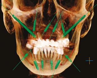
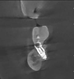
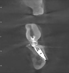
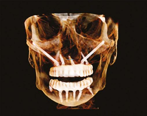
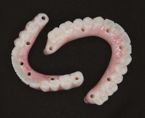
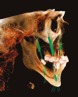
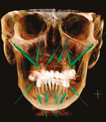
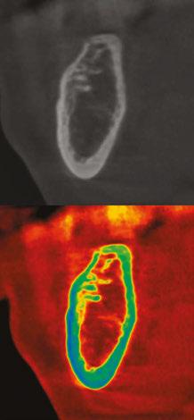
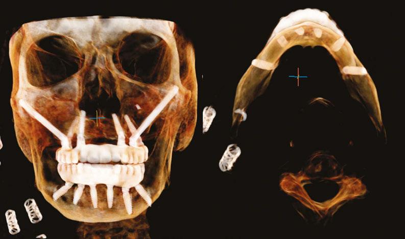
Full arch implant placement is becoming increasingly common. Techniques have advanced significantly even over the past 10 years. Most of these advancements have focused on atrophic cases of the maxilla allowing restoration of full dentition without cantilevers. These same advancements have lagged for atrophic mandibles. Posterior Lateral Mandibular Cortical Stabilization (PLMCS) and Posterior Medial Mandibular Cortical Stabilization (PMMCS) are techniques that were developed for extending AP spread, cantilever elimination, and increasing insertional torque in the posterior mandibular region. They do this by engaging the lateral or medial posterior mandibular cortex (Figures 1-3) regions that are present even on severely atrophic cases, while avoiding the inferior alveolar nerve. As bone in these cortices is type I, excellent immediate stability can be achieved primarily. This allows reliably high composite torque scores and immediate loading of the temporary prothesis.
Surgical technique
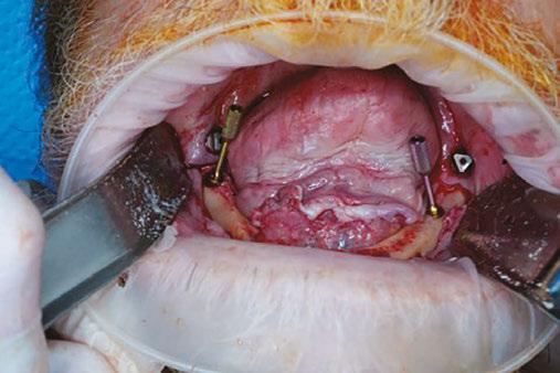
One of the principle advantages of this technique is it follows the same principles of angled implant placement that any fullarch surgeon is comfortable with. In the author’s practice, fullarch cases are performed under general intubated anesthesia. Placement begins with a standard full thickness mucoperiosteal flap elevation. The flap is continued distally with a buccal release for PLMCS or tapering slightly lingual for PMMCS. One must be aware that the lingual nerve extends to the level of the ridge or above in up to 17% of cases, in some studies, and the incision should never track past the lingual cortex.1 Dissection is taken to the buccal or lingual in a subperiosteal fashion depending on the technique chosen. This is particularly important using PMMCS as violation of the soft tissue will result in prolapse of the sub-
Sami Nizam II, MD, DMD, is a dual degree and double boardcertified oral and maxillofacial surgeon and facial cosmetic surgeon. He owns and operates Alabama Surgical Arts, an outpatient surgical center located in Montgomery, Alabama that is accredited by the American Association for Accreditation of Ambulatory Surgery Facilities (AAAASF). Follow Dr. Nizam on Instagram, Facebook, and Tiktok @Alabamasurgicalarts.
Educational aims and objectives
This self-instructional course for dentists aims to discuss the Posterior Lateral Mandibular Cortical Stabilization (PLMCS) and Posterior Medial Mandibular Cortical Stabilization (PMMCS) techniques for implant placement.
Expected outcomes
Implant Practice US subscribers can answer the CE questions by taking the quiz online at implantpracticeus.com to earn 2 hours of CE from reading this article. Correctly answering the questions will demonstrate the reader can:
• Realize some advantages of PLMCS and PMMCS.
• Realize some of the disadvantages of PLMCS and PMMCS.
• Recognize some clinical issues that can arise during these procedures.
• Identify some safety measures that can be taken when using these techniques.
lingual or submandibular gland. This can be an intraoperative annoyance and lead to a sialocele blocking the duct over the long term.
The author usually makes the decision on which technique to use based on which direction gives better implant-to-bone contact area. Cadaveric dissections have revealed the inferior alveolar nerve travels from medial to lateral in a horizontal plane
Figure
as it traverses the body of the mandible anteriorly.2 Due to this typical trajectory, PLMCS is selected approximately two-thirds of the time. This allows a longer/wider implant with resultant greater bone-to-implant contact area. Retraction of the soft tissue is typically with Minnesota retractor on the buccal. For the lingual, the tongue is retracted with a Sweetheart (SurgiMac) and soft tissues subperiosteally with a Seldin or similar retractor. This retraction can be challenging in a poorly sedated patient and cause airway embarrassment (airway compromise) from posterior tongue pressure.
Next, the implant preparation is begun with a pilot bur visualizing the apex of the bur exiting out the cortex. This is much like a zygomatic implant. For added safety with this technique, viewing the proposed entry and exit point on a coronal CT and using measurements if desired can help ensure the nerve is not penetrated. As is common in the posterior mandible in atrophic cases, the marrow space is of type 3 or 4 bone. Densification burs can be used to compact the bone. They give the added benefit of guiding over the canal as it is surrounded in type 1 or 2 bone.
An additional safety measure one could take, if the clinician desires, would be active or static navigation. The implant is then placed and countersunk. A bone mill is used to remove excess bone around the coronal portion. If the tip is overextended, it can be cut with copious irrigation, again much like a zygomatic implant. Caution must be exercised so that the bur does not catch the lingual or buccal tissue as significant tissue damage can occur. Copious irrigation is also obligatory as heat generation can be significant. Typically a 30 degree multiunit is then placed and torqued to specification (Figure 4). On occasion, a more acute angle multiunit abutment may be needed for the final restoration, as the max divergence for monolithic zirconia is no more than 25-30 degrees. If this cannot be achieved, then a custom multiunit (or 45 degree multiunit if available for the brand being used) may be necessary. The author has had this issue occur once, and it was resolved with a custom multiunit. Attached gingiva can be sparse in this region.
Literature and clinical experience have revealed a lack of attached gingiva can lead to peri-implantitis, recession, and bone loss.3 A plan for a free gingival graft with the goal of having 2 mm or greater of keratinized gingiva should be considered once integration has occurred. The author has found this the best time to place free gingival grafts as the temporary can be used as an effective healing barrier with simple composite addition. One can also evaluate the amount of tissue needed, demonstrate the deficit to the patient with intraoral photography, and plan if other sites will be needed before executing the procedure.
Advantages
Protocols such as PATZI (Pterygoid, Angled Anterior, Transnasal, Zygomatic) have revolutionized the treatment of the atrophic maxilla.4 Benefits have been clear. Using PLMCS and PMMCS in the mandible brings many of the same benefits including greater composite torque scores, larger AP spreads, ability to load, and the ability to restore to second molar even in the temporary (Figure 5). It is the author’s opinion that patients should receive second molars when feasible in a temporary. Distributing
Figure 3: CT of PLMCS and PMMCS
Figure 4: Angulated abutments
Figure 5: Temp and access holes
forces over a larger area decreases force at any point. Remembering the law of physics, pressure=force/area. As area (the denominator) in the formula increases, with force (the numerator) held constant, pressure then decreases. This is very similar to the concept of a bed of nails. One cannot lie on a single nail; however, one can lie on a bed of nails without harm. This concept has been born out in the literature. Decreasing distal cantilevers causes less stress on the prosthesis distal junction as well as less crestal forces on the distal implant.5 Although there is no exact agreement on cantilever length, it is accepted it should never be greater than two times the AP spread with 1.5 being most commonly used in practice.6 PLMCS and PMMCS allow complete elimination of any cantilever.
Other challenging clinic scenarios are patients who are Class III, who have anterior mental foramen, or U-shaped mandibles. These scenarios can limit the ability to replace a second molar and sometimes even a full first molar if implants are placed anterior to the foramen using standard All-on-4 procedures. Figures 6 and 7 demonstrate such a scenario. The patient has a Class I occlusion, usual anatomic exit of the mental foramen, however, has a U-shaped mandible. This causes a small AP spread of only 7 mm in this case. The average male Caucasian molar is approximately 11.5 mm in MD width.7 In this scenario, one molar would be pushing the AP spread limits, and two would be prohibitive. This logically does not make sense when we have the ability to provide a second molar on the maxilla using PATZI protocols.
Another clinical problem that occurs is what to do in cases of traditional All-on-4 with a distal tilted implant anterior foramen that fails. Depending on when it occurs and the amount of bone loss, adequate bone stock may not be available to place another implant, particularly immediately. PLMCS and PMMCS provide a “bail out” to keep the patient in a fixed prosthesis in these scenarios. There also is the theoretical advantage of applying forces into the body of an atrophic mandible to avoid atrophy in this region. The author has seen fractures occur in this region from falls on patients with traditional All-on-4s. These techniques have the potential to mitigate resorption in this region.
The body region of the mandible is often of type 3 or 4 bone in edentulous cases. Figure 8 demonstrates a case in which extreme type 4 bone was present in the body extending to the
Figure 6 (left): Lateral view poor ap spread. Figure 7 (right): Anterior view poor ap spread
Figure 8 (left): Extreme cancellus type 4 marrow space. Figure 9 (right): PLMCS used in posterior and parasymphysis regions
parasymphysis region. Traditional implants with densification burs yielded no primary stability in the angled or posterior implant. Engaging type 1 bone of the cortex buccally in these regions allowed for excellent primary stability and case loading seen in Figure 9. Finally, practice differentiation is key in today’s market. The ability to fully restore dentition immediately, reliably, and with long term success can be a strong practice builder and marketing tool.
Disadvantages
As with any increased AP spread and arch length impression, accuracy can be affected.8 Accuracy of digital or traditional impression techniques becomes more important to avoid misfits. Additionally, there is the theoretical disadvantage of mandibular flexure and long spans causing fracture in the temp or final.9 It is recommended that if PLMCS or PMMCS is used, it be used with four anterior implants for better biomechanical resistance. In severely atrophic mandibles, fracture could be a theoretical disadvantage, especially in the case of a tapered implant that is overtorqued during placement. This is fortunately unlikely as the opposite cortex as well as the inferior border remains intact.
Milling difficulties can occur in the final, particularly monolithic zirconia. This can occur if the implant is placed at too great of an angle and cannot be up righted with a multiunit abutment. In this case, a custom multiunit abutment would need to be fabricated. Inferior alveolar nerve injury is also a potential sequela. Techniques listed above such as visualization, measurement on the CT, feel, and densification burs are usually adequate. If one prefers, active or static navigation techniques could be implemented to further mitigate risk.
Injury to soft tissue on the buccal could theoretically result in injury to the facial artery or vein as they cross the inferior border of the mandible. If this occurs, localization and ligation would be required. Injury to the lingual tissue could result in injury to the lingual nerve, submandibular gland, or sublingual gland depending on region of placement. Glandular injury could lead to sialocele. Conservative injection of neuromodulator at the time of injury, if witnessed, would offer a simple solution. If a sialocele is noted late, marsupialization would offer the simplest solution. As with any complications, the easiest solution is avoidance, and good retraction with the patient asleep is a prerequisite for these techniques. The author has experienced none of the above complications to-date.
A final disadvantage is adding of cost and time in comparison to a traditional All-on-4. This should be factored into the cost presented to the patient. The author has found that when
One of the principle advantages of this technique is it follows the same principles of angled implant placement that any fullarch surgeon is comfortable with.
the advantages are discussed using CTs and printed temporaries, it becomes a great selling point.
Conclusion
As full-arch implant placement becomes more mainstream, practice differentiation becomes crucial. PLMCS and PMMCS give the ability to restore a full dentition in the temporary, increase composite torque scores, eliminate cantilevers, better distribute occlusal forces, and provide better long term outcomes even in the most atrophic cases. It is the author’s sincere opinion that this technique is a valuable tool to anyone involved in the practice of restoring full arches.
REFERENCES
1. Nizam SA 2nd, Ziccardi VB. Trigeminal Nerve Injuries: Avoidance and Management of Iatrogenic Injury. Oral Maxillofac Surg Clin North Am. 2015 Aug;27(3):411-424. doi: 10.1016/j.coms.2015.04.006. Epub 2015 Jun 2.
2. Yeh AYE, Finn BP, Jones RHB, Goss AN. The variable position of the inferior alveolar nerve (IAN) in the mandibular ramus: a computed tomography (CT) study. Surg Radiol Anat. 2018 Jun;40(6):653-665. doi: 10.1007/s00276-018-1973-9. Epub 2018 Jan 20.
3. Ramanauskaite A, Schwarz F, Sader R. Influence of width of keratinized tissue on the prevalence of peri-implant diseases: A systematic review and meta-analysis. Clin Oral Implants Res. 2022 Jun;33 Suppl 23:8-31. doi: 10.1111/clr.13766.
4. Ponnusamy S, Gonzalez J, Holtzclaw D. A Systematic Approach to Restoring Full Arch Length with Maxillary Fixed Implant Reconstruction: The PATZi Protocol. Int J Oral Maxillofac Implants. 2023 Oct 17;38(5):996-1004. doi: 10.11607/jomi.10153.
5. Khorshid HE, Issa NO, Ekram AM. Effect of implant diameter and cantilever length on the marginal bone height changes and stability of implants supporting screw retained prostheses: A randomized double blinded control trial. J Adv Prosthodont. 2023 Jun;15(3):101-113. doi: 10.4047/jap.2023.15.3.101. Epub 2023 Jun 28.
6. Wang Q, Zhang ZZ, Bai SZ, Zhang SF. Biomechanical analysis of stress around the tilted implants with different cantilever lengths in all-on-4 concept. BMC Oral Health. 2022 Nov 5;22(1):469. doi: 10.1186/s12903-022-02520-8. PMID: 36335327;
7. Singh SP, Goyal A. Mesiodistal crown dimensions of the permanent dentition in North Indian children. J Indian Soc Pedod Prev Dent. 2006 Dec;24(4):192-196. doi: 10.4103/0970-4388.28076.
8. Zhang YJ, Qian SJ, Lai HC, Shi JY. Accuracy of photogrammetric imaging versus conventional impressions for complete arch implant-supported fixed dental prostheses: A comparative clinical study. J Prosthet Dent. 2023 Aug;130(2):212-218. doi: 10.1016/j. prosdent.2021.09.035. Epub 2021 Nov 12.
9. Gao J, Li X, He J, Jiang L, Zhao B. The effect of mandibular flexure on the design of implant-supported fixed restorations of different facial types under two loading conditions by three-dimensional finite element analysis. Front Bioeng Biotechnol. 2022 Aug 29;10:928656. doi: 10.3389/fbioe.2022.928656.
1. Posterior Lateral Mandibular Cortical Stabilization (PLMCS) and Posterior Medial Mandibular Cortical Stabilization (PMMCS) are techniques that were developed for ________.
a. extending AP spread
b. cantilever elimination
c. increasing insertional torque in the posterior mandibular region
d. all of the above
2. One must be aware that the lingual nerve extends to the level of the ridge or above in up to _______ of cases, in some studies, and the incision should never track past the lingual cortex.
a. 17%
b. 36%
c. 45%
d. 52%
3. The author usually makes the decision on which technique to use based on which direction gives better implant-to-bone contact area.
a. True
b. False
4. Cadaveric dissections have revealed the _______ travels from medial to lateral in a horizontal plane as it traverses the body of the mandible anteriorly.
a. inferior alveolar nerve
b. facial nerve
c. hypoglossal nerve
d. glossopharyngeal nerve
5. (Regarding irrigation) On occasion, a more acute angle multiunit abutment may be needed for the final restoration, as the max divergence for monolithic zirconia is no more than _______ degrees.
a. 10-15 degrees
b. 25-30 degrees
c. 40-45 degrees
d. 50-55 degrees
6. Literature and clinical experience have revealed a lack of attached gingiva can lead to _______.
a. peri-implantitis
b. recession
c. bone loss
d. all of the above
Each article is equivalent to two CE credits. Available only to paid subscribers. Free subscriptions do not qualify for the CE credits. Subscribe and receive up to 16 CE credits for only $149; call 866-579-9496, or visit https://implantpracticeus.com/ subscribe/ to subscribe today.
n To receive credit: Go online to https://implantpracticeus.com/continuingeducation/, click on the article, then click on the take quiz button, and enter your test answers.
AGD Code: 690
Date Published: April 15, 2026
Expiration Date: April 15, 2029
2 CE CREDITS
7. Using PLMCS and PMMCS in the mandible brings many of the same benefits (as the PATZI (Pterygoid, Angled Anterior, Transnasal, Zygomatic Protocol) including greater composite torque scores, larger AP spreads, ability to load, and the ability to restore to second molar even in the temporary.
a. True
b. False
8. Distributing forces over a larger area _______ force at any point.
a. increases
b. decreases
c. has no effect on
d. creates a harmful
9. It is recommended that if PLMCS or PMMCS is used, it be used with _______ anterior implant(s) for better biomechanical resistance.
a. one
b. two
c. three
d. four
10. PLMCS and PMMCS give the ability to restore a full dentition in the temporary, increase composite torque scores, eliminate cantilevers, better distribute occlusal forces, and provide better long term outcomes even in the most atrophic cases.
a. True
b. False
To provide feedback on CE, please email us at education@medmarkmedia.com
Legal disclaimer: Course expires 3 years from date of publication. The CE provider uses reasonable care in selecting and providing accurate content. The CE provider, however, does not independently verify the content or materials. Any opinions expressed in the materials are those of the author and not the CE provider. The instructional materials are intended to supplement, but are not a substitute for, the knowledge, skills, expertise and judgement of a trained healthcare professional.
Progressive Dental Marketing wins Suncoast Emmy for “The Weight of a Smile”
Progressive Dental Marketing’s video production team clinched the prestigious Suncoast Emmy in the Community Service Category for their impactful video, “The Weight of a Smile,” showcasing transformative dental care stories with Dr. Burak Taskonak.
This accolade, presented by the Suncoast Chapter of the National Academy of Television Arts & Sciences, recognizes outstanding work in the television industry, highlighting the quality and impact of storytelling.
“The Weight of a Smile” captures the transformative dental journeys of patients under the care of Dr. Burak Taskonak, emphasizing the profound impact that dental care can have on individuals’ lives. Produced by Pipo Wagner, the video is a testament to Progressive Dental Marketing’s commitment to showcasing the emotional and life-changing aspects of dental care through compelling narrative and high-quality production.
Winning the Suncoast Emmy not only acknowledges the exceptional talent of Progressive Dental Marketing’s video production team but also underscores the company’s dedication to community service and its ability to communicate powerful stories that resonate with audiences. This recognition is expected to elevate the company’s brand reputation in the dental marketing industry and attract more practices that aim to share their transformative patient stories.
As a full-service dental marketing company, Progressive Dental Marketing leverages the power of storytelling to help dental practices grow by closing high-value cases and increasing production per patient. Through end-to-end digital marketing campaigns, videography, and practice coaching, the company aims to convert marketing activities into measurable results.
This Emmy win reinforces the effectiveness of Progressive Dental Marketing’s approach and highlights the importance of impactful content in engaging and inspiring both dental professionals and patients.
About Progressive Dental Marketing
Progressive Dental Marketing is a full-service dental marketing and coaching firm focused on helping dental practices grow by closing high-value cases. The company delivers comprehensive marketing solutions through digital campaigns, video production, and strategic coaching, enhancing practice efficiency and profitability. Led by President and CEO Bart Knellinger and Vice President Gerritt Cora, Progressive Dental Marketing is committed to sustainable growth and education within the dental industry. IP
Members of the Progressive Dental team at the 2025 Suncoast Regional Emmy Awards. Pictured (L-R): Pipo Wagner, Shelby Dorvil, Ian Crenshaw, Skylor Elliott, and Sebastian Ruiz
The 2025 Suncoast Regional Emmy® Award (left), presented to Progressive Dental and Video Producer, Pipo Wagner, for “The Weight of a Smile.” The Progressive Dental team (right), following their 2025 Suncoast Regional Emmy® Award win, with Dr. Burak Taskonak holding the award at the center
Revolutionizing oral health with BLISS: a biofluorescence-led approach to chronic disease management
Dental caries and periodontal disease are the two most prevalent chronic conditions in the oral cavity. These diseases develop early in life and persist throughout a person’s lifetime, often exacerbating systemic health conditions and diminishing overall quality of life. Traditionally, dental care has focused on treating symptoms after they appear, with radiographic imaging as the primary diagnostic tool. However, such methods detect damage only after it occurs, offering limited insight into disease origin.
The true culprit behind these conditions is pathogenic biofilm — a structured bacterial community that forms on oral surfaces. Understanding its location and extent is critical for managing disease proactively. Disclosing solutions have long been used to visualize plaque, but they bind to proteins nonspecifically and do not differentiate between harmless and pathogenic bacteria. Additionally, their use can be time-consuming and uncomfortable for patients.
Our solution is BLISS (Biofluorescence-Led Intelligent Screening System), a non-invasive imaging technology that visualizes biofilm in real time by detecting its natural fluorescence. Pathogenic bacteria, especially late colonizers, produce a metabolic byproduct called porphyrin. When exposed to 405nm blue light, porphyrins absorb energy and emit red fluorescence, while healthy tooth surfaces fluoresce green. This allows clinicians to distinguish harmful biofilm without the need for dyes or rinses.
Notably, pathogenic biofilm typically forms after 3 days of plaque accumulation and is associated with both caries and periodontal disease. BLISS can detect this early, enabling personalized hygiene feedback and better preventive care. The technology is especially useful around implants, where peri-implantitis often involves biofilm hidden between threads or in deep pockets — areas not visible with radiographs. In these cases, BLISS can confirm whether biofilm removal is complete after treatment.
Being radiation-free, BLISS is safe for use in various settings such as clinics, schools, elder care facilities, and even at home. Its portability and ease of use support routine hygiene checks
without requiring specialized infrastructure. This expands the scope of dental care from reactive treatment to proactive disease prevention and long-term health maintenance.
BLISS also serves as a powerful communication tool. Patients can see their own oral condition in real time, which fosters trust, improves compliance, and boosts motivation for home care. Clinicians report higher treatment acceptance and return rates when using BLISS, benefiting dentists, hygienists, and patients alike.
Incorporating BLISS alongside traditional radiography allows for earlier, more accurate diagnosis of chronic oral diseases. This integration marks a shift from symptom-driven intervention to evidence-based prevention. By visualizing what was once invisible, BLISS empowers both professionals and patients to work together toward lifelong oral health.
Let’s end the silent burden of chronic oral disease — starting with BLISS. For more information, visit Aiobio.com.
Prosthetic restorations exhibit darker fluorescence than natural teeth; red fluorescence reveals areas of inadequate plaque control
Red fluorescence on the implant surface indicates biofilm accumulation
What increases the value of an endodontic practice and why it matters for your future
Introduction
For endodontists, practice valuation is not just a number at the end of a financial statement. It is a direct reflection of the strength of the practice’s systems, the relationships with referring dentists, and the trust patients place in their care. Endodontic practices are uniquely referral-driven, which means their value is heavily influenced by the health of these networks. At the same time, investments in modern technology and well-designed operations make a substantial impact on valuation. Understanding these factors ensures that endodontists are prepared not only for future transitions but also for long-term growth.
At Specialized Dental Partners, we work alongside endodontists to maximize these drivers. By focusing on both the business and human aspects of a practice, we help specialists strengthen their practices today while positioning them for long-term success and transition planning.
Referral networks as a cornerstone
The majority of endodontic practices depend on referrals from general dentists, making the strength of those relationships central to practice value. Strong communication, timely patient updates, and collaborative relationships with general dentists not only secure referrals but also demonstrate professionalism, which enhances long-term value.
Technology as a value multiplier
Endodontics has been at the forefront of dental innovation for years. CBCT imaging, digital workflows, and advanced operating microscopes are now viewed as standard investments in a thriving practice. These tools improve diagnostic accuracy, streamline treatments, and reassure both patients and referring doctors of clinical excellence. For valuation, technology demonstrates a commitment to quality and positions the practice as a leader, not a follower.
Operational efficiency and team strength
Valuation is also tied to how smoothly a practice operates day-to-day. Efficient scheduling systems, well-managed patient flow, and reduced no-show rates all demonstrate strong management. A stable and well-trained team is equally critical. Turn-
over is costly both financially and reputationally. A cohesive team that delivers consistent patient experiences raises practice value significantly. Endodontists who focus on leadership and staff development ultimately protect and grow the longterm value of their practice.
Why this matters for the future
Understanding practice valuation benefits endodontists long before retirement. For those considering partnership, valuation provides a baseline for fair and transparent discussions. For those in mid-career, it highlights areas where investment can increase both day-to-day success and long-term financial stability. Even for newer specialists, paying attention to these valuation drivers creates flexibility, ensuring that when opportunities arise, the practice is well-positioned to respond. By focusing on referrals, technology, and operations, endodontists ensure that their practices remain attractive to partners, buyers, and patients alike.
Conclusion
Endodontic practice valuation is a reflection of relationships, technology, and leadership. By strengthening these areas, specialists not only secure the immediate success of their practices but also build lasting value that will serve them for decades to come.
Are you ready to position your endodontic practice for long-term growth? Discover how Specialized Dental Partners can help endodontists build enduring value while preserving clinical independence. Learn more or connect with our team at specializeddental.com.
IP
This information was provided by Specialized Dental Partners.
Dr. Helena Becker, Endodontist Partner in Renton, WA
Premium implants, premium patients: how to attract clients who value quality over price
Javeria Rameez Naqvi discusses how perception affects treatment acceptance
Too many implant dentists believe the only way to increase case acceptance is to lower their prices. It’s a race to the bottom — one that erodes profits, attracts the wrong patients, and forces you to work harder for less.
But here’s the truth: Patients don’t say no to implants because of price. They say no because they don’t see the value.
The problem isn’t cost — it’s perception. And the busiest implant dentists don’t compete on price. They make price irrelevant by clearly demonstrating why implants are the best choice.
Here’s how to shift the conversation from “How much does it cost?” to “How soon can we start?”
1. Patients pay for what they truly value
Think about the last time a patient rejected implants because of cost. Was the real issue the price — or was it that they didn’t fully understand the long-term benefits?
Patients make big financial decisions every day:
• Buying a car that costs more than a full-arch case
• Spending thousands on home renovations
• Taking out loans for vacations, weddings, and luxury items
Why? Because they see the value in those purchases.
Shift the patient’s perspective
Instead of:
“Implants cost $X.”
Say:
“Imagine eating your favorite foods again without worry. Imagine never having to deal with dentures slipping or discomfort. That’s what implants give you — not just teeth, but a lifetime of confidence and function.”
When patients feel the value on a personal level, price becomes secondary.
Real-world insight: Here’s a challenge a dentist shared with us: “Ethically, I’m obviously obligated to tell them all their options
Javeria Rameez Naqvi is an internet marketing expert, author, and entrepreneur who helps dental practice owners attract more high-value implant cases. With extensive experience in digital marketing, she specializes in strategies that bring in pre-qualified, ready-to-pay patients — without the struggle of chasing leads or price shoppers. You can email her at: javeria@7figurepractices.com.
earnestly. I do an implant here and there, but many patients go for the cheapest and quickest option even if I tell them the pitfalls. Oddly enough, the delayed gratification of the implant is often a pretty big deterrent as well. I’m trying to figure out some verbiage to show people it’s worth the time and money.”
The key takeaway? Your words matter. Patients need to hear why waiting or choosing a cheaper option will cost them more in the long run.
2. Stop attracting price shoppers
If your marketing focuses on free consultations, or discounts, you’re unintentionally attracting the wrong crowd — patients looking for the cheapest option rather than the best solution.
The problem with price-focused marketing
• It turns implants into a commodity. Patients start comparing you to every other dentist based on cost alone.
• It brings in unqualified leads. Patients who hope insurance will cover everything or those who will never actually move forward.
• It forces you into a sales role. You spend more time “selling” than practicing dentistry.
Real-world insight: “I am the one that price shoppers end up using. Ironically, being in that position has made me quite a lot
Implant Practice US
Opportunities
Live and on-demand webinars
Bimonthly enewsletters
Digital editions
Exclusive online content
Special event invitations
Podcasts, and more
BECOME
https://implantpracticeus.com/subscribe/
better than the other dentists in town that are charging half again as much as I do.”
The key takeaway? Lower prices don’t necessarily lead to better patient loyalty or higher profits. They just lead to more haggling and less appreciation for your expertise.
Instead, attract patients who value quality
When your marketing shifts from price-driven to value-driven, you attract patients who see implants as a necessity, not a luxury.”
Successful implant dentists position themselves as the go-to expert, not the cheapest option. Here’s how:
• Pre-educate before the consult. Use content, videos, and patient testimonials to explain why implants are the superior solution.
• Be upfront about costs. Make it clear that implants are a long-term investment so only serious patients book a consultation.
• Highlight real patient success stories. Show life-changing transformations, not just price tags.
When your marketing shifts from price-driven to valuedriven, you attract patients who see implants as a necessity, not a luxury.
3. Make the cost conversation easy
The biggest mistake dentists make is bringing up cost before establishing value.
If the first thing a patient hears is “This will cost $5,000,” they instantly go into sticker shock — without understanding what they’re paying for.
The right way to present cost
“We have three ways to replace a missing tooth. Dentures are removable and have limitations. Bridges require altering healthy teeth. Implants are the closest thing to a real tooth — they preserve bone, last a lifetime, and don’t require extra maintenance. Which one sounds like the best long-term option for you?”
Once the patient chooses implants, then discuss cost.
“Great choice! The investment for an implant starts at $X, and we offer flexible payment options to make it easy for you.”
First-hand experience: Educate, and work with them financially when possible. I emphasize that with correct placement, even in catastrophic failure of natural dentition, that implants more times than not are “convertible,” but primary emphasis is on keeping treatment of edentulous spaces “individualized.” Know the timeline on treatment; yes, expense too, but that expense is over months, not days.
When a patient sees the value first, they’re much more open to discussing financing instead of immediately rejecting treatment.
Too many implant dentists waste time on consults with patients who:
• Expect insurance to cover everything
• Are just “curious” but not ready to commit
• Don’t have the ability or willingness to pay
Real-world strategy: “My consult isn’t free. If they don’t show up, I’ve got a lot of easy money. Or at least I’ve got the treatment planning covered. It also incentivizes the patient to accept treatment and scares off people who will likely not continue.”
The solution? Pre-qualify before they book an appointment.
Three simple pre-qualifying questions
Before scheduling a consult, ask:
1. “Are you looking for a permanent tooth replacement, or just exploring options?”
• Filters out those who aren’t serious about treatment.
2. “Are you aware that implants are an investment, starting at $X?”
• Prepares them for the financial commitment.
3. “If you’re a good candidate, how soon are you looking to start treatment?”
• Identifies motivated patients who are ready to move forward.
When you pre-qualify, you only meet with serious patients — no more wasted consults.
5. Position yourself as the trusted expert
Patients don’t pick a provider based on price alone. They choose the dentist they trust the most.
How to build authority and trust
• Showcase your expertise: Use patient testimonials, before-and-after photos, and educational content to demonstrate your success.
• Create a patient-centered experience: Make sure every interaction — online, on the phone, and in-person — reassures patients they’re in expert hands.
• Be the guide, not the salesperson: Educate patients so they feel confident in their decision, rather than pressured into treatment.
When patients trust you, price becomes a secondary concern.
The bottom line: price isn’t the problem — perceived value is
If you keep hearing “It’s too expensive,” it’s not a sign to lower your prices. It’s a sign that patients don’t fully understand the value of implants.
By focusing on education, pre-qualification, and positioning yourself as the expert, you can:
• Attract high-value patients who are ready to commit
• Stop wasting time on price shoppers and unqualified leads
• Increase case acceptance — without lowering your fees Because the busiest implant dentists don’t compete on price. They create demand by making price irrelevant.
• In-Network options with major payors and plans
• Out-of-Network when appropriate
• Reliable, timely, predictable results with best rates
• Access to the Nexus Specialty Physician Network
• Nexus billing portal ensures error-free claim submission and tracking
• Personal billing representative
• Complete solution for:
º Implants
º Surgical Guides
º Bone Grafts
º Extractions
º CBCT
Bill Nexus Bill Makes Reimbursement for Medically Necessary Oral Care Simple, Easy and Reliable
Getting reimbursement from medical insurance can be a nightmare for dentists. With the Nexus Bill system, you can avoid the common pitfalls of inconsistent payments, endless delays, constantly fighting with insurance companies, and patients refusing care because you are not “In-Network” for medical.
One connection fits ALL.
TAG Dental Implants is where innovation meets simplicity. Our unique single-connection platform is designed to streamline implantology, making it easier, faster, and more predictable for every procedure. With one connection across our entire implant system, dentists can rely on seamless compatibility, improved efficiency, and uncompromised strength. This approach not only reduces inventory and simplifies restorative workflows but also delivers long-term confidence and peace of mind—for both clinicians and their patients.