Endodontic Practice US Winter 2025/Spring 2026 Vol 19 No 1
Microscope endodontic workflow: an ergonomic systemic approach
Dr. Juan Carlos Ortiz Hugues
A life-saving exam
Dr. Brett Gilbert and Jonathan Gegerson
Five things to consider when selling your dental practice
Attorneys Casey Gocel and Jonathan S. Rhone
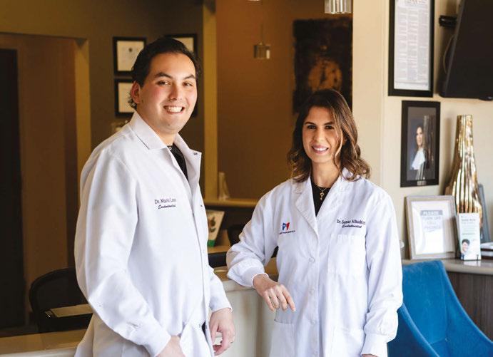
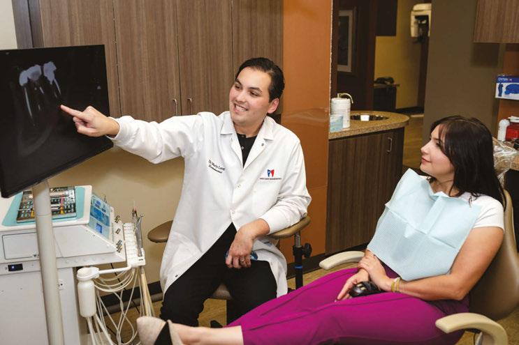
Drs. Samar Alhashimi and Mario Leon Paredes
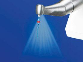
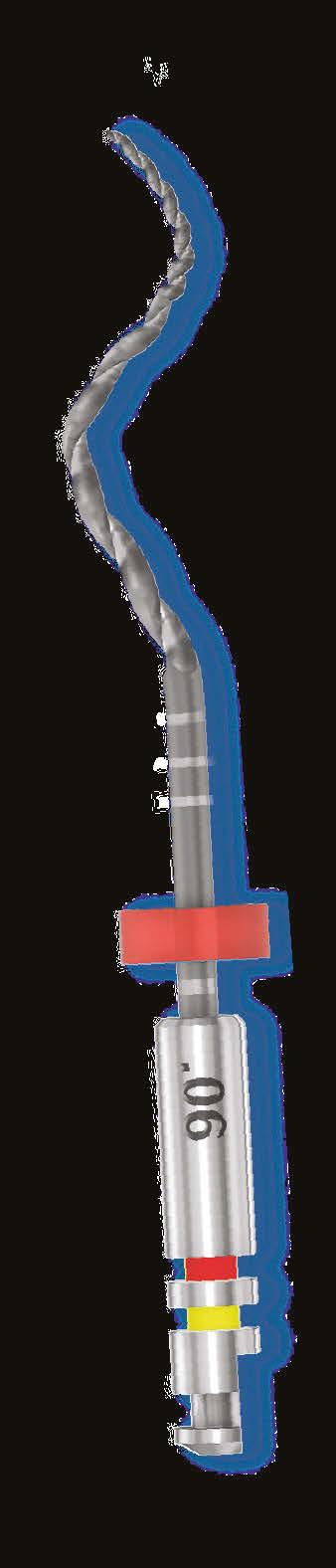
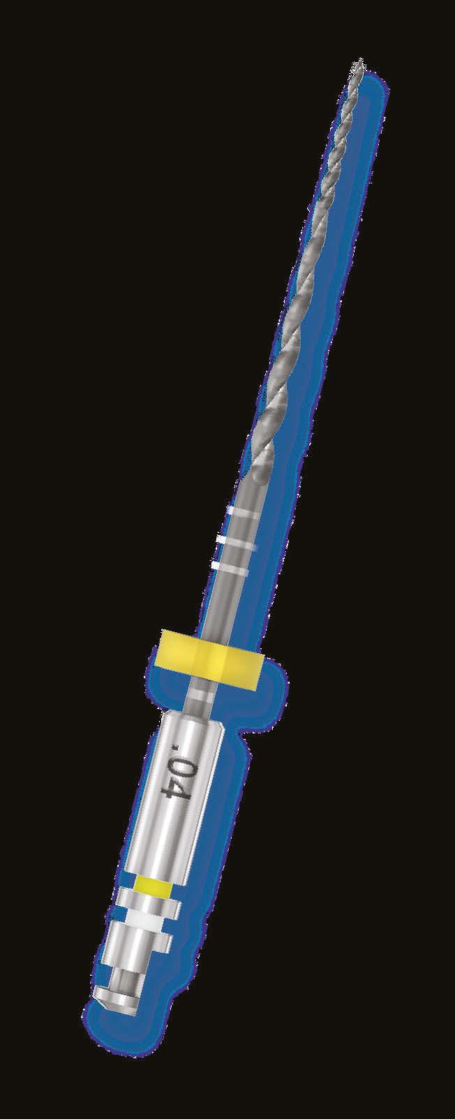
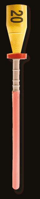
Designed to enable shaping without an initial glide path in most cases.*
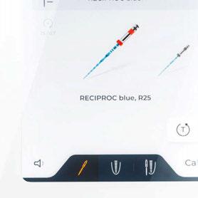
X-Smart® Pro+ provides the genuine reciprocating motion, enabling Reciproc™ Blue – an instrument designed for one file endo.
* Zuolo, M.L., M.C. Carvalho, and G. De-Deus, Negotiability of Second Mesiobuccal Canals in Maxillary Molars Using a Reciprocating System. J Endod, 2015. 41(11): p. 1913-7. Based on treatment of MB2 canals of more than 300 patients. The aim was to assess the frequency in which Reciproc R25 was able to directly scout and reach working lengths in comparison with hand fi les. In the hand fi le group working length was successfully reached in 57.48%. In the Reciproc R25 group the working length was successfully reached in 85.63% of cases.
PERFECT MATCH Longing for one file endo?
Winter 2025/Spring 2026 n Vol 19 No 1
Editorial Advisors
Dennis G. Brave, DDS
David C. Brown, BDS, MDS, MSD
L. Stephen Buchanan, DDS, FICD, FACD
Gary B. Carr, DDS
Arnaldo Castellucci, MD, DDS
Gordon J. Christensen, DDS, MSD, PhD
Stephen Cohen, MS, DDS, FACD, FICD
Samuel O. Dorn, DDS
Josef Dovgan, DDS, MS
Luiz R. Fava, DDS
Robert Fleisher, DMD
Marcela Fridland, DDS
Gerald N. Glickman, DDS, MS
Jeffrey W Hutter, DMD, MEd
Syngcuk Kim, DDS, PhD
Kenneth A. Koch, DMD
Gregori M. Kurtzman, DDS, MAGD, FPFA, FACD, DICOI
Joshua Moshonov, DMD
Richard Mounce, DDS
Yosef Nahmias, DDS, MS
David L. Pitts, DDS, MDSD
Louis E. Rossman, DMD
Stephen F. Schwartz, DDS, MS
Ken Serota, DDS, MMSc
E Steve Senia, DDS, MS, BS
Michael Tagger, DMD, MS
Martin Trope, BDS, DMD
Peter Velvart, DMD
Rick Walton, DMD, MS
John West, DDS, MSD
CE Quality Assurance Board
Bradford N. Edgren, DDS, MS, FACD
Fred Stewart Feld, DMD
Gregori M. Kurtzman, DDS, MAGD, FPFA, FACD, FADI, DICOI, DADIA
Circulation Disclosure: Total Circulation May Vary. Publisher retains the right to adjust circulation based on a number of factors including but not limited to: print and digital distribution by mail, email, and website for industry tradeshows, educational events, including nonpaid bulk copies and/or digital access provided to events, clients and educational institutions.
ISSN number 2372-6245
A reflection on patientcentered endodontics
Ihave the privilege of both practicing and teaching endodontics. One of the lessons I continually try to impart to students and colleagues is that while our focus as clinicians is often on the technical demands of the procedure, and endodontics can indeed be a highly challenging discipline requiring intense focus, we must never forget that the tooth we are treating is attached to a human being with complex emotions, fears, and expectations.
While we may be preoccupied with delivering the highest-quality root canal using the most advanced technology available, our patients are seeking something deeper. Providing exceptional care requires us to step out of our purely technical mindset and into the realm of patient management and empathy.
First and foremost, patients want to be heard. They want to feel listened to, acknowledged, and understood. We may glance at a radiograph and quickly arrive at a diagnosis and plan. We may also have a waiting room full of other patients who need our attention. But for the individual sitting in our chair, their tooth, and their story, is unique. Taking the time to honor that individuality requires patience and presence. A colleague of mine, an endodontist with years of experience defending dentists in malpractice cases, once told me that the most effective way to avoid lawsuits is to be consistently kind and generous with patients.
I always explain to patients that I don’t simply work in their mouth; I bring them into the process. I explain what I am doing, why I am doing it, and I review all of their treatment options in detail. While I am the doctor providing recommendations, I recognize that many of the cases that come to me are complex and far from blackand-white. Treatment plans often require adaptation and thoughtful decision-making, and I want my patients fully involved in that journey.
Another commonly overlooked aspect of care is the administration of local anesthesia. For us, the injection may seem like an insignificant, routine step, something to “get out of the way” before the real work begins. For patients, however, it is often the most stressful part of the entire visit. When we acknowledge this, it shifts our perspective. Mastering the art of a slow, gentle, and painless injection becomes just as important as our technical proficiency within the tooth. For many patients, that moment defines the overall experience.
Similarly, the rubber dam can be a new and sometimes intimidating experience for patients. Taking the time to explain its purpose and benefits goes a long way. We often underestimate how claustrophobic it can feel, which is why we never leave a patient under a rubber dam unattended.
Following up with patients after a procedure can also have a powerful impact. Many of our cases involve collaboration with general dentists, oral surgeons, or other specialists. When we communicate with those providers and then update the patient, it reinforces that their care is coordinated and that we are fully invested in their wellbeing. Returning calls promptly often eases anxieties and builds trust.
Much has been written about the advanced technologies that allow us to perform sophisticated endodontic procedures with remarkable precision. Far less attention is given to the patient-centered care that transforms a technically successful root canal into a truly meaningful treatment experience. Ultimately, our skill with instruments must be matched by our skill with people. When we honor both, we not only save teeth, we elevate the entire practice of endodontics.
Joseph C. Stern, DDS, completed his endodontic residency at Columbia University, where he earned his DDS. A Diplomate of the American Board of Endodontics, he directs endodontics at Touro Dental School and maintains a practice in Clifton, NJ.
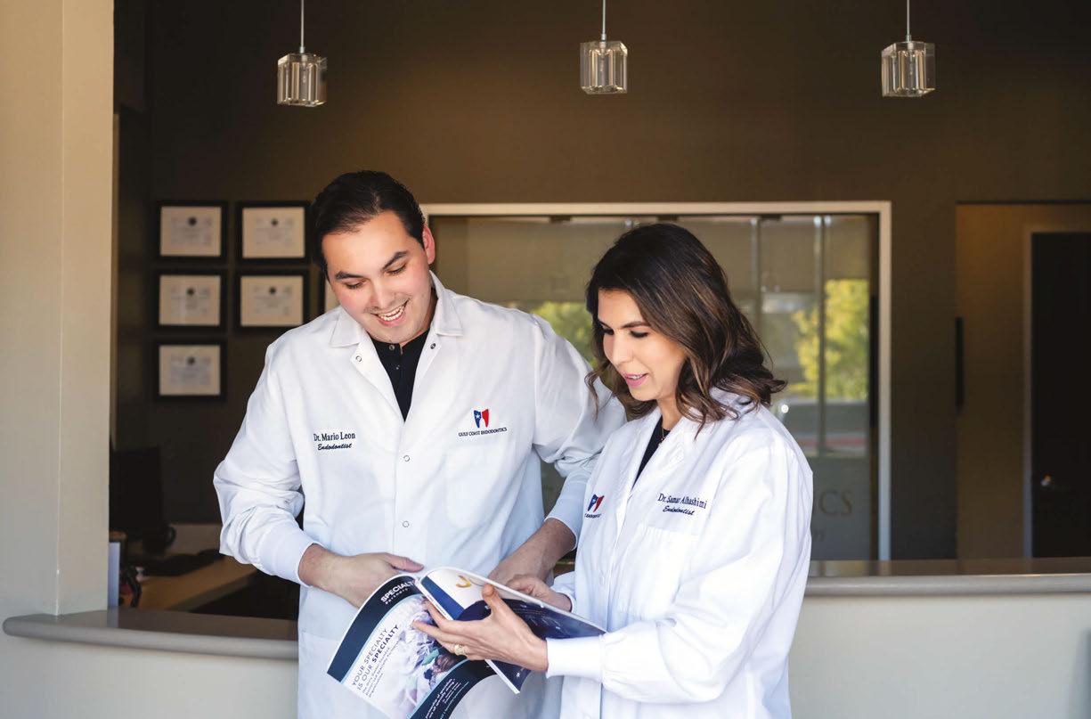
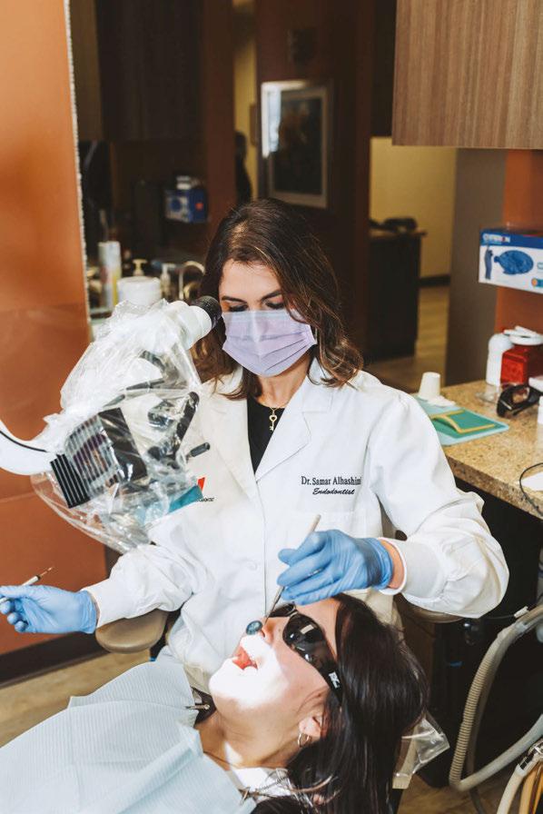
Drs. Samar Alhashimi and Mario Leon Paredes find fulfillment in the science and art of healing
Cover image of Drs. Alhashimi and Paredes courtesy of Specialty1 Partners.
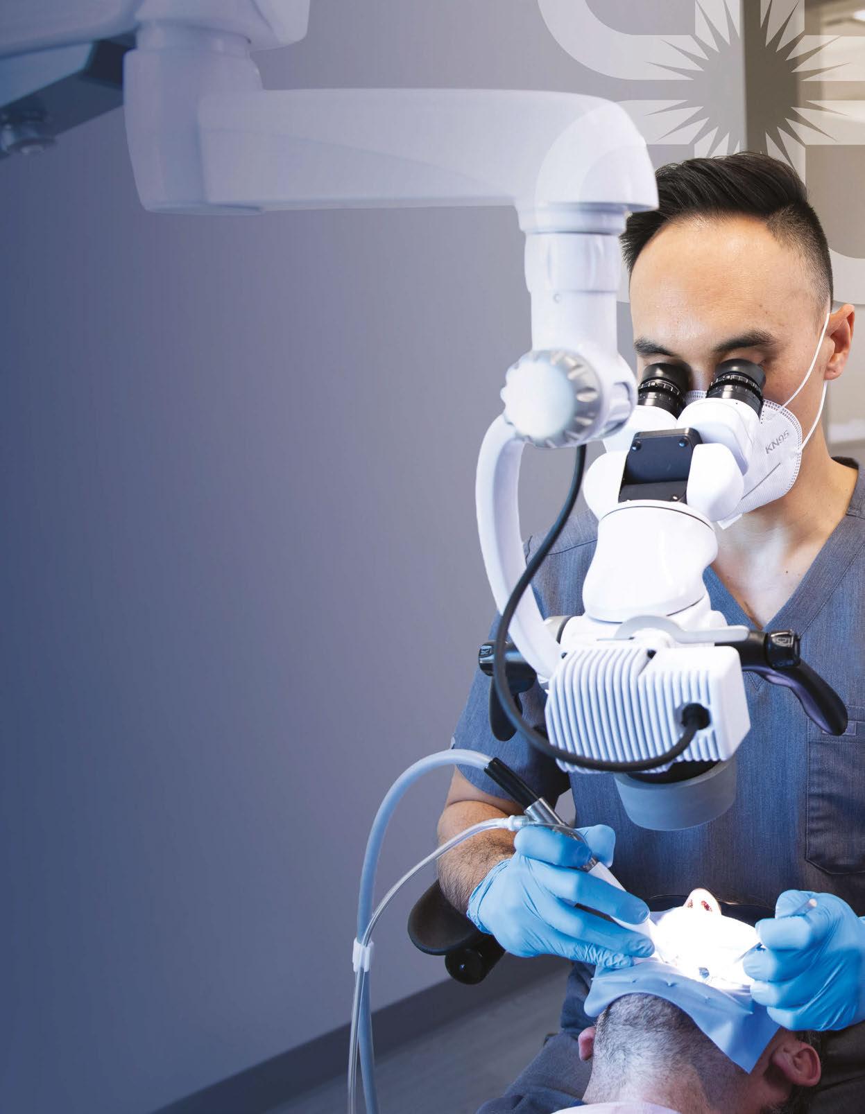
Microscope endodontic workflow: an ergonomic systematic approach
Dr. Juan Carlos Ortiz Hugues discusses the advantages of proper ergonomic use of the dental microscope
Attorneys Casey Gocel and Jonathan
Drs. Joel C. Small and Edwin McDonald
Published by
Director of Publishing Amanda Culver amanda@medmarkmedia.com
Editor in Chief
Mali Schantz-Feld, MA, CDE mali@medmarkmedia.com Tel: (727) 515-5118
Director of Sales & Business Development Ty Maggard ty@medmarkmedia.com Tel: (910) 476-2784
eMedia Coordinator Scott Peterson emedia@medmarkmedia.com
Social Media Manager Felicia Vaughn socialmedia@medmarkmedia.com
Website Support Eileen Kane webmaster@medmarkmedia.com
Subscription Rate 1 year (4 issues) $149 https://endopracticeus.com/subscribe/ CONNECT with us on social media
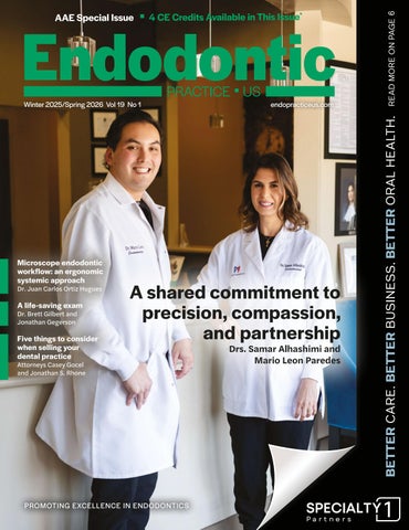
A shared commitment to precision, compassion, and partnership
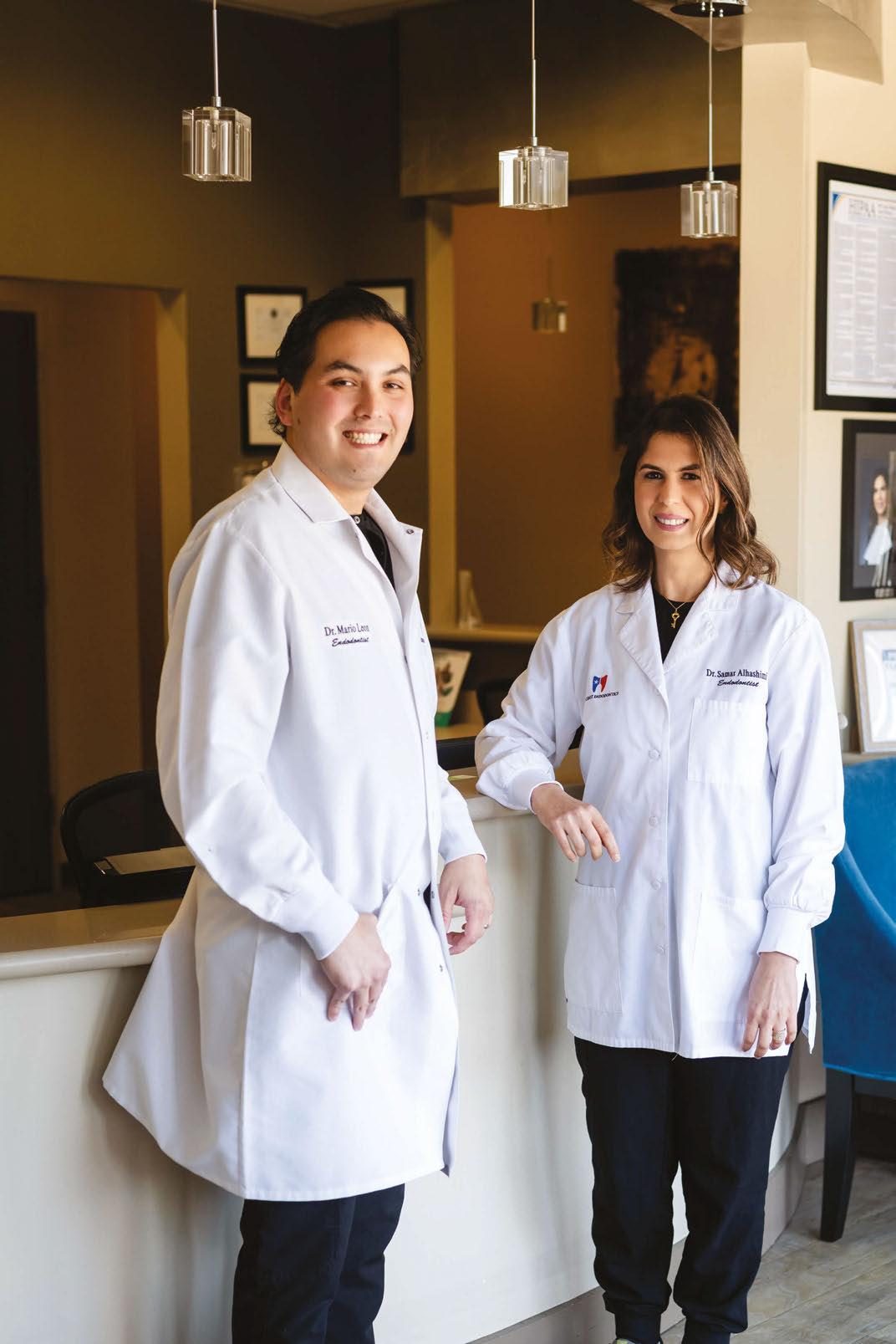
Drs. Samar Alhashimi and Mario Leon Paredes find fulfillment in the science and art of healing
Every patient who walks through the doors of Gulf Coast Endodontics in Houston, Texas brings a story, often one marked by pain, anxiety, or uncertainty. For Drs. Samar Alhashimi and Mario Leon Paredes, that moment is where science meets empathy and where their shared philosophy begins: healing with precision and compassion.
Both clinicians discovered their passion for dentistry early on, drawn by its blend of science, artistry, and human connection. Dr. Alhashimi earned her Bachelor of Dental Surgery from Baghdad University in Iraq, later completing a prosthodontic internship and endodontic training at Loma Linda University in California, where she also earned a master’s degree in Endodontics. Her research exploring the effects of Omega-3 on dentinal tissue regeneration reflected a lifelong curiosity for how biology and technology can converge to restore what once seemed lost.
Dr. Leon Paredes’ path began in Guatemala City at Universidad Francisco Marroquín. Inspired by a family of healthcare professionals and mentored by one of Guatemala’s pioneering endodontists, Dr. Estuardo Mata, he saw firsthand how dentistry could transform lives. His journey continued through an AEGD residency at the University of Connecticut and advanced specialty training at the University of Texas Health Science Center at Houston.
Though their origins are worlds apart, both doctors were shaped by the same truth: excellence in endodontics demands both intellect and heart.
The journey to Gulf Coast Endodontics
For Dr. Alhashimi, joining Gulf Coast Endodontics marked the next step in a career built on dedication and purpose. She
began as an associate, quickly earning the trust of patients, colleagues, and referring dentists through her calm demeanor and meticulous approach. “My transition into partnership wasn’t a leap — it was a natural progression,” she recalls. “It came from consistency, communication, and aligning with a team that shares the same values.”
Dr. Leon Paredes’ path to partnership reflected a similar loyalty and vision. “From the start, I knew this was where I wanted to build my career,” he says. “The practice’s founders, Drs. Matthew and Mark (Haddad), welcomed me with trust and mentorship. I wanted to give that same dedication back to the practice and our team.”
Today, the two share leadership within a practice known for its collaborative spirit and cutting-edge care. Gulf Coast Endodontics has grown into one of the most respected endodontic groups in Texas, with a reputation built on advanced technology, clinical precision, and a deeply patient-centered culture.
That growth has also been strengthened by their partnership with Specialty1 Partners, a leading dental specialty partnership organization supporting specialists across the country. “Specialty1 has been instrumental in allowing us to focus on what matters most — our patients,” says Dr. Leon Paredes. “They’ve helped streamline operations, modernize systems, and provide access to shared resources that have enhanced our efficiency and team development.” Dr. Alhashimi adds, “Partnering with Specialty1 Partners has elevated how we manage our practice while preserving the culture and autonomy that make Gulf Coast Endodontics special.”
A shared clinical philosophy grounded in empathy
Both doctors view endodontics as a discipline defined by evolution — driven by research, refined by technology, and grounded in empathy.
Dr. Alhashimi’s dual training in prosthodontics and endodontics gives her a unique perspective. “Understanding how to restore and preserve teeth from a prosthetic standpoint allows me to see the full picture,” she explains. “My goal isn’t just to relieve pain, it’s to protect the long-term function and beauty of every tooth.”
Her use of 3D imaging, laser-assisted irrigation, and biocompatible materials reflects her belief in minimally invasive, biologically driven care. “Every tooth is unique,” she says, “but following precise clinical protocols, supported by technology, ensures predictable, comfortable outcomes.”
Dr. Leon Paredes echoes that philosophy, emphasizing evidence-based care as the foundation of his approach. “Technology enhances what we do, but it’s our understanding of the science behind it that truly drives better results,” he notes. His adoption of advanced laser disinfection and research-guided techniques has elevated efficiency and patient comfort while reducing treatment times.
A passion for research and education
Throughout his professional career, Dr. Leon Paredes has contributed to the field of endodontics in ways that extend
beyond the operatory. “One of the highlights of my career has been the opportunity to present lectures at the American Association of Endodontists,” he shares. “Presenting my research and exchanging knowledge with colleagues from across the nation reinforced my commitment to advancing our specialty through education and collaboration.”
Dr. Leon Paredes has also received awards for his work in two pivotal areas of endodontics — regenerative endodontics and pulpal response following restorative treatments. These achievements, he says, represent not just personal milestones, but a continuing mission to merge science and clinical practice for the benefit of patients everywhere. “Every study, every lecture, every discussion adds another layer of understanding to how we can better preserve natural teeth.”
Collaboration and connection
The partnership between Drs. Alhashimi and Paredes thrives on mutual respect and a shared belief that teamwork extends beyond the operatory. Within the practice, they collaborate closely on complex cases, exchanging perspectives that draw from their unique educational backgrounds and experiences.
Dr. Samar Alhashimi
Their teamwork also extends outward to general dentists and specialists across the region. “We maintain open communication with every referring doctor,” says Dr. Alhashimi. “Detailed case reports, imaging, and outcome notes ensure seamless continuity of care.”
Dr. Leon Paredes agrees: “Endodontics doesn’t happen in isolation. Collaboration leads to stronger outcomes and strengthens the trust our community has in us.”
That community trust is evident not only in their clinical results but also in their reputation for warmth and accessibility. The Gulf Coast Endodontics team takes pride in creating an environment where patients feel seen, heard, and respected from the first phone call to the final follow up.
Leadership, mentorship, and practice culture
As partners, both doctors see their role as more than clinicians — they are mentors, educators, and advocates for growth. Dr. Alhashimi is passionate about teaching, having served as both clinician and professor, sharing her knowledge of advanced techniques with students and colleagues alike. “Seeing patients get better and students succeed reminds me why continuous learning matters,” she says.
The support of Specialty1 Partners has further empowered that leadership, offering structure, training, and strategic insight to help Gulf Coast Endodontics continue to thrive without compromising its patient-first philosophy.”
Dr. Leon Paredes’ leadership style centers on empowerment. He works closely with the office manager and marketing coordinator to strengthen operational efficiency and patient engagement. “Our team is like family,” he explains. “Supporting them, celebrating their wins, helping them problem solve, is just as important as the clinical side of what we do.”
The support of Specialty1 Partners has further empowered that leadership, offering structure, training, and strategic insight to help Gulf Coast Endodontics continue to thrive without compromising its patient-first philosophy.
Innovation and the future of endodontics
Both doctors are optimistic about the future of their specialty. With rapid advances in regenerative endodontics, biologic materials, and imaging precision, they see a horizon where saving natural teeth becomes even more predictable.
“Endodontics is moving toward a biologically driven, technology-enhanced model,” Dr. Alhashimi explains. “Regenerative approaches and digital workflows will continue to redefine what’s possible.”
Dr. Leon Paredes adds, “The future will belong to practices that balance science with empathy — where innovation is always guided by what’s best for the patient.”
Their shared goal is to ensure that Gulf Coast Endodontics, supported by Specialty1 Partners, remains at the forefront of that evolution, combining cutting edge care with the timeless values of compassion, education, and service.
A practice built on purpose
At its core, Gulf Coast Endodontics is a story of partnership — between doctors, patients, and the community they serve.
Drs. Samar Alhashimi and Mario Leon Paredes exemplify that ideal, blending skill with empathy and vision with humility.
“Every day, we have the opportunity to relieve pain and restore confidence,” says Dr. Alhashimi. “That’s not just dentistry; that’s impact.”
Dr. Leon Paredes agrees: “Our goal isn’t just to fix a tooth; it’s to change how patients feel about their health, their smile, and their experience in our care.”
Together, with the ongoing support of Specialty1 Partners, they are shaping the next chapter of Gulf Coast Endodontics — rooted in teamwork, trust, and a shared commitment to excellence. EP
Dr. Mario Leon Paredes
Nor th America’s most respected, doc tor- l ed , multi-sp e cialty p artn ershi p organization for gr o wth and long-term success.
“It’s
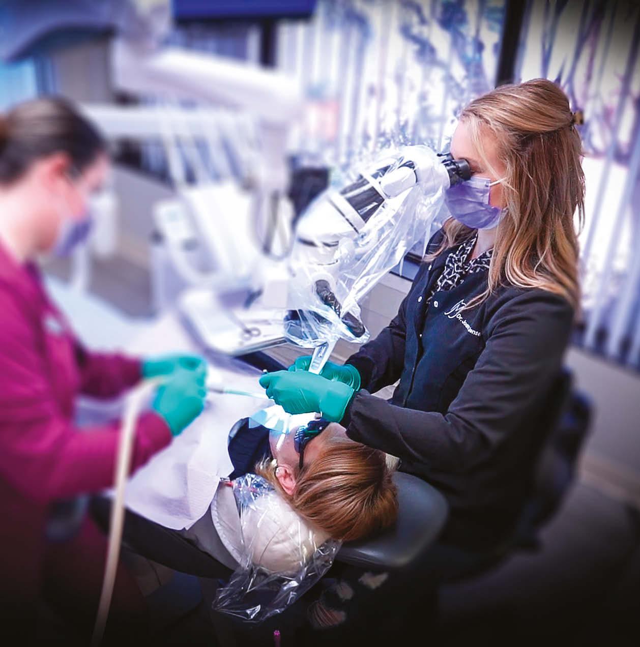
Dr. Joanne Jensen Endodontic Artistry
Crown preparation and endodontic therapy on a painful and carious tooth
Dr. David King treats a tooth that was interfering with quality of life
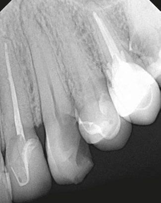
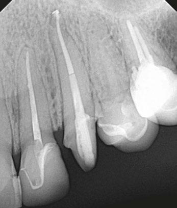
The patient presented for examination of pain that had persisted over the previous few days in the upper left (UL) area, specifically tooth No. 11. The patient described that the pain was moderate and interfering with certain aspects of life. Radiographs were taken to evaluate for carious and non-carious pathology. After examining the radiographs, it was determined that tooth No.14 was non-restorable, and tooth No. 11 needed endodontic treatment, a core, and crown to restore health (Figures 1 and 2).
After discussing all treatment options, the patient opted for endodontic therapy and buildup. Nitrous oxide was declined, so not used. Treatment began with local anesthetic that included: 1.7cc (1 carpule) 4% Articaine HCl w/1:100k epi. infiltration.
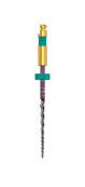
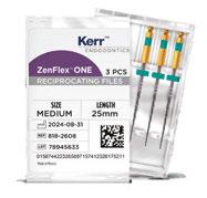
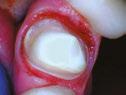
Further examination noted that decay had progressed into the nerve. The decay was removed and the canal accessed, after which the canal was cleaned and shaped with Kerr ZenFlex™ ONE (Figure 3).
After cleaning and shaping, the canal, which was determined to be 23.5 mm in length, was rinsed, irrigated, and agitated with 3ccs each of NaOCl and EDTA. The canal was sized and shaped with a ZenFlex™ ONE Medium (Green 35.06/25 mm) file to working length using crown-down tech-
David King, DDS, was born in Utah, but grew up in Montana, Idaho, and the Kansas City area. In 2006, immediately after graduating from UMKC School of Dentistry, he started a dental practice, and shortly thereafter purchased another practice to create his own small dental group practice. Dr. King practiced for a total of just over 7 years in the Kansas City area including in Liberty, Independence, Bonner Springs, and Lee’s Summit. After selling his small group of practices to a larger Kansas City-based dental group, he was recruited and relocated to the Albuquerque, New Mexico area where he joined the family of Pacific Dental Services-supported doctors and dental practices. After spending nearly 4 years in Rio Rancho and Albuquerque, he was given an opportunity to relocate back to his home state of Missouri to assist in the growth of several dental practices there, and he partnered in multiple practices including in O’Fallon, St. Peter’s, and St. Charles, Missouri. Currently, Dr. King spends his free time in general dentistry as a regional partner doctor with Lumio Dental where he enjoys service to others by providing high quality comprehensive dental care including Invisalign® , restorative, fixed, and removable prosthodontic dental care, endodontic treatment, and complex dental implant dentistry, all while devoting his primary time to serving as a husband, father, flower farmer, and doing his best to serve in his local church (The Church of Jesus Christ of Latter-Day Saints) congregation and as a public speaker and educator.
nique and substantial irrigation. The anatomical location, near the anatomic apex, was slightly distally angulated.
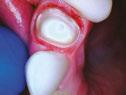
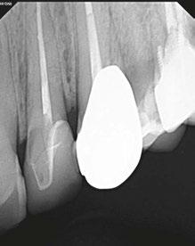
The canal was dried, BC sealer was placed, and the canal was obturated with warm condensation. Then, the space was prepared for a FibreKleer™ 4x Tapered 1.5 mm post (Figure 3). The post was put in place and bonded with Optibond™ Universal (Figure 4).
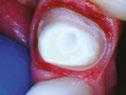
The patient had opted in for a crown because the caries/failing restoration had extended to greater than two-thirds of the occlusal table, and endodontic treatment had just been completed. The tooth was reduced, and occlusion clearance and margin height were checked (Figure 5). The final crown, in shade A2, was milled in house and seated same day (Figure 6). The final crown was cemented with Optibond™ Universal bond and Maxcem Elite™ Cement. This case study was provided by Kerr.
Figure 1 (left) and Figure 2 (right)
Figure 3 (left), Figure 4 (center), and Figure 5 (right)
Figure 6
Enhance
Excellent Flexibility
Increased Performance
High Cutting Efficiency
Reach More
Scan the QR Code or visit go.kerrdental.com/zenflexcm
in comprehensive tip and taper configurations sizes (04/06, 13-30)
Tooth No. 3 obturated with SimpliCore™
Dr. David King treats a failing restoration
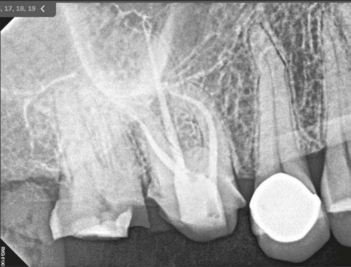
The patient came in for a crown re-do on tooth No. 2 and pain on tooth No. 3 (Figure 1).
The patient presented with a failing restoration on tooth No. 2 and caries/decay extending greater than two-thirds occlusal table. The procedure consisted of removal of gross decay, any fractured enamel or old restorations, replacement of tooth structure with a buildup, and placement of a crown to cover and protect the tooth and ensure the patient can return to normal function.
The structure of tooth No. 2 was reduced using a high-speed handpiece, and a provisional crown was fabricated and secured with temporary cement material.
The patient returned for root canal treatment on tooth No. 3. The doctor discussed the severity of conditions and what treatments were medically necessary to eliminate the decay. Treatment options were discussed, as well as advantages and disadvantages of each treatment option, and the patient opted for endodontic therapy.
The decay was removed, and access to the pulpal canals was exposed with the pulpal chamber completely unroofed.
1. Rotary and hand files were used to debride and shape the root canals.
2. The canals were rinsed with NaOCI and EDTA.
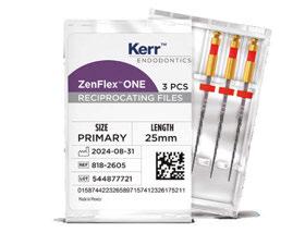
3. ZenFlex™ ONE Primary Files (.25/.06/25 mm) were used to instrument to working length (Figure 4).
David King, DDS, was born in Utah, but grew up in Montana, Idaho, and the Kansas City area. In 2006 immediately after graduating from UMKC School of Dentistry, he started a dental practice and shortly thereafter purchased another practice in the makings of his own small dental group practice. Dr. King practiced for a total of just over 7 years in the Kansas City area including in Liberty, Independence, Bonner Springs, and Lee’s Summit. After selling his small group of practices to a larger Kansas City-based dental group, he was recruited and relocated to the Albuquerque, New Mexico area where he joined the family of Pacific Dental Services-supported doctors and dental practices. After spending nearly 4 years in Rio Rancho and Albuquerque, he was given an opportunity to relocate back to his home state of Missouri to assist in the growth of several dental practices there and there partnered in multiple practices including in O’Fallon, St. Peter’s, and St. Charles Missouri.
Currently, Dr. King spends his free time in general dentistry as a regional partner doctor with Lumio Dental where he enjoys service to others by providing high quality comprehensive dental care including Invisalign, restorative, fixed, and removable prosthodontic dental care, endodontic treatment and complex dental implant dentistry, all while devoting his primary time to serving as a husband, father, flower farmer, and doing his best to serve in his local church (The Church of Jesus Christ of Latter-Day Saints) congregation and as a public speaker and educator.
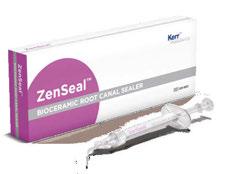
4. The canals were dried before placing ZenSeal™ Bioceramic Sealer (Figure 4).
5. The canals were obturated using SimpliCore™ for ZenFlex™ ONE Primary (Figures 2 and 3).
A core build-up was placed on tooth No. 3 to prevent bacterial reinfection, provide essential support for a final crown, and reduce increased risk of tooth fracture or RCT failure. This case study was provided by Kerr.
Figure 1 (left) and Figure 2 (right)
Figure 3
Figure 4
Crafted for Simplicity. Molded for Perfection.
SimpliCore™
Gutta Percha Carrier Obturators
Compatible with most file systems on the market, SimpliCore is designed to integrate into your existing workflow. Featuring a solid gutta percha core, our obturators are precisely crafted for ease-of-use and predictable obturation outcomes.
ISO Indicator Handle
Solid Gutta Percha Core with Length Verification Markers
Promo Code: SCMO25
Gutta Percha
*Promotional goods must be of equal or lesser value and may not be combined with any other offers. Valid only for obturator packs. Purchase must be made on one invoice between 10/01/2025 and 3/31/2026 unless otherwise noted. Redeem by 4/30/2026. To receive your promotional goods, email or fax a copy of your authorized invoice noting promo code SCMO25 and product(s) desired to: kerrpromo@kerrdental.com or FAX: 888.727.2614 Limit to 3 redemptions. Incomplete submissions will not be processed. Allow 8 weeks for delivery. Offer valid in the 50 United States and the District of Columbia only. Promotions are subject to change or cancellation without notice. Offer void if purchased product is returned. Note that you may have an obligation under federal, state or local law to reflect discounts on product given pursuant to this promotion on any cost report forms submitted to a federal or state government or private payer who provides reimbursement for that product.
1-800-KERR-123 | kerrdental.com
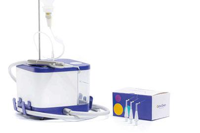
OdneClean – hydrodynamic cavitation using sterile water
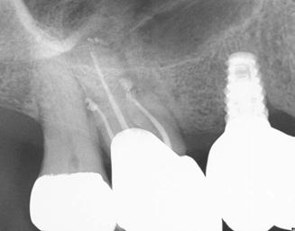
Rapid radiographic healing of a large periapical lesion: a six‑month case review
Managing necrotic pulps with symptomatic apical periodontitis remains a frequent yet clinically demanding challenge in endodontic practice. Optimal outcomes rely heavily on effective disinfection and removal of biofilm and necrotic debris from the complex root canal anatomy. This case highlights the healing potential that can be achieved when standard instrumentation is combined with Odne®Clean, an advanced irrigation device that uses sterile water as its primary debridement medium delivered through an ultra-thin and flexible tip that is designed to optimize cleaning in the apical third. The result was an impressive reduction of a large periapical lesion within only 6 months, an encouraging outcome for both clinician and patient.
About OdneClean
Odne®Clean creates a hydro-dynamic cavitation cloud inside the root canal using sterile water as the main irrigation medium. With its 190 µm tip, the thinnest dental fluid-delivery tip on the market, the cavitation jet effectively cleans complex root canal geometries and apical thirds and increases the effect of the final standard needle disinfection rinse with NaOCl.1 Odne®Clean de-risks the procedure by significantly reducing the use of harsh disinfectants and eliminating the need for its activation.2,3 It also supports minimally invasive root canal treatments enabling endodontists and dentists to preserve as much tooth structure as possible. Visit https://odne.co/odne-clean/ to learn more.
Case presentation
An 89-year-old male patient presented with unprovoked pain and clear pain on percussion. Clinical testing supported a diagnosis of pulp necrosis with symptomatic apical periodontitis, confirmed by radiographic evaluation. The preoperative radiograph showed a large, well-defined periapical radiolucency, consistent with a PAI score of 5, indicating severe apical periodontitis (Figures 1A-1C).
Treatment protocol
After establishing access and confirming working length, canal instrumentation was carried out to size 35/.04 in all three canals. Shaping was performed using standard solutions, 5% NaOCl and 17% EDTA, followed by OdneClean`s clinical protocol.
Given the necrotic status of the tooth and the large lesion, calcium hydroxide was placed as an interim dressing. At the subsequent visit, the canals were irrigated again with OdneClean activation to remove the CaOH and dried before obturation. Obturation was completed using bioceramic BC Sealer™. A postoperative X-ray can be seen in Figure 2.
Follow-up and healing
At the 6-month follow-up, the patient was asymptomatic, and percussion testing was normal. Radiographic evaluation revealed a clear reduction in the periapical radiolucency, with the PAI score improving from 5 to 3, indicating significant healing (Figures 2A-2D).
The visual reduction in lesion size was substantial, going from large to small. Periapical lesions of this magnitude often require extended healing periods. Large scale studies report that 85% of lesion cases heal within 48 months, and 42.5% show clear healing or signs of healing at 6 months.4 Another study observed that, additionally, healing is strongly influenced by the initial lesion size and patient age.5 They reported that patients with fully healed lesions had a mean age of 41.2 years, compared with 52.8 years in unhealed cases. Healing time increased by 4.9% per year of age and 0.3% per mm³ of lesion volume.5 Remarkably, only 8.9% of large lesions healed completely within 6 months.5 Considering this evidence, the degree of healing observed in this 89-year-old patient is impressive.
Conclusion
This case highlights the successful management of a large periapical lesion associated with pulp necrosis and symptomatic apical periodontitis. The combination of standard instrumentation, calcium hydroxide dressing, and mechanical debridement using OdneClean contributed to significant healing within 6 months. The outcome reflects how thoughtful irrigation strategies can elevate everyday endodontic procedures and produce predictable, patient-centered results.
Clinical case courtesy of Dr. Elena Kurtz, Exclusively Endodontics, and provided by Odne. References available online.
Figures 1A–1C: Preoperative X‑ray (A) and CBCT (B, C) showing the large periapical radiolu cent lesion (red arrows)
A. B. C.
Figure 2A–2D: Postoperative X‑ray (A), 6‑month follow‑up X‑ray (B), and CBCT (C, D) show ing radiographic healing. Red arrows indicate the periapical radiolucent lesion
A.
B. C. D.
A life-saving exam
Dr. Brett Gilbert and Jonathan Gegerson provide a call to action for head and neck screening in dental practice
Abstract
This article underscores the critical role of the dental professional in the early detection of head and neck cancers through a systematic Conventional Visual and Tactile Examination (CVTE). Inspired by the personal story of my close friend, Jonathan, whose cancer diagnosis may have been expedited by routine dental screening, this article explores the components of the head and neck exam, its clinical rationale, and the current evidence supporting its implementation as a standard of care in dental settings.
Jonathan’s story in his own words: a missed opportunity at the dental chair
Educational aims and objectives
This self-instructional course for dentists looks at the critical role of the dental professional in the early detection of head and neck cancers.
Expected outcomes
Endodontic Practice US subscribers can answer the CE questions by taking the quiz online at endopracticeus.com to earn 2 hours of CE from reading this article. Correctly answering the questions will demonstrate the reader can:
• Recognize the importance of the Conventional Visual and Tactile Examination (CVTE) in routine dental visits for early detection of head and neck cancers.
• Identify and describe each component of a systematic extraoral and intraoral head and neck exam.
• Understand the current evidence-based guidelines that support the implementation of CVTE in dental practice.
• Apply practical strategies for incorporating the CVTE into clinical workflows to enhance patient outcomes.
There I sat waiting for the doctor to examine my concern of a lump on the right side of my neck. I assumed it was nothing to be concerned about. The doctor entered the room, and I showed her the area. She felt around my neck on both sides, felt under my arms, and then asked a couple questions. “Have you had any dental work recently?” I responded, “No.” She then asked, “Have you had any infections in the mouth, or a root canal or anything?” Once again, my response was, “No.” She then looked at me and said, “It may be cancer.” I was stunned, and my body and mind froze in that moment, as that was the last thing I thought I was going to hear. Other than the lump on my neck, I was in perfect health. I worked out constantly, ate a Keto diet, never smoked, and limited all fatty foods and alcohol. There was no reason I should have cancer — at least that is what I thought.
Brett E. Gilbert, DDS, FICD, graduated from the University of Maryland Dental School (DDS, 2001, Endo, 2003). He is a professor in the Department of Endodontics at the University of Illinois at Chicago and a Diplomate of the American Board of Endodontics. He is the founder of the Access Endo Impact Academy, a global direct mentorship continuing education platform. He is a fellow in the International College of Dentists and a contributing consultant for Oral Health Journal. Dr. Gilbert lectures nationally and internationally on clinical endodontics and personal wellness and is the host of the On The Cusp podcast. Dr. Gilbert is a global key opinion leader in the area of endodontics and new technology. He is a partner in Specialized Dental Partners and has a private practice, King Endodontics PLLC, limited to Endodontics in Niles, Illinois.
Jonathan Gegerson, cancer survivor, has many dentists in his family tree — his great uncle, father, and brother were all dentists. He worked for Envista with Kerr as the Global Trainer and then with Orascoptic as a Regional Manager. His 14-year career in the dental industry ended in 2021 when he could no longer perform his duties because of cancer. Since then, he has written a book Perspective C, (available on Amazon), to inspire and help people going through an illness. Gegerson has personally endured seven surgeries, 67 sessions of radiation, and over 200 sessions of chemotherapy. He believes that the biggest lesson is to approach everything with a desired outcome predetermined and to keep smiling. jonathangegerson@gmail.com
Since my initial diagnosis in May 2019, I have received over 200 rounds of chemotherapy, 67 sessions of radiation, and six surgeries. The side effects from all these treatments have been overwhelming at times. I lost my ability to chew and must be on a soft, liquid diet. My mouth opens just wide enough for a spoon. I was on a feeding tube for 6 months. I have constant lymphedema of my face, tongue, and throat which creates speaking problems, swallowing concerns, and at times, vision issues. I have radiation scars on my face, and one of my vocal cords was paralyzed during my recent 17-hour surgery, which was to help correct lymphedema as my airway was being constricted. I ended up in the ICU twice due to airway constriction. My body was literally suffocating itself.
I am the second patient to receive the type of surgery I had for head and neck lymphedema. My recovery took 7 months, and for two of those months, I was in the hospital. I share this because there were opportunities to catch the cancer sooner. Those opportunities were in my own power as well as the power of my dentist and hygienist. I mention my hygienist and dentist as I would see them regularly — at least every 6 months. If the cancer was caught sooner, it may have prevented such an
extensive treatment plan, limited all my side effects, and resulted in a better lifestyle after treatment.
I would rather have been told there was a concern 3 or 6 months before by my dentist or hygienist and addressed the concern with my primary doctor immediately. Early detection results in better patient outcomes. I was informed by my oncologist that out of 4 million people, 10 to 15 people get the type of cancer I was diagnosed with: Salivary Duct Carcinoma HER2 Positive. I was also told that I have been beating the odds, and I am one of a kind. I would rather be many of a kind when it comes to living through and after cancer. This is one of my motivations to encourage all hygienists and dentists to perform a cancer screening exam that includes the neck. Most people with my diagnosis are not here to tell their story. I intend to purposefully speak for them and myself as I encourage all dental professionals to perform a head and neck exam on every adult patient, every time they see them!
The clinical imperative
The dental setting provides a unique, often untapped opportunity to detect early signs of head and neck malignancies. The Conventional Visual and Tactile Examination (CVTE) is a simple, low-cost, evidence-based procedure that can uncover abnormalities before they become life-threatening.
The American Dental Association (ADA) recommends that clinicians perform a systematic CVTE for all adult dental patients, including inspection and palpation of the face, neck, and regional lymph nodes to detect tissue changes, masses, or asymmetry.1 This recommendation is echoed by the American Academy of Otolaryngology–Head and Neck Surgery, which emphasizes evaluation of the skin, salivary glands, thyroid, and lymph nodes using bimanual palpation techniques.2
Despite this guidance, implementation in general practice remains inconsistent.3
Step-by-step: performing the Conventional Visual and Tactile Examination (CVTE)
General observation
Observe facial symmetry, skin color/lesions, swelling, and visible masses. Note signs of asymmetry, facial droop, or skin changes.
Lymph node palpation
Preauricular and postauricular, submental and submandibular, cervical chain (anterior and posterior), supraclavicular nodes. Use gentle, circular pressure with the pads of your fingers, bilaterally when appropriate.
Salivary glands
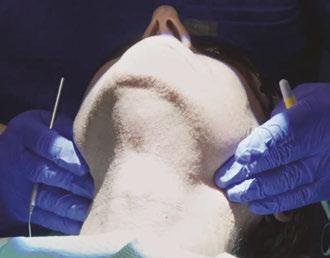
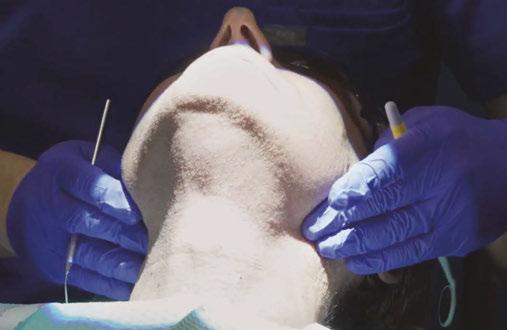
Palpate parotid, submandibular, and sublingual glands for enlargement, tenderness, or firmness (Figure 1).
Thyroid gland
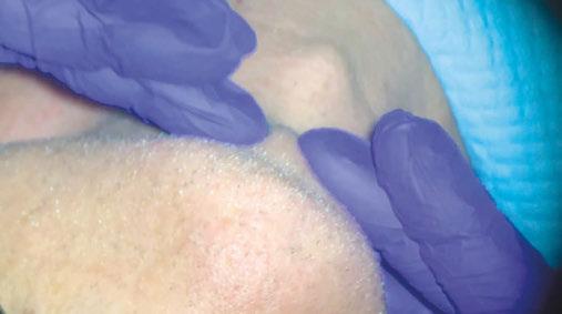
Visually inspect for enlargement while the patient swallows. Palpate the thyroid gently from behind the patient using both hands (Figure 2).
Figure 2: Thyroid exam
Figure 1: Salivary gland extraoral exam
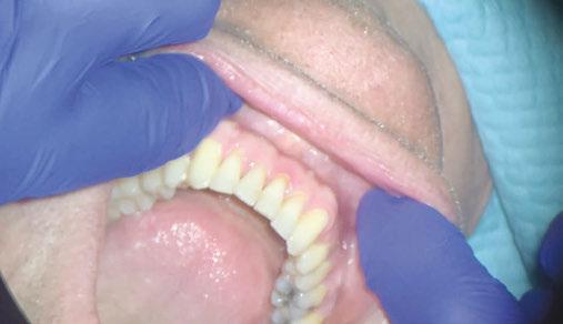
Figure 3: Intraoral vestibule exam
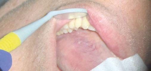
Figure 4: Lateral tongue exam
Floor of mouth (Bimanual palpation)
Place one gloved hand inside the mouth and one under the chin. Gently compress to assess for firm masses or nodularity.
Intraoral exam
Thorough inspection of the lips, buccal mucosa, gingiva, hard/soft palate, tongue (dorsal, lateral, and ventral), and oropharynx. Use gauze to pull and manipulate the tongue for complete visualization (Figures 3-4).
A case from my chair: CVTE in action
While Jonathan’s story reflects the devastating consequences of a missed opportunity for early detection, I also carry with me the opposite experience — one in which a thorough Conventional Visual and Tactile Examination (CVTE) led to the early diagnosis of a life-threatening malignancy.
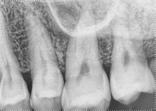
In 2016, a patient presented to my endodontic clinic for evaluation of a sore area on the palatal tissue adjacent to tooth No. 14 (Figure 5). As with every patient encounter, I performed a standard diagnostic endodontic evaluation alongside a systematic CVTE.
Clinical Findings
• Percussion: Negative
• Palpation: Positive on the palatal surface of tooth No. 14
• Periodontal Probing: Within normal limits
• Mobility: Within normal limits
• Pulpal Sensitivity (Cold): Normal response
These results pointed toward an endodontic diagnosis for tooth No. 14 of normal pulp with symptomatic apical periodontitis (due to a positive finding of palpation on the palate). But what caught my attention was not the tooth — it was the tissue.
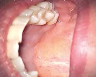
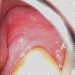
During the CVTE, I noted a small raised swelling on the palatal surface (Figure 6) and a separate white nodular lesion on the buccal gingiva adjacent to tooth No. 14 (Figure 7). Importantly, these findings did not align with any odontogenic pathology typically associated with tooth No. 14. That clinical inconsistency provided the moment to pause and widen the diagnostic lens.
Referral and diagnosis
Given the suspicious nature of these findings, I referred the patient to an oral and maxillofacial surgeon for biopsy. Oral and Maxillofacial (OMS) surgeons are often the first line of defense for dentists who may be unsure about a diagnosis that does not appear to be odontogenic in origin. The term “non-leo” refers to a lesion of non-endodontic origin. As a dental specialist, determination that findings are not adding up to a diagnosis of dental origin must be referred immediately.
OMS is the first line of referral for dentists as these specialists often have had extensive medical and hospital training. A dentist must understand that when they detect an irregular finding, no matter how small, it is critical to make this referral. Immediate referral will shorten the time between detection and diagnosis.
In many cases, the referral may seem like it was excessive if the OMS does not diagnose a problem. As clinicians, we should never allow doubt to creep into our minds by thinking that the finding is likely not significant. In fact, a non-significant diagnosis is the hope of the referral! The conversation we have with a patient in this moment should be calm in tone and decisive. I often will have my clinical team call the OMS office to set up a consultation appointment while the patient is still in our office. We must consider that a patient may listen to your concern but not act by making an appointment which could decrease the chances of a good outcome if a serious medical diagnosis is confirmed.
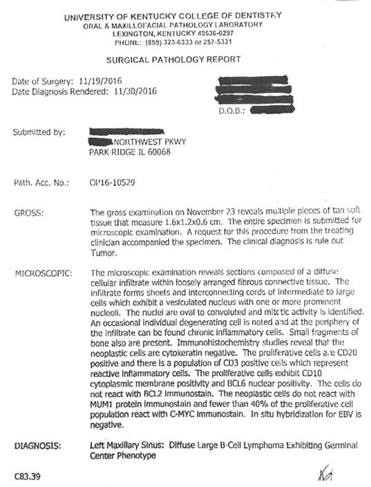
This patient was compliant and presented to the OMS the next day. The OMS performed their own examination and determined that a biopsy was necessary. The biopsy results revealed:
“Diffuse large B-cell lymphoma of the left maxillary sinus, germinal center phenotype” (Figure 8).
The patient was subsequently referred to oncology and underwent successful treatment for which they were extremely grateful that the detection of such a devastating systemic illness was detected early in the dental chair.
Clinical takeaway
This case illustrates the critical importance of integrating CVTE with routine diagnostic protocols. If I had focused solely
on the dental findings, the underlying malignancy may have been missed. It was the intentional soft tissue assessment through CVTE that revealed the warning signs.
When something doesn’t “add up” between tooth-level findings and soft tissue presentation, it’s time to:
• Pause and reassess
• Expand the differential beyond odontogenic causes
• Refer promptly to an oral and maxillofacial surgeon
Head and neck cancers often masquerade as benign or dental conditions — or remain entirely silent. A few extra moments of systematic examination can create a critical bridge to life-saving intervention.
Evidence basis: why it matters
CVTE remains the gold standard in dental practice for early detection of head and neck malignancy,1,2,3 according to multiple high-level guidelines and reviews. Some literature shows that adjunctive screening tools (e.g., fluorescence imaging, salivary biomarkers) have not demonstrated sufficient evidence to replace or supplement CVTE in routine practice.4,5 However, any device or screening tool that reminds a clinician how important it is to conduct these exams are worthy. CVTE should be considered a critical part of the dental examination and can be used in conjunction with any other screening tool that a clinician feels is valuable. Recent systematic reviews confirm that visual inspection and palpation of the head, neck, and lymph nodes are the most frequently implemented and most reliable methods for early cancer detection in dental settings.6,7
Emerging test for high detection of oropharyngeal cancers is under investigation
Recent studies, including Das, et al., (2024, 2025),8,9 demonstrate that the HPV-DeepSeek assay — using whole-genome sequencing of circulating tumor HPV DNA (ctHPV-DNA) — achieves high sensitivity (up to 96–99%) and specificity (up to 99%) for detecting HPV-positive oropharyngeal squamous cell carcinoma, with detection possible up to 7.8–10 years before clinical diagnosis in some cases.8,9,10,11 This supports the claim that HPV-DeepSeek is a highly accurate, non-invasive liquid biopsy with potential for early cancer detection.
However, the clinical utility of HPV-DeepSeek as a routine screening tool in the dental setting remains investigational11,12,13 While the test shows promise for early detection and could theoretically complement head and neck examinations in dentistry, there is currently no guideline or regulatory approval for its use in routine screening or as a standard adjunct in dental practice. The National Comprehensive Cancer Network (NCCN) guidelines emphasize that blood-based ctHPV-DNA assays are not yet part of standard screening or diagnostic protocols and that their performance and actionable implications outside of clinical trials are still being evaluated.11
Closing thoughts: a moral and clinical duty
For Jonathan, the absence of a head and neck exam may have cost him an earlier diagnosis, a simpler treatment, and a better quality of life. He has beaten the odds and desperately
wants to share his story as a motivation and wake up call for dental professionals. His story is a call to action — for dentists and hygienists to go beyond the minimum, to reclaim our critical role in early cancer detection, and to never underestimate the power of a few minutes of intentional, hands-on examination.
Working in conjunction with other dental specialists to get a second opinion or to perform more advanced testing procedures, such as a biopsy or other imaging, represents best practice in dentistry. As dental professionals, we often have more opportunities to see and examine our patients than a primary physician. We cannot become complacent and avoid performing the CVTE just because the patient appeared healthy at prior visits. Thorough review of patient health history, medications, and any acknowledgement of oral habits such as smoking, oral placement of tobacco, and even consumption of alcohol should further motivate clinicians to remember to perform this life-saving examination.
As dentists, we do not have to take on the burden of making a definitive diagnosis on cases that present with abnormal findings. Our duty and responsibility are only to make a timely and appropriate referral for further examination and testing. By making the CVTE a routine, non-negotiable part of every adult dental visit, we honor our patients, our profession, and the principle that dentistry is, at its core, a healing art.
Figure 8: Biopsy report
REFERENCES
1. Lingen MW, Abt E, Agrawal N, Chaturvedi AK, Cohen E, D’Souza G, Gurenlian J, Kalmar JR, Kerr AR, Lambert PM, Patton LL, Sollecito TP, Truelove E, Tampi MP, Urquhart O, Banfield L, Carrasco-Labra A. Evidence-based clinical practice guideline for the evaluation of potentially malignant disorders in the oral cavity: A report of the American Dental Association. J Am Dent Assoc. 2017 Oct;148(10):712-727.e10. doi: 10.1016/j.adaj.2017.07.032.
A few extra moments of systematic examination can create a critical bridge to life-saving intervention.”
2. Pynnonen MA, Gillespie MB, Roman B, Rosenfeld RM, Tunkel DE, Bontempo L, Brook I, Chick DA, Colandrea M, Finestone SA, Fowler JC, Griffith CC, Henson Z, Levine C, Mehta V, Salama A, Scharpf J, Shatzkes DR, Stern WB, Youngerman JS, Corrigan MD. Clinical Practice Guideline: Evaluation of the Neck Mass in Adults Executive Summary. Otolaryngol Head Neck Surg. 2017 Sep;157(3):355-371. doi: 10.1177/0194599817723609.
3. Pynnonen MA, Gillespie MB, Roman B, Rosenfeld RM, Tunkel DE, Bontempo L, Brook I, Chick DA, Colandrea M, Finestone SA, Fowler JC, Griffith CC, Henson Z, Levine C, Mehta V, Salama A, Scharpf J, Shatzkes DR, Stern WB, Youngerman JS, Corrigan MD. Clinical Practice Guideline: Evaluation of the Neck Mass in Adults. Otolaryngol Head Neck Surg. 2017 Sep;157(2_suppl):S1-S30. doi: 10.1177/0194599817722550.
4. Moyer VA; U.S. Preventive Services Task Force. Screening for oral cancer: U.S. Preventive Services Task Force recommendation statement. Ann Intern Med. 2014 Jan 7;160(1):55-60. doi: 10.7326/M13-2568.
5. Huber MA. Adjunctive Diagnostic Techniques for Oral and Oropharyngeal Cancer Discovery. Dent Clin North Am. 2018 Jan;62(1):59-75. doi: 10.1016/j.cden.2017.08.004. Epub 2017 Oct 16.
6. Louredo BVR, de Lima-Souza RA, Pérez-de-Oliveira ME, Warnakulasuriya S, Kerr AR, Kowalski LP, Hunter KD, Prado-Ribeiro AC, Vargas PA, Santos-Silva ARD. Reported physical examination methods for screening of oral cancer and oral potentially malignant disorders: a systematic review. Oral Surg Oral Med Oral Pathol Oral Radiol. 2024 Feb;137(2):136-152. doi: 10.1016/j.oooo.2023.10.005. Epub 2023 Oct 16.
7. Sykes EA, Weisbrod N, Rival E, Haque A, Fu R, Eskander A. Methods, Detection Rates, and Survival Outcomes of Screening for Head and Neck Cancers: A Systematic
Review. JAMA Otolaryngol Head Neck Surg. 2023 Nov 1;149(11):1047-1056. doi: 10.1001/jamaoto.2023.3010.
8. Das D, Hirayama S, Aye L, Bryan ME, Naegele S, Zhao B, Efthymiou V, Mendel J, Fisch AS, Kröller L, Michels BE, Waterboer T, Richmon JD, Adalsteinsson V, Lawrence MS, Crowson MG, Iafrate AJ, Faden DL. Blood-based screening for HPV-associated cancers. medRxiv [Preprint]. 2024 Feb 2:2024.01.04.24300841. doi: 10.1101/2024.01.04.24300841.
9. Bryan ME, Aye L, Das D, Hirayama S, Al-Inaya Y, Mendel J, Naegele S, Efthymiou V, Alzumaili B, Faquin WC, Sadow PM, Lin D, Varvares MA, Feng AL, Deschler DG, Chan AW, Paly J, Park JC, Roberts T, Merkin R, Mishra SK, Kröller L, Michels B, Iafrate AJ, Wirth LJ, Adalsteinsson VA, Crowson M, Waterboer T, Mirabello L, Lawrence MS, Guan Z, Fisch AS, Richmon JD, Faden DL. Direct Comparison of Alternative Blood-Based Approaches for Early Detection and Diagnosis of HPV-Associated Head and Neck Cancers. Clin Cancer Res. 2025 Aug 14;31(16):3483-3493. doi: 10.1158/1078-0432. CCR-24-2525.
10. National Comprehensive Cancer Network. Head and Neck Cancers. Practice Guideline. Updated August 12, 2025. https://www.nccn.org/guidelines/guidelinesdetail?category=1&id=1437.
11. Poljak M, Cuschieri K, Alemany L, Vorsters A. Testing for Human Papillomaviruses in Urine, Blood, and Oral Specimens: an Update for the Laboratory. J Clin Microbiol. 2023 Aug 23;61(8):e0140322. doi: 10.1128/jcm.01403-22. Epub 2023 Jul 13.
12. Araujo M, Bouassaly J, Farshadi F, Hier M, Mascarella M, Mlynarek A, Alaoui-Jamali M, da Silva SD. Current status of circulating tumor DNA and circulating cell alterations in HPV-associated head and neck cancer. Oral Oncol. 2025 Aug;167:107417. doi: 10.1016/j.oraloncology.2025.107417. Epub 2025 Jun 13.
Endodontic Practice US Webinars
LEARN about the lastest techniques and technology from industry leaders with our free live and archived educational webinars. Our online seminars are a convenient way to access great information and upskill. Check out our most recent webinars:
• Implementing Laser Assisted Endodontics with Understanding & Confidence: Using the EdgePro for Initial RCTs and Retreatments with Dr. Fernando Meza
• Minimally Invasive Endodontics: Is it a Paradigm Shift or Myth with host Dr. Mustafa Gündoğar
• Anatomically Centered Endodontics with host Dr. Chafic Safi
Continuing Education Quiz
A life-saving exam GILBERT/GEGERSON
1. The _______ is a simple, low-cost, evidence-based procedure that can uncover abnormalities before they become life-threatening.
a. Conventional Visual and Tactile Examination (CVTE)
b. Eccles Index (EI)
c. Four-Finger Test (FFT)
d. Miller Index (MI)
2. ________ recommends that clinicians perform a systematic CVTE for all adult dental patients, including inspection and palpation of the face, neck, and regional lymph nodes to detect tissue changes, masses, or asymmetry.
a. American Heart Association
b. The American Dental Association (ADA)
c. U.S. Preventive Services Task Force
d. World Health Organization
3. Upon general observation, during a CVTE, clinicians should observe ________ and visible masses, and note signs of asymmetry, facial droop, or skin changes.
a. facial symmetry
b. skin color/lesions
c. swelling
d. all of the above
4. ________ glands should be palpated for enlargement, tenderness, or firmness.
a. Parotid
b. Submandibular
c. Sublingual
d. All of the above
5. Clinicians can examine the floor of the mouth by placing one gloved hand inside the mouth and one under the chin and gently compressing to assess for firm masses or nodularity.
a. True
b. False
6. _______ are often the first line of defense for dentists who may be unsure about a diagnosis that does not appear to be odontogenic in origin.
a. Oncologists
b. Radiologists
c. Oral and Maxillofacial (OMS) surgeons
d. Dermatologists
Each article is equivalent to two CE credits. Available only to paid subscribers. Free subscriptions do not qualify for the CE credits. Subscribe and receive up to 16 CE credits for only $149; call 866-579-9496, or visit https://endopracticeus.com/ subscribe/ to subscribe today.
n To receive credit: Go online to https://endopracticeus.com/continuingeducation/, click on the article, then click on the take quiz button, and enter your test answers.
AGD Code: 730
Date Published: April 5, 2026
Expiration Date: April 5, 2029
7. When something doesn’t “add up” between tooth-level findings and soft tissue presentation, it’s time to: _________
a. pause and reassess
b. expand the differential beyond odontogenic causes
c. refer promptly to an oral and maxillofacial surgeon
d. all of the above
8. Head and neck cancers ________.
a. are often of endodontic origin
b. often masquerade as benign or dental conditions — or remain entirely silent
c. are always painful and obvious
d. can not be felt with the gloved hand
9. __________ guidelines emphasize that blood-based ctHPV-DNA assays are not yet part of standard screening or diagnostic protocols and that their performance and actionable implications outside of clinical trials are still being evaluated.
a. American Cancer Society
b. American Association of Oral Surgeons
c. The National Comprehensive Cancer Network (NCCN)
d. American Dental Education Association
10. Working in conjunction with other dental specialists to get a second opinion or to perform more advanced testing procedures, such as a biopsy or other imaging, represents best practice in dentistry.
a. True
b. False
To provide feedback on CE, please email us at education@medmarkmedia.com
Legal disclaimer: Course expires 3 years from date of publication. The CE provider uses reasonable care in selecting and providing accurate content. The CE provider, however, does not independently verify the content or materials. Any opinions expressed in the materials are those of the author and not the CE provider. The instructional materials are intended to supplement, but are not a substitute for, the knowledge, skills, expertise and judgement of a trained healthcare professional.
Microscope endodontic workflow: an ergonomic systematic approach
Dr. Juan Carlos Ortiz Hugues discusses the advantages of proper ergonomic use of the dental microscope
Abstract
The dental microscope has been a key technology that has improved outcomes in root canal procedures since its incorporation in the 1990s. However, despite its mandatory status in endodontic postgraduate programs at dental schools, it is still underused or improperly utilized. This is primarily because operators often find themselves adapting to the technology rather than the technology adapting to them.
By applying ergonomic principles to endodontic practice and gaining a better understanding of the microscope, dental professionals can integrate this tool more effectively into their practices. This integration can enhance workflow during endodontic treatments, resulting in fewer interruptions and greater ease of use. Ultimately, this not only improves the outcomes of root canal treatments but also enhances operator comfort and performance in the long term, safeguarding both their physical and mental well-being.
Introduction
Ergonomics and human factors are critical yet often neglected areas in dentistry, focusing on optimizing the fit between the job and the worker, both physically and cognitively. These disciplines have been pivotal in enhancing various industries, including manufacturing and aviation, which face significant physical and cognitive challenges.1
The demanding nature of dentistry is marked by factors such as awkward postures, physical forces, prolonged tasks, and cognitive demands like decision-making and motivation.2 With studies showing over 80% of dentists suffering from musculoskeletal disorders and a 13% burnout rate, it is essential for educational institutions to emphasize ergonomic training to address these issues.3,4
Juan Carlos Ortiz Hugues, DDS, CEAS II, Endodontist, is a Master of the Academy of Microscope Enhanced Dentistry, AEP, President of the Academy of Microscope Enhanced Dentistry, and author of the book Ergonomics Applied To Dental Practice (Quintessence Publishing). Dr. Hugues provides lectures, training, and advice in advanced dental ergonomics in United States , Latin America, and Asia. web page: www.dentalmicroscopetraining.com; e-mail: ohendodoncia@hotmail.com
Disclosure: Juan Carlos Ortiz Hugues has no financial interest in any of the companies mentioned in this article and received no compensation for writing this article.
Educational aims and objectives
This self-instructional course for dentists looks at the importance of using the dental microscope in endodontics and ergonomic ways to enhance comfort and performance.
Expected outcomes
Endodontic Practice US subscribers can answer the CE questions by taking the quiz online at endopracticeus.com to earn 2 hours of CE from reading this article. Correctly answering the questions will demonstrate the reader can:
• Identify the physical issues that endodontists face when ergonomic principles are not followed.
• Identify the advantages of using the dental microscope for the dentist and the team.
• Recognize the importance of using a proper Microscope Depth of Field Setting.
• Realize the role of mirror-handling skills.
• Receive some patient positioning tips.
• Realize the crucial role that an assistant plays in enhancing ergonomics.
2 CE CREDITS
Consistently applying ergonomic principles can significantly improve several key areas in dental practice:5
• Posture and positioning: Maintaining spinal alignment to reduce strain.
• Four-handed dentistry: Enhancing collaboration with assistants for efficiency.
• Office organization: Structuring the workspace for optimal workflow.
• Productivity: Streamlining tasks through better organization.
• Cognitive functions: Improving focus and decision-making capabilities.
• Equipment selection and use: Choosing tools that promote ergonomic safety.
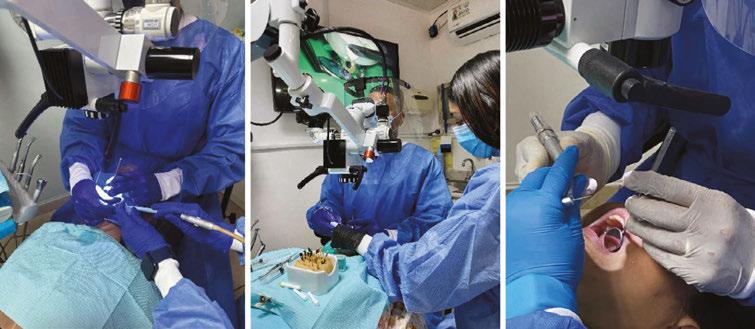
The dental microscope, introduced by Gary Carr in the 1990s, has transformed endodontics, becoming essential in postgraduate training.6,7 Its benefits extend beyond magnification and lighting, significantly impacting ergonomics when used correctly. However, simply owning a microscope does not ensure its effective use; proper training is vital.
Effective use of the dental microscope can enhance:8
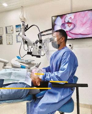
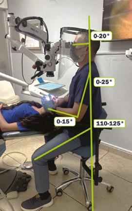
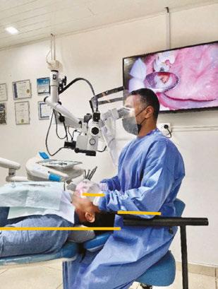
• Neutral posture: Reducing physical strain during procedures (Figure 1).
• Focus and concentration: Minimizing errors and improving precision.
• Controlled movements: Conserving energy and reducing fatigue.
• Improved visibility: Leading to better performance outcomes.
• Team collaboration: Allowing each team member to focus on their specific roles.
• Overall performance and efficiency: Enhancing job satisfaction and patient care quality.
By prioritizing ergonomic practices and proper training in advanced tools like the dental microscope, dental professionals can mitigate the challenges of their demanding field, promoting both their well-being and the quality of care they provide.
Establishing a smooth workflow with the dental microscope is necessary to avoid interruptions, which can have detrimental effects not only on the physical capabilities of the operator and assistant but also on cognitive executive functions. This can impact decision-making processes in the prefrontal cortex, leading to errors, dissatisfaction, and demoralization.9
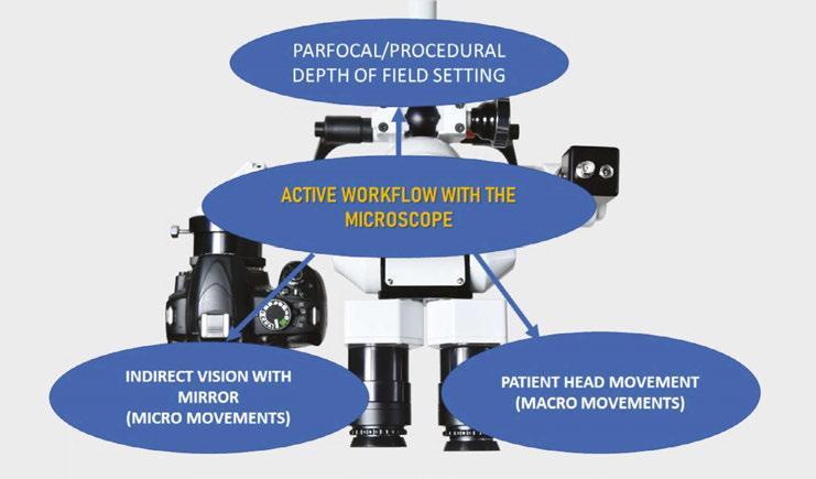
To enhance microscope endodontics workflow during active tasks involving the mirror — such as using burs, endodontic files, filling materials, or conducting surgical procedures — it is vital to control the following factors (Figure 2):
• Microscope depth of field setting
• Mirror handling skills (micromovements)
• Patient positioning and head movements (macromovements)
Microscope Depth of Field
Magnification Depth of Field of View Setting (MDOFS) presents a significant challenge for new microscope users, particularly in the realm of stereoscopic microscopy. A solid understanding of magnification and optics is crucial for a smoother experience.10
Once the operator is optimally positioned at the 12 o’clock position, and the patient is comfortably situated, adjustments to the microscope for interpupillary distance and parfocality should be made. This process is typically performed only once if the same user consistently operates the microscope. The operator should start by targeting the tooth at lower magnifications to ensure a broader field of view before starting the procedure. After successfully identifying the tooth, they can transition to higher magnification and utilize the microfocal knob to achieve a sharp image. This initial focus at high magnification ensures that all lower levels remain in clear view.
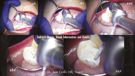
A common mistake in endodontics is the reliance on high magnification throughout procedures, which can lead to a loss of references and perspective due to the restricted field of view. Instead, it is advisable to predominantly use lower magnification to maintain visibility of essential reference points such as the fingertips, file rubber stops, cusp tips, and the axial inclination of the handpiece or file in relation to the tooth, reserving higher magnification for more detailed tasks (Figure 3).
Figure 1 (left) : Operator neutral posture in microscope endodontics. Figure 2 (right): Active workflow with the microscope, factors
Figure 3: Indirect vision, magnification steps, information, and details
Mirror handling skills (micromovements)
Research highlights the crucial role of line of vision in muscle activity, posture, and joint fatigue, particularly in dentistry.11 Mastering mirror handling during procedures is essential for enhancing dentists’ physical and cognitive skills.12 Incorporating this training into dental school curricula is vital for developing the neuromotor capabilities necessary for effective mirror use, especially with microscopes.13
When the operator and microscope are stationary, only the mirror and patient’s head can be adjusted. Neglecting these adjustments can lead to unintentional shifts in the microscope’s position, causing disruptions and awkward postures that can result in discomfort or injuries.
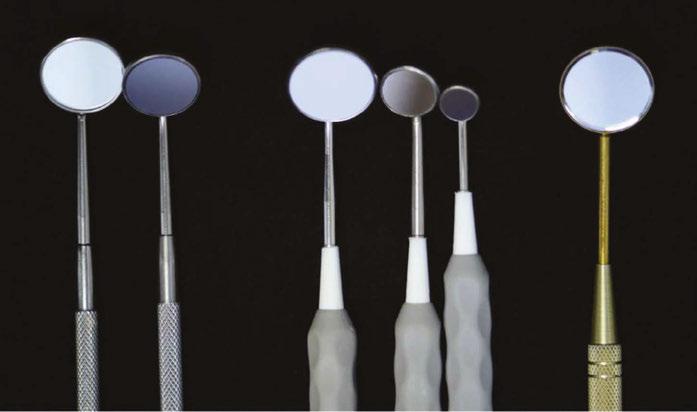
Utilizing mirror motions with intraoral fulcrum points is key for stable control, enabling precise movements and better visualization of difficult angles.14 Selecting a front surface mirror with rhodium or HD coatings offers sharper reflections, enhancing visual quality when paired with coaxial lighting and magnification15 (Figure 4).
Honing mirror skills allows dentists to achieve optimal focal sharpness, improving focus without needing to reposition the microscope or themselves. This proficiency not only elevates the quality of care provided but also promotes greater comfort for both dentist and patient, making it an invaluable skill in modern dentistry.
Patient positioning and head movements (macromovements)
By following a systematic approach to positioning, the operator can effectively control their posture once positioned at 12 o’clock behind the patient’s head, which impacts their interaction with the microscope.16
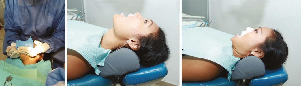
Patients should be placed horizontally in the dental chair, ensuring their mouth is directly below the microscope’s objective for ideal visibility. The patient’s head needs to be positioned at the end of the headrest, minimizing distance to maintain the operator’s posture and the microscope’s proper settings. Proper positioning also prevents forward-leaning postures that can strain the lower back and neck17 (Figure 5).
Figure 5: Patient horizontal position in the chair, patient mouth height in relation to operator’s elbows
Figures 6A-6C (left): Patient lateral head movements. 6A. Verbal indications; 6B. Chin-up. 6C. Chin to the chest
Figures 4A-4C: Mirror sizes and surface types. 4A. Standard Dental Mirrors Miltex #4 and #3, poor reflectivity. 4B. HD Extra Bright Mirrors Hu-Freidy #4, #0, and Micro; twice as bright as standard; great for microscope users. 4C. Crystalmark, abrasion-resistant, for use when using air abrasion
A. B.
C.
A. B.
C.
For optimal ergonomics, adjust the patient’s height so their mouth is at or slightly above the operator’s elbows. This helps align the microscope’s binoculars correctly, promoting a neutral head posture, wrist, and forearm alignment. Avoid inclining the backrest too much, as this can raise the mouth too high and lead to wrist extension and neck hyperextension. Conversely, if positioned too low, the binoculars may need to exceed safe angles, causing neck flexion.
Once positioned, use verbal instructions to guide the patient for clear views of occlusal, vestibular, palatal, and lingual surfaces by adjusting their chin or head position. These cues enhance visibility while minimizing interruptions, allowing for better focus and stamina for the operator18 (Figure 6).
Patient Positioning Tips
• Position the patient horizontally.
• Keep the head at the end of the headrest.
• Adjust the height to align the mouth with the operator’s elbows.
• Use verbal cues to manage the patient’s head position for optimal visibility.
• Proper positioning protects the operator’s posture during procedures.
• The position of the patient’s mouth sets the microscope’s alignment with the operator’s head.
Four-handed dentistry enhances posture, positioning, movements, and overall workflow
To achieve 100% working time in microscope dentistry, both a trained assistant and effective organizational practices are essential. An assistant plays a crucial role in enhancing ergonomics, as dentistry is a team-oriented profession. They should receive the same level of training as the dentist, focusing on ergonomic principles to maintain both physical and mental stamina.19
Microscope dentistry requires the operator to maintain focus while working in the patient’s mouth, making an assistant vital.
Without one, interruptions can occur when the operator needs to search for tools, which can negatively impact posture and lead to discomfort in key areas like the neck and back. This can also impair cognitive functions, leading to errors and wasted time.20,21
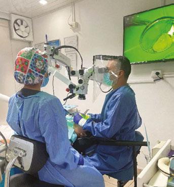
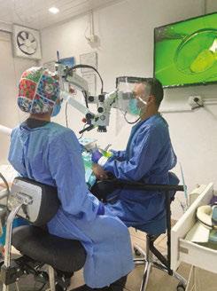
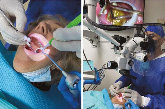
The assistant should be strategically positioned to optimize efficiency, keeping frequently used items within elbow reach while less frequently used supplies can be stored on a movable cart. This pre-organization facilitates swift instrument transfer, allowing the dentist to concentrate on their work while the assistant manages the surroundings22, 23 (Figures 7 and 8).
Effective motion control by the assistant helps the dentist adhere to ergonomic principles,24 maximizing endurance and precision during endodontic lengthy procedures. Additionally, the assistant is responsible for maintaining visual clarity by managing any obstructions on the microscope’s mirror, typically using a triple syringe and high-vacuum suction.
The organized arrangement of dental instruments, along with the expert motion control and transfer by the dental assistant, allows the dentist to adhere to Carr’s Ergonomics Laws of Motion, particularly Classes I and II.6 This approach enables effortless instrument retrieval, enhancing endurance and precision during
lengthy endodontic procedures while maintaining a smooth workflow. A crucial responsibility of the assistant in microscope dentistry is to keep the mirror free from water, dust, or fog. This meticulous task is vital, as it ensures the dentist can utilize indirect vision effectively, minimizing interruptions and allowing for greater focus and accuracy on the tooth (Figure 9).
Think of the dental microscope as your safety seatbelt — an essential tool that upholds treatment quality and protects both the patient and practitioner from injury not occasionally, but always.”
To ensure the assistant’s well-being, ergonomic stools with features like height adjustment, negative seat inclination, and supportive backrests are recommended. A healthy assistant contributes significantly to overall productivity and efficiency in the practice.
Conclusion
Workflow is vital in all dental disciplines, particularly in endodontics, where professionals face challenges related to task complexity, static posture, decision-making, and the need for visual information. Endodontics has embraced technology more extensively than other specialties, yet the focus should extend beyond basic tasks like endodontic access or locating the MB2 canal. Endodontists should strive to use the microscope consistently throughout their practice.
This requires discipline and training within an ergonomic framework, evolving work practices to prioritize precision alongside the biomechanics of the human body and the cognitive advantages of the microscope. Proper use of this tool promotes neutral posture, improves motion efficiency, and enhances teamwork.
Think of the dental microscope as your safety seatbelt — an essential tool that upholds treatment quality and protects both the patient and practitioner from injury not occasionally, but always.
REFERENCES
1. Fidelis Mne ChiaA. Comprehensive Review of Ergonomics Principles and Applications on Optimizing Workplace Performance and Well-being. IRE Journals. Aug 2024;8(2). https://www.irejournals.com/paper-details/1706185. Accessed September 30, 2025.
2. Gebhardt JS, Harth V, Groneberg DA, Mache S. Job Demands and Resources Perceived by Dentists in a Digital Dental Workplace and Perceived Effects on Job Satisfaction and Stress: A Qualitative Study. Clin Pract. 2025 May 12;15(5):92. doi: 10.3390/ clinpract15050092.
3. Kumar M, Pai KM, Vineetha R. Occupation-related musculoskeletal disorders among dental professionals. Med Pharm Rep. 2020 Oct;93(4):405-409. doi: 10.15386/mpr1581. Epub 2020 Oct 25.
4. Negucioiu M, Buduru S, Ghiz S, Kui A, Șoicu S, Buduru R, Sava S. Prevalence and Management of Burnout Among Dental Professionals Before, During, and After the COVID-19 Pandemic: A Systematic Review. Healthcare (Basel). 2024 Nov 26;12(23):2366. doi: 10.3390/healthcare12232366.
6. Carr GB, Murgel CA. The use of the operating microscope in endodontics. Dent Clin North Am. 2010 Apr;54(2):191-214. doi: 10.1016/j.cden.2010.01.002.
7. American Association of Endodontics. Microscopes in Endodontics. https://www.aae. org/specialty/clinical-resources/microscopes-in-endodontics/. Accessed September 30, 2025.
8. Bud M, Jitaru S, Lucaciu O, Korkut B, Dumitrascu-Timis L, Ionescu C, Cimpean S, Delean A. The advantages of the dental operative microscope in restorative dentistry. Med Pharm Rep. 2021 Jan;94(1):22-27. doi: 10.15386/mpr-1662. Epub 2021 Jan 29.
9. Aurtenetxe S, García-Pacios J, Del Río D, López ME, Pineda-Pardo JA, Marcos A, Delgado Losada ML, López-Frutos JM, Maestú F. Interference Impacts Working Memory in Mild Cognitive Impairment. Front Neurosci. 2016 Oct 13;10:443. doi: 10.3389/ fnins.2016.00443.
10. Kumar RSM, Jain R, Vekaash V. Capturing precision: A guide to dental photomicrography. J Conserv Dent Endod. 2024 Oct;27(10):1085-1087. doi: 10.4103/JCDE. JCDE_653_24. Epub 2024 Oct 5.
11. Katano K, Nakajima K, Saito M, Kawano Y, Takeda T, Fukuda K. Effects of Line of Vision on Posture, Muscle Activity and Sitting Balance During Tooth Preparation. Int Dent J. 2021 Oct;71(5):399-406. doi: 10.1016/j.identj.2020.12.025. Epub 2021 Feb 18. Erratum in: Int Dent J. 2022 Oct;72(5):731-733. doi: 10.1016/j.identj.2022.06.013.
12. Garcia PPNS, Pugliesi PMS, Wajngarten D, Neves TDC, Pazos JM, Dovigo LN. Development and assessment of an indirect vision training programme for operatory dentistry: Effects on working posture. Eur J Dent Educ. 2022 Feb;26(1):36-44. doi: 10.1111/eje.12670. Epub 2021 Mar 18.
13. Yıldırım YA, Süsgün Yıldırım Z, Ergun N. Evaluation of manual dexterity when using a mirror of dental students and academicians with increasing professional experience. Eur J Dent Educ. 2024 May;28(2):430-437. doi: 10.1111/eje.12965. Epub 2023 Nov 7.
14. Stormon N. Shetty S. Instrumentation. The University of Queensland. https://uq.pressbooks.pub/dentistryenvironment/chapter/instrumentation. Accessed September 30, 2025.
15. Bishayi D, Suvarna K, Singh S, Dutta A, Thomas MS. Technique Tips: A Reflection on Mouth Mirrors: Types, Usage and Modifications. Dental Update. October 2023;50(9):792-797.
16. Valachi B. Practice Dentistry Pain-Free: Evidence-based Ergonomic Strategies to Prevent Pain and Extend Your Career. Posturedontics Press; June 15, 2008.
17. Griegel-Morris P, Larson K, Mueller-Klaus K, Oatis CA. Incidence of common postural abnormalities in the cervical, shoulder, and thoracic regions and their association with pain in two age groups of healthy subjects. Phys Ther. 1992 Jun;72(6):425-431. doi: 10.1093/ptj/72.6.425.
18. Pîrvu C, Pătraşcu I, Pîrvu D, Ionescu C. The dentist’s operating posture - ergonomic aspects. J Med Life. 2014 Jun 15;7(2):177-182. Epub 2014 Jun 25.
19. Alnasser MY, Alsuliman S, AlYami DM. The impact of dental assistants in modern oral healthcare: A literature review. Advances in Oral and Maxillofacial Surgery. September 2025;19;100572. https://doi.org/10.1016/j.adoms.2025.100572.
20. Chen Y, Fang W, Guo B, Bao H. Fatigue-Related Effects in the Process of Task Interruption on Working Memory. Front Hum Neurosci. 2021 Nov 17;15:703422. doi: 10.3389/fnhum.2021.703422.
21. Abd-Elfattah HM, Abdelazeim FH, Elshennawy S. Physical and cognitive consequences of fatigue: A review. J Adv Res. 2015 May;6(3):351-358. doi: 10.1016/j. jare.2015.01.011. Epub 2015 Feb 24.
22. Gupta A, Bhat M, Mohammed T, Bansal N, Gupta G. Ergonomics in dentistry. Int J Clin Pediatr Dent. 2014 Jan;7(1):30-34. doi: 10.5005/jp-journals-10005-1229. Epub 2014 Apr 26.
23. Yang J, Abdel-Malek K. Human reach envelope and zone differentiation for ergonomic design. Human Factors and Ergonomics in Manufacturing & Service Industries. 2009;19 (1):15–34. Wiley Online Library. doi.org/10.1002/hfm.20135.
24. FDI World Dental Federation. Ergonomics and posture guidelines for oral health professionals. https://www.fdiworlddental.org/sites/default/files/202104/FDI_HSDW_ ergonomics_and_posture_guidelines_eng_2021.pdf. Accessed September 30, 2025.
Continuing Education Quiz
Microscope endodontic workflow: an ergonomic systematic approach HUGUES
1. With studies showing over _______ of dentists suffering from musculoskeletal disorders and a 13% burnout rate, it is essential for educational institutions to emphasize ergonomic training to address these issues.
a. 10%
b. 45%
c. 68%
d. 80%
2. Effective use of the dental microscope can enhance: neutral posture, focus and concentration, __________, and overall performance and efficiency.
a. controlled movements
b. improved visibility
c. team collaboration
d. all of the above
3. (Regarding microscope depth-of-field setting) Once the operator is optimally positioned at the position, and the patient is comfortably situated, adjustments to the microscope for interpupillary distance and parfocality should be made.
a. 12 o’clock
b. 2 o’clock
c. 3 o’clock
d. 5 o’clock
4. Utilizing mirror motions with intraoral fulcrum points is key for stable control, enabling precise movements and better visualization of difficult angles.
a. True
b. False
5. One of the patient positioning tips recommends: Adjust the height to align the mouth with the operator’s ________.
a. shoulders
b. elbows
c. neck
d. head
6. Regarding four-handed dentistry, the assistant should receive ________, focusing on ergonomic principles to maintain both physical and mental stamina.
a. no training on the microscope
b. a lower level of training than the dentist
c. the same level of training as the dentist
d. only training on physical positioning for the patient
Each article is equivalent to two CE credits. Available only to paid subscribers. Free subscriptions do not qualify for the CE credits. Subscribe and receive up to 16 CE credits for only $149; call 866-579-9496, or visit https://endopracticeus.com/ subscribe/ to subscribe today.
n To receive credit: Go online to https://endopracticeus.com/continuingeducation/, click on the article, then click on the take quiz button, and enter your test answers.
AGD Code: 070
Date Published: April 5, 2026
Expiration Date: April 5, 2029
7. The assistant is responsible for maintaining visual clarity by managing any obstructions on the microscope’s mirror, typically using a ________.
a. triple syringe
b. high-vacuum suction
c. paper towel
d. both a and b
8. The organized arrangement of dental instruments, along with the expert motion control and transfer by the dental assistant, allows the dentist to adhere to _______, particularly Classes I and II.
a. Carr’s Ergonomics Laws of Motion
b. Newton’s Three Laws of Motion
c. Kepler’s Laws of Motion
d. Hooke’s Law of Motion
9. A crucial responsibility of the assistant in microscope dentistry is to keep the mirror free from _______.
a. water
b. dust
c. fog
d. all of the above
10. Dentists should think of the dental microscope as their safety seatbelt an essential tool that upholds treatment quality and protects both the patient and practitioner from injury not occasionally, but always.
a. True
b. False
To provide feedback on CE, please email us at education@medmarkmedia.com
Legal disclaimer: Course expires 3 years from date of publication. The CE provider uses reasonable care in selecting and providing accurate content. The CE provider, however, does not independently verify the content or materials. Any opinions expressed in the materials are those of the author and not the CE provider. The instructional materials are intended to supplement, but are not a substitute for, the knowledge, skills, expertise and judgement of a trained healthcare professional.
Why do we need two types of sealers in endodontics?
As a dentist, the choice between bioceramic sealers, such as ProRoot® Bio Sealer, and resin-based sealers, such as AH Plus® Ribbon®, can depend on the specific clinical situation. Both are suitable for everyday use, but each offers distinct properties that could be beneficial in different treatment scenarios.
Bioceramic sealers, as ProRoot Bio Sealer, offer bioactive properties that can be highly beneficial in specific clinical contexts. These materials release calcium ions, which facilitate the formation of hydroxyapatite and present a high pH through the release of hydroxide ions. Clinical studies suggest that bioceramic sealers may promote faster healing of periapical lesions, which is possibly due to these properties.1 Therefore, they could be beneficial when there is a periapical lesion. Additionally, they could also be favorable in cases of complex anatomy. Unlike resin-based sealers, bioceramics are hydrophilic and set in the presence of moisture, making them particularly valuable in cases where complete dryness of the canal is difficult to achieve.2 Their dimensional stability and hydrophilic nature make them well-suited for use with single cone technique, also referred to as sealer-based obturation. This method leverages the sealer’s properties to create a three-dimensional seal through hydraulic pressure, simplifying the obturation. In addition, this technique allows avoiding excessive vertical or lateral compaction forces and thereby the risk of vertical root fracture.3 Still, ProRoot Bio Sealer is also compatible with warm obturation techniques. In contrast, resin-based sealers are commonly used in techniques in which the primary seal of the root canal is provided by the gutta percha. One of the main advantages of these sealers is their excellent sealing ability. Resin-based sealers, such as AH Plus® Ribbon®, demonstrate adhesion to both dentin and gutta percha.4,5 They also present low solubility, ensuring a long-term hermetic seal of the root canal system, preventing microorganisms and their by-products from leaking into the periapical region.6 This results in a tight seal that helps reduce the risk of leakage and microbial recontamination. Additionally, resin-based sealers offer a longer working time, making them particularly useful in complex or time-consuming procedures. Furthermore, AH Plus® Ribbon® presents outstanding radiopacity,9 allowing to clearly distinguish the sealer from surrounding anatomical structures to easily control the obturation radiographically. Resin-based sealers do not require moisture for setting which makes them less technique sensitive, and they can be used with all obturation techniques, including warm vertical compaction, carrier-based obturation, cold lateral condensation, and cold single cone. Regarding the latter technique, and contrary to what was thought for a long time for resin-based sealers, the newly reformulated AH Plus® Ribbon® exhibits slight expansion after setting, making it suitable for use with the cold single-cone technique.7
Due to their long-standing use in endodontics, there is substantial clinical evidence supporting the comparable success rates of resin-based and bioceramic sealers. In a recent systematic review and meta-analysis on this topic, the clinical success rate for AH Plus ranged from 86.2% to 100% (3-24 months of follow-up) versus 75% to 100% for bioceramic sealers.8
Dr. David Landwehr explains, “With the two sealers, I now have the best of both worlds. With the new and improved AH Plus Ribbon, I will continue to get all of the benefits I have appreciated from a resin-based sealer over my 25 years as a clinician, but now with increased consistency in the mix. The ProRoot Bio Sealer gives me greater versatility when I feel a bioceramic alternative provides a clinical benefit in challenging cases and very complicated anatomies filled with a single-cone obturation.”
In sum, both resin-based and bioceramic sealers have important and largely overlapping roles in endodontic practice and allow clinicians to achieve a successful treatment. Understanding their respective strengths allows clinicians to select the most appropriate material based on the clinical demands of each case.
REFERENCES
1. Khandelwal A, Janani K, Teja K, Jose J, Battineni G, Riccitiello F, Valletta A, Palanivelu A, Spagnuolo G. Periapical Healing following Root Canal Treatment Using Different Endodontic Sealers: A Systematic Review. Biomed Res Int. 2022 Jul 8;2022:3569281. doi: 10.1155/2022/3569281.
2. Küçükkaya Eren S. Clinical applications of calcium silicate-based materials: a narrative review. Aust Dent J. 2023 Jun;68 Suppl 1:S96-S109. doi: 10.1111/adj.12986. Epub 2023 Oct 26.
3. Al-Hiyasat AS, Sawallha AM, Taha NA. The effect of sealer type and obturation technique on the fracture resistance of endodontically treated roots. Clin Oral Investig. 2023 Dec;27(12):7359-7367. doi: 10.1007/s00784-023-05326-2. Epub 2023 Nov 3.
4. Maharti ID, Larasputri I, Herdianto N, Margono A, Tasomara R, Rosseti R. A comparative analysis of adhesion abilities between AH Plus® Bioceramic, Ceraseal® and AH Plus® on root canal dentine surfaces. J Conserv Dent Endod. 2025 Sep;28(9):881-885. doi: 10.4103/JCDE.JCDE_406_25. Epub 2025 Sep 5.
5. De-Deus G, Oliveira DS, Cavalcante DM, Simões-Carvalho M, Belladonna FG, Antunes LS, Souza EM, Silva EJNL, Versiani MA. Methodological proposal for evaluation of adhesion of root canal sealers to gutta-percha. Int Endod J. 2021 Sep;54(9):1653-1658. doi: 10.1111/iej.13549. Epub 2021 Jun 10.
6. Silva EJ, Perez R, Valentim RM, Belladonna FG, De-Deus GA, Lima IC, Neves AA. Dissolution, dislocation and dimensional changes of endodontic sealers after a solubility challenge: a micro-CT approach. Int Endod J. 2017 Apr;50(4):407-414. doi: 10.1111/ iej.12636. Epub 2016 Apr 17.
7. Dimensional stability comparison testing was conducted according to the test method in ADA#57 R2012 5.8, with adapted acceptance criteria. Data on file.
8. Zamparini F, Lenzi J, Duncan HF, Spinelli A, Gandolfi MG, Prati C. The efficacy of premixed bioceramic sealers versus standard sealers on root canal treatment outcome, extrusion rate and post-obturation pain: A systematic review and meta-analysis. Int Endod J. 2024 Aug;57(8):1021-1042. doi: 10.1111/iej.14069. Epub 2024 Apr 12.
9. Radiopacity [mmAI/mm] comparison test result according to ISO 6876-2012. Data on file.
For more information, contact Consumables-Data-Requests@dentsplysirona.com.
This information was provided by Dentsply Sirona.
Five things to consider when selling your dental practice
Attorneys Casey Gocel and Jonathan S. Rhone offer
tips to prepare for one of the biggest
transactions in a dentist’s career
If you are considering selling your dental practice, but do not know where to begin, you have come to the right place. Selling your dental practice will likely be the biggest financial transaction in your career, so it is important that you are properly prepared to sell and properly protected after the sale occurs. The work you do upfront can potentially save you money and headaches in the future.
Before selling your practice, it is important to have your house in order. That means that your practice is in good financial, legal, and operational condition. A potential buyer will likely conduct extensive due diligence on your practice and will ask to review your tax returns, financial statements, insurance policies, associate contracts, vendor agreements, provider agreements, accounts receivable history, real estate leases, and more. If you conduct your own diligence ahead of time, you will be much better prepared by knowing the status of your practice and potential issues that need to be addressed before the time comes to enter into an agreement to sell. Below is an outline of five key items that you should consider reviewing, preparing, and understanding before exploring a sale of your dental practice in earnest.
1. Know what kind of buyer you are targeting
The type of buyer you are targeting will greatly impact your preparation for, and negotiation of, a sale. There are three different potential buyers for your dental practice: a DSO, an outside dentist, or one of your associates. Typically, a sale to a DSO yields the highest total sale price; however, DSOs pay selling dentists using more complex payment structures that could result in less cash in your pocket at closing, and DSOs usually place very restrictive non-competes and employment requirements on their dentists. Let’s consider an example where a DSO values your practice at $1.5 million, while another dentist or one of
Casey
Gocel is a Partner and Co-Chair of the National Dental Law Group at Mandelbaum Barrett PC in Roseland, New Jersey. She can be contacted at: cgocel@mblawfirm.com.
Jonathan
S. Rhone is an Associate in the National Dental Law Group at Mandelbaum Barrett PC.
your associates is prepared to buy your practice for $1 million. While it is enticing to take the higher offer, there are several considerations you must take into account before deciding which offer to take.
DSO sales often include non-cash consideration such as rollover equity, earnouts, holdbacks and indemnity escrows. Generally speaking, a DSO buyer will pay at least 30% of your total purchase price in the form of rollover equity, meaning that 70% (or less) of the total sale price will be paid in cash. Using the example above, your closing cash payment only will be $1.05 million of the $1.5 million valuation. While the DSO equity may be a fruitful investment that pays in the future, it is not guaranteed, and often times, doctors are left with illiquid or even worthless rollover equity.
Further, some of the sale price may be tied to an earnout. An earnout is a mechanism by which the DSO will pay you a sum of money if certain metrics are attained over time. The earnout criteria will likely be drawn out over several years. Sticking with our $1.5 million valuation example, if the DSO offers an earnout worth $250,000, that amount will also be reduced from your payment at closing, with no guarantee that you will hit the earnout metrics. Your closing cash payment is now reduced to $800,000. Additionally, DSOs often require an indemnity escrow, which is money that the DSO will hold back, usually for 1 year, to be able to protect itself from any lawsuits or liabilities that arise from your former ownership of the practice. If this money is not used, it will be returned to you. The indemnity escrow further reduces the cash paid to you at closing. To recap, in this example, the seller will only get $700,000 of the $1.5 mil-
lion purchase price in the form of cash at closing, with the remaining amounts being allocated to the rollover equity, earnout, and indemnity escrow.
In a sale to an outside dentist or an associate, the closing payment is usually paid fully in cash at the closing. Therefore, the $1-million private sale valuation could yield more cash in your pocket at closing than a $1.5-million DSO valuation. Keep in mind that the upside of selling to a DSO has the potential to be more profitable. If you are able to hit the earnout metrics, and the DSO is well run and able to increase the value of your rollover equity, your total consideration for the sale could be significantly higher.
Selling your dental practice will likely be the biggest financial transaction in your career, so it is important that you are properly prepared to sell and properly protected after the sale occurs.”
Another factor to keep in mind is that DSOs typically require selling dentists to sign a 5-year employment contract, with severe financial penalties imposed if the doctor leaves before the 5-year term expires. Further, as a holder of rollover equity, the selling doctor becomes bound by the term of the DSO’s operating agreement. These operating agreements typically contain very restrictive non-competes that prevent the selling dentist from setting up a new practice or working elsewhere. Meanwhile, if you sell your practice to an outside dentist or an associate, the buyer may only require a short transitional employment period, typically 6 to 12 months, to transition the practice smoothly. The restrictive covenants that a dentist buyer places on a selling dentist are typically far less restrictive and give the seller much more flexibility in the future. Your individual circumstances and future plans are a key part of deciding who should buy your practice.
2. Know what you are worth
Having accurate and complete financial reports is critical because this is how potential buyers will value your practice. You should know what you are worth and how much debt your practice is carrying before entertaining offers from buyers, otherwise you will have less negotiating leverage because you will not know whether you are being valued fairly.
If you are thinking of selling to a DSO, we recommend hiring an accounting firm to perform a sale-side quality of earnings (QofE) analysis to determine your earnings before interest, taxes, depreciation, and amortization (EBITDA) and, ultimately, the value of your practice. If you are thinking of selling to an outside dentist or an associate, a simpler valuation can be prepared by your accountant based on historical collections.
You should also run lien searches on yourself and your practice to determine what debts will need to be paid off at closing. This is also a great way to find out if you have old liens on the practice that should have been terminated. Tracking down old creditors to get these liens removed can be very time consuming, and it’s best to clean this up prior to engaging in a sale transaction.
3. Assess how you are classifying your associates
It is important to make sure that all associate dentists are properly classified as employees or independent contractors. DSOs and other buyers will examine all staff members in the
practice to determine if they are properly classified. If a dentist is improperly classified as an independent contractor when they should be an employee, the buyer will make an adjustment in their projections based on the expense of that employee going forward. Misclassification could have a negative impact on your EBITDA (i.e., your purchase price), not to mention that it may cause problems with both state and federal regulatory agencies.
4. Understand your lease or real estate situation and whether it’s part of the sale
The real estate aspects of a transaction often disrupt dental deals because if a seller leases its property, the buyer will need to assume the lease. That almost certainly means that the parties must obtain the landlord’s consent to assign the lease to the buyer. It is important to review leases early in the sale process to understand what rights the landlord has with respect to assigning a lease, if there are requirements for personal or corporate guarantees, and if the landlord or lease terms provide for unreasonable demands to assign the lease. Oftentimes, a landlord will request to review the financials of the buyer before consenting to the assignment, which could take time. If the lease is not addressed early in the process, and the parties move towards the closing date without landlord’s consent to assign the lease, the entire deal can unravel if the lease cannot be assigned.
If you are the owner of the real estate, you must decide if you wish to sell the real estate or lease it to the buyer. In each case, you will want to have a clear understanding of the value of the property (if you are selling) or the fair market rent for the property (if you are leasing) prior to commencing negotiations.
5. Conduct reputational due diligence
Reputation matters. It is important to understand what publicly available information exists about you before pursuing a sale. DSOs and other buyers typically do basic research on Google, ChatGPT, and social media sites on selling doctors, and may even run background checks. That means you should remove provocative and inappropriate pictures from social media and be aware of any negative articles that exist about you. It is important to be truthful and forthcoming about potentially damaging publicity. In one case, a selling doctor, when asked by a DSO, informed them that he had never been arrested. The doctor, in fact, was arrested several years earlier, but the arrest had since been expunged from his record. However, a news article from many years ago was unearthed by the DSO through a Google search. The DSO terminated the transaction, not because the doctor was arrested, but because he lied about the arrest.
It is never too early to start thinking about your exit strategy. Whether you sell to a DSO, and outside doctor, or an associate, preparation is the key to maximizing your purchase price.
The industry standard for endodontic troughing
and
discovery. Munce burs trough the isthmus, core-out fiber and metal posts, expose separated instruments, dissect cement-line around posts, plus uncover hidden and calcified canals.
Available in 4-packs, 6-packs, and variety packs, starting at just $62!
“The development of these unique, endo-specific burs was born of the necessity for a longer-than-surgical-length, slow speed, round troughing bur with a small-diameter, yet stiff shaft. In the first 25 years of my own clinical experience, I found that despite the need, no such bur existed. So I began making them prn at the chairside, and later at the urging of colleagues who also needed them, I undertook to develop and make them available to dentists and endodontists worldwide... You will find that, because these are carbide burs, they are very efficient cutters and exceptionally durable.”
– C. John Munce, D.D.S., F.I.C.D.
Is “just good enough” okay?
Drs. Joel C. Small and Edwin McDonald discuss the importance of honing leadership skills
Ioften encounter colleagues who commit significant time and resources to developing their technical capabilities yet suffer from a lack of leadership and communication skills. Despite being at the peak of their technical game, their practices remain stuck at a suboptimal level.
This scenario reminds me of the fable of the strong and powerful lumberjack who never takes the time or effort to sharpen his axe, and consequently wonders why his production suffers. There is no question that technical ability is important, but like the lumberjack, we must sharpen other skills to create an optimal and peak performing practice and team.
According to Bill Adams, the cofounder of The Leadership Circle, when the leader plateaus, so does the organization. This concept applies as much for a dental practice as it does for a multi-national corporation.
Leadership is what takes an organization from good to great according to Jim Collins, author of numerous seminal leadership books. So why do we prioritize clinical skill while overlooking leadership and communication skills? Perhaps this is a major blind spot caused by our lack of training and knowledge. While corporate America prioritizes leadership training, dentistry lags behind. While corporations spend literally billions of dollars developing leaders, dental school curricula are sorely insufficient in this area. Hopefully this is changing.
Another explanation is that we often become complacent because being “just good enough” becomes comfortable over time, and we therefore see no compelling reason to develop other non-clinical skills. Michael Gerber, the author of The E Myth, would say that we are primarily technicians who create businesses that simply allow us to utilize our technical skills and nothing else. Gerber discusses the fallacy of this scenario, pointing out the many problems that arise from this mindset.
Doctors who lack leadership and communication skills are more likely to suffer from work/life imbalance
Drs. Joel C. Small and Edwin (Mac) McDonald have a total of over 75 years of dental practice experience. Both doctors are trained and certified Executive Leadership Coaches. They have joined forces to create Line of Sight Coaching, a business dedicated to helping their fellow dentists discover a better and more enjoyable way to create and lead a highly productive clinical dental practice. Through their work, clients experience a better work/life balance, find more joy in their work, and develop a strong practice culture and brand that positively impact their bottom line. To receive their free ebook, 7 Surprising Steps to Grow Your Practice Through Leadership, go to www.lineofsightcoaching.com.
No matter the format, investing in leadership development may prove to be one of your best investments.”
because they have never developed their team to effectively delegate tasks to staff. Michael Gerber likens this to a juggler who has too many balls in the air until eventually one of the balls falls, and the others follow. Doctors are notorious for juggling too much because their lack of leadership prevents them from developing a team that they trust to handle the many tasks that should be delegated. Over time, this burdensome and unsustainable workload takes a toll, resulting in unnecessary long hours in the office and eventual burnout.
Practices that lack effective leadership and communication also experience more costly staff turnover and diminished staff motivation. Doctors who express frustration due to their team’s lack of motivation must examine the underlying cause which is likely due to the doctor’s inability to inspire their staff, gain staff commitment, and clearly communicate expectations, all of which are leadership issues.
The profound irony is that these negative scenarios are both unnecessary and completely avoidable. Clients that have recognized the necessity to lead and communicate effectively have also found that practicing is much easier, and that investing the time and effort in their personal leadership development has paid long-term dividends for them and their team.
Leadership and effective communication are not effective without each other. All great leaders are great communicators who possess a high degree of emotional intelligence that allows them to engage more effectively with their teams. They understand the needs of the team at any given moment and how to self-regulate so they “show up” in the best possible manner for those they lead.
Acquiring this skillset takes an understanding of the critical importance of leadership in taking a “just good enough” practice to a great practice and a commitment to self-improvement.
Leadership training is available in many forms. Personal leadership coaching is extremely effective because the one-onone experience allows coaches to focus on specific needs, thus expediting the learning process while finding solutions for actual real-time practice issues in need of attention. There are also leadership courses offered online and through universities. No matter the format, investing in leadership development may prove to be one of your best investments.
Advanced Endodontic File System
Precision, Power, and Adaptabilityin Every Procedure
Strong to significantly increase resistance to cyclic fatigue.
Heat treated NiTi enables the EXIMIA7™ file extreme flexibility reducing shape memory, preserving canal anatomy e ortlessly following the canal as you shape.
Heat Treated NiTi
Parabolic Cross Section
Available in 3 lengths: 21, 25 & 29 mm
Maximum flute diameter 1 mm
• Heat treatment process improves strength and flexibility
• Available in .04 and .06 Constant Taper - Variable Pitch
• Maximum flute diameter 1mm allows for minimally invasive preparation
• Parabolic Cross Section non cutting tip maximizes file cutting efficiency
• ISO tip size 17-45
• Available lengths: 21, 25 & 29 mm
PURE-Ca-SEAL™ comes premixed in (1) 2g Syringe and Includes 20 applicator tips
The non-resorbable hydrophilic nature, dimensional stability, biocompatibility, antibacterial property, bioactivity, and ease of delivery makes PURE-Ca-SEAL™ BioCeramic Endodontic Sealer a promising NEW option.