Instrumentation / Education
n
4 CE Credits Available in This Issue*
Fall 2025 Vol 18 No 3
endopracticeus.com
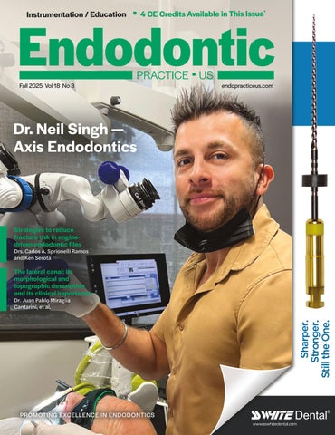
Dr. Neil Singh — Axis Endodontics
Strategies to reduce fracture risk in enginedriven endodontic files
Drs. Carlos A. Sprionelli Ramos and Ken Serota
The lateral canal: its morphological and topographic description and its clinical importance
Sharper. Stronger. Still the One.
Dr. Juan Pablo Miraglia Cantarini, et al.
PROMOTING EXCELLENCE IN ENDODONTICS www.sswhitedental.com