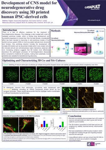
DevelopmentofCNSmodelfor neurodegenerativedrug discoveryusing3Dprinted humaniPSC-derivedcells
MatthieuTrigano,Anna-LenaZepernick,EveCorrie,EmmaV.Jones MedicinesDiscoveryCatapult,Block35,AlderleyPark,Cheshire,SK104ZF,UK
Methods
Thereisalackofeffectivemedicinesforthetreatmentof neurodegenerativediseases.Thischallengeisalsocoupledwithalackof validatedpreclinicalmodels,whichislimitingdrugdevelopmentand successfulprogressiontoclinicaltrial.Overthepastdecade,considerable efforthasbeendevotedtoimprovinginvitropreclinicalmodelsofthe centralnervoussystem(CNS)tomoreaccuratelyreplicatebrainstructure andfunction.Onepromisingdirectionincludesthedevelopmentof complex3Dmodelssuchasbio-printedmodelswhichmorecloselymimic theintricatecell-cellandcell-extracellularmatrixinteractionsobservedin vivo.Increasingevidencesuggeststhat3Dmodelsmoreeffectively reproducethepathologicalprocessesofneurodegenerativediseases whencomparedtotraditional2Dsystems,makingthempotentiallymore predictiveandvaluablefortranslationaldrugdiscovery.


OptimizingandCharacterizing3DCo-andTri-Cultures


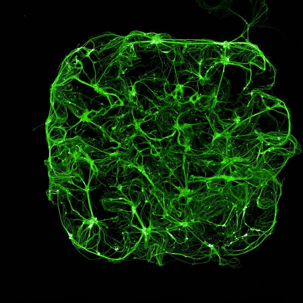
Calceinlabellingofneuron-astrocytecoculturesatmultipletimepointsshowedrobustcellviabilityandincreasedculturecomplexityovertime.
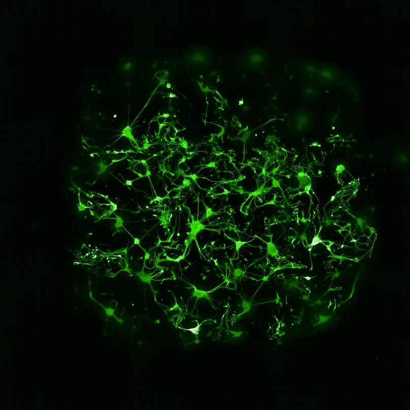
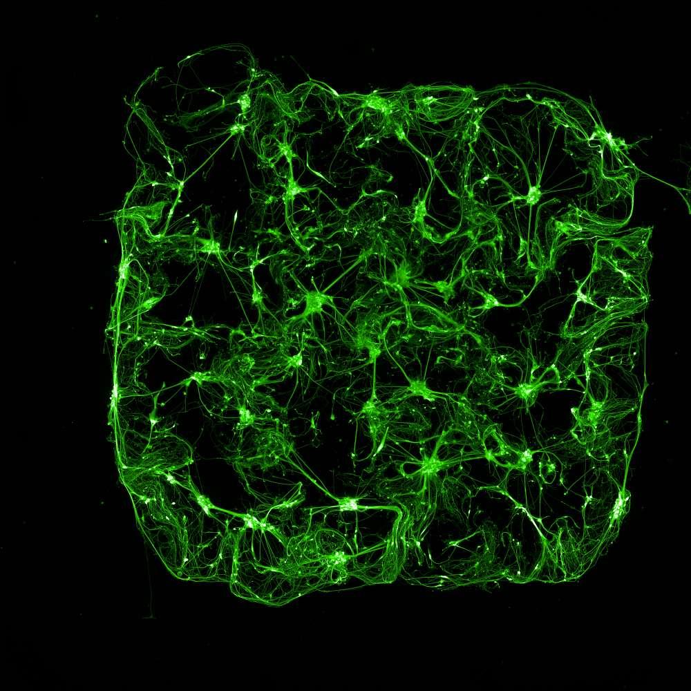
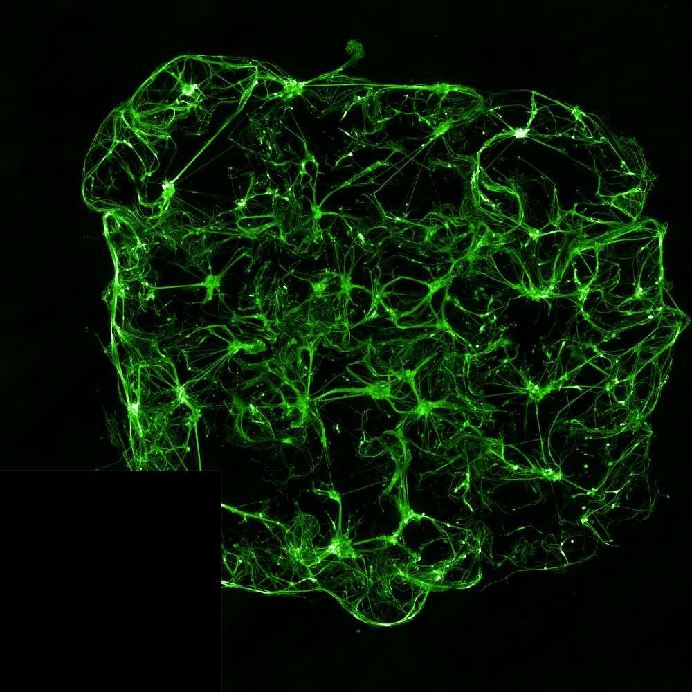
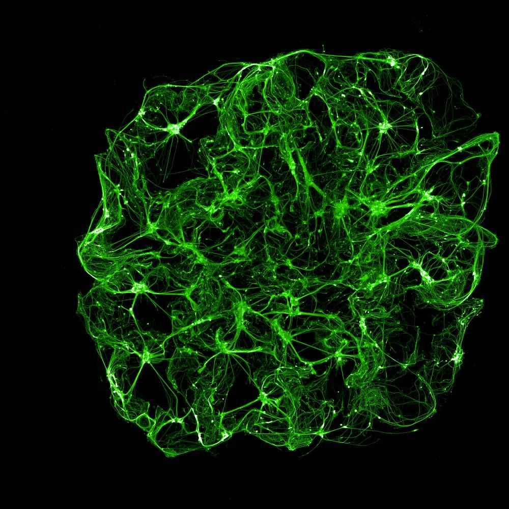
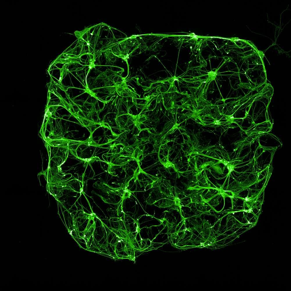
1week7weeks
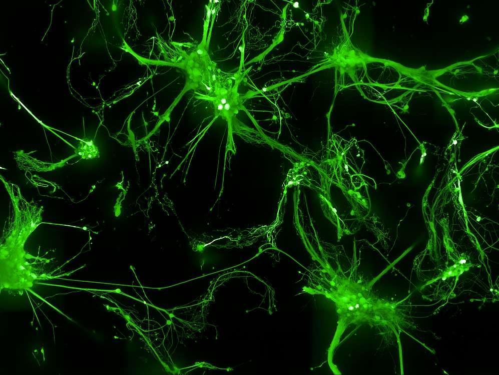
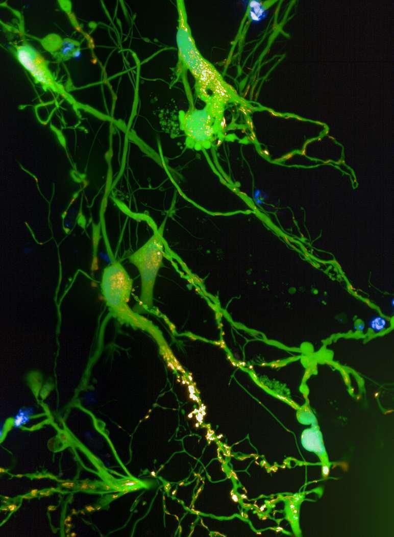
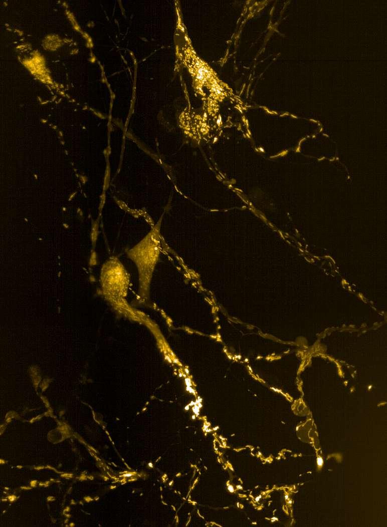
Todistinguishneuronsfromastrocytes,co-culturesweretransducedwith NeuroLightlentivirus,encodinganmRubyfluorescentproteindrivenbythe synapsinpromoter.Theseresultsdemonstratethatthehydrogelmatrixisamenable toefficienttransductionbylentiviruses.
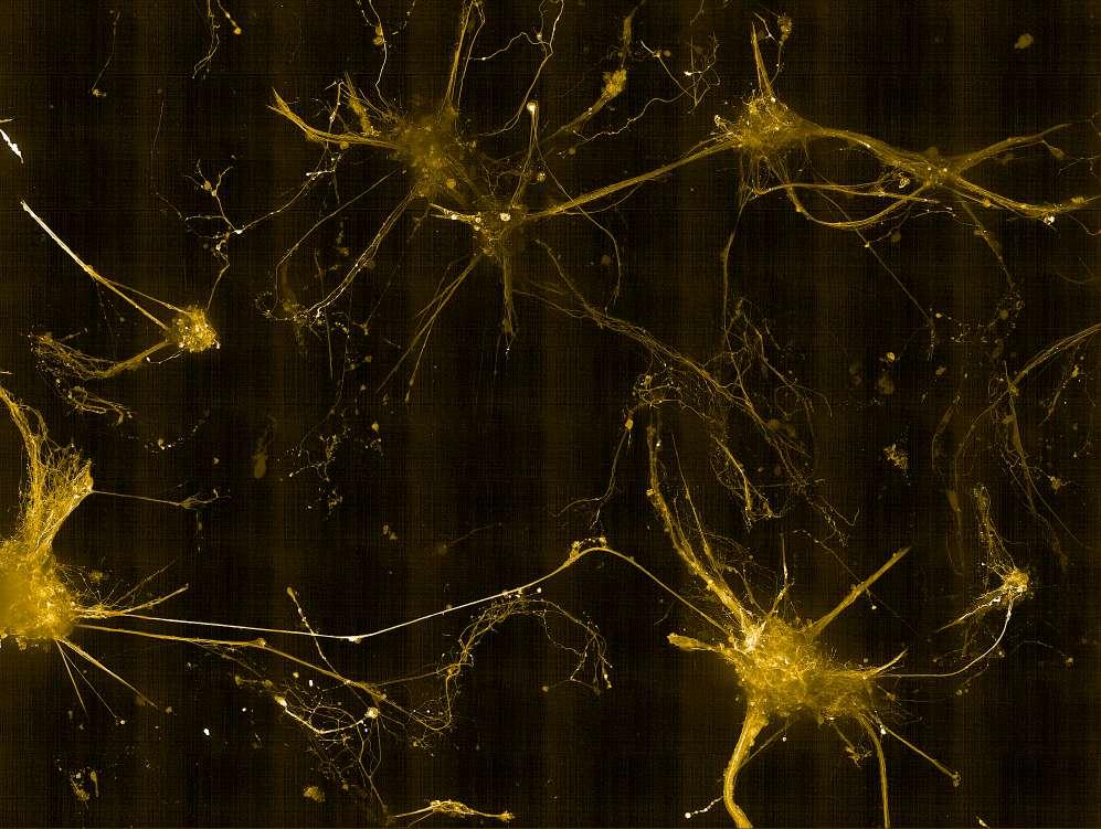

mitochondrialmembranepotential(ΔΨm)in3Dneuron–astrocyte cultures.
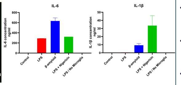
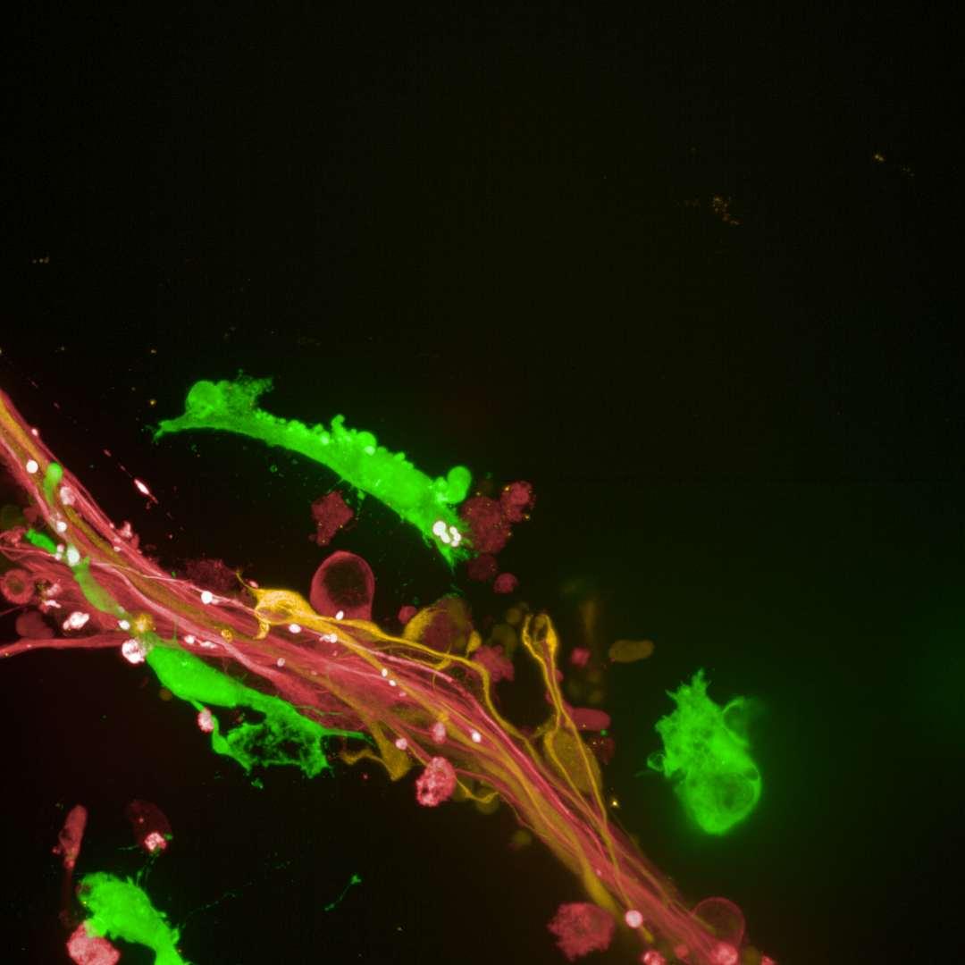
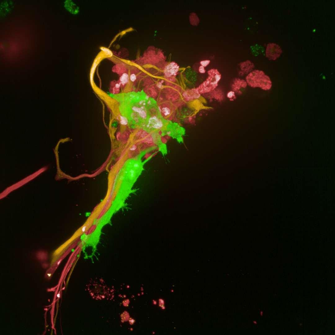
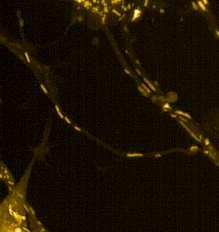
LPSandAβ42inducedmicroglia-dependentIL-6release.LPS+NigericintriggeredarobustIL-1βsecretionwhich isconsistentwithinflammasomeactivation.ThereisnodetectableIL-6inLPS-stimulatedcultureslackingmicroglia confirmingmicrogliaasaprimarysourceofinflammatorycytokines. Usingimmunocytochemicalmethodsandconfocalimaging,weobservedmicroglia(GFP)incloseproximityofneurons (β3-tubulin)andastrocytes(GFAP).
Conclusion
•Wehavesuccessfullydemonstratedthata3Dmodelof neuronsandastrocytesareabletogrowandremain viableforupto8weeks.
•The3Dmodelisamenableforbothfixedandlivecell imaging(e.g.viability,neuronalactivity,mitochondrial function).
•WehaveestablishedaCNStriculturemodelinwhich microgliaareinvadingthegelandmigratingtowards neuronsandastrocyte.
•Usingdifferentstimuli,wehaveshownthat3DCNStriculturescanserveasamodeltoinvestigate neuroinflammation.