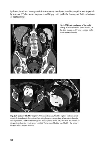
hydronephrosis and subsequent inflammation, or to rule out possible complications, especially abscess. CT also serves to guide renal biopsy or to guide the drainage of fluid collections or nephrostomy. Fig. 2.57 Renal carcinoma of the right kidney. Renal carcinoma (black arrows) in the right kidney on CT scan (coronal multiplanar reconstruction).
Fig. 2.58 Urinary bladder rupture. CT scan of urinary bladder rupture on transversal (on the left) and sagittal (on the right) multiplanar reconstructions. Contrast medium in urinary bladder (MM) leaks through the defect (white arrow, left) out from the bladder to the peritoneal cavity (white arrows, right). The urinary bladder was filled by the urinary catheter with contrast medium.
98
Ukázka elektronické knihy, UID: KOS506547
MRI MRI is not a method of the first choice for kidney, ureteral or bladder examinations. It does not use ionizing radiation and provides good tissue contrast that can be further improved by using intravenously-applied contrast media or using special sequences. However, most renal lesions can be diagnosed more quickly and accurately by ultrasound or CT, or a combination of both methods. In addition, MRI is more time-consuming, the predisposition to degradation by motion artifacts is greater and the availability less compared to CT or ultrasound.
MRI of the prostate In contrast, MRI is the only imaging method capable of detecting early stages of prostate cancer (Fig. 2.59) with sufficient sensitivity and is also used as a screening method for this disease (PI-RADS, Prostate Imaging Reporting and Data System). A very beneficial technique is also the fusion of imaging data obtained from previous prostate MRI examination with ultrasound examination. This facilitates biopsy of suspected lesions that were detected on MRI using trans-rectal ultrasound, although the lesions may not be visible on ultrasound. Minimal knowledge: • Low-dose native CT is suitable for finding stones in patients with clinical signs of renal colic. • Patients with acute scrotum are urgently examined by ultrasound, which is the imaging method of choice. • In case of suspicion of renal injury, CT is the method of choice. • When searching for prostate cancer, MRI is the method of choice, this is the only method which is capable of detecting early-stage prostatic cancer.
Fig. 2.59 Prostate cancer (white arrow) on MRI examination in three different sequences (T2 WI, DWI, and contrast enhanced T1 WI with fat saturation). Prostate cancer is on T2 WI (left picture) hypointense, with the signs of restriction of diffusion (middle picture) and enhanced after contrast administration (right picture).
99
Ukázka elektronické knihy, UID: KOS506547
2.5 INTRODUCTION TO NEURORADIOLOGY Neuroradiology is a branch of radiology that is concerned with the central nervous system and with vascular and non-vascular interventions following diagnostic radiology exams. The brain, due to its localization, was not directly visualized before the invention of CT and could only be imaged indirectly. Moreover, only MRI is capable visualizing the spinal cord; until the introduction of MRI in daily practice, the spinal cord was not directly visualized despite CT developments. Currently, MRI is a primary diagnostic imaging method in neuroradiology, CT is reserved for imaging in acute conditions such as trauma or stroke, and is also used for intervention guidance. From the remainder of diagnostic imaging methods, ultrasound, X-ray as well as digital subtraction angiography (DSA) are also used. Today, plain X-ray images are indicated almost exclusively to show degenerative changes in the spine and are always made in two perpendicular planes (anterior-posterior and lateral). The advantage of lateral spine plain film images is the possibility of performing dynamic images (images obtained in a standing position and when leaning forward and backward), where the mobility of individual segments and signs of possible instability in each segment can be assessed. X-ray of the skull, unlike native images of the spine, is now considered as an obsolete method whose diagnostic value is minimal; however, it is still needed for the assessment of skull shape and for the assessment of cranial sutures in children and evaluation of possible craniosynostosis. Doppler ultrasound is a very useful method for vessel evaluation (extracranial arteries or intracranial-transcranial ultrasound, mostly in hands of neurologists). Moreover, ultrasound is very successful in the examination of the neonatal and infant brain, as the brain is very easily accessible through open fontanelles. Indications for head ultrasound are as follows: • routine head ultrasound for all premature neonates; • suspicion of brain anomalies on antenatal neonates; • any sick neonate in whom brain pathology is implicated (searching for hemorrhage, tumors or hydrocephalus); • head trauma in neonates to exclude intracranial bleeding; • a neonate that had not been screened prenatally (which is very uncommon in developed countries). Diagnostic DSA is currently reserved for vascular disease with unclear imaging findings, such as when searching for arteriovenous malformations or aneurysms. Please see also Chapter 1.2 and 1.6 of this textbook. CT was a strategic breakthrough in neuroimaging. Soon, however, MRI assumed a dominant role. In the following paragraphs pay attention to the basic differences between these two methods in neuroradiology (CT vs MRI). CT: • It is generally easily accessible in the Czech Republic, available 24 hours per day in each district town. • It is fast and relatively inexpensive. • There are no absolute contraindications to CT. • The tissue contrast compared with MRI is clearly worse. • Its sensitivity and specificity are lower, very low in some indications. • Displays bone structures and calcifications well. 100 Ukázka elektronické knihy