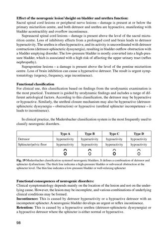
Effect of the neurogenic lesionʼsheight on bladder and urethra function: Sacral spinal cord lesions or peripheral nerve lesions – damage is present at or below the primary micturition centre, and both detrusor and urethra are hypoactive, manifesting with bladder acontractility and overflow incontinence. Suprasacral spinal cord lesions – damage is present above the level of the sacral micturition centre. Loss of inhibitory effects from a prolonged cord and brain leads to detrusor hyperactivity. The urethra is often hyperactive, and its activity is uncoordinated with detrusor contractions (detrusor-sphincteric dyssynergia), resulting in bladder outflow obstruction with a bladder emptying disorder. The low-pressure bladder is mostly converted into a high-pressure bladder, which is associated with a high risk of affecting the upper urinary tract (reflux nephropathy). Suprapontine lesions – a damage is present above the level of the pontine micturition centre. Loss of brain inhibition can cause a hyperactive detrusor. The result is urgent symptomatology (urgency, frequency, urge incontinence). Functional classification For clinical use, this classification based on findings from the urodynamic examination is the most practical. Treatment is guided by urodynamic findings and includes a range of different aetiological factors. According to this classification, the detrusor may be hyperactive or hypoactive. Similarly, the urethral closure mechanism may also be hyperactive (detrusorsphincteric dyssynergia – obstruction) or hypoactive (urethral sphincter incompetence – it leads to incontinence). In clinical practice, the Madersbacher classification system is the most frequently used to classify neurogenic disorders.
Detrusor
Sphincter/pelvic floor
Type A
hyperactivity
hyperactivity
Type B
hyperactivity hypoactivity
Type C
hypoactivity
hyperactivity
Type D
hypoactivity
hypoactivity
Fig. 19 Madersbacher classification systemof neurogenic bladders. It defines a combination of detrusor and sphincter dysfunctions: The thick line indicates a high-pressure bladder or subvesical obstruction at the sphincter level. The thin line indicates a low-pressure bladder or well-relaxing sphincter
Functional consequences of neurogenic disorders: Clinical symptomatology depends mainly on the location of the lesion and not on the underlying cause. However, the lesion may be incomplete, and various combinations of underlying clinical conditions may be formed. Incontinence: This is caused by detrusor hyperactivity or a hypoactive detrusor with an incompetent sphincter. A neurogenic bladder develops an urgent or reflex incontinence. Retention: This is caused by a hyperactive urethra (detrusor-sphincteric dyssynergia) or a hypoactive detrusor where the sphincter is either normal or hyperactive. 98
Ukázka elektronické knihy, UID: KOS290576
As well as the effect on the bladder, upper urinary tract dilatation (megaurethers) may occur in patients with high intravesical pressure due to detrusor hyperactivity and detrusor-sphincteric dyssynergia. This may be associated with vesico-ureteral reflux, especially in children, for example, with congenital spinal cord anomalies. The transfer of high pressure to the kidneys leads to kidney damage and chronic renal failure, which, after years, may be the cause of death in patients with spinal cord injury. Diagnosis and treatment of a neurogenic bladder Investigation: Determining medical history, physical examination, neurological consultative examination. Urine culture, urine cytology and determining urea, creatinine and glucose in serum. IVU, ultrasound (including measurement of post-micturition residue), voiding diary, urodynamic or video-urodynamic testing. Principles of treatment Treatment depends on an accurate diagnosis, namely diagnosis in terms of the resulting dysfunction of the urinary tract. I. Incontinence Detrusor hyperactivity – see the chapter on urgent incontinence. Incompetent sphincter – see the chapter on stress incontinence. II. Urinary retention Hypoactive detrusor or detrusor-sphincteric dyssynergia: The previously used Credé manoeuvre is now abandoned because of the risk of vesicoureteral reflux and no longer recommended. The Credé manoeuvre consisted in emptying the bladder by increasing intraabdominal pressure throught ension of the abdominal wall and hand pressure in the suprapubic region. Cholinergics. These are drugs that increase intravesical pressure. They include acetylcholine agonists that stimulate muscarinic cholinergic receptors. One of the acetylcholine agonists is, for example, bethanechol, but is not registered in the Czech Republic. For this reason, cholinesterase inhibitors such as distigmine (Ubretid®) are used in the Czech Republic. These, however, can be used only to a limited extent, and cases of imperfect sphincter relaxation during micturition have a risk of reflux. Clean intermittent catheterization: Currently, the gold standard in cases of neurogenic bladder emptying disorders is clean intermittent catheterization (CIC) or clean intermittent self-catheterization (CISC). The patient disinfects the external orifice of the genitals, injects the gel into the urethra and then introduces a disposable sterile urinary catheter. In domestic conditions, washing the hands and genitals with soap without disinfectingis sufficient, and the use of gel can be replaced by modern disposable catheters already coated with a gel layer. This minimizes the risk of the patient introducing pathogens into the urinary tract. After learning the CIC technique at the urological outpatient clinic, the patient self-catheterizes regularly approximately 5 times a day. In children, catheterization is performed by their parents. The use of disposable catheters instead of a permanent catheter increases the patientʼs quality of life and reduces the risk of inflammatory complications in a neurogenic bladder. Hyperactive urethral sphincter: α-adrenergic blockers (see BPH) are administered to relax the lissosphincter in combination with central myorelaxants in order to relax the rhabdos99
Ukázka elektronické knihy, UID: KOS290576
phincter (e.g., baclofen). Endoscopic sphincterotomy is performed in severe neurological disorders (e.g., spinal dysraphisms and spinal traumas). If these methods are not effective, CIC (CISC) or less suitable permanent urine derivation (permanent urinary catheter or epicystostomy) is required. Spinal trauma From a urological point of view, spinal cord trauma is specific mainly due to dynamic functional changes during spinal shock and will therefore be discussed in more detail. Although the definitive activity of detrusor and urethra depends on the level of the spinal cord lesion, the initial period is always accompanied by spinal shock, with detrusor paralysis and urinary retention occurring. Spinal shock is a period of decreased excitability of spinal segments below and at the level of a spinal cord lesion. Incidence: Approximately 14 spinal cord injuries/1000000 people occur annually in the world. In 2005, 2.5 million people in the world were registered living after spinal cord trauma. In the Czech Republic, about 300–350 new patients with spinal cord trauma are recorded annually. Immediate measures in cases of spinal shock: Bladder hyperdistension can lead to damage to bladder contractility. The most preferred regimen is intermittent catheterization (every 4 – 6 hours), or a temporarily established suprapubic catheter (puncture epicystostomy). A permanent urinary catheter is not suitable for completely immobile patients because of the risk of urethral decubitus, infection and later urethral stricture or paraurethral abscess. Bladder drainage is necessary until the spinal shock subsides. Subsequent treatment: The re-appearance of detrusor activity is variable, but it usually is around 4–5 (2–12) weeks after the accident. The next steps depend on the location of the lesion. Upper motor neuron lesion: When the spinal cord is damaged above the sacral micturition centre (S2, S3), the pontine micturition centre is disabled and thus the damping of provoked bladder contractions is disabled. The centre of the spinal cord and thus micturition reflex are functional. Detrusor hyperactivity develops here, with the occurrence of undamped contractions at a certain filling and certain intravesical pressure. Often, detrusor-sphincteric dyssynergia, i.e., subvesical obstruction, is also present. This is a type of neurogenic bladder that is a risk to the upper urinary tract. Treating a hyperactive detrusor and adequate urine voiding with intermittent catheterization are primarily indicated. Lower motor neuron lesion: A lesion in or below spinal cord segments S2or S3. The micturition reflex is completely disabled. Bladder acontractility develops, and depending on the condition of the sphincters, also bladder distension or incontinence. Without aids, the patient can only urinate by applying the abdominal press manoeuvre. For the derivation of urine, clean intermittent catheterization is primarily indicated. Incomplete and degenerative spinal cord lesions, for example, in renal failure, different combinations of the above mentioned may occur. Serious urological complications of spinal cord trauma (uroinfection, megaureters, renal insufficiency, urolithiasis) may occur with a longer time interval. Therefore, regular urological monitoring by sonography, excretory urography, urine culture, and serum creatinine are required.
100
Ukázka elektronické knihy