4
ULTRASOUND EXAMINATION OF THE LOWER LIMBS 1. oligosymptomatic DVT 2. the risk of future DVT through spread of SVT 3. therapeutic work‑up related to 1 and 2 above The deciding technique is color duplex sonography. Compression ultrasound (B‑mode) determines the extent of the superficial vein thrombosis, highlights the risk of propagation to deep veins and excludes DVT. Color flow mapping and dopplerometry are of ancillary importance: to clarify the extent of thrombotic obliteration and determine reflux sites in insufficient venous segments (Chapter 4.4.2) (Musil, 2013). D‑dimer assessment is of little value. In most patients with SVT, D‑dimer levels are markedly increased but determining them does not enable a distinction to be made between a primary thrombotic from a primary inflammatory process. D‑dimer values cannot predict the spread of thrombosis or other thrombotic complications (see above). Investigation of other coagulation parameters (e.g. factor VIII concentrations) has not to date, been introduced into clinical practice (Blättler, 2008). Venography here has been replaced by ultrasound. Venography relying on contrast medium used during the examination can induce thrombophlebitis. Screening for thrombophilia is not routinely performed; it should be considered in patients with recurring primary thrombophlebitis. Screening for occult primary illness (e.g. malignancy or vasculitis) is carried out if there are suspected clinical symptoms and/or signs of given diseases. or disease with respect to migratory, recurrent and/or multifocal non‑varicose vein SVT, recurrent varicose phlebitis, or current extensive DVT not directly related to superficial thrombosis. Screening for risk factors. These are generally similar: older age and extensive, long term untreated varices or chronic venous insufficiency (varices and trophic changes of the skin and subcutaneous tissue), local physical damage, heat, wearing
→
↑
↑
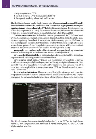
Fig. 4.3 Organized thrombus with calcified deposits ( ↑) in the GSV on the thigh, lumen width 7.2 mm (longitudinal and transverse, B-mode, linear probe 5.7 and 7.0 MHz, display depth 6 and 5 cm) 98
Ukázka elektronické knihy, UID: KOS260136
Superficial vein thrombosis (SVT)
4
inappropriate clothing, long sitting, etc. Varicophlebitis is quite common in pregnancy and the post‑natal period. Hormone contraception and hormone replacement therapy as risk factors are a complex issue that is outside the scope of this volume (Musil, 2013).
4.4
Ultrasound diagnosis of SVT
There are at least three main reasons why patients with clinically clear SVT should be referred for an ultrasound scan of the superficial and deep veins of both lower limbs: 1. Clinical examination cannot exclude the proximal spread of a thrombus in the saphenous trunk. 2. New studies using sonography have described the frequent coincidence of SVT with DVT, especially in ascending phlebitis of the GSV or SSV. SVT of the GSV and SSV is associated with DVT in 10–30 % of cases (average 15–19 %, Becker, 2006). 3. Significant local pain in SVT may mask DVT and make it asymptomatic. SVT is, for these reasons, a clear indication for duplex sonography.
4.4.1 Setting the ultrasound machine Ultrasound scanning for this indication is simple at first glance but there are a few things to learn. The settings of the US machine must be selected. The pre‑set is for soft superficial tissues with a higher frequency of 7–8 MHz. In CFM, compared with the basic soft tissue setting (PRF 1000–1200 Hz) the frequency range has to be increased. Otherwise the image is distorted by a large number of artifacts. Pulse Wave Doppler (PW Doppler) should initially be set to a low velocity 15– 20 cm/s and the filter of incoming ultrasound signal to 50 Hz to remove the the low frequencies. Adjust the depth of view and focus. Signal gain and compression are determined by the patient’s body build and amount of subcutaneous fat. It is advisable to have the settings of the ultrasound machine prepared in advance and only adjusted during the investigation. With good imaging, the superficial veins can be seen filled with thrombotic matter which is layered and resembles “cut onions” (Figures 4.1 and 4.4). The structure of the vein wall may also be visible, Digital zoom can be used to investigate veins measuring up to 1 mm. The disadvantage, however, is the considerable grain size of the image. The examination is performed in longitudinal and transverse views. The extent of thrombosis is examined at the outset, especially the distance of thrombus from the SFJ and SPJ and then 5–7 days later, checking for any extension to the deep veins.
4.4.2 Chief contribution of ultrasound to investigation of SVT 1. Determination of the extent of thrombosis in the superficial veins (compression sonography, B‑mode, CFM) – particularly, the proximal end of the thrombus in the saphenous vein from the SFJ and SPJ. 99
Ukázka elektronické knihy, UID: KOS260136
4
ULTRASOUND EXAMINATION OF THE LOWER LIMBS
→
→
A.
B.
→
C.
Fig. 4.4 Changes in ultrasound image of varicophlebitis over time – A. acute varicophlebitis about 1–7 days after onset, inside the vein is a central anechoic region (→) surrounded by concentrated echogenic layers along the vein wall, B. subacute varicophlebitis more than 10 days after onset. The anechoic segments inside the vein alternate with echogenic segments, C. long-term chronic changes after varicose phlebitis (months, years), echogenic, well-defined deposits of fibrous thrombus within the vein, locally (→) calcifications its with ultrasound shadow (transverse and longitudinal imaging, B-mode, linear probe 7,0 MHz, display depth 5 cm)
2. Detection of impaired vein segment connecting superficial and deep vein systems (compression sonography, B‑mode) – thigh and calf perforators, SFJ and SPJ. 3. Exclusion of DVT (compression sonography, B‑mode) – SVT of the GSV and SSV is associated with DVT in 10–30 % of cases (average 15–19 %, Becker, 2006), The deep vein system is affected more frequently in non‑varicose than in varicose thrombophlebitis: DVT may occur in both affected and unaffected lower limb on the other side. 4. Monitoring the development of SVT over time – monitoring intervals should be determined by the clinical picture and subjective complaints, at least once a week until the onset of clinical recession. 5. Demonstration of thrombosis in asymptomatic segments of superficial veins – thrombi can spread through the superfical veins further than can be expected from the clinical examination.
Literature ASCHER E, HANSON JN, SALLES‑CUNHA S, HINGORANI A.: Lesser saphenous vein thrombophlebitis: its natural history and implication for management. Vasc Endovasc Surg 2003; 37(6): 421–427. BECKER F: Dictionary of vascular medicine terms, vol 2. Elsevier SAS, 2006, 68–70. BLÄTTER W, SCHWARZENBACH B, LARGIADÉR J: Superficial vein thrombophlebitis serious concern or much ado about little? Vasa 2008; 37: 31–38. 100
Ukázka elektronické knihy, UID: KOS260136