foramen coecum
Rathke’s pouch Seessel’s pouch stomodeum 1 oral membrane
2
hyoid chondoblastema thyroglossal duct
hyoid cart 3
carotis
pyramidal lobe thyroid carotis
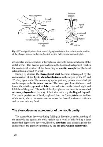
Fig. 52 The thyroid primordium named thyroglossal ducts descends from the midline of the pharynx toward the larynx. Sagittal section (left), frontal section (right).
invaginates and descends as a thyroglossal duct into the mesenchyme of the distal arches. The thyroid primordium in the human development reaches the anatomical position of the branching of carotid complex of the heart arterial trunk around 7th week. During its descent the thyroglossal duct becomes interrupted by the condensation of the hyoid chondroblastema in the region of the 2nd and 3rd pharyngeal arch. The remaining upper part may persist as a blind pit on the tongue – the foramen caecum. The lower part loses its lumen and forms the middle pyramidal lobe, situated between the lateral right and left lobe of the gland. The cells of the thyroglossal duct can form so called accessory thyroids on the way of their descent – e.g. the lingual thyroid. The partial persistence of the thyroglossal duct can form cysts in the midline of the neck, which can sometimes open on the dermal surface as a fistula and secrete salivary fluid.
The stomodeum as a precursor of the mouth cavity The stomodeum develops during folding of the embryo and expanding of the amniotic sac against the yolk vesicle. As a result of this folding a deep stomodeal depression develops, lined by ectoderm and closed against the endoderm of the primitive pharynx by the oro-pharyngeal membrane. – 98 –
Ukázka elektronické knihy, UID: KOS257538
The stomodeal cavity is situated in the region of the first pharyngeal arch. This arch becomes divided in early ontogeny into mandibular and maxillary portions, which then frame the oral entrance. Whereas the mandibular branches come early into contact with each other, the maxillary processes are shorter and do not reach the midline. Between them a frontal process from above enters, later forming the premaxillary part of the upper jaw. Both lateral branches of the maxillary processes expand later into the palatal processes, which together with the outgrowth from the premaxilla form the secondary palate. The unification of the palate occurs in the 7th–8th week, and if it is incomplete, a cleft palate (palatoschisis) occurs. The formation of the complete secondary palate causes the partition of the original primitive oropharyngeal cavity and the nasopharynx (epipharynx) has been formed. These developmental processes concern the development of the human face, which is flat in contrast to other mammals, in which the face projects in a snout. The development of the face will be described in detail in the chapter on skeletal development. The development of the pituitary gland
Just in front of the oropharyngeal membrane in the roof of stomodeum the ectoderm starts to evaginate into a small pouch. called Rathke’s pouch, which is the primordium of the pituitary gland. This pouch deepens against the forebrain, which reacts on the Rathke’s pouch by formation of
c ien
Rathke’s pouch
d
um
e
od
m sto
n
alo
h ep
notochord
um ke’s ibul Rath ch infund pou
adenohypophysis notochord
Seessel’s pouch
oral thyroid membrane
neurohypophysis
Fig. 53 The pituitary gland originates from two different primordia. The ectodermal Rathke’s pouch gives rise to the endocrine epithelium of the adenohypophysis, while the projection of the hypothalamus develops into the neurohypophysis. Sagittal section. – 99 –
Ukázka elektronické knihy, UID: KOS257538
a diencephalic floor diverticulum of the 3rd ventricle. This infundibular recess grows down against Rathke’s pouch and the two fuse at about the 5th week. The stem of Rathke’s pouch atrophies and the end of pouch gradually loses its connection with the foregut. Its cavity becomes reduced and the tissue forms the intermediate and tuberalis part of the anterior lobe of the pituitary gland – the adenohypophysis. The brain-derived infundibulum becomes the stalk and pars nervosa of the neurohypophysis.
Development of the teeth The tooth represents a complex structure which consists in addition to the soft pulp of three different hard tissues: dentin, enamel and cement. To understand the microscopic structure of the tooth let us consider at least the basic principles of its development. The first sign of tooth development appears at about the 6 week of pregnancy as a thickening of the oral ectodermal epithelium which forms finally a labio-gingival groove, separating the lip from the gum. This groove epithelium proliferates into gingival mesenchyme forming tooth buds in labio-gingival groove dental lamina
2
1
lip
enamel organ
3 4 5
labio-gingival groove
gum
1st dentition 2nd dentition outer enamel epithelium
reticular reticulum
preameoblasts dental papilla
bud of permanent tooth ameloblasts odontoblasts
dental follicle
Fig. 54 Formation of the enamel organ. The ectodermal tooth buds interact with the underlying mesenchyme. – 100 –
Ukázka elektronické knihy, UID: KOS257538