98
2. THYROID GLAND Table 2.10 Causes of hyperthyroidism More common causes Graves-Basedow disease Toxic adenoma Multinodular toxic goiter De Quervain thyroiditis Painless thyroiditis Iatrogenic causes, medication (e.g. amiodarone) thyrotoxicosis Less common causes Initial phases of Hashimoto’s thyroiditis Postpartum thyroiditis Hyperemesis gravidarum Struma ovarii, trophoblastic tumors TSH secreting pituitary adenoma Metastatic thyroid cancer
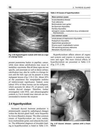
Fig. 2.36 Hypoechogenic nodule with halo as a sign of a benign lesion
present psammoma bodies in papillary cancer, while more dense calcifications may occur in medullary carcinoma. But all these signs are not specific, and punctuate as well as dense solitary calcification may also be visible in a benign lesion and the halo sign can be present in some malignant lesions (Fig. 2.33–2.36). About 20% of the population has nonpalpable nodules of indeterminate significance. Indeed, some (about 10%) of these lesions can be malignant, which accounts for about 2% of persons with nodular thyroid changes. Therefore, further investigation with FNA, and ultrasonographic controls in 3 to 6 month time intervals are recommended to monitor the situation.
Hyperthyroidism affects almost all organs and systems and express in numerous symptoms and signs. The main clinical effects of hyperthyroidism are presented in Table 2.11 (Fig. 2.37, 2.38).
2.4 Hyperthyroidism Increased thyroid hormone production is predominantly caused by pathological changes within the thyroid gland itself, most frequently by Graves-Basedow disease. The other common causes of hyperthyroidism are: toxic nodule, toxic multinodular goiter, and subacute thyroiditis. The differential diagnosis of disorders leading to hyperthyroidism is listed in Table 2.10.
Fig. 2.37 Graves’ disease – patient with a heart failure
Ukázka elektronické knihy, UID: KOS196184
99
2.4 Hype r t hyr oidis m
Laboratory evaluation of hyperthyroidism: • TSH (thyroid stimulating hormone) is low in all causes of hyperthyroidism except the central one (TSH producing adenoma) • fT3 (free triiodothyronine) and fT4 (free thyroxine) levels are increased • TSH receptor antibodies are positive in patients with Graves-Basedow disease • Anti-TPO and antithyroglobulin antibodies may be positive or negative Other biochemical findings: • A liver test may be found to be mildly increased, as well as alkaline phosphatase (bone isophorm) • The calcium level can be elevated, as well as calciuria, due to high bone turnover • In the blood count, normochromic anemia and relative lymphocytosis may be present • The level of albumin and some vitamins may also be low • Patients with subacute (De Quervain) thyroiditis have a high sedimentation rate Indication for imaging methods: • Primary thyroid pathology is examined with ultrasonography – this method and a laboratory assessment is usually sufficient to confirm the diagnosis • CT scan and MR imaging may be necessary in the evaluation of the size and extent of large toxic goiters
Fig. 2.38 Graves’ disease – pretibial myxedema
• Radionuclide methods (scintigraphy) are used to confirm the “hot” toxic nodule within the thyroid gland for to examine thyrotoxic patient prone to radioiodine therapy
Table 2.11 Clinical symptoms and signs of hyperthyroidism Cardiovascular
Tachycardia, palpitations, atrial fibrillation, high output heart failure, angina pectoris, increased pulse pressure, systolic murmur on heart apex
Neuromuscular
Nervousness, irritability, anxiety, depression, lability, insomnia, fatigue, fine tremor of fingers and tongue, weakness of proximal muscles, periodic paralysis, increased neuromuscular reflexes
Eyes
Conjunctivitis, lagophthalmos, chemosis, palpebral edema, exophthalmos, ophthalmoplegia, diplopia, optic nerve involvement
Skin
Hyperhidrosis, warmness, alopecia, hyperpigmentation, onycholysis, acropachy, pretibial myxedema, vitiligo
Gastrointestinal
Hyperphagia, diarrhea or increased frequency of stools, elevated liver tests
Osseous
Osteoporosis, increased osseous ALP, mild hypercalcemia
Sexual
Irregular menses, amenorrhea, decreased fertility, gynecomastia in men
Other
Heat intolerance, weight loss, anemia, lymphadenopathy, lymphocytosis
Ukázka elektronické knihy, UID: KOS196184
100
2. THYROID GLAND
on a preexisting adenomatous goiter (about 5%) (Fig. 2.41, 2.42). On the contrary, the size of the thyroid gland may be within normal limits in some patients (Fig. 2.43). In patients with a long standing disease, the echo structure could be nodular.
Fig. 2.39 Active Graves-Basedow disease
2.4.1 Diffuse toxic goiter – Graves-Basedow disease Graves-Basedow disease (GBD) is an auto immune disorder, most common in young women (female to male ratio 5:1), which is caused by thyroid-stimulating hormone receptor (TSHR) antibodies binding to the TSH receptor. This receptor stimulation leads to excessive thyroid hormone production and to thyroid gland enlargement in the majority of, but not all, patients. The clinical symptoms and signs of GBD do not differ from other causes of thyreotoxicosis, except the presence of ophthalmopathy (30–60%), and more rare pretibial myxedema and acropachy (about 5%). The goiter, if present, is diffuse and firm, and in some patients a bruit over frontal area of the neck may be audible (Fig. 2.39). Imaging Ultrasonography (US) shows an increase in glandular size with a homogenous structure and rarely calcifications without nodules. The findings are not specific for any particular type of diffuse thyroid disease. The thyroid gland has a hypoechogenic pattern and hyperemia can be seen on color Doppler examination (Fig. 2.40). Contrary to that, the blood flow is decreased in a patient with exogenous thyrotoxicosis – thyrotoxicosis facticia or iatrogenic. Incidentally, nodules in GBD may be visible superimposed
Fig. 2.40 Duplex Doppler image of Graves-Basedow disease in a nodular goiter
Fig. 2.41 Graves-Basedow disease – enlarged thyroid with a small nodule
Fig. 2.42 Graves-Basedow disease in a nodular goiter
Ukázka elektronické knihy, UID: KOS196184