Regions of the upper limb, regiones membri superioris
98
The lymph nodes collecting the lymph from the upper limb and part of the chest wall surround the trunk of axillary artery and its major branches: the brachial nodes, the interpectoral nodes, the subscapular nodes, and the apical and central axillary lymph nodes. The lymph from all these lymph nodes is drained further to the supraclavicular nodes and ultimately to the subclavian lymphatic trunk. The branches providing blood supply and innervation for the surrounding muscles arise from the neurovascular bundle in the axilla. The thoracodorsal nerve and vessels provide supply for the latissimus dorsi muscle. The serratus anterior is innervated from the long thoracic nerve and obtains blood supply from the lateral thoracic artery. The subscapular nerve and vessels serve for the subscapularis muscle. The posterior axillary wall is also pierced by nerves
and vessels. The circumflex scapular vessels pass through the triangular space to the dorsal scapular surface. The axillary nerve and the posterior circumflex humeral vessels pass through the quadrangular space at the level of the surgical neck of the humerus. Both structures continue further to the inferior surface of the deltoid muscle.
4.4
Anterior and posterior region of the arm
The proximal boundary line runs at the level of the axillary folds; the distal boundary line is established, by convention, 3 centimeters above the humeral 2
1 21
3
20
19 4
18
17 16
15
14
5
13
12 6 11 7
8
10 9
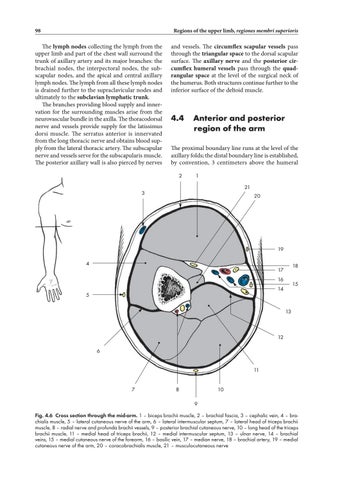
Fig. 4.6 Cross section through the mid-arm. 1 – biceps brachii muscle, 2 – brachial fascia, 3 – cephalic vein, 4 – brachialis muscle, 5 – lateral cutaneous nerve of the arm, 6 – lateral intermuscular septum, 7 – lateral head of triceps brachii muscle, 8 – radial nerve and profunda brachii vessels, 9 – posterior brachial cutaneous nerve, 10 – long head of the triceps brachii muscle, 11 – medial head of triceps brachii, 12 – medial intermuscular septum, 13 – ulnar nerve, 14 – brachial veins, 15 – medial cutaneous nerve of the forearm, 16 – basilic vein, 17 – median nerve, 18 – brachial artery, 19 – medial cutaneous nerve of the arm, 20 – coracobrachialis muscle, 21 – musculocutaneous nerve
Ukázka elektronické knihy, UID: KOS172329
Elbow regions
epicondyles. The surface anatomy of these regions is dependent on the muscular masses of the flexors and extensors of the arm. There are two shallow grooves visible on the surface, the lateral and medial bicipital grooves, between the flexor and extensor muscle groups on both sides of the arm. The brachial fascia is slightly drawn in here by the lateral and medial intermuscular septa. The fascia and septa attached to the humerus divide the entire volume of the arm in two osteofascial compartments – the anterior and posterior compartment of arm. The skin on the ventral and medial side of the arm is fine and less pigmented, while the skin covering the dorsal side is thicker. The superficial cephalic vein runs initially laterally under the skin from the cubital fossa, turning above the biceps brachii to the medial side of the arm to reach the deltopectoral groove. The basilic vein ascends on the medial side of the arm and penetrates the fascia approximately at the half-length of the arm. The medial antebrachial cutaneous nerve passes through the same opening in the fascia, the basilic hiatus, subcutaneously. Small cubital lymph nodes can be found in the proximity of the basilic hiatus. The dorsal part of brachial fascia is also penetrated by minor branches of the radial nerve providing sensitive innervation to the skin (via the posterior brachial cutaneous nerve and the lateral inferior brachial cutaneous nerve). The brachial fascia covers the muscles of this region and is continuous with the fascias of surrounding regions. The intermuscular septa are connected with the brachial fascia. The lateral intermuscular septum extends from the deltoid muscle attachment to the lateral epicondyle of the humerus. The radial nerve pierces this septum and descends from its posterior position around the bone in a spiral groove anteriorly. It is accompanied by the deep brachial vessels, in the proximal half of the arm. The medial intermuscular septum extends from the coracobrachialis attachment site to the medial epicondyle of the humerus. The ulnar nerve penetrates this septum as it passes to the posterior region of the elbow. The arm is divided into anterior and posterior compartment (Fig. 4.6) by the medial and lateral intermuscular septum, the humerus and the brachial fascia. The anterior brachial compartment contains the biceps brachii, the brachialis and the coracobrachialis muscles; the triceps brachii is found in the posterior compartment. The brachial neurovascular bundle passes just anterior along the medial intermuscular septum. The bundle consists of the centrally located brachial artery accompanied by two brachial veins. The median nerve is anterior to the artery in the proximal part of arm; in the distal part it moves into
99
a medial position. The ulnar nerve together with the superior ulnar collateral artery penetrates the medial intermuscular septum above the half-length of the arm and descends along the dorsal surface of the septum to the ulnar groove on the posterior surface of the medial epicondyle. The radial nerve originates from the posterior cord of the brachial plexus and descends laterodistally. Together with the deep brachial vessels it enters the radial groove on the humerus and takes the spiral course around its posterior aspect between the lateral and medial head of the triceps brachii muscle. The musculocutaneous nerve comes off the lateral cord and penetrates the coracobrachialis muscle. It then descends in the cleft between the coracobrachialis and the biceps brachii muscles, innervating both of them. After turning laterally and superficially, and proximal to the elbow, the musculocutaneous nerve penetrates the brachial fascia and continues further in the subcutaneous tissue as the lateral antebrachial cutaneous nerve.
4.5
Elbow regions
The boundaries of the elbow region are defined by two planes passing through the extremity approximately 3 fingers above and below the epicondyles of the humerus. A shallow depression, the cubital fossa, is visible on the ventral aspect. Both bicipital grooves passing from the arm converge towards the cubital fossa. The fossa shrinks during bending into a transverse flexion crease, which contains three muscular prominences. The superior one is formed by the biceps brachii muscle and its tendon; the two lateral prominences are formed by groups of muscles arising from the epicondyles. The olecranon is a prominent landmark under the skin in the dorsal aspect.
Anterior cubital region The skin of the anterior region is very fine with prominent subcutaneous veins. Basilic and cephalic vein are connected by the variable median cubital vein. The medial antebrachial cutaneous nerve follows the course and branching pattern of the basilic vein. The lateral antebrachial cutaneous nerve follows the cephalic vein in a similar fashion. The veins in this region are commonly used for intravenous administration of medicaments. Accidental para venous application can be associated with significant pain due to irritation of cutaneous nerves surrounding the vessels.
UkĂĄzka elektronickĂŠ knihy, UID: KOS172329
Regions of the upper limb, regiones membri superioris
100
The cubital fascia is reinforced near the insertion of the accessory biceps tendon, the bicipital aponeurosis, lacertus fibrosus, and above the origins of muscles arising from the epicondyles of the humerus. The major content of the cubital fossa proper (Fig. 4.7), from lateral to medial, are: the tendon of the biceps brachii muscle, the brachial vessels and the median nerve. These structures are situated under the cubital fascia. The brachial artery is palpable medial to the biceps brachii tendon and the median nerve lies immediately medial to the artery. The brachial artery leaves the fossa by pass-
ing under the bicipital aponeurosis and branches distally into the radial and ulnar arteries. The radial artery runs between the pronator teres and the brachioradialis muscles on the surface of the supinator muscle. The ulnar artery is larger than the radial artery and passes first deeply to the pronator teres muscle and then continues distally to the forearm between the flexor digitorum profundus and the flexor carpi ulnaris muscles. The branches of these arteries form the articular network of the elbow. The median nerve descends on the forearm passing through a slit between the two heads of the pronator
8 9
10
7 6
5 11
4 3
2
1
12
13
Fig. 4.7 Anterior region of the elbow. 1 – radial artery, 2 – brachioradialis muscle, 3 – deep branch of radial nerve, 4 – superficial branch of radial nerve, 5 – lateral cutaneous nerve of the forearm, 6 – cephalic vein, 7– biceps brachii muscle, 8 – median nerve, 9 – brachial artery, 10 – ulnar nerve, 11 – basilic vein and medial cutaneous nerve of the forearm, 12 – ulnar artery, 13 – pronator teres muscle
Ukázka elektronické knihy, UID: KOS172329