ADVANCED GENOMICS FOR CANCER PATIENTS IN RURAL MAINE
ON THE COVER
JAX’s Maine Cancer Genomics Initiative is the first statewide program of its kind. It works with every oncology practice across healthcare systems throughout Maine to bring advanced tools and precision cancer care to patients across the state. Inside cover: Dendritic cells like the one shown in the colored micrograph above play a crucial role in the initiation of immune responses in humans.
90 YEARS OF JAX — ONE VISION FOR A HEALTHIER FUTURE
Earlier this year, The Jackson Laboratory (JAX) celebrated its 90 th birthday. On May 4, 1929, Clarence Cook Little established the Laboratory with the help of philanthropic support, for the purpose of conducting research on cancer.
Ninety years later, JAX’s mission has evolved and expanded, but we are still motivated by the same vision that inspired C.C. Little almost a century ago: a future free of disease.
This issue of Search highlights two distinct projects in which JAX is playing a leadership role in transforming the future of human health: the JAX ME/CFS Collaborative Research Center, a multi-institution effort led
Join us
Learn more about our innovative scientific research in a fun and interactive way. JAX hosts a variety of special events in Connecticut and Maine throughout the year.
by Professor Derya Unutmaz, M.D., and Assistant Professor Julia Oh, Ph.D., and the Maine Cancer Genomics Initiative, launched with the generous support of The Harold Alfond® Foundation.
Each of these programs highlights our approach to science characterized by collaboration, creativity and commitment to solving difficult medical problems, and to ensuring that patients can benefit from those scientific advances.
As we expand the frontier of genomics in ways that bring us closer to patients, we continue to do the type of basic science that has defined the Laboratory since our founding. Our expertise in fundamental research remains at the core of who we are, as is evident in the profile of Assistant Professor Beth Dumont, Ph.D., and her work
on the molecular mechanisms that drive genetic diversity. Fourteen years after she first came to JAX as a student in our Summer Student Program, Dumont joined our faculty because, as she says, she “couldn’t do this work anywhere else.”
Thank you for being a part of the first 90 years of JAX. We can’t wait to see what the future holds, and we are grateful to all whose support for our mission makes our work possible.
Edison Liu, M.D. President and CEO, The Jackson Laboratory
LEARN MORE ABOUT JAX’S 90 TH BIRTHDAY Visit www.jax.org/jax90
JAXTAPOSITION EVENT SERIES
Conversations with JAX faculty and senior leadership on how diseases like cancer, cardiac disease, immunological disorders and more can affect you and your family. Discussions will cover new research in these areas, as well as preventative approaches to disease.
PUBLIC TOURS
Experience guided walking tours of our campuses located in Bar Harbor, Maine, and Farmington, Conn. Each tour is hosted by a JAX postdoctoral researcher and includes a behind‑the‑scenes look at our cutting edge research.
FORUM FOR DISCOVERY
JAX’s annual Forum for Discovery event will take place on Thursday, July 11 in Bar Harbor, Maine. Join scientists and JAX leaders for an update on how the Laboratory is changing the future of human health.
Learn more or register at www.jax.org/give/events
Questions? Contact Advancement Events at advancementevents@jax.org
JAX DONORS LAUNCH LEADERSHIP FUND FOR EMPLOYEES
The Charles E. Hewett, Ph.D., Leadership Excellence Endowment was established in August 2018 in honor of former Jackson Laboratory Executive Vice President and Chief Operating Officer Charles (Chuck) Hewett, Ph.D.
Thanks to more than 30 inaugural donors, JAX has raised nearly $1 million toward leadership development programs for Laboratory employees.
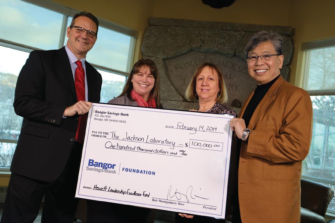
In February, representatives from Bangor Savings Bank visited the Laboratory’s Bar Harbor, Maine campus to present their gift of $100,000 toward the fund.
“We are proud to support Chuck Hewett’s legacy at The Jackson Laboratory,” says Bob Montgomery‑Rice, president and CEO of Bangor Savings Bank. “He has played an important role in both of our organizations, helping to shape our vision and values. This gift highlights our shared belief that workforce development is critical to the state’s success, and we are thrilled to support Chuck, his vision and his deep rooted commitment to the people of Maine.”
To make a donation to this endowment, visit www.jax.org/chuck-hewett-fund
MAINE STATE SCIENCE FAIR
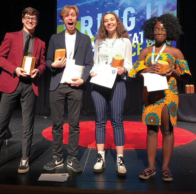
The 73rd annual Maine State Science Fair, organized by The Jackson Laboratory and Maine Mathematics and Science Alliance, took place in March and included 250 high school students. Participants competed for more than $548,000 in scholarships and awards. This year’s big winners include:
1st Grand Award — Tyler Delargy, Bangor High School, “Developing Three Dimensional Spatial Cognition for the Visually Impaired Using Computational Depth Mapping and Vibro Tactile Display”
2 nd Grand Award — Antonina Zakorchemna and Artem Laptiev, Fryeburg Academy, “Product Development of an Alternative Low Cost Braille E Reader”
3 rd Grand Award — Amara Ifeji, Bangor High School, “Testing the Effectiveness of Mycorrhizae in the Phytoremediation of Heavy Metals from Stormwater”
above with their awards are, from left, Delargy, Laptiev, Zakorchemna and Ifeji.
OPENING DOORS TO SCIENCE
Students from the Maine School of Science and Mathematics recently visited JAX to learn about genetics, genomics and personalized medicine.
They collected their own DNA samples and used polymerase chain reaction to prepare the samples for sequencing. After sequencing the DNA, they analyzed the results to determine whether they had the “wild type” version of a gene or one of several other variations. The students were introduced to computational biology concepts and learned about the many different JAX mouse strains.
The students ended the day with a presentation and meet and greet with JAX Assistant Professor Ron Korstanje, Ph.D.
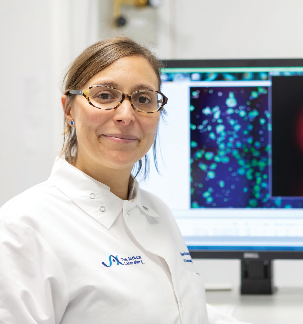
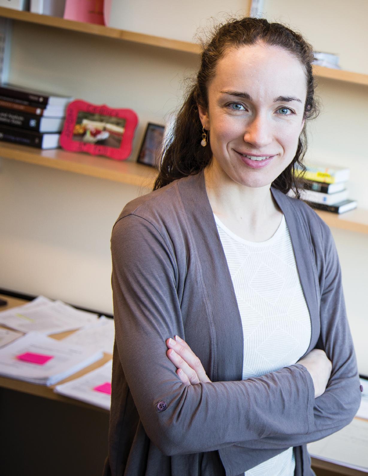
Pictured
Elise Courtois, Ph.D.
BY GRACE SCOTT | PHOTOGRAPHY BY CHARLES CAMARDA
A close friendship sparked interest in Elise Courtois to explore endometriosis, a common gynecological disease that is still misunderstood by so many women, society and even some clinicians.
Elise Courtois is a research scientist in the laboratory of Paul Robson at The Jackson Laboratory in Connecticut. Armed with a Discovery Award from the Department of Defense, she has launched a research project to study the genetics of endometriosis, with hopes of developing better diagnoses and treatment options for patients.
Endometriosis arises when tissue that usually lines the inside of the uterus — the endometrium — grows outside of the womb, creating complex ectopic lesions. The endometrium like lesions are painful and
can severely affect quality of life. Surgical removal of lesions can provide relief, but they usually recur. Hysterectomy — removal of the uterus — provides more permanent relief, but puts an end to fertility and is not ideal for the majority of afflicted reproductive aged women.
Courtois is looking for answers to help women afflicted by this disease, like her close friend Patricia, who is a post‑operative endometriosis patient.
“We have strong clinical collaborators and cutting edge technologies, so I’m really hoping this moves forward into a full blown line of endometriosis research,” Courtois says.
Making a difference
for rural cancer patients
BY JOYCE DALL’ACQUA PETERSON
PHOTOGRAPHY BY AARON BOOTHROYD & THOMAS FOUCHEREAUX
Jens Rueter and the Maine Cancer Genomics Initiative are providing advanced genomic tools to oncology practices throughout Maine.
Oncologist Rachit Kumar, M.D., is deep in discussion about his patient with stage 4 pancreatic cancer that had metastasized to multiple organs. Around the conference room table and on the speakerphone, fellow oncologists and experts in cancer genomics and clinical trials discuss the genetic profile of the patient’s cancer. A genetic counselor and two student interns diligently take notes as the team weighs in on advanced treatment options.
If you’re picturing this scene at a big-city university medical center, think again. This tumor board meeting is taking place at the MaineGeneral Medical Center’s Harold Alfond Center for Cancer Care in Augusta, the capital of Maine, the state with the lowest population density on the East Coast. And when it comes to cancer, Mainers are at a particular disadvantage: They are not only older (with the nation’s highest median age of 44.5 years), but also have a higher- than - average cancer rate even after adjusting for age and smoking.
JAX designed the Maine Cancer Genomics Initiative (MCGI) to provide access to the latest advances in precision cancer care to the state’s cancer patients, most of whom are treated at small community hospitals. Founded in 2016 with a grant from The Harold Alfond® Foundation, MCGI has already enrolled every oncology practice in Maine, and most of their oncologists, including Kumar.
Participating oncologists submit patient tumor samples to be sequenced and profiled for genes known to be associated with various cancers, and with response or resistance to U.S. Food and Drug Administration-approved targeted therapies or new drugs in development. The MCGI team takes advantage of JAX’s clinical genomics capabilities, including complex tumor profiling assays from the JAX Clinical Laboratory Improvement Amendments (CLIA) Lab and the JAX Clinical Knowledgebase, an interactive online encyclopedia that allows both researchers and clinicians to interpret complex cancer genomic profiles.
Several tumor profiles, and the most appropriate treatment options for them, are reviewed at each monthly tumor board meeting in Augusta. Seated next to Kumar and directing the patient discussion is the founding medical director of MCGI, Jens Rueter, M.D.
Kumar reads the first case summary aloud: “The patient was started first line with three cycles of FOLFIRINOX” — the combination chemotherapy that is the current standard for advanced pancreatic cancer — “but tolerated treatment poorly even with dose reduction. The patient and I were interested in genomic testing for further treatment options.”
The patient’s cancer shows a genetic variant that is rare in pancreatic cancer: unusually high levels of PD-L1, a protein that protects cancer cells from immune attack. On speakerphone, the external clinical advisor comments, “Due to the high PD-L1 expression, a clinical trial investigating a PD-L1 therapy would be a good option for this patient, and off-label use of PD - L1 therapy may be an option as well.”
Based on the discussion, Kumar says he will continue to treat the patient using a standard of care but will consider immunotherapy on a clinical trial or off - label basis based on the test results discussed at the tumor board for future therapy.
clinician has prescribed a new medication based on the genomic information of the tumor; in others, the patient is enrolled in a clinical trial.”
Born and raised in Germany, Rueter received his undergraduate and medical degrees from Humboldt University in Berlin. He completed his residency in internal medicine at Tulane University and fellowship training in hematology and oncology at the University of Pennsylvania. In 2010 he joined the medical staff at Eastern Maine Medical Center (now known as Northern Light Health) Cancer Care in Brewer, and St. Joseph Hospital in Bangor, and has courtesy staff privileges at Mount Desert Island Hospital in Bar Harbor. He became an adjunct member of the JAX faculty in 2012.
We already have a number of success stories of patients’ therapies being guided by genomic profiling.
– Jens Rueter
Rueter steers the meeting to focus on the case of a patient with lung cancer and a history of smoking, and then that of a man with metastatic carcinoma of the bile ducts.
To date MCGI enrolls mostly patients with stage 4 cancer, meaning that their tumors have metastasized to other organs of the body and that their mortality rate is higher than that of patients with stage 1, 2 or 3 cancer.
“We already have a number of success stories of patients’ therapies being guided by genomic profiling,” Rueter says. “In some cases, the
MCGI Program Director
Andrey Antov, Ph.D., works with Rueter in developing the initiative, liaising with the Maine oncology community and overseeing the clinical design and implementation of the study protocols. “We were very fortunate to find Jens as our medical director. We were looking for someone who really understands the molecular aspects of cancer. And he has been in Maine for nine years and really knows the Maine medical community,” says Antov.
Kumar adds, “Jens is a great leader, and I think his role is very important in connecting the experts. He has a background as a clinician and also as a researcher, so he brings both sides to the table. Connecting the two is where he really shines.”
With the early success of MCGI in Maine, JAX is taking the first steps to explore expanding community genomic medicine initiatives in New England. JAX is a National Cancer
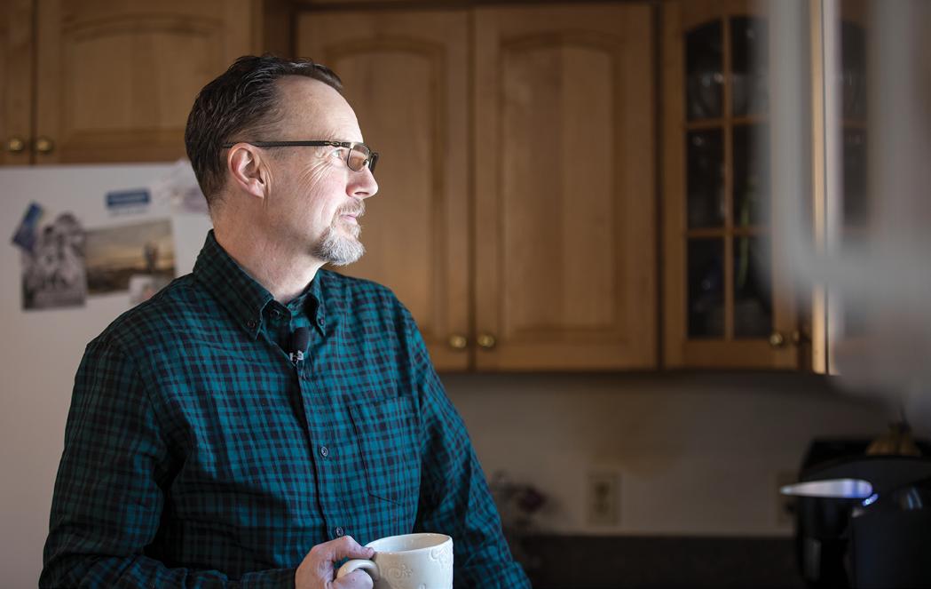
Medical Director Jens Rueter meets with a colleague in Bar Harbor.
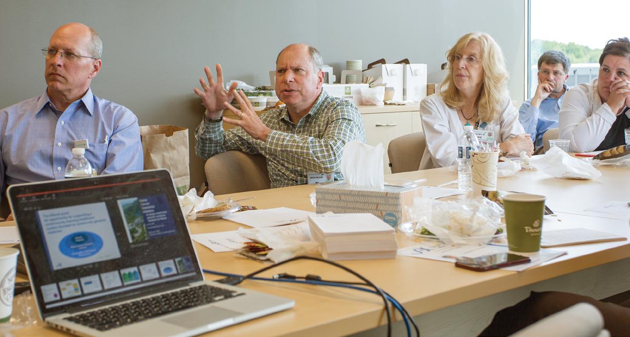
Medical professionals from across Maine gather in Augusta to learn more about MCGI.
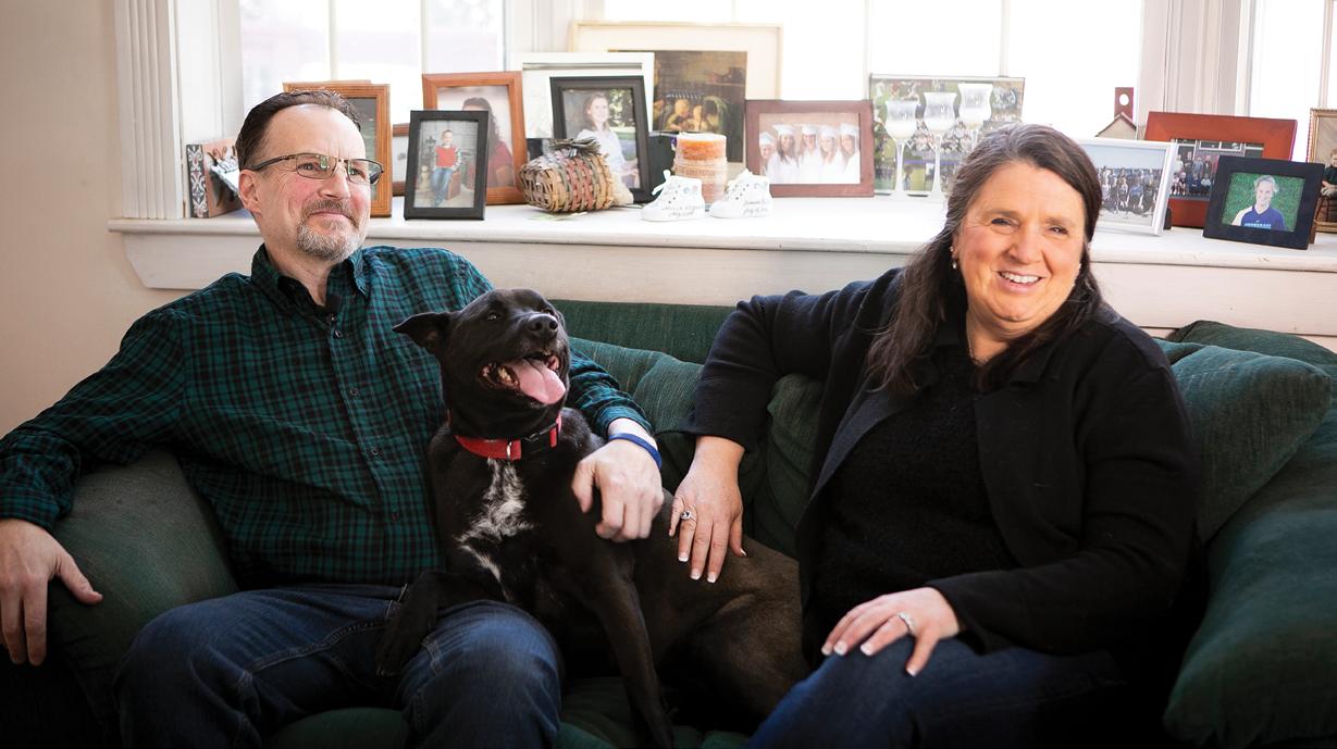
Rob Leighton, MCGI patient, his wife, Charlene, and their dog, Annie, at their home in Westbrook, Maine (above). Rob was diagnosed with colon cancer in 2016. Testing through MCGI revealed specific genetic mutations in his cancer sample. Armed with the results of the test, he was able to consult with his doctor and make the informed decision to participate in a clinical trial that delivers a treatment specific to his genetic mutation. After just the first nine weeks of the trial, a scan showed that all of his tumors had shrunk by an average of 36 percent. His tumors are now about half the size as when he started his trial.
That’s why we do what we do — to help our patients. That’s why I became a doctor. And if I’m able to make a difference along with The Jackson Laboratory, that’s all that I can ask for.
– Rachit Kumar
Institute-designated Cancer Center, and in 2016 Rueter was appointed the Cancer Center’s associate director for regional translational research partnerships, leading outreach and partnerships with community hospitals in the region.
MCGI, Kumar says, “is the sort of project that should be available in every state and every country. It’s already helped improve outcomes for a few of my patients. That’s why we do what we do — to help our patients. That’s why I became a doctor. And if I’m able to make a difference along with The Jackson Laboratory, that’s all that I can ask for.”
Kumar notes that while the tumor board meetings provide guidance for new approaches to treating individual cancer patients, “the most important thing is how this program can help to change how cancer care is done, for all patients.”
“Now that I have been an oncologist for almost a decade,” Rueter says, “the fact that I can help cancer patients deal with a devastating disease means a lot. I have found it most gratifying that patients are thankful even if I can’t cure their cancer.”
He recounts attending a JAX event at the Jesup Memorial Library in Bar Harbor last summer. “A number of patients, and relatives of deceased patients, came up to me to thank me. It’s so humbling. It also helps me put my own life in perspective, be more thankful for what I have, that I’m healthy at the moment.”
With your support, we can make a difference in the lives of Maine cancer patients. Learn more about the Maine Cancer Genomics Initiative and how you can contribute to this life - changing work by visiting www.jax.org/mcgi
From tragedy, hope and closure
BY MARK WANNER | ILLUSTRATION BY JANE CHA
One day in 2011, Charles Keller, M.D., faced a grim task. In the afternoon, he had to perform an autopsy on a 14 year old girl who had died from rhabdomyosarcoma, a rare cancer that develops in the muscles of patients. But first, he had a meeting with some visitors from JAX, including Susie Airhart (now JAX’s senior director, strategic research initiatives), whom he had known for quite a while.
Keller, who led the pediatric cancer program at Oregon Health and Science University (OSHU) at the time, lacked effective treatments for many of his young patients. But Airhart came to ask for his assistance in building a research tool that could help improve the situation. JAX had been successful in setting up arrangements to obtain adult tumors to
build a patient derived xenograft (PDX) mouse program, but they also wanted to include pediatric cancers. Keller jumped at the idea, and provided the first rhabdomyosarcoma sample that day. And it built from there.
“We published on that model, and over time I worked with 17 different xenografts, most of them obtained posthumously,” says Keller. “The models became highly valuable for drug testing, and with that many posthumous samples — coming from untreatable cancers — if you get a signal in the research, it’s likely to be real.”
Keller, who has since left OSHU to run the Children’s Cancer Therapy Development Institute, recently had an exciting result from
Dr. Charles Keller, a specialist in pediatric cancers, teams with JAX to find therapies for cancers that are now incurable, bringing hope for future patients and closure for the families of those who have been lost.
his mouse‑based testing. Entinostat, a drug previously fast tracked but not yet approved by the U.S. Food and Drug Administration for certain breast cancers, has anti tumor effects against a highly aggressive, treatment resistant form of rhabdomyosarcoma. The results, published in Science Signaling, provide hope for patients who previously had none, and entinostat continues to be evaluated in clinical trials.
“JAX partnered with us to test a series of drugs in the PDX mice, but it was difficult and expensive,” says Keller. “The first three compounds looked good in theory, but they all failed. Jim went out on a limb and did the studies again. And now it’s led to the progress with entinostat.”
The work provides more than tangible hope for current patients and their families. It also brings comfort for those whose children could not be saved.
“The families would come back to visit when we had a PDX model in the early stages of development. Their child had died, but the mouse, and the benefits it could provide, gave it some kind of meaning. For many of them, it brought closure.”
Why do researchers work with mice? Learn more at www.jax.org/why-the-mouse
BY MARK WANNER PHOTOGRAPHY BY TIFFANY LAUFER
Solving the mystery of ME/CFS
Maybe it starts with a cold or case of the flu. It’s hard to know.
Patients with myalgic encephalomyelitis (ME, commonly called ME/CFS because of its original name, chronic fatigue syndrome) often report that their ordeal began with a bug or infection of some kind. But then their life gets disrupted. Severe, flu-like symptoms become constant, with muscle aches and trouble thinking clearly. Standing for any length of time is difficult, and exertion that they could previously handle without breaking a sweat, such as climbing a flight of stairs, becomes overwhelming and hard to recover from. Weeks turn to months, and still the symptoms persist. Disease progression varies in severity, but the worst cases can leave patients bedridden and even unable to feed themselves.
But why? No one knows. And that’s a huge problem. There is no biomarker yet that definitely identifies an ME/ CFS patient. There’s also no ME/CFS specific treatment and no cure to help them. Indeed, doctors often misdiagnose patients or don’t diagnose them at all. They may prescribe exercise, a common and helpful recommendation for patients with depression. For those with ME / CFS, however, exercise is intolerable, exacerbating rather than improving their condition. Or doctors may push patients to seek mental health treatment, again on the assumption that the symptoms are rooted in mental, not physical, causes.
The research frontier at JAX
In part because of the misunderstandings and stigma, research into ME/CFS initially lagged. There were also some well- documented missteps and controversies that hindered discovery. More resources and effort are now being applied, however, and data are accumulating that at last point to a suspect: the immune system, perhaps in concert with microbiome dysfunction.
To thoroughly explore the possible connection, Professor Derya Unutmaz, M.D., and Assistant Professor Julia Oh, Ph.D., are leading a systems-based research program at The Jackson Laboratory. Funded by a major grant from the National Institutes of Health (NIH), the JAX ME/ CFS Collaborative Research Center (CRC) is pursuing both research and clinical projects to generate a comprehensive picture of ME/CFS at the molecular level. The goal is to understand the biology of ME/CFS in unprecedented detail.
Working with ME/CFS medical specialists, the researchers are rigorously analyzing samples from patients and healthy people to identify how they differ. They will look to define the disease mechanisms, with a focus on the microbiome as a possible immune trigger. Success will lead to consistent, confident disease diagnosis, and therapeutic targets for potential drug development.
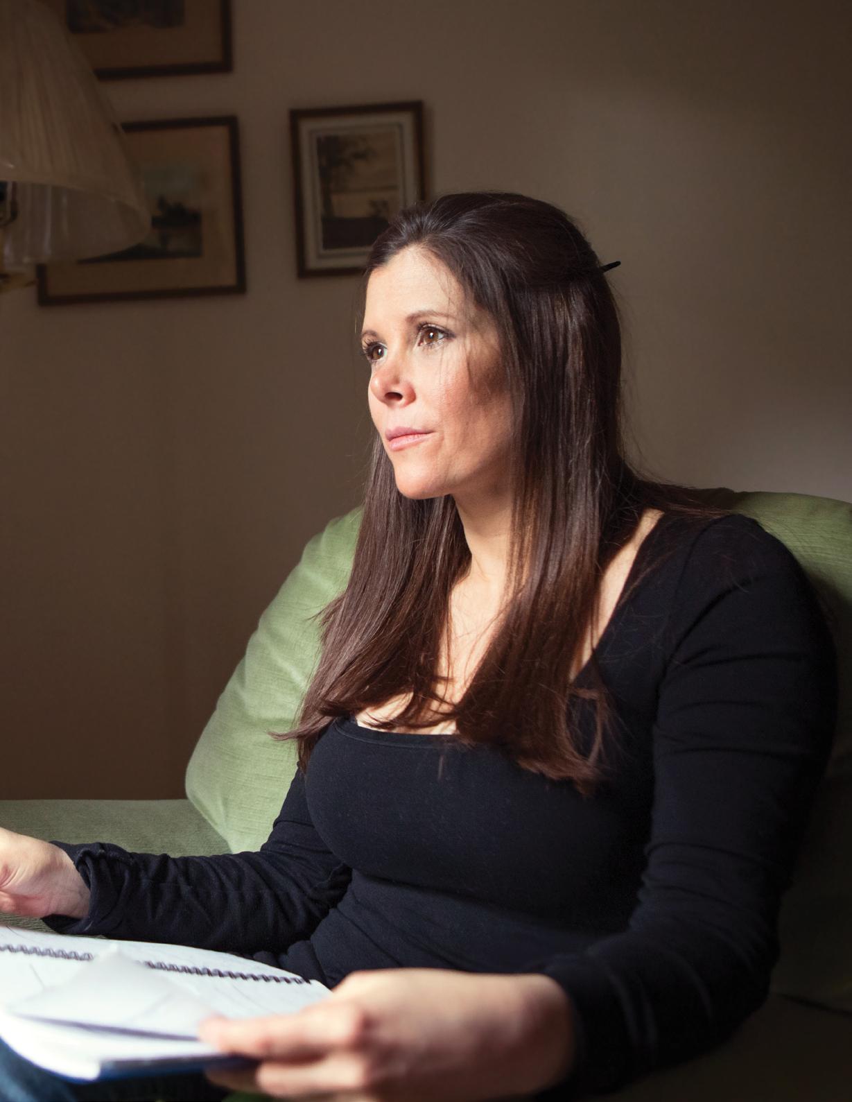
ME/CFS
For decades, no one has known what causes ME/CFS. As a result, this debilitating disease has been very difficult to diagnose and even harder to treat. Now, a collaborative JAX research center led by Derya Unutmaz is diving deep into the biology of ME/CFS patients like Shannon, pictured here, to find a root cause of the disease. Unutmaz believes the answer may lie in their own immune systems.
Immunologist Derya Unutmaz and Research Assistant Lindsey Placek
Microbiome researcher Julia Oh
“We need to be able to develop the tools and the methods that can be used in a clinical setting that allows physicians to say ‘yes, this is ME/CFS,’ or ‘no, this is something else,’” says Suzanne Vernon, Ph.D., the research director of the Bateman Horne Center (BHC), the core clinical collaborator for the CRC. “Because right now, they’ve got nothing.”
The patient’s biology
When looking for something that has been invisible for decades, it’s hard to know where to start. Hence the first “C” in CRC, “collaborative,” is a cornerstone of the effort. To acquire the insight needed to know where to look, then the data needed for the search, it’s crucial to have a variety of perspectives and resources. The CRC has scientists from a number of different specialties, led by Unutmaz, a human immunologist, and Oh, a microbiome expert. But the CRC reaches well beyond the laboratory as well to engage clinicians, including those who work with the disease and the patients at the BHC, and patient activists whose voices were so often muted in the past. Importantly, three NIH - funded
What’s in a name?
Three decades ago, the Centers for Disease Control and Prevention (CDC) named a mysterious, debilitating disease with no known biological cause “chronic fatigue syndrome,” or CFS. It turned out to be a somewhat vague and unfortunate choice, for several reasons. Many people are chronically tired because of stress and perhaps poor sleep habits, but they don’t have the disease and may be more inclined to think “well, I’m tired too,” dismissing the much more severe symptoms of those who do. Fatigue is also a prominent component of diseases such as depression, implying a possible
CRCs have now been established, with major collaborations developing between JAX, Cornell University and Columbia University.
“We’re sequencing thousands of species of bacteria. We’re determining hundreds of different populations of immune cells in the same person. We’re also analyzing their metabolism and determining hundreds of different metabolites in their blood. We’re trying to put the patient’s biology back together,” explains Unutmaz. Using an integrated analysis that requires an incredible amount of computation and technology, he and his group are combining these data with clinical data in order to look for hallmarks of the disease.
Unutmaz says they are already seeing profound differences in the immune systems of ME/ CFS patients compared to controls. “We’re very excited about that,” he says. “In my mind, there’s no question that there’s an immunological basis for this disease.” Finding a biological basis for the disease, Unutmaz says, would enable a physician to easily diagnose ME/ CFS by identifying the corresponding biomarker.
psychological component. Finally, those struggling with the disease face symptoms and challenges that go far beyond simple “fatigue.”
The drive for a better, more accurate name yielded “myalgic encephalomyelitis,” or ME, a tongue twister connoting central nervous system inflammation and muscle pain. It avoids the fatigue label, but is itself not a perfect descriptor, as the inflammation seen in many patients has not yet been proven to be a factor in the actual disease. Perhaps as a compromise, many clinical organizations now refer to the disease as ME/CFS. In 2015,
a medical panel attempted to further refine the name, suggesting “systemic exertion intolerance disease” because of patients’ inability to persist with, or recover from, ordinary physical or cognitive effort. But while “systemic exertion intolerance” is a reasonably good characterization of a disease hallmark, it has been unsuccessful in replacing ME/CFS as a name.
Visit www.jax.org/mecfs to watch videos about the challenges faced by patients, doctors and scientists alike in confronting ME/CFS.
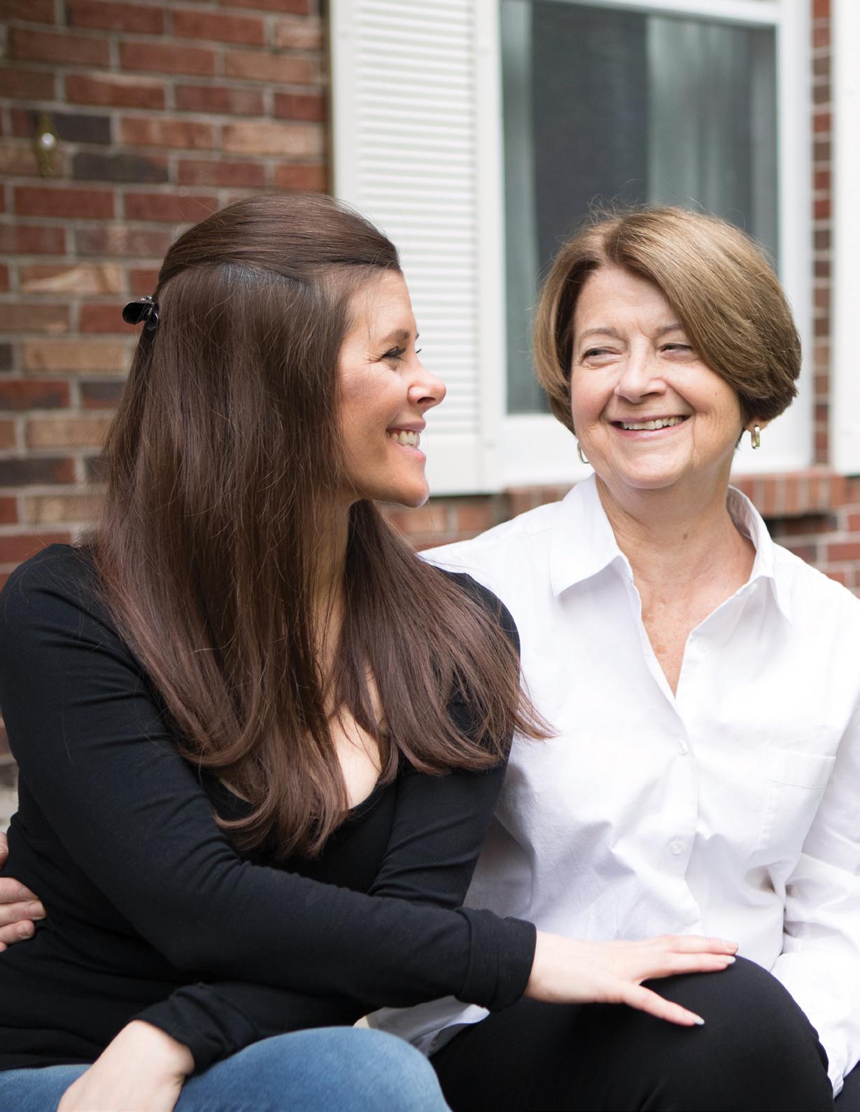
The clinical perspective
Lucinda Bateman, M.D., is the “Bateman” in the BHC’s name. She was drawn to the ME/CFS field when her older sister fell ill in 1987. At that time Bateman was training in internal medicine at Johns Hopkins University, but the stigma associated with the disease was still strong. After a decade of practicing primary care, she decided to focus on ME / CFS, and her practice grew rapidly. Ultimately, in 2014, she spearheaded the formation of the BHC to address both the clinical needs of her patients and the clinical research so lacking elsewhere.
“The biggest barrier in the field has been a knowledge gap in the practice of medicine,” she says. “The science has moved slowly, mostly because it’s underfunded. But it is picking up speed. One of the reasons I’m so excited about working with Derya is that I feel like our collaboration — so far and in the future — will be very substantial in terms of scientific results we can take back to get this illness mainstreamed into regular science.”
Also in the clinical equation are, of course, patients and patient advocates. Mary Dimmock became involved with ME/CFS advocacy when her son fell ill after contracting giardia during a backpacking trip. He recovered from the initial infection, but his health continued to decline. A long, difficult road followed, and Dimmock saw firsthand the misunderstanding and even hostility her son faced in the medical community. Her experience working at the pharmaceutical company Pfizer provided her with knowledge about research and disease, but it didn’t prepare her for what many ME/CFS patients face.
“I can appreciate that as a researcher, it may be easy to lose track of the urgency that patients feel. But for ME/CFS, there are no tests or medicines and patients still struggle to get any help from their medical providers,” she says. “There’s an urgent need to deliver a diagnostic test and treatments as quickly as possible. Even treatments that provide only partial relief could make a huge difference in a patient’s quality of life.”
A microscopic culprit?
The mystery underlying ME/CFS may have its roots in a larger mystery about human life: the microbiome. We have long known we share our lives with huge numbers of microbes (the latest estimate puts the number near 30 trillion, though precision is understandably elusive), but we’ve focused largely on the relatively few pathogenic varieties until recently. Now we know that the entire microbiome plays a large role in our health
and in at least some diseases. Research is still in its early days, and exactly what happens and why is still under investigation, but some early results are highly intriguing. One new concept is that even if two people have the same species of bacteria in them, different strains of that species can have an impact on how they interact with the individual immune systems.
“What Derya and I have seen is that not only does immunogenicity differ by species, it can also differ by strain,” says Oh, CRC associate director. “And this is very significant for disease, because we think that ME/CFS patients may have different species composition, but they could also have different strains that could be what is underlying their disease in terms of immune interactions. And if we could get an idea from the initial patient cohorts of what some of the differences are between them and healthy controls, that would be awesome.”
Pushing microbiome research forward is one part of a highly innovative research program. Another is the computational component, which is vitally important for working with the massive amounts of data generated, and involves novel tools developed by JAX Professor Peter Robinson, M.D. Managing the data for analysis will involve machine learning and deep learning methods developed by Robinson, which will integrate the component data sets into a cohesive whole.
The new research tools and methods have the potential to finally shed light on ME/ CFS’s mysterious pathology. “Technology is advancing very fast,” says Unutmaz. “Things we can do now weren’t even possible five or 10 years ago.”
The excitement around this kind of innovation has everyone associated with the CRC optimistic about its future impact on ME/CFS understanding and treatment. “With all the data we generate, we hope to achieve two things,” says Unutmaz. “One is to generate new hypotheses about ME/CFS and determine the disease mechanism. The second is to come up with a variety of biomarkers to enable biology-based diagnosis of patients rather than having to rely on clinical symptoms. We have the opportunity to address the great unmet needs of both patients and physicians dealing with this disease.”
An ME/CFS diagnosis is within reach, with your support. Your gift can help us harness the incredible power of the immune system. Help ME/CFS patients and others by visiting www.jax.org/immunology-research
BY MARK WANNER | PHOTOGRAPHY BY WIKIIMAGES FROM PIXABAY
Neuroscience is strikingly similar to astrophysics.
Why? Well, our own galaxy, the Milky Way, is pretty big as galaxies go, containing 250 billion stars, give or take 100 billion or so. Our brains contain about 90 – 100 billion neurons. So you get the idea. There’s the numerical equivalent of a smallish galaxy in each of our skulls. But that’s not all. Each neuron has, on average, about 7,000 synaptic connections with other neurons. That puts the synapse count in the neighborhood of 600 trillion. The system of interconnections almost defies comprehension.
Which brings us to late onset Alzheimer’s disease (LOAD). LOAD is actually pretty simple for a neurological disease, at least on the surface. In LOAD, neurons die over time. After quite a while — usually decades — neuron death affects memory and cognition, and ultimately the damage becomes so extensive that the patient dies. Hallmarks of LOAD include accumulation of protein aggregations, called beta - amyloid plaques and tau tangles, thought to disrupt neuron function.
But scratch the surface and it’s not at all simple. The environmental and behavioral contributions to LOAD, such as diet and exercise (or lack thereof), accumulate over an extremely long time. Even sleep habits early in life are coming under scrutiny. Some recent attention has also been focused on immune function and blood vessel health as factoring into disease progression. Genetically speaking, there is a strong connection between a certain gene subtype, known as ApoE4, and LOAD risk, but no one is quite sure how the mechanism works. All in all, LOAD remains poorly understood for such a serious disease.
The clinical perspective on therapy development has focused on the theory that the beta-amyloid plaques seen in patients hold the key to therapy development. Most of the therapies tested to date were developed to reduce or eliminate the plaques, but none have impeded disease progression. Recent insight suggests the reason may be timing: it’s possible that plaque formation leads to downstream effects, and eliminating them too late in disease progression doesn’t help. Perhaps if plaque formation can be prevented or reversed at an early stage, however, neurodegeneration would be mitigated.
But how do you know who’s in the pre - symptomatic stages of LOAD? And how do you recreate a disease with so many variables and such a long pre - symptomatic period in the lab? It will definitely take more than one lab, or one group, but JAX is combining human and mouse data — including imaging data of human patient brains — to develop more useful mouse models. There is also mouse-based research with genetically diverse mice carrying LOAD-associated mutations. Human patients have a lot of variability in their susceptibility to Alzheimer’s disease, so it follows that background genetics can play a large role in LOAD incidence and speed of onset.
The upshot, it seems, is that it’s no surprise that LOAD has been so difficult to address clinically. The good news, though, is that the field is rising to meet its challenges, and the near future holds exciting potential. It took the Hubble Space Telescope to peer far enough into space to learn about the universe’s origins. We may now have the tools we need to peer deep into our own brains and learn enough about how they work that we can also learn how to keep them working well.
THE ENGINES OF GENETIC DIVERSITY
BY EMILY POWERS | PHOTOGRAPHY BY TIFFANY LAUFER
JAX Assistant Professor Beth Dumont, Ph.D., is working to better understand the processes that give rise to genetic diversity, and how specific variations can contribute to disease susceptibility.
The earliest genetics lesson that Beth Dumont can remember introduced her to Gregor Mendel and his pea plants. Through selective breeding and crossbreeding of generations of his plants, Mendel discovered that certain simple traits — such as seed shape and flower color — are passed from parent to offspring in predictable patterns. Mendelian rules of inheritance further explain that every child receives one set of chromosomes from each of his or her parents, making up a new genome.
But for Dumont, the exceptions to Mendel’s model are far more interesting than the rules.
“We know that chromosomes don’t just get transmitted fully intact from one generation to the next — they’re subject to mutations, they undergo the process of recombination and these are processes that are vital for driving evolution as well,” Dumont explains. “They’re
the processes that generate all the genetic diversity that we see in the natural world, both on short timescales and on longer timescales.”
A mutation occurs when a genetic sequence is altered or changed through additions, deletions or substitutions. Recombination, on the other hand, is the rearrangement of pre-existing genetic material. Dumont has dedicated her career to better understanding these two molecular processes, which geneticists consider “the engines of genetic diversity.”
AN INTEREST IN DIVERSITY
Dumont grew up in the woods of Maine, where she learned to appreciate natural diversity at a young age. She spent her days outside, no matter the weather, turning over rocks and inspecting pine cones. The inexhaustible shapes, sizes and patterns that she observed in her back yard inspired curiosity for the natural world that has only grown throughout her academic and professional career.
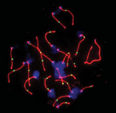
THE DUMONT LAB
Researching the mechanisms that generate genetic diversity through the lens of evolution
Using fluorescence to visualize recombination allows researchers to locate a DNA repair protein (green) during meiosis and estimate the number of crossover events in the cell. (Approx. 1000x magnification.)
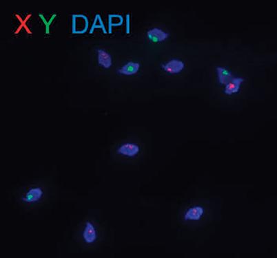
After meiosis, each sperm carries either an X or Y sex chromosome. But do they have different survival properties, and if so, what underlies them? Differential labeling of the two categories of sperm may provide answers. (Approx. 1000x magnification.)
After her first year at Cornell University, Dumont attended The Jackson Laboratory’s Summer Student Program, where she was mentored by Malcolm Lyons, Ph.D., in the laboratory of Beverly Paigen, Ph.D. Dumont’s exposure to science had been limited before that summer. Looking back, she credits her time in the Summer Student Program as “life-changing.”
“At the end of the summer, what seized me the most was this recognition that the approach we were using to identify physiological differences between strains [of mice] rested on the fact that they were genetically distinct from one another,” recalls Dumont. “That really got me interested in understanding where genetic diversity came from in the first place.”
Dumont graduated summa cum laude and Phi Beta Kappa from Cornell with a dual degree in biology and anthropology. She went on to complete her Ph.D. work at the University of Wisconsin-Madison Laboratory of Genetics. Her dissertation identified several loci that contribute to species differences in recombination rate. Loci are specific, unchanging regions of a chromosome, much like a street address on a map. By identifying these loci, Dumont came one step closer to elucidating the cause of vastly divergent recombination rates between species.
“Beth excels at finishing projects, even when the results are complicated,” reports Dumont’s doctoral mentor, Bret Payseur. “Having Beth as my first graduate student was like winning the lottery on the first try.”
Hoping to expand upon her Ph.D. minor in biostatistics following her doctoral work, Dumont took a senior research fellow position in the laboratory of Evan Eichler, Ph.D., at the University of Washington Department of Genome Sciences, followed by a distinguished postdoctoral research fellow position at the North Carolina State University Department of Biological Sciences.
During Dumont’s Ph.D. years, genome sequencing technologies became faster and cheaper, leading to their increased use in genetics laboratories across the United States.
“I realized if I was ever going to be competitive in harnessing those emergent technologies, I really needed to expand my computational skill set,” says Dumont. This realization motivated her to take positions in labs known for their massive data sets and their focus on computational genomics.
“It’s not sufficient to just generate data,” Dumont continues. “You need to know how to handle it and draw inferences from it, too.”
JAX HOMECOMING
In 2016, 14 years after first setting foot at JAX, Dumont joined the faculty as an assistant professor. She now leads the Dumont Lab and mentors summer students of her own.
“So much of what motivates the questions I ask, and my day-to-day existence, is just this innate curiosity about how inheritance works and basic questions about evolutionary mechanisms and mechanisms of mutation and recombination,” says Dumont.
Her lab at JAX takes this curiosity-driven approach and uses it to guide research questions about how exactly genetic diversity is generated. Why do recombination and mutation rates vary between species and individuals? What causes this variation? To what degree is this propensity for diversity encoded in our DNA?
Using the wild-derived inbred strains of mice at JAX, Dumont’s lab is finding that the tendency to engage in diversity-generating processes may actually be embedded in an individual’s genetic code. This means that while mutation and recombination account for all new variation within populations, these very same variations can encode for the frequency of mutation and recombination.
By understanding these complex interactions, geneticists can begin to unravel how genetic variants cause disease.
“Every disease has to start with a mutation somewhere,” explains Dumont. “And I’m really interested in understanding how those mutations pop up, why they pop up in specific places in the genome, and why they pop up at different rates in different individuals.”
IMPLICATIONS IN DISEASE RESEARCH
Genetic diversity accounts not only for the visible differences between us — eye color, height, freckles, widow’s peak — but also for our varying risk of developing cancer, diabetes and other diseases.
“If we’re going to understand how specific genetic variances contribute to disease, it helps to have an understanding of where a particular genetic variance came from in the first place. That’s where my work comes in,” says Dumont.
Dumont’s research focuses on mutation and recombination in the germline (the testes and ovaries), which could tie into infertility research and treatment down the road. Further, the work being done on mutations in the Dumont Lab may have implications for cancer research. By better understanding mutations and how they arise, scientists and doctors can assess who is at risk for the mutations in somatic tissue that lead to cancer.
Reflecting on the future for her lab and her research, Dumont comments, “I couldn’t do this work anywhere other than at JAX.”
Like many other accomplished scientists, Beth Dumont launched her scientific career during her summer of learning and discovery at the JAX Summer Student Program. You can contribute to the next generation of scientific leaders by supporting JAX education programs at www.jax.org/give .
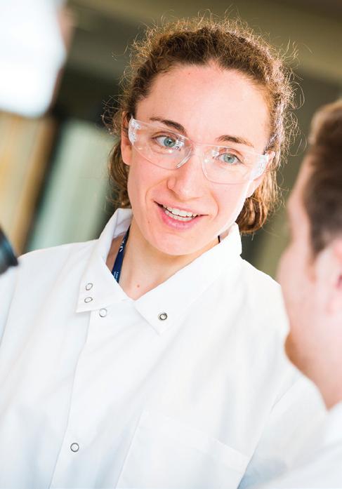
Assistant Professor Beth Dumont
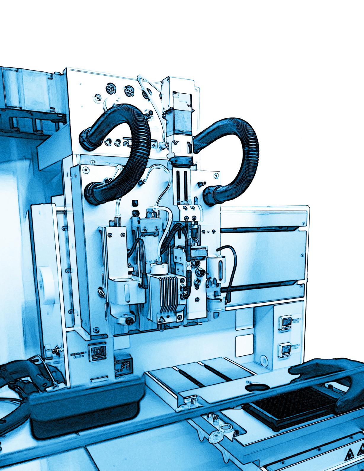
BIOPRINTING cancer
BY JOYCE DALL’ACQUA PETERSON PHOTOGRAPHY BY CHARLES CAMARDA ILLUSTRATION BY ZOË REIFSNYDER
BIOPRINTING and its immune environment
New 3D bioprinting technique creates a platform for precision immunotherapy study.
The next generation of in vitro study of cancer cells is here, and it’s live, high def and in 3D.
It’s now possible to 3D print an exact copy of a patient’s tumor, right down to the various immune and other cells that surround it (known as the tumor’s microenvironment) and the capillaries that supply blood to it. This revolutionary technology is the brainchild of Jackson Laboratory immunologist Derya Unutmaz, M.D., and Ibrahim Tarik Ozbolat, Ph.D., a 3D bioprinting pioneer at Penn State University.
Cancer immunotherapy — harnessing the power of the immune system to thwart cancer — holds promise for patients but has had both successes and failures.
“The interactions between tumor and immune cells in this microenvironment are thought to play a critical role in cancer development, progression and control,” Unutmaz says. “Our limited understanding of this complex interplay is a major barrier to improving immunotherapeutic strategies and defining predictive biomarkers for clinical benefit.”
The new platform will enable the researchers to observe, in action and in 3D, some of cancer’s tricks, such as co opting immune cells in the microenvironment to suppress an anti‑tumor immune response, thus tricking the body into ignoring a foreign invader.
“We hypothesize that cellular and molecular interactions between immune and tumor cells in 3D environments will differ from monolayer cultures and more closely recapitulate the in vivo state,” Unutmaz states.
Tumors start from a single cancer cell, but a mass of these cells can only continue to grow when it develops blood vessels to feed off the patient’s blood, eventually developing its own circulation and ultimately invading the patient’s circulatory system, enabling metastasis.
“We are able to recapitulate this process in a little device,” Ozbolat says. Bioprinted breast cancer cells, chosen from a widely used research cell line, are injected into a gel and connected to bioprinted capillaries. Once this living, circulating tumor is established, he says, “we insert bioengineered immune cells that circulate through the blood vessels and test whether they will kill the tumor.”
With the two year grant from the National Cancer Institute, Unutmaz says, “we will establish a proof of principle that paves the way for a fast, accurate and clinically relevant platform for developing individualized immunotherapies for many kinds of cancers.”
600 Main Street
Bar Harbor, ME 04609-1523
Forwarding service requested
A PUBLICATION OF THE JACKSON LABORATORY
Mission
We discover precise genomic solutions for disease and empower the global biomedical community in our shared quest to improve human health.
Locations
Bar Harbor, Maine
Ellsworth, Maine
Farmington, Connecticut
Sacramento, California Shanghai, China
Editor Joseph Blanchette
Design & art direction
Jane Cha
Karen Davis
Danielle Meier
Zoë Reifsnyder
Rebecca Hope Woods
Copy editors
Joyce Dall’Acqua Peterson
Carol Lamb
Rebecca Hope Woods
This publication was produced by JAX Creative. Printed June 2019