
International Research Journal of Engineering and Technology (IRJET) e-ISSN: 2395-0056
Volume: 13 Issue: 02 | Feb 2026 www.irjet.net p-ISSN: 2395-0072
International Research Journal of Engineering and Technology (IRJET) e-ISSN: 2395-0056
Volume: 13 Issue: 02 | Feb 2026 www.irjet.net p-ISSN: 2395-0072
Ashish Cha nd1 , Raj Kumar2
1Assistant Professor, Dept. of Electrical, Electronics & Communication Engineering, RIMT University, Punjab, India
2Assistant Professor, Dept. of Civil Engineering, RIMT University, Punjab, India
Abstract - Braintumorclassificationplaysacrucialrolein early diagnosis, treatment planning, and patient prognosis. Magnetic Resonance Imaging (MRI) is the most widely used non-invasive imaging modality for detecting and analyzing brain tumors due to its high soft-tissue contrast. However, manual interpretation of MRI scans is time-consuming, subjective, and prone to inter-observer variability. To overcome these challenges, automated brain tumor classification systems based on Machine Learning (ML) and Deep Learning (DL) techniques have gained significant attention. This review paper presents a comprehensive analysis of traditional image processing methods, machine learningalgorithms,andstate-of-the-artdeeplearningmodels used for brain tumor classification. The paper discusses commonly used datasets, preprocessing techniques, feature extraction methods, classification strategies, evaluation metrics, and current challenges. Finally, future research directions and emerging trends in intelligent brain tumor diagnosissystems arehighlighted.
Key Words: Brain Tumor Classification, MRI, Machine Learning, Deep Learning, CNN, Medical Image Analysis
Brain tumors are among the most life-threatening neurological disorders, characterized by abnormal and uncontrolledcellproliferationwithinthebrain.Accordingto the World Health Organization (WHO), brain tumors are broadlyclassifiedintobenignandmalignantcategories,with gliomas, meningiomas, and pituitary tumors representing themostfrequentlyoccurringtypes[1],[14],[17].Earlyand accurateclassificationofthesetumorsiscriticalforeffective clinicaldecision-making,treatmentplanning,andimproving patientsurvivalrates[2],[15].MagneticResonanceImaging (MRI)hasbecometheimagingmodalityofchoiceforbrain tumordiagnosisduetoitssuperiorsoft-tissuecontrastand absenceofionizingradiation.However,despitesignificant advancementsinMRItechnology,manualinterpretationof brainMRIscansremainsachallengingtaskforradiologists. Tumor heterogeneity, variations in size and shape, and overlapping intensity patterns between normal and abnormal tissues often lead to diagnostic uncertainty and inter-observer variability [1], [3], [18].To overcome these limitations, researchers have increasingly focused on automated brain tumor classification systems. In recent years, Machine Learning (ML) and Deep Learning (DL) techniques particularly Convolutional Neural Networks
(CNNs), residual networks, hybrid models, and transfer learning approaches have demonstrated remarkable successinmedicalimageanalysisbyenablingrobustfeature extractionandaccurateclassification[2],[4],[6],[9],[10], [11].Severalstudieshavereportedimprovedclassification performance using data augmentation techniques, hybrid deep learning architectures, and large-scale public MRI datasetssuchasTCIAandKagglerepositories[4],[16],[19], [21]. Consequently, this review systematically examines existing ML- and DL-based approaches for brain tumor classification, highlighting their methodologies, strengths, limitations,andemergingresearchtrends[1],[14],[17].
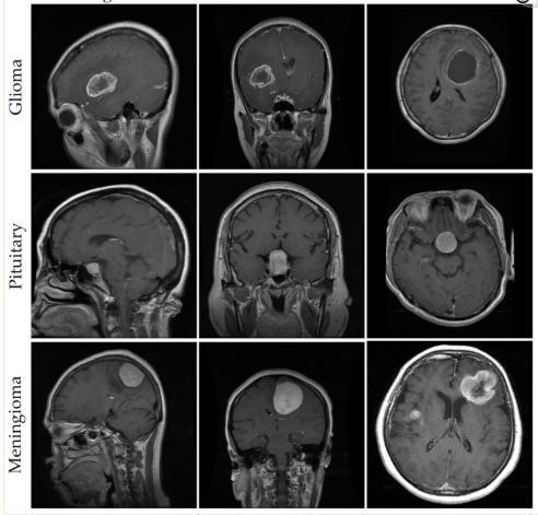
Brain tumors are characterized by abnormal cell growth within the brain and are broadly classified based on their origin,growthbehavior,anddegreeofmalignancy.InMRIbased brain tumor classification research, certain tumor types are more frequently studied due to their clinical prevalence,distinctradiologicalfeatures,andavailabilityin public datasets. Among these, gliomas, meningiomas, pituitarytumors,andhealthy(notumor)casesarethemost commonlyinvestigatedcategories[1],[14],[17].
Gliomasaremalignantbraintumorsthatoriginatefromglial cells,includingastrocytes,oligodendrocytes,andependymal cells. They represent the most aggressive and frequently occurringprimary brain tumorsinadults.Gliomasexhibit highlyinfiltrativegrowthpatterns,makingcleardelineation fromsurroundinghealthytissueparticularlychallenging.On MRI scans, gliomas often display heterogeneous intensity patterns,irregulartumorboundaries,edema,andnecrotic regions, especially in high-grade cases. These complex characteristics make glioma detection and classification a criticalyetchallengingtaskforautomatedmachinelearning anddeeplearningsystems[2],[11],[15],[21].
Meningiomasare generally benigntumors thatarise from themeninges,theprotectivemembranescoveringthebrain andspinalcord.Theyaretypicallyslow-growingandwellcircumscribed, which facilitates their detection and
International Research Journal of Engineering and Technology (IRJET) e-ISSN: 2395-0056
Volume: 13 Issue: 02 | Feb 2026 www.irjet.net p-ISSN: 2395-0072
classificationcomparedtomalignanttumors.OnMRIimages, meningiomas often appear as well-defined, extra-axial masses with homogeneous signal intensity and strong contrast enhancement. Due to their relatively uniform structureanddistinctappearance,meningiomashavebeen widelyusedinearlyandcontemporarymachinelearninganddeeplearning-basedbraintumorclassificationstudies [1],[4],[10],[14].
Pituitarytumorsdevelopinthepituitarygland,asmallbut vital endocrine organ located at the base of the brain. Although most pituitary tumors are benign, they can significantlydisrupthormonalregulationandneurological function. In MRI scans, pituitary tumors are typically observedinthesellarandsuprasellarregionsandmaycause compression or displacement of adjacent anatomical structures.Theirrelativelysmallsize,centrallocation,and subtleintensityvariationsnecessitatehigh-resolutionMRI imaging and robust feature extraction techniques for accurateclassification[6],[8],[9],[19].
The“NoTumor”orhealthybraincategoryconsistsofMRI scans that do not exhibit any pathological abnormalities. Thisclassservesasanessentialreferenceinbothbinaryand multi-classbraintumorclassificationframeworks.Healthy brain MR images generally demonstrate symmetrical anatomical structures and consistent tissue intensity distributions. Inclusion of this category enhances model generalization,improvesrobustness,andhelpsreducefalsepositivetumordetectionsinautomateddiagnosticsystems [1],[5],[16],[19].
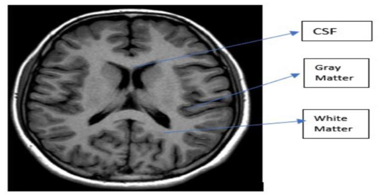
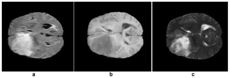
MagneticResonanceImaging(MRI)isthemostwidelyused imagingmodalityforbraintumordiagnosisandanalysisdue to its superior soft-tissue contrast, multiplanar capability, andnon-invasivenature.DifferentMRIsequencesemphasize distinct tissue characteristics, and their combined use provides complementary diagnostic information that is essential for accurate tumor detection, segmentation, and classificationinautomatedsystems[1],[14],[17].
T1-weightedMRIimagesprovidehighanatomicaldetailand clearvisualizationofnormalbrainstructures.InT1images, cerebrospinalfluid(CSF)appearsdark,whilewhitematter appearsbrighterthangraymatter.Althoughtumorregions maynotalwaysbedistinctlyvisibleinT1imagesalone,these scans are crucial for anatomical reference, structural integrity assessment, and initial tumor localization. Consequently, T1-weighted images are frequently used as baseline inputs in multi-modal brain tumor classification frameworks[1],[10],[18].
T1-weighted contrast-enhanced images are acquired following the administration of contrast agents such as gadolinium,whichaccentuateareaswithadisruptedblood–brain barrier. Tumorous regions often exhibit strong enhancementinT1cimages,enablingimprovedvisualization of tumor boundaries, vascularization, and active tumor regions.Duetotheirabilitytohighlightmalignanttissue,T1c imagesareextensivelyusedinbothtumorsegmentationand classification tasks, particularly in deep learning-based approaches[2],[6],[11],[16].
T2-weightedMRIimagesarehighlysensitivetovariationsin water content and are effective in visualizing edema and fluid-richregionssurroundingtumors.InT2images,fluids appearbright,makingthismodalityvaluableforidentifying tumor-associated swelling and peritumoral edema. T2weightedscansprovidecomplementaryinformationtoT1 and T1c images and are commonly integrated into multimodal classification systems to improve diagnostic performance[4],[9],[15],[21].
FLAIR images suppress cerebrospinal fluid signals while maintaining sensitivity to pathological tissues, thereby enhancing the visibility of lesions adjacent to fluid-filled regions.ThispropertymakesFLAIRparticularlyeffectivefor
International Research Journal of Engineering and Technology (IRJET) e-ISSN: 2395-0056
Volume: 13 Issue: 02 | Feb 2026 www.irjet.net p-ISSN: 2395-0072
detectinginfiltrativetumorregionsandedemathatmaybe obscured in T1 or T2 images. FLAIR sequences are extensivelyusedingliomaanalysisandclassificationstudies, astheyprovidecleardelineationoftumorinfiltrationzones critical for accurate diagnosis and treatment planning [1], [11],[17].
Publicly available datasets have played a crucial role in advancing brain tumor classification research by enabling standardized training and evaluation of machine learning anddeeplearningmodels.TheBrainTumorSegmentation (BraTS) dataset is one of the most widely used resources, offeringmultimodalMRIscanssuchasT1,T1-contrast,T2, and FLAIR images along with expert annotations. The availability of multiple MRI modalities allows models to learn comprehensive tumor characteristics; however, models trained solely on BraTS may show limited generalizationwhenappliedtosimplerorsingle-modality datasets [1], [11], [17]. The Figshare brain tumor dataset primarily contains T1-weighted contrast-enhanced MRI images and is commonly employed for multi-class classificationofglioma,meningioma,andpituitarytumors. While this dataset is easy to use and often produces high classificationaccuracy,itsrelianceonasingleMRImodality restrictsitsabilitytocapturereal-worldimagingvariability
[4], [10], [20]. The Br35H (Brain Tumor Detection 2020) dataset is mainly designed for binary classification and includesMRIscanslabeledastumorornotumor.Although usefulforbenchmarkingtumordetectionmodels,itdoesnot support detailed tumor subtype classification [12], [19]. TCIAprovideslarge,heterogeneousMRIdatasetscollected frommultipleinstitutions,makingitvaluablefordeveloping models with improved clinical generalization, though extensivepreprocessingisoftenrequired[16],[17].Kaggle MRIdatasetsarewidelyusedduetoeasyaccessibilitybut may lack sufficient diversity to fully represent clinical scenarios[5],[19].Overall,datasetsize,modalitydiversity, andlabelingstrategysignificantlyinfluencemodelaccuracy andgeneralizationperformance[1],[14],[21].
Dataset
BraTS Multimodal Segmentation & Classification Moderate Medium
Figshare Singlemodal
Br35H Singlemodal
TCIA Multimodal
Kaggle MRI Singlemodal
Classification
Classification
& Prognosis
Table -1: Comparative Analysis and Generalization Challenges
DatasetssuchasBraTS,Figshare,Br35H,TCIA,andKaggle MRI collections have significantly advanced brain tumor classificationresearch.However,variationsindatasetsize, modality, labeling strategy, and clinical diversity strongly influencemodelperformance.
Preprocessingisacriticalstageinbraintumorclassification pipelines,asitenhancesimagequalityandenableslearning of discriminative features by machine learning and deep learningmodels.NoiseremovaltechniquessuchasGaussian and median filtering are widely employed to suppress unwanteddistortionswhilepreservingessentialstructural information in MRI images [1], [14]. Skull stripping is performedtoeliminatenon-braintissues,includingtheskull andscalp,ensuringthatmodelsfocusexclusivelyonrelevant brainregionswheretumorsoccur[11],[18].
Intensitynormalizationisappliedtominimizevariationsin brightness and contrast caused by different MRI scanners and acquisition protocols, thereby improving dataset
International Research Journal of Engineering and Technology (IRJET) e-ISSN: 2395-0056
Volume: 13 Issue: 02 | Feb 2026 www.irjet.net p-ISSN: 2395-0072
consistency and model generalization [1], [17]. Image resizing and spatial normalization are commonly used to standardizeinputdimensions,allowingcompatibilitywith convolutionalneuralnetworkarchitectures[10],[15].
To address data scarcity and class imbalance, data augmentationtechniquessuchasrotation,flipping,scaling, andtranslationareextensivelyusedtoincreasetrainingdata diversityandreduceoverfitting[4],[9],[21].Additionally, contrastenhancementtechniquesimprovetumorvisibility by amplifying intensity differences between normal and abnormal tissues, facilitating more accurate tumor discrimination [3], [18]. Overall, effective preprocessing significantly improves classification accuracy, robustness, and generalization across diverse MRI datasets [1], [14], [21].
Traditional machine learning–based brain tumor classification approaches rely on handcrafted feature extraction from MRI images, followed by conventional classifiers for tumor identification. Texture-based feature extractiontechniquessuchastheGrayLevelCo-occurrence Matrix (GLCM) are widely used to quantify spatial relationships between pixel intensities, enabling effective characterization of tumor texture patterns [1], [18], [20].
LocalBinaryPatterns(LBP)capturelocaltexturevariations bycomparingeachpixelwithitsneighboringpixelsandare effective in highlighting fine-grained tumor details [18]. Histogram of Oriented Gradients (HOG) focuses on edge orientationsandstructuralinformation,makingitusefulfor capturingtumorboundariesandshapecharacteristics[20].
WaveletTransform–basedfeaturesdecomposeMRIimages intomultiplefrequencysub-bands,allowingsimultaneous analysis of spatial and frequency information relevant to tumor characterization [1], [21]. In addition, shape and statistical texture features such as area, perimeter, smoothness, contrast, and entropy provide valuable informationabouttumormorphologyandappearance[14], [20].
Followingfeatureextraction,machinelearningclassifiersare employed for decision-making. Support Vector Machines (SVMs)areamongthemost widelyusedclassifiersdue to their effectiveness in handling high-dimensional feature spacesandachievingrobustclassificationperformance[5], [15].K-NearestNeighbors(KNN)classifiestumorsbasedon similaritymeasuresandissimpletoimplement,particularly for small datasets [14]. Random Forest (RF) classifiers combinemultipledecisiontreestoenhancerobustnessand reduce overfitting [21]. Naïve Bayes classifiers leverage probabilistic learning and perform well on limited data, whileDecisionTreesofferinterpretableclassificationrules [1], [14]. Although these traditional methods are computationallyefficientandeffectiveforsmallerdatasets,
their performance is highly dependent on the quality of handcraftedfeaturesandlacksscalabilityforcomplextumor heterogeneity[1],[17].
Deep learning–based approaches have gained significant attentioninbraintumorclassificationduetotheirabilityto automaticallylearnhierarchicalanddiscriminativefeatures directlyfromMRIimages,therebyeliminatingtheneedfor manual feature engineering. Among these approaches, ConvolutionalNeuralNetworks(CNNs)arethemostwidely adoptedbecauseoftheirstrongcapabilitytocapturespatial andstructuralpatternsinmedicalimages[1],[14],[17].
EarlyCNNarchitecturessuchasAlexNetdemonstratedthe effectiveness of deep learning for image classification by employing multiple convolutional and pooling layers to extractlow- andhigh-level features[10],[15].VGG16and VGG19introduceddeeperyetuniformnetworkarchitectures usingsmallconvolutionalfilters,enablingtheextractionof fine-grainedtumorfeaturesfromMRIscans[2],[9].ResNet addressedthevanishinggradientproblemthroughresidual or skip connections, allowing very deep networks to be trained efficientlyand resultinginimprovedclassification accuracy for complex tumor patterns [2], [11]. DenseNet furtherenhancedfeaturereusebyconnectingeachlayerto everyotherlayer,promotingefficientinformationflowand reducingoverfittinginlimitedmedicaldatasets[12],[21]. Inception-based architectures employed parallel convolutionalfiltersofvaryingsizeswithinthesamelayer, allowing multi-scale feature extraction that is particularly beneficial for tumors with heterogeneous structures [10], [15]. Collectively, these CNN-based architectures have significantly improved the accuracy, robustness, and reliabilityofautomatedbraintumorclassificationsystems [1],[17],[21].
Performance evaluation metrics play a critical role in assessing the effectiveness of brain tumor classification models. Accuracy measures the overall proportion of correctly classified MRI images and provides a general assessment of model performance [14], [15]. Precision quantifiestheproportionofcorrectlypredictedtumorcases among all predicted tumor cases, helping to reduce falsepositive diagnoses [1]. Recall, also known as sensitivity, evaluates the model’s ability to correctly identify tumor cases and is particularly important in medical diagnosis, wheremisseddetectionscanhavesevereconsequences[2], [9].Specificitymeasurestheabilityofthemodeltocorrectly classifynon-tumorcases,minimizingunnecessarymedical interventions [14], [17]. The F1-score combines precision andrecallintoasinglemetric,offeringabalancedevaluation when class distributions are imbalanced [15], [21]. Additionally, the Area Under the Receiver Operating
International Research Journal of Engineering and Technology (IRJET) e-ISSN: 2395-0056
Volume: 13 Issue: 02 | Feb 2026 www.irjet.net p-ISSN: 2395-0072
Characteristic Curve (AUC) assesses the model’s discriminative capability across different decision thresholds, with higher AUC values indicating superior classificationperformance[10],[17].
Despite significant progress in brain tumor classification, severalchallengesandlimitationsstillexist.Onemajorissue isthelimitedavailabilityofannotatedmedicaldatasets,as expert labeling by radiologists is time-consuming and expensive, resulting in small training datasets. Another challengeisclassimbalance,wherecertaintumortypeshave farfewersamplesthanothers,causingmodelstobebiased toward majority classes and reducing accuracy for rare cases. Deep learning models also suffer from overfitting, especiallywhentrainedonlimitedorrepetitivedata,leading topoorperformanceonunseenimages.Inaddition,thehigh computationalcostrequiredfortraininganddeployingdeep models demands powerful hardware and long processing times,whichmaynotbefeasibleinallhealthcaresettings. The lack of model interpretability is another concern, as manydeeplearningsystemsfunctionasblackboxes,making it difficult for clinicians to understand and trust their predictions. Finally, clinical validation and deployment remain challenging because models trained on public datasets may not perform consistently in real hospital environments due to variations in scanners, imaging protocols,andpatientpopulations.
Futureresearchinbraintumorclassificationisexpectedto focusonseveralpromisingdirectionstoimproveaccuracy, reliability, and clinical usability. One important area is ExplainableAI(XAI),whichaimstomakemodeldecisions transparent and understandable for doctors, thereby increasing trust in AI-assisted diagnosis. Multimodal MRI fusion is another key direction, where information from different MRI sequences is combined to capture complementarytumor featuresandenhance classification performance. Developing lightweight and efficient models will support real-time diagnosis and make AI systems suitable for deployment in resource-constrained clinical settings.Federatedlearningisgainingattentionasitenables collaborative model training across multiple hospitals withoutsharingpatientdata,thusensuringdataprivacyand security. Integration of AI models with clinical decision supportsystemswillhelpdoctorsbyprovidingtimelyand accurate diagnostic assistance within existing healthcare workflows. Additionally, self-supervised and semisupervised learning methods are expected to reduce dependencyonlarge labeleddatasetsbyeffectivelylearning fromunlabeledorpartiallylabeledmedicalimages.
Thisreviewpresentedacomprehensiveoverviewofbrain tumorclassificationtechniquesusingmachinelearningand deeplearningapproaches.WhiletraditionalMLmethodslaid thefoundation,deeplearningmodels particularlyCNNs have achieved remarkable performance improvements. However, challenges related to data availability, interpretability,andclinicalapplicabilitypersist.Addressing these issues will be key to developing reliable and deployableautomatedbraintumordiagnosissystemsinthe future.
[1] Zine-dine,I.,Riffi,J.,ElFazazy,K.,ElBatteoui,I.,Mahraz, M.A.,&Tairi,H.Areview:Machinelearningtechniquesof brain tumor classification and segmentation. Machine Graphics&Vision,34(3),31–55,2025.
[2] Zahoor, M. M., Khan, S. H., Alahmadi, T. J., et al. Brain TumorMRIClassificationUsingaNovelDeepResidualand RegionalCNN.Biomedicines,12(7):1395,2024.
[3]Refining neural network algorithms for accurate brain tumorclassificationinMRIimagery.BMCMedicalImaging, 24:118,2024.
[4]ClassificationofBrainMRITumorImagesBasedonDeep LearningPGGANAugmentation.PubMed(2021).
[5] Brain tumor magnetic resonance images classification basedmachinelearningparadigms.PubMed,2022.
[6]BrainTumor/MassClassificationFrameworkUsingMRIBasedTransferDeep-LearningModel.PubMed.
[7] Brain tumor classification using MRI images and deep learningtechniques.PubMed(2025).
[8]T1-weightedMRI-basedbraintumorclassificationusing hybriddeeplearningmodels.PubMed(2025).
[9] Brain Tumor Classification from MRI Using Image EnhancementandCNNTechniques.PubMed(2024).
[10] Brain Tumor Classification From MRI Using Deep LearningandCNNTechniques.MDPIarticle(2023).
[11] Díaz-Pernas, F. J., Martínez-Zarzuela, M., AntónRodríguez, M., & González-Ortega, D. A Deep Learning ApproachforBrainTumorClassificationandSegmentation UsingaMultiscaleCNN.arXiv,2024.
[12] Shah, M. A., Alam, M. M., & Khan, S. H. Tumor Aware DenseNetSwinHybridLearningforLarge-ScaleBrainMRI Classification.arXiv,2026.
International Research Journal of Engineering and Technology (IRJET) e-ISSN: 2395-0056
Volume: 13 Issue: 02 | Feb 2026 www.irjet.net p-ISSN: 2395-0072
[13] Filvantorkaman, M., Piri, M., & Filvan Torkaman, M. Fusion-Based Brain Tumor Classification Using Deep LearningandExplainableAI.arXiv(2025).
[14] Toufiq, D. M., Sagheer, A. M., & Veisi, H. A Review on BrainTumorClassificationinMRIImages.TurkishJournalof ComputerandMathematicsEducation(TURCOMAT),2021.
[15]Raza,M.U.,etal.ClassifyingBrainTumorsonMRIUsing CNNs.Electronics,12(4),2023.
[16]TheCancerImagingArchive(TCIA).PublicMRItumor imaging repository widely used in classification and segmentationresearch.
[17] Investigating brain tumor classification using MRI: scientometric analysis (2015–2024). Springer Nature publication.
[18]Abbasi,S.,&Tajeripour,F.Detectionofbraintumorin 3D MRI images using local binary patterns and histogram orientation gradient. Neurocomputing 219:526-535, 2017 (reviewedinbroadersurvey).
[19] Brain Tumor MRI Dataset on Kaggle widely referenced public dataset used in numerous classification studies.
[20]Cheng,J.,etal.ClassificationofBrainTumorsfromMRI Using Handcrafted and Deep Features. MDPI/Applied Sciences(earlierdatabaseusers).
[21] Gumaei, A., et al. Hybrid feature extraction and classification for brain tumor MRI images. Brain Sciences article(2024).