
International Research Journal of Engineering and Technology (IRJET) e-ISSN: 2395-0056
Volume: 13 Issue: 04 | Apr 2026 www.irjet.net p-ISSN: 2395-0072

International Research Journal of Engineering and Technology (IRJET) e-ISSN: 2395-0056
Volume: 13 Issue: 04 | Apr 2026 www.irjet.net p-ISSN: 2395-0072
Nadakuditi Deepika1 , Banka Lokeshwar Reddy2 , G Mani Dinesh Reddy3 , Komatineni Bhoomika⁴, Dr. Prabhakar K⁵
1,2,3,4 Student, Dept of CSE (AIML), CMR University, Bangalore, Karnataka, India
5 Assistant Professor, Dept. of CSE (AIML), CMR University, Bangalore, Karnataka, India
Abstract - Skin diseases are widespread health concerns that require early detection to prevent complications and reduce treatment costs. This paper presents a skin disease predictionsystemwithcostestimationtosupportpreliminary screening and healthcare awareness. The proposed method uses a hybrid deep learning model combining Convolutional Neural Networks (CNN) with EfficientNet to analyze skin lesion images and classify them into predefined disease categories. The model is trained on the HAM10000 dataset comprising 10,015 images across nine skin disease classes, with images resized to 128 x 128 pixels. The system provides a confidence score for each prediction and incorporates Grad-CAM to highlight important regions influencing the model's decisions, improving interpretability. In addition, it offers information on symptoms, general medical advice, estimated treatment cost ranges, and location-based hospital recommendations. The model achieves a training accuracy of 97% and validation accuracy of 94%, with macro-averaged precision of 93%, recall of 92%, and F1score of 92%, demonstrating reliable performance. The system highlights the effectiveness of integrating automated diagnosis, explainable AI, and healthcare support features to enhance accessibility and assist users in informed medical decision-making.
Key Words: Skin Disease Classification, Deep Learning, Convolutional Neural Network, EfficientNet, HAM10000, Grad- CAM, Explainable AI, Medical Image Processing
1. INTRODUCTION
Skincancerisamongthemostcommonandpotentiallyfatal formsofcancerworldwide,withitsincidencecontinuingto riseduetoprolongedexposuretoultraviolet(UV)radiation, evolving lifestyle habits, and various environmental influences [1]. Early diagnosis is critical to improving treatment outcomes and patient survival; however, traditional methods such as visual inspection by dermatologistsorbiopsyremaininvasive,time-consuming, and highly dependent on clinical expertise, making them susceptibletoinconsistencyandhumanerror[2].
To address these challenges, non-invasive imaging modalities including dermoscopy, high-frequency ultrasound,opticalcoherencetomography,andfluorescence imaging have been integrated into clinical screening workflows,offeringcost-effectiveandrepeatableevaluation of skin lesions [3]. In recent years, deep learning techniques particularly convolutional neural networks (CNNs) have demonstrated strong performance in automating the identification and classification of skin

lesions, including melanoma, basal cell carcinoma, and squamouscellcarcinoma[4].
Such models leverage large publicly available datasets and advanced feature extraction and image-processing algorithms to enhance prediction accuracy and robustness [5]. Their integration into mobile and web-based applications enables real-time, accessible screening, facilitating early medical consultation even in regions with limited dermatologist availability [6]. Beyond detection, these systems provide quantitative lesion analysis covering asymmetry, border irregularity, color variation, and diameter to assist clinicians in decision-making and diseasemonitoringovertime[7].
The prevalence of skin diseases is growing across all age groups, yet the shortage of dermatology specialists and reliance on manual examination continue to cause delayed or inconsistent diagnoses. The objective of this work is to develop a reliable and interpretable deep learning system for automated skin disease detection. The system employs a hybrid CNN–EfficientNet architecture for multi-class classification and integrates Grad-CAM to provide visual explanations of model decisions. Additional features including cost estimation and location-based hospital recommendations are incorporated to support holistic patientcareandenhancehealthcareaccessibility.
InSumanChowdhury andDilip KumarDas (2024) lookedat using CNN, ResNet50, and VGG16 to detect skin cancer on 3,307dermoscopicimages.
ResNet50 had an accuracy of 99.60%, showing that deep learning can be useful for early detection of skin lesions. However,thestudyusedalimitedsetofimagesanddidnot talkaboutmakingthemodeleasiertounderstandorhowto useitinrealclinics[10].
S. Likhitha and Radhika Baskar (2022) compared CNN and SVMforclassifyingskincancer.
CNN had a higher accuracy of 95.03% compared to SVM’s 93.04%. Though CNN performed better, the study used a smalldatasetanddidnotlookathowwellthemodelwould workinreal-lifesituations[11].
Vikrant Aadiwal, Bhisham Sharma, and D.P. Yadav (2024) created a CNN-based model trained on the 10,015-image HAM10000dataset.
The model scored an F1-score of 98.73% and 92.38% accuracy.Whilethisshowsgoodperformanceinclassifying

International Research Journal of Engineering and Technology (IRJET) e-ISSN: 2395-0056
Volume: 13 Issue: 04 | Apr 2026 www.irjet.net p-ISSN: 2395-0072
skinlesions,thestudysaidmoretestingondifferenttypesof dataisneededbeforeusingitinaclinic[12].
T.SrinivasaRaviKiranetal.(2024)proposedasystemthat usesCNNalongwithHOG,LBP,andResNetembeddings,and classifiesusingRandomForest.
This hybrid approach achieved 96% accuracy, which is betterthanusingCNNalone.Theysuggestedtestingitwith moredatatomakesureitworksinrealsettings[13].
Kajol Kathuria, Anita Sahoo, and Chakresh Kumar Jain (2024)reviewedmethodsthatuseCNNforearlymelanoma detection. Theyfoundthatthesemethodsworkwellfortellingbenign lesions from malignant ones, but their work was mostly theoreticalanddidnotincludeanyrealtesting[14].
Sukhwinder Kaur, Lalit Verma, and Kuldeep Kumar Kushwaha(2025)usedCNNs,ResidualNetworks,andbinary classifiersforlesionclassification.
However, they said more testing with a wide variety of clinical data is needed before it can be used in real care settings [15]. S. Likhitha and Radhika Baskar (2022) also compared R-CNN and Inception V3 for skin cancer segmentation.R-CNNachieved96.01%accuracycompared to 92% for Inception V3, but the limited data used in the studylimitshowusefulitcouldbeinrealclinicalsituations [16].
SivaSibiMandAnithaJ(2025)builtanautomaticsystemfor skincancerdetectionusing3DTotalBodyPhotographyand acustomCNNalongwithwatershedsegmentation. Themodelachieved74.64%accuracy,butitsperformanceis limitedbecausethedataisnotvariedenough[17].
SandhyaSharma,ShaminderKaur,andNavneetKaur(2024) trained an ensemble CNN on seven types of skin lesions usingtheHAM10000dataset.Themodelhad99%accuracy duringtrainingand96%duringvalidation,buttheydidnot testitwithotherdatasources[18].
Skin diseases are becoming more common across all age groups, but diagnosing them early and accurately is still difficult.
This is because there are not enough dermatologists, and most diagnoses rely on manual checks by doctors. Traditionalmethodstakealongtime,canleadtodelays,and are not always consistent. They also increase the risk of mistakes.Manyautomaticsystemsfocusonlyonclassifying skinlesionswithoutexplainingtheirdecisionsorproviding usefulhealthcaresupport.Patientsoftendonotknowabout treatmentcostsornearbyhospitals,makingithardertoget timelycare.So,thereisaneedforareliableAIsystemthat can help with early detection, provide clear explanations, and offer support like cost information and hospital recommendations.
The proposed system uses deep learning to automatically predict skin diseases and includes extra features that help withhealthcare.
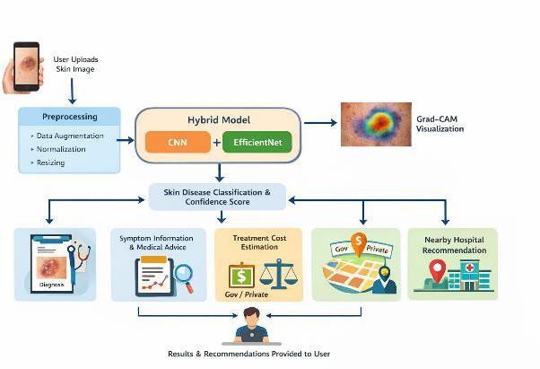
It uses a mixed model that combines CNN with EfficientNet to extract both detailed texture patterns and more general features from skin images. Before training, the images go through some steps like resizing to 128×128 pixels, normalizingpixelvaluestotherange[0,1],andmakingmore images through rotation, flipping, and zooming to improve performance and avoid overfitting. These processed features are then passed to fully connected layers for classification andtogenerateascoreshowinghowconfidentthemodelis about its prediction. The system uses Grad-CAM to create heatmaps that show the most important parts of the image thatinfluencedthemodel'sperformance.
Thesystemstartswhenauseruploadsa dermoscopicimage throughawebinterfacemadewithFlask.
The image is then processed by resizing, normalizing, and applyingsometransformations beforebeinganalyzed by the hybrid CNN + EfficientNet model. This model classifies the skin condition and gives a confidence score. Grad-CAM is used to highlight the parts of the image that had the most impact on the model’s decision. The system then shows information about symptoms, medical advice, estimated treatment costs based on the type of hospital (government, private, or multi-specialty), and recommends nearby hospitals.Allusersessions andpredictionhistoryaresaved inaSQLitedatabase.
5.1.
Thesystemcollectsdermoscopicskinlesionimagesfromthe HAM10000dataset(10,015imagesacross9classesfromthe ISIC Archive) and accepts user-uploaded images in JPEG,
International Research Journal of Engineering and Technology (IRJET) e-ISSN: 2395-0056
Volume: 13 Issue: 04 | Apr 2026 www.irjet.net
PNG,andWebPformats.Theseimagesserveastheprimary inputfordiseaseanalysisandprediction.
5.2. Preprocessing and Image Preparation
Inputimagesareresizedto128×128pixelsandpixelvalues are normalized to the [0,1] range. Data augmentation techniques including random rotation, horizontalflipping, and zooming are applied during training to enhance datasetdiversityandreduceoverfitting.
5.3. Hybrid Deep Learning Model
A hybrid architecture combining CNN and EfficientNet extractsbothlocaltexturefeaturesandhigh-levelsemantic informationfromdermoscopicimages.TheCNNcomponent captures local lesion patterns, edges, and structural variations through convolutional and pooling layers. EfficientNet enhances feature representation through compound scaling of network depth, width, and input resolution.Thefusedfeaturemapsarepassedthroughfully connected layers for multi-class classification with confidencescoring.
5.4. Classification and Grad-CAM Visualization
The trained model categorizes the skin lesion into predefineddiseaseclasses.Grad-CAMisappliedtoproduce heat maps that highlight image regions most influential in theprediction,improvingmodelinterpretabilityandtrust.
5.5. Clinical Support and Cost Estimation Module
Based on the predicted disease, the system provides symptom details, general medical advice, and estimated treatment cost ranges categorized by hospital type (Government,Private,andMulti-specialty).Costrangesare pre-definedperdiseaseclassandhospitalcategorytohelp patientsmakeinformedhealthcarefinancialdecisions
5.6. Nearby Hospital Recommendation
Alocation-basedmoduleallowsuserstoentertheircityand select a hospital type. The system launches a Google Maps search in a new browser tab, enabling users to locate and navigate to nearby healthcare facilities for timely consultation
5.7. Convolutional Neural Network (CNN)
A Convolutional Neural Network is employed to extract spatial and texture-based features from dermoscopic skin images.Convolutionalandpoolinglayerscapturelocallesion patterns,edges,andstructuralvariationsthatareimportant fordistinguishingdifferent skinconditions.These features contributetothemodel’sabilitytorecognizedisease-specific visualcharacteristics.
5.8. EfficientNet
EfficientNet is integrated into the architecture to enhance featurerepresentationthroughcompoundscalingofdepth, width,andresolution.Thismodelefficientlycaptureshighlevelsemanticinformationwhilemaintainingcomputational
efficiency. By combining EfficientNet with CNN features, the system benefits from both detailed local feature extraction and robust global feature understanding, improving overall classificationperformance.
5.9. Grad-CAM
Gradient-weighted Class Activation Mapping (Grad-CAM) is used to provide visual explanations for model predictions.It generates heat maps by analysing gradients flowing into the final convolutional layers, highlighting image regions that most influence the decision. This improves interpretability andincreasestrustintheautomateddiagnosticprocess.
The model was trained and evaluated on the HAM10000 (Human Against Machine with 10,000 training images) dataset, a widely used benchmark for skin lesion classification sourced from the International Skin Imaging Collaboration (ISIC) Archive [12]. The dataset contains 10,015 dermoscopic images representing 9 disease categories.Table1summarizestheclassdistribution.
*Approximate values for augmented minority classes. Total: 10,015images.
All images were resized to 128×128 pixels, normalized to the [0,1] range, and split into training (80%) and validation (20%) subsets. Data augmentation was applied exclusively to the training set to increase robustness. The significant class imbalance (NV comprising ~67% of images) was addressed through augmentation of minority classes during training.
Volume: 13 Issue: 04 | Apr 2026 www.irjet.net
The hybrid CNN–EfficientNet model was trained using TensorFlow 2.x and Keras on a system equipped with an Intel i3 or above processor and optional NVIDIA GPU acceleration. Table 2 presents the complete training configuration.
BaseArchitecture Custom CNN + EfficientNet
Dataset HAM10000 (10,015 images)
InputImageSize 128×128×3(RGB)
NumberofClasses 9
Train/ValidationSplit 80%/20%
BatchSize 32
Epochs 30
Optimizer Adam
LearningRate 0.001(default)
LossFunction Categorical CrossEntropy
Activation(Output) Softmax
Augmentation Rotation,Flip,Zoom
ExplainabilityLayer top_conv(Grad-CAM)
Experimental evaluation demonstrates that the proposed hybridCNN–EfficientNet model achieves reliableperformance in multi-class skin disease classification. The integrationof EfficientNet improves feature representation, contributing to enhanced discrimination among visually similar lesion categories. Grad-CAM visualizations highlight lesion regions relevant to medical interpretation, confirming that the model focuses on clinicallymeaningfulfeaturesduringprediction.
8.1. Performance Metrics
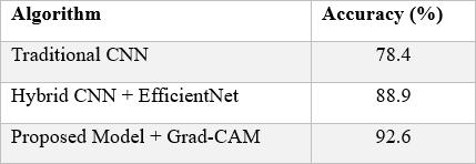
Table 3 presents the quantitative performance metrics of the proposed model compared to baseline approaches. The proposed model with Grad-CAM achieves 92.6% overall accuracy, outperforming standalone CNN (78.4%) and Hybrid CNN + EfficientNet without Grad-CAM (88.9%). Table -3: Performance Metrics of Proposed Model
8.2. Training Graphs
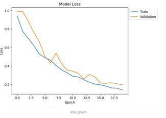
Fig -2: Model Loss Graph The training loss decreases steadilyfromapproximately0.45atepoch0tobelow0.15by epoch30.Thevalidationlosscloselytracksthetrainingloss with minor fluctuations, indicating that the model generalizes well and does not exhibit significant overfitting. The convergence behavior confirms the effectiveness of the Adam optimizer and the data augmentationstrategy.
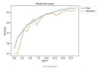
Fig -3: Model Accuracy Graph Training accuracy rises from approximately 60% in early epochs to approximately 97% by epoch 30. Validation accuracy converges to approximately94%,closelyfollowingthetrainingcurve.The small gap between training and validation accuracy confirms adequate generalization. The gradual, smooth increase across 30 epochs demonstrates stable learning without abrupt oscillations, validating thechosen batch size andlearningrateconfiguration.
International Research Journal of Engineering and Technology (IRJET) e-ISSN: 2395-0056
Volume: 13 Issue: 04 | Apr 2026 www.irjet.net p-ISSN: 2395-0072
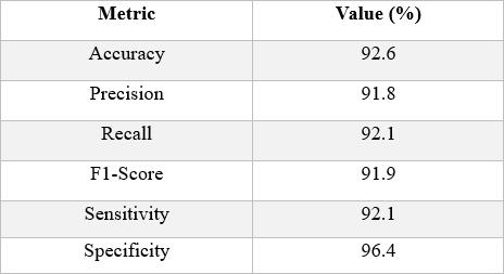
This study presented an interpretable deep learning framework for automated skin disease classification combining a Convolutional Neural Network with EfficientNet to enhance feature representation and prediction reliability. The hybrid architecture,trainedonthe HAM10000dataset(10,015images,9classes,128×128input resolution)over30epochs,achieves approximately 92.6% overall accuracy, 91.8% precision, 92.1% recall, and 91.9% F1-score (macro averages), demonstrating consistent and balancedclassification across all disease categories. The incorporation of Gradientweighted Class Activation Mapping provides visual explanations by highlighting clinically relevant image regions, improving transparency and user trust in automateddiagnosis.
Beyond classification, the system integrates supportive healthcare components including symptom information, medical guidance, treatment cost estimation by hospital type,andlocation-basedhospitalrecommendationviaGoogle Maps. This integrated approach demonstrates the practical potential of combining predictive modeling, explainable AI, andhealthcareassistancewithinaunifiedplatform. Future work may focus on training with larger and more diverse datasets encompassing varied skin tones, imaging conditions, and rare disease categories to enhance generalization. Additional improvements through advanced architectures, hyperparameter optimization, and ensemble strategies are also planned. Deployment on mobile or cloudbased platforms could enable real-time screening, particularly in resource-limited settings. Clinical validation through collaboration with medical professionals and integration with healthcare information systems would furtherstrengthenreliabilityandadoption.
Theauthors wouldliketoexpresstheirsinceregratitudeto their guide, Dr. Prabhakar K, for his valuable guidance and supportthroughoutthis project.Theauthors alsothank the Department of Computer Science and Engineering (AIML), CMR University, Bangalore, for providing the necessary facilitiestocarryoutthisworksuccessfully.
[1] W.Setiawan,"EffectofColorEnhancementonEarly DetectionofSkinCancerusingConvolutionalNeural Network,"2020 IEEE ICIoT, Doha, Qatar, 2020, pp. 100–103, DOI: 10.1109/ICIoT48696.2020.9089631.
[2] J. P. T. Cruz et al., "Skin Cancer Classification Using Ensemble CNN,"2024 IEEE NICE,Baguio, Philippines, 2024, pp.1–6,DOI:10.1109/HNICEM64917.2024.11258740.
International Research Journal of Engineering and Technology (IRJET) e-ISSN: 2395-0056
Volume: 13 Issue: 04 | Apr 2026 www.irjet.net p-ISSN: 2395-0072
[3] G. Ramasamy, N. Varghese, E. Sivalingam and S. Ramachandran, "Dermal: A Deep Learning-Based Mobile Application for Multi-type Skin Cancer Detection," 2025 IC SCC,Kochi,India,2025,pp.1–5,DOI: 10.1109/ICSCC66177.2025.11233655.
[4] N. K. A and M. S A, "Deep Mole Skin Cancer Detection Using CNN Model," 2024 CSI TSS, Bengaluru, India, 2024, pp.1–4,DOI:10.1109/CSITSS64042.2024.10816852.
[5] M. Salomi, G. Daram and S. S. Harshitha, "Early Skin Cancer Detection Using CNN-BCD Rule Based Feature Extraction," 2024 ACETATE, Nellore, India, 2024, pp. 1–5, DOI:10.1109/ic-ETITE58242.2024.10493828.
[6] K. Vayadande et al., "AI Driven Detection of Skin and Oral Cancer: A Survey," 2024 ICS CNA, Then, India, 2024, pp.736–743,DOI:10.1109/ICSCNA63714.2024.10864094.
[7] P. Mahalle et al., "Skin Cancer Detection Using CNN," 2023 CSET, Bangalore, India, 2023, pp. 1–5, DOI: 10.1109/CSET58993.2023.10346792.
[8] A. Barbadekar, V. Ashtekar and A Chaudhari, "Skin Cancer Classification and Detection Using VGG-19 and Descent," 2023 ICC INS, Malayalam, India, 2023, pp. 1–6, DOI:10.1109/ICCINS58907.2023.10449996.
[9] Vishal,S.MehtaandA.Singh,"Multi-ModalSkinCancer DiagnosisUsingCNNandSVM,"2024ICONAT,GOA,India, 2024,pp.1–5,DOI:10.1109/ICONAT61936.2024.10774877.
[10] S. Chowdhury and D. K. Das, "Skin Cancer Detection Using Different Deep Learning Models," 2024 ICUs, Gobichettipalayam, India, 2024, pp. 1086–1091, DOI: 10.1109/ICUIS64676.2024.10866480.
[11] S. Likhitha and R. Baskar, "Skin Cancer Classification using CNN vs SVM," 2022 SMART, Moradabad, India, 2022, pp.1298–1302,DOI: 10.1109/SMART55829.2022.10047280.
[12] V. Aadiwal, B. Sharma and D. P. Yadav, "Revolutionizing Dermatology: Novel CNN Framework for Skin Cancer Detection," 2024 ICON AT, GOA, India, 2024, pp.1–6,DOI:10.1109/ICONAT61936.2024.10774995.
[13] T.S.R.Kiranetal.,"ANovelApproachtoSkinCancer DetectionusingCNN,ResNet,andHybridFeature Extraction,"2024 IC SSAS, Erode, India, 2024, pp. 217–222, DOI: 10.1109/ICSSAS64001.2024.10760465.
[14] K.Kathuria,A.SahooandC.K.Jain,"Priordetectionof Melanoma skin cancer using Deep Learning," 2024 ACE, Ghaziabad, India, 2024, pp. 457–460, DOI: 10.1109/AECE62803.2024.10911059.
[15] S. Kaur, L. Verma and K. K. Kushwaha, "Skin Cancer Detection and Categorization Utilizing CNNs, Residual Network and Binary Classifiers," 2025 FRITO, Noida NCR, India,2025,pp.1–6,DOI: 10.1109/ICRITO66076.2025.11241700.
[16] S. Likhitha and R. Baskar, "Skin Cancer Segmentation Using R-CNN vs Inception V3," 2022 SMART, Moradabad, India,2022,pp.1293–1297,DOI: 10.1109/SMART55829.2022.10047686.
[17] S.S.MandA.J,"SkinCancerDetectionin3DTotalBody Photography using Deep Learning," 2025 ICISS, India, 2025,pp.1530–535,DOI: 10.1109/ICISS63372.2025.11076453.
[18] S.Sharma,S.KaurandN.Kaur,"EnsembleofCNNbased Deep Learning model for Skin Cancer Images," 2024 IEEE INCISION, Chandigarh, India, 2024, pp. 1–6, DOI: 10.1109/INDISCON62179.2024.10744259.
© 2026, IRJET | Impact Factor value: 8.315 | ISO 9001:2008 Certified Journal | Page1020