
International Research Journal of Engineering and Technology (IRJET) e-ISSN: 2395-0056
Volume: 13 Issue: 01 | Jan 2026 www.irjet.net p-ISSN: 2395-0072
International Research Journal of Engineering and Technology (IRJET) e-ISSN: 2395-0056
Volume: 13 Issue: 01 | Jan 2026 www.irjet.net p-ISSN: 2395-0072
Dr. K. L. Senthil kumar1 , Dr. P. D. Gokulan2 , Mr. S. Venkateshwaran3, G. Praveen Kumar4, S. Priyadharshini5, M. Punith Kumar6
1 Principal, Sri Vijay Vidyalaya College of Pharmacy, Dharmapuri, Tamilnadu, India
2 Head of the Department of Pharma ceutical Chemistry, Sri Vijay Vidyalaya College of Pharmacy, Dharmapuri, Tamil nadu, India)
3* Associate professor, Sri Vijay Vidyalaya College of Pharmacy, Dharmapuri, Tamil Nadu (India)
*4, 5, 6 B. Pharm Students, Sri Vijay Vidyalaya College of Pharmacy, Dharmapuri, Tamil Nadu (India)
Abstract- Aim: The aim of this study was to analyze the different bioactive compounds found in the leaves of Ruellia tuberosa collected from the Dharmapuri district. The study focused on identifying the bioactive constituents present in the chloroform extract and ethanol extracts of the Ruellia tuberosa leaves using the GC-MS technique along with phytochemical screening.
Materials and methods: The collected leaves of Ruellia tuberosa were thoroughly washed, air-dried, and ground into a coarse powder. The leaf extract was prepared by using the Soxhlet extraction method. The selection of solvents is based on their polarity.
Result: The phytochemical screening and GC-MS analysis confirmed the presence of various bioactive compounds in Ruellia tuberosa leaves. The study revealed several active chemical constituents, such as alkaloid, glycosides, flavonoids, proteins, tannins, terpenoids, etc.
Conclusion: The present study shows that Ruellia tuberosa leaves contain a rich source of secondary metabolites.
Key words: Ruellia tuberosa, extraction, pharma cogenetic studies, phytochemical analysis, and GC-MS technique.
Introduction
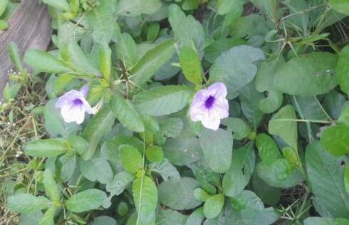
Ruellia tuberosa is a medicinal plant that has been traditionally used since ancient times. It is an erect, perennial herb belongingtothefamilyAcantharean.Theplantisknownbyvarious names,suchasMinnieroot,feverroot,snapdragon root, andsheeppotato.Itiscommonlyreferredtoasthe“crackerplant.”Theplantgrowsneartheroadsideingrasslands,cultivated fields, and even in a dry(xerophilic)area It is a short-lived perennial plant with striking funnel-shaped violet bracteates flowersondichotomouscymes.Thefruitisasub-cylindricalpuberulentcapsulehavingmoreorless 20seedsperlocule.The plant also has thick, fusiform tuberous roots that grow in clusters. Ruellia tuberosa L. is a tropical plant that is commonly found in many parts of Asia, including Indonesia, Malaysia, and India. The plant flowers and produces fruits from June to February. It can be easily recognized by its attractive, trumpet-shaped, violet-colored flowers with a five-lobed corolla. The plantis mainlygrowninmoistand shady environments.Theplant attracts manypollinators,especiallybutterfliesandother insects.Theflowersappearlightbluish-purpleduetothepresenceofanthocyanin, a flavonoidpigmentthatisalsousedasa natural dye in industries. The plant contains several important phytochemical constituents, such as alkaloids, glycosides, flavonoids, tannins, proteins, terpenoids, diterpenes, sterols, etc. This study aims to investigate the extraction of bioactive compoundsfromRuelliatuberosausingdifferentsolventsandevaluatethephytochemicalcontentusingGaschromatographymassspectrometryanalysis
International Research Journal of Engineering and Technology (IRJET) e-ISSN: 2395-0056
Volume: 13 Issue: 01 | Jan 2026 www.irjet.net p-ISSN: 2395-0072
Traditionally, the plant is used for anti-diabetic, anti-inflammatory, ant nociceptive, antipyretic, analgesic, antioxidant, antihypertensive,insecticidal,anticancer,andantidotaltoxicagents.
TheleafextractofRuelliatuberosaL.hasanti-diuretic,anti-cancer,andanti-oxidantactivity.
Ruelliatuberosarootextractisusedforanti-diureticactivity,reducingbloodsugarlevelsandkidneydisease.
Itisusedinancientdaysformanytraditionalpurposes.
Vernacular name:
Assamese:Chapatti
Bangladesh:Potpati
Marathi :Ruwel
SriLankan:Heenamukkara
Tamil:Kiranthinayakam,Pattaskai
Telugu:Jurbulagadda
Taxonomic Hierarchy:
Kingdom:Plantae
Subkingdom:Viridiplantae
Infrakingdom: Streptophyta
Superdivision:Embryophyta
Class : Magnoliopsida
Superorder:Asternae
Order : Lamiales
Family : Acanthaceae
Genus : RuelliaL.
Species:RuelliatuberosaL.
Pharmacognostical Studies:
Methodology
1. Macro soppy(SOP No. PCOG-004-SOP): The external feature of the test sample was documented using a Nikon D-5600 digitalcamera.
2. Micro scopy(SOP No. PCOG-005-SOP): Thesample was preserved in fixative FAA for more than 48 h. The preserved specimens were cut into thin transverse sections using a sharp blade, and the sections were stained with 0.8% safranin and 0.5% Astra blue. Transverse sections were photographed using an Axiolab5 trinocular microscope attached to a Zeiss Axiocam208colordigitalcameraunderbrightfieldlight.Magnificationswereindicatedbyascalebar.
© 2026, IRJET | Impact Factor value: 8.315 | ISO 9001:2008 Certified Journal | Page419
Result:
International Research Journal of Engineering and Technology (IRJET) e-ISSN: 2395-0056
Volume: 13 Issue: 01 | Jan 2026 www.irjet.net p-ISSN: 2395-0072
Microscopy:
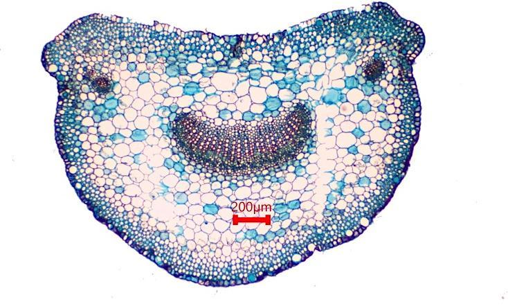
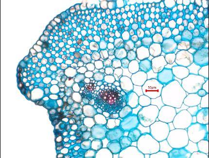
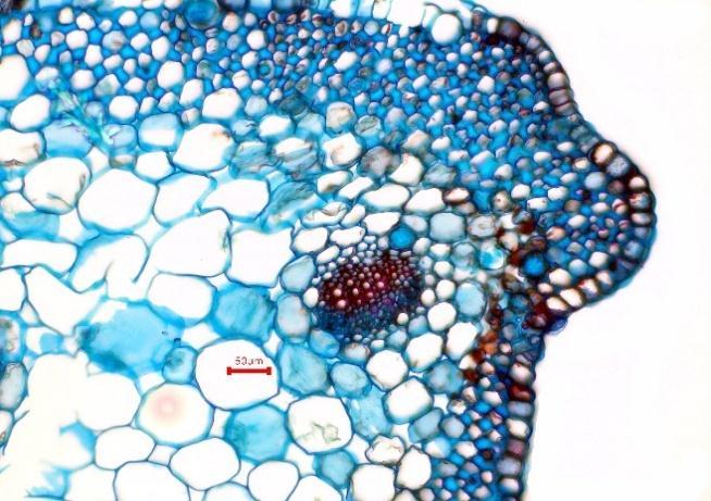
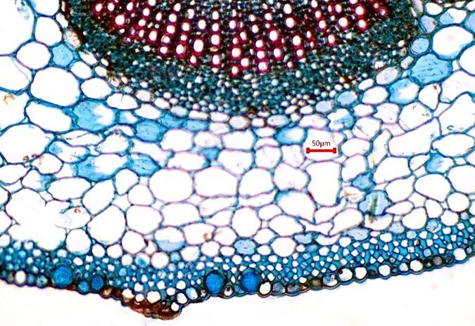
The transverse section of the petiole is urn-shaped. The lower surface is convex, while the upper surface is slightly concave and bears lateral wings. The outermost layer is a single-layered epidermis covered with a cuticle. Beneath it, 4-5 layers of collenchymatous tissue are present, which continue with the parenchymatous ground tissue. At the center, an arc-shaped vascularbundleisseen.Thevascularbundlesareconjoint,collateral,closed,andcomposedofnormalvasculartissues.Xylem isorientedtowardstheuppersideand Phloemtowardsthelowerside.Twotracebundlescanbeobservedoneachuppersidebelowthewingregion.Cystolithsare foundinepidermalcells,andstarchgrainsaredistributedintheinnerparenchymacells.

International Research Journal of Engineering and Technology (IRJET) e-ISSN: 2395-0056
Volume: 13 Issue: 01 | Jan 2026 www.irjet.net p-ISSN: 2395-0072
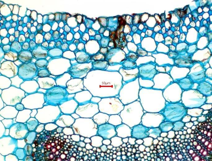
Upper portion enlarged
Figure no: 02 Micro scopy of Ruellia tuberosa petiole
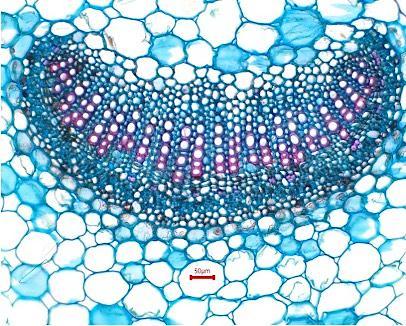
Enlarged view of vascular region
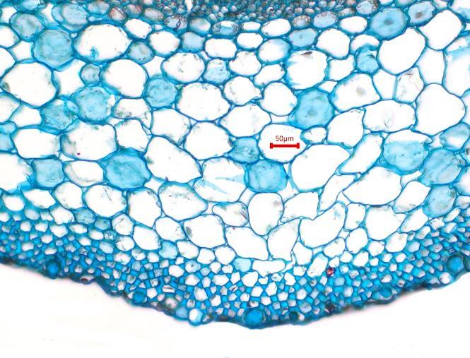
Enlarged view of lower portion
Col -collenchyma; Cys -cystolith; E -epidermis; Pa -parenchyma; Ph -phloem; SG -starchgrain; TB -tracebundle; V -vessel; VB -vascularbun
Leaf
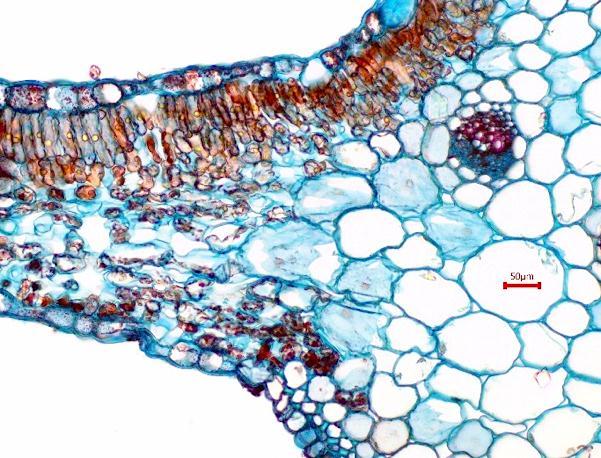
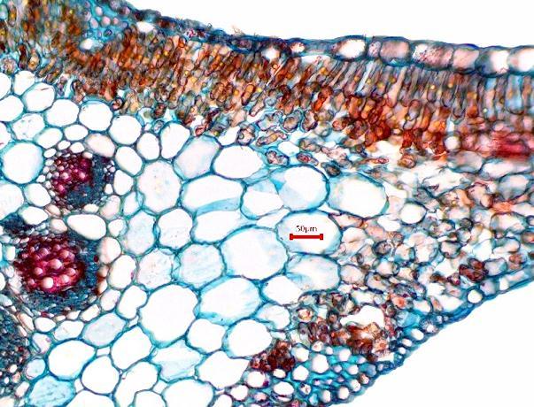
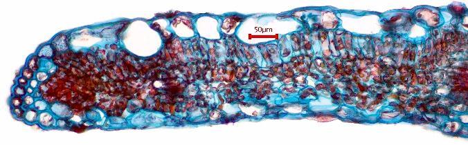
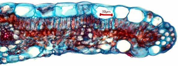
TheTSoftheleafisdorsiventralwithaflatlowersurfaceandaslightlyelevateduppersurfaceandshowsribbon-likelateral laminarextensions.
Midrib
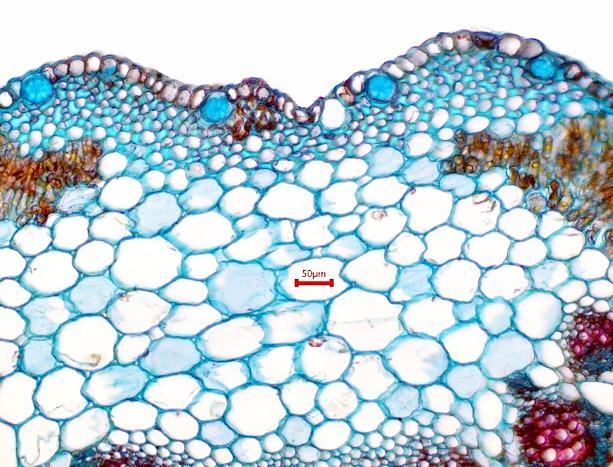
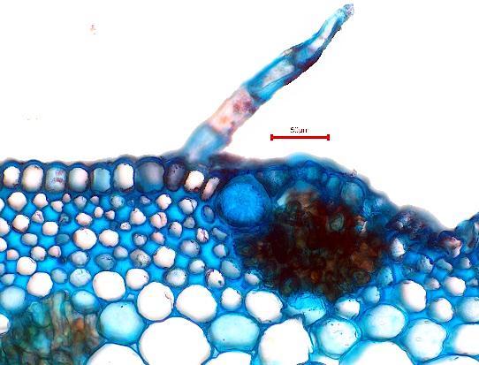
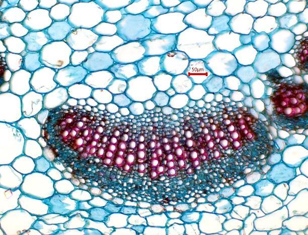
TS of the midrib shows an outer single-layered upper and lower epidermis covered by cuticle and bears several covering trichomes; 3 to 4 layers of collenchymatous hypodermal layer are present, followed by parenchymatous ground tissue embedded with a crescent-shaped conjoint, collateral, and closed vascular bundle at the center; xylem is placed towards the uppersideandphloemtowardsthelowerside;xylemisformedofvessels,tracheids,fibers,andparenchyma,whereasphloem is made up of sieve elements and phloem parenchyma; two or three trace bundles can be seen on the upper side facing towardsthelamina.
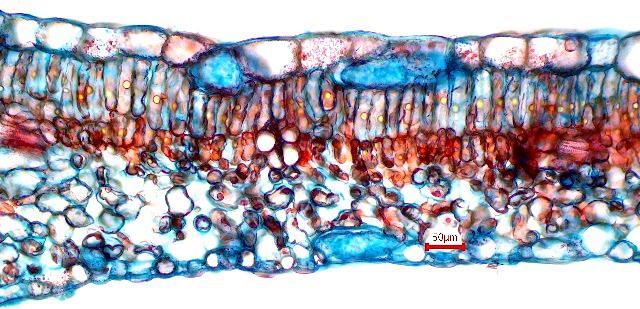
Lamina
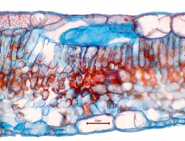
The transverse section of the lamina shows that both the upper and lower epidermis are single-layered and covered with a cuticle. These layers also bear covering trichomes. The mesophyll tissue is well differentiated into an upper, tightly packed single layer of palisade cells,followed by four to five layers of loosely arrangedspongy parenchyma cells.Veins run through themesophylltissue.Cystolithsarefoundinspecializedepidermalcellsonbothsurfacesofthelamina,whileoilglobulesare scatteredthroughoutthelamina.Afewcollenchymacellsarealsopresentneartheleafmargin.
© 2026, IRJET | Impact Factor value: 8.315 | ISO 9001:2008 Certified Journal | Page421
International Research Journal of Engineering and Technology (IRJET) e-ISSN: 2395-0056
Volume: 13 Issue: 01 | Jan 2026 www.irjet.net p-ISSN: 2395-0072

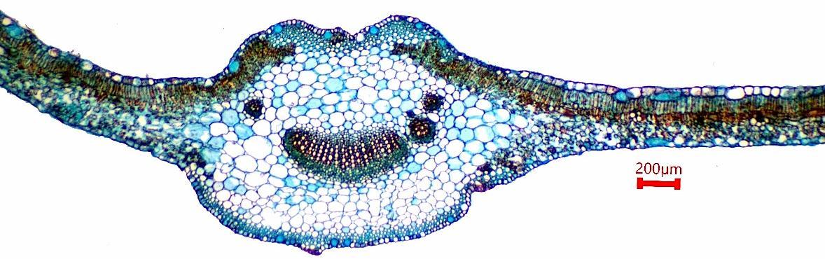






no: 03 Microscopy of Ruellia tuberosa leaf passing through midrib
TS of leaf
International Research Journal of Engineering and Technology (IRJET) e-ISSN: 2395-0056
Volume: 13 Issue: 01 | Jan 2026 www.irjet.net p-ISSN: 2395-0072
International Research Journal of Engineering and Technology (IRJET) e-ISSN: 2395-0056
Volume: 13 Issue: 01 | Jan 2026 www.irjet.net p-ISSN: 2395-0072
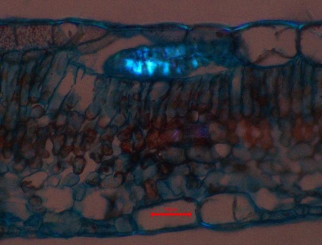
withcystolith Underapolarizedfield
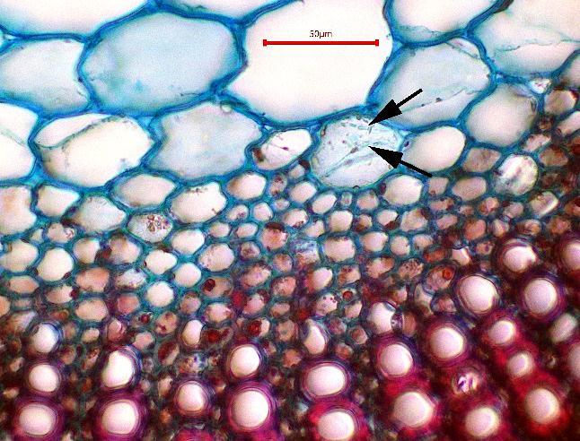
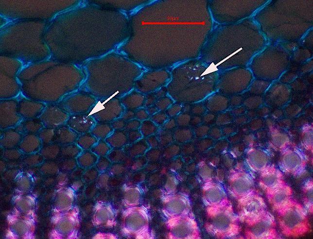
Col - collenchyma; Cu - cuticle; Cys - cystolith; LE - lower epidermis; MCr- micro sphenoidal crystals; OG - oil globule; Paparenchyma; Pal - palisade; Ph - phloem’s - spongy parenchyma - trichome; TB - trace bundle; UE - upper epidermis; Vvessel; VB -vascularbundle; Ve –vein
PHYTOCHEMICAL STUDIES:
The obtained extracts were subjected to phytochemical analysis. This qualitative study was carried out to identify the presenceofsecondarymetabolitessuchasalkaloids,glycosides,flavonoids,terpenoids,diterpenes,proteins,tannins, sterols, etc.
1. Test for Alkaloids
Dragendorff’s test
To 1 ml of extract, add 1 ml of Dragendorff’s reagent (potassium bismuth iodide solution). An orange-red precipitate indicatesthepresenceofalkaloids.
© 2026, IRJET | Impact Factor value: 8.315 | ISO 9001:2008 Certified Journal | Page424
International Research Journal of Engineering and Technology (IRJET) e-ISSN: 2395-0056
Volume: 13 Issue: 01 | Jan 2026 www.irjet.net p-ISSN: 2395-0072
Mayers Test
To 1 ml of extract, add 1 ml of Mayer’s reagent (potassium mercuric iodide solution). Whitish yellow or cream-colored precipitateindicatesthepresenceofalkaloids.
2. Test for Glycosides
Test solution: Itwaspreparedbydissolvingthesampleinalcohol.
Keller-Killiani Test
To a test solution, a few dropsof ferricchloridesolutionand concentrated sulphuricacid wereadded; the formation of two layersoccurred,alowerlayerofreddish-browncolorandanupperlayerofbluish-greencolorsimultaneously.
3. Test for Triterpenoids
Test solution: Itwaspreparedbydissolvingtheextractsinchloroform.
Salkowski test
Afewdropsofconcentratedsulfuricacidwereaddedtothesolutionandallowedtostandforsometime.Thelackofformation ofyellowcolorinthelowerlayerindicatestheabsenceoftriterpenoids.
4. Test for Carbohydrate
The Molisch testto 2-3ml of the test solution, added few drop Molish reagent solution and was shaken. Concentrated sulphuricacidwasaddedfromthesideofthetesttube.Avioletringwasformedatthejunctionoftwolipids.
5. Test for Protein
Test solution: Itwaspreparedbydissolvingextractinwaterandmakingaqueousextract.
Biuret test
To about 3 ml of the extract,40% sodium hydroxide solution and a few drops of 1% copper solution were added.White producesbluecolor.
6. Test for Anthraquinone
Borntrager’s Test:Add10mlof10%ammoniasolutionwithafewmlof3mlofAq.Theextractisshakenwithmlofbenzene and filtered (shaken vigorously 30 mins) appears as a pink, violet, or red-colored solution, it shows the presence of anthraquinones.
7. Test for Flavonoids
Test solution:A small amount of extract and 2M HCL was added and heated for about 30-40 mins. The extract was cooled downandagainextractedwithethylacetate,whichwasfurtherconcentratedtodrynessandreadytobeusedasatestsample.
Lead acetate test: Toasmallquantityofextract,leadacetatewasadded.Formationofyellow-colored precipitateshowsthe presenceofflavonoids
8. Test for Quinone
Conc. HCl test
Plantextractwithconc.HClappearsgreenincolor;itshowsthepresenceofquinones.
International Research Journal of Engineering and Technology (IRJET) e-ISSN: 2395-0056
Volume: 13 Issue: 01 | Jan 2026 www.irjet.net p-ISSN: 2395-0072
9. Test for Tannins
Test solution: Thetestsolutionwaspreparedbydissolvingtheextractinwaterandalcohol.
Ferric chloride solution test
To 1 ml of the extract, ferric chloride was added. The formation of a dark blur or greenish-blackcolor shown in the presenceindicatesthetannins.
10. Test for Cholesterol
Add2mlofchloroformwith10dropsofacetic anhydrideandadd2-3dropsofconc.H₂SO₄. Theappearanceof aredrosecolorshowstheabsenceofcholesterol.
11. Test for Diterpenes
Plant extract is dissolved in distilled water, and 3-4 drops of copper acetate solution are added. It appears emerald green incolor;itshowsthepresenceofditerpenes.
12. Test for Terpenoids
Add2mlofchloroformwith5mlofplantextractandadd3mlofconc.H₂SO₄(boiledinawaterbath).Donotappear inagrey-coloredsolutionintheabsenceofterpenoids.
13. Test for Saponins
Test solution: Itwaspreparedbydissolvingextractinwaterandmakingaqueousextract.
Foam Test
The drug extract was vigorously shaken with water Persistent foam formation does not indicate the absence of saponins.
14. Test for Fat
Solubility test
Fatsareinsolubleinwaterandethanol.Butsolubleinchloroform,benzene,andether.ItshowsthepresenceofFat.
15. Test for sterols
Liberman-Burchard Test
1gmofthetestsubstancewasdissolvedinafew dropsofchloroform,3mlofaceticanhydrideand3mlofglacialaceticacid wereadded,anditwaswarmedandcooledunderthetap,anddropsofconc.H₂SO₄wereaddedalongthesideofthetesttube. Thenon-appearanceofabluish-greencolorshowstheabsenceofsterols.
Table 1: Phytochemical analysis of Ruellia tuberosa
International Research Journal of Engineering and
Volume: 13 Issue: 01 | Jan 2026 www.irjet.net
PHYTOCHEMICAL SCREENING: (+) Presence (-)/Absence
TheextractofRuelliatuberosaL.wassubjectedtophytochemicalanalysis.Theselectionofsolventisbasedontheirpolarity. The solvents used for the extraction were chloroform, ethanol, and water. The phytochemical screening of secondary metabolites such as alkaloids, glycosides, flavonoids, triterpenoids, diterpenes, carbohydrates,proteins, tannins, cholesterol, andsterolswasdetermined.
GAS CHROMATOGRAPHY AND MASS SPECTROPHOTOMETRY
TheRuelliatuberosaleavescontainarichsourceofsecondarymetabolites.Theextractionandcharacterizationofbiologically activecomponentsfrommedicinalplantshaveresultedinthetreatmentforvariousdiseases.Thebioactivecompoundpresent intheRuelliatuberosawasseparatedbyusinggaschromatographyandmassspectrometry.
GC-MS analysis of ruellia tuberosa
PROCEDURE
Sample preparation:
Weigh1gofhomogenizedsample(chloroformandethanol)intoa15mlplastictube.Add1mlofethylacetateandvortex for5minutes.
Thentransfertoa1mlvialforinjectionofµlofthesampleintoGC-MS
INSTRUMENT CONDITIONS:
C–MS analysis was performed using a Shimadzu QP2020 gas chromatograph–mass spectrometer equipped with a split/splitlessinjectorandanelectronimpactionizationsourceoperatinginEI⁺mode.SeparationwasachievedonanHP-5MS capillary column (30 m × 0.32 mm, film thickness 0.25 µm). An injection volume of 1.0 µL was employed, with the injector temperaturemaintainedat285°C.
Helium (99.999% purity) wasused asthe carrier gasat a constant flow rate of1.0 mL/min. The oven temperatureprogram was set with an initial temperature of 50°C, which was gradually increased to 290°C and held for 10 minutes to ensure complete elution of all components. The split valve was opened 3 minutes after injection to purge the injector. All injections werecarriedoutusinga10µLmicrosyringe.
International Research Journal of Engineering and Technology (IRJET) e-ISSN: 2395-0056
Volume: 13 Issue: 01 | Jan 2026 www.irjet.net
International Research Journal of Engineering and Technology (IRJET) e-ISSN: 2395-0056
Volume: 13 Issue: 01 | Jan 2026 www.irjet.net p-ISSN: 2395-0072
Table 2: Phytochemical screening by using chloroform extract
Conclusion:
The extraction of Ruellia tuberosa using solvents of varying polarity resulted in different percentage yields. Phytochemical screening was carried out to identify the presence of bioactive constituents in each extract. The detection of these phytochemicalsthroughpreliminaryanalysissubstantiatesthemedicinalsignificanceoftheplant.
International Research Journal of Engineering and Technology (IRJET) e-ISSN: 2395-0056
Volume: 13 Issue: 01 | Jan 2026 www.irjet.net p-ISSN: 2395-0072
The plant extract was subjected to gas chromatography–mass spectrometry (GC–MS) analysis under standardized experimentalconditions.Theseparationofcompoundswasachievedbasedontheirretentiontimeandrelativepeakarea,and detection was performed using a suitable detector such as a flame ionization detector (FID). The resulting chromatogram exhibited multiple peaks corresponding to diverse bioactive constituents. GC analysis further confirmed the chemical heterogeneity of the extract by effectively resolving volatile compounds. The identified constituents may contribute to the therapeuticpotentialof Ruellia tuberosa
REFERENCE
[1]Juswardi, Nur Salamah, Nina Tanzerina, Kamila Alawiyah, Syafrina Lamin, and Mustafa Kamal.Metabolite profiling of Minnieroot(RuelliatuberosaL.)leavesusinggaschromatography-massspectrometry(GC-MS),2025
[2]SandhyaSharma,JelineRaniJ,SohanLal,HarpalSinghandSugimaniMarndiRuelliatuberosaL.:amedicinalplantofIndia, 2024
[3] Gokulakrishnan S, Dharanya M, and Dr. K. B. Ilango. STANDARDIZATION AND SOLVENT COMPARATIVE STUDY OF RUELLIATUBEROSAL.,2023
[4] Thakur Adhika Govindsinh, M. H. Ghante, N. B. Ghiware, S. K. Sarje. Pharmacological evaluation of Ruellia tuberosa stem barkonantianxietyactivity,2020
[5] M.N.L.C. Harika and P. Radhika, Department of Biochemistry.Phytochemical Analysis of Ruellia tuberosaTuber Ethanolic ExtractUsingUV-VIS,FTIR,andGC-MSTechniques,2019
[6] Ghaidaa Jihadi Md., Aseel Mohammad Omran, and HaiderM.H., Antibacterial and Phytochemical Analysis of Piper nigrum usingGasChromatography-MassspectrumandFouriertransforminfrareddchromatography,2016
[7]SrinivasanK.,SivasubramanianS.,KumaravelS.,PhytochemicalprofilingandGCMSstudyAdhathodavasicaleaves,2014
[8]SenthilkumarP,SambathR,andVasantharaj.AntimicrobialPotentialandScreeningofAntimicrobialcompoundsofRuellia tuberoseUsingGC-MS,2013
[9]Arirudran,A.Saraswathy,andVijayalakshmiKrishnamurthy.AntimicrobialactivityofRuelliatuberosaL.2011