
International Research Journal of Engineering and Technology (IRJET) e-ISSN: 2395-0056
Volume: 13 Issue: 04 | Apr 2026 www.irjet.net p-ISSN: 2395-0072

International Research Journal of Engineering and Technology (IRJET) e-ISSN: 2395-0056
Volume: 13 Issue: 04 | Apr 2026 www.irjet.net p-ISSN: 2395-0072
Dr.G. Nagarjuna Reddy 1 , P. Nandini2 ,K. Muralidhar3 , K. Nageswara Rao 4
1 ASSOCIATE PROFESSOR, 2,3,4 UG STUDENTS
Department of Electronics and Communication Engineering, N.B.K.R. INSTITUTE OF SCIENCE AND TECHNOLOGY
ABSTRACT
Theearlyandaccuratedetectionofpulmonarydiseases is critical for effective treatment and improved patient outcomes. This project presents a deep learning-based system for multi-disease classification using chest radiographs.TheproposedmodelistrainedontheChestX6 dataset, which contains six categories of chest X-ray images: Normal, Pneumonia-Bacterial, Pneumonia- Viral, COVID-19, Tuberculosis, and Emphysema. To achieve robust performance, multiple convolutional neural network (CNN) architectures such as CNN, MobileNet, VGG16 and DenseNet are employed and compared. The dataset is preprocessed to enhance image quality and ensure balanced class representation. The system is implemented using Python in Google Colab with T4 GPU support. Experimental results demonstrate that the proposed deep learning models can effectively distinguish between multiple lung diseases with high accuracy, supportingradiologistsindiagnosticdecision-making.This workhighlightsthepotentialofdeeplearningandmedical imaging integration to advance automated healthcare diagnostics
KEYWORDS
Deep Learning, Chest X-ray, Lung Disease Classification, Multi-class Classification, Convolutional Neural Networks (CNN), Transfer Learning, MobileNet, VGG16, DenseNet, Medical Image Analysis, COVID-19, Pneumonia, Tuberculosis, Emphysema, Image Preprocessing
Respiratory diseases represent a significant global healthchallenge,withmillionsofcasesreportedannually. Conditions such as pneumonia, tuberculosis, COVID-19, andemphysemaseverelyaffectlungfunctionandcanlead to life-threatening complications if not diagnosed at an earlystage.Earlydetectionandaccurateclassificationof thesediseasesarecrucialfortimelymedicalintervention and improved patient outcomes. Medical imaging techniques, particularly chest X-ray imaging, play a vital role in the diagnosis and treatment planning of pulmonary diseases due to their ability to provide essentialanatomicalinformation[1],[2].
Accurate classification of lung diseases is critical in clinical practice to determine appropriate treatment strategies.However,thistaskremainschallengingdueto the similarity in radiographic patterns among different diseases. For instance, viral and bacterial pneumonia oftenexhibitoverlappingvisualcharacteristics,makingit difficultforradiologiststodistinguishbetweenthem[3]. Whilemedicalexpertsplayacrucialroleindiagnosis,the availability of automated supportive tools for rapid and reliable assessment can significantly enhance diagnostic efficiency [4]. Currently, computer-aided diagnostic systems based on chest X-ray imaging offer a promising solution for lung disease detection, with X-rays being preferred due to their affordability, speed, and accessibility[5].
Artificial intelligence (AI), particularly deep learning models,hasshownsignificantpotentialinimprovingthe accuracy and efficiency of medical image analysis. Machine learning and deep learning techniques are increasingly being applied to lung disease detection and classification tasks. However, challenges such as limited datasetsize,classimbalance,andloweraccuracyinsome existing models remain critical issues. Deep Convolutional Neural Networks (CNNs) have demonstrated strong performance in detecting and classifying lung diseases from chest X-ray images, although they often require substantial computational resourcesandtrainingtime.
To address these challenges, transfer learning using pre-trained deep learning models has emerged as an effectiveapproach.Pre-trainedarchitecturessuchasVGG, DenseNet,MobileNet, and EfficientNet have been widely adopted due to their ability to improve accuracy while reducing training time. These models have proven successfulnotonlyinmedicalimagingbutalsoinvarious domains such as image recognition, speech processing, andpatternanalysis.
This study focuses on the implementation and comparative analysis of multiple deep learning models, including a custom CNN, VGG16, and DenseNet121, for multi-class classification of lung diseases using chest Xray images. The proposed system aims to classify six categories: Normal, Bacterial Pneumonia, Viral Pneumonia,COVID-19,Tuberculosis,andEmphysema.A transfer learning approach is utilized to enhance model
International Research Journal of Engineering and Technology (IRJET) e-ISSN: 2395-0056
Volume: 13 Issue: 04 | Apr 2026 www.irjet.net p-ISSN: 2395-0072
performance while minimizing computational complexity. Additionally, data preprocessing and augmentationtechniquesareappliedtoimprovedataset qualityandaddressclassimbalanceissues.
Theobjectiveofthisworkistodevelopanefficientand accurate computer-aided diagnostic system that can assisthealthcareprofessionalsintheearlydetectionand classificationoflungdiseases,therebyimprovingclinical decision-makingandpatientoutcomes.
A multi-class classification approach was proposed by Nahiduzzaman et al. [6] for identifying multiple lung diseasesusingchestX-rayimages.Themethodcombined convolutionalneuralnetworkswithanextremelearning machinetoimproveclassificationperformance,achieving highaccuracyacrossmultiplediseasecategories.
Gunraj et al. [7] developed COVIDNet-CT, a deep convolutional neural network tailored for detecting COVID-19 from CT images, demonstrating reliable performance in medical diagnosis. Cohen et al. [8] introduced a COVID-19 image dataset that has been widely used for training and evaluating deep learning models.
Gozes et al. [9] proposed an artificial intelligence-based system for automated detection and monitoring of COVID-19 using CT image analysis, highlighting the effectiveness of deep learning in real-time clinical applications.Narinetal.[10]applieddeep convolutional neural networks for automatic detection of COVID-19 using chest X-ray images, achieving high classification accuracy.
Zhangetal.[11]introducedananomalydetection-based deep learning approach for COVID-19 screening, improving efficiency in cases with limited labeled data. Panwaretal.[12]developednCOVnet,afastandefficient modelforCOVID-19detectionfromX-rayimages.
Minaee et al. [13] proposed Deep-COVID, a transfer learning-based framework that improved classification performance by leveraging pretrained models. Rajaraman and Antani [14] focused on tuberculosis detection using deep learning ensembles, enhancing robustnessandaccuracy.
Caliman Sturdza et al. [15] presented a comprehensive review of deep learning techniques for COVID-19 detection, discussing existing challenges and future directions.Foundationaldeeplearningarchitecturessuch asResNet[16],VGG[17],DenseNet[18],andAlexNet[19] havesignificantlycontributedtoadvancementsinimage classification and are widely used in medical imaging applications.
Rajpurkaretal.[20]introducedCheXNet,adeeplearning model that achieved radiologist-level performance in
pneumoniadetection,demonstratingthepotentialofAIin clinicaldiagnosis.
Accordingtotheliteraturereview,severalstudieshave already been conducted; however, the detection of lung diseasesremainsahighlychallengingissue.
The methodologyofthe proposedwork focusesonthe automatedclassificationoflungdiseasesfromchestX-ray images using deep learning techniques. The system employs Convolutional Neural Networks (CNN), VGG16, and DenseNet121 models to perform both binary and multi-class classification. The overall methodology consists of data collection, preprocessing, feature extraction, classification, and performance evaluation.These approaches are widely supported by previous research in medical image analysis and deep learningapplications.
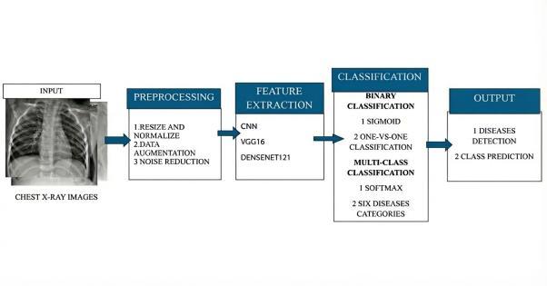
Fig. 1 presents the overall workflow of the proposed methodology for lung disease classification using deep learning techniques. Initially, chest X-ray images are collectedandpreprocessedtoenhanceimagequalityand ensure consistency in input size. The preprocessed imagesarethenfedintodeeplearningmodelsforfeature extraction and classification. Based on the extracted features, the system predicts the corresponding lung diseasecategory.
A.Data Collection
Chest X-ray images were collected from publicly available medical imaging datasets. The dataset comprisessixlungdiseaseclasses,includingbothnormal and abnormal pulmonary conditions. All images were annotated according to their corresponding disease labels.
B.Data Preprocessing
Toensureuniformityandimprovelearningefficiency, allX-rayimageswereresizedto224×224pixels.Image normalizationwasappliedtoscalepixelvalues,anddata augmentation techniques such as rotation, horizontal flipping, and zooming were employed to reduce overfittingandenhancemodelgeneralization.
International Research Journal of Engineering and Technology (IRJET) e-ISSN: 2395-0056
Volume: 13 Issue: 04 | Apr 2026 www.irjet.net p-ISSN: 2395-0072
C.Model Architecture
Deep features were extracted using three different architectures:
● CNN: A custom CNN model was designed to automatically learn hierarchical features from chestX-rayimages
● VGG16: A pretrained VGG16 model was finetunedusingtransferlearningtocapturedetailed lungdiseasepatterns.
● DenseNet121: DenseNet121 was utilized for its dense connectivity, enabling effective feature reuseandimprovedgradientflow.
D.Binary Classification Approach
Binary classification was performed using a one-vsreststrategyforeachlungdiseaseclass.Inthisapproach, one disease category was treated as the positive class, whiletheremainingcategorieswereconsiderednegative. A sigmoid activation function was used in the output layer, and binary cross-entropy loss was employed to optimizetheclassificationperformance.
E.Multi-Class Classification Approach
For multi-class classification, the extracted deep featureswerepassedtoaSoftMaxclassifiertocategorize chestX-rayimagesintooneofthesixlungdiseaseclasses simultaneously.Categoricalcross-entropylosswasused to train the models, enabling a unified framework for comprehensivelungdiseasediagnosis.
F.Performance Evaluation
The performance of CNN, VGG16, and DenseNet121 modelswasevaluatedusingstandardevaluationmetrics, including accuracy, precision, recall, F1-score, and confusion matrix analysis. These metrics provide a detailed assessment of the effectiveness of both binary andmulti-classclassificationapproaches.
ADVANTAGES & APPLICATIONS
ADVANTAGES
● EarlyDiseaseDetection
● DualClassificationCapability
● HighClassificationAccuracy
● ReducedManualEffort
● EfficientUseofMedicalData
● ScalableFramework
● Cost-EffectiveSolution
APPLICATIONS
● HospitalandDiagnosticCenters
● COVID-19andPneumoniaScreening
● TelemedicinePlatforms
● MedicalResearch
● EducationalandTrainingTools
● PublicHealthMonitoring
● ClinicalDecisionSupportSystems
Themodelsweretrainedformultipleepochsusingthe prepared dataset of 15,000 chest X-ray images. After training,evaluationwasperformedonthetestdatasetto measurerealperformance.
Amongthethreemodels,DenseNet121producedthebest resultswithanaccuracyof90%.Thevalidationaccuracy improved steadily during training and became stable towards the final epochs. This indicates that the model was able to learn meaningful features from the dataset withoutsignificantoverfitting.
TheVGG16modelachieved82%accuracy.Itperformed consistently but showed slightly higher validation loss compared to DenseNet121. Some fluctuations were noticedduringearlyepochs,but performance stabilized aftertuningthefinallayers.
ThecustomCNNmodelachieved81%accuracy.Whileit was able to classify most images correctly, it showed difficulty in distinguishing between classes with similar radiographic patterns, especially bacterial and viral pneumonia.
During multi-class classification, most predictions were correct across all six categories. The confusion matrix showed that misclassification mainly occurred between pneumonia-related classes, which share similar visual features.
In binary classification experiments, the performance slightly improved because each model focused on identifying one disease at a time. DenseNet121 again showed more reliable and balanced predictions comparedtotheothermodels.
Overall, the results indicate that deeper pretrained models perform better for medical image classification compared to a basic CNN trained from scratch. Similar performance trends have been observed in previous studies on deep learning-based medical image classification
International Research Journal of Engineering and Technology (IRJET) e-ISSN: 2395-0056
Volume: 13 Issue: 04 | Apr 2026 www.irjet.net p-ISSN: 2395-0072
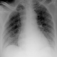
Fig.2 showstheinputimageusedintheproposedsystem. The chest X-ray image is provided as the initial input to themodel.Itisthenpassedthroughpreprocessingsteps suchasresizingandnormalizationbeforeclassification.
TRAINING AND VALIDATION LOSS
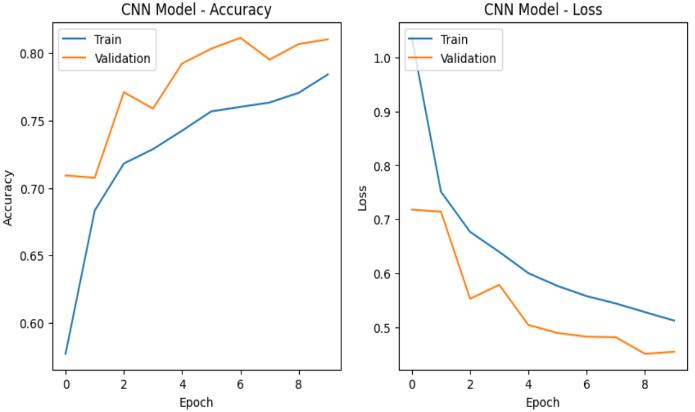
Fig.3 showsthetrainingandvalidationaccuracyandloss curves of the CNN model. The increasing training accuracy and decreasing loss indicate effective learning, while the validation performance reflects the model’s abilitytogeneralizetounseenlungdiseaseimages.
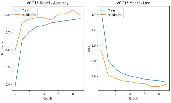
Fig.4 showsthetrainingandvalidationaccuracyandloss curvesoftheVGG16model.Theincreasingaccuracyand decreasing loss indicate effective learning, while the validation results demonstrate good generalization performanceonlungdiseaseimages.
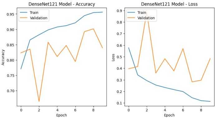
Fig.5 showsthetrainingandvalidationaccuracyandloss curves of the DenseNet121 model. The increasing accuracy and decreasing loss indicate effective learning, while the validation results reflectstrong generalization performanceforlungdiseaseclassification.
Table 1 presents the comparative performance of CNN, VGG16, and DenseNet121 using standard evaluation metrics, including accuracy, precision, recall, and F1score. The results demonstrate that DenseNet121 achieves superior performance in lung disease classification.
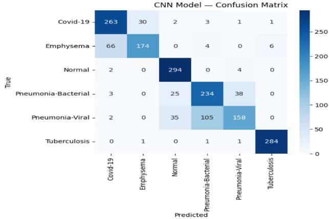
Fig. 6 shows the confusion matrix of the CNN model for lung disease classification. The matrix compares actual classlabelswithpredictedlabels.Thediagonalelements representcorrectlyclassifiedsamples,whileoff-diagonal elements indicate misclassifications. This visualization provides insight into the classification performance acrossdifferentlungdiseasecategories.
International Research Journal of Engineering and Technology (IRJET) e-ISSN: 2395-0056
Volume: 13 Issue: 04 | Apr 2026 www.irjet.net p-ISSN: 2395-0072
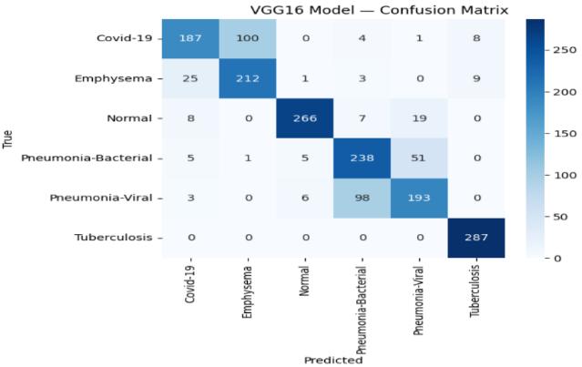
Fig.7illustratestheconfusionmatrixoftheVGG16model forlungdiseaseclassification.Thematrixcomparestrue andpredictedclasslabels,where higherdiagonal values indicate accurate predictions and off-diagonal values representmisclassifications.
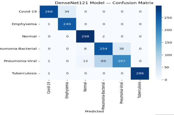
Fig. 8 presentstheconfusionmatrixoftheDenseNet121 model for lung disease classification. The matrix comparesactualandpredictedclasslabels,wherestrong diagonal values indicate accurate predictions. This suggests that DenseNet121 achieves superior classificationperformancecomparedtotheothermodels.
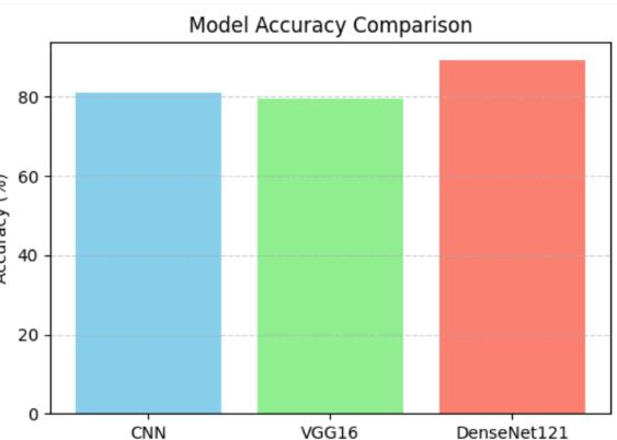
Fig. 9 shows the accuracy comparison of different deep learning models for lung disease classification. The results indicate that DenseNet121 achieves the highest accuracy, demonstrating superior performance comparedtoCNNandVGG16.
BINARY CLASSIFICATION
(i) BINARY CLASSIFICATION PERFORMANCE OF CNN MODEL
Table 2 presentsthebinaryclassificationperformanceof the CNN model using the one-vs-rest strategy across six lung disease classes. Each class was considered independently as a binary problem. The results are evaluated using standard metrics, including accuracy, precision,recall,andF1-score.
Viral vs Emphasema
vs Tuberculosis
vs PneumoniaBacterial
vs PneumoniaViral
vs Tuberculosis
PneumoniaBacterial vs PneumoniaViral
PneumoniaBacterial vs Tuberculosis
PneumoniaViral vs Tuberculosis
(ii) BINARY CLASSIFICATION PERFORMANCE OF VGG16 MODEL ClassPairs Accuracy
vs Emphasema
vs Covid-19
International Research Journal of Engineering and Technology (IRJET) e-ISSN: 2395-0056
Volume: 13 Issue: 04 | Apr 2026 www.irjet.net p-ISSN: 2395-0072
PneumoniaBacterial vs Covid-19
PneumoniaViral vs Covid-19
vs Tuberculosis
vs Emphasema
Bacterial vs Emphasema
Viral vs Emphasema
vs Tuberculosis
Normal vs PneumoniaBacterial
vs PneumoniaViral
Bacterial vs PneumoniaViral
Bacterial vs Tuberculosis
Viral vs Tuberculosis
Table 3 presents the binary classification results of the VGG16 model using the one-vs-rest strategy across six lung disease classes. Each class was considered independently as a binary problem. The performance is evaluated using standard metrics, including accuracy, precision,recall,andF1-score.
(iii) BINARY
Pairs
vs Emphasema
vs Covid-19
Bacterial vs Covid-19
Viral vs Covid-19
Viral vs Emphasema
Bacterial vs PneumoniaViral
Bacterial vs Tuberculosis
Viral vs Tuberculosis
Table 4 presents the binary classification results of the DenseNet121modelusingtheone-vs-reststrategyacross six lung disease classes. Each class was considered independently as a binary problem. The performance is evaluated using standard metrics, including accuracy, precision,recall,andF1-score.
This study evaluated the performance of CNN, VGG16, andDenseNet121modelsforlungdiseasedetectionusing chest X-ray images. Both multiclass and binary classification approaches were implemented to analyze the effectiveness of the models. In multiclass classification,themodelsweretrainedtoclassifysixlung disease categories. The results show that all models achieved good classification performance. For binary classification, a One-vs-One strategy was applied to evaluate each disease pair separately. Most class pairs achieved high accuracy, precision, recall, and F1-score. However, some pairs such as Pneumonia-Bacterial and Pneumonia-Viral were more challenging to distinguish. Amongthethreemodels,DenseNet121demonstratedthe bestoverallperformance.Itachievedhigheraccuracyand more consistent results across most disease pairs. Therefore, DenseNet121 is the most effective model for lungdiseaseclassificationinthisstudy.
[1]X. Wang, Y. Peng, L. Lu, Z. Lu, M. Bagheri and R. M. Summers, "ChestX-Ray8: Hospital-Scale Chest X-Ray Database and Benchmarks on Weakly-Supervised Classification and Localization of Common Thorax Diseases," 2017 IEEE Conference on Computer Vision and Pattern Recognition (CVPR), Honolulu, HI, USA, 2017,pp.3462-3471,doi:10.1109/CVPR.2017.369.
International Research Journal of Engineering and Technology (IRJET) e-ISSN: 2395-0056
Volume: 13 Issue: 04 | Apr 2026 www.irjet.net p-ISSN: 2395-0072
[2] Apostolopoulos,IoannisD,andTzaniA Mpesiana. “Covid-19:automaticdetectionfromX-rayimages utilizing transfer learning with convolutional neural networks.” Physical and engineering sciences in medicine vol. 43,2 (2020): 635-640. doi:10.1007/s13246-020-00865-4
[3] Khan, Asif Iqbal et al. “CoroNet: A deep neural network for detection and diagnosis of COVID-19 from chest x-ray images.” Computer methods and programs in biomedicine vol. 196(2020):105581. doi:10.1016/j.cmpb.2020.105581
[4] Rahman, Tawsifur, Muhammad E. H. Chowdhury, AmithKhandakar,KhandakerR.Islam,Khandaker F.Islam,ZaidB.Mahbub,MuhammadA.Kadir,and SaadKashem.2020."TransferLearningwithDeep Convolutional Neural Network (CNN) for Pneumonia Detection Using Chest X-ray" Applied Sciences 10, no. 9: 3233. https://doi.org/10.3390/app10093233.
[5] Singh,AjayPaletal.“EvaluationofDeepLearning Methods for Pulmonary Disease Classification.” Current medical imaging vol. 21 (2025): e15734056388107. doi:10.2174/0115734056388107250710120917
[6] Nahiduzzaman, Md et al. “Parallel CNN-ELM: A multiclass classification of chest X-ray images to identifyseventeenlungdiseasesincludingCOVID19.” Expert systems with applications vol. 229 (2023): 120528. doi:10.1016/j.eswa.2023.120528.
[7] Gunraj, Hayden et al. “COVIDNet-CT: A Tailored Deep Convolutional Neural Network Design for Detection of COVID-19 Cases From Chest CT Images.” Frontiers in medicine vol. 7 608525. 23 Dec.2020,doi:10.3389/fmed.2020.608525
[8] Cohen,Joseph&Morrison,Paul&Dao,Lan&Roth, Karsten & Duong, Tim & Ghassemi, Marzyeh. (2020). COVID-19 Image Data Collection: Prospective Predictions Are the Future. 10.48550/arXiv.2006.11988.
[9] Gozes, Ophir & Frid-Adar, Maayan & Greenspan, Heather & Browning, Patrick & Zhang, Huangqi & Ji, Wenbin & Bernheim, Adam & Siegel, Eliot. (2020). Rapid AI Development Cycle for the Coronavirus(COVID-19)Pandemic:InitialResults for Automated Detection & Patient Monitoring using Deep Learning CT Image Analysis. 10.48550/arXiv.2003.05037.
[10] Narin,Alietal.“Automaticdetectionofcoronavirus disease (COVID-19) using X-ray images and deep convolutional neural networks.” Pattern analysis andapplications:PAA vol.24,3(2021):1207-1220. doi:10.1007/s10044-021-00984-y.
[11] Zhang, Jianpeng & Li, Yi & Shen, Chunhua & Xia, Yong.(2020).COVID-19 Screeningon ChestX-ray Images Using Deep Learning based Anomaly Detection.10.48550/arXiv.2003.12338.
[12] Panwar,Harshetal.“Applicationofdeeplearning for fast detection of COVID-19 in X-Rays using nCOVnet.” Chaos, solitons, and fractals vol. 138 (2020):09944.doi:10.1016/j.chaos.2020.109944.
[13] Minaee, Shervin et al. “Deep-COVID: Predicting COVID-19 from chest X-ray images using deep transfer learning.” Medical image analysis vol. 65 (2020): 101794. doi:10.1016/j.media.2020.101794
[14] Rajaraman, Sivaramakrishnan, and Sameer K Antani. “Modality-specific deep learning model ensemblestowardimprovingTBdetectioninchest radiographs.” IEEE access : practical innovations, open solutions vol. 8 (2020): 27318-27326. doi:10.1109/access.2020.2971257
[15] Caliman Sturdza, Olga Adriana et al. “Deep Learning Network Selection and Optimized Information Fusion for Enhanced COVID-19 Detection: A Literature Review.” Diagnostics (Basel, Switzerland) vol. 15,14 1830.21Jul.2025,doi:10.3390/diagnostics151418 30
[16] K. He, X. Zhang, S. Ren and J. Sun, "Deep Residual Learning for Image Recognition," 2016 IEEE Conference on Computer Vision and Pattern Recognition(CVPR),LasVegas,NV,USA,2016,pp. 770-778,doi:10.1109/CVPR.2016.90.
[17] Simonyan, Karen & Zisserman, Andrew. (2014). VeryDeepConvolutionalNetworksforLarge-Scale ImageRecognition.arXiv1409.1556.
[18] Huang, Gao & Liu, Zhuang & van der Maaten, Laurens & Weinberger, Kilian. (2017). Densely Connected Convolutional Networks. 10.1109/CVPR.2017.243.
[19] Hinton, Geoffrey & Krizhevsky, Alex & Sutskever, Ilya & Rachmad, Yoesoep. (2012). ImageNet Classification with Deep Convolutional Neural Networks. Advances in Neural Information Processing Systems.1097-1105. 10.1145/3065386.[19].
[20] Rajpurkar,Pranav&Irvin,Jeremy&Zhu,Kaylie& Yang, Brandon & Mehta, Hershel & Duan, Tony & Ding, Daisy & Bagul, Aarti & Langlotz, Curtis & Shpanskaya, Katie & Lungren, Matthew & Ng, Andrew. (2017). CheXNet: Radiologist-Level Pneumonia Detection on Chest X-Rays with Deep Learning.doi:10.48550/arXiv.1711.05225.