International Research Journal of Engineering and Technology (IRJET) e-ISSN: 2395-0056

Volume: 09 Issue: 08 | Aug 2022 www.irjet.net p-ISSN: 2395-0072
International Research Journal of Engineering and Technology (IRJET) e-ISSN: 2395-0056
Volume: 09 Issue: 08 | Aug 2022 www.irjet.net p-ISSN: 2395-0072
1Mr. Satish D. Kale, 2Dr. S. B. More,
1PG Student, 2Professor, 1-2Department of Computer Engineering, 1-2Aditya Engineering College, Beed, Maharashtra, India. ***
Abstract - Diabetic retinopathy (DR) is a retinal condition that affects people with diabetes and is the leading cause of blindness among the elderly. Changes in blood vessels might lead them to bleed or leak fluid, producingvisual distortion. As a result, blood vessel extraction is critical in assisting ophthalmologists in early detection of this condition and preventing vision loss. Diabetes Retinopathy is a severe chronic condition that is one of the primary causes of blindness and visual impairment among diabetic individuals in affluent nations. According to studies, 90 percent of instances may be avoided with early identification and treatment. Physicians utilize retinal imaging to detect lesions associated with this illness during eye screening. The amount of photos that must be manually evaluated is growing costly because to the rising number of diabetics. Furthermore, training new staff for this form of image-based diagnosis takes a long time because it requires daily practice to gain skill. The review of retinopathy categorization for diabetic patients is discussed in this research utilizing several approaches using computer vision i.e. image processing with artificial intelligence.
Key Words: Artificial Intelligence, Computer Vision, DiabeticRetinopathy,MachineLearning,DeepLearning
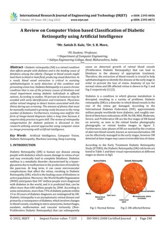
Diabetic Retinopathy (DR) is human eye disease among peoplewithdiabeticswhichcausesdamagetoretinaofeye and may eventually lead to complete blindness. Diabetes mellitusisa metabolicdisorder characterized bya hyperglycaemiaduetomalfunctionintheproductionofinsulinby the pancreas. At long term, it can cause microvascular complications that affect the retina, resulting in Diabetic Retinopathy(DR),whichistheleadingcauseofblindnessin activepopulation.Moreover,theWorldHealthOrganization (WHO)anticipatesthat347millionpeoplewerediagnosed withdiabetesintheworld,anditispredictedthat,canbe affectmorethan640millionpeopleby2040.Accordingto someestimations,morethan75%ofdiabeticpatientswithin 15to20yearsofdiabetesdiagnosisareendangeredbyDR. Diabeticretinopathyisanasymptomaticretinaldiseaseand primarilyaconsequenceofdiabetes,whichinvolveschanges tobloodvessels,resultinginmicroaneurysms,hemorrhages, exudates, malformation and vascular tortuosity (NonProliferative Diabetic Retinopathy) that can subsequently
cause an abnormal growth of retinal blood vessels (Proliferative Diabetic Retinopathy) that can lead to blindness in the absence of appropriate treatment. Therefore,theextractionofbloodvesselsiscrucialtohelp ophthalmologiststoidentifythisdiseaseattheearlystagein order to prevent the loss of vision. Anatomy of eye for normalretinaandDR-affectedretinaisshowninFig-1and Fig-2respectively[1][2].
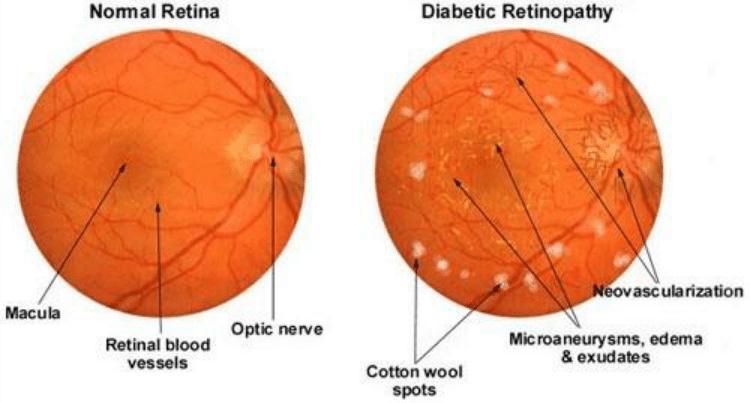
Diabetes is a condition in which glucose metabolism is disrupted, resulting in a variety of problems. Diabetic retinopathy(DR)isadisorderinwhichbloodvesselsinthe rear of the retina get damaged. According to the International Diabetes Federation (IDF), approximately millionpeopleworldwidehavediabetes,androughlyonethirdofthemhaveindicationsofDR.NoDR,Mild,Moderate, Severe,andProliferativeDRarethefivestagesofDRbased on severity, as seen in the retinal fundus photography photographs or retinal fundus images in figure 3. Furthermore,laterphasesofDRaremarkedbythecreation ofaberrantbloodvessels,knownasneovascularization.DR canbeeffectivelymanagedintheearlystages,howeverDR detectedatlaterstagesmaycauseirreversiblelossofvision.
According to the Early Treatment Diabetic Retinopathy Study(ETDRS),theDiabeticRetinopathy(DR)risklevelsare listedinTable1andtheirvisualrepresentationatdifferent stagesasshowninfig3
Fig-1:NormalRetina Fig-2:DR-affectedRetina
International Research Journal of Engineering and Technology (IRJET) e-ISSN: 2395-0056
Volume: 09 Issue: 08 | Aug 2022 www.irjet.net p-ISSN: 2395-0072
Researchershavedevisedorappliedeffectivetechniques for diagnosing diabetic retinopathy in two ways: binary classificationandmulticlassification,asshownbelow.
Fig3:StagesofDiabeticRetinopathy
Table1:DiabeticRetinopathyrisklevels
NoDR Nolesions
MildNPDR PresenceofMA
Moderate NPDR
PresenceofMAandHM
PresenceofCottonwoolspotsand Exudates
SevereNPDR Anyofthesymptoms
Venousbeadingin2quadrants
PresenceofMAandextensiveHMin4 quadrants
Intraretinalmicrovascular abnormalitiesin1quadrant
PDR Neovascularization
Presenceofpreretinal&vitreousHM
Ophthalmologistsurgediabeticpeopletohavetheirfundus medicallyscreened ona regularbasistodetectDRs early. Nonetheless, diabetic retinopathies are often overlooked untilsignificantdamagetothepatient'sfundushasoccurred (typically manifested as worsening or loss of vision). The proper identification and categorization of DR phases can assist clinicians in deciding on appropriate intervention techniques. Diabetic patients all over the world require regular screening to aid in early detection and treatment delivery.Nearly90%ofdiabetesindividualscanbedetected with early illness detection and adequate screening, and disease development can be slowed by avoiding future repercussions. The main issue is that DR does not reveal characteristicsymptomsuntilthediseasehasprogressedto an advanced stage [3]. To avoid difficulties, periodic eye examinationsandregularcheck-upsareencouraged.Human evaluation of retinal characteristics and morphological differencesinfundusimages,ontheotherhand,isatedious andtime-consumingoperation.Toaddressthisshortcoming, numerousautomatedcomputer-aideddiagnostictoolshave recentlybeendeveloped,whichassistophthalmologistsin examiningretinalabnormalities.
Several techniques for detecting microaneurysms, hemorrhages, and exudates are discussed [1] for ultimate detection of non-proliferative diabetic retinopathy. Blood vessels detection techniques are also discussed for the diagnosisofproliferativediabeticretinopathy.Anumberof imageprocessingtechniquesapplicabletowhitelightretinal fundus images have been proposed in the literature [2], whichwereusedtodesignscreeningsystemsforthisretinal disorder. A common prerequisite step used in all the approachesisthebloodvesselnetworkextraction.Basedon theretinalimageprocessingtechniquesused,thescreening systemscanbefurthercategorizedasthosewhichareusedto designDRreferralsystemsfocusingonlocalizationofasingle symptomandthoseDRreferralsystemsfocusingonisolation of multiple symptoms. Various conventional and deep learning-based diabetic retinopathy disease detection and classification methods are reviewed [3] and analyzed to provideaclearinsightandfuturedirections.MeherMadhu Dharmanaet.al.[4]proposedmethodwhichhasaneffective feature extraction technique based on blob detection followed by classification of different stages of diabetic retinopathyusingmachinelearningtechnique.Thisfeature extractiontechniquecouldhelpautomaticcharacterizationof retinaimagesfordiabeticretinopathywithanaccuracyof83 per cent with the most efficient machine learning classification algorithm, which would help specialists to handilyrecognizethepatient’sconditioninaprogressively precise manner. Messadi Mohamed et.al. [5] presented approachisbasedonthesegmentationofbloodvesselsand extractsthegeometricfeatures,whichareusedintheearly detectionofdiabeticretinopathy.Theproposedsystemwas testedontheDRIVEandMessidordatabasesandachievedan averagesensitivity,specificityandaccuracyof89%,99%and 96%,respectivelyforthesegmentationofretinalvesselsand 91%, 100% and 93%, respectively for the classification of diabetic retinopathy. Doshna Umma Reddy et.al. [6] consideredaconvolutionalneural network whichusesthe VGG- 16 model as a pre-trained neural network for finetuning,and,therebyclassifyingtheseverityofDR.Themodel alsouses efficient deeplearning techniquesincluding data augmentation,batchnormalization,dropoutlayersandlearnrateschedulingonhighresolutionimagestoachievehigher levelsofaccuracy
J.Anithaet.al.[7]developedCADtechniquesareanalyzed withrespecttoperformanceevaluationandthechallenges are discussed, some suitable solutions are suggested for improving the system to be more accurate. R. Subhashini et.al. [8] constructed a graphical user interface that can integrateimageprocessingtechniquestogetherinorderto predict whether the input fundus/retinal image received
International Research Journal of Engineering and Technology (IRJET) e-ISSN: 2395-0056
Volume: 09 Issue: 08 | Aug 2022 www.irjet.net p-ISSN: 2395-0072
fromthepatientisaffectedwithDiabeticRetinopathyornot; if affected, the graphical user interface will display the severity along with the required action needed to be undertakenbytheuser/patient.ManojKumarBeheraet.al. [9] has proposed research two well-known predefined feature extraction techniques scale invariant feature transform (SIFT) and speeded up robust features (SURF) have been used simultaneously on each retinal images to capturetheExudatesregions.TheseExudatesofeachimage stored in a feature matrix and used by the support vector machine(SVM)classifierforpredictionofDR.KaranBhatia et.al.[10]focusedondecisionaboutthepresenceofdisease by applying ensemble of machine learning classifying algorithms on features extracted from output of different retinal imageprocessingalgorithms,likediameter ofoptic disk,lesionspecific(microaneurysms,exudates),imagelevel (prescreening,AM/FM,qualityassessment).Decisionmaking for predicting the presence of diabetic retinopathy was performedusingalternatingdecisiontree,Ada-Boost,Naïve Bayes, Random Forest and SVM. Masoud Khazaee Fadafen et.al. [11] proposed method on the DIARETDB1 database, which includes 89 selected images for the diagnosis of diabetic retinopathy, was tested and with four models of methodsavailableforrecognizingsaliencies,frequencytuned method (FT) model, the spectral residual approach (SR) model,theSDSPmodel:anovelsaliencydetectionmethodby combiningsimplepriorhasbeencompared.Toevaluatethe performance of the proposed method with other methods using Ground truth images, the ROC curve and the AUC calculation were used. SumeshE P et.al. [12]createda DR detectiontechnique,involvingdigitalimageprocessing,has been developed by utilizing retinal image, where fundus imagehasbeenobtainedfrompatient’sretina.Thisproposed work aims at segmenting the fundus image into Exudates, Microaneurysm,OpticalDiskandhemorrhageandexamine whether the retinal condition is in Proliferative / Non Proliferative DR stage. Various performance measures has been utilized in validating the proposed technique. From thoseperformanceanalysis,wecouldobserve98%accuracy indetectingPDRandNPDRwithin39seconds(halfminute).
Ali Shojaeipour et.al. [13] developed system in which the Gaussian filter is used to enhance images and separate vessels with a high brightness intensity distribution. Next, wavelets transform is used to extract vessels. After that according to some criteria such as vessels density, the locationofopticdiscwasdetermined.Thenafteropticdisc extraction,exudatesregionsweredetermined.Finallythey classifiedtheimageswithaboostingclassifier.Withutilizing the boosting algorithm, the suggested system can have a power classifier. Mirthula Balaji et.al. [14] implemented a semanticanalysisthatutilizesforportrayingtheDR.Inour proposed methodology, an innovative framework to overcometheissuesoftraditionalmethodology.TheGLCM aneffectivefeatureischosenforextractingthefeatureswith theco-occurrencematrix.Afterextractingthefeatures,the classificationprocessisperformedusingProbabilisticNeural
value:
Network(PNN)whichprovidesaneffectiveclassifieroutput. It is concluded that this novel vessel segmentation frameworkacquiredbetteraccuracy,sensitivity,Fmeasures, specificityandprecisionfromthisexperiment.YuhanisYusof et.al. [15] focuses on classification of fundus image that contains with or without signs of DR and utilizes artificial neuralnetwork(NN)namelyMulti-layeredPerceptron(MLP) trained by Levenberg-Marquardt (LM) and Bayesian Regularization (BR) to classify the data. Nineteen features havebeenextractedfromfundusimageandusedasneural networkinputsfortheclassification.ItislearnedthatMLP trainedwithBRprovidesabetterclassificationperformance with72.11%(training)and67.47%(testing)ascomparedto the use of LM. Shailesh Kumar et.al. [16] presents an improved diabetic retinopathy detection scheme by extractingaccurateareaandatenumberofmicroaneurysm fromcolorfundusimages.Diabeticretinopathy(DR)isaneye diseasewhichoccursduetodamageofretinaasaresultof long illness of diabetic mellitus. The recognition of MA at primary stage is very crucial and it is the first step in inhibitingDR.Avarietyofmethodshavebeenproposedfor detectionanddiagnosisofDR.ClassificationofDRhasbeen donebylinearSupportvectormachine(SVM).Thesensitivity andspecificityofDRdetectionsystemareobservedas96% and 92% respectively. Bhavani Sambaturu et.al. [17] proposedanovelmethodtodetecthardexudateswithhigh accuracy with respect to lesion level. They tested our algorithm on publicly available DiaretDB database, which containsthegroundtruthforallimages.Theyachievedhigh performanceresultssuchassensitivityof0.87andF-Scoreof 0.78 and Positive Predict Value (PPV) of 0.76 for hard exudatelesionleveldetection,comparedtotheexistingstate ofarttechniques.TanapatRatanapakornet.al.[18]hasthe automated software for screening and diagnosing DR, by usingthecombinationofdigitalimageprocessingtechniques, hasbeendeveloped.Thissoftwareyieldsthegoodaccuracy forthedetectionofDRfromfundusphotographs.Itcanbe used asan alternative or adjunctive tool for DR screening, especiallyintheremoteareawhereophthalmologistisnot available or in the rural area where ophthalmologist has manytaskoverloads.
Although diabetic retinopathy cannot be healed, laser analysiscanhelppreventvisionlossifdonebeforetheretina isnegativelyaffected.Thesurgicalremovalofvitreous gel can enhance eyesight if the retina has not been severely damaged. This research aids in the early diagnosis of retinopathy,whichcanleadtoirreversiblevisuallossifnot treatedpromptly.Thisstudydetailedtheauthors'studiesfor detecting diabetic retinopathy. Technical people and researcherswhoneedtoleverageongoingresearchinthis fieldwouldbenefitfromoureffort.Variousapproachesfor detecting and treating diabetic retinopathy patients have been developed, including the categorization of different
International Research Journal of Engineering and Technology (IRJET) e-ISSN: 2395-0056 Volume: 09 Issue: 08 | Aug 2022 www.irjet.net p-ISSN: 2395-0072
phases of diabetic retinopathy employing using artificial intelligence.
[1] JaveriaAmin,MuhammadSharif,MussaratYasmin,"A Review on Recent Developments for Detection of DiabeticRetinopathy",Scientifica,vol.2016,ArticleID 6838976, 20 pages, 2016. https://doi.org/10.1155/2016/6838976
[2] Sandhya Soman, Jayashree R, “A Survey of Image Processing Techniques for Diabetic Retinopathy”, International Conference on Advances in computer ScienceandTechnology(IC-ACT’18)–2018,ISSN:23951303
[3] ValarmathiS,Dr.R.Vijayabhanu,“AReviewonDiabetic RetinopathyDiseaseDetectionandClassificationusing ImageProcessingTechniques”,InternationalResearch JournalofEngineeringandTechnology(IRJET),Volume: 07Issue:09|Sep2020,pp456-455.
[4] M.M.DharmanaandA.M.S.,"Pre-diagnosisofDiabetic Retinopathy using Blob Detection," 2020 Second International Conference on Inventive Research in Computing Applications (ICIRCA), Coimbatore, India, 2020, pp. 98-101, doi: 10.1109/ICIRCA48905.2020.9183241.
[5] E.Z.Aziza,L.MohamedElAmine,M.MohamedandB. Abdelhafid,"DecisiontreeCARTalgorithmfordiabetic retinopathy classification," 2019 6th International Conference on Image and Signal Processing and their Applications(ISPA),Mostaganem,Algeria,2019,pp.1-5, doi:10.1109/ISPA48434.2019.8966905.
[6] N. B. Thota and D. Umma Reddy, "Improving the AccuracyofDiabeticRetinopathySeverityClassification withTransferLearning,"2020IEEE63rdInternational Midwest Symposium on Circuits and Systems (MWSCAS),Springfield,MA,USA,2020,pp.1003-1006, doi:10.1109/MWSCAS48704.2020.9184473.
[7] A. G. P. H., J. Anitha and J. N. R. J., "Computer Aided Diagnosis Methods for Classification of Diabetic RetinopathyUsingFundusImages,"2018International ConferenceonCircuitsandSystemsinDigitalEnterprise Technology(ICCSDET),Kottayam,India,2018,pp.1-4, doi:10.1109/ICCSDET.2018.8821200.
[8] R.Subhashini,T.N.R.Nithin,U.M.S.Koushik,“Diabetic RetinopathyDetectionusing ImageProcessing(GUI)”, International Journal of Recent Technology and Engineering(IJRTE),ISSN:2277-3878,Volume-8,Issue2S3,July2019
[9] M.K.BeheraandS.Chakravarty,"DiabeticRetinopathy Image Classification Using Support Vector Machine," 2020 International Conference on Computer Science, EngineeringandApplications(ICCSEA),Gunupur,India, 2020, pp. 1-4, doi: 10.1109/ICCSEA49143.2020.9132875.
[10] K.Bhatia,S.AroraandR.Tomar,"Diagnosisofdiabetic retinopathy using machine learning classification algorithm,"20162ndInternationalConferenceonNext GenerationComputingTechnologies(NGCT),Dehradun, 2016,pp.347-351,doi:10.1109/NGCT.2016.7877439.
[11] Fadafen,Masoud&Mehrshad,Nasser&Razavi,Seyyed. (2018). Detection of diabetic retinopathy using computational model of human visual system. Biomedical Research (India). 29. 1956-1960. 10.4066/biomedicalresearch.29-18-551.
[12] Hamood Ali Hamood Al shamaly, Sumesh E P, VidhyalavanyaR,JayakumariC,“DiabeticRetinopathy Detection Using Matlab”, International Journal Of Scientific & Technology Research Volume 8, Issue 11, November2019
[13] A.Shojaeipour,M.J.NordinandN.Hadavi,"Usingimage processingmethodsfordiagnosisdiabeticretinopathy," 2014 IEEE International Symposium on Robotics and Manufacturing Automation (ROMA), Kuala Lumpur, 2014,pp.154-159,doi:10.1109/ROMA.2014.7295879.
[14] V. Govindaraj, M. Balaji, T. a. Mohideen and S. a. F. J. mohideen,"Eminentidentificationandclassificationof Diabetic Retinopathy in clinical fundus images using ProbabilisticNeuralNetwork,"2019IEEEInternational Conference on Intelligent Techniques in Control, OptimizationandSignalProcessing(INCOS),Tamilnadu, India, 2019, pp. 1-6, doi: 10.1109/INCOS45849.2019.8951349.
[15] N. H. Harun, Y. Yusof, F. Hassan and Z. Embong, "Classification of Fundus Images For Diabetic RetinopathyusingArtificialNeuralNetwork,"2019IEEE Jordan International Joint Conference on Electrical Engineering and Information Technology (JEEIT), Amman, Jordan, 2019, pp. 498-501, doi: 10.1109/JEEIT.2019.8717479.
[16] S.KumarandB.Kumar,"DiabeticRetinopathyDetection byExtractingAreaandNumberofMicroaneurysmfrom Colour Fundus Image," 2018 5th International Conference on Signal Processing and Integrated Networks (SPIN), Noida, 2018, pp. 359-364, doi: 10.1109/SPIN.2018.8474264.
[17] K.K.PalavalasaandB.Sambaturu,"AutomaticDiabetic RetinopathyDetectionUsingDigitalImageProcessing," 2018InternationalConferenceonCommunicationand
© 2022, IRJET | Impact Factor value: 7.529 | ISO 9001:2008 Certified Journal | Page243
International Research Journal of Engineering and Technology (IRJET) e-ISSN: 2395-0056
Volume: 09 Issue: 08 | Aug 2022 www.irjet.net p-ISSN: 2395-0072
Signal Processing (ICCSP), Chennai, 2018, pp. 00720076,doi:10.1109/ICCSP.2018.8524234.
[18] Ratanapakorn,Tanapat&Daengphoonphol,Athiwath& Eua-Anant, Nawapak & Yospaiboon, Yosanan. (2019). Digital image processing software for diagnosing diabeticretinopathyfromfundus photograph.Clinical Ophthalmology. Volume 13. 641-648. 10.2147/OPTH.S195617.
© 2022, IRJET | Impact Factor value: 7.529 | ISO 9001:2008 Certified Journal | Page244