Bone Cancer Detection using AlexNet and VGG16
 Dr Sunitha M.R1 , Huda Rahman2 , Gopinath C.B3 , Sathyabhama R4
1Professor, Computer Science and Engineering, Adichunchanagiri Institute of Technology, Chikmagalur, Karnataka, India.
24th Sem M.tech Computer Science and Engineering Adichunchanagiri Institute of Technology Chikmagalur, Karnataka, India
3Assistant Professor, Computer Science and Engineering, Adichunchanagiri Institute of Technology, Chikmagalur, Karnataka, India.
4Assistant Professor, Computer Science and Engineering, Adichunchanagiri Institute of Technology, Chikmagalur, Karnataka, India. ***
Abstract In bio medical field the discernment oftumor in premature stages is a hot research theme while most of the malignancy specifies the tumor untimely. Encountering and spotting the tumor requires loads of determined attempts. The controversial debate is to perceive out the identification and the detection framework which has to be rapid and authentic. Utilizing the distinct Machine Learning techniques and the CT scan images of bone this work has been used to communicate a point of view for bone cancer detection. This writing is particularly devoted to discover bone cancer/tumor by making use of AlexNet and VGG16.
Key Words: Bone Tumor, AlexNet, VGG 16, Smoothing Filter, Averaging Filter, Canny Edge
1. INTRODUCTION
Body’s hollow helping skeleton is the bone. In contemplationtoavertcalciumsaltsfrombeenlaiddown, the exterior surface of bone is comprised if rigid tissues. Thetrabecularboneontheinsideofthefirmexternallayer ismasked bycortical bone, while the external of the bone is guarded of the bone is guarded by periosteum. The mushy tissues known as bone marrow is founded in the medullary cavity of certain bones which are supposed to be hollow. As a tissue barrier, endosteum serves. A patch ofcartilage,amildertypeofbone liketissue,canbefound through each end of the bone. Cartilage is a componentof the bone that has been constituted of a fibrous tissue framework blended with a substance that resembles gel butcontainslesscalciumthanthatofbone.Themajorityof bone ends as cartilage. After then, the body deposits calcium mostly on cartilage to become bone. A few cartilage may remain somewhere at extremities following bone evolutionto assist like a bridge connecting bones. A joint is formed when all this cartilage, together with ligaments and additionalstructures, combines two bones. Boneisafairlymuscularandrigidelement.12,000pounds persquareinchisthemaximumloadcapacityofbone.The thigh bone must becompressed between 1,200and 1,800 poundsinordertobreakit.Twotypesofcellsareavailable
with in bone. The osteoclast is a varietyof cell that not only produces new bone and weakens existing bone. Several bones have oily tissue mostly in marrow. Other bones' marrow accommodatesa blendof fat cells and blood forming cells. Uncontrolled cell multiplication caused by cancer can partition the cells, multiply erratically, form evil tumours, and attack adjacent organs. The digestive, neurological, and circulatory systems can becomeobstructedbysuchatumourasitgrows,andthen they can also release hormones which alter how the body processes.
Cellssomeofwhichareregardedcanceroussince they damage DNA. Whenever DNA is broken in a typical cell, the cell must repair the damage or it will die. If the injuredDNA isn't fixed, the cells will continue to divide needlesslyuntiltheydie.
The body's cancerous cells frequently spread to otherregionswheretheystarttoformtumoursthatrevert to healthy tissue. We refer to this phenomenon as metastasis. Tumor cells then enter the body's lymphatic system or bloodstream. The human body can harbour several distinct kinds of cancer. Bone tumoris indeed the term for a condition where a tumour directly influencesa bone.
The majority of bone cancers are either primary orsecondary.Theboneisthesiteofprimarybonecancer. On the other hand, secondary bone cancer can develop elsewherewithinthebody.
2. LITERATURE SURVEY
Mokhled S. Al Tarawneh [1] proposed Lung Cancer determination technique. The noise in the image was removed by utilizing Gabor filter and segmentation of imagewascarriedoutintwostrategiestheyarethreshold techniqueand marker controlled watershed.Utilization of Binarization the features were extracted to identify the disease.
Volume: 09 Issue: 07 | July 2022 www.irjet.net p ISSN: 2395 0072
MaduriAvula [2] proposed methodology to discover bone ailment in the MR images by applying mean pixel power. The noise removal in the informative MR image is done inorder to set apart the tumor section and also the computation of K means clustering is carried out. To characterizr whether the tumor is benign or malignant meanpixelpowercomesintoservice.
KishorKumarReddy[3]offeredanewapproachusinga local evolving computation to estimate tumour measurementsand bone malignant expansionorganisation. The procedure used locale establishing calculations to divide the subject of concern. The size of the tumour is determined by the amount of pixels in the recovered tumour section. The actual pixel value is used to accept the illness pattern. It's really challengingtoselectanaccurateseedlocationwhileusing theimage.
Abdulmuhssin Binhssan [4], This suggested procedure outlines a strategy to spot enchondroma tumours. Image dataisdenoisedbyusingatwo sidedfilterinadditiontoa standard filter. The two sided filter seems to have a numberofissues.RemovalofnoiseinDenoisingtheimage requires a higher outlay of funds. The regularfilter achieves better performance than the reciprocal filter. Threshold segmentation is employed to divide the image, and morphological actions are connected to enhance the tumourarea.
Sunitha M. R. [5], proposed a system to segment the region of interest based on hierarchical graph based method. This method segments the region of interest efficiently.
Sunitha M.R. et. al. presented bone sarcoma detection method to detect irregular growth of tissue within bone using different preprocessing technique and detection usingVGG16methodandobtainedgoodaccuracy.
3. METHODOLOGY
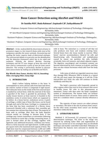
Figure1showsthemethodologyforDetectionofBone TumorusingAlexNetandVGG 16.
Figure 1 Methodology for Detection of Bone Tumor using AlexNet and VGG 16
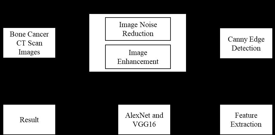
Image Acquisition
In any image analysis system, collecting the imagesis the most important step. CT scans, MRIs, ultrasounds, and X rays are examples of different image modalities. CT scan imagesareusedasinputtothedefinedmodel.
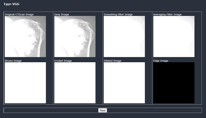
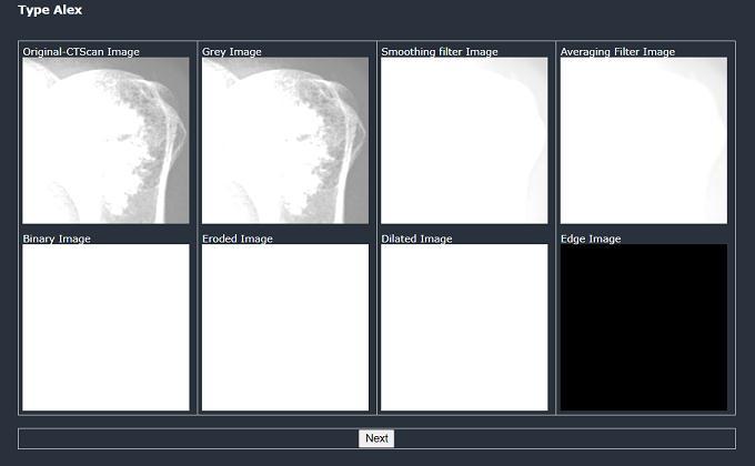
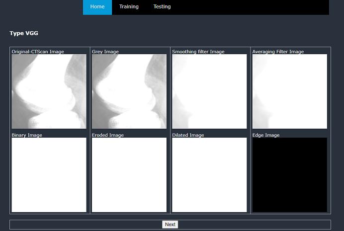
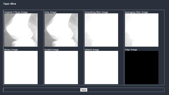
Preprocessing
An optical inquiry of unwavering consistency. Pre processing is a crucial step in improving the quality of an image. The sifting process kicks off the image preparation phase. Smoothing, honing, and removing clamor are some of the applications for image sifting. Sifting eliminates noiseandotherminorvariationsintheimage.Asa result, thesesoundsshouldbedenoised.Themiddlefilterisused inthismethodtoeliminatenoiseandsmoothoutdamaged images. As compared to other filters, the main advantage aboutthefilter isthatitachievesamazingnoisereduction with less disorientation. Following sifting, the dim change isthenextstage.ThisistheprocedureforconvertingRGB level pixels to the dim dimension. The procedure for shading an image is more complicated. As a result, the grayscale picture must be modified. By keeping the luminance,themodificationprimarilyremovesthetintand immersiondata.
For noise removal smoothing and averaging filters are used. Smoothing is sometimes used to minimize image noise. Image smoothing is a primary image enhancement techniqueforremovingnoisefromimages.Asaresult,itis requiredin many image processing processes. Image smoothing is a technique for enhancing image quality. Filter2D()isthesmoothingfilterused.
Imagesofteningisaccomplishedusingtheaverage filteringmethodology by reducing the contrast between nearbypixels.Asitadvancespixelbypixelthroughoutthe image, the average filter substitutes every item also with theaverage itemof its neighbours, including itself. The averagefilteremployedisblur().
Volume: 09 Issue: 07 | July 2022 www.irjet.net p ISSN: 2395 0072
After filtering, the image is transformed to gray image.
Then, the morphological operator’s dilation and erosionareapplied.Dilationincludespixelstothelimitsof object in an image, whereas erosion eliminates pixels on object limits. The size and structureof the constructing factor used to improve the image quality determines the amount of pixels includedor excludedfrom the objects in theimage.
Edge Detection
A limitation among two regions with slightly specific dim dimension properties is obtained using an edge indicator. Edge detection is a technique for removing importantfeatures that aid in theidentificationof malignant development. A clever edge indicator is used in this suggested methodology to discern an image's edge. The edges are known and distinguished as the points where picture brilliance shifts. Great identification, great restriction, and negligible reaction are all features of the vigilantedgeindicator.
Canny Edge Detection is used in which the first step is to smoothen the image to eliminate the noise. Then, image gradient is calculated to highlight the areas with higher spatial derivatives. Using non maximum suppression the pixelsthatarenotatmaximumaresuppressed.Hysteresis is used to minimize the gradient array even further, removingstreakingandthinningtheedges.
Feature Extraction
In image processing, image feature extraction is a critical system. It plays a significant role in disease detection by utilizingimage processing. To detect malignancy, features from the portioned image are segregated. The final result isto predict malignancy and not malignancyof an image are referred to as feature extraction. Feature extraction reduces the number of assets needed to represent a complex arrangement of data. It's the process of identifying and extracting specific elements of interest from an image for further processing. The element is shown as the image's most delegated data. Each component represents a measurable feature of an entity and is documented with the intention of evaluating the object'sessentialcharacteristics.
Classification
Thefinalandmostcriticalstageofourproposedschemeis classification. The classifier distinguishes between different stages of bone tumor that is stage 0, stage 1 or stage2. FortheclassificationAlexNetandVGG 16 isused. AlexNet and VGG 16 is a name for Convolutional Neural Network(CNN).
AlexNet
The resolution of the input image to be fed into the AlexNetnetworkis227x227pixelsinRGBformat.Thefirst layer in the AlexNet network is the convolution layer.There are 96 different types of kernels in the first layer, each of which is 11x11 and has a stride of 4. Since there are 96 different kernels and each feature map includes features of size 55 x 55, the performance of the firstlayeryields96differentchannelsorfeaturemaps.
ThesecondlayerisaMaxPoolLayer,withstrideequalto2 and max pooling performed over a 3 x 3 window. The feature map's size is decreased to 27 x 27 after this pooling, but the number of feature channels is retained at 96.
ThethirdlayerisaConvolutionLayerwithakernelsizeof 5x5andapaddingof2toensurethattheperformanceof the convolution layer matches the input feature size. As a result,thesizeoffeaturemapscreatedbythisconvolution layeris27x27,andthenumberofkernelsincludedinthis scenario is256,so256differentchannelsorfeature maps are obtainedfrom this convolution layer's output, each of whichwillbe27x27insize.
Thefourthlayerisanother MaxPoolLayer,thistimewith max pooling performed over a window of 3 x 3, stride equal to 2, and result of this pool is 13 x 13 feature maps and256channelsareobtained.
Then, there are three continuous Convolution Layers. In that the first convolution layer has a kernel size of 3 x 3 withpaddingof1and384kernels,resultingin384feature maps with a size of 13 x 13 that move through the next convolution layer. The performance of the second convolution layer will have 384 channels or 384 feature maps, each of which is 13 x 13 pixels in size. Since thepaddingisequalto1fora3x3kernelsize,thesizeof any feature map at the result of this convolution layer remains the same as the size of the feature maps that are fedintotheconvolutionlayer.
VGG16
For the conv1 layer 224x224 RGB image is fed as input. There are considerable convolutional layers onto which the images have been fed. Following certain convolution layersdimentionalpoolingiscarriedoutbythefivelayers layersofmax pooling.
After subsequent convolutional layer stacks are succeeded with 3 fully connected layers. Last layer comprises of soft max layer. Then the layers in the network are fully connected and which also has a hidden ReLUlayer.
4. RESULTS AND DISCUSSIONS
The discussion of results is that same image is taken as inputforbothAlexNetandVGG16andeventuallyitcanbe concludedthatwhichmethodhasgoodaccuracy.
Figure
Figure
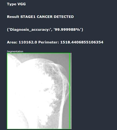
Figure 6 and Figure 7 depicts the result as cancerous at stage 1. The accuracy for AlexNet is 72.28% and for VGG16 is 99.998%. VGG16 has great accuracy in comparisonwithAlexNet.
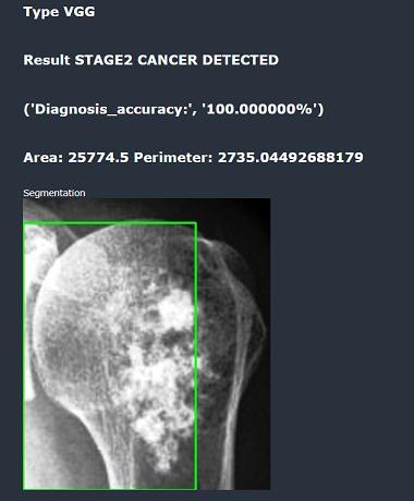
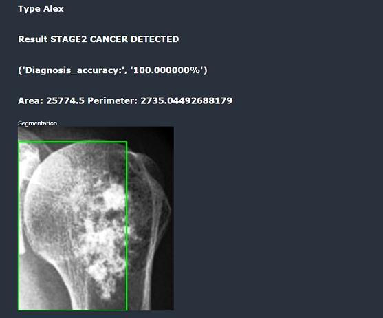
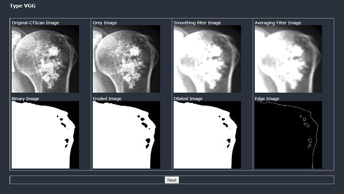
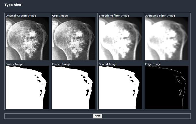
Figure8andFigure9depictsthepreparationstepsbefore the classification. The results of this step is same in both VGG16andAlexNet.
Figure 10: Result in AlexNet for Stage2
Figure 11: Result in VGG16 for Stage2
Figure 10 and Figure 11 depicts the result as cancerous at stage 2. The accuracy for AlexNet is 100% and for VGG16 is 100%. Both VGG16 and AlexNet have sameaccuracy.
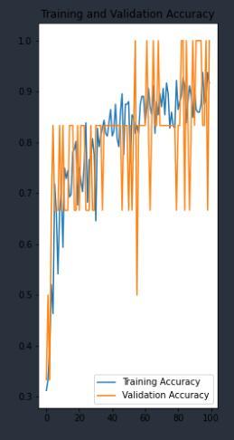
Figure 12: AlexNet Accuracy Graph
Figure 12 shows the training and validation accuracy graphofAlexNet.InX axisthetrainingdataaretakenand in the Y axis validation data is taken. The accuracy of the system is 97.3%%. The graph has been plotted with train and test data. The model has been tested with validation setofdataandtotrainthemodeltrainingdataisutilized.
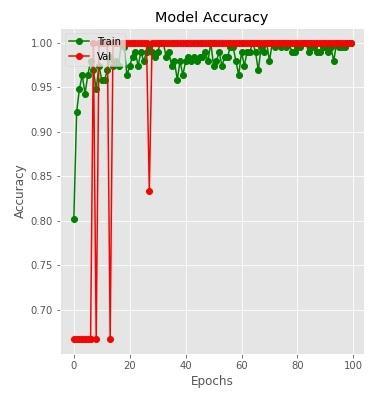
Figure 13 depicts VGG16 graph with model accuracy The X axis follows Epochs and Y axis follows Accuracy The graphhasbeenplottedwithtrainandtestdata.Themodel hasbeentestedwithvalidationsetofdataandtotrainthe modeltrainingdataisutilized.
5. CONCLUSION
The most life threatening ailment is bone cancer in whichthesignsareunnoticedinthepre maturestages.To overcome the fatality rate detection in the early stages is veryimportant.
IncomparisonwithAlexNet,VGG16yieldsbetterresults asdiscussedearlierinsection4bylookingintothevarious results.
REFERENCES
MokhledS.Al tarawneh,“Lungcancerdetectionusing imageprocessingtechniques”,Leonardoelectronicjournal ofpracticesandtechnologies,20,147 158,2012.
[1] Maduri Avula, Narasimha Prasad Lakkakula, Murali Prasad raja, “Bone cancer detection from MRI scan imagery using Mean Pixel Intensity, Asia modeling symposium,2014
[2] Kishor Kumar Reddy, Anisha PR,RajuGVS,“A novel approachfordetectingthetumorsizeandbonecancer stage using region growing algorithm”, International Conference on Computational Intelligence and CommunicationNetworks,2015.
[3] Abdulmuhssin Binhssan, “Enchondroma tumor Detection”,Internationaljournalofadvancedresearch in computer and communication Engineering, 4(6), june2015.
[4] Sunitha Madasi Ramachandra, Haradagere Siddaramaiah Jayanna, Ramegowda, “Hierarchical graph based segmentation and consensusbased human tracking technique” Journal of information processingsystems,Vol.15,No.1,pp.67 90,Feb2019.
[5] Sunitha M.R., Huda Rahman, Varun E, “Bone Sarcoma DetectionusingVGG16”,Vol.8,No.8,JETIR,2021