International Research Journal of Engineering and Technology (IRJET) e ISSN:2395 0056

International Research Journal of Engineering and Technology (IRJET) e ISSN:2395 0056
1,2,3,4,5 Department of Computer Science and Engineering, Dayananda Sagar University, Bangalore, Karnataka, India ***
Abstract Compared to most other tissues, lungs are directly exposed to oxygen concentrations. Lung diseases are one of the leading causes of death. There are many different lung diseases, some of which are caused by viral, bacterial, or fungal infections. Other lung diseases are associated with environmental factors, including COVID19, tuberculosis, bronchitis, pneumonia, etc. Deep learning has shown great potential when applied to medical images for disease detection including lung disease. We build and compare two pre trained models, MobileNet and VGG16 architectures using the Transfer learning approach. We have also used Supervised Machine Learning algorithms like Random forest, Decision Trees, Support Vector Machines, and Logistic Regression. This paper provides the analysis which we have performedon the different algorithms.
Key Words: Fungal infections, Pneumonia, MobileNet, VGG16, Transfer Learning, Supervised Machine Learning algorithms
Lung disorders often called respiratory diseases, are illnesses that affect the lungs' airways and other components.Pneumonia,TB,andCoronavirusDiseaseare allexamplesoflungdiseases(COVID 19).Accordingtothe Forum of International Respiratory Societies, around 334 million people have asthma, tuberculosis kills 1.4 million people eachyear, lungcancerkills1.6 millionpeople,and pneumoniakillsmillions.COVID 19wasaglobalpandemic that infected millions of individuals and put a strain on healthcareservices.Lungillnessesarewithoutadoubtone oftheworld'sleadingcausesofdeathanddisability.Early identification is crucial for boosting long term survival rates and enhancing the possibilities of recovery. Lung disease is usually discovered by a physical exam & skin tests,bloodtests,sputumsampletests,chestX rayexams, andcomputedtomography(CT)scanexamsareallusedto identify cancer. Deep learning has recently shown considerable promise in disease identification using medicalpictures,particularlylungdisease.
The threat of lung illnesses is enormous, in particular in growing and low middle income countries, wherein tens of thousands and thousands of humans are dealing with poverty and air pollutants. According to the
estimation of WHO, over four million untimely deaths arise yearly from household air pollutants associated illnesses, together with asthma, and pneumonia. Hence, it's far more important to take important steps to lessen air pollutants and carbon emissions. It is likewise vital to enforce green diagnostic structures that can help in detecting lung illnesses. Since December 2019, a singular coronavirus sickness 2019 (COVID 19) has been inflicting critical lung harm and respiration problems. In addition, pneumonia,aformoflungsickness,maybebecauseofthe causativevirusofCOVID 19orcanbebecauseofdifferent viral orbacterial infections.Hence,earlydetectionoflung illnesses has turned out to be more essential than ever. Recently, the virtual era has turned out to be a greater essential worldwide. This challenge can offer medical doctors and different researchers a course for detecting lung sickness with the assistance of a deep studying methodology. A huge quantity of lung X ray pics are used asadataset
Machine Learning performs an essential position in clinical systems. Lung illnesses are one of the main reasonsfordeath.Theearlyidentityandpredictionoflung illnesseshave emergedasa need withinside the research, as it may facilitate the subsequent scientific control of patients. Machine Learning-primarily based totally selection help systems offers the contribution to the medical doctors of their analysis decisions. The mission taken into consideration is the class of lung illnesses like Pneumonia, Tuberculosis, Lung cancer, and Covid 19. MachineLearning and Deep Learning are used to manage informationinadditiontocreatingfashionsfordiagnosing patients. Combining the processing of affected person statisticswithinformationfromchestX-raysandCTscans, using CNN with the famous pre-skilled model, These Neural networks for information of this shape are the techniquesusedforthismissiontopickoutlungillnesses.
A fully CNN has been proposed in Ref. [6] to reduce the false positive rate in classifying the lung nodules. This methodcanonlyanalyzethenatureoftheCTscanimages inordertoreducetheprobabilityofawrongdiagnosis.
Volume: 09 Issue: 05 | May 2022 www.irjet.net p ISSN:2395 0072 © 2022, IRJET | Impact Factor value: 7.529 | ISO 9001:2008 Certified Journal | Page 2976
Mohammed Maaz Ahmed Khan1, Mohammed Siddiq S2, Mythri J L3, Naveen A4 , Dr. Rajesh T M5International Research Journal of Engineering and Technology (IRJET) e ISSN:2395 0056
Volume: 09 Issue: 05 | May 2022 www.irjet.net p ISSN:2395 0072
In Ref. [7], a framework for deep learning is proposed to predict lung cancer and pneumonia offering two deeplearning methods.Initially,they used modified AlexNetforthediagnosisofchestX rays.Moreover,inthe modified AlexNet, SVM is implemented for the purpose of classification.
Deep learning methods are also proposed in Ref. [8] where several transfer learning methods such as DenseNet121, AlexNet, Inception V3, etc., are used for pneumonia diagnosis. However, the parameter tuning for theirimplementedmethodsisverycomplex.
The Software requirements used for performing the experiments are as follows, Transfer learning, React framework, Tensorflow, Python3, OpenCV, Keras, Numpy, Matplotlib,Scikit learn,andTkinter
In order to successfully run the project the following hardware requirements are needed CPU Intel i3+, RAM 2GB+, Storage Space At least 2GB, Mobile phone (Android &iPhone)
Constructing dataset for Tuberculosis, pneumonia, lung cancer, and COVID 19, Model portable on IOS and Androidplatform,Recognitionoflung relateddiseases
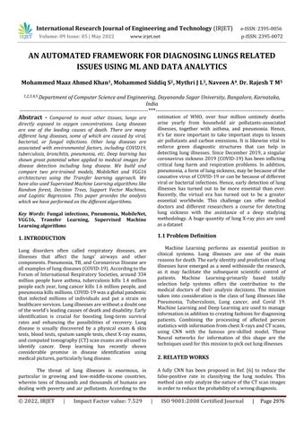
The first step is to acquire images. To produce a classification model, the computer needs to learn by example. The computer needs to view many images to recognize an object. Other types of data, such as time series data and voice data, can also be used to train deep learning models. In the context of the work used in this project, the relevant data required to detect lung disease willbeimages.ImagesthatcouldbeusedincludechestX ray,andCTscan.Theoutputofthisstepisimagesthatwill laterbeusedtotrainthemodel.
The second step is preprocessing. Here, the image could be enhanced or modified to improve image quality. Image modification such as lung segmentation and bone eliminationcouldbeusedtoidentifytheregionofinterest (ROI), whereby the detection of the lung disease can then be performed on the ROI. Edge detection could also be used to provide an alternate data representation. Data augmentation could be applied to the images to increase theamountofavailabledata.Featureextractioncouldalso be conducted so that the deep learning model could identify important features to identify a certain object or class. The output of this step is a set of images whereby thequalityoftheimagesisenhanced,orunwantedobjects havebeenremoved.Theoutputofthisstepisimagesthat were enhanced or modified that will later be used in training.
In the third step, namely training, three aspects could be considered. These aspects are the selection of deep learning algorithms, usage of transfer learning, and usage of an ensemble. There are numerous deep learning algorithms, for example, multilayer perceptron neural network (MPNN), recurrent neural network (RNN), and the aforementioned CNN. Different algorithms have differentlearningstyles.CNNworksparticularlywellwith images.Adeeplearningalgorithmshouldbechosenbased onthenatureofthedata athand.Transferlearningrefers to the transfer of knowledge from one model to another. Ensemble refers to the usage of more than one model during classification. Transfer learning and ensemble are techniques used to reduce training time, improve classification accuracy and reduce overfitting. The output ofthisstepismodelsgeneratedfromthedatalearned.
In the fourth and final step, which is classification, the trained model will predict which class an image belongs to. For example, if a model was trained to differentiate X ray images of healthy lungs and tuberculosis infected lungs, it should be able to correctly classify new images (images that are never seen by the model before) into healthy lungs or tuberculosis infected lungs. The model willgiveaprobabilityscorefortheimage.Theprobability score represents how likely an image belongs to a certain class. At the end of this step, the image will be classified basedontheprobabilityscoregiventoitbymodel
International Research Journal of Engineering and Technology (IRJET) e ISSN:2395 0056
Volume: 09 Issue: 05 | May 2022 www.irjet.net p ISSN:2395 0072
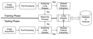
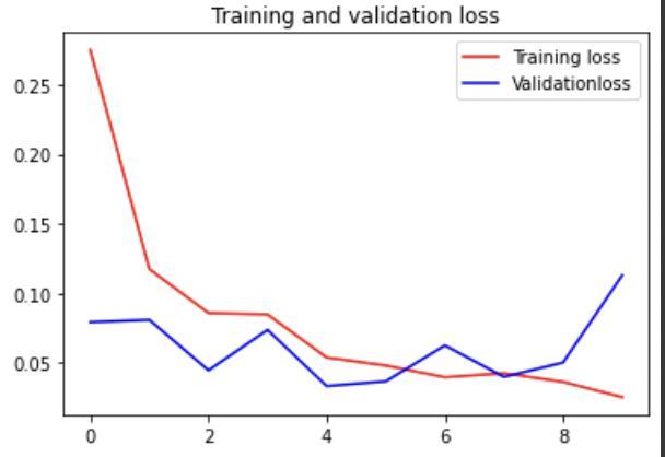
Chart -1:Summaryoftrainingevaluation&lossofImage Segmentation
Table -1: SummaryofDataset Summary of Dataset Category Training dataset Testing Dataset Pneumonia 6119 2623 Normal 4965 2128
Table -1: SummaryofSupervisedandTransferlearning experimentationresults
Summary of Supervised and Transfer learning experimentation results Algorithm Training Accuracy Testing Accuracy
Randomforest 86.3% 84.1% Logistic Regression 80.2% 75.1% DecisionTrees 80.1% 79.1% SVM 79.22% 67.88% MobileNet 99.83% 99.83% VGG16 99% 98.24%
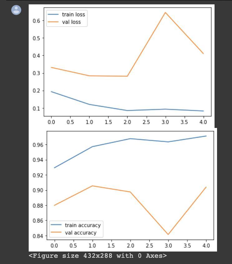
Chart 2:Summaryoftrainingevaluation&lossofVGG Classifier
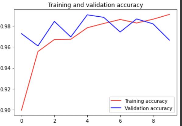
Chart 3:Summaryoftrainingevaluation&lossof MobileNet
The final product is a fully integrated desktop application that can classify various lung based diseases through X Rays and CT Scans, these include diseases such as Pneumonia,Lungcancer,Covid 19,andTuberculosis.This product is designed in such a way that any person with basicknowledgecanuploadtheirX RayorCT Scanstoget adetailedanalysis.
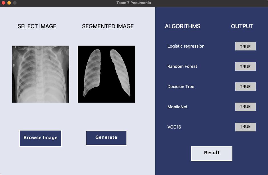

TheApplicationwill beusednotonlyasa second opinion but also as a way of initial screening and Cross Verification among radiologists, as detection of diseases from a Chest X ray and CT Scan is complex and may go unnoticed.
This is a real time application that uses machine learningandartificialintelligence(AI)basedalgorithmsto assess the condition of the lungs, return the health of the lungs and the percentage of infection (if affected by the diseases mentioned above). The final application is
projected to be implemented once the development and initialtestingaredone.
This is a project under the collaboration of Dayananda SagarUniversity,CDSIMER,andSagarGroupofHospitals.
Itisa greatpleasureforus toacknowledgetheassistance and the support of many individuals who have been responsible for the successful completion of this project work.
First, we take this opportunity to express our sincere gratitude to the School of Engineering & Technology, Dayananda Sagar University for providing us with a great opportunity to pursue our Bachelor’s degree inthisinstitution.
We would like to thank Dr. A Srinivas. Dean, School of Engineering & Technology, Dayananda Sagar University for his constant encouragement and expert advice. It is a matter of immense pleasure to express our sincere thanks to Dr. Girisha G S, Department Chairman, Computer Science, and Engineering, Dayananda Sagar University,forprovidingtherightacademicguidancethat madeourtaskpossible.
We wouldliketo thank our guide Dr.RajeshT M, Associate Professor, Dept. of Computer Science and Engineering, Dayananda Sagar University, for sparing his valuable time to extend help in every step of our project work, which paved the way for smooth progress and the fruitfulculminationoftheproject.
We would like to thank our Project Coordinator Dr. Meenakshi Malhotra and all the staff members of ComputerScienceandEngineeringfortheirsupport.
Wearealsogratefultoourfamilyandfriendswho provided us with every requirement throughout the course.Wewouldliketothankoneandallwhodirectlyor indirectlyhelpedusintheProjectwork.
[1] [1] Qin,Z.Z.,Ahmed,S.,Sarker,M.S.,Paul,K.,Adel,A. S. S., Naheyan, T., … Creswell, J. (2021). Tuberculosis detection from chest x rays for triaging in a high tuberculosis burden setting: an evaluation of five artificial intelligence algorithms. The Lancet Digital Health, 3(9), e543 e554. doi:10.1016/s2589 7500(21)00116 3
[2] [2] Pasa,F.,Golkov,V.,Pfeiffer,F.etal.EfficientDeep Network Architectures for Fast Chest X Ray Tuberculosis Screening and Visualization. Sci Rep 9, 6268 (2019). https://doi.org/10.1038/s41598 019 42557 4
International Research Journal of Engineering and Technology (IRJET) e ISSN:2395 0056 Volume: 09 Issue: 05 | May 2022 www.irjet.net p ISSN:2395 0072
[3] [3] Yoo,SeungHoon;Geng,Hui;Chiu,TinLok;Yu,Siu Ki; Cho, Dae Chul; Heo, Jin; Choi, Min Sung; Choi, Il Hyun;CungVan,Cong;Nhung,NguenViet;Min,Byung Jun; Lee, Ho (2020). Deep Learning Based Decision Tree Classifier for COVID 19 Diagnosis From Chest X ray Imaging. Frontiers in Medicine, 7(), 427 doi:10.3389/fmed.2020.00427
[4] [4] Chandra T.B., Verma K. (2020) Pneumonia Detection on Chest X Ray Using Machine Learning Paradigm. In: Chaudhuri B., Nakagawa M., Khanna P., Kumar S. (eds) Proceedings of 3rd International ConferenceonComputerVisionandImageProcessing. Advances in Intelligent Systems and Computing, vol 1022. Springer, Singapore. https://doi.org/10.1007/978 981 32 9088 4_3
[5] [5] van Cleeff, M., Kivihya Ndugga, L., Meme, H. et al. The role and performance of chest X ray for the diagnosisoftuberculosis:Acost effectivenessanalysis in Nairobi, Kenya. BMC Infect Dis 5, 111 (2005). https://doi.org/10.1186/1471 2334 5 111
[6] [6] SetioAAA, Traverso A,de Bel T, BerensMSN,van denBogaardC,CerelloP,ChenH,Dou Q,Fantacci ME, Geurts B, et al. Validation, comparison, and combination of algorithms for automatic detection of pulmonary nodules in computed tomography images: the LUNA16 challenge. Med Image Anal 2017;42: 1 13.
[7] [7] Bhandary Abhir, et al. Deep learning framework to detect lung abnormality a study with chest X Ray and lung CT scan images. Pattern Recogn Lett January 2020;129:271 8 https://doi.org/10.1016/j.patrec.2019.11.013.
[8] [8] Chouhan V, et al. A novel transfer learning based approach for pneumonia detection in chest X ray images. Appl Sci 2020;10(2):559. https://doi.org/10.3390/app10020559.

Mr Mohammed Maaz Ahmed Khan is pursuing a Bachelor of Technology degree in the DepartmentofComputerScience andEngineeringfromDayananda Sagar University, located in Bangalore,Karnataka,India.Heis currently in his final year of Engineering and will be graduating from Dayananda SagarUniversityintheyear2022.


Mr. Mohammed Siddiq S is pursuing a Bachelor of Technology degree in the DepartmentofComputerScience andEngineeringfromDayananda Sagar University, located in Bangalore,Karnataka,India.Heis currently in his final year of Engineering and will be graduating from Dayananda SagarUniversityintheyear2022.


Miss Mythri J L is pursuing a BachelorofTechnologydegreein the Department of Computer Science and Engineering from Dayananda Sagar University, located in Bangalore, Karnataka, India Sheiscurrentlyinherfinal year of Engineering and will be graduating from Dayananda SagarUniversityintheyear2022.
Mr. Naveen A is pursuing a BachelorofTechnologydegreein the Department of Computer Science and Engineering from Dayananda Sagar University, located in Bangalore, Karnataka, India He is currently in his final year of Engineering and will be graduating from Dayananda SagarUniversityintheyear2022.
Dr Rajesh T M is an Associate Professor in the Department of Computer Science and Engineering from Dayananda Sagar University, located in Bangalore,Karnataka,India.